


Abstract
Background: The decision to perform biventricular repair (BVR) in neonates and infants presenting with either single or multiple left ventricle outflow obstructions (LVOTOs) and a borderline left ventricle (BLV) is subject to extensive discussion, and limited information is known regarding the long-term outcomes. As a result, the objective of this study is to critically assess and summarize the available data regarding the prognosis of neonates and infants with LVOTO and BLV who underwent BVR. Methods: In February 2023, we conducted a review study with three different medical search engines (the National Library of Medicine, Science Direct, and Cochrane Library) for Medical Subject Headings and free text terms including “congenital heart disease”, “outcome”, and “borderline left ventricle”. The search was refined by adding keywords for “Shone’s complex”, “complex LVOT obstruction”, “hypoplastic left heart syndrome/complex”, and “critical aortic stenosis”. Results: Out of a total of 51 studies, 15 studies were included in the final analysis. The authors utilized heterogeneous definitions to characterize BLV, resulting in considerable variation in inclusion criteria among studies. Three distinct categories of studies were identified, encompassing those specifically designed to evaluate BLV, those focused on Shone’s complex, and finally those on aortic stenosis. Despite the challenges associated with comparing data originating from slightly different cardiac defects and from different eras, our results indicate a favorable survival rate and clinical outcome following BVR. However, the incidence of reintervention remains high, and concerns persist regarding residual pulmonary hypertension, which has been inadequately investigated. Conclusions: The available data concerning neonates and infants with LVOTO and BLV who undergo BVR are inadequate and fragmented. Consequently, large-scale studies are necessary to fully ascertain the long-term outcome of these complex defects.
1. Background
The decision whether to perform biventricular repair (BVR) or univentricular palliation (UVP) in neonates and infants affected by multiple left heart obstructive lesions and borderline left ventricle (BLV) is always challenging [1,2,3,4,5,6,7,8,9,10,11,12,13,14,15,16], and knowledge regarding the long-term prognosis of these patients remains scarce. Various scores utilizing echocardiography data have been proposed as a means of predicting the efficacy of BVR in BLV [2,3,4,5,9,14]; however, these are solely based on short-term results [3,4,14], with a mean follow-up period of 5–6 years. Furthermore, these scoring systems are not without limitations, which include heterogeneity in the echocardiographic parameters and outcome measures evaluated, as well as the retrospective design during the score development [2,3,4,5,9,14]. Additionally, significant variations in inclusion criteria were observed among the studies from differing authors [2,3,4,5,9,14]. Moreover, the term “borderline left ventricle” encompasses a broad spectrum of complex cardiac defects characterized by one or multiple left-sided obstructions and diminutive left sections, with significant uncertainty regarding the optimal choice between BVR and UVP. The absence of a universally accepted definition of “borderline left ventricle” has resulted in the use of various definitions, such as critical left ventricular outflow (LVOT) stenosis, hypoplastic left heart complex, multiple left heart obstructive lesions, and small left heart structures. Additionally, neonates and infants with Shone’s complex [17,18,19,20,21,22,23,24,25,26,27] have typically been evaluated separately, even though these congenital heart defects (CHDs) are also characterized by multiple left-sided obstructions and borderline left structures. Despite the high volume of literature on surgical and percutaneous aortic stenosis (AS) valvuloplasty in pediatric patients [28,29,30,31,32,33,34,35,36,37,38,39,40,41,42,43,44,45,46,47,48,49,50,51,52,53,54], studies have not specifically focused on BLV despite neonatal AS being typically associated with a diminutive left ventricle (LV). Data regarding the long-term outcomes of neonates and infants with Shone’s complex [17,18,19,20,21,22,23,24,25,26,27] and critical neonatal AS [28,29,30,31,32,33,34,35,36,37,38,39,40,41,42,43,44,45,46,47,48,49,50,51,52,53] undergoing BVR are also limited. The aim of the present study is to systematically review data pertaining to the long-term outcomes of neonates and infants with one or multiple LVOT obstructions and BLV who have undergone a biventricular correction.
2. Methods
In February 2023, we conducted a review study within 3 different medical search engines (the National Library of Medicine, Science Direct, and Cochrane Library) for Medical Subject Headings and free text terms including “congenital heart disease”, “outcome”, and “borderline left ventricle”. The search was refined by adding keywords for “Shone’s complex”, “complex LVOT obstruction”, “hypoplastic left heart syndrome/complex”, and “critical aortic stenosis”.
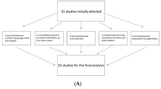
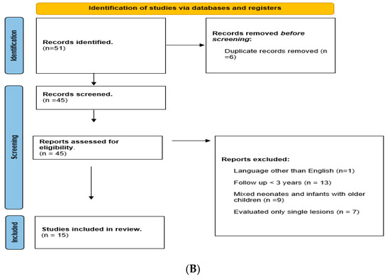
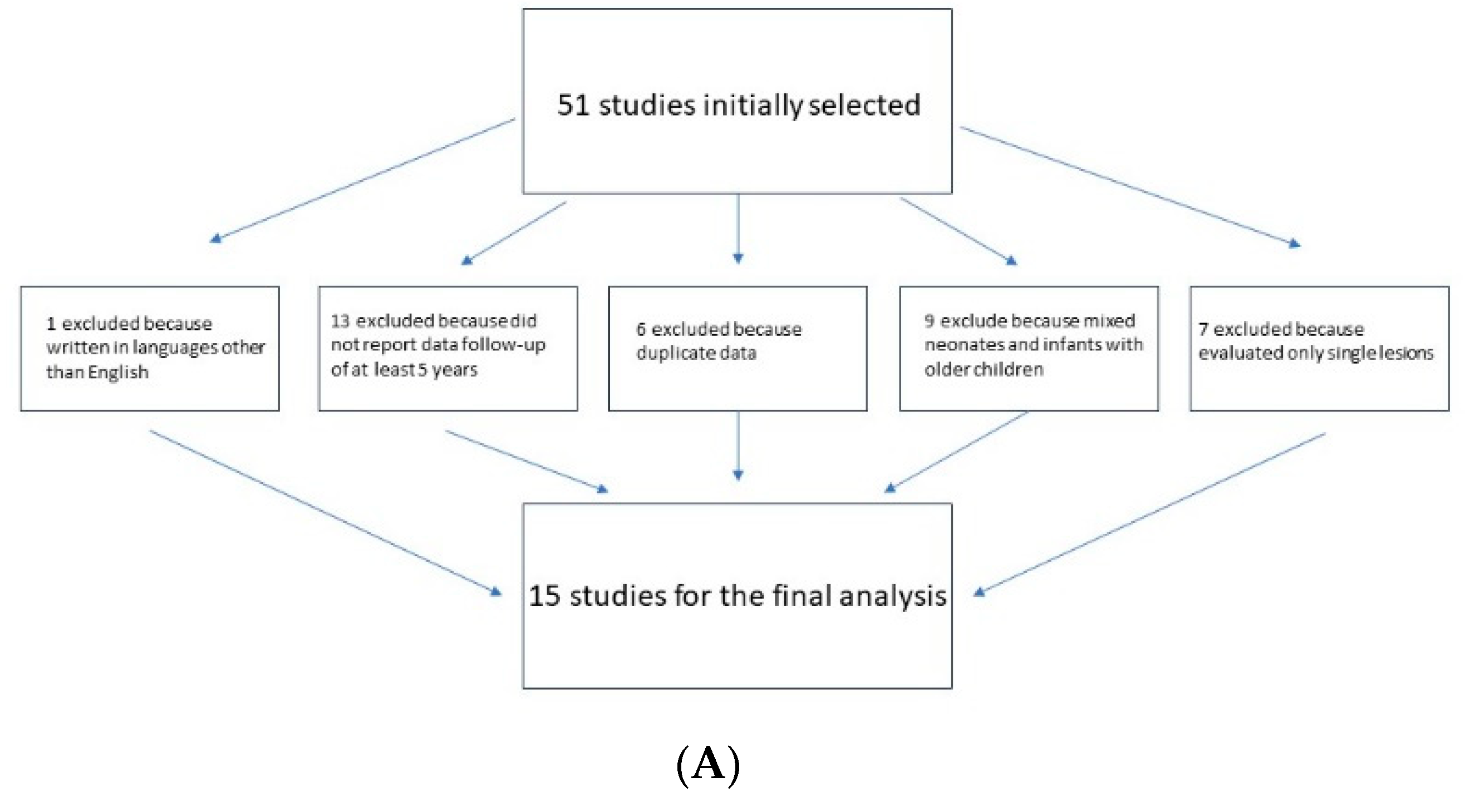
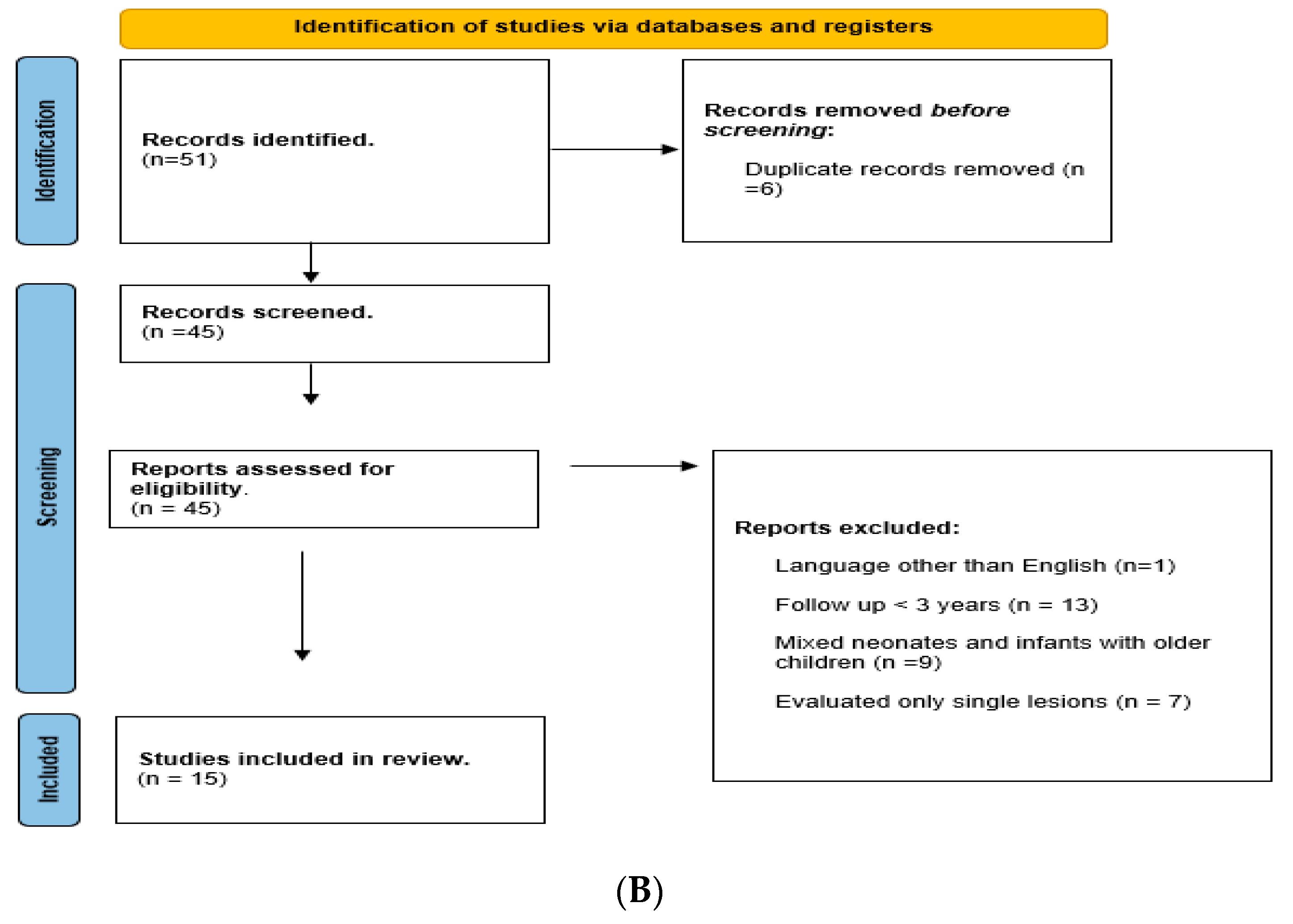
The titles and abstracts of articles identified by this strategy were evaluated and excluded if (i) the reports were written in languages other than English (1 study); (ii) studies did not report a data follow-up of at least 3 years (13 studies); (iii) there were duplicate data (6 works); (iv) the reports mixed neonates and infants with older children (9 studies); (v) the reports evaluated only single lesions (e.g., isolated aortic stenosis, excluding multiple stenosis and borderline LV) (7 studies) (Figure 1).
Figure 1.
(A) Diagraph of inclusion/exclusion criteria. (B) Flow diagram for new review.
3. Results
Of 51 studies, 36 were excluded for the above-mentioned criteria, leaving 15 studies for the final analysis. Three broad categories of studies were identified: those specifically designed to evaluate neonates and infants with BLV [1,3,4,6,7,10], those on Shone’s complex [22,23,24], and those on aortic stenosis [28,30,31,43,47].
3.1. CHD Definition and Groups
Various definitions have been utilized to describe LV obstruction with borderline LV, including BLV [1] (Table 1), critical left ventricular outflow (LVOT) stenosis [3,4], hypoplastic left ventricle with mitral stenosis [6], hypoplastic left heart complex [8,11], multiple left heart obstructive lesions [7,10], and small left heart structures [9]. Two main categories of BLV definitions may be identified: one includes in BLV a series of defects characterized by two or more diminutive left-sided lesions (e.g., mitral valve-MV, aortic valve AoV, LVOT, LV) [1,6,7,10,11,55], while the other is focused on the presence of a critical LVOT obstruction [3,4,8] (Table 1). While LV hypoplasia was a criterion of inclusion for most authors [1,3,6,7,8,10,11], not all authors used this inclusion criterion [4,9]. Notably, one study had the presence of aortic coarctation [9] as the primary inclusion criterion, while others excluded patients with isolated aortic arch obstruction [1]. Z-scores were employed to determine the degree of mitral and aortic valve hypoplasia, but different Z-score sources were employed by various authors [56,57,58], and at times the nomograms employed were not indicated at all [6,10,11]. Established exclusion criteria were discordant atrio-ventricular and ventricular arterial connections [1,3,4,7,9], aortic or mitral atresia [1,3,4,6,8,10], atrioventricular septal defects [1,6,7,10], or other complex associated CHDs [3,7,8,10].
Table 1.
(A) Borderline LV definition according to different authors. (B) Shone’s complex definition according to major studies.
The definition of Shone’s complex is also not consistent throughout the available literature. It has been defined as mitral stenosis plus at least one [19,22,24] or two [23] LVOT obstructions. Furthermore, the literature on aortic stenosis is also greatly heterogenous [54], with articles focused on the results of surgical valvuloplasty [28,30,31] or percutaneous valvuloplasty [43,47]. Finally, there is a challenge obtaining data on children with BLV since they have often been excluded [5,28,30,32,49,52,53] or grouped together with older children [31,41]. Several studies used fixed cut-off values to exclude patients from BVR [5,28,30,49], such as aortic valve annulus < 5 mm [28,30] or <4.5 mm [5], MV annulus < 7 mm, LV end-diastolic volume < 20 mL/m2 [30], and LV long axis < 80% of the LV [49]. Other studies utilized more generic exclusion criteria, such as borderline LV [38,53] or multiple LVOT obstruction [32,52]. The present study focuses on three articles on surgical valvuloplasty performed in neonates and infants with critical AS [28,30,31] and two articles on balloon valvuloplasty [43,47].
3.2. Follow-Up Duration
Our investigation revealed the finding of very limited data on long-term follow-up of BLV (Table 2), with only seven studies [1,3,4,6,7,8,10] reporting follow-up data of at least 5 years. However, not all the studies [7,8,9,10] completed the 5-year follow-up for all subjects [1,7,8,9,10]. The longest follow-up interval was, on average, 8 years [1], but once again, not all the subjects completed the follow-up. Follow-up intervals ranged from 0.1 to 16.4 years [1]. Some studies had a very limited sample size (e.g., <20 subjects) [6,8,11], while others had moderate to large sample sizes (ranging from 39 to 71 subjects) [1,7,9,10] or very large sample sizes (e.g., >100 subjects) [3,4]. All the studies, except for two [3,4], had a retrospective, single-center design.
Table 2.
(A) Major studies on outcomes in borderline LV attempting BVR. (B) Major studies on outcomes in Shone undergoing BVR. (C) Major studies on outcomes in neonates and infants with aortic stenosis undergoing either surgical/percutaneous valvuloplasty.
In the studies pertaining to the Shone complex, only three retrospective studies conducted at single centers [22,23,24] with sample sizes ranging from 27 [23] to 121 subjects [22] reported follow-up data for at least 5 years. The duration of follow-up was longer compared to studies pertaining to BLV, varying from 4.4 ± 4 years [23] up to 7.9 years [24].
The included studies on aortic stenosis were all single-center with retrospective design [28,30,31,43,47]. Here, mean sample sizes varied from 37 [30] to 84 subjects [28], while follow-up varied from a mean of 3.2 years [43] to >8 years [31], with a total duration of follow-up of 17.7 years in some cases [28].
3.3. Survival/Transplant-Free Survival for BVR
In the context of BLV, the definition of successful BVR varies among authors. Some describe it as “survival alone” [3,4,7,8], while others use “transplant-free survival” [1,9,10]. The reported rates of successful BVR ranged from 70% [3] to 82% [1] at 5 years. There are only a couple of studies [1,6] reporting data on survival at 10 years of follow-up, with a survival rate ranging from 77% [1] (transplant-free survival) to 88.5% (survival alone) [6]. Studies evaluating Shone’s complex reported transplant-free survival rates ranging from 86% [22] to 61.3% [24] at 10 years. In studies on AS [28,30,31,43,47], different time intervals have been evaluated to estimate survival rate and freedom from reintervention. Survival at 10 years varied from 42% [47] to 86% [43], and at 15 years the survival rates varied from 85% [28] to 64% [31]. Late deaths were rare, with survival rates being very similar at 1 year and 10 years [30,43].
3.4. Freedom from Reintervention
Reports on reintervention rates in borderline LV studies have been inconsistent or not reported at all [1]. Even when reported, obtaining a real estimate is difficult due to very limited [7,9,11] or varying [8] follow-up duration. Freedom from reintervention at 1 year varied from 50% [7] to 61% [9], while at 3 years, it ranged from 25% [11] to 50% [5]. Multiple interventions were frequently required [4,5,6,10], with a series [10] of 72 hypoplastic left heart complexes with a mean follow-up of 5.9 years (range 2.0 to 12.1 years) requiring an estimated 1.9 interventions per patient [10]. In the Cardiac Heart Surgery Society (CHSS) series, 19.2% required two reinterventions and 5.7% required three reinterventions [5]. Studies pertaining to Shone’s complex evaluated freedom from reintervention on longer-term follow-up, with freedom from reintervention estimates varying from 72% [22] to 61.3% [24] at 10 years. In all the studies, reinterventions on the mitral valve (repair or replacement) were the most common, along with subaortic membrane resection or more invasive interventions on the LVOT such as Konno or Ross operations [22,23,24].
Finally, in aortic stenosis freedom from reintervention varied from 21% [47] to 65% [28] at 10 years and from 18% [28] to 64% [31] at 15 years. The limited available data make it difficult to compare surgical and percutaneous valvuloplasty. However, a recent review and meta-analysis [54] demonstrated no significant difference in survival between surgical and percutaneous valvuloplasty for congenital aortic stenosis, although the incidence of reintervention was higher in percutaneous valvuloplasty (p < 0.001).
3.5. Z-Scores Increase at Follow-Up
All the studies demonstrated a significant growth of left ventricular structures and their Z- scores after BVR. Left ventricle end-diastolic diameter Z-scores increased significantly [6,8,11,22] (p from <0.02 to <0.001), as well as LV mass (p from 0.11 to <0.005) [6,7] and LV/RV ratio (p < 0.01) [11]. Aortic annulus Z-scores (p from <0.05 to <0.001) [7,9,22] and MV Z-scores (p all <0.001) also increased [8,9,11,19,22,23].
3.6. Clinical Outcome, Incidence of Pulmonary Hypertension, Need of Medications
Limited data are available on New York Heart Association (NYHA) class and the need for medication during follow-up. However, in the available studies, most patients were in class NYHA I at the last follow-up examination [6,7,10]. In a small series of eight children with hypoplastic left ventricle and mitral stenosis with a mean follow up of 6.5 years (±4.5 years), most cases were in class NYHA I or II, with only three patients requiring medication. Another study [7] of over 39 hypoplastic left heart complexes, with a mean follow-up f of 34 months (range 177 months), reported that 74% of patients were in class NYHA I, while the remaining 26% were in class NYHA II, and only 19% of all patients required medications.
A study on a series of 43 patients with Shone’s complex demonstrated that at 10 years, 82.6% of survivors were in NYHA I, with mild or less mitral regurgitation present in 66.7%, normal LV function was observed in 79.2%, and 91.7% were free of any LVOTO [24]. Similarly, another series reporting on 27 Shone cases found that at 15 years, all survivors were either NYHA class I or II, 30% had moderate LV dysfunction, and 70% were free of residual obstruction [23].
The occurrence of pulmonary hypertension has been infrequently reported in the literature [1,6,7]. In the included studies, the incidence varies from 7.1% [1] (24 hypoplastic left heart with a mean follow-up of 8 years) to 44.1% [22] (122 Shone’s complex patients with a mean follow-up of 7.2 years), and even as high as 57.1% [6] (8 hypoplastic left ventricle+ mitral stenosis with a mean follow-up of 6.5 years).
3.7. Risk Factors for Poor Outcome and Reintervention
Several echocardiographic parameters have been evaluated to determine which ones are more predictive of poor outcomes following BVR. Studies have demonstrated that a lower aortic annulus Z-score [28,43,47], MV annulus Z-score [43,47], LV end-diastolic Z-scores [6,31,47], and LV dysfunction [28,47] were all predictors for successful BVR. Both the presence and the degree of endocardial fibroelastosis (EFE) were risk factors for poor outcome on univariate analysis [3,4,28,31]. Moderate or severe EFE was either the only factor that remained significant on multivariate analysis [43] or the combination of moderate or severe EFE with either lower aortic valve Z-scores and younger age [3] or LVOT diameters < 4 mm [4]. EFE has been found to be a predictor of death, even in cases of adequate LV function [4]. Furthermore, EFE has been shown to be a predictor for reintervention [28] along with ventricular dysfunction [28], the presence of multilevel stenosis [31], a small aortic annulus Z-score [4,43], a lower aortic root, and LV end-diastolic diameters [43]. In a study of 72 hypoplastic left heart complexes [10], a moderate to large ventricular septal defect (odds ratio-OR- = 0.22, p = 0.001) was found to be the strongest predictor of BVR failure, followed by a unicommisural valve (OR = 16, p = 0.006) and mitral valve Z-score (OR = 2.2, p = 0.002).
4. Discussion
Our critical assessment of the literature outlined how comprehensive data on the long-term outcome of neonates and infants with BLV undergoing BVR are limited and fragmented [2,3,4,5,9,10,11,12,13,14,17,18,19,20,21,22,23,24,25,28,29,30,31,32,33,34,35,36,37,38,39,40,41,42,43,44,45,46,47,48,49,50,51,52,53]. Although many studies have focused on medium/short-term results (e.g., 5 years survival) [1,3,4,6,7,8,9,10,11], limited knowledge exists on the long-term prognosis of these critical patients, not only regarding survival but also regarding the incidence of reintervention, complications, or quality of life. Additionally, the lack of standardization of disease definition posed a challenge to both the search and the comparison of data, leading to significant heterogeneity in the inclusion criteria. The term “borderline left ventricle” is generally employed to describe a series of CHDs characterized by one or multiple inflow and/or outflow left-sided stenosis and/or small left heart structures [1,3,4,6,7,8,9,10,11] including critical aortic stenosis [3,4], aortic coarctation (with or without aortic arch hypoplasia) [9], and Shone’s complex [22,23,24]. Different studies, however, have been focused on specific conditions including aortic stenosis and/or LVOT obstruction [3], or aortic coarctation [7], as the main criteria of inclusion. Shone’s complex has been generally considered separately [22,23,24], despite belonging to the same spectrum of disease. Furthermore, even the definition of Shone’s complex is not completely standardized yet. Shone’s complex was originally described as a parachute mitral valve, supra-annular mitral ring, subaortic stenosis, and coarctation of the aorta [22,23,24]. Most series, however, have defined the complex as any mitral valve disease of varying categories plus at least one additional level of left heart obstruction [22,23,24]. While there is a vast literature [54] on aortic stenosis in the pediatric age, even encompassing critical aortic stenosis, it remains a challenge to derive data specifically on children with borderline LV, since they have often been excluded [5,28,30,32,49,52,53].
Discrepancies in the inclusion criteria among different studies hamper the possibility to compare data and to perform a meta-analysis. Furthermore, it is difficult to compare data originating from different eras, with time periods from the late 1990s [3,4,10,11] to the present day [1] accompanied by their different outcomes due to the improvement of diagnostic interventional strategies. Even Z-scores employed for echocardiographic diagnosis and disease severity estimation employed by older works [56,57,58] presented significant limitations that have been overcome by more recent works [59,60,61]. Older Z-scores [58] tend to overestimate the disease severity [59]. Thus, authors who utilized older nomograms [3,7,10] probably included even milder forms of LV obstructive diseases in the definition of “borderline left ventricle”, which may have contributed to a more favorable outcome.
Transplant-free survival rates are acceptable at 5 years, ranging from 70% [3] to 82% [1]. Despite the limited data, transplant-free survival at 10 years also shows promising results, ranging from 61.3% [24] to 88.5% [6], with a survival reduction between 5 to 10 years follow-up of around 4–5% [1,19,22,23]. Recent studies [1,6,8,22] have shown slightly better outcomes than older series [10,11,31], suggesting that progress in surgical techniques may have contributed to improved outcomes. Similarly, it is also true that progress has been made regarding surgical techniques for univentricular correction, and consequently lifetime expectancy has increased [26]. A recent multicenter Australian registry [26] with over 301 lateral tunnel Fontan procedures performed between 1980 and 2014 demonstrated that overall survival at 15 and 25 years was 90% (95% confidence interval [CI]: 86–93%) and 80% (95% CI: 69–91%), respectively. Although biventricular Fontan procedures (such as those performed in borderline LV) have usually demonstrated more favorable results, no significant difference emerged in this study [26] among the outcomes of single- or biventricular total cavo-pulmonary connection.
Neonates and infants with borderline LV who have undergone BVR continue to face a significant burden of reintervention. One or multiple reinterventions are often required [6,7,8,9,10,11], with up to 50% [7] or 60% [8,9] of patients requiring it within the first year of life. Early reintervention may indicate inappropriate decision making and is often associated with poor outcomes [5]. In fact, a short interval after the first reintervention, particularly within the first 30 days, has been identified [5] as a risk factor for mortality. As stated previously, multiple reinterventions are often necessary [5,10] and the available data indicate that freedom from reintervention in long-term follow-up is limited to 20% of cases [47] and up to 18% at 15 years [28].
Several echocardiographic markers have been identified to predict BVR failure. These markers include lower aortic and mitral valve annulus [28,43,47], LV end-diastolic volume [6,31,47], LV dysfunction [28,47], and endocardial fibroelastosis [3,4,28,31,43]. Endocardial fibroelastosis has been found to be a strong predictor of death [4,43] and the need for reintervention [28]. Despite limited evidence, the available data suggest that most patients who underwent BVR for BVL are in good clinical status [6,7,10,23,24]. Furthermore, long-term medications are typically only required in a minority of cases.
Incidence of pulmonary hypertension, which is a worrisome complication even in successful BVR without significant residual stenosis [34], was rarely described [1,6,7], varying from 44.1% [22] up to 57.1% [6] at medium-term follow-up. Despite the normalization of Z-scores of left sections that has been described by multiple authors [6,7,8,9,11,22], even in the lack of significant residual stenosis a moderate LV systolic [23,24] and diastolic dysfunction may persist and contribute to the maintenance or development of pulmonary hypertension. Unfortunately, however, little to no investigation has been conducted on LV diastolic function.
Strength and Limitations
This paper has several strengths. This is one of the first studies that has tried to evaluate together pathologies that have usually been considered separately (hypoplastic left heart complexes, Shone’s anomalies, critical aortic stenosis) despite belonging to the same spectrum of the disease (e.g., one or multiple LVOT obstruction and BLV). This is also one of the few papers that has tried to address the long-term outcome of BLV, including some aspects that have been poorly evaluated so far (e.g., the clinical status, the presence of pulmonary hypertension).
This paper also has some limitations, including the lack of homogenous definitions of the same disease and the fact that the choice of different end points hampered the possibility to perform a metanalysis of the current data. Even comparisons among such different data (and coming from different eras) resulted in difficulties. Large, multicenter studies with clear and uniform disease definitions and well-defined outcomes are advised for a better understanding of the long-term outcomes of complex CHDs characterized by one or multiple LVOT obstruction and BLV.
5. Conclusions
Data on medium- and long-term outcomes of neonates and infants with multiple LVOT obstruction and borderline LV undergoing BVR are limited and fragmented. While survival rates have improved, patients who underwent BVR remain burdened by high reintervention rates. It is necessary to conduct large studies with standardized criteria for inclusion and exclusion, outcomes, and evaluation of clinical and echocardiographic variables, as well as with longer and more uniform follow-up intervals, to gain a better understanding of the long-term outcomes in this specific patient population. Our critical appraisal of the available literature may help guide clinicians in both parental counseling and clinical decision making, while also laying the foundation for future standardized studies regarding BVR for neonates and infants with BLV and multiple LVOT obstructions.
Author Contributions
Conceptualization, M.C. and R.G.; data curation, M.C., P.M., C.V., A.P. and R.G.; formal analysis, M.C.; investigation, G.S., E.F., P.M. and N.A. methodology, M.C., R.G., N.A. and V.J.; supervision, S.K. and V.J.; validation, M.C., S.K. and R.G.; writing—original draft, M.C., P.M. and R.G. All authors have read and agreed to the published version of the manuscript.
Funding
This research received no external funding.
Institutional Review Board Statement
Not applicable.
Informed Consent Statement
Not applicable.
Data Availability Statement
Not applicable.
Conflicts of Interest
The authors declare no conflicts of interest.
References
- Kang, S.L.; Chaturvedi, R.R.; Wan, A.; Cheung, K.; Haller, C.; Howell, A.; Barron, D.J.; Seed, M.; Lee, K.J. Biventricular Repair in Borderline Left Hearts: Insights from Cardiac Magnetic Resonance Imaging. JACC Adv. 2022, 1, 100066. [Google Scholar] [CrossRef] [Scilit]
- Colan, S.D.; McElhinney, D.B.; Crawford, E.C.; Keane, J.F.; Lock, J.E. Validation and re-evaluation of a discriminant model predicting anatomic suitability for biventricular repair in neonates with aortic stenosis. J. Am. Coll. Cardiol. 2006, 47, 1858–1865. [Google Scholar] [CrossRef] [Scilit]
- Lofland, G.K.; McCrindle, B.W.; Williams, W.G.; Blackstone, E.H.; Tchervenkov, C.I.; Sittiwangkul, R.; Jonas, R.A. Critical aortic stenosis in the neonate: A multi-institutional study of management, outcomes, and risk factors. J. Thorac. Cardiovasc. Surg. 2001, 21, 10–27. [Google Scholar] [CrossRef] [Scilit]
- Hickey, E.J.; Caldarone, C.A.; Blackstone, E.H.; Lofland, G.K.; Yeh, T., Jr.; Pizarro, C.; Tchervenkov, C.I.; Pigula, F.; Overman, D.M.; Jacobs, M.L.; et al. Critical left ventricular outflow tract obstruction: The disproportionate impact of biventricular repair in borderline cases. J. Thorac. Cardiovasc. Surg. 2007, 134, 1429–1436. [Google Scholar] [CrossRef] [Scilit] [PubMed]
- Hickey, E.J.; Caldarone, C.A.; Blackstone, E.H.; Williams, W.G.; Yeh, T., Jr.; Pizarro, C.; Lofland, G.; Tchervenkov, C.I.; Pigula, F.; McCrindle, B.W. Biventricular strategies for neonatal critical aortic stenosis: High mortality associated with early reintervention. J. Thorac. Cardiovasc. Surg. 2012, 144, 409–417. [Google Scholar] [CrossRef] [Scilit] [PubMed]
- Shimada, M.; Hoashi, T.; Nakata, T.; Ozawa, H.; Kurosaki, K.; Kitano, M.; Ichikawa, H. Surgical Outcomes of Biventricular Repair for Hypoplastic Left Ventricle with Congenital Mitral Valve Stenosis. World J. Pediatr. Congenit. Heart Surg. 2019, 10, 11–17. [Google Scholar] [CrossRef] [Scilit]
- Cavigelli-Brunner, A.; Bauersfeld, U.; Prêtre, R.; Kretschmar, O.; Oxenius, A.; Valsangiacomo Buechel, E.R. Outcome of biventricular repair in infants with multiple left heart obstructive lesions. Pediatr. Cardiol. 2012, 33, 506–512. [Google Scholar] [CrossRef] [Scilit] [PubMed]
- Freund, J.E.; den Dekker, M.H.; Blank, A.C.; Haas, F.; Freund, M.W. Midterm Follow-Up After Biventricular Repair of the Hypoplastic Left Heart Complex. Ann. Thorac. Surg. 2015, 99, 2150–2156. [Google Scholar] [CrossRef] [Scilit]
- Plymale, J.M.; Frommelt, P.C.; Nugent, M.; Simpson, P.; Tweddell, J.S.; Shillingford, A.J. The Infant with Aortic Arch Hypoplasia and Small Left Heart Structures: Echocardiographic Indices of Mitral and Aortic Hypoplasia Predicting Successful Biventricular Repair. Pediatr. Cardiol. 2017, 38, 1296–1304. [Google Scholar] [CrossRef] [Scilit]
- Schwartz, M.L.; Gauvreau, K.; Geva, T. Predictors of outcome of biventricular repair in infants with multiple left heart obstructive lesions. Circulation 2001, 104, 682–687. [Google Scholar] [CrossRef] [Scilit]
- Tchervenkov, C.I.; Tahta, S.A.; Jutras, L.C.; Béland, M.J. Biventricular repair in neonates with hypoplastic left heart complex. Ann. Thorac. Surg. 1998, 66, 1350–1357. [Google Scholar] [CrossRef] [Scilit]
- Tchervenkov, C.I. Indications, criterions, and principles for biventricular repair. Cardiol. Young 2004, 14 (Suppl. S1), 97–100. [Google Scholar] [CrossRef] [Scilit]
- Steele, J.M.; Komarlu, R.; Worley, S.; Alsaied, T.; Statile, C.; Erenberg, F.G. Short-term results in infants with multiple left heart obstructive lesions. Congenit. Heart Dis. 2019, 14, 1193–1198. [Google Scholar] [CrossRef] [Scilit]
- Cantinotti, M.; Marchese, P.; Giordano, R.; Franchi, E.; Assanta, N.; Koestenberger, M.; Jani, V.; Duignan, S.; Kutty, S.; McMahon, C.J. Echocardiographic scores for biventricular repair risk prediction of congenital heart disease with borderline left ventricle: A review. Heart Fail. Rev. 2023, 28, 63–76. [Google Scholar] [CrossRef] [Scilit]
- Grosse-Wortmann, L.; Yun, T.J.; Al-Radi, O.; Kim, S.; Nii, M.; Lee, K.J.; Redington, A.; Yoo, S.J.; van Arsdell, G. Borderline hypoplasia of the left ventricle in neonates: Insights for decision-making from functional assessment with magnetic resonance imaging. J. Thorac. Cardiovasc. Surg. 2008, 136, 1429–1436. [Google Scholar] [CrossRef] [Scilit]
- Delius, R.E.; Rademecker, M.A.; de Leval, M.R.; Elliott, M.J.; Stark, J. Is a high-risk biventricular repair always preferable to conversion to a single ventricle repair? J. Thorac. Cardiovasc. Surg. 1996, 112, 1561–1568. [Google Scholar] [CrossRef] [Scilit] [PubMed]
- Shone, J.D.; Sellers, R.D.; Anderson, R.C.; Adams, P., Jr.; Lillehei, C.W.; Edwards, J.E. The developmental complex of “parachute mitral valve,” supravalvar ring of left atrium, subaortic stenosis, and coarctation of aorta. Am. J. Cardiol. 1963, 6, 714–725. [Google Scholar] [CrossRef] [Scilit]
- Aslam, S.; Khairy, P.; Shohoudi, A.; Mercier, L.A.; Dore, A.; Marcotte, F.; Miró, J.; Avila-Alonso, P.; Ibrahim, R.; Asgar, A.; et al. Shone Complex: An Under-recognized Congenital Heart Disease with Substantial Morbidity in Adulthood. Can. J. Cardiol. 2017, 33, 253–259. [Google Scholar] [CrossRef] [Scilit] [PubMed]
- Delmo Walter, E.M.; Van Praagh, R.; Miera, O.; Hetzer, R. Repair of left ventricular inflow tract lesions in Shone’s anomaly: Valve growth and long-term outcome. Ann. Thorac. Surg. 2013, 95, 948–955. [Google Scholar] [CrossRef] [Scilit] [PubMed]
- Brown, J.W.; Ruzmetov, M.; Vijay, P.; Hoyer, M.H.; Girod, D.; Rodefeld, M.D.; Turrentine, M.W. Operative results and outcomes in children with Shone’s anomaly. Ann. Thorac. Surg. 2005, 79, 1358–1365. [Google Scholar] [CrossRef] [Scilit]
- Brauner, R.A.; Laks, H.; Drinkwater, D.C., Jr.; Scholl, F.; McCaffery, S. Multiple left heart obstructions (Shone’s anomaly) with mitral valve involvement: Long-term surgical outcome. Ann. Thorac. Surg. 1997, 64, 721–729. [Google Scholar] [CrossRef] [Scilit]
- Nicholson, G.T.; Kelleman, M.S.; De la Uz, C.M.; Pignatelli, R.H.; Ayres, N.A.; Petit, C.J. Late outcomes in children with Shone’s complex: A single-centre, 20-year experience. Cardiol. Young 2017, 27, 697–705. [Google Scholar] [CrossRef] [Scilit]
- Delmo Walter, E.M.; Komoda, T.; Siniawski, H.; Miera, O.; Van Praagh, R.; Hetzer, R. Long-term surgical outcome of mitral valve repair in infants and children with Shone’s anomaly. Eur. J. Cardiothorac. Surg. 2013, 43, 473–481. [Google Scholar] [CrossRef] [Scilit]
- Malhotra, S.P.; Lacour-Gayet, F.; Campbell, D.N.; Miyamoto, S.; Clarke, D.R.; Dines, M.L.; Ivy, D.D.; Mitchell, M.B. Outcomes of reparative and transplantation strategies for multilevel left heart obstructions with mitral stenosis. Ann. Thorac. Surg. 2008, 86, 1305–1309. [Google Scholar] [CrossRef] [Scilit]
- Marathe, S.P.; Zannino, D.; Shi, W.Y.; du Plessis, K.; Kehr, J.; Perumal, G.; Sun, J.; Gentles, T.L.; Ayer, J.; Sholler, G.F.; et al. Two Ventricles Are Not Better Than One in the Fontan Circulation: Equivalent Late Outcomes. Ann. Thorac. Surg. 2019, 107, 852–859. [Google Scholar] [CrossRef] [Scilit] [PubMed]
- Wilson, T.G.; Shi, W.Y.; Iyengar, A.J.; Winlaw, D.S.; Cordina, R.L.; Wheaton, G.R.; Bullock, A.; Gentles, T.L.; Weintraub, R.G.; Justo, R.N.; et al. Twenty-Five Year Outcomes of the Lateral Tunnel Fontan Procedure. Semin. Thorac. Cardiovasc. Surg. 2017, 29, 347–353. [Google Scholar] [CrossRef] [Scilit] [PubMed]
- Emani, S.M.; McElhinney, D.B.; Tworetzky, W.; Myers, P.O.; Schroeder, B.; Zurakowski, D.; Pigula, F.A.; Marx, G.R.; Lock, J.E.; del Nido, P.J. Staged left ventricular recruitment after single-ventricle palliation in patients with borderline left heart hypoplasia. J. Am. Coll. Cardiol. 2012, 60, 1966–1974. [Google Scholar] [CrossRef] [Scilit] [PubMed]
- Galoin-Bertail, C.; Capderou, A.; Belli, E.; Houyel, L. The mid-term outcome of primary open valvotomy for critical aortic stenosis in early infancy—A retrospective single center study over 18 years. J. Cardiothorac. Surg. 2016, 11, 116. [Google Scholar] [CrossRef] [Scilit]
- Schlein, J.; Ebner, B.E.; Geiger, R.; Simon, P.; Wollenek, G.; Moritz, A.; Gamillscheg, A.; Base, E.; Laufer, G.; Zimpfer, D. Long-term outcomes after the paediatric Ross and Ross-Konno procedures. Interact. Cardiovasc. Thorac. Surg. 2021, 33, 455–461. [Google Scholar] [CrossRef] [Scilit] [PubMed]
- Hawkins, J.A.; Minich, L.L.; Tani, L.Y.; Day, R.W.; Judd, V.E.; Shaddy, R.E.; McGough, E.C. Late results and reintervention after aortic valvotomy for critical aortic stenosis in neonates and infants. Ann. Thorac. Surg. 1998, 65, 1758–1762. [Google Scholar] [CrossRef] [Scilit] [PubMed]
- Brown, J.W.; Ruzmetov, M.; Vijay, P.; Rodefeld, M.D.; Turrentine, M.W. Closed transventricular aortic valvotomy for critical aortic stenosis in neonates: Outcomes, risk factors, and reoperations. Ann. Thorac. Surg. 2006, 81, 236–242. [Google Scholar] [CrossRef] [Scilit]
- McElhinney, D.B.; Lock, J.E.; Keane, J.F.; Moran, A.M.; Colan, S.D. Left heart growth, function, and reintervention after balloon aortic valvuloplasty for neonatal aortic stenosis. Circulation. 2005, 111, 451–458. [Google Scholar] [CrossRef] [Scilit]
- Ren, Q.; Yu, J.; Chen, T.; Qiu, H.; Ji, E.; Liu, T.; Xu, X.; Cen, J.; Wen, S.; Zhuang, J.; et al. Long-term outcomes of primary aortic valve repair in children with congenital aortic stenosis—15-year experience at a single center. Front. Cardiovasc. Med. 2022, 9, 1029245. [Google Scholar] [CrossRef] [Scilit]
- Munesh, T. Persistence of pulmonary arterial hypertension after relief of left sided obstructive lesions in small infants: Our experience. Images Paediatr. Cardiol. 2017, 19, 1–7. [Google Scholar] [PubMed]
- Alexiou, C.; Langley, S.M.; Dalrymple-Hay, M.J.; Salmon, A.P.; Keeton, B.R.; Haw, M.P.; Monro, J.L. Open commissurotomy for critical isolated aortic stenosis in neonates. Ann. Thorac. Surg. 2001, 7, 489–493. [Google Scholar] [CrossRef] [Scilit] [PubMed]
- Han, R.K.; Gurofsky, R.C.; Lee, K.J.; Dipchand, A.I.; Williams, W.G.; Smallhorn, J.F.; McCrindle, B.W. Outcome and growth potential of left heart structures after neonatal intervention for aortic valve stenosis. J. Am. Coll. Cardiol. 2007, 50, 2406–2414. [Google Scholar] [CrossRef] [Scilit] [PubMed]
- Hill, G.D.; Ginde, S.; Rios, R.; Frommelt, P.C.; Hill, K.D. Surgical Valvotomy Versus Balloon Valvuloplasty for Congenital Aortic Valve Stenosis: A Systematic Review and Meta-Analysis. J. Am. Heart Assoc. 2016, 5, e003931. [Google Scholar] [CrossRef] [Scilit] [PubMed]
- Bhabra, M.S.; Dhillon, R.; Bhudia, S.; Sethia, B.; Miller, P.; Stumper, O.; Wright, J.G.; De Giovanni, J.V.; Barron, D.J.; Brawn, W.J. Surgical aortic valvotomy in infancy: Impact of leaflet morphology on long-term outcomes. Ann. Thorac. Surg. 2003, 76, 1412–1416. [Google Scholar] [CrossRef] [Scilit] [PubMed]
- Crespo, D.; Miro, J.; Vobecky, S.J.; Poirier, N.; Lapierre, C.; Zhao, N.N.; Dahdah, N. Experience in a single centre with percutaneous aortic valvoplasty in children, including those with associated cardiovascular lesions. Cardiol. Young 2009, 19, 372–382. [Google Scholar] [CrossRef] [Scilit] [PubMed]
- Elshershari, H.; Alehan, D.; Demircin, M.; Pasaoglu, I.; Bilgic, A. Surgical outcome of congenital valvar aortic stenosis. Turk. J. Pediatr. 2002, 44, 304–311. [Google Scholar] [PubMed]
- Ewert, P.; Bertram, H.; Breuer, J.; Dahnert, I.; Dittrich, S.; Eicken, A.; Emmel, M.; Fischer, G.; Gitter, R.; Gorenflo, M.; et al. Balloon valvuloplasty in the treatment of congenital aortic valve stenosis—A retrospective multicenter survey of more than 1000 patients. Int. J. Cardiol. 2011, 149, 182–185. [Google Scholar] [CrossRef] [Scilit]
- Hamidi-Manesh, L.; Tibby, S.M.; Herman, R.; Rosenthal, E.; Qureshi, S.A.; Krasemann, T. Influence of balloon size on aortic regurgitation in neonates undergoing balloon aortic valvuloplasty—A retrospective study over an 11-year period. J. Interv. Cardiol. 2013, 26, 200–207. [Google Scholar] [CrossRef] [Scilit]
- Vida, V.L.; Bottio, T.; Milanesi, O.; Reffo, E.; Biffanti, R.; Bonato, R.; Stellin, G. Critical aortic stenosis in early infancy: Surgical treatment for residual lesions after balloon dilation. Ann. Thorac. Surg. 2005, 79, 47–51. [Google Scholar] [CrossRef] [Scilit]
- Hochstrasser, L.; Ruchat, P.; Sekarski, N.; Hurni, M.; von Segesser, L.K. Long-term outcome of congenital aortic valve stenosis: Predictors of reintervention. Cardiol. Young 2015, 25, 893–902. [Google Scholar] [CrossRef] [Scilit]
- Jindal, R.C.; Saxena, A.; Kothari, S.S.; Juneja, R.; Shrivastava, S. Congenital severe aortic stenosis with congestive heart failure in late childhood and adolescence: Effect on left ventricular function after balloon valvuloplasty. Catheter. Cardiovasc. Interv. 2000, 51, 168–172. [Google Scholar] [CrossRef] [Scilit]
- Kim, D.W.; Raviele, A.A.; Vincent, R.N. Use of a 3 French system for balloon aortic valvuloplasty in infants. Catheter. Cardiovasc. Interv. 2005, 66, 254–257. [Google Scholar] [CrossRef] [Scilit] [PubMed]
- Latiff, H.A.; Sholler, G.F.; Cooper, S. Balloon dilatation of aortic stenosis in infants younger than 6 months of age: Intermediate outcome. Pediatr. Cardiol. 2003, 24, 17–26. [Google Scholar] [CrossRef] [Scilit] [PubMed]
- Loomba, R.S.; Bowman, J.L.; Cao, Y.; Tweddell, J.; Dearani, J.A.; Simpson, P.M.; Cetta, F.; Pelech, A.N. Is aortic valve leaflet morphology predictive of outcome in pediatric aortic valve stenosis? Congenit. Heart Dis. 2015, 10, 552–560. [Google Scholar] [CrossRef] [Scilit] [PubMed]
- Miyamoto, T.; Sinzobahamvya, N.; Wetter, J.; Kallenberg, R.; Brecher, A.M.; Asfour, B.; Urban, A.E. Twenty years experience of surgical aortic valvotomy for critical aortic stenosis in early infancy. Eur. J. Cardiothorac. Surg. 2006, 30, 35–40. [Google Scholar] [CrossRef] [Scilit] [PubMed]
- Prijic, S.M.; Vukomanovic, V.A.; Stajevic, M.S.; Bjelakovic, B.B.; Zdravkovic, M.D.; Sehic, I.N.; Kosutic, J.L. Balloon dilation and surgical valvotomy comparison in non-critical congenital aortic valve stenosis. Pediatr. Cardiol. 2015, 36, 616–624. [Google Scholar] [CrossRef] [Scilit] [PubMed]
- Robinson, B.V.; Brzezinska-Rajszys, G.; Weber, H.S.; Ksiazyk, J.; Fricker, F.J.; Fischer, D.R.; Ettedgui, J.A. Balloon aortic valvotomy through a carotid cutdown in infants with severe aortic stenosis: Results of the multi-centric registry. Cardiol. Young 2000, 10, 225–232. [Google Scholar] [CrossRef] [Scilit]
- Rossi, R.I.; Manica, J.L.; Petraco, R.; Scott, M.; Piazza, L.; Machado, P.M. Balloon aortic valvuloplasty for congenital aortic stenosis using the femoral and the carotid artery approach: A 16-year experience from a single center. Catheter. Cardiovasc. Interv. 2011, 78, 84–90. [Google Scholar] [CrossRef] [Scilit] [PubMed]
- Siddiqui, J.; Brizard, C.P.; Galati, J.C.; Iyengar, A.J.; Hutchinson, D.; Konstantinov, I.E.; Wheaton, G.R.; Ramsay, J.M.; d’Udekem, Y. Surgical valvotomy and repair for neonatal and infant congenital aortic stenosis achieves better results than interventional catheterization. J. Am. Coll. Cardiol. 2013, 62, 2134–2140. [Google Scholar] [CrossRef] [Scilit] [PubMed]
- Brown, J.W.; Rodefeld, M.D.; Ruzmetov, M.; Eltayeb, O.; Yurdakok, O.; Turrentine, M.W. Surgical valvuloplasty versus balloon aortic dilation for congenital aortic stenosis: Are evidence-based outcomes relevant? Ann. Thorac. Surg. 2012, 94, 146–153. [Google Scholar] [CrossRef] [Scilit] [PubMed]
- Mart, C.R.; Eckhauser, A.W. Development of an echocardiographic scoring system to predict biventricular repair in neonatal hypoplastic left heart complex. Pediatr. Cardiol. 2014, 35, 1456–1466. [Google Scholar] [CrossRef] [Scilit] [PubMed]
- Pettersen, M.D.; Du, W.; Skeens, M.E.; Humes, R.A. Regression equations for calculation of z scores of cardiac structures in a large cohort of healthy infants, children, and adolescents: An echocardiographic study. J. Am. Soc. Echocardiogr. 2008, 21, 922–934. [Google Scholar] [CrossRef] [Scilit]
- Sluysmans, T.; Colan, S.D. Theoretical and empirical derivation of cardiovascular allometric relationships in children. J. Appl. Physiol. 2005, 99, 445–457. [Google Scholar] [CrossRef] [Scilit]
- Daubeney, P.E.; Blackstone, E.H.; Weintraub, R.G.; Slavik, Z.; Scanlon, J.; Webber, S.A. Relationship of the dimension of cardiac structures to body size: An echocardiographic study in normal infants and children. Cardiol. Young 1999, 9, 402–410. [Google Scholar] [CrossRef] [Scilit]
- Cantinotti, M.; Kutty, S.; Franchi, E.; Paterni, M.; Scalese, M.; Iervasi, G.; Koestenberger, M. Pediatric echocardiographic nomograms: What has been done and what still needs to be done. Trends Cardiovasc. Med. 2017, 27, 336–349. [Google Scholar] [CrossRef] [Scilit]
- Cantinotti, M.; Giordano, R.; Scalese, M.; Murzi, B.; Assanta, N.; Spadoni, I.; Maura, C.; Marco, M.; Molinaro, S.; Kutty, S.; et al. Nomograms for two-dimensional echocardiography derived valvular and arterial dimensions in Caucasian children. J. Cardiol. 2017, 69, 208–215. [Google Scholar] [CrossRef] [Scilit]
- Lopez, L.; Colan, S.; Stylianou, M.; Granger, S.; Trachtenberg, F.; Frommelt, P.; Pearson, G.; Camarda, J.; Cnota, J.; Cohen, M.; et al. Relationship of Echocardiographic Z Scores Adjusted for Body Surface Area to Age, Sex, Race, and Ethnicity: The Pediatric Heart Network Normal Echocardiogram Database. Circ. Cardiovasc. Imaging 2017, 10, e006979. [Google Scholar] [CrossRef] [Scilit] [PubMed]

Disclaimer/Publisher’s Note: The statements, opinions and data contained in all publications are solely those of the individual author(s) and contributor(s) and not of MDPI and/or the editor(s). MDPI and/or the editor(s) disclaim responsibility for any injury to people or property resulting from any ideas, methods, instructions or products referred to in the content. |
© 2024 by the authors. Licensee MDPI, Basel, Switzerland. This article is an open access article distributed under the terms and conditions of the Creative Commons Attribution (CC BY) license (https://creativecommons.org/licenses/by/4.0/).