


Abstract
X-ray imaging is the most widely used modality for fracture diagnosis in clinical practice due to its efficiency and accessibility. However, automated X-ray fracture detection faces two major challenges. First, fracture regions often contain subtle and low-contrast crack patterns, making it difficult for models to capture essential fine details. Second, fractures exhibit strong directional variability, while conventional detection frameworks have limited capacity to model rotation changes. To address these issues, we propose FDEA-Net, an enhanced detection framework tailored for fracture analysis. It integrates two lightweight improvement modules. The Fracture Detail Enhancer (FDE) strengthens high-frequency textures and fine-grained structural cues that are closely associated with fracture lines. The Rotation Aware Encoder (RAE) encodes rotation-sensitive representations, improving recognition under diverse fracture orientations. Experiments on a large-scale X-ray fracture dataset show clear performance gains, achieving an mAP50 of 0.742 and an F1-score of 0.738. These findings verify the effectiveness of combining detail enhancement with rotation-aware feature modeling. FDEA-Net provides an efficient and generalizable solution for reliable detection of subtle fractures in medical imaging.
Keywords:
bone fracture detection; X-ray images; YOLO11; rotation-aware encoding; detail enhancement MSC:
68U10; 68T45; 68T07
1. Introduction
X-ray imaging is the most widely used modality for fracture diagnosis due to its low cost, rapid acquisition, and controllable radiation dose [1]. However, fracture regions often appear subtle, low-contrast, and texturally ambiguous [2]. These characteristics make manual reading time-consuming and highly dependent on clinical experience. It also increases the risk of fatigue-induced errors [3]. In recent years, deep-learning-based object detection methods have shown strong capability in medical image analysis [4]. It provides an effective way to improve both the efficiency and accuracy of automated fracture detection [5].
Despite these advances, fracture detection in medical imaging still faces two challenges. First, there is insufficient detail representation [6]. X-ray images often have low signal levels, overlapping anatomical structures, and weak contrast. Under these conditions, fracture lines appear as very fine, high-frequency, and highly localized textures [7]. Conventional convolutional neural networks (CNNs) can extract local patterns. However, they easily lose sharp edges and subtle structures in low-contrast areas [8]. This leads to blurred fracture boundaries and missing fine-scale cues. Second, modeling orientation information remains limited in existing systems [9]. Fractures may appear in many directions due to variations in loading forces and bone rotation [10]. Conventional CNNs use isotropic kernels, which cannot capture direction-sensitive patterns [11]. Transformer-based detectors provide long-range feature modeling. However, their position encoding is not well suited for rotational deformation in medical images [12]. As a result, existing models often fail to recognize fractures with uncommon or oblique orientations, leading to missed detections or incorrect predictions [13].
Recent studies on fracture detection generally evolve along two research themes. One theme focuses on improving general detection frameworks, such as Faster R-CNN, FCOS, DETR, and their variants [14]. These models extend global perception through feature pyramids, multi-scale fusion, and Transformer-based designs [15]. The other theme draws on the characteristics of medical imaging. It enhances fine structural cues using attention mechanisms, texture amplification, and frequency-domain feature processing [16]. Although these approaches have advanced detection performance, two fundamental issues remain unresolved. Most existing methods still struggle to highlight subtle fracture lines, which are easily overshadowed by complex background textures [17]. At the same time, they lack explicit modeling of orientation and rotation cues, resulting in unstable detection when fractures appear in diverse directional patterns [18]. These limitations constitute core obstacles to achieving reliable and robust X-ray fracture detection [19].
To address the challenges of subtle fracture textures and orientation variability, we developed FDEA-Net, an enhanced framework for X-ray fracture detection. The model integrates two plug-and-play modules with complementary functions to strengthen feature representation. The first module, the Fracture Detail Enhancer (FDE), uses direction-selective convolutions and structured noise suppression. It amplifies high-frequency cues and fine-scale textures around cortical disruptions. As a result, low-contrast fracture regions become more distinguishable in the feature space [20]. The second module, the Rotation Aware Encoder (RAE), combines lightweight rotational encoding with local directional interaction. It provides stable modeling of fractures appearing in multiple orientations. This improves the model’s consistency when detecting oblique, rotated, or non-standard fracture patterns [21]. FDE and RAE provide complementary capabilities. FDE enhances detail visibility, while RAE improves directional sensitivity. Their synergy enables more comprehensive extraction and representation of fracture-related structural cues. Experiments on a large-scale X-ray fracture dataset show clear improvements. FDEA-Net achieves an mAP50 of 0.742 and an F1-score of 0.738. These results demonstrate the effectiveness and practical value of combining detail enhancement with orientation modeling in clinical settings.
The main contributions of this work are as follows:
- We propose the FDE module, which enhances fine-grained fracture textures and improves structural representation in low-contrast regions.
- We introduce the RAE module, which uses lightweight rotation-aware encoding to achieve robust modeling of fractures appearing in diverse orientations.
- We develop the FDEA-Net framework, which achieves an mAP50 of 0.742 and an F1-score of 0.738 on the X-ray fracture dataset, demonstrating the effectiveness of combining detail enhancement with orientation modeling.
The remainder of this paper is organized as follows: Section 2 reviews the development of fracture detection and related key techniques, with emphasis on fine-grained texture modeling and orientation-aware representation. Section 3 describes the proposed FDEA-Net framework in detail, including the design principles and implementation of the FDE and RAE modules. Section 4 presents experiments on a large-scale X-ray fracture dataset and evaluates the model through quantitative metrics, visual analysis, and ablation studies. Section 5 discusses potential directions for future research. Section 6 concludes the paper.
2. Related Work
2.1. Traditional Machine-Learning-Based Fracture Analysis
Before the rise in deep learning, X-ray fracture detection relied mainly on machine learning techniques. The typical pipeline consisted of four steps: image preprocessing, region-of-interest extraction, hand-crafted feature description, and final classification [6,12,22]. In the preprocessing stage, various enhancement techniques such as adaptive histogram equalization, Wiener filtering, and wavelet-based denoising were used to strengthen cortical edges and suppress imaging noise [11]. For feature representation, earlier studies depended on manually designed texture and structural descriptors, including Local Binary Patterns, Gray-Level Co-occurrence Matrices, Histograms of Oriented Gradients, curvature features, and geometric shape descriptors [2]. These features aimed to capture discontinuities along fracture lines and abrupt changes in cortical boundaries [23]. The extracted features were then fed into classical machine learning classifiers such as Support Vector Machines (SVM), random forests, decision trees, or K-Nearest Neighbors. Among these, SVM often achieved relatively strong performance on public datasets [24].
However, these methods have well-known limitations. First, hand-crafted features are highly sensitive to imaging angle, noise, occlusion, and anatomical variability, which leads to poor generalization [23]. Second, X-ray fracture patterns are subtle and low-contrast, making it difficult for traditional descriptors to capture the high-dimensional variations needed for accurate recognition [25]. Third, the lack of end-to-end modeling limits their ability to adapt to complex clinical scenarios [26]. With the availability of large annotated datasets and the rapid progress of deep learning, these traditional approaches have been gradually replaced by more expressive neural network models [27].
2.2. Deep Learning for Fracture Classification and Detection
The introduction of deep neural networks shifted fracture analysis from hand-crafted features to end-to-end feature learning [9]. Early studies used CNN models such as VGG, ResNet, and DenseNet to perform fracture classification. These networks learned multi-level semantic features that capture both soft-tissue texture and cortical discontinuities [28]. They outperformed traditional methods across hand, wrist, hip, and long-bone fracture datasets [7,10,28]. To further improve clinical applicability, many studies adopted transfer learning. Pretrained ImageNet models were adapted to fracture analysis to mitigate the lack of annotated medical data. Several works reported notable performance gains [7]. As research shifted from simple classification to joint localization and recognition, object detection frameworks became the mainstream solution. Two-stage detectors such as Faster R-CNN used a region proposal network to identify candidate fracture regions and achieved strong localization performance on multi-site X-ray datasets [1]. One-stage detectors such as SSD and the YOLO family provided real-time detection and end-to-end inference, making them suitable for clinical assistance systems [29,30]. YOLOv3 to YOLOv8 showed strong robustness in detecting fractures of the hip, wrist, ribs, and other anatomical regions, especially for fine-grained and small-scale structures [2,5,8,25]. In addition, several studies incorporated attention mechanisms, feature pyramids, multi-scale fusion, and Transformer encoders to strengthen the modeling of low-contrast fracture textures and complex orientations [27].
Despite these advances, current deep detection models still face two limitations. CNN architectures may lose fracture details in weak-texture regions [4]. Existing detection frameworks also lack structured modeling of orientation and rotation [7]. As a result, their ability to handle directional variation is inadequate. Fractures with oblique or rotated orientations are therefore more likely to be missed or incorrectly localized [5]. These limitations motivate the design of the FDE and RAE modules proposed in this work.
3. Method
3.1. Overview of FDEA-Net Architecture
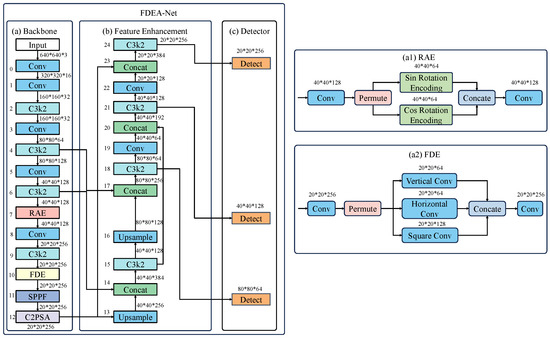
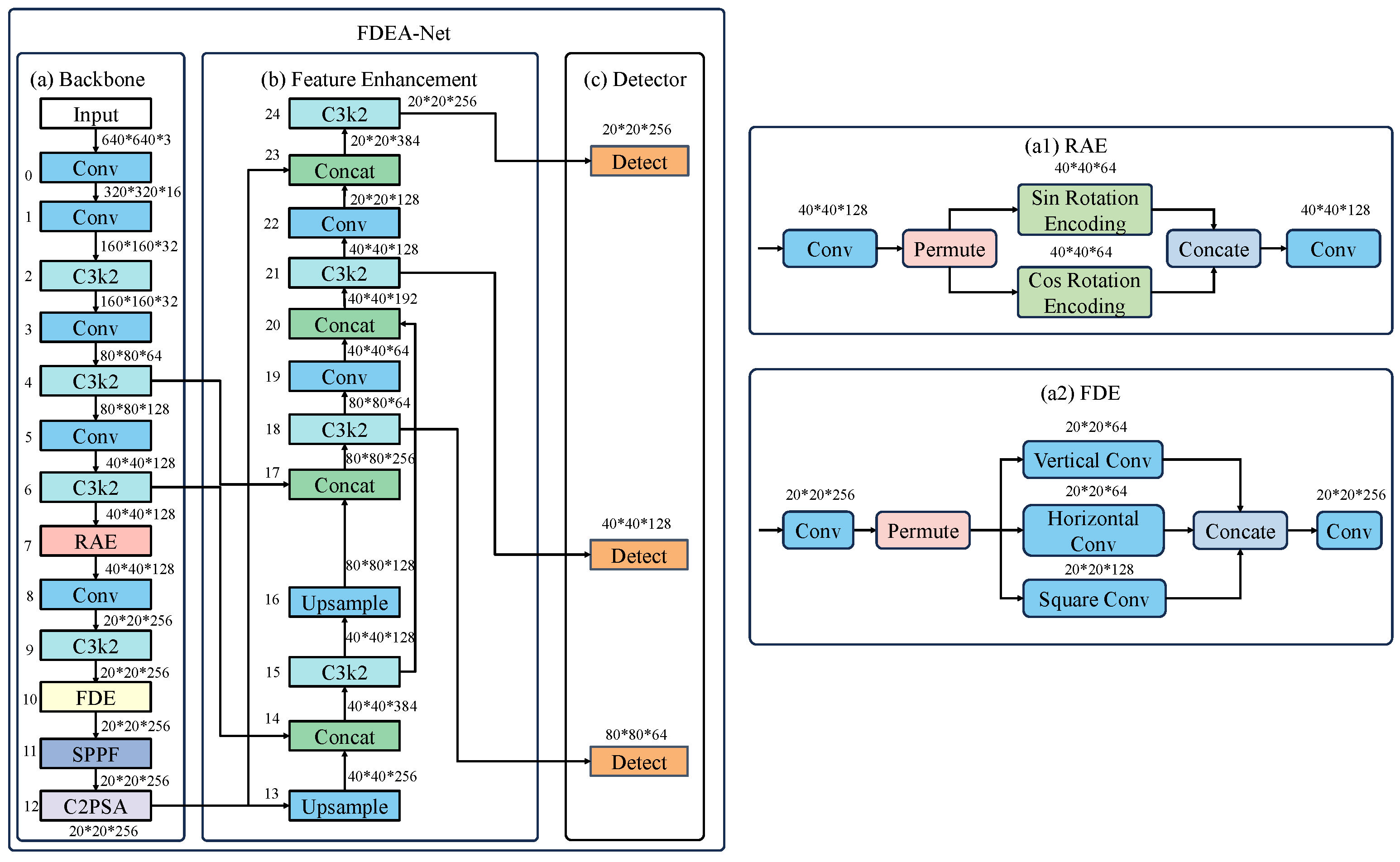
The overall architecture of FDEA-Net consists of three components: the backbone, the feature enhancement module, and the Detector. Its framework is shown in Figure 1. The backbone is built on the multi-scale pyramid structure of YOLO11. For fracture detection, shallow features play a crucial role in capturing fine fracture patterns. Therefore, we insert the RAE module into the shallow stages of the backbone to improve the modeling of orientation-varying fracture structures. After multi-scale feature extraction, the network enters the feature enhancement stage. In this stage, the FDE module is integrated into the high-resolution branch to strengthen weakened textures that may occur during early convolution processing. The enhanced features are then fed into the detection head for bounding box regression and classification. By inserting RAE and FDE at different depths, FDEA-Net establishes a complementary mechanism that combines orientation-aware encoding with detail enhancement. This strategy improves robustness to diverse fracture shapes while increasing sensitivity to subtle fracture structures.
Figure 1.
The framework of FDEA-Net.
3.2. RAE Module
The RAE Module is designed to improve fracture detection under large orientation variability. Standard convolution layers are translation-invariant, but they are not explicitly sensitive to rotation. This often leads to unstable responses when fracture traces appear oblique, rotated, or under non-standard positioning. A direct solution is to adopt rotation-invariant or rotation-equivariant networks, such as Group Equivariant CNNs (G-CNNs). However, such methods typically require group convolutions over multiple discrete orientations and group feature maps, which increases computational and memory cost and introduces non-trivial integration overhead in modern multi-scale detectors. In contrast, RAE injects rotation sensitivity into the feature space through a learnable complex rotation, while keeping the backbone and detection pipeline unchanged. This design targets robustness to orientation changes rather than strict equivariance. It is more suitable for real clinical X-rays where rotations are continuous and often coupled with scale, noise, and anatomical overlap.
Given an input feature tensor,
the channels are divided into real and imaginary parts:
To model rotation, RAE learns spatially adaptive sine and cosine parameters:
The complex rotation encoding is defined as
Expanding the real and imaginary components yields
The final rotation-aware representation is reconstructed by concatenating the rotated components:
Compared with group convolutions that replicate features over orientations, RAE preserves the channel dimensionality and performs only element-wise mixing within each complex pair. Its additional cost is dominated by a small number of pointwise operations, which is negligible relative to the backbone computation. After rotation encoding, the fused features are passed through convolutions to enhance channel interactions and generate the final output. Overall, RAE explicitly models orientation changes in the feature space and encourages consistent structural responses under rotated fracture patterns, while remaining lightweight and easy to integrate into multi-scale detection frameworks.
3.3. FDE Module
The FDE module is designed to explicitly model the anisotropic and high-frequency characteristics of fracture patterns in X-ray images. Unlike generic attention mechanisms or texture enhancement modules that rely on global reweighting or implicit feature selection, FDE introduces a structurally constrained, direction-aware enhancement strategy tailored to the morphological properties of fracture lines.
In radiographic images, fracture manifestations are typically characterized by elongated, weakly contrasted cortical discontinuities with strong directional preference. These patterns differ fundamentally from general textures or salient regions targeted by conventional attention modules. Standard convolutional layers and channel-wise attention often smooth or suppress such fine structures, especially in low-contrast regions. To address this issue, FDE explicitly decomposes feature enhancement along dominant spatial orientations and selectively amplifies fracture-related high-frequency responses.
Let the input feature map to be
After a permutation operation that strengthens spatial dependencies, the feature map is passed through three directional convolution paths. The first branch employs a vertical convolution with a kernel to emphasize vertically oriented fracture textures:
The second branch applies a horizontal convolution with a kernel, enhancing horizontal fracture structures:
The third branch uses a square convolution to supplement local contextual information and reinforce small-scale structural consistency:
The outputs of the three branches are concatenated to form an aggregated representation:
A convolution is then used to integrate the directional responses, followed by a residual connection that preserves the original semantic information:
By explicitly encoding orientation-constrained high-frequency responses rather than relying on global attention reweighting, FDE enhances fracture-specific structural details while maintaining robustness to background noise and anatomical overlap. This design differentiates FDE from existing attention-based or texture-oriented modules and enables more reliable detection of subtle fracture patterns in challenging radiographic conditions.
4. Experiment
4.1. Experimental Setup
The experiments were conducted on an Intel(R) i9-13900HX workstation equipped with an NVIDIA RTX 4090 GPU with 24 GB of memory. The software environment was Ubuntu 18.04, and the development platform was PyCharm 2019. All models were implemented in Python 3.8 using the PyTorch 2.0.1 deep learning framework. The number of training epochs was set to 300, and the batch size was set to 8. The momentum coefficient was fixed at 0.937. During training, all X-ray images were resized to 640 × 640 pixels, following the standard input resolution of YOLO-based detectors to balance efficiency and accuracy. Although clinical X-ray scans are high-resolution, fracture lines typically span multiple pixels as continuous cortical discontinuities rather than single-pixel patterns. Therefore, their essential structural characteristics are largely preserved after resizing. To further alleviate potential information loss caused by downsampling, FDEA-Net is explicitly designed with detail- and orientation-aware mechanisms.
4.2. Dataset
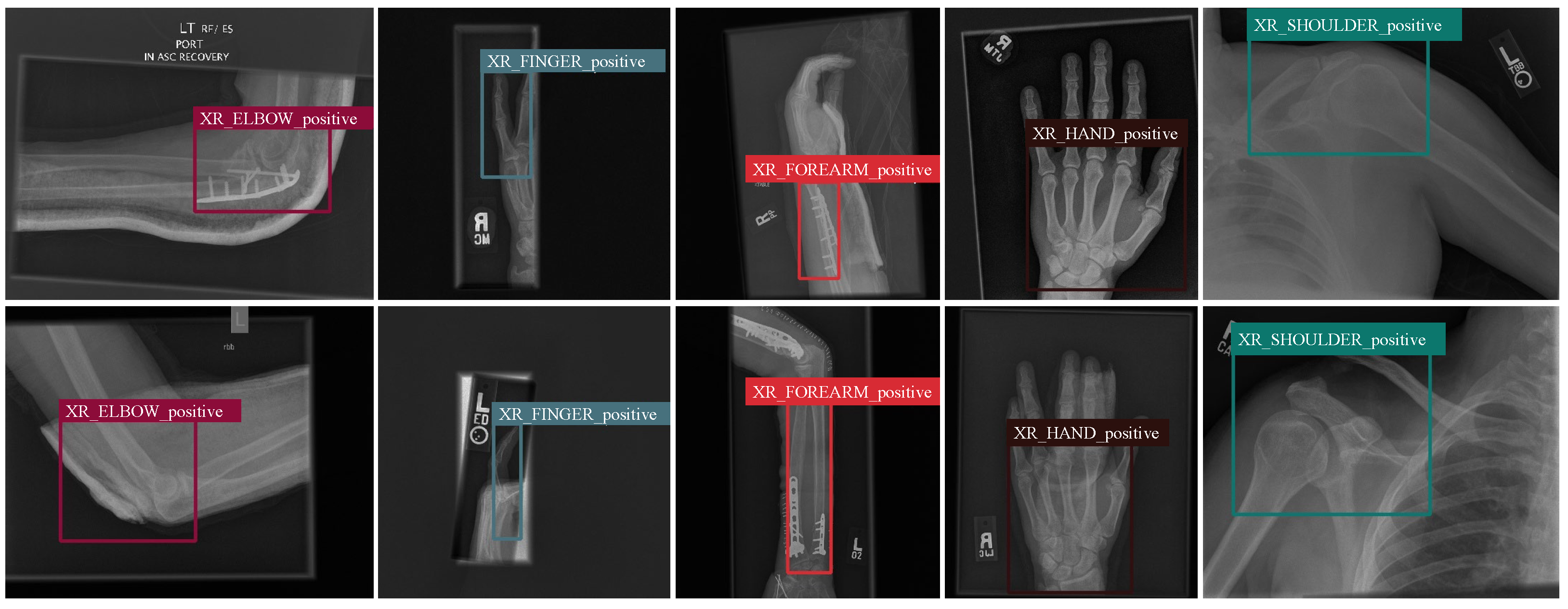
To comprehensively evaluate the performance of FDEA-Net in real clinical scenarios, we constructed a large-scale X-ray fracture dataset covering multiple anatomical sites and imaging conditions. All images were collected from routine clinical examinations. A total of 6412 X-ray images were gathered, involving fractures of the elbow, finger, forearm, hand, and shoulder. These cases exhibit subtle crack patterns, cortical discontinuities, overlapping soft tissues, and low-contrast appearance—factors that greatly increase the difficulty of automated detection. To ensure robust training and strong generalization, the dataset was divided into training, validation, and test sets using an 8:1:1 ratio. This resulted in 5129 training images, 642 validation images, and 641 testing images. Such a split preserves data diversity while reducing the risk of overfitting, allowing the final evaluation to better reflect real-world performance.
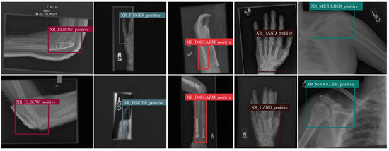
Figure 2 illustrates representative samples from different categories. The examples show significant variability in fracture shape, orientation, contrast, and background complexity. This diversity highlights the necessity of enhancing fine details and modeling directional information within the detection framework. The constructed dataset provides a realistic, challenging foundation for experimental evaluation, enabling a reliable assessment of the clinical applicability of FDEA-Net.
Figure 2.
Examples of bone fracture X-ray images.
4.3. Comparative Experiments
To systematically evaluate the performance of the proposed FDEA-Net on X-ray fracture detection, we compared it with several representative object detectors. The comparison covers two-stage models (Faster-RCNN), Transformer-based detectors (Deformable-DETR and DINO), anchor-free one-stage methods (FCOS), and the latest YOLO families (YOLOv11, YOLOv12, YOLOv13). All models were trained using the same dataset split, optimization strategy, and evaluation metrics. This ensures fair and consistent comparisons across methods.
Table 1 reports the results on five key indicators: Precision, Recall, mAP50, mAP50–95, and F1-score. FDEA-Net achieves the best performance on all metrics. It obtains a Precision of 0.732, a Recall of 0.745, and an mAP50 of 0.742. They are the highest among all detectors. Under the stricter mAP50–95 metric, the model reaches 0.312, outperforming the baseline models by a clear margin. Its F1-score reaches 0.738, indicating a better balance between sensitivity and specificity.
Table 1.
Comparison results of different models. The bolded numbers represent the best-performing experimental results in each comparison group.
As shown in the updated Table 1, FDEA-Net achieves the best detection performance while maintaining a very compact model size of 5.23 M parameters, which is significantly smaller than most Transformer-based detectors and even smaller than the YOLO series. This demonstrates that the proposed FDE and RAE modules introduce only minimal parameter overhead while providing substantial performance gains. The detection accuracy for elbow, finger, forearm, hand, and shoulder fractures is 0.741, 0.735, 0.747, 0.758, and 0.729, respectively. These results indicate that the performance variation across anatomical sites remains within a narrow range. This suggests that FDEA-Net maintains stable detection capability across different anatomical structures and does not show strong site-dependent bias.
These improvements arise from the complementary strengths of the two proposed modules. The FDE module enhances the visibility of fine fracture lines and strengthens local high-frequency patterns. It makes subtle fractures more distinguishable in the feature space. The RAE module encodes rotation-sensitive patterns, enabling robust recognition across diverse fracture orientations. Together, they allow FDEA-Net to handle fractures of varying shapes, directions, and contrast levels. The model can extract diagnostic cues even under complex backgrounds and overlapping anatomical structures.
Overall, the comparative results demonstrate that FDEA-Net offers clear advantages for X-ray fracture detection. Its strong performance and stable behavior suggest good potential for extension to other medical imaging tasks.
4.4. Qualitative Comparison
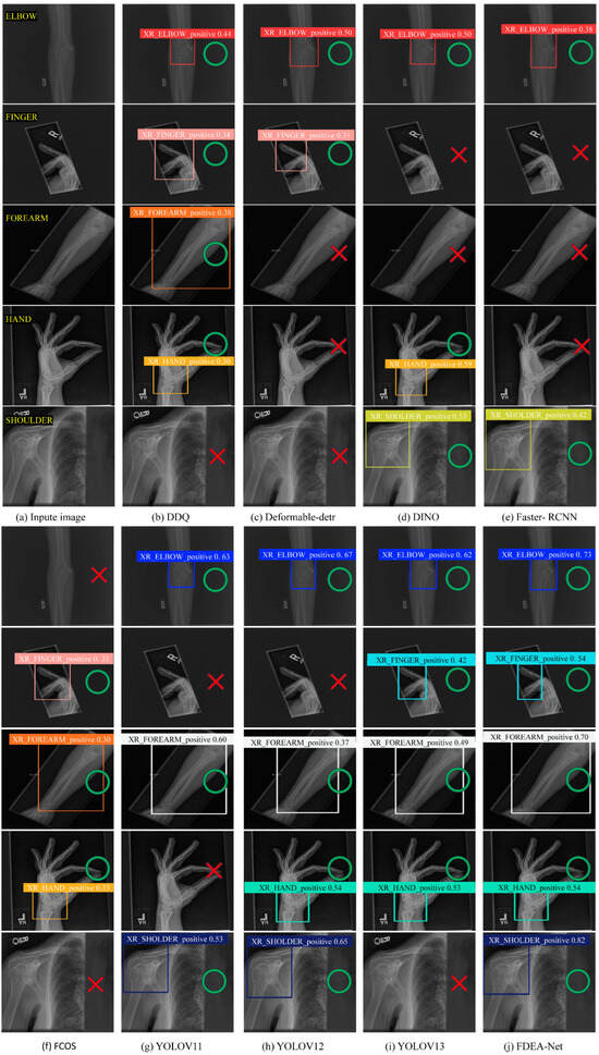
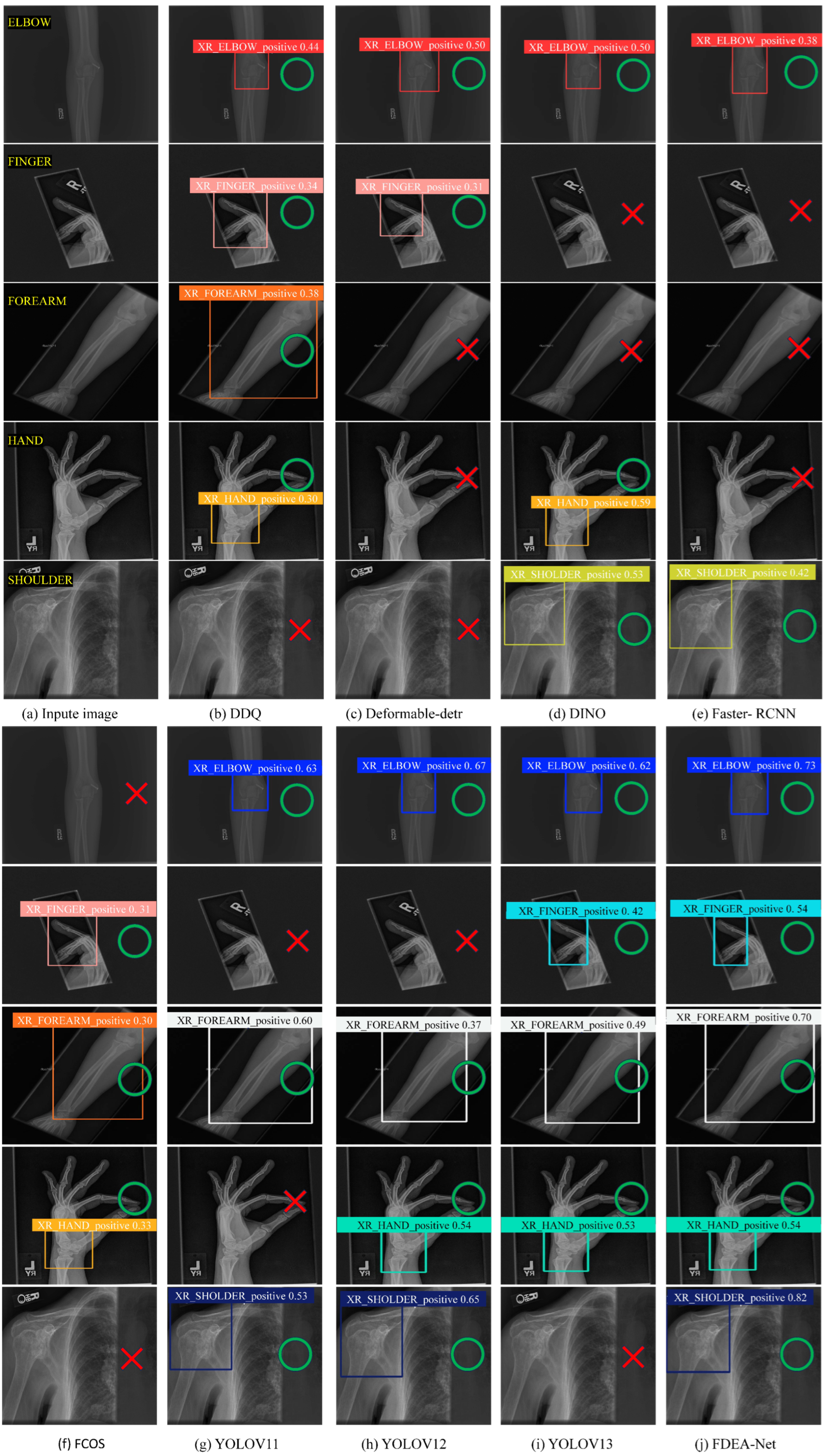
To further demonstrate the advantages of FDEA-Net in modeling fine-grained fracture structures, we present qualitative comparisons across five representative anatomical categories: ELBOW, FINGER, FOREARM, HAND, and SHOULDER. As shown in Figure 3, the left column displays the original X-ray images, followed by the predictions of competing methods and the results of our FDEA-Net.
Figure 3.
Comparison of different models. Green circles indicate a true positive prediction. Red X marks indicate a false negative prediction.
The visual comparison reveals several consistent patterns. Traditional Transformer-based or anchor-free detectors struggle under challenging conditions. These failures occur when fracture textures are blurred, orientations vary widely, or background noise is complex. For example, DINO and FCOS fail to detect fractures in the FOREARM cases, while Deformable-DETR shows clear localization shifts in the FINGER samples. Although the YOLO series performs more robustly, it still struggles to reliably capture fracture regions under low contrast or non-standard viewing angles.
In contrast, FDEA-Net produces more accurate, clearer, and more stable detections across all anatomical categories. In ELBOW and FOREARM images, the framework successfully identifies subtle cortical disruptions even under weak contrast. In FINGER and HAND cases, it captures slight, multi-directional fracture traces with high consistency. For SHOULDER images, FDEA-Net outputs tighter bounding boxes with substantially higher confidence.
4.5. Ablation Study
To further evaluate the individual contributions and joint benefits of the proposed FDE and RAE modules, we conduct step-by-step ablation experiments by integrating each module into the original YOLO11 framework. Table 2 reports the detailed performance metrics, including Precision, Recall, mAP50, mAP50–95, and F1-score. As shown, the original YOLO11 baseline (P = 0.696, R = 0.716, mAP50 = 0.714, F1 = 0.706) shows consistent improvement when either module is added. Incorporating the RAE module significantly enhances the model’s ability to recognize fractures under varied orientations, increasing mAP50 to 0.723 and F1-score to 0.721. This indicates that rotation-aware encoding effectively mitigates the intrinsic limitation of convolutional layers in modeling directional variations.
Table 2.
Ablation study of the proposed modules. The symbol ✓ indicates that the corresponding module is enabled, while blank entries denote the baseline YOLO11. The last row with both ✓ marks corresponds to the full FDEA-Net. The bolded numbers represent the best-performing experimental results in each comparison group.
Adding the FDE module yields even greater gains in fine-detail and texture representation. The mAP50 rises to 0.731, and the F1-score improves to 0.722, demonstrating that the direction-sensitive convolution in FDE effectively highlights subtle fracture structures. When both FDE and RAE are combined, the full FDEA-Net achieves the best overall performance, reaching an mAP50 of 0.742, an mAP50–95 of 0.312, and an F1-score of 0.738. These results strongly confirm the complementary nature of detail enhancement and rotation-aware feature modeling in X-ray fracture detection.
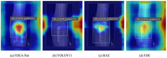
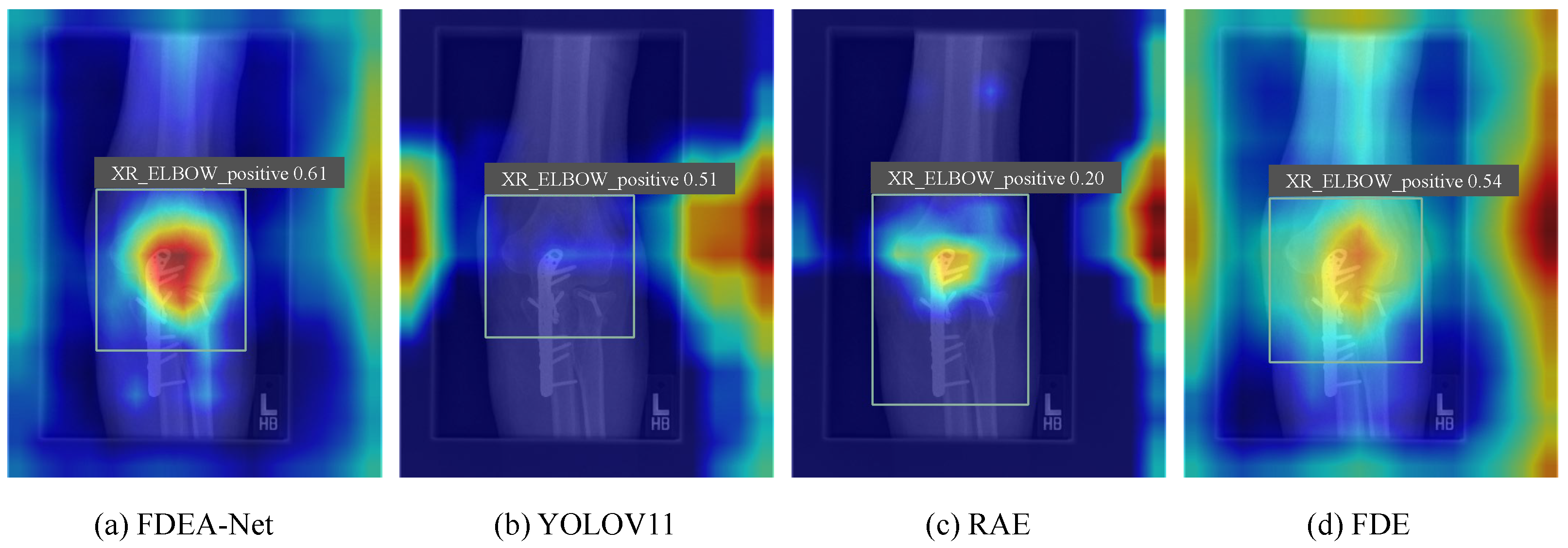
Figure 4 further illustrates the complementary effects of the proposed modules through heatmap visualizations. The YOLO11 baseline shows dispersed and weak activation, failing to capture subtle fracture patterns. With the addition of RAE, the network becomes more sensitive to directional cues, but its attention remains locally constrained. FDE enhances high-frequency fracture textures and focuses activation along crack lines, yet lacks robustness to orientation changes. When both modules are combined, FDEA-Net produces the most concentrated and continuous activation, simultaneously strengthening detail representation and directional awareness. These visual results confirm that detail enhancement and orientation modeling address complementary aspects of fracture detection.
Figure 4.
Ablation result of the models.
4.6. Comparative Experiments on Different Datasets
To further evaluate the generalization capability of the proposed FDEA-Net, we conducted additional experiments on an external public dataset, namely the Human Bone Fracture Multi-modal Image Dataset (HBFMID). Unlike the self-constructed clinical dataset used in previous experiments, HBFMID provides a more diverse data distribution and imaging conditions, making it suitable for assessing cross-dataset robustness [38].
The HBFMID contains a total of 1539 human skeletal images, covering both X-ray and MRI modalities. It includes multiple anatomical sites such as the elbow, finger, forearm, humerus, shoulder, femur, tibia, knee, condyle, wrist, spine, as well as normal cases. This diversity introduces significant variations in imaging modality, anatomical structure, and fracture appearance, posing a challenging generalization scenario.
For this experiment, FDEA-Net was trained on the original clinical X-ray fracture dataset and directly evaluated on the X-ray subset of HBFMID without fine-tuning. The same evaluation metrics were adopted for consistency. As shown in Table 3, FDEA-Net achieves a Precision of 0.961, a Recall of 0.963, an mAP50 of 0.975, and an mAP50-95 of 0.664, demonstrating strong generalization performance under unseen data distributions.
Table 3.
Generalization performance comparison on the HBFMID. The bolded numbers represent the best-performing experimental results in each comparison group.
Compared with other representative detection models, FDEA-Net consistently outperforms the baselines on the HBFMID. These results indicate that the proposed detail-enhancement and rotation-aware mechanisms effectively capture fracture-related structural cues that remain stable across datasets and imaging conditions. The strong performance on an external public dataset further confirms the robustness and general applicability of FDEA-Net in real-world clinical scenarios.
5. Discussion
FDEA-Net targets two key challenges in X-ray fracture detection: weak visibility of fine fracture textures and instability caused by orientation variability. Experimental and visual results show that the FDE and RAE modules provide complementary feature enhancements. FDE strengthens high-frequency fracture cues through direction-aware convolutions and multi-branch aggregation, leading to clearer cortical edges and subtle crack patterns. The resulting improvements in mAP50 and mAP50–95 indicate better recall and localization accuracy. RAE, in contrast, improves sensitivity to directional changes by explicitly encoding rotation-aware features, producing more stable responses across varying imaging angles. When combined, FDE and RAE achieve the largest performance gains, confirming that reliable fracture detection requires both detail enhancement and orientation-aware modeling. This complementary design enables FDEA-Net to remain robust under low contrast, background noise, and irregular imaging postures.
Despite these advantages, several limitations remain. Extremely subtle micro-fractures with contrast levels close to imaging noise, fractures heavily obscured by overlapping anatomical structures, and ambiguous cortical irregularities that resemble projection artifacts may still pose challenges. These cases largely reflect inherent limitations of single-view X-ray imaging rather than deficiencies of the proposed framework. Future work will explore multi-scale rotation modeling and the integration of anatomical priors to further improve robustness, generalization, and its applicability as a computer-aided clinical decision-support tool.
6. Conclusions
This study presents FDEA-Net, a fracture detection framework that integrates FDE and RAE to address the loss of subtle crack details and variations in fracture orientation. By reinforcing high-frequency textures and encoding rotation-sensitive features, the model achieves robust and accurate detection. Experiments on a large-scale X-ray dataset demonstrate clear performance gains. The results confirm that combining detail enhancement with rotation modeling is effective for complex clinical scenarios. The proposed modules are lightweight and can be applied to other medical imaging tasks. Future work will explore multi-scale rotation modeling and broader clinical generalization.
Author Contributions
X.Y.: Writing—review and editing, Writing—original draft, Visualization, Validation, Software, Methodology, Investigation, Conceptualization. M.W.: Writing—review and editing, Resources, Project administration, Methodology, Funding acquisition, Conceptualization. C.H.: Writing—review and editing, Project administration, Methodology, Conceptualization. All authors have read and agreed to the published version of the manuscript.
Funding
This work was supported by Science Research Project of Hebei Education Department Research on Image Denoising Method Based on Diffusion Model under Grant ZC2025081.
Data Availability Statement
The data used in this study are derived from actual clinical practice. Due to privacy and ethical restrictions, the data cannot be made publicly available and can only be provided upon reasonable request.
Conflicts of Interest
The authors declare no conflicts of interest.
Abbreviations
The following abbreviations are used in this manuscript:
| FDEA-Net | Fracture Detail Enhancement and Rotation-Aware Encoding Network |
| FDE | Fracture Detail Enhancer |
| RAE | Rotation Aware Encoder |
| CNN | Convolutional Neural Network |
| mAP50 | mean Average Precision at Intersection over Union 50 |
| IoU | Intersection over Union |
| GPU | Graphics Processing Unit |
| YOLO | You Only Look Once |
| DETR | Detection Transformer |
| FCOS | Fully Convolutional One-Stage Object Detection |
| SVM | Support Vector Machine |
References
- Zhao, X.; Dong, Y.H.; Xu, L.Y.; Shen, Y.Y.; Qin, G.; Zhang, Z.B. Deep bone oncology Diagnostics: Computed tomography based Machine learning for detection of bone tumors from breast cancer metastasis. J. Bone Oncol. 2024, 48, 100638. [Google Scholar] [CrossRef] [Scilit]
- Ahmed, K.D.; Hawezi, R. Detection of bone fracture based on machine learning techniques. Meas. Sens. 2023, 27, 100723. [Google Scholar] [CrossRef] [Scilit]
- Nguyen, H.P.; Hoang, T.P.; Nguyen, H.H. A deep learning based fracture detection in arm bone X-ray images. In 2021 International Conference on Multimedia Analysis and Pattern Recognition (MAPR); IEEE: New York, NY, USA, 2021; pp. 1–6. [Google Scholar]
- Tariq, M.; Choi, K. YOLO11-Driven Deep Learning Approach for Enhanced Detection and Visualization of Wrist Fractures in X-Ray Images. Mathematics 2025, 13, 1419. [Google Scholar] [CrossRef] [Scilit]
- Tanzi, L.; Vezzetti, E.; Moreno, R.; Moos, S. X-ray bone fracture classification using deep learning: A baseline for designing a reliable approach. Appl. Sci. 2020, 10, 1507. [Google Scholar] [CrossRef] [Scilit]
- Elsheikh, A.M.; Elhag, A.A. Machine learning-based analysis of multi-region bone fracture detection and classification using biomedical images. Alex. Eng. J. 2025, 128, 186–199. [Google Scholar] [CrossRef] [Scilit]
- Unal, M.; Unlu, R.; Uppuganti, S.; Nyman, J.S. Prediction of biomechanical properties of ex vivo human femoral cortical bone using Raman spectroscopy and machine learning algorithms. Bone Rep. 2025, 26, 101870. [Google Scholar] [CrossRef] [Scilit]
- Nkonda, G.F.; Selvaraj, J.; Umapathy, S. Bone Fracture Identification in Upper Limbs of Children Using Deep Learning Techniques. In 2024 International Conference on Distributed Systems, Computer Networks and Cybersecurity (ICDSCNC); IEEE: New York, NY, USA, 2024; pp. 1–6. [Google Scholar]
- Zhang, S.; Wang, X.; Wang, J.; Pang, J.; Lyu, C.; Zhang, W.; Luo, P.; Chen, K. Dense distinct query for end-to-end object detection. In Proceedings of the IEEE/CVF Conference on Computer Vision and Pattern Recognition, Vancouver, BC, Canada, 17–24 June 2023; pp. 7329–7338. [Google Scholar]
- Medaramatla, S.C.; Samhitha, C.V.; Pande, S.D.; Vinta, S.R. Detection of hand bone fractures in X-ray images using hybrid yolo nas. IEEE Access 2024, 12, 57661–57673. [Google Scholar] [CrossRef] [Scilit]
- Franco, P.N.; Maino, C.; Mariani, I.; Gandola, D.G.; Sala, D.; Bologna, M.; Franzesi, C.T.; Corso, R.; Ippolito, D. Diagnostic performance of an AI algorithm for the detection of appendicular bone fractures in pediatric patients. Eur. J. Radiol. 2024, 178, 111637. [Google Scholar] [CrossRef] [Scilit]
- Hossen, M.S.; Hoque, A.; Islam, M.T.; Kirawanich, P.; Baharuddin, M.H.; Alsaif, H.; Soliman, M.S. Dng metamaterial-inspired slotted stub antenna with enhanced gain, efficiency, and distributed current for early stage bone fracture detection applications. IEEE Sens. J. 2024, 24, 37932–37946. [Google Scholar] [CrossRef] [Scilit]
- Kutbi, M. Artificial intelligence-based applications for bone fracture detection using medical images: A systematic review. Diagnostics 2024, 14, 1879. [Google Scholar] [CrossRef] [Scilit]
- Verma, A.; Kumar, V.; Sharmila; Yadav, R. Humerus bone fracture detection utilizing YOLOv4 algorithm: A deep learning approach. In 2024 2nd International Conference on Disruptive Technologies (ICDT); IEEE: New York, NY, USA, 2024; pp. 1191–1196. [Google Scholar]
- Wang, M.; Li, M. CASIA-Net: An indoor work site smoking detection framework. Comput. Ind. 2025, 173, 104383. [Google Scholar] [CrossRef] [Scilit]
- Meena, T.; Roy, S. Bone fracture detection using deep supervised learning from radiological images: A paradigm shift. Diagnostics 2022, 12, 2420. [Google Scholar] [CrossRef] [Scilit] [PubMed]
- Parvin, S.; Rahman, A. A real-time human bone fracture detection and classification from multi-modal images using deep learning technique: S. Parvin and A. Rahman. Appl. Intell. 2024, 54, 9269–9285. [Google Scholar] [CrossRef] [Scilit]
- Sun, H.; Wang, X.; Li, Z.; Liu, A.; Xu, S.; Jiang, Q.; Li, Q.; Xue, Z.; Gong, J.; Chen, L.; et al. Automated rib fracture detection on chest X-ray using contrastive learning. J. Digit. Imaging 2023, 36, 2138–2147. [Google Scholar] [CrossRef] [Scilit] [PubMed]
- Alam, A.; Al-Shamayleh, A.S.; Thalji, N.; Raza, A.; Morales Barajas, E.A.; Thompson, E.B.; de la Torre Diez, I.; Ashraf, I. Novel transfer learning based bone fracture detection using radiographic images. BMC Med. Imaging 2025, 25, 5. [Google Scholar] [CrossRef] [Scilit] [PubMed]
- Wu, H.Z.; Yan, L.F.; Liu, X.Q.; Yu, Y.Z.; Geng, Z.J.; Wu, W.J.; Han, C.Q.; Guo, Y.Q.; Gao, B.L. The Feature Ambiguity Mitigate Operator model helps improve bone fracture detection on X-ray radiograph. Sci. Rep. 2021, 11, 1589. [Google Scholar] [CrossRef] [Scilit]
- Liu, Y.; Cheng, X.; Xu, N.; Wang, L.; Wang, X.; Zhong, X. MFAE-YOLO: Multi-Feature Attention-Enhanced Network for Remote Sensing Images Object Detection. IEEE Trans. Geosci. Remote Sens. 2025, 63, 5631214. [Google Scholar] [CrossRef] [Scilit]
- Sutradhar, D.; Fahad, N.M.; Raiaan, M.A.K.; Jonkman, M.; Azam, S. Cervical spine fracture detection utilizing YOLOv8 and deep attention-based vertebrae classification ensuring XAI. Biomed. Signal Process. Control 2025, 101, 107228. [Google Scholar] [CrossRef] [Scilit]
- Nouri Moqadam, A.; Kazemi, R. Design of a novel dual-polarized microwave sensor for human bone fracture detection using reactive impedance surfaces. Sci. Rep. 2023, 13, 10776. [Google Scholar] [CrossRef] [Scilit]
- Ju, R.Y.; Cai, W. Fracture detection in pediatric wrist trauma X-ray images using YOLOv8 algorithm. Sci. Rep. 2023, 13, 20077. [Google Scholar] [CrossRef] [Scilit]
- Alwzwazy, H.A.; Alzubaidi, L.; Zhao, Z.; Gu, Y. FracNet: An end-to-end deep learning framework for bone fracture detection. Pattern Recognit. Lett. 2025, 190, 1–7. [Google Scholar] [CrossRef] [Scilit]
- Zou, J.; Arshad, M.R. Detection of whole body bone fractures based on improved YOLOv7. Biomed. Signal Process. Control 2024, 91, 105995. [Google Scholar] [CrossRef] [Scilit]
- Scutelnicu, L.A.; Forna, N.; Luca, M. An overview of techniques for automatic detection of bone fractures. Procedia Comput. Sci. 2025, 270, 1557–1567. [Google Scholar] [CrossRef] [Scilit]
- Patel, Y.; Patel, D.; Patel, H.; Gupta, R.; Tanwar, S. Faster R-CNN based Framework For Bone Fracture Detection in Telesurgery Systems in Healthcare 4.0. In 2024 5th IEEE Global Conference for Advancement in Technology (GCAT); IEEE: New York, NY, USA, 2024; pp. 1–6. [Google Scholar]
- Song, D.; Peng, Y.; Gu, X.; U, K. A Lightweight YOLOv8-Based Network for Efficient Corn Disease Detection. Mathematics 2025, 13, 4002. [Google Scholar] [CrossRef] [Scilit]
- Peng, Y.; Li, H.; Zhang, Y.; Sun, X.; Wu, F. Scene adaptive sparse transformer for event-based object detection. In Proceedings of the IEEE/CVF Conference on Computer Vision and Pattern Recognition, Seattle, WA, USA, 16–22 June 2024; pp. 16794–16804. [Google Scholar]
- Peng, Y.; Li, H.; Wu, P.; Zhang, Y.; Sun, X.; Wu, F. D-FINE: Redefine Regression Task in DETRs as Fine-grained Distribution Refinement. arXiv 2024, arXiv:2410.13842. [Google Scholar]
- Li, F.; Zhang, H.; Liu, S.; Guo, J.; Ni, L.M.; Zhang, L. Dn-detr: Accelerate detr training by introducing query denoising. In Proceedings of the IEEE/CVF Conference on Computer Vision and Pattern Recognition, New Orleans, LA, USA, 18–24 June 2022; pp. 13619–13627. [Google Scholar]
- Ren, S.; He, K.; Girshick, R.; Sun, J. Faster R-CNN: Towards Real-Time Object Detection with Region Proposal Networks. IEEE Trans. Pattern Anal. Mach. Intell. 2017, 39, 1137–1149. [Google Scholar] [CrossRef] [Scilit]
- Tian, Z.; Shen, C.; Chen, H.; He, T. FCOS: A Simple and Strong Anchor-Free Object Detector. IEEE Trans. Pattern Anal. Mach. Intell. 2022, 44, 1922–1933. [Google Scholar] [CrossRef] [Scilit]
- Khanam, R.; Hussain, M. YOLOv11: An overview of the key architectural enhancements. arXiv 2024, arXiv:2410.17725. [Google Scholar] [CrossRef] [Scilit]
- Tian, Y.; Ye, Q.; Doermann, D. YOLOv12: Attention-Centric Real-Time Object Detectors. arXiv 2025, arXiv:2502.12524. [Google Scholar]
- Lei, M.; Li, S.; Wu, Y.; Hu, H.; Zhou, Y.; Zheng, X.; Ding, G.; Du, S.; Wu, Z.; Gao, Y. YOLOv13: Real-Time Object Detection with Hypergraph-Enhanced Adaptive Visual Perception. arXiv 2025, arXiv:2506.17733. [Google Scholar]
- Jamil, M.; Farooq, N.; Gilani, S.O. Ground truth annotation, analysis and release of data set of radiographic images of bone fractures. Pak. J. Eng. Technol. 2020, 3, 6–11. [Google Scholar]

Disclaimer/Publisher’s Note: The statements, opinions and data contained in all publications are solely those of the individual author(s) and contributor(s) and not of MDPI and/or the editor(s). MDPI and/or the editor(s) disclaim responsibility for any injury to people or property resulting from any ideas, methods, instructions or products referred to in the content. |
© 2026 by the authors. Licensee MDPI, Basel, Switzerland. This article is an open access article distributed under the terms and conditions of the Creative Commons Attribution (CC BY) license.