


Abstract
Plant endophytes can regulate host plant growth, improve stress resistance, and facilitate the biosynthesis of secondary metabolites, with great research value and application potential. However, traditional approaches for the isolation, cultivation and identification of plant endophytes are constrained by low culturability, limited species diversity, and loss of their original ecological functions inside host tissues. In recent years, integrated multi-omics strategies combining metagenomics, metatranscriptomics, and metaproteomics have exhibited the greatest potential to mitigate culturability limitations by enabling genome-guided targeted strain isolation and in situ functional activity profiling, among which the cultivation and targeted isolation of endophytes benefit most from omics integration. These approaches drive a paradigm shift from conventional blind screening to precise targeted isolation, and from generic medium culture to omics-guided rational cultivation, greatly improving the accuracy of strain identification and functional characterization. Nevertheless, current omics-based strategies still face inherent limitations including high experimental costs, complex operational procedures, and challenging data interpretation. The most critical future direction lies in establishing standardized experimental protocols and shared resource databases, combined with microfluidic platforms and artificial intelligence-assisted bioinformatics analysis, to address the core bottlenecks restricting endophyte isolation, cultivation and identification. This review is the first to systematically summarize research progress on traditional approaches, omics technologies and emerging strategies for plant endophyte isolation, cultivation and identification, highlights prevailing challenges and developmental trends in this field, and provides methodological references for the efficient exploitation and sustainable utilization of plant endophyte resources.
1. Introduction
Plant endophytes refer to microorganisms that colonize the tissues of healthy plants without causing harm to the host, including bacteria, fungi, and actinobacteria, and represent a crucial component of plant-associated microbial resources [1]. The biological functions of plant endophytes have garnered widespread academic attention, as they play key roles in promoting plant growth, enhancing stress resistance, and regulating host metabolism [2]. However, the functions of many strains remain insufficiently explored [3], making the development and utilization of endophytic microbial resources challenging. Therefore, the isolation, cultivation, and identification of plant endophytes are widely regarded as crucial foundational steps for conducting functional analysis and studies exploring underlying functional mechanisms.
Although traditional research methods have established a mature technical system, they are constrained by limitations such as low isolation efficiency, a limited range of cultivable microbial species, and low identification accuracy [2,4]. These bottlenecks make it difficult to meet the growing demands for the in-depth exploration of endophytic microbial resources. In recent years, omics technologies have increasingly become essential tools for studying the complex biological characteristics of plant endophytes. Guided by omics approaches, new research paradigms have been developed to enhance the cultivability of endophytes and integrate accurate identification methods. These advancements provide new insights and support for the deeper exploration of plant endophytes, shifting from community structure analysis to the elucidation of functional interactions. Single-omics technologies, including genomics, transcriptomics, metabolomics, and proteomics, are generally fundamental approaches that allow for the analysis of endophyte characteristics from multiple levels, such as genetics, gene expression, protein, and metabolism. On this basis, multi-omics research strategies combining metagenomics, metatranscriptomics, and metabolomics are employed to characterize endophytic microbial community structures, offering a comprehensive analysis of the metabolic networks, genetic backgrounds, and expression regulation patterns of plant endophytes [5,6]. In particular, metabolomics can unravel metabolic dependencies among endophytes and clarify plant–endophyte chemical crosstalk [7]. Furthermore, emerging omics technologies, such as single-cell omics and spatial omics, provide new perspectives for the refined and precise study of plant endophytes [8,9]. Among spatial omics tools, MALDI-MSI has been widely applied to in situ localization of metabolites secreted by endophytes, while spatial transcriptomics maps microscale symbiotic niches. In contrast, single-cell omics applications for general endophytes remain scarce, with most studies confined to mycorrhizal and nodule symbiosis [10].
The innovative application of omics technologies has provided new perspectives for analyzing the growth, metabolism, and interaction mechanisms of plant endophytes, offering strong technical support for targeted isolation, efficient cultivation, and accurate identification of these microbes. Xiong et al. used proteomics and transcriptomics to deeply explore the interaction mechanisms between the rice endophyte Streptomyces hygroscopicus OsiSh-2 and its host [11]. Li et al. integrated multiple omics technologies to successfully isolate nitrogen-fixing endophytes from Miscanthus [12]. Deciphering these interaction mechanisms reveals the in situ nutritional demands of strains, which facilitates medium formulation and enhances endophyte culturability. The innovation of omics technologies has facilitated the development of a technical system for targeted screening, optimized cultivation design, and precise species and functional identification of plant endophytes. This has provided key technical support for the exploration and utilization of endophytic microbial resources, while also clarifying future research directions. The core technological processes of isolation, cultivation, and identification using traditional methods and omics technologies are illustrated in Figure 1.
Figure 1.
Overview of isolation, cultivation, and identification methods for plant endophytes. Blue region: stepwise workflow of traditional isolation and cultivation methods. Purple region: sequential omics-driven workflow from multi-omics detection and data analysis to optimization feedback and targeted isolation. Green region: identification methods (upper: traditional; lower: omics-driven). Arrows indicate workflow direction.
This paper aims to provide a comprehensive review of the technical advances in the isolation, cultivation, and identification of plant endophytes. To the best of our knowledge, this work is the first to systematically synthesize and compare traditional approaches, omics technologies, and emerging strategies along the full technical pipeline for the isolation, cultivation, and identification of plant endophytes. It outlines the optimization strategies for these techniques in the field, discusses future development directions and challenges, and aims to offer technical insights for the exploration and development of plant endophyte resources.
2. Plant Endophytes
Plant endophytes exhibit high species diversity, primarily including bacteria (including actinobacteria) and fungi, as well as archaea and protists [2,13,14]. Among them, the primary bacterial groups include common genera such as Bacillus, Streptomyces, Pseudomonas, and Rhizobium [15]. The main fungal endophytes consist of genera such as Colletotrichum, Aspergillus, Alternaria, and Nigrospora [16]. In addition, some microalgae and viruses are also classified as endophytes, yet archaea, protists, microalgae and viruses receive far less research focus compared with bacteria and fungi [17]. Plant endophytes exert diverse biological functions, including improving host stress tolerance and regulating ecological processes. Endophytes enhance plant growth and tolerance to biotic and abiotic stresses via nutrient activation, hormone regulation and immune modulation [18,19]. They also participate in ecological nutrient cycling through hyphal networks and metabolic pathways, facilitating soil nutrient activation and supporting the restoration of degraded grassland ecosystems [20,21]. Given their broad physiological and ecological capabilities, efficient pipelines for isolating, culturing and accurately identifying endophytes must be established to tap into the potential of these valuable functional strains.
In recent years, plant endophytes have become a research hotspot in botany, microbiology, ecology and related interdisciplinary fields due to their diverse functions and broad application prospects [22,23]. Endophytes are deemed critical for developing biofertilizers and biological control strategies in agriculture [24]. The sugarcane-derived endophyte Serratia marcescens SM exhibits notable insecticidal activity [25]. Endophytes are increasingly recognized as an important source of natural bioactive compounds, and have the capacity to efficiently produce medicinal metabolites such as paclitaxel, vincristine and camptothecin [26]. They can also improve rhizosphere microenvironments and help enhance the cadmium phytoremediation capacity of hyperaccumulator plants [27].
Plants and endophytes have formed stable obligate symbiosis over long-term coevolution, which represents the distinctive natural history of the endophytic microbiome. Endophytes colonize plant tissues through vertical seed transmission and environmental vector-mediated horizontal colonization, assembling intricate communities composed of dominant core strains and abundant low-abundance rare strains [2,28]. Most endophytes develop stringent nutritional requirements during long-term symbiosis with host plants, such that only a small fraction can be recovered via artificial cultivation. Furthermore, closely related strains show minimal divergence in morphological features and conventional molecular markers; traditional identification approaches cannot resolve species boundaries, and evolutionary clues for undescribed novel lineages remain insufficient [16]. Although endophytes hold broad potential in agriculture, pharmacy and environmental restoration, their in-depth research and industrial use are greatly limited by traditional isolation, cultivation and identification technologies.
3. Traditional Methods
3.1. Isolation: The Dilemma of Precision and the Loss of Endophytic Microbial Diversity
The isolation of traditional plant endophytes mainly includes plant tissue selection, surface sterilization, tissue processing, strain isolation and purification [29]. Surface sterilization is widely regarded as fundamental to the successful isolation of endophytes. However, traditional surface sterilization techniques often face a core bottleneck of surface sterilization bias: the dual dilemma of over-sterilization and insufficient sterilization, both of which can compromise the effectiveness of the isolation process. Chemical surface sterilization methods typically combine 75% ethanol with suitable disinfectants [30]. However, disinfectant concentration and sterilization duration are hard to calibrate, easily leading to incomplete surface decontamination or over-sterilization. In studies on tea plants, high sodium hypochlorite concentrations reduce leaf endophyte diversity yet maintain stem bacterial richness, demonstrating varying tissue tolerances to disinfection and the necessity of tissue-specific disinfection strategies [31,32]. In addition, host-tissue specificity has been widely reported to significantly shape endophytic community composition, with strong effects of host plant species, genotype, tissue type and developmental stage. Differences in host genetics and tissue microhabitats select different microbial populations, and community composition changes continuously during plant growth. These variations result in highly variable recovery rates of culturable strains, even under the same isolation conditions [33]. The resulting large variations in endophyte richness and abundance may further determine suitable surface sterilization intensity, isolation strategies and culture conditions.
After surface sterilization of the explants, the selection of an appropriate endophyte isolation method generally depends on the characteristics of the host plant, the type of target strains, and the research objectives. Traditional endophyte isolation methods are mainly based on two strategies: the direct plating of small surface-sterilized plant explant segments, with pure single colonies isolated and purified as microorganisms grow at the tissue edges; the coating of agar plates with homogenates of ground surface-sterilized plant tissues, with pure single colonies obtained via the dilution spread plate method [34,35]. These methods have remained classic in the field of plant endophyte research due to their simplicity and wide applicability. The blue area in Figure 1 illustrates the operational steps involved in the traditional tissue homogenization method for isolating plant endophytes. However, these methods also have several limitations, such as poor targeting and susceptibility to contamination, which may significantly hinder the comprehensive development and utilization of plant endophyte resources [16,36]. Traditional isolation methods lack targeted isolation strategies and rely solely on conventional protocols, which may limit the efficient recovery of low-abundance and difficult-to-culture plant endophytes. Metabarcoding detected 5.8-fold more endophytic taxa (n = 88) than PDA plate isolation (n = 15); more than 90% of these taxa were rare low-abundance fungal lineages that failed to grow on standard artificial media [37]. This is regarded as one of the key reasons why such methods fail to fully represent the authentic composition of plant endophyte communities.
3.2. Cultivation: Limitations of Culture Medium and Lack of Microenvironment Simulation
Traditional cultivation of plant endophytes commonly employs standard media such as LB, PDA, MS, and NB. However, these media present inherent culture-medium bias and two critical limitations that lead to poor culturability: nutritional dependence and signaling dependence [4]. High-nutrient media strengthen resource competition and suppress oligotrophic endophytes [38]. Standard media are rich in carbon and nitrogen sources and tend to favor the growth of fast-growing endophytes. However, they are less effective at cultivating specific endophytes that thrive in low-nutrient environments. Some endophytes isolated from tomato seeds, including Kosakonia, Ralstonia, and Sphingobium, show limited growth on standard media but can be effectively restored by supplementing tomato tissue extract [39]. This indicates that standard media may lack essential nutrients. For such nutritionally dependent endophytes, culturability can be improved by optimizing medium components or adding specific nutrients.
Additionally, many endophytes rely on symbiotic relationships with their host plants to obtain nutrients or growth signaling molecules, which makes their cultivation more challenging under standard conditions. Dormant propagules of endophytes require plant signals to initiate germination, and nutrient supplementation alone cannot break dormancy [39]. Studies have shown that ethylene and abscisic acid secreted by sugarcane can promote and regulate the colonization of endophytic nitrogen-fixing bacteria [40]. Studies have also confirmed that adding plant tissue extracts to the culture medium can effectively improve the isolation and regrowth efficiency of recalcitrant endophytes [41]. This supports the idea that the growth of some endophytes requires signaling molecules for regulation. Therefore, incorporating plant-derived signaling compounds into standard culture media is likely to satisfy the growth regulatory needs of these endophytes.
Furthermore, loss of symbiotic function during in vitro cultivation is another critical bottleneck. Endophytes may gradually lose their essential interactive functions with plants when cultured in the laboratory [42]. The Macrophomina phaseolina F80 endophytic fungus from Arabidopsis roots is unable to convert and produce iron-chelated feruloyl-coumarin in the absence of coumarin secreted by the host plant, thereby losing its core function in mediating plant iron nutrition [43]. Traditional culture conditions often fail to replicate the microenvironment and symbiotic characteristics inside plant tissues. Owing to their simplistic nutrient composition and lack of host signaling molecules, these conditions can greatly restrict the resuscitation and normal growth of recalcitrant endophytes. Although traditional cultivation techniques are well established and easy to operate, they recover as little as 1% of total endophyte diversity, meaning over 99% of endophytes cannot be captured via standard culture approaches [44]. These methods are limited by low cultivability and the loss of in situ functionality, which may significantly hinder the in-depth exploration of plant endophyte resources.
3.3. Identification Technology: Low Resolution and Incomplete Database
Traditional endophyte identification methods are primarily based on morphological identification, physiological and biochemical tests, and early molecular techniques (Figure 1) [4]. This system is intuitive and easy to operate, allowing preliminary classification of strains based on colony, spore morphology, and structural features. However, phenotypic identification presents critical limitations including high subjectivity and frequent taxonomic misclassification, leading to low taxonomic resolution in traditional phenotypic identification methods [45,46,47].
The emergence of early molecular identification techniques improved accuracy to some extent; however, these methods, which rely on sequence conservancy and polymorphism, still suffer from insufficient resolution. The commonly used 16S rRNA for endophyte identification can suffer from high sequence similarity among closely related species and insufficient resolution at the species level [48]. Studies have shown that the classification results based on the 16S rRNA gene exhibit significant discrepancies when compared to those derived from genomic evolutionary relationships [49]. As the universal DNA barcode for fungi, ITS exhibits insufficient species-level resolution for certain plant-associated and endophytic fungal taxa. Furthermore, marked length variation and pronounced primer amplification bias may further compromise the reliability of fungal molecular identification [50,51]. Beyond the above two ribosomal markers, various alternative molecular markers are available for endophyte identification, including single-gene loci, functional genes and fingerprinting profiles [36]. These markers perform well for preliminary classification at the genus level. However, long-term plant–endophyte coevolution creates highly conserved gene sequences among closely related strains, so such markers lack sufficient polymorphic sites to distinguish cryptic species within the same genus.
With respect to database incompleteness, most of the endophyte gene sequences currently available in public databases belong to common bacterial phyla, with sequences from rare or habitat-specific endophytes being incomplete or even absent. Some sequences lack reference strains to verify their taxonomic accuracy, and insufficient sequence coverage may significantly impact the accuracy of annotations [2,52]. Since public databases lack type-strain reference sequences for endophytic Mollisia, rare endophytic OTUs of this group can only be roughly assigned, failing to discriminate closely related species [53]. Overall, identification relying on phenotypic traits and single conserved genes has inherent limitations, and it is difficult to achieve accurate taxonomic delineation of closely related and rare endophyte species.
In summary, traditional research paradigms face issues such as the lack of standardized processes, low cultivability of endophytes, and difficulty in analyzing in situ characteristics, which have widely become key bottlenecks limiting the research and development of endophytes. Therefore, there is a growing need to build upon traditional research techniques and integrate powerful, culture-independent multi-omics-driven technologies, offering a crucial direction for overcoming the limitations in endophyte research. To systematically analyze the technical characteristics of traditional methods, omics-driven technologies, and cutting-edge techniques in the isolation, cultivation, and identification of plant endophytes, the related technological systems are summarized in Table 1.
Table 1.
Summary of methods for isolation, cultivation and identification of plant endophytes.
4. Omics-Driven Methods
Omics technologies have provided new strategies for the isolation, cultivation, and identification of plant endophytes. Currently, numerous studies have indicated that research on plant endophytes has gradually shifted from traditional methods to omics-driven approaches. Technologies such as metagenomics, metatranscriptomics, and metabolomics have been widely applied in endophyte studies, with their development timeline illustrated in Figure 2. Various omics technologies have demonstrated great potential in plants such as rice, wheat, and sugarcane. For example, Walitang et al. used proteomics to reveal the interaction mechanisms between rice and its endophytic microbes, as well as their stress response mechanisms [58]. Xia et al. employed metagenomics and metabolomics to uncover the interaction and defense mechanisms between maize and its endophytic microbes [65]. Elucidating plant–endophyte interactions and stress defense mechanisms can provide theoretical references and research insights for the targeted isolation, cultivation optimization, and functional characterization of related endophytes. A summary of the applications of relevant omics technologies in plant endophytes is provided in Table 2. Multi-omics technologies have increasingly emerged as the mainstream in plant endophyte research, addressing key limitations of traditional approaches.
Figure 2.
Timeline of omics applications in plant endophyte research, separated into the Single-Omics era (2005–2017) and Multi-Omics era (2017–2025) by horizontal arrows. Purple boxes mark the introduction of key omics technologies, and blue boxes show their corresponding representative applications in endophyte studies. All technological milestones and research cases presented in this figure are compiled from references [11,60,63,66,67,68,69,70,71,72,73,74,75].
Table 2.
List of omics-based investigations of endophytes from diverse plant sources.
4.1. Targeted Isolation: From Blind Screening to Precise Screening
Traditional methods for isolating plant endophytes are typically based on random screening and cultivation dependent approaches, which are not only inefficient but also prone to losing key endophytes involved in host interactions. Omics-driven targeted strain selection involves extracting and sequencing the total DNA of the host plant’s endophytic microbial community, allowing for the identification of relevant core genes. The workflow proceeds sequentially: raw reads are assembled and binned into MAGs, followed by dRep dereplication, quality evaluation, and functional annotation via KEGG and COG databases; SIP and FISH further assist in screening metabolically active endophytes in planta [85,86]. This is followed by constructing a candidate strain library and selecting core candidate strains based on omics data. The quality of recovered MAGs is evaluated following MIMAG benchmarks, which define medium-quality genomes with completeness ≥50% and contamination <10% [87]. Considering the frequent fragmentation of plant endophyte genomes, a higher completeness cutoff should be applied. Next, qualified MAGs are utilized to pinpoint functional genes, which guide subsequent targeted isolation and experimental validation to recover viable target strains, improving the efficiency of isolating desired strains.
The metagenomic-based functional prediction isolation strategy involves high-throughput sequencing of plant endophyte communities, combined with binning techniques to obtain high-quality MAGs of endophytes. Key features of this method are listed in Table 1. This approach constructs a non-redundant gene set of the endophytic microbiome and identifies key functional sequences. Carrión et al. sequenced the metagenome of beetroot root endophytes and applied binning techniques to obtain high-quality MAGs [88]. They successfully identified functional genes related to disease resistance and biosynthesis, and isolated endophytic strains with disease-resistant capabilities. Nevertheless, whether the functional genes of isolated endophytes are expressed requires further validation through metatranscriptomics. Metatranscriptomics, using the DESeq2 differential analysis tool combined with the apeglm algorithm to correct log2FC values, identified significant upregulation of target genes involved in root microbiome phosphorus transport and energy metabolism. These gene expressions were further validated through targeted experiments, providing technical support for the validation of active gene expression in microbial communities [89].
For some hard-to-isolate microorganisms, single-cell genomics focuses on culturing individual endophytic cells, overcoming the limitations of cultivation-dependent methods. SM et al. successfully constructed the genome of rare endophytes from the roots of poplar using single-cell genomic sequencing [71], providing a methodological basis for isolating rare endophytes. Cross et al. [90] proposed a reverse genomics isolation technique, where relevant targets are selected based on single-cell genomic or metagenomic data. Specific antibodies are then prepared, fluorescently labeled, and combined with flow cytometry for targeted isolation of the desired microorganisms. This strategy provides a practical paradigm for translating sequence information directly into culturable target strains. Even so, single-cell sequencing is hampered by uneven whole-genome amplification, which introduces obvious sequence bias and often results in incomplete genome assembly [71]. Furthermore, tools like dRep, which perform dereplication, can classify genomes and eliminate redundant sequences, thereby removing interference from duplicate sequences. This may significantly improve the reliability of functional gene and metabolic pathway annotations [91], providing more robust molecular support for the precise isolation of endophytes.
After identifying the core functional gene clusters, further annotation of the target genes is performed using databases such as KEGG and COG, allowing for the clarification of the metabolic potential of the target endophytes. Wang et al. [92] performed KEGG pathway analysis on the whole genome of the potato endophyte Q2H1 and identified genes related to siderophores and auxin synthesis. By integrating KEGG pathways, they further annotated and categorized its functional roles, providing precise targets for subsequent functional validation. Metagenomic high-throughput sequencing combined with binning techniques can identify key gene clusters, while metatranscriptomics validates the expression of functional genes. dRep ensures the reliability of annotations, and KEGG/COG analyses clarify metabolic potential, all of which can support the precise isolation of endophytes. Overall, the combination of omics technologies and bioinformatics tools holds the potential to establish a complete technical workflow for precise isolation of endophytes.
However, in situ functional verification of endophytes still requires further investigation. Stable isotope probing (SIP) and fluorescence in situ hybridization (FISH) are generally regarded as effective in situ functional verification techniques for validating the functions of endophytes [93,94]. By integrating SIP-based functional activity identification with FISH-derived spatial distribution data, specific primers can be designed to establish a high-throughput PCR pre-screening system. This approach allows for the rapid identification of positive clones from plant tissues, thereby enhancing the throughput of functional endophyte isolation. In studies focusing on Miscanthus sinensis, the combination of 15N2-labeled DNA-SIP technology with FISH successfully localized the ecological niche of active nitrogen-fixing bacteria [12], thereby accelerating the screening efficiency of target endophytes. The semi-automated high-throughput stable isotope technology developed by Nuccio et al. significantly enhances sample throughput and experimental reproducibility [95]. This targeted localization screening approach, originally applied to arbuscular mycorrhizal fungi, can also be adapted and applied to the isolation of plant endophytes. Thus, the integration of in situ validation techniques and omics data helps further refine the system for screening and localization of functional endophytes.
4.2. Cultivation Optimization: From General Cultivation to Rational Design Cultivation
The core prerequisite for the successful cultivation of endophytes largely lies in providing appropriate nutrition and a suitable growth microenvironment. Endophytic community structure is generally shaped by host plant species, genotype, tissue type and developmental stage, which leads to differences in nutritional requirements and growth characteristics of strains. Such host-related background characteristics should also be considered when designing culture conditions based on omics data. Unlike traditional general-purpose cultivation methods, the application of omics technologies can reveal the nutritional requirements [96], interaction mechanisms with the host plant [97], and environmental adaptation mechanisms [98] of endophytes, facilitating the shift from cultivation-dependent to design-driven cultivation. The growth factors provided by the host plant’s internal environment to endophytes can be predicted through omics data analysis, including metagenomics, metatranscriptomics, proteomics, and metabolomics. This allows for the design of growth nutrients and environments that closely resemble their natural endophytic conditions, which can enhance the cultivability and diversity of plant endophytes.
Omics technologies provide a scientific methodology for designing the cultivation of endophytes. Before medium formulation, omics annotations predict metabolic pathways, and substrate control cultures coupled with metabolite quantification validate these pathways to guide medium optimization [99]. By using metagenomic sequencing, all microbial DNA from plant tissues can be directly extracted, allowing for the assembly of metagenomes and the genomes of hard-to-culture endophytes. By using binning techniques to analyze sequence information, the key nutritional carbon sources, nitrogen sources, and growth factors required for these microorganisms can be inferred [100]. This approach enables the design of specific media, potentially overcoming the cultivation bottlenecks of hard-to-culture endophytes. Prasannakumar et al. [101] used shotgun sequencing to analyze the functional genes of the sorghum endophytic community. By integrating KEGG pathway annotations, they identified the specific carbon and nitrogen requirements of the microorganisms, providing a basis for optimizing culture media for specific endophytes. Li et al. used binning techniques to assemble a genomic draft from Miscanthus roots, identifying nifH (nitrogenase gene), czcA (heavy metal resistance gene), and acdS (plant growth-promoting gene) [12]. They further validated the metabolic activity of the endophytes through DNA-SIP, successfully isolating nitrogen-fixing endophytes.
The accuracy of genome binning has improved the targeted identification of microorganisms from host plants and enhanced the efficiency of microbial isolation and cultivation. Sieber et al. [102] proposed the DAS tool, which incorporates dereplication, aggregation, and scoring strategies. This tool, when applied to microbial genome analysis, yields a significantly higher number of endophyte types compared to other tools. It has reported the highest number of nearly complete and draft-level bins in human gut, oil seep, and soil samples. By integrating the advantages of multiple algorithms, the DAS tool achieves precise optimization of metagenomic binning, providing critical technical support for high-quality identification of metagenomic drafts. Omics technologies enable the prediction of strain nutritional preferences at the genomic level, thereby greatly reducing trial-and-error in medium formulation.
For symbiotic endophytes that rely on host plant secondary metabolites, metabolomics directly analyzes the metabolic products in plant tissues to identify the substances that influence the growth and metabolic activities of plant endophytes. Harbort et al. [103] found that coumarin compounds produced in Arabidopsis roots under iron deficiency significantly promote the growth of beneficial endophytes. Metabolomics can detect plant-microbe interactions and elucidate the key nutrients required for microbial growth. In a study on the medicinal plant Elephantorrhiza elephantina, RNA-seq-guided medium supplementation with 1% olive oil and 0.5% yeast extract increased the number of cultivable endophytic species from 4 to 17. Xu et al. performed transcriptomic analysis and found that hypoxia-response genes were highly induced in the symbiotic tuberoids of Dendrobium officinale, suggesting that endophytic fungi in the roots may exist in a low-oxygen environment [104]. This provides a basis for controlling oxygen concentration in the in vitro cultivation of endophytes. Dissection of host metabolism and environmental signals offers a critical foundation for recapitulating the native growth conditions of endophytes.
Proteomics and metaproteomics can reflect the in situ physiological status and metabolic activity of endophytes at the protein level. By identifying highly expressed functional enzymes, nutrient transporters, and signaling proteins in native environments, these approaches clarify the nutritional requirements and growth-limiting factors of endophytes, thereby providing critical support for culturomics-based high-throughput culture condition optimization and improving the culturability of recalcitrant endophytes. Yue et al. performed quantitative proteomic analysis on the wheat endophyte Bacillus sp. WR13, identifying upregulated proteins including siderophore synthetases, iron transporters, and iron-dependent functional enzymes, and confirming iron as a critical growth-limiting factor [105]. Li et al. used proteomics to characterize Mycoavidus cysteinexigens, identifying key transporters and TCA enzymes that revealed its carbon and nitrogen preferences, with nitrogen as the primary growth-limiting factor [106]. These findings indicate that proteomics facilitates the identification of nutritional limitations and targeted optimization of culture conditions, thereby enhancing the culturability of endophytes.
In addition, spatial omics enables in situ analysis of the spatial distribution and interactions between cells and molecules in tissues, preserving authentic micro-niche information. This can provide supplementary guidance for culturomics-based culture environment simulation. However, its application in plant endophytes remains limited. Serrano et al. used spatial co-transcriptomics and single-nucleus RNA sequencing to characterize the in situ spatial relationship between arbuscular mycorrhizal fungi and host roots [8]. This strategy can be applied to plant endophyte cultivation to clarify spatial interactions and support the optimization of culture systems.
Culturomics has emerged as a key strategy for design-driven cultivation of endophytes. By simulating the in situ growth environment via high-throughput methods, it can effectively improve microbial culturability and diversity [107]. However, culturomics differs from omics-guided cultivation. Culturomics aims for high-throughput strain isolation, while omics data predict microbial metabolism and nutritional needs to direct medium development. Flores-Duarte et al. [108] employed a culturomics strategy in the halophyte Mesembryanthemum crystallinum. Using host-derived medium to simulate the in situ growth environment and adopting a high-throughput culturomics workflow for microbial isolation, they significantly enhanced endophytic culturability and diversity, and characterized the plant growth-promoting, metal-resistant, and metabolic traits of the isolated strains. These studies indicate that genomic and transcriptomic data provide critical guidance for defining the nutritional requirements and rational media design of endophytes. Importantly, culturomics relies on multi-omics integration rather than blind trial and error, supporting the efficient development of tailored media for beneficial endophytes.
However, many uncultivable endophytes are closely linked to microbial interactions [75]. Microbial dark matter refers to the unknown microbial taxa and functional genes that are widely present in the environment, detectable by sequencing, but difficult to isolate and pure-culture using traditional laboratory methods [109]. Co-cultivation strategies are widely regarded as a crucial pathway for unlocking the microbial dark matter. By constructing artificial microbial communities to simulate natural ecological environments, co-cultivation activates silent biosynthetic gene clusters (BGCs) to produce novel metabolites, enhancing the yield of specific target metabolites and unlocking the metabolic potential of endophytes [110]. For example, in a co-culture system of hazelnut cells and the endophytic fungus Epicoccum nigrum, the paclitaxel yield reached 404.5 µg/L, 136.6 times higher than in monoculture [111]. Beyond traditional monoculture, co-culture has become an important supplementary technique for the targeted cultivation of endophytes. Based on intermicrobial interactions, the co-culture strategy can effectively activate the metabolic potential of endophytes, offering a novel approach to help break through the bottleneck of their unculturability.
4.3. Identification: Endophyte Identification and Functional Analysis
Omics technologies provide systematic and precise technical support for the identification and functional characterization of endophyte strains. They can help overcome the limitations of traditional identification methods, which are inefficient and restricted, enabling comprehensive analysis from the genetic to the metabolic level. Metagenomics, the FastANI algorithm, multilocus sequence analysis, and single-cell genomic sequencing can enable precise strain identification of endophytes. Meanwhile, metagenomics, metatranscriptomics, proteomics, and metabolomics can progressively advance functional characterization and mechanism elucidation (Figure 1). Metagenomics enables the acquisition of complete microbial genomic information. By comparing genomes, conducting phylogenetic analysis, and integrating functional specificity, it allows for precise strain-level identification of endophytes [76,112]. The high-throughput tool FastANI enables rapid and accurate analysis of endophyte genomic data. By combining the ANI ≥ 95% species delineation threshold with phylogenetic analysis, FastANI can efficiently distinguish closely related endophyte groups. While ANI ≥ 95% serves as the standard species cutoff for prokaryotes, it is limited by an 83–95% ambiguous range covering 0.21% of genomic comparisons and taxonomic exceptions like Escherichia-Shigella complexes [113,114]. Thus, it provides crucial support for precise strain identification and addresses the low computational efficiency of traditional BLAST methods (NCBI BLAST+ v2.10.0). To overcome the limitations of using a single gene for strain identification, multilocus sequence analysis (MLSA) selects 5–10 specific genes for PCR amplification and sequencing. These genes are mostly evolutionarily conserved housekeeping genes with rare horizontal gene transfer and moderate genetic polymorphism, which can reliably distinguish closely related strains. By integrating the variation information from multiple functional genes, MLSA significantly enhances the resolution of strain identification. Pusz-Bochenska et al. conducted MLSA of core genes such as cpn60 and tuf in hard-to-culture plant pathogenic phytoplasmas, successfully distinguishing different pathogenic strains [115]. The rise in single-cell genomic sequencing and gene-editing-assisted validation has further enhanced the precision of strain identification, extending it to the level of individual endophytes. Lundberg et al. successfully isolated uncultivable endophytes from Arabidopsis roots using single-cell genomic sequencing, performing precise species classification [116]. Compared with the high throughput of FastANI and high resolution of MLSA, single-cell genome sequencing enables precise identification of unculturable endophytes. It can compensate for the inability of the two methods to analyze individual strains, and is particularly suitable for distinguishing closely related endophytes in communities.
The application of omics technologies has addressed the long-standing dilemma that the dissection of functional mechanisms in plant endophytes was heavily reliant on cultivation, driving endophyte-related research from superficial functional prediction towards precise characterization of growth and metabolic traits. Through functional annotation against databases including KEGG and COG, metagenomics allows for systematic mining of metabolic-associated genes in uncultured endophytes, prediction of their nutritional preferences such as carbon and nitrogen utilization and substance biosynthesis, and provides theoretical support for the targeted design of culture medium formulations. Tian et al., by integrating metagenomics with KEGG pathway analysis, identified numerous genes related to plant polysaccharide degradation, carbohydrate and protein metabolism, and biological nitrogen fixation in the microbial communities of tomato root nodules [117]. It can generally be concluded that the nutritional metabolic genetic features of root endophytes are highly compatible with their in situ growth niches, which can potentially be directly employed as a critical criterion for the design of in vitro isolation and culture systems. However, the accurate expression of these functional genes requires validation through combined metatranscriptomics and proteomics analysis [118], which is key to understanding the functional mechanisms.
Metatranscriptomics reflects gene expression levels. In contrast, metaproteomics enables direct capture and identification of functional proteins expressed by endophytes in situ, including metabolic enzymes, transporters, secreted proteins, and stress-responsive proteins. It reveals metabolic activity and symbiotic phenotypes at the protein level, providing key evidence for functional mechanism analysis and helping the critical leap from “gene prediction” to “functional validation” [119,120]. Bao et al. [121] applied metaproteomics to characterize functional proteins of endophytes in rice roots. They identified key enzymes involved in nitrogen fixation and methane oxidation, confirming their in situ metabolic activities. This approach can enable precise and direct determination of endophytic functions in the native host environment.
Metabolomics directly captures the metabolic state of endophytes in their native environment and under artificial culture conditions, enabling the prediction of nutritional requirements and the optimization of culture media. Profiling metabolites in host tissues or culture systems via UPLC-MS and LC-MS/MS reveals key nutrients, growth factors, and metabolic constraints, offering direct guidance for tailored medium design. Using metabolomics and proteomics, Zhao et al. profiled the metabolic traits of the wheat endophyte Bacillus sp. WP-6, identifying key enrichment in amino acid, porphyrin, and chlorophyll metabolism under stress conditions [122]. Such metabolic traits mirror the strain’s nutritional preference, facilitating tailored medium formulation for better isolation and cultivation. Furthermore, in situ metabolomics enables the identification of host-derived growth factors; incorporating these compounds into culture media mimics native niches and can help improve the culturability of recalcitrant endophytes.
4.4. Multi-Omics Integration: Enabling Precise Isolation, Cultivation and Identification
In plant endophyte research, the in-depth application of omics technologies such as metagenomics, metatranscriptomics, metabolomics, proteomics, and single-cell genomics aims to overcome the limitations of traditional methods, including low isolation efficiency, narrow cultivation range, and insufficient identification accuracy. These advancements can provide critical support for breaking through traditional bottlenecks in endophyte research. Notably, the structure and functional characteristics of endophytic communities are generally significantly influenced by host plant species, genotype, tissue type and developmental stage. Endophytes under different host backgrounds exhibit distinct biological traits, which may further increase the difficulty of comprehensive interpretation. However, single-omics technologies still face limitations, such as only being able to predict functional potential, difficulty in elucidating the gene-function-phenotype relationship, inability to dynamically monitor endophyte growth, and low identification resolution [44]. These limitations may hinder research on rare endophytes and could fail to fully meet the technical demands. Therefore, combining multiple omics technologies, supplemented with innovative techniques, is likely to offer new strategies to overcome the restrictions in plant endophyte isolation, cultivation, and identification research [123].
The efficient development of endophyte resources largely relies on the integrated application of multi-omics and traditional cultivation. Omics technologies quickly uncover functional targets, while traditional techniques optimize cultivation conditions based on omics data to obtain pure endophytes. This aligns with the logic of culturomics and metagenomics complementarity. The combination of both approaches can hold the potential to overcome the challenges of having functional targets without endophytes or having endophytes with unclear functions. It is widely regarded as a core pathway for the utilization of endophyte resources [124]. The low cultivability of endophytes has long been recognized as a challenge in microbiological research. Transcriptomics, by analyzing the gene expression profiles of uncultivable endophytes, helps identify the substances required for their growth, thereby providing targeted guidance for endophyte isolation and significantly improving isolation efficiency [107]. Bai et al., based on transcriptomic data from Arabidopsis roots and leaves, designed tissue-specific culture media, which increased the number of cultivable endophyte species by more than five times, enriching the diversity of endophytes [125]. Huang et al., by combining metabolomics and multi-omics analyses, discovered triterpenoid compounds specifically synthesized by plant roots, which can help promote the colonization of certain beneficial microorganisms in the roots [57]. Clarifying the key nutritional requirements of the strain provides a basis for the targeted development of specialized culture media.
Integrated multi-omics analysis of plant endophytes first conducts normalization and batch-effect correction on multidimensional data from amplicon sequencing, metagenomics, metatranscriptomics, and metabolomics to ensure data comparability. Their functions, limitations, resolution, sample requirement, best use case and other relevant indicators are detailed in Table 1. Correlation analysis, multivariate statistical methods, and co-expression network analysis are then applied to reveal intrinsic associations among omics layers. Finally, functional enrichment and joint annotation are performed to achieve systematic integration and biological interpretation of the multidimensional data [126]. Lopez-Barbera et al. [127] used yeast to conduct cross-analysis integrating genomic, transcriptomic and metabolomic data for metabolic pathway verification, validating this layered integration paradigm. This strategy can also be applied to plant endophytes. However, discordant expression patterns are often observed across different omics layers including metatranscriptomics and metaproteomics, which can be corrected via statistical approaches such as weighted integration, consistency filtering and cross-validation [128]. A reasonable data correction algorithm is generally regarded as a key prerequisite to ensure that multi-omics results conform to the real physiological state of the strain. This integrated multi-omics analytical workflow is schematically illustrated in Figure 3.
Figure 3.
Schematic workflow illustrating the multi-omics integration strategy for guiding the isolation, cultivation, and identification of plant endophytes. The pipeline starts with multi-omics raw data (Step 1), followed by data preprocessing and normalization (Step 2), individual omics analysis (Step 3), and cross-omics integration using tools such as WGCNA, MetaboAnalyst 5.0, and mixOmics (Step 4). Conflicting signals between different omics layers are then calibrated (Step 5), and integrated functional interpretation is performed to identify key targets (Step 6). These results guide targeted strain isolation, medium optimization, and precise identification (Step 7), followed by experimental validation and closed-loop iteration (Step 8).
Currently, bioinformatics tools and statistical frameworks including WGCNA, MetaboAnalyst 5.0, mixOmics and iOmicsPASS are generally widely applied in plant endophyte research to conduct correlative analysis of metagenomic, metatranscriptomic and metabolomic data, thereby supporting the isolation, screening, culture optimization and taxonomic identification of endophytes. Among these approaches, WGCNA, as a classic multi-omics integration algorithm, can enable the construction of gene-metabolite co-expression networks and the identification of key co-expression modules and core regulatory factors closely associated with the growth characteristics, biological functions, isolation, cultivation and taxonomic identification of plant endophytes [60]. MetaboAnalyst 5.0 supports metabolite annotation and pathway enrichment analysis, contributing to the screening of pivotal metabolic signatures linked to strain growth and functional traits [129]. The mixOmics framework adopts multivariate statistical algorithms to achieve joint dimensionality reduction, feature screening and module correlation analysis for multi-omics data, allowing researchers to mine key factors governing microbial cultivability from high-dimensional omics datasets [130]. Furthermore, iOmicsPASS realizes cross-omics integration and functional prediction at the pathway level, and can identify core regulatory units that modulate the isolation and cultivation efficiency of endophytic strains [131]. Nevertheless, the above tools have inherent limitations. WGCNA lacks reliable cross-omics integration capacity, mixOmics is unable to resolve hierarchical regulatory networks, and iOmicsPASS entails high computational costs with limited applications in endophyte research. Some tools have high requirements for data distribution characteristics and sample size. The biological interpretation of cross-omics association results still requires manual verification, and complex data analysis processes have certain demands for computing resources and bioinformatics skills.
Multi-omics technologies can cross-validate three key dimensions: genetic basis, active expression, and functional products, providing technical support for the targeted isolation of endophytes. Taking the isolation of nitrogen-fixing endophytes as an example, metagenomics is first used to predict functional genes, followed by the construction of a plant endophyte gene set using Illumina sequencing technology, and the prediction of the nifH gene [132]. Subsequently, metagenomic and metatranscriptomic analyses are combined to examine the expression changes in nifH under different low-nitrogen conditions [133]. Then, 15N-SIP and FISH technologies are used to localize nitrogen-fixing bacteria. For example, Wang et al. employed these techniques to identify active nitrogen-fixing bacteria in rice paddy soil and validated their distribution within root cortical cells [134]. A reasonable data correction algorithm is a key prerequisite to ensure that multi-omics results conform to the real physiological state of the strain.
Integrated multi-omics analyses enable multi-level cross-validation, overcoming the limitations of single-omics approaches. Corroboration across multi-dimensional data allows precise characterization of endophyte metabolism and growth traits, providing robust molecular support for targeted isolation, culture optimization, and species identification. The integration of multi-omics with traditional culture experiments is increasingly regarded as an important trend in plant endophyte research. Currently, databases such as GenBank, KEGG, and IMG/M have amassed a wealth of omics data on plant endophytes, providing essential support for resource exploration and functional analysis. To facilitate the subsequent implementation of omics experiments, the core databases, application areas, and website links for several omics technologies are summarized in Table 3.
Table 3.
Compilation of core omics methods resources in plant endophytes research.
5. Challenges and Future Directions
Although omics technologies have significantly advanced the depth of plant endophyte research, they also face numerous challenges. Lately, the cost of high-throughput sequencing has dropped dramatically. For example, metagenomics-based sample pooling strategies can further reduce the costs of sequencing and library preparation for plant endophytic communities [145]. Notably, the cost of whole-genome sequencing has decreased markedly in recent years compared with the high pricing observed a decade ago, showing a clear downward trend across mainstream sequencing platforms. However, with the exponential growth in the output of multi-omics sequencing data, subsequent bioinformatics analysis, high-performance computing resources, professional personnel, and data storage still may present high economic and technical barriers [146,147]. These may continue to pose substantial limitations for resource-limited laboratories.
The plant–endophyte system features complex compositions and diverse metabolic backgrounds, while multi-omics data are characterized by large volumes and significant dimensional heterogeneity, imposing stringent requirements for data integration and standardized analysis. Existing multi-omics analysis tools still have limitations in terms of efficiency and accuracy when handling large volumes of data. In addition, the integration and analysis of omics data lack standardized workflows. For instance, there are variations in the preprocessing methods for metabolomic data, such as those for the microbiome, between different laboratories, making effective data comparison challenging [148].
Most current plant endophyte studies rely on in vitro cultivation or tissue extraction methods, which disrupt the original spatial distribution of plant endophytes and their interactions with plant-associated microbes. This may fail to accurately reflect the functional roles of endophytes under natural conditions. To systematically analyze the spatial distribution and interactions of different plant endophytes within plant tissues, both spatial dimension and bioinformatics approaches must be employed for in-depth analysis [149]. However, this also increases the complexity of data integration and interpretation. Therefore, there is a growing need to develop more efficient, accurate, and standardized research strategies.
The establishment of standardized experimental protocols and resource databases is fundamental to advancing the field of plant endophyte research. Currently, the lack of standardized procedures greatly restricts the collaborative development of research in this area [4]. Significant differences in the methods used by different laboratories in processing tissue data have led to difficulties in making cross-comparisons of research findings [150]. Therefore, the pre-treatment of experimental materials and the establishment of standardized laboratory standard operating procedures (SOPs) are widely considered particularly crucial. The standardized SOPs should include detailed steps for plant endophyte isolation, such as sample selection, surface sterilization, sterile procedures, strain purification and separation, and strain preservation, thereby providing a solid theoretical foundation for researchers. Novak et al. [151] developed standardized SOPs for key experimental procedures, including surface sterilization, aseptic manipulation, and culture medium sterilization. Based on these protocols, five laboratories conducted 210 aseptic experimental assessments, yielding a contamination rate of <1%, which improved the reliability of the research outcomes to some extent. Zhou et al. [152] constructed the standardized sequence alignment databases reprDB and panDB. When applied to the annotation of highly complex microbial communities, the sequencing read annotation rates reached 49.7% and 75.1%, significantly improving the accuracy of microbial identification. The development of plant endophyte resource databases could draw on the construction strategies employed in these standardized databases, such as reprDB and panDB. By integrating sequence data of plant endophytes derived from the same tissues across different host plants, it becomes possible to facilitate the rapid acquisition of relevant and informative datasets [153]. However, issues related to the standardization and ownership of plant endophyte databases still require further clarification.
The innovative application of microfluidic technologies is expected to open new avenues for the cultivation and isolation of plant endophytes, thereby promoting deeper exploration in this field. Richter et al. [154] summarized the Fungi-on-a-Chip platform, which enables the precise cultivation of fungal single cells. In particular, the double emulsion droplet-based microfluidic system can establish a micro-cultivation platform that integrates internal nutrient supply, intermediate target metabolite capture, and external stable encapsulation to simulate the microenvironment within plant tissues, thereby enabling the in situ cultivation of host-dependent fungi. This technique is particularly suited for plant endophytic fungi that are difficult to recover using conventional plate culture, and can significantly improve the culturability of these recalcitrant taxa. McCully et al. [61] developed the GrowMiDE platform, which enables the efficient enrichment of rare and difficult-to-culture microorganisms and may improve the culturability of plant endophytes. This platform utilizes double emulsion microencapsulation to entrap individual microbial cells with trace nutrients, constructing a stable microscale culture environment and further enhancing the isolation efficiency of slow-growing and oligotrophic endophytes. Liu et al. [155] established a microfluidic high-throughput screening platform that enables the co-cultivation of plant endophytes and host plants. Using this system, they successfully isolated the plant endophyte Bacillus paramycoides (PE1), which increased the root growth rate of the hyperaccumulator plant Phytolacca acinosa by 54.31% and enhanced the removal efficiency of the heavy metal Cd by 55.49%. Such a setup circumvents the limitations of conventional methods that cannot simultaneously monitor plant growth and dynamic endophyte–host interactions, providing an efficient technical strategy for the targeted screening of functional endophytes.
For difficult-to-cultivate endophytes with extremely low abundance and those highly dependent on host interactions, single-cell genomics is expected to overcome the resolution limitations of conventional omics. Fluorescence-activated cell sorting (FACS) enables targeted sorting using FISH probes, as well as label-free sorting based on cellular physical properties for high-throughput identification and isolation of single cells [156]. Endophyte cultivation guided by single-cell Raman spectroscopy-sequencing combined technology is generally more efficient and targeted [157]. The integration of microfluidic single-cell cultivation with single-cell omics can encapsulate individual endophytes in picolitre-scale droplets, allowing real-time monitoring of cellular metabolic activity and precise capture of functionally active endophytes predicted by metatranscriptomics, thereby alleviating the loss of functional endophytes in traditional cultivation to a certain extent [158]. The combination of microfluidics and mass spectrometry allows rapid detection of secondary metabolites within droplets, providing important technical support for the study of microbial secondary metabolites in droplet-based systems [159]. Notably, microfluidic techniques are generally limited by complex operation and high cost, which may restrict their widespread application.
The integrative analysis of multi-omics datasets faces substantial challenges due to the large data volume and the heterogeneity in data dimensions. Against this background, artificial intelligence (AI) may provide intelligent analytical support for endophyte research [160]. Through intelligent analysis of colony phenotypic characteristics, AI-based approaches could assist the precise screening of diverse plant endophytes [161]. Diao et al. constructed an AI-RACS platform combining microfluidic trapping and machine learning to efficiently screen rare metabolically active endophytes from plant samples [162]. By integrating microfluidic technology with AI-based visual analysis, a three-pronged platform integrating static array single-cell isolation, dual deep learning-based multi-modal phenotyping and LIB contactless sorting was built for high-throughput single-cell strain screening [62]. This technological framework could be further applied to research on plant endophytes. Evangelisti et al. [63] developed AM Finder, which adopts a two-stage hierarchical recognition mechanism: CNN1 segments images into tiles to judge root infection status and quantify overall colonization levels, while CNN2 further analyses infected regions to identify intracellular fungal structures for fine qualitative analysis. It can automatically identify and quantify the colonization of arbuscular mycorrhizal fungi in plant roots. Although AM Finder was originally built exclusively for arbuscular mycorrhizal fungi, its deep-learning image recognition framework can be transferred to the morphological screening of plant endophytic colonies. In addition, AI-driven integration of multi-omics data has helped enable the establishment of a “Design–Build–Test–Learn-Predict” research framework (DBTLP), which can utilize AI for data learning and prediction, with continuous experimental feedback to retrain the model and can be further extended to predict the optimal culture conditions for endophytes and assist in medium optimization and strain screening [64]. Notably, AI analysis relies on large-scale, high-quality annotated datasets, which remain scarce in the endophyte research field. Meanwhile, the recognition accuracy of models needs to be improved in highly complex plant-microbial communities, and a gap still exists between proof-of-concept validation and reliable practical application.
The application of omics technologies is expected to promote further advances in the isolation, cultivation, and identification of plant endophytes. Building on standardized SOPs and database development, the innovative application of microfluidic technologies could facilitate omics-guided targeted isolation and screening, combined with AI-assisted techniques to integrate massive omics data analysis. Together, these strategies may drive the exploration of plant endophyte resources, help elucidate the key influencing factors of microbial cultivation and the complex mechanisms of plant-microbe interactions, and steadily advance the exploration of plant endophyte resources toward targeting, precision, and high cultivability.
6. Conclusions
During the isolation, cultivation, and identification of plant endophytes, several persistent bottlenecks remain, including low culturability, loss of in situ functions, and limited taxonomic resolution during identification. These challenges have long constrained the in-depth advancement of research on plant endophytes. Advances in microbial research urgently require innovative technological support. Omics-based approaches are widely regarded as providing essential technical foundations for the targeted isolation, rational cultivation design, and precise identification of plant endophytes. For instance, metagenomics enables the prediction of microbial functional genes, metabolomics facilitates the detection of microbial metabolites to clarify their nutritional requirements, and metatranscriptomics verifies the expression activity of functional genes. Meanwhile, the integration of emerging omics approaches, such as culturomics, single-cell omics, and spatial omics, may markedly improve the precision of isolation and cultivation of plant endophytes, thereby facilitating the efficient exploration of rare plant endophyte resources.
In the future, omics-driven strategies for the isolation, cultivation, and identification of plant endophytes could focus on two key directions. On the one hand, efforts might be directed toward developing cost-effective experimental workflows and integrated data analysis platforms, thereby providing an important foundation for the broader application of omics technologies. On the other hand, it is worth noting that the multidimensional integration of multi-omics technologies is likely to remain a central theme in future plant endophyte research. Coupling these approaches with techniques such as reverse genomics, microfluidic cultivation, and in situ cultivation may provide new avenues for addressing key challenges in the study of plant endophytes. For the exploration and utilization of rare and difficult-to-culture plant endophytes, future efforts may focus on the ecological characteristics of their habitats and their specific nutritional requirements, thereby facilitating the development of more mature and advanced technological frameworks.
Author Contributions
Conceptualization, Writing—original draft, Investigation, X.C.; Writing—original draft, Writing—review and editing, J.W.; Writing—review and editing, L.T., Z.Z., D.G., Y.Y., L.Q., Y.X. and H.Y.; Writing—review and editing, Supervision, Project administration, B.Y. All authors have read and agreed to the published version of the manuscript.
Funding
This study was supported by the Yuelushan Laboratory Breeding Program (Grant No. YLS-2025-ZY02030), the Scientific Research Fund of Hunan Provincial Education Department (No. 22A0151), the Hunan Province Graduate Student Scientific Research Innovation Project (No. CX20251060), the Joint Talent Introduction Program of Yuelushan Laboratory (No. 2024RC2052), the National Natural Science Foundation of China (No. 31800076), and Natural Science Foundation of Hunan province, China (No. 2019JJ50245).
Data Availability Statement
The data of this study are available from the correspondence author upon reasonable request.
Conflicts of Interest
The authors declare no conflicts of interest.
References
- Vimal, S.R.; Singh, J.S.; Kumar, A.; Prasad, S.M. The Plant Endomicrobiome: Structure and Strategies to Produce Stress Resilient Future Crop. Curr. Res. Microb. Sci. 2024, 6, 100236. [Google Scholar] [CrossRef] [Scilit] [PubMed]
- Hardoim, P.R.; Van Overbeek, L.S.; Berg, G.; Pirttilä, A.M.; Compant, S.; Campisano, A.; Döring, M.; Sessitsch, A. The Hidden World within Plants: Ecological and Evolutionary Considerations for Defining Functioning of Microbial Endophytes. Microbiol. Mol. Biol. Rev. 2015, 79, 293–320. [Google Scholar] [CrossRef] [Scilit] [PubMed]
- Zhao, C.; Onyino, J.; Gao, X. Current Advances in the Functional Diversity and Mechanisms Underlying Endophyte–Plant Interactions. Microorganisms 2024, 12, 779. [Google Scholar] [CrossRef] [Scilit] [PubMed]
- Dos Reis, J.B.A.; Lorenzi, A.S.; Do Vale, H.M.M. Methods Used for the Study of Endophytic Fungi: A Review on Methodologies and Challenges, and Associated Tips. Arch. Microbiol. 2022, 204, 675. [Google Scholar] [CrossRef] [Scilit] [PubMed]
- Kaul, S.; Sharma, T.; Dhar, M.K. “Omics” Tools for Better Understanding the Plant–Endophyte Interactions. Front. Plant Sci. 2016, 7, 955. [Google Scholar] [CrossRef] [Scilit] [PubMed]
- Mataigne, A.; Vannier, N.; Vandenkoornhuyse, P.; Hacquard, S. Multi-Genome Metabolic Modeling Predicts Functional Inter-Dependencies in the Arabidopsis Root Microbiome. Microbiome 2022, 10, 217. [Google Scholar] [CrossRef] [Scilit] [PubMed]
- Mishra, S.; Priyanka; Sharma, S. Metabolomic Insights into Endophyte-Derived Bioactive Compounds. Front. Microbiol. 2022, 13, 835931. [Google Scholar] [CrossRef] [Scilit] [PubMed]
- Serrano, K.; Bezrutczyk, M.; Goudeau, D.; Dao, T.; O’Malley, R.; Malmstrom, R.R.; Visel, A.; Scheller, H.V.; Cole, B. Spatial Co-Transcriptomics Reveals Discrete Stages of the Arbuscular Mycorrhizal Symbiosis. Nat. Plants 2024, 10, 673–688. [Google Scholar] [CrossRef] [Scilit] [PubMed]
- Somoza, S.C.; Bonfante, P.; Giovannetti, M. Breaking Barriers: Improving Time and Space Resolution of Arbuscular Mycorrhizal Symbiosis with Single-Cell Sequencing Approaches. Biol. Direct 2024, 19, 67. [Google Scholar] [CrossRef] [Scilit] [PubMed]
- Li, Y.; Vigil, J.; Pradhan, R.; Zhu, J.; Libault, M. Integrating Single-Cell and Spatial Multi-Omics to Decode Plant–Microbe Interactions at Cellular Resolution. Microorganisms 2026, 14, 380. [Google Scholar] [CrossRef] [Scilit] [PubMed]
- Gao, Y.; Ning, Q.; Yang, Y.; Liu, Y.; Niu, S.; Hu, X.; Pan, H.; Bu, Z.; Chen, N.; Guo, J.; et al. Endophytic Streptomyces hygroscopicus OsiSh-2-Mediated Balancing between Growth and Disease Resistance in Host Rice. mBio 2021, 12, e01566-21. [Google Scholar] [CrossRef] [Scilit] [PubMed]
- Li, Y.; Yang, R.; Häggblom, M.M.; Li, M.; Guo, L.; Li, B.; Kolton, M.; Cao, Z.; Soleimani, M.; Chen, Z.; et al. Characterization of Diazotrophic Root Endophytes in Chinese Silvergrass (Miscanthus sinensis). Microbiome 2022, 10, 186. [Google Scholar] [CrossRef] [Scilit] [PubMed]
- Chow, C.; Padda, K.P.; Puri, A.; Chanway, C.P. An Archaic Approach to a Modern Issue: Endophytic Archaea for Sustainable Agriculture. Curr. Microbiol. 2022, 79, 322. [Google Scholar] [CrossRef] [Scilit] [PubMed]
- Nguyen, B.T.; Dumack, K.; Trivedi, P.; Islam, Z.; Hu, H. Plant Associated Protists—Untapped Promising Candidates for Agrifood Tools. Environ. Microbiol. 2023, 25, 229–240. [Google Scholar] [CrossRef] [Scilit] [PubMed]
- Aloo, B.N.; Mbega, E.R.; Makumba, B.A.; Tumuhairwe, J.B. Life Inside Plants: Insights into the Lifestyle, Diversity, and Metabolites of Endophytic Bacteria Involved in Plant Defense Against Phytopathogens. In Microbial Biocontrol: Sustainable Agriculture and Phytopathogen Management; Kumar, A., Ed.; Springer International Publishing: Cham, Switzerland, 2022; pp. 79–111. [Google Scholar]
- Liao, C.; Doilom, M.; Jeewon, R.; Hyde, K.D.; Manawasinghe, I.S.; Chethana, K.W.T.; Balasuriya, A.; Thakshila, S.A.D.; Luo, M.; Mapook, A.; et al. Challenges and Update on Fungal Endophytes: Classification, Definition, Diversity, Ecology, Evolution and Functions. Fungal Divers. 2025, 131, 301–367. [Google Scholar] [CrossRef] [Scilit]
- Aleynova, O.A.; Kiselev, K.V. Interaction of Plants and Endophytic Microorganisms: Molecular Aspects, Biological Functions, Community Composition, and Practical Applications. Plants 2023, 12, 714. [Google Scholar] [CrossRef] [Scilit] [PubMed]
- Cui, K.; Xu, T.; Chen, J.; Yang, H.; Liu, X.; Zhuo, R.; Peng, Y.; Tang, W.; Wang, R.; Chen, L.; et al. Siderophores, a Potential Phosphate Solubilizer from the Endophyte Streptomyces sp. CoT10, Improved Phosphorus Mobilization for Host Plant Growth and Rhizosphere Modulation. J. Clean. Prod. 2022, 367, 133110. [Google Scholar] [CrossRef] [Scilit]
- Xiong, X.; Zeng, J.; Ning, Q.; Liu, H.; Bu, Z.; Zhang, X.; Zeng, J.; Zhuo, R.; Cui, K.; Qin, Z.; et al. Ferroptosis Induction in Host Rice by Endophyte OsiSh-2 Is Necessary for Mutualism and Disease Resistance in Symbiosis. Nat. Commun. 2024, 15, 5012. [Google Scholar] [CrossRef] [Scilit] [PubMed]
- Behie, S.W.; Bidochka, M.J. Ubiquity of Insect-Derived Nitrogen Transfer to Plants by Endophytic Insect-Pathogenic Fungi: An Additional Branch of the Soil Nitrogen Cycle. Appl. Environ. Microbiol. 2014, 80, 1553–1560. [Google Scholar] [CrossRef] [Scilit] [PubMed]
- Xu, Y.; Zhang, F.; Chen, X.; Zhang, M.; Chen, Z. Synergistic Interactions between Nitrogen Fixation and Phosphorus Uptake in Legumes: Insights from the Root Nodule Bacterium En1. J. Agric. Food Chem. 2025, 73, 31773–31782. [Google Scholar] [CrossRef] [Scilit] [PubMed]
- Vorholt, J.A.; Vogel, C.; Carlström, C.I.; Müller, D.B. Establishing Causality: Opportunities of Synthetic Communities for Plant Microbiome Research. Cell Host Microbe 2017, 22, 142–155. [Google Scholar] [CrossRef] [Scilit] [PubMed]
- Afzal, I.; Shinwari, Z.K.; Sikandar, S.; Shahzad, S. Plant Beneficial Endophytic Bacteria: Mechanisms, Diversity, Host Range and Genetic Determinants. Microbiol. Res. 2019, 221, 36–49. [Google Scholar] [CrossRef] [Scilit] [PubMed]
- Orozco-Mosqueda, M.D.C.; Flores, A.; Rojas-Sánchez, B.; Urtis-Flores, C.A.; Morales-Cedeño, L.R.; Valencia-Marin, M.F.; Chávez-Avila, S.; Rojas-Solis, D.; Santoyo, G. Plant Growth-Promoting Bacteria as Bioinoculants: Attributes and Challenges for Sustainable Crop Improvement. Agronomy 2021, 11, 1167. [Google Scholar] [CrossRef] [Scilit]
- Lin, D.-J.; Zhou, J.; Ali, A.; Fu, H.; Gao, S.; Jin, L.; Fang, Y.; Wang, J. Biocontrol Efficiency and Characterization of Insecticidal Protein from Sugarcane Endophytic Serratia marcescens (SM) against Oriental Armyworm Mythimna separata (Walker). Int. J. Biol. Macromol. 2024, 262, 129978. [Google Scholar] [CrossRef] [Scilit] [PubMed]
- Gupta, S.; Chaturvedi, P.; Kulkarni, M.G.; Van Staden, J. A Critical Review on Exploiting the Pharmaceutical Potential of Plant Endophytic Fungi. Biotechnol. Adv. 2020, 39, 107462. [Google Scholar] [CrossRef] [Scilit] [PubMed]
- Liu, C.; Li, B.; Dong, Y.; Lin, H. Endophyte Colonization Enhanced Cadmium Phytoremediation by Improving Endosphere and Rhizosphere Microecology Characteristics. J. Hazard. Mater. 2022, 434, 128829. [Google Scholar] [CrossRef] [Scilit] [PubMed]
- Mesny, F.; Hacquard, S.; Thomma, B.P. Co-evolution within the Plant Holobiont Drives Host Performance. EMBO Rep. 2023, 24, e57455. [Google Scholar] [CrossRef] [Scilit] [PubMed]
- Zhang, J.; Liu, Y.-X.; Guo, X.; Qin, Y.; Garrido-Oter, R.; Schulze-Lefert, P.; Bai, Y. High-Throughput Cultivation and Identification of Bacteria from the Plant Root Microbiota. Nat. Protoc. 2021, 16, 988–1012. [Google Scholar] [CrossRef] [Scilit] [PubMed]
- Sahu, P.K.; Tilgam, J.; Mishra, S.; Hamid, S.; Gupta, A.; Jayalakshmi, K.; Verma, S.K.; Kharwar, R.N. Surface Sterilization for Isolation of Endophytes: Ensuring What (Not) to Grow. J. Basic. Microbiol. 2022, 62, 647–668. [Google Scholar] [CrossRef] [Scilit] [PubMed]
- Yu, Y.; Chen, Z.; Xie, H.; Feng, X.; Wang, Y.; Xu, P. Overhauling the Effect of Surface Sterilization on Analysis of Endophytes in Tea Plants. Front. Plant Sci. 2022, 13, 849658. [Google Scholar] [CrossRef] [Scilit] [PubMed]
- Musa, Z.; Ma, J.; Egamberdieva, D.; Abdelshafy Mohamad, O.A.; Abaydulla, G.; Liu, Y.; Li, W.-J.; Li, L. Diversity and Antimicrobial Potential of Cultivable Endophytic Actinobacteria Associated With the Medicinal Plant Thymus roseus. Front. Microbiol. 2020, 11, 191. [Google Scholar] [CrossRef] [Scilit] [PubMed]
- Yang, H.; Zhang, X.; Yan, Z.; Wang, Y.; Wang, Q.; Lu, B.; Chen, J.; Wu, X. Diversity and Function of Strawberry Endophytic Bacterial Communities Associated with Host Genotype and Niche. Curr. Microbiol. 2025, 82, 244. [Google Scholar] [CrossRef] [Scilit] [PubMed]
- Rodriguez, R.J.; White, J.F., Jr.; Arnold, A.E.; Redman, R.S. Fungal Endophytes: Diversity and Functional Roles. New Phytol. 2009, 182, 314–330. [Google Scholar] [CrossRef] [Scilit] [PubMed]
- Singh, R.; Pandey, K.D.; Singh, M.; Singh, S.K.; Hashem, A.; Al-Arjani, A.-B.F.; Abd_Allah, E.F.; Singh, P.K.; Kumar, A. Isolation and Characterization of Endophytes Bacterial Strains of Momordica charantia L. and Their Possible Approach in Stress Management. Microorganisms 2022, 10, 290. [Google Scholar] [CrossRef] [Scilit] [PubMed]
- Mametja, N.M.; Ramadwa, T.E.; Managa, M.; Masebe, T.M. Recent Advances and Developments in Bacterial Endophyte Identification and Application: A 20-Year Landscape Review. Plants 2025, 14, 2506. [Google Scholar] [CrossRef] [Scilit] [PubMed]
- Durán, M.; San Emeterio, L.; Canals, R.M. Comparison of Culturing and Metabarcoding Methods to Describe the Fungal Endophytic Assemblage of Brachypodium rupestre Growing in a Range of Anthropized Disturbance Regimes. Biology 2021, 10, 1246. [Google Scholar] [CrossRef] [Scilit] [PubMed]
- Hansen, Z.A.; Fulcher, M.R.; Wornson, N.; Spawn-Lee, S.A.; Johnson, M.; Song, Z.; Michalska-Smith, M.; May, G.; Seabloom, E.W.; Borer, E.T.; et al. Soil Nutrient Amendment Increases the Potential for Inter-Kingdom Resource Competition among Foliar Endophytes. ISME Commun. 2024, 4, ycae130. [Google Scholar] [CrossRef] [Scilit] [PubMed]
- Shaik, S.P.; Thomas, P. In Vitro Activation of Seed-Transmitted Cultivation-Recalcitrant Endophytic Bacteria in Tomato and Host–Endophyte Mutualism. Microorganisms 2019, 7, 132. [Google Scholar] [CrossRef] [Scilit] [PubMed]
- Nong, Q.; Malviya, M.K.; Solanki, M.K.; Solanki, A.C.; Lin, L.; Xie, J.; Mo, Z.; Wang, Z.; Song, X.-P.; Huang, X.; et al. Sugarcane Root Transcriptome Analysis Revealed the Role of Plant Hormones in the Colonization of an Endophytic Diazotroph. Front. Microbiol. 2022, 13, 924283. [Google Scholar] [CrossRef] [Scilit] [PubMed]
- Eevers, N.; Gielen, M.; Sánchez-López, A.; Jaspers, S.; White, J.C.; Vangronsveld, J.; Weyens, N. Optimization of Isolation and Cultivation of Bacterial Endophytes through Addition of Plant Extract to Nutrient Media. Microb. Biotechnol. 2015, 8, 707–715. [Google Scholar] [CrossRef] [Scilit] [PubMed]
- Alain, K.; Querellou, J. Cultivating the Uncultured: Limits, Advances and Future Challenges. Extremophiles 2009, 13, 583–594. [Google Scholar] [CrossRef] [Scilit] [PubMed]
- Van Dijck, L.; Esposto, D.; Huelsmann, C.; Malisic, M.; Piro, A.; Giehl, R.F.H.; Balcke, G.U.; Tissier, A.; Parker, J.E. Cooperation between a Root Fungal Endophyte and Host-derived Coumarin Scopoletin Mediates Arabidopsis Iron Nutrition. New Phytol. 2025, 248, 857–871. [Google Scholar] [CrossRef] [Scilit] [PubMed]
- Riva, V.; Mapelli, F.; Bagnasco, A.; Mengoni, A.; Borin, S. A Meta-Analysis Approach to Defining the Culturable Core of Plant Endophytic Bacterial Communities. Appl. Environ. Microbiol. 2022, 88, e02537-21. [Google Scholar] [CrossRef] [Scilit] [PubMed]
- Ko Ko, T.W.; Stephenson, S.L.; Bahkali, A.H.; Hyde, K.D. From Morphology to Molecular Biology: Can We Use Sequence Data to Identify Fungal Endophytes? Fungal Divers. 2011, 50, 113–120. [Google Scholar] [CrossRef] [Scilit]
- Hawksworth, D.L.; Lücking, R. Fungal Diversity Revisited: 2.2 to 3.8 Million Species. Microbiol. Spectr. 2017, 5, 10-128. [Google Scholar] [CrossRef] [Scilit] [PubMed]
- Franco-Duarte, R.; Černáková, L.; Kadam, S.; Kaushik, K.S.; Salehi, B.; Bevilacqua, A.; Corbo, M.R.; Antolak, H.; Dybka-Stępień, K.; Leszczewicz, M.; et al. Advances in Chemical and Biological Methods to Identify Microorganisms—From Past to Present. Microorganisms 2019, 7, 130. [Google Scholar] [CrossRef] [Scilit] [PubMed]
- Bartoš, O.; Chmel, M.; Swierczková, I. The Overlooked Evolutionary Dynamics of 16S rRNA Revises Its Role as the “Gold Standard” for Bacterial Species Identification. Sci. Rep. 2024, 14, 9067. [Google Scholar] [CrossRef] [Scilit] [PubMed]
- Parks, D.H.; Chuvochina, M.; Waite, D.W.; Rinke, C.; Skarshewski, A.; Chaumeil, P.-A.; Hugenholtz, P. A Standardized Bacterial Taxonomy Based on Genome Phylogeny Substantially Revises the Tree of Life. Nat. Biotechnol. 2018, 36, 996–1004. [Google Scholar] [CrossRef] [Scilit] [PubMed]
- Schoch, C.L.; Seifert, K.A.; Huhndorf, S.; Robert, V.; Spouge, J.L.; Levesque, C.A.; Chen, W.; Fungal Barcoding Consortium; Fungal Barcoding Consortium Author List; Bolchacova, E.; et al. Nuclear Ribosomal Internal Transcribed Spacer (ITS) Region as a Universal DNA Barcode Marker for Fungi. Proc. Natl. Acad. Sci. USA 2012, 109, 6241–6246. [Google Scholar] [CrossRef] [Scilit] [PubMed]
- Nilsson, R.H.; Anslan, S.; Bahram, M.; Wurzbacher, C.; Baldrian, P.; Tedersoo, L. Mycobiome Diversity: High-Throughput Sequencing and Identification of Fungi. Nat. Rev. Microbiol. 2019, 17, 95–109. [Google Scholar] [CrossRef] [Scilit] [PubMed]
- Doilom, M. Can ITS Sequence Data Identify Fungal Endophytes from Cultures? A Case Study from Rhizophora apiculata. Mycosphere 2017, 8, 1869–1892. [Google Scholar] [CrossRef] [Scilit]
- Tanney, J.B.; Seifert, K.A. Mollisiaceae: An Overlooked Lineage of Diverse Endophytes. Stud. Mycol. 2020, 95, 293–380. [Google Scholar] [CrossRef] [Scilit] [PubMed]
- Samson, R.A.; Visagie, C.M.; Houbraken, J.; Hong, S.-B.; Hubka, V.; Klaassen, C.H.W.; Perrone, G.; Seifert, K.A.; Susca, A.; Tanney, J.B.; et al. Phylogeny, Identification and Nomenclature of the Genus Aspergillus. Stud. Mycol. 2014, 78, 141–173. [Google Scholar] [CrossRef] [Scilit] [PubMed]
- Franzosa, E.A.; McIver, L.J.; Rahnavard, G.; Thompson, L.R.; Schirmer, M.; Weingart, G.; Lipson, K.S.; Knight, R.; Caporaso, J.G.; Segata, N.; et al. Species-Level Functional Profiling of Metagenomes and Metatranscriptomes. Nat. Methods 2018, 15, 962–968. [Google Scholar] [CrossRef] [Scilit] [PubMed]
- Gupta, S.; Schillaci, M.; Roessner, U. Metabolomics as an Emerging Tool to Study Plant–Microbe Interactions. Emerg. Top. Life Sci. 2022, 6, 175–183. [Google Scholar] [CrossRef] [Scilit] [PubMed]
- Huang, A.C.; Jiang, T.; Liu, Y.-X.; Bai, Y.-C.; Reed, J.; Qu, B.; Goossens, A.; Nützmann, H.-W.; Bai, Y.; Osbourn, A. A Specialized Metabolic Network Selectively Modulates Arabidopsis Root Microbiota. Science 2019, 364, eaau6389. [Google Scholar] [CrossRef] [Scilit] [PubMed]
- Walitang, D.I.; Roy Choudhury, A.; Subramanian, P.; Lee, Y.; Choi, G.; Cho, K.; Yun, S.H.; Jamal, A.R.; Woo, S.-H.; Sa, T. Microbe-Responsive Proteomes During Plant–Microbe Interactions Between Rice Genotypes and the Multifunctional Methylobacterium oryzae CBMB20. Rice 2023, 16, 23. [Google Scholar] [CrossRef] [Scilit] [PubMed]
- Wu, E.; Mallawaarachchi, V.; Zhao, J.; Yang, Y.; Liu, H.; Wang, X.; Shen, C.; Lin, Y.; Qiao, L. Contigs Directed Gene Annotation (ConDiGA) for Accurate Protein Sequence Database Construction in Metaproteomics. Microbiome 2024, 12, 58. [Google Scholar] [CrossRef] [Scilit] [PubMed]
- Liu, C.; Bai, Z.; Luo, Y.; Zhang, Y.; Wang, Y.; Liu, H.; Luo, M.; Huang, X.; Chen, A.; Ma, L.; et al. Multiomics Dissection of Brassica napus L. Lateral Roots and Endophytes Interactions under Phosphorus Starvation. Nat. Commun. 2024, 15, 9732. [Google Scholar] [CrossRef] [Scilit] [PubMed]
- McCully, A.L.; Loop Yao, M.; Brower, K.K.; Fordyce, P.M.; Spormann, A.M. Double Emulsions as a High-Throughput Enrichment and Isolation Platform for Slower-Growing Microbes. ISME Commun. 2023, 3, 47. [Google Scholar] [CrossRef] [Scilit] [PubMed]
- Diao, Z.; Peng, Q.; Luo, S.; Kan, L.; Ge, A.; Gao, W.; Li, R.; Bao, W.; Wang, X.; Ji, Y.; et al. AI-Powered High-Throughput Digital Colony Picker Platform for Sorting Microbial Strains by Multi-Modal Phenotypes. Nat. Commun. 2025, 16, 8769. [Google Scholar] [CrossRef] [Scilit] [PubMed]
- Evangelisti, E.; Turner, C.; McDowell, A.; Shenhav, L.; Yunusov, T.; Gavrin, A.; Servante, E.K.; Quan, C.; Schornack, S. Deep Learning-based Quantification of Arbuscular Mycorrhizal Fungi in Plant Roots. New Phytol. 2021, 232, 2207–2219. [Google Scholar] [CrossRef] [Scilit] [PubMed]
- Gong, X.; Zhang, J.; Gan, Q.; Teng, Y.; Hou, J.; Lyu, Y.; Liu, Z.; Wu, Z.; Dai, R.; Zou, Y.; et al. Advancing Microbial Production through Artificial Intelligence-Aided Biology. Biotechnol. Adv. 2024, 74, 108399. [Google Scholar] [CrossRef] [Scilit] [PubMed]
- Xia, X.; Wei, Q.; Wu, H.; Chen, X.; Xiao, C.; Ye, Y.; Liu, C.; Yu, H.; Guo, Y.; Sun, W.; et al. Bacillus Species Are Core Microbiota of Resistant Maize Cultivars That Induce Host Metabolic Defense against Corn Stalk Rot. Microbiome 2024, 12, 156. [Google Scholar] [CrossRef] [Scilit] [PubMed]
- Güimil, S.; Chang, H.-S.; Zhu, T.; Sesma, A.; Osbourn, A.; Roux, C.; Ioannidis, V.; Oakeley, E.J.; Docquier, M.; Descombes, P.; et al. Comparative Transcriptomics of Rice Reveals an Ancient Pattern of Response to Microbial Colonization. Proc. Natl. Acad. Sci. USA 2005, 102, 8066–8070. [Google Scholar] [CrossRef] [Scilit] [PubMed]
- Krause, A.; Ramakumar, A.; Bartels, D.; Battistoni, F.; Bekel, T.; Boch, J.; Böhm, M.; Friedrich, F.; Hurek, T.; Krause, L.; et al. Complete Genome of the Mutualistic, N2-Fixing Grass Endophyte Azoarcus Sp. Strain BH72. Nat. Biotechnol. 2006, 24, 1384–1390. [Google Scholar] [CrossRef] [Scilit] [PubMed]
- Lery, L.M.S.; Hemerly, A.S.; Nogueira, E.M.; Von Krüger, W.M.A.; Bisch, P.M. Quantitative Proteomic Analysis of the Interaction Between the Endophytic Plant-Growth-Promoting Bacterium Gluconacetobacter diazotrophicus and Sugarcane. MPMI 2011, 24, 562–576. [Google Scholar] [CrossRef] [Scilit] [PubMed]
- Sessitsch, A.; Hardoim, P.; Döring, J.; Weilharter, A.; Krause, A.; Woyke, T.; Mitter, B.; Hauberg-Lotte, L.; Friedrich, F.; Rahalkar, M.; et al. Functional Characteristics of an Endophyte Community Colonizing Rice Roots as Revealed by Metagenomic Analysis. MPMI 2012, 25, 28–36. [Google Scholar] [CrossRef] [Scilit] [PubMed]
- Qawasmeh, A.; Obied, H.K.; Raman, A.; Wheatley, W. Influence of Fungal Endophyte Infection on Phenolic Content and Antioxidant Activity in Grasses: Interaction between Lolium perenne and Different Strains of Neotyphodium lolii. J. Agric. Food Chem. 2012, 60, 3381–3388. [Google Scholar] [CrossRef] [Scilit] [PubMed]
- Utturkar, S.M.; Cude, W.N.; Robeson, M.S.; Yang, Z.K.; Klingeman, D.M.; Land, M.L.; Allman, S.L.; Lu, T.-Y.S.; Brown, S.D.; Schadt, C.W.; et al. Enrichment of Root Endophytic Bacteria from Populus Deltoides and Single-Cell-Genomics Analysis. Appl. Environ. Microbiol. 2016, 82, 5698–5708. [Google Scholar] [CrossRef] [Scilit] [PubMed]
- Morales-Cruz, A.; Allenbeck, G.; Figueroa-Balderas, R.; Ashworth, V.E.; Lawrence, D.P.; Travadon, R.; Smith, R.J.; Baumgartner, K.; Rolshausen, P.E.; Cantu, D. Closed-reference Metatranscriptomics Enables in Planta Profiling of Putative Virulence Activities in the Grapevine Trunk Disease Complex. Mol. Plant Pathol. 2018, 19, 490–503. [Google Scholar] [CrossRef] [Scilit] [PubMed]
- Chen, T.; Nomura, K.; Wang, X.; Sohrabi, R.; Xu, J.; Yao, L.; Paasch, B.C.; Ma, L.; Kremer, J.; Cheng, Y.; et al. A Plant Genetic Network for Preventing Dysbiosis in the Phyllosphere. Nature 2020, 580, 653–657. [Google Scholar] [CrossRef] [Scilit] [PubMed]
- Saarenpää, S.; Shalev, O.; Ashkenazy, H.; Carlos, V.; Lundberg, D.S.; Weigel, D.; Giacomello, S. Spatial Metatranscriptomics Resolves Host–Bacteria–Fungi Interactomes. Nat. Biotechnol. 2024, 42, 1384–1393. [Google Scholar] [CrossRef] [Scilit] [PubMed]
- Yan, C.; Owen, J.S.; Seo, E.-Y.; Jung, D.; He, S. Microbial Interaction Is Among the Key Factors for Isolation of Previous Uncultured Microbes. J. Microbiol. 2023, 61, 655–662. [Google Scholar] [CrossRef] [Scilit] [PubMed]
- Xu, J.; Shi, R.; Cheng, Y.; Xie, G.; Zhu, Y.; Qin, M. Metabolomic Association Analysis Reveals Defense Related Secondary Metabolic Reprogramming in Callus of Scutellaria baicalensis Georgi Induced by Endophytic Pseudomonas Sp. 2B. Ind. Crops Prod. 2022, 187, 115409. [Google Scholar] [CrossRef] [Scilit]
- Kimotho, R.N.; Maina, S. Unraveling Plant–Microbe Interactions: Can Integrated Omics Approaches Offer Concrete Answers? J. Exp. Bot. 2024, 75, 1289–1313. [Google Scholar] [CrossRef] [Scilit] [PubMed]
- Wu, Q.; Lin, X.; Li, S.; Liang, Z.; Wang, H.; Tang, T. Endophytic Bacillus Sp. AP10 Harboured in Arabis Paniculata Mediates Plant Growth Promotion and Manganese Detoxification. Ecotoxicol. Environ. Saf. 2023, 262, 115170. [Google Scholar] [CrossRef] [Scilit] [PubMed]
- Zhang, H.; Hou, K.; Liang, X.; Lin, W.; Ma, R.; Zang, Y.; Zhan, X.; Wang, M.; Feng, S.; Ying, Q.; et al. Sex-Specific Responses of Taxus mairei to UV-B Radiation Involved Altering the Interactions between the Microbiota Assembly and Host Secondary Metabolism. Microbiome 2024, 12, 165. [Google Scholar] [CrossRef] [Scilit] [PubMed]
- Green, K.A.; Berry, D.; Feussner, K.; Eaton, C.J.; Ram, A.; Mesarich, C.H.; Solomon, P.; Feussner, I.; Scott, B. Lolium perenne Apoplast Metabolomics for Identification of Novel Metabolites Produced by the Symbiotic Fungus Epichloë festucae. New Phytol. 2020, 227, 559–571. [Google Scholar] [CrossRef] [Scilit] [PubMed]
- Cao, X.; Yuan, Q.; Hu, C.; Zhang, H.; Sun, X.; Yan, B.; Ma, X.; Zhang, L.; Huang, L.; Li, S.; et al. Wild Wisdom Meets Cultivation: Comparative Rhizomicrobiome Analysis Unveils the Key Role of Paraburkholderia in Growth Promotion and Disease Suppression in Coptis chinensis. Microbiome 2025, 13, 150. [Google Scholar] [CrossRef] [Scilit] [PubMed]
- Ahmed, A.; Liu, Y.; Khan, R.; He, P.; He, P.; Wu, Y.; Munir, S.; He, Y. Molecular Insights from Integrated Metabolome-Transcriptome into Endophyte Bacillus subtilis L1-21 Surfactin against Citrus Huanglongbing. Microbiol. Res. 2025, 290, 127942. [Google Scholar] [CrossRef] [Scilit] [PubMed]
- Sun, X.; Kong, T.; Huang, D.; Chen, Z.; Kolton, M.; Yang, J.; Huang, Y.; Cao, Y.; Gao, P.; Yang, N.; et al. Arsenic (As) Oxidation by Core Endosphere Microbiome Mediates As Speciation in Pteris vittata Roots. J. Hazard. Mater. 2023, 454, 131458. [Google Scholar] [CrossRef] [Scilit] [PubMed]
- Cernava, T.; Erlacher, A.; Soh, J.; Sensen, C.W.; Grube, M.; Berg, G. Enterobacteriaceae Dominate the Core Microbiome and Contribute to the Resistome of Arugula (Eruca sativa Mill.). Microbiome 2019, 7, 13. [Google Scholar] [CrossRef] [Scilit] [PubMed]
- Liu, S.; Moon, C.D.; Zheng, N.; Huws, S.; Zhao, S.; Wang, J. Opportunities and Challenges of Using Metagenomic Data to Bring Uncultured Microbes into Cultivation. Microbiome 2022, 10, 76. [Google Scholar] [CrossRef] [Scilit] [PubMed]
- Huang, W.E.; Ferguson, A.; Singer, A.C.; Lawson, K.; Thompson, I.P.; Kalin, R.M.; Larkin, M.J.; Bailey, M.J.; Whiteley, A.S. Resolving Genetic Functions within Microbial Populations: In Situ Analyses Using rRNA and mRNA Stable Isotope Probing Coupled with Single-Cell Raman-Fluorescence In Situ Hybridization. Appl. Environ. Microbiol. 2009, 75, 234–241. [Google Scholar] [CrossRef] [Scilit] [PubMed]
- The Genome Standards Consortium; Bowers, R.M.; Kyrpides, N.C.; Stepanauskas, R.; Harmon-Smith, M.; Doud, D.; Reddy, T.B.K.; Schulz, F.; Jarett, J.; Rivers, A.R.; et al. Minimum Information about a Single Amplified Genome (MISAG) and a Metagenome-Assembled Genome (MIMAG) of Bacteria and Archaea. Nat. Biotechnol. 2017, 35, 725–731. [Google Scholar] [CrossRef] [Scilit] [PubMed]
- Carrión, V.J.; Perez-Jaramillo, J.; Cordovez, V.; Tracanna, V.; De Hollander, M.; Ruiz-Buck, D.; Mendes, L.W.; Van Ijcken, W.F.J.; Gomez-Exposito, R.; Elsayed, S.S.; et al. Pathogen-Induced Activation of Disease-Suppressive Functions in the Endophytic Root Microbiome. Science 2019, 366, 606–612. [Google Scholar] [CrossRef] [Scilit] [PubMed]
- Vannier, N.; Mesny, F.; Getzke, F.; Chesneau, G.; Dethier, L.; Ordon, J.; Thiergart, T.; Hacquard, S. Genome-Resolved Metatranscriptomics Reveals Conserved Root Colonization Determinants in a Synthetic Microbiota. Nat. Commun. 2023, 14, 8274. [Google Scholar] [CrossRef] [Scilit] [PubMed]
- Cross, K.L.; Campbell, J.H.; Balachandran, M.; Campbell, A.G.; Cooper, C.J.; Griffen, A.; Heaton, M.; Joshi, S.; Klingeman, D.; Leys, E.; et al. Targeted Isolation and Cultivation of Uncultivated Bacteria by Reverse Genomics. Nat. Biotechnol. 2019, 37, 1314–1321. [Google Scholar] [CrossRef] [Scilit] [PubMed]
- Olm, M.R.; Brown, C.T.; Brooks, B.; Banfield, J.F. dRep: A Tool for Fast and Accurate Genomic Comparisons That Enables Improved Genome Recovery from Metagenomes through de-Replication. ISME J. 2017, 11, 2864–2868. [Google Scholar] [CrossRef] [Scilit] [PubMed]
- Wang, Y.; Zhao, Q.; Sun, Z.; Li, Y.; He, H.; Zhang, Y.; Yang, X.; Wang, D.; Dong, B.; Zhou, H.; et al. Whole-Genome Analysis Revealed the Growth-Promoting Mechanism of Endophytic Bacterial Strain Q2H1 in Potato Plants. Front. Microbiol. 2022, 13, 1035901. [Google Scholar] [CrossRef] [Scilit] [PubMed]
- Worsley, S.F.; Macey, M.C.; Prudence, S.M.M.; Wilkinson, B.; Murrell, J.C.; Hutchings, M.I. Investigating the Role of Root Exudates in Recruiting Streptomyces Bacteria to the Arabidopsis thaliana Microbiome. Front. Mol. Biosci. 2021, 8, 686110. [Google Scholar] [CrossRef] [Scilit] [PubMed]
- Schmidt, H.; Gorka, S.; Seki, D.; Schintlmeister, A.; Woebken, D. Gold-FISH Enables Targeted NanoSIMS Analysis of Plant-associated Bacteria. New Phytol. 2023, 240, 439–451. [Google Scholar] [CrossRef] [Scilit] [PubMed]
- Nuccio, E.E.; Blazewicz, S.J.; Lafler, M.; Campbell, A.N.; Kakouridis, A.; Kimbrel, J.A.; Wollard, J.; Vyshenska, D.; Riley, R.; Tomatsu, A.; et al. HT-SIP: A Semi-Automated Stable Isotope Probing Pipeline Identifies Cross-Kingdom Interactions in the Hyphosphere of Arbuscular Mycorrhizal Fungi. Microbiome 2022, 10, 199. [Google Scholar] [CrossRef] [Scilit] [PubMed]
- Qin, Y.; Xie, X.-Q.; Khan, Q.; Wei, J.-L.; Sun, A.-N.; Su, Y.-M.; Guo, D.-J.; Li, Y.-R.; Xing, Y.-X. Endophytic Nitrogen-Fixing Bacteria DX120E Inoculation Altered the Carbon and Nitrogen Metabolism in Sugarcane. Front. Microbiol. 2022, 13, 1000033. [Google Scholar] [CrossRef] [Scilit] [PubMed]
- Mishra, A.K.; Sudalaimuthuasari, N.; Hazzouri, K.M.; Saeed, E.E.; Shah, I.; Amiri, K.M.A. Tapping into Plant–Microbiome Interactions through the Lens of Multi-Omics Techniques. Cells 2022, 11, 3254. [Google Scholar] [CrossRef] [Scilit] [PubMed]
- Hung, S.-H.W.; Yeh, P.-H.; Huang, T.-C.; Huang, S.-Y.; Wu, I.-C.; Liu, C.-H.; Lin, Y.-H.; Chien, P.-R.; Huang, F.-C.; Ho, Y.-N.; et al. A Cyclic Dipeptide for Salinity Stress Alleviation and the Trophic Flexibility of Endophyte Provide Insights into Saltmarsh Plant–Microbe Interactions. ISME Commun. 2024, 4, ycae041. [Google Scholar] [CrossRef] [Scilit] [PubMed]
- Khan, S.; Mathur, A. Genome Insight and Factorial Design to Elucidate the Regulation of the Tryptophan-Mediated IAA Biosynthetic Pathway in an Endophyte. Sci. Rep. 2026, 16, 10376. [Google Scholar] [CrossRef] [Scilit] [PubMed]
- Salam, N.; Xian, W.-D.; Asem, M.D.; Xiao, M.; Li, W.-J. From Ecophysiology to Cultivation Methodology: Filling the Knowledge Gap between Uncultured and Cultured Microbes. Mar. Life Sci. Technol. 2021, 3, 132–147. [Google Scholar] [CrossRef] [Scilit] [PubMed]
- Prasannakumar, M.K.; Mahesh, H.B.; Desai, R.U.; Kunduru, B.; Narayan, K.S.; Teli, K.; Puneeth, M.E.; Rajadurai, R.C.; Parivallal, B.; Babu, G.V. Metagenome Sequencing of Fingermillet-Associated Microbial Consortia Provides Insights into Structural and Functional Diversity of Endophytes. 3 Biotech 2020, 10, 15. [Google Scholar] [CrossRef] [Scilit] [PubMed]
- Sieber, C.M.K.; Probst, A.J.; Sharrar, A.; Thomas, B.C.; Hess, M.; Tringe, S.G.; Banfield, J.F. Recovery of Genomes from Metagenomes via a Dereplication, Aggregation and Scoring Strategy. Nat. Microbiol. 2018, 3, 836–843. [Google Scholar] [CrossRef] [Scilit] [PubMed]
- Tlou, M.; Ndou, B.; Mabona, N.; Khwathisi, A.; Ateba, C.; Madala, N.; Serepa-Dlamini, M.H. Next Generation Sequencing-Aided Screening, Isolation, Molecular Identification, and Antimicrobial Potential for Bacterial Endophytes from the Medicinal Plant, Elephantorrhiza elephantina. Front. Microbiol. 2024, 15, 1383854. [Google Scholar] [CrossRef] [Scilit] [PubMed]
- Xu, Z.-X.; Zhu, X.-M.; Yin, H.; Li, B.; Chen, X.-J.; Fan, X.-L.; Li, N.-Q.; Selosse, M.-A.; Gao, J.-Y.; Han, J.-J. Symbiosis between Dendrobium catenatum Protocorms and Serendipita indica Involves the Plant Hypoxia Response Pathway. Plant Physiol. 2023, 192, 2554–2568. [Google Scholar] [CrossRef] [Scilit] [PubMed]
- Yue, Z.; Liu, Y.; Chen, Y.; Chen, C.; Zhang, J.; He, L.; Ma, K. Comprehensive Genomics and Proteomics Analysis Reveals the Multiple Response Strategies of Endophytic Bacillus Sp. WR13 to Iron Limitation. Microorganisms 2023, 11, 367. [Google Scholar] [CrossRef] [Scilit] [PubMed]
- Li, Z.; Yao, Q.; Dearth, S.P.; Entler, M.R.; Castro Gonzalez, H.F.; Uehling, J.K.; Vilgalys, R.J.; Hurst, G.B.; Campagna, S.R.; Labbé, J.L.; et al. Integrated Proteomics and Metabolomics Suggests Symbiotic Metabolism and Multimodal Regulation in a Fungal-endobacterial System. Environ. Microbiol. 2017, 19, 1041–1053. [Google Scholar] [CrossRef] [Scilit] [PubMed]
- Lagier, J.-C.; Armougom, F.; Million, M.; Hugon, P.; Pagnier, I.; Robert, C.; Bittar, F.; Fournous, G.; Gimenez, G.; Maraninchi, M.; et al. Microbial Culturomics: Paradigm Shift in the Human Gut Microbiome Study. Clin. Microbiol. Infect. 2012, 18, 1185–1193. [Google Scholar] [CrossRef] [Scilit] [PubMed]
- Flores-Duarte, N.J.; Pajuelo, E.; Mateos-Naranjo, E.; Navarro-Torre, S.; Rodríguez-Llorente, I.D.; Redondo-Gómez, S.; Carrasco López, J.A. A Culturomics-Based Bacterial Synthetic Community for Improving Resilience towards Arsenic and Heavy Metals in the Nutraceutical Plant Mesembryanthemum crystallinum. IJMS 2023, 24, 7003. [Google Scholar] [CrossRef] [Scilit] [PubMed]
- Zha, Y.; Chong, H.; Yang, P.; Ning, K. Microbial Dark Matter: From Discovery to Applications. Genom. Proteom. Bioinform. 2022, 20, 867–881. [Google Scholar] [CrossRef] [Scilit] [PubMed]
- Wu, J.; Chen, D.; Li, Q.; Feng, T.; Xu, J. Metabolomics-Guided Discovery of New Dimeric Xanthones from Co-Cultures of Mangrove Endophytic Fungi Phomopsis asparagi DHS-48 and Phomopsis Sp. DHS-11. Mar. Drugs 2024, 22, 102. [Google Scholar] [CrossRef] [Scilit] [PubMed]
- Salehi, M.; Moieni, A.; Safaie, N.; Farhadi, S. New Synergistic Co-Culture of Corylus avellana Cells and Epicoccum nigrum for Paclitaxel Production. J. Ind. Microbiol. Biotechnol. 2019, 46, 613–623. [Google Scholar] [CrossRef] [Scilit] [PubMed]
- Blanco-Míguez, A.; Beghini, F.; Cumbo, F.; McIver, L.J.; Thompson, K.N.; Zolfo, M.; Manghi, P.; Dubois, L.; Huang, K.D.; Thomas, A.M.; et al. Extending and Improving Metagenomic Taxonomic Profiling with Uncharacterized Species Using MetaPhlAn 4. Nat. Biotechnol. 2023, 41, 1633–1644. [Google Scholar] [CrossRef] [Scilit]
- Chung, M.; Munro, J.B.; Tettelin, H.; Dunning Hotopp, J.C. Using Core Genome Alignments To Assign Bacterial Species. mSystems 2018, 3, e00236-18. [Google Scholar] [CrossRef] [Scilit] [PubMed]
- Jain, C.; Rodriguez-R, L.M.; Phillippy, A.M.; Konstantinidis, K.T.; Aluru, S. High Throughput ANI Analysis of 90K Prokaryotic Genomes Reveals Clear Species Boundaries. Nat. Commun. 2018, 9, 5114. [Google Scholar] [CrossRef] [Scilit]
- Pusz-Bochenska, K.; Perez-Lopez, E.; Wist, T.J.; Bennypaul, H.; Sanderson, D.; Green, M.; Dumonceaux, T.J. Multilocus Sequence Typing of Diverse Phytoplasmas Using Hybridization Probe-Based Sequence Capture Provides High Resolution Strain Differentiation. Front. Microbiol. 2022, 13, 959562. [Google Scholar] [CrossRef] [Scilit] [PubMed]
- Lundberg, D.S.; Lebeis, S.L.; Paredes, S.H.; Yourstone, S.; Gehring, J.; Malfatti, S.; Tremblay, J.; Engelbrektson, A.; Kunin, V.; Rio, T.G.D.; et al. Defining the Core Arabidopsis thaliana Root Microbiome. Nature 2012, 488, 86–90. [Google Scholar] [CrossRef] [Scilit] [PubMed]
- Tian, B.-Y.; Cao, Y.; Zhang, K.-Q. Metagenomic Insights into Communities, Functions of Endophytes and Their Associates with Infection by Root-Knot Nematode, Meloidogyne incognita, in Tomato Roots. Sci. Rep. 2015, 5, 17087. [Google Scholar] [CrossRef] [Scilit] [PubMed]
- Delogu, F.; Kunath, B.J.; Evans, P.N.; Arntzen, M.Ø.; Hvidsten, T.R.; Pope, P.B. Integration of Absolute Multi-Omics Reveals Dynamic Protein-to-RNA Ratios and Metabolic Interplay within Mixed-Domain Microbiomes. Nat. Commun. 2020, 11, 4708. [Google Scholar] [CrossRef] [Scilit] [PubMed]
- Salvato, F.; Hettich, R.L.; Kleiner, M. Five Key Aspects of Metaproteomics as a Tool to Understand Functional Interactions in Host-Associated Microbiomes. PLoS Pathog. 2021, 17, e1009245. [Google Scholar] [CrossRef] [Scilit] [PubMed]
- Kumar, D.; Sattiraju, K.S.; Reddy, M.S. A Systems Approach to Endophyte-Mediated Plant Holobiont and Microbiome Dynamics. Plants 2026, 15, 802. [Google Scholar] [CrossRef] [Scilit] [PubMed]
- Bao, Z.; Okubo, T.; Kubota, K.; Kasahara, Y.; Tsurumaru, H.; Anda, M.; Ikeda, S.; Minamisawa, K. Metaproteomic Identification of Diazotrophic Methanotrophs and Their Localization in Root Tissues of Field-Grown Rice Plants. Appl. Environ. Microbiol. 2014, 80, 5043–5052. [Google Scholar] [CrossRef] [Scilit] [PubMed]
- Zhao, Y.; Zhang, F.; Mickan, B.; Wang, D.; Wang, W. Physiological, Proteomic, and Metabolomic Analysis Provide Insights into Bacillus Sp.-Mediated Salt Tolerance in Wheat. Plant Cell Rep. 2022, 41, 95–118. [Google Scholar] [CrossRef] [Scilit] [PubMed]
- Lewis, W.H.; Tahon, G.; Geesink, P.; Sousa, D.Z.; Ettema, T.J.G. Innovations to Culturing the Uncultured Microbial Majority. Nat. Rev. Microbiol. 2021, 19, 225–240. [Google Scholar] [CrossRef] [Scilit] [PubMed]
- Xu, M.-Q.; Pan, F.; Peng, L.-H.; Yang, Y.-S. Advances in the Isolation, Cultivation, and Identification of Gut Microbes. Mil. Med. Res. 2024, 11, 34. [Google Scholar] [CrossRef] [Scilit] [PubMed]
- Bai, Y.; Müller, D.B.; Srinivas, G.; Garrido-Oter, R.; Potthoff, E.; Rott, M.; Dombrowski, N.; Münch, P.C.; Spaepen, S.; Remus-Emsermann, M.; et al. Functional Overlap of the Arabidopsis Leaf and Root Microbiota. Nature 2015, 528, 364–369. [Google Scholar] [CrossRef] [Scilit] [PubMed]
- Xu, L.; Pierroz, G.; Wipf, H.M.-L.; Gao, C.; Taylor, J.W.; Lemaux, P.G.; Coleman-Derr, D. Holo-Omics for Deciphering Plant-Microbiome Interactions. Microbiome 2021, 9, 69. [Google Scholar] [CrossRef] [Scilit] [PubMed]
- Lopez-Barbera, A.; Abasolo, N.; Torrell, H.; Canela, N.; Fernández-Arroyo, S. Integrative Transcriptomic and Target Metabolite Analysis as a New Tool for Designing Metabolic Engineering in Yeast. Biomolecules 2024, 14, 1536. [Google Scholar] [CrossRef] [Scilit] [PubMed]
- Morabito, A.; De Simone, G.; Pastorelli, R.; Brunelli, L.; Ferrario, M. Algorithms and Tools for Data-Driven Omics Integration to Achieve Multilayer Biological Insights: A Narrative Review. J. Transl. Med. 2025, 23, 425. [Google Scholar] [CrossRef] [Scilit] [PubMed]
- Pang, Z.; Zhou, G.; Ewald, J.; Chang, L.; Hacariz, O.; Basu, N.; Xia, J. Using MetaboAnalyst 5.0 for LC–HRMS Spectra Processing, Multi-Omics Integration and Covariate Adjustment of Global Metabolomics Data. Nat. Protoc. 2022, 17, 1735–1761. [Google Scholar] [CrossRef] [Scilit] [PubMed]
- Rohart, F.; Gautier, B.; Singh, A.; Lê Cao, K.-A. mixOmics: An R Package for ‘omics Feature Selection and Multiple Data Integration. PLoS Comput. Biol. 2017, 13, e1005752. [Google Scholar] [CrossRef] [Scilit] [PubMed]
- Koh, H.W.L.; Fermin, D.; Vogel, C.; Choi, K.P.; Ewing, R.M.; Choi, H. iOmicsPASS: Network-Based Integration of Multiomics Data for Predictive Subnetwork Discovery. npj Syst. Biol. Appl. 2019, 5, 22. [Google Scholar] [CrossRef] [Scilit] [PubMed]
- Bigatton, E.D.; Verdenelli, R.A.; Haro, R.J.; Ayoub, I.; Barbero, F.M.; Martín, M.P.; Dubini, L.E.; Jorrín Novo, J.V.; Lucini, E.I.; Castillejo, M.Á. Metagenomic Analysis to Assess the Impact of Plant Growth-Promoting Rhizobacteria on Peanut (Arachis hypogaea L.) Crop Production and Soil Enzymes and Microbial Diversity. J. Agric. Food Chem. 2024, 72, 22385–22397. [Google Scholar] [CrossRef] [Scilit] [PubMed]
- Li, F.; Hitch, T.C.A.; Chen, Y.; Creevey, C.J.; Guan, L.L. Comparative Metagenomic and Metatranscriptomic Analyses Reveal the Breed Effect on the Rumen Microbiome and Its Associations with Feed Efficiency in Beef Cattle. Microbiome 2019, 7, 6. [Google Scholar] [CrossRef] [Scilit] [PubMed]
- Wang, X.; Bei, Q.; Yang, W.; Zhang, H.; Hao, J.; Qian, L.; Feng, Y.; Xie, Z. Unveiling of Active Diazotrophs in a Flooded Rice Soil by Combination of NanoSIMS and 15N2-DNA-Stable Isotope Probing. Biol. Fertil. Soils 2020, 56, 1189–1199. [Google Scholar] [CrossRef] [Scilit]
- Sayers, E.W.; Bolton, E.E.; Brister, J.R.; Canese, K.; Chan, J.; Comeau, D.C.; Connor, R.; Funk, K.; Kelly, C.; Kim, S.; et al. Database Resources of the National Center for Biotechnology Information. Nucleic Acids Res. 2022, 50, D20–D26. [Google Scholar] [CrossRef] [Scilit] [PubMed]
- Chen, I.-M.A.; Chu, K.; Palaniappan, K.; Ratner, A.; Huang, J.; Huntemann, M.; Hajek, P.; Ritter, S.; Varghese, N.; Seshadri, R.; et al. The IMG/M Data Management and Analysis System v.6.0: New Tools and Advanced Capabilities. Nucleic Acids Res. 2021, 49, D751–D763. [Google Scholar] [CrossRef] [Scilit] [PubMed]
- Huerta-Cepas, J.; Szklarczyk, D.; Heller, D.; Hernández-Plaza, A.; Forslund, S.K.; Cook, H.; Mende, D.R.; Letunic, I.; Rattei, T.; Jensen, L.J.; et al. eggNOG 5.0: A Hierarchical, Functionally and Phylogenetically Annotated Orthology Resource Based on 5090 Organisms and 2502 Viruses. Nucleic Acids Res. 2019, 47, D309–D314. [Google Scholar] [CrossRef] [Scilit] [PubMed]
- The UniProt Consortium; Bateman, A.; Martin, M.-J.; Orchard, S.; Magrane, M.; Ahmad, S.; Alpi, E.; Bowler-Barnett, E.H.; Britto, R.; Bye-A-Jee, H.; et al. UniProt: The Universal Protein Knowledgebase in 2023. Nucleic Acids Res. 2023, 51, D523–D531. [Google Scholar] [CrossRef] [Scilit] [PubMed]
- Caspi, R.; Billington, R.; Ferrer, L.; Foerster, H.; Fulcher, C.A.; Keseler, I.M.; Kothari, A.; Krummenacker, M.; Latendresse, M.; Mueller, L.A.; et al. The MetaCyc Database of Metabolic Pathways and Enzymes and the BioCyc Collection of Pathway/Genome Databases. Nucleic Acids Res. 2016, 44, D471–D480. [Google Scholar] [CrossRef] [Scilit] [PubMed]
- Kanehisa, M.; Furumichi, M.; Sato, Y.; Matsuura, Y.; Ishiguro-Watanabe, M. KEGG: Biological Systems Database as a Model of the Real World. Nucleic Acids Res. 2025, 53, D672–D677. [Google Scholar] [CrossRef] [Scilit] [PubMed]
- Athar, A.; Füllgrabe, A.; George, N.; Iqbal, H.; Huerta, L.; Ali, A.; Snow, C.; Fonseca, N.A.; Petryszak, R.; Papatheodorou, I.; et al. ArrayExpress Update—From Bulk to Single-Cell Expression Data. Nucleic Acids Res. 2019, 47, D711–D715. [Google Scholar] [CrossRef] [Scilit] [PubMed]
- Clough, E.; Barrett, T.; Wilhite, S.E.; Ledoux, P.; Evangelista, C.; Kim, I.F.; Tomashevsky, M.; Marshall, K.A.; Phillippy, K.H.; Sherman, P.M.; et al. NCBI GEO: Archive for Gene Expression and Epigenomics Data Sets: 23-Year Update. Nucleic Acids Res. 2024, 52, D138–D144. [Google Scholar] [CrossRef] [Scilit] [PubMed]
- Liu, Z.; Kong, X.; Long, Y.; Liu, S.; Zhang, H.; Jia, J.; Cui, W.; Zhang, Z.; Song, X.; Qiu, L.; et al. Integrated Single-Nucleus and Spatial Transcriptomics Captures Transitional States in Soybean Nodule Maturation. Nat. Plants 2023, 9, 515–524. [Google Scholar] [CrossRef] [Scilit] [PubMed]
- Richardson, L.; Allen, B.; Baldi, G.; Beracochea, M.; Bileschi, M.L.; Burdett, T.; Burgin, J.; Caballero-Pérez, J.; Cochrane, G.; Colwell, L.J.; et al. MGnify: The Microbiome Sequence Data Analysis Resource in 2023. Nucleic Acids Res. 2023, 51, D753–D759. [Google Scholar] [CrossRef] [Scilit] [PubMed]
- Teufel, M.; Sobetzko, P. Reducing Costs for DNA and RNA Sequencing by Sample Pooling Using a Metagenomic Approach. BMC Genom. 2022, 23, 613. [Google Scholar] [CrossRef] [Scilit] [PubMed]
- Carper, D.L.; Appidi, M.R.; Mudbhari, S.; Shrestha, H.K.; Hettich, R.L.; Abraham, P.E. The Promises, Challenges, and Opportunities of Omics for Studying the Plant Holobiont. Microorganisms 2022, 10, 2013. [Google Scholar] [CrossRef] [Scilit] [PubMed]
- Berger, B.; Yu, Y.W. Navigating Bottlenecks and Trade-Offs in Genomic Data Analysis. Nat. Rev. Genet. 2023, 24, 235–250. [Google Scholar] [CrossRef] [Scilit] [PubMed]
- Di Guida, R.; Engel, J.; Allwood, J.W.; Weber, R.J.M.; Jones, M.R.; Sommer, U.; Viant, M.R.; Dunn, W.B. Non-Targeted UHPLC-MS Metabolomic Data Processing Methods: A Comparative Investigation of Normalisation, Missing Value Imputation, Transformation and Scaling. Metabolomics 2016, 12, 93. [Google Scholar] [CrossRef] [Scilit] [PubMed]
- Cao, Z.; Zuo, W.; Wang, L.; Chen, J.; Qu, Z.; Jin, F.; Dai, L. Spatial Profiling of Microbial Communities by Sequential FISH with Error-Robust Encoding. Nat. Commun. 2023, 14, 1477. [Google Scholar] [CrossRef] [Scilit] [PubMed]
- Hu, B.; Canon, S.; Eloe-Fadrosh, E.A.; Anubhav; Babinski, M.; Corilo, Y.; Davenport, K.; Duncan, W.D.; Fagnan, K.; Flynn, M.; et al. Challenges in Bioinformatics Workflows for Processing Microbiome Omics Data at Scale. Front. Bioinform. 2022, 1, 826370. [Google Scholar] [CrossRef] [Scilit] [PubMed]
- Novak, V.; Andeer, P.F.; King, E.; Calabria, J.; Fitzpatrick, C.; Kelm, J.M.; Wippel, K.; Kosina, S.M.; Bowen, B.P.; Daum, C.; et al. Breaking the Reproducibility Barrier with Standardized Protocols for Plant–Microbiome Research. PLoS Biol. 2025, 23, e3003358. [Google Scholar] [CrossRef] [Scilit] [PubMed]
- Zhou, W.; Gay, N.; Oh, J. ReprDB and panDB: Minimalist Databases with Maximal Microbial Representation. Microbiome 2018, 6, 15. [Google Scholar] [CrossRef] [Scilit] [PubMed]
- Xu, H.-Q.; Xiao, H.; Bu, J.-H.; Hong, Y.-F.; Liu, Y.-H.; Tao, Z.-Y.; Ding, S.-F.; Xia, Y.-T.; Wu, E.; Yan, Z.; et al. EMNPD: A Comprehensive Endophytic Microorganism Natural Products Database for Prompt the Discovery of New Bioactive Substances. J. Cheminform 2023, 15, 115. [Google Scholar] [CrossRef] [Scilit] [PubMed]
- Richter, F.; Bindschedler, S.; Calonne-Salmon, M.; Declerck, S.; Junier, P.; Stanley, C.E. Fungi-on-a-Chip: Microfluidic Platforms for Single-Cell Studies on Fungi. FEMS Microbiol. Rev. 2022, 46, fuac039. [Google Scholar] [CrossRef] [Scilit] [PubMed]
- Liu, C.; Lin, H.; Li, B.; Dong, Y.; Qiu, Y. Screening Endophyte with Capability to Improve Phytoremediation Efficiency from Hyperaccumulators: A Novel and Efficient Microfluidic Method. Chemosphere 2022, 286, 131723. [Google Scholar] [CrossRef] [Scilit] [PubMed]
- Oberpaul, M.; Brinkmann, S.; Marner, M.; Mihajlovic, S.; Leis, B.; Patras, M.A.; Hartwig, C.; Vilcinskas, A.; Hammann, P.E.; Schäberle, T.F.; et al. Combination of High-throughput Microfluidics and FACS Technologies to Leverage the Numbers Game in Natural Product Discovery. Microb. Biotechnol. 2022, 15, 415–430. [Google Scholar] [CrossRef] [Scilit] [PubMed]
- Zhang, P.; Ren, L.; Zhang, X.; Shan, Y.; Wang, Y.; Ji, Y.; Yin, H.; Huang, W.E.; Xu, J.; Ma, B. Raman-Activated Cell Sorting Based on Dielectrophoretic Single-Cell Trap and Release. Anal. Chem. 2015, 87, 2282–2289. [Google Scholar] [CrossRef] [Scilit] [PubMed]
- Dong, L.; Chen, D.-W.; Liu, S.-J.; Du, W. Automated Chemotactic Sorting and Single-Cell Cultivation of Microbes Using Droplet Microfluidics. Sci. Rep. 2016, 6, 24192. [Google Scholar] [CrossRef] [Scilit] [PubMed]
- Sun, W.-H.; Wei, Y.; Guo, X.-L.; Wu, Q.; Di, X.; Fang, Q. Nanoliter-Scale Droplet–Droplet Microfluidic Microextraction Coupled with MALDI-TOF Mass Spectrometry for Metabolite Analysis of Cell Droplets. Anal. Chem. 2020, 92, 8759–8767. [Google Scholar] [CrossRef] [Scilit] [PubMed]
- Liu, R.; Peng, X.-P.; Newman, D.J.; Purchase, D.; Li, G.; Kusari, S. Unlocking the Metabolic Potential of Endophytic Fungi through Epigenetics: A Paradigm Shift for Natural Product Discovery and Plant–Microbe Interactions. Nat. Prod. Rep. 2025, 42, 1690–1716. [Google Scholar] [CrossRef] [Scilit] [PubMed]
- Huang, Y.; Sheth, R.U.; Zhao, S.; Cohen, L.A.; Dabaghi, K.; Moody, T.; Sun, Y.; Ricaurte, D.; Richardson, M.; Velez-Cortes, F.; et al. High-Throughput Microbial Culturomics Using Automation and Machine Learning. Nat. Biotechnol. 2023, 41, 1424–1433. [Google Scholar] [CrossRef] [Scilit] [PubMed]
- Diao, Z.; Jing, X.; Hou, X.; Meng, Y.; Zhang, J.; Wang, Y.; Ji, Y.; Ge, A.; Wang, X.; Liang, Y.; et al. Artificial Intelligence-Assisted Automatic Raman-Activated Cell Sorting (AI-RACS) System for Mining Specific Functional Microorganisms in the Microbiome. Anal. Chem. 2024, 96, 18416–18426. [Google Scholar] [CrossRef] [Scilit] [PubMed]
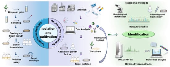
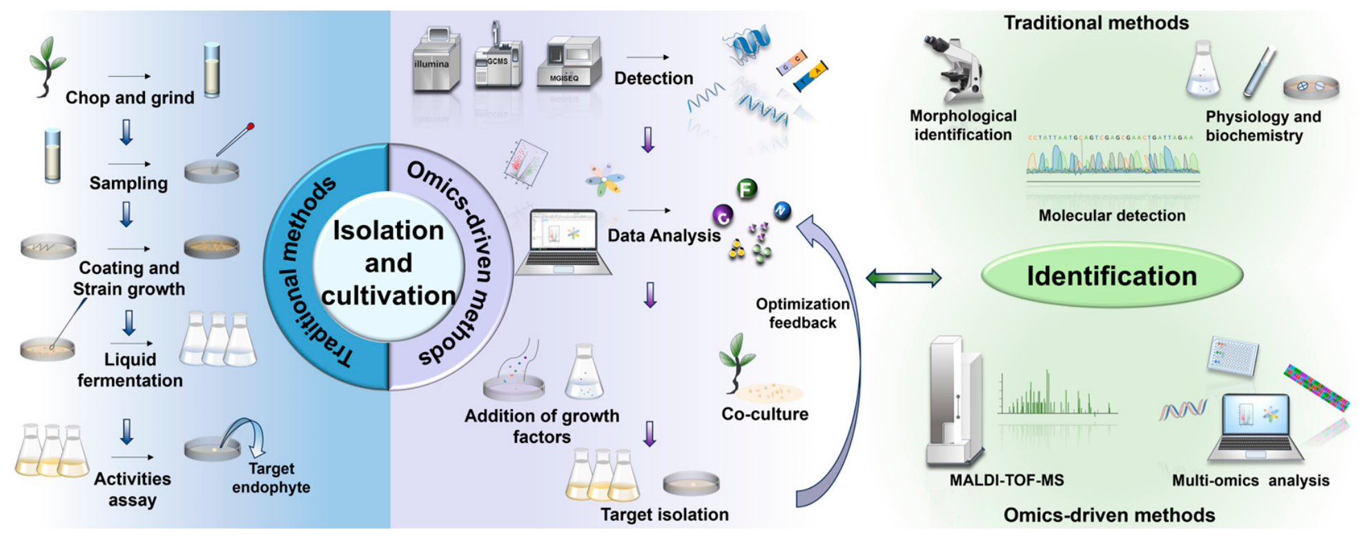
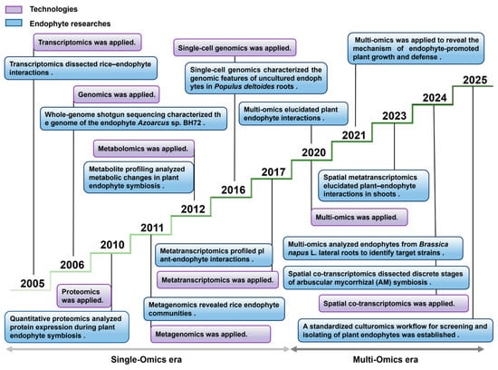
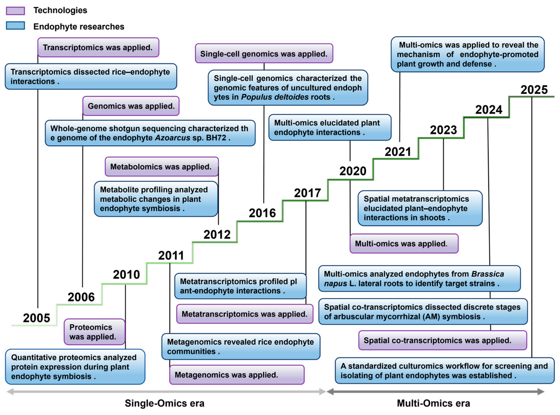
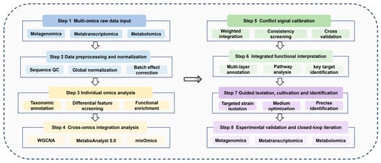
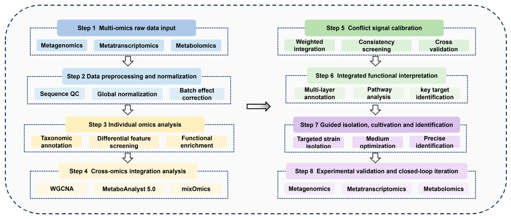

Disclaimer/Publisher’s Note: The statements, opinions and data contained in all publications are solely those of the individual author(s) and contributor(s) and not of MDPI and/or the editor(s). MDPI and/or the editor(s) disclaim responsibility for any injury to people or property resulting from any ideas, methods, instructions or products referred to in the content. |
© 2026 by the authors. Licensee MDPI, Basel, Switzerland. This article is an open access article distributed under the terms and conditions of the Creative Commons Attribution (CC BY) license.