


Abstract
Leaf architecture critically impacts crop yield. The Auxin Response Factor (ARF) family is a key regulator of leaf development, yet remains uncharacterized in the important legume crop chickpea (Cicer arietinum L.), which bears pinnate compound leaves. Here, we performed a genome-wide identification and analysis of ARF genes in chickpea. We identified 33 CaARF genes and resolved their phylogenetic structure through comparison with six other key dicot species. The analysis revealed a deeply conserved core set of ARF proteins across species, all sharing the N-terminal DNA-binding domain (DBD), with most the C-terminal PB1 domain, connected by a middle region (MR). We also uncovered instances of lineage-specific expansion, e.g., a chickpea-specific ARF clade, which is characterized by the absence of the C-terminal PB1 domain. Expression profiling using public transcriptome data and qRT-PCR revealed distinct spatiotemporal expression patterns for CaARF genes across tissues and during compound leaf development. Detailed in situ hybridization analysis for selected candidates, chosen based on phylogenetic proximity to known leaf-development-related ARFs in other species, localized their transcripts to specific regions within compound leaf primordia. Focusing on CaARF5, the closest ortholog of Arabidopsis MONOPTEROS/ARF5, we confirmed its nuclear localization and dynamic expression during chickpea leaf development. Functional complementation assays demonstrated that CaARF5 could restore developmental defects in the Arabidopsis mp mutant. Our study establishes an evolutionary and molecular framework for the chickpea ARF family, highlighting conserved features and species-specific innovations, and provides essential resources for future research on auxin-mediated leaf development and ARF-targeted legume breeding.
1. Introduction
Chickpea (Cicer arietinum L.) is one of the most important legume crops globally, valued as a rich source of protein, carbohydrates, minerals, vitamins, and health-promoting fatty acids. It serves as a primary source of plant-based protein and forage, particularly in arid and semi-arid regions [1]. Beyond its primary harvest as a dry pulse, the chickpea holds significant value as a vegetable crop: in many regions, the immature green seeds and pods are widely consumed as a nutritious vegetable, featuring in diverse culinary preparations such as salads, sautés, and stews [2]. A defining morphological trait of the chickpea plant is its pinnate compound leaf, whose architecture directly influences photosynthetic efficiency and yield potential [3]. The morphogenesis of this compound structure—a process involving the precise initiation, patterning, and outgrowth of multiple leaflets from a common primordium—represents a sophisticated developmental program [4]. Unraveling the molecular networks governing this process is not only a fascinating question in plant developmental biology but also holds substantial promise for targeted genetic improvement aimed at optimizing canopy architecture and enhancing light capture efficiency.
The phytohormone auxin acts as a master regulator of plant growth and organogenesis, with its transcriptional outputs largely mediated by a family of plant-specific transcription factors known as Auxin Response Factors (ARFs) [5]. ARF proteins function as the executive arm of the auxin pathway by directly binding to Auxin Response Elements (AuxREs, typically TGTCNN) in the promoters of target genes, thereby activating or repressing their transcription to coordinate developmental processes [6,7,8]. Canonical ARF proteins are modular, comprising three primary domains: an N-terminal B3-type DNA-binding domain (DBD), a highly variable middle region (MR) that often dictates transcriptional activity (glutamine-rich for activators, serine/proline-rich for repressors), and a conserved C-terminal domain (CTD), now recognized as a Phox and Bem1 (PB1) domain that mediates protein–protein interactions, notably with Aux/IAA repressors [5,9,10]. In the prevailing model of nuclear auxin pathway (NAP), auxin promotes the degradation of Aux/IAA proteins, releasing ARFs from repression and allowing them to modulate downstream gene networks [11,12]. Phylogenetic and functional studies indicate that ARFs have diverged into three functionally different classes: A, B and C. Class A ARFs (A-ARFs) are primarily regulated by auxin through the NAP and acting as transcriptional activators. In contrast, B- and C-ARFs are considered transcriptional repressors [13]. Extensive research across model plants like Arabidopsis thaliana has elucidated specialized biological roles for specific ARFs: A-ARF5/MONOPTEROS regulates embryo patterning and vascular development [14,15]; A-ARF7 and A-ARF19 coordinate lateral root formation and tropisms [16,17]; B-ARF3/ETTIN and B-ARF4 govern abaxial-adaxial identity in lateral organs [18]; ARF6 and ARF8 integrate hormonal signals during reproductive development [19,20,21]; ARF17 coordinates the development of different sporophytic cell layers in anthers [22]. Beyond Arabidopsis, ARFs in crops such as rice, maize, tomato, and cotton have been implicated in stress responses, nutrient homeostasis, inflorescence structure, fruit set, and fiber development, underscoring their functional versatility and agricultural relevance [23,24,25,26,27,28,29].
Previous studies had shown that multiple ARF members regulate different aspects of leaf developmental processes. In Arabidopsis, ARF5/MP serves as a master regulator during early leaf development. Its functions are multifaceted: it is indispensable for leaf initiation, contributes to the establishment of leaf dorsoventral (adaxial-abaxial) polarity, and activates downstream genes such as WUSCHEL-RELATED HOMEOBOX 1 (WOX1) and PRESSED FLOWER (PRS) to promote lamina outgrowth and flattening [30]. Other A-ARFs, such as ARF7 and ARF19, are known to cooperatively influence leaf cell expansion [31,32]. In tomato, different A-ARFs (SlMP, SlARF19a, SlARF19b) contribute to leaflet formation with varying potency, largely correlated with their expression levels; the slmp slarf19b double mutant fails to initiate leaflet primordia, forming needle-like leaves [33]. In multiple species, B-ARFs, particularly ARF3 and ARF4 orthologs, are tightly integrated into core leaf developmental networks. ARF3 and ARF4 transcripts were specifically accumulated in the abaxial domains of developing leaf primordia. They collaborate with factors like KANADI to establish and maintain leaf polarity [18]. In Medicago truncatula, adaxially expressed ARGONAUTE7 (MtAGO7) facilitates ta-siRNA biosynthesis to negatively regulate the abaxially enriched MtARF3 [34]. The plants overexpressing a TAS3 ta-siRNA-insensitive mutant of MtARF3 (MtARF3m) exhibits increased leaflet number [35]. In tomato wiry mutant (slago7), reduced ta-siRNA levels and SlARF3/4 accumulation lead to fewer, nearly radial leaflets, and occasional needle-like leaves, whereas reducing SlARF3 and SlARF4 levels can rescue the wiry leaf lamina, and increased activity of either can phenocopy wiry leaves [36]. In summary, leaf morphogenesis is finely tuned by a complex network of interactions mediated by ARFs across different classes, integrating transcriptional activation, repression, and small RNA-mediated post-transcriptional regulation.
In this study, we systematically identified and characterized the ARF gene family in chickpea, with a focus on their potential roles in the development of the species’ characteristic odd-pinnate compound leaves. A total of 33 CaARF genes were identified from the genome. Subsequent comprehensive analyses—including phylogenetics, protein domain architecture, gene structure, chromosomal distribution, duplication events, and promoter cis-element analysis—defined the family’s genomic organization and potential regulatory features. Expression profiling via transcriptomics and qRT-PCR revealed that several class A and class B CaARF genes are highly expressed in the shoot apical meristem and young leaf primordia, implicating them in early leaf morphogenesis. Functional analysis focused on CaARF5, which showed specific accumulation in early leaf primordia and partially rescued the developmental defects of the Arabidopsis mp mutant, demonstrating functional conservation. While ARF families have been described in some other legumes [37,38,39], a dedicated analysis in chickpea was previously absent. Our work fills this gap and provides a multifaceted resource that integrates evolutionary, molecular, and expression data. This foundation establishes the chickpea ARF family as a key system for probing the mechanisms of auxin-mediated leaf development and identifies potential targets for legume improvement strategies.
2. Results
2.1. Identification of 33 Members of ARF Family Genes in Chickpea
Based on genome-wide analysis, a total of 33 ARF family members were identified in the chickpea genome (cv. CDC Frontier, version v2.0.a1), which were sequentially designated as CaARF1 to CaARF32 primarily according to their phylogenetic relationships with Arabidopsis ARF proteins (Supplemental Table S2). Prediction of physicochemical properties revealed substantial variation in the protein lengths, ranging from 132 (CaARF32) to 1125 amino acids (CaARF19c), corresponding to a molecular weight range of approximately 14.76 to 126.33 kDa. The isoelectric point (pI) values ranged from 5.55 (CaARF5) to 10.22 (CaARF29). The majority of the family members (26/33) had a theoretical pI below 7.0, indicating that the CaARF family is predominantly acidic; only seven proteins (CaARF9b, CaARF10a, CaARF10b, CaARF16a, CaARF16b, CaARF16c, and CaARF29) displayed basic characteristics. The grand average of hydropathicity (GRAVY) was negative for 31 members, suggesting an overall hydrophilic nature. The two exceptions, CaARF31 and CaARF32, had positive GRAVY values. In terms of instability, all members exhibited instability index values exceeding 40, indicating that the CaARF family proteins are unstable. The aliphatic index ranged from 65.43 to 107.05, with CaARF31 and CaARF32 surpassing 100, implying that these two proteins may possess relatively higher thermal and structural stability (Supplemental Table S3).
In summary, the chickpea CaARF family exhibits substantial diversity in key physicochemical properties, including length, molecular weight, and charge, while being collectively characterized by acidic pI, hydrophilicity, and predicted instability.
2.2. Phylogenetic Analysis and Classification of the ARF Family in Chickpea
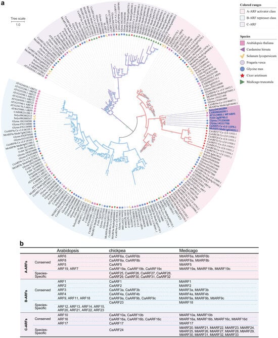
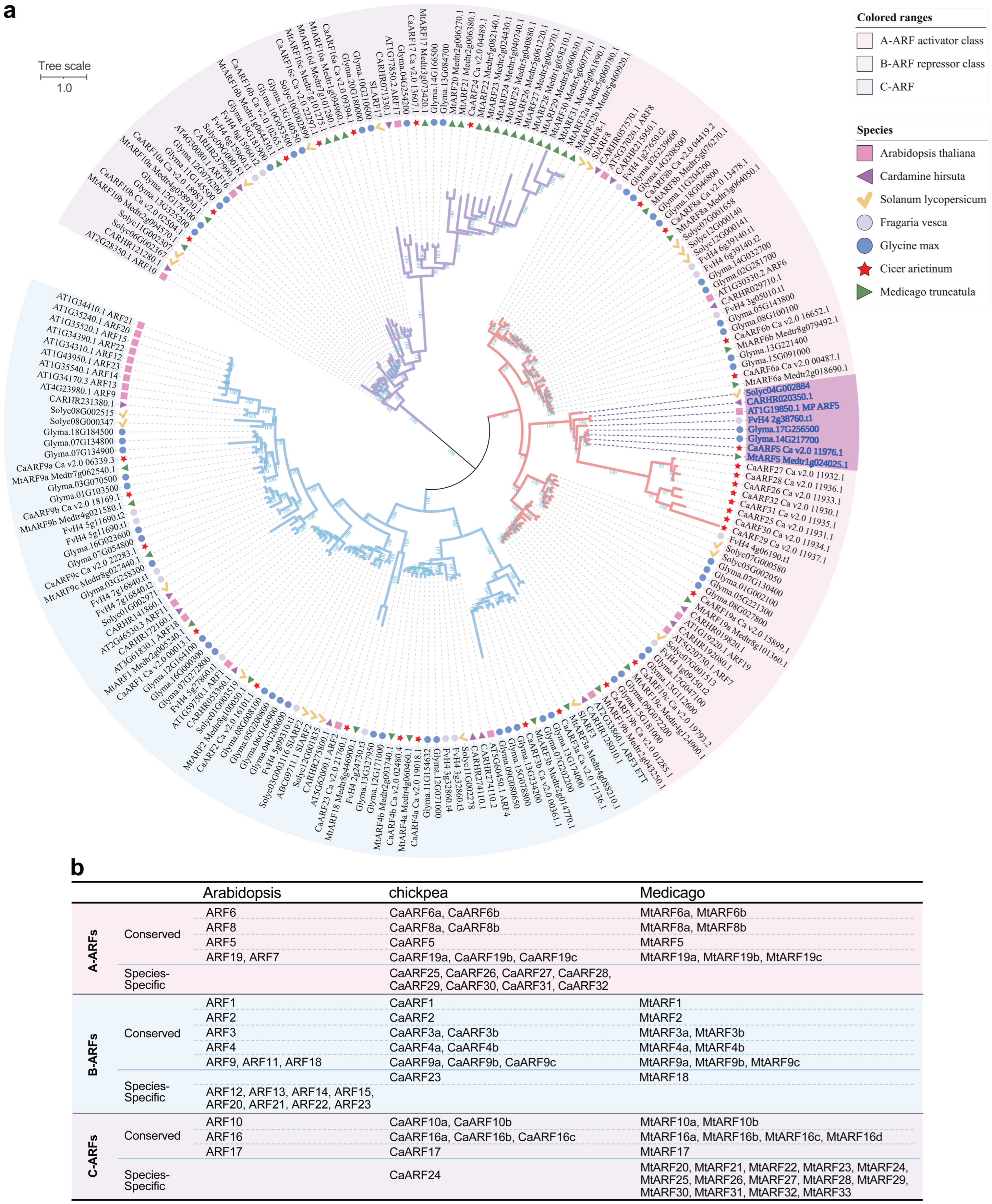
To elucidate the evolutionary relationships of the chickpea ARF family, we performed a phylogenetic analysis using ARF protein sequences from chickpea, Medicago truncatula, soybean (Glycine max), Arabidopsis thaliana, and three other compound-leafed species—Fragaria vesca, Solanum lycopersicum, and Cardamine hirsuta. The resulting midpoint-rooted tree resolves all ARF proteins into three major, well-supported subfamilies, corresponding to the A-ARFs, B-ARFs, and C-ARFs (Figure 1a). Each subfamily comprises multiple distinct branches that predominantly consist of orthologous proteins from the seven species analyzed. Statistical analysis of orthologous groups revealed that, compared to Arabidopsis, certain branches contain a notably larger number of members in chickpea and other legumes. For instance, the orthologous groups containing AtARF9, AtARF16, and AtARF19 are significantly expanded in legumes, suggesting lineage-specific gene family expansion events during legume genome evolution (Figure 1b).
Figure 1.
Phylogenetic analysis of the ARF protein family in chickpea and other plant species. (a) Phylogenetic tree constructed using ARF protein sequences from chickpea (Cicer arietinum) and six representative species: Arabidopsis thaliana, Glycine max, Medicago truncatula, Fragaria vesca, Solanum lycopersicum, and Cardamine hirsuta. the corresponding bootstrap values are indicated at the branch nodes, and the branch length scale bar represents 1.0. The dark blue font indicates the ARF5 homologous gene. The three major evolutionary subfamilies are indicated by distinct colored regions: A-ARF (Pink), B-ARF (Blue), and C-ARF (Purple). (b) Statistical distribution of member numbers within the A-ARF, B-ARF, and C-ARF subfamilies in Arabidopsis thaliana, Cicer arietinum, and Medicago truncatula.
We also identified a limited number of species-specific ARF branches belonging to different subfamilies. Chickpea possesses a specific clade within the A-ARF subfamily that is phylogenetically most closely related to the ARF5 clade, while Arabidopsis thaliana and Medicago truncatula each possesses a distinct, species-specific clade within the B-ARF and C-ARF subfamilies, respectively (Figure 1b). These unique clades likely reflect functional divergence arising from distinct biological adaptations in each species. Collectively, the phylogenetic framework established here provides a crucial foundation for elucidating functional diversification and molecular evolution of the ARF gene family in chickpea.
2.3. Gene Structure and Conserved Domain Analysis of the ARF Family in Chickpea
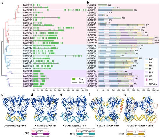
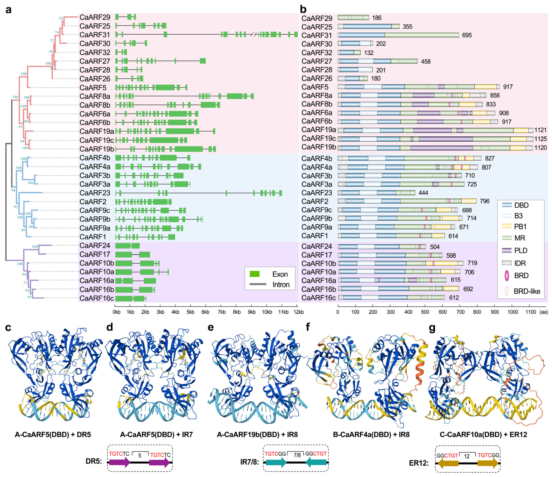
Analysis of gene structures revealed that members within the same phylogenetic group generally share similar exon-intron patterns, with the notable exceptions of the chickpea-specific A-ARF clade members and CaARF23. Excluding these exceptions, members of the A and B-ARF subfamilies typically contain the higher number of exons (11–14), whereas C-ARF members possess significantly fewer (2–4). This pattern is consistent with the hypothesis that the A/B and C subfamilies diverged early in their evolutionary history (Figure 2a).
Figure 2.
Analysis of Gene Structure and Protein Conserved Domains in the Chickpea ARF Gene Family. (a) Gene structure of the chickpea ARF genes. Green rectangles represent exons, and introns are indicated by black lines. (b) Schematic diagram of ARF domains in chickpea. The number of amino acid residues for each ARF protein is labeled. DBD, DNA-binding domain; B3, B3 DNA-binding domain; PB1, protein–protein interaction domain; MR, middle region. PLD, prion-like domain. IDR, intrinsic disordered region; BRD, B3 repression domain. A scale bar is shown at the bottom of the chart. (c) A-CaARF5(DBD) binds to the inverted repeat DR5. (d) A-CaARF5(DBD) binds to the inverted repeat IR7. (e) A-CaARF19b(DBD) binds to the inverted repeat IR8. (f) B-CaARF4a(DBD) binds to the inverted repeat IR8. (g) C-CaARF10a(DBD) binds to the everted repeat ER12. In panels (c)–(g), the blue regions in the double helix structure indicate high confidence, while the yellow regions indicate medium confidence. Different colors within the dashed boxes represent distinct AuxRE sequences, specifically magenta for the DR5 motif, cyan for the IR7/8 motif, and orange for the ER12 motif.
Domain architecture analysis shows that the majority of chickpea ARF members contain the canonical B3-type DNA-binding domain (DBD), Middle Region (MR), and C-terminal Phox and Bem1 (PB1) domain. However, the composition and presence of these domains exhibit significant divergence among subfamilies. Within the A-ARF subfamily, the eight members constituting the chickpea-specific clade have markedly shorter protein sequences and uniformly lack the PB1 domain. Their gene structures are also atypical and highly variable, suggesting these members may have diverged functionally from canonical ARF proteins. In contrast, the remaining A-ARF members outside this specific clade exhibit a complete domain architecture, comprising the DBD, B3, MR, and PB1 domains, and frequently contain PLD (Prion-like domain) and IDR (intrinsically disordered region) sequences (Figure 2b).
In the B-ARF subfamily, most members possess the DBD, B3, MR, and PB1 domains. However, the evolutionarily conserved pair CaARF3a/3b and the species-specific member CaARF23 lack the PB1 domain. Within the C-ARF subfamily, four of the seven members lack the PB1 domain, while three retain it, indicating considerable variation in PB1 domain retention among C-ARFs. Additionally, a B3 repression domain (BRD, R/K/VLFG sequences) was found to be widely present in B- and C-ARF subfamily members, found in few A-ARF members (Figure 2b).
We employed AlphaFold3 to model the interaction between chickpea ARF DBDs and AuxREs, testing whether the clade-specific binding rules defined in Arabidopsis are conserved. Structural predictions for representative clade A (CaARF5, CaARF19b), B (CaARF4a), and C (CaARF10a) members confirm this conservation (Figure 2c–g). As anticipated, the clade A DBDs bind a broad spectrum of AuxRE configurations, the clade B DBD preferentially interacts with an IR8 motif, and the clade C DBD exhibits a preference for an ER13 motif. These models validate that the core principle of an ARF-AuxRE recognition code—where phylogeny dictates binding specificity—is maintained in chickpea, structurally defining the potential for divergent transcriptional outputs from a conserved ARF subfamily.
2.4. Chromosomal Localization and Synteny Analysis of CaARFs
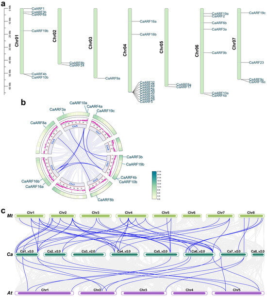
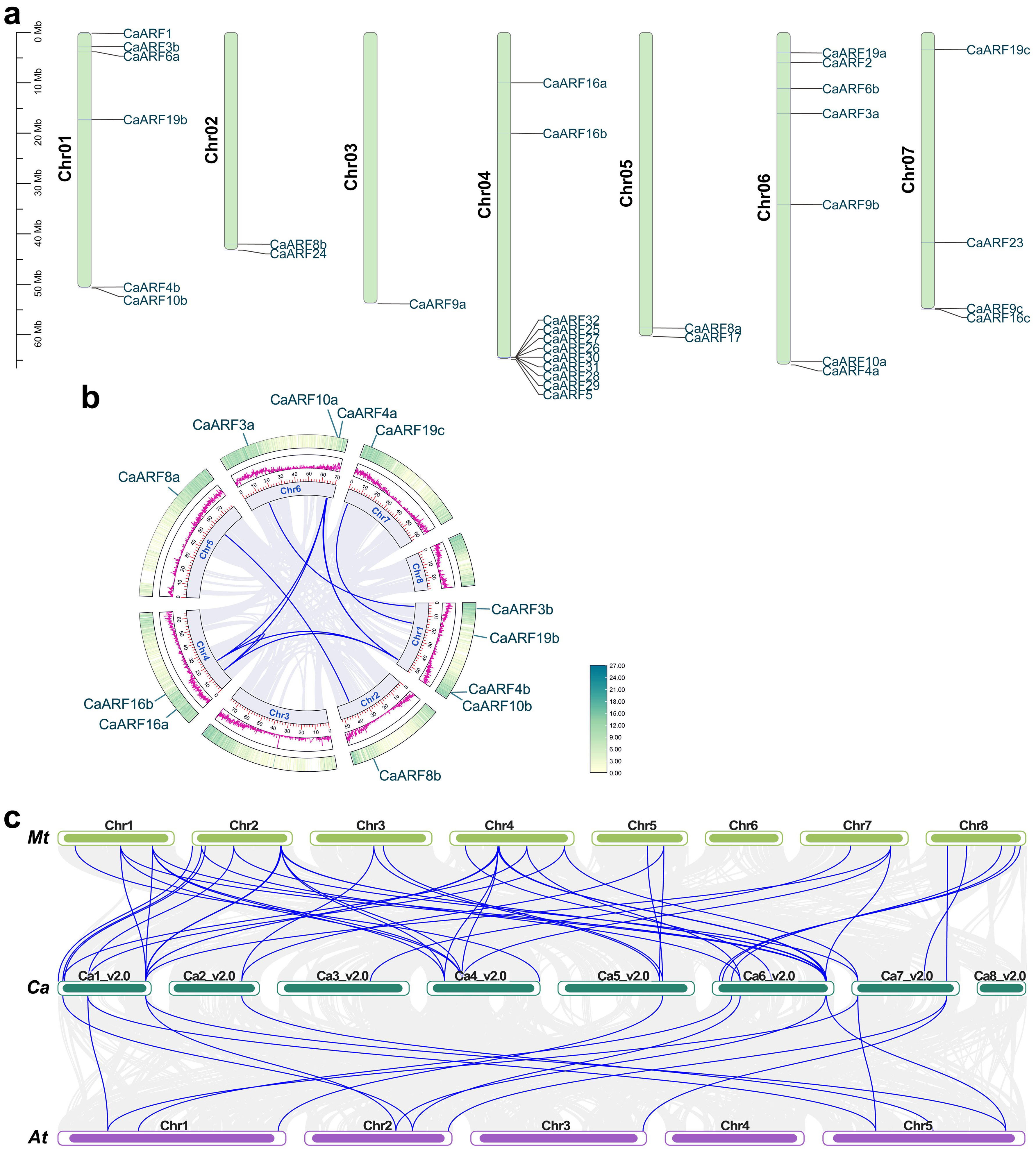
Based on the chromosomal localization results of the chickpea (Cicer arietinum) ARF genes (Figure 3a; Supplemental Table S5), the CaARF family members exhibit a pronounced non-uniform distribution pattern across the chromosomes. Chromosome 4 harbors the highest number of CaARF genes. Notably, this includes three typical ARF members (CaARF5, CaARF16a, CaARF16b) and all members of the chickpea-specific clade, which together with CaARF5 are arranged in a tight tandem array at the distal end of the chromosome, strongly suggesting evolution through recent tandem duplication events. Regarding the distribution pattern, certain genes, such as CaARF19b, CaARF16b, and CaARF9b, tend to be localized to the middle chromosomal regions or near the centromere, whereas the majority of the remaining genes are located on the distal regions of the chromosome arms.
Figure 3.
Chromosomal localization and synteny analysis. (a) Chromosomal localization of the ARF gene family in chickpea. The left scale indicates chromosome length (Mb), and the positions of ARF genes are marked on the right side of each chromosome. (b) Synteny analysis within the ARF gene family of chickpea. The middle circular magenta lines denote gene density. (c) Interspecific synteny of the ARF gene family between chickpea and model species.
To further explore their evolutionary relationships, we performed synteny analysis and identified a total of 10 segmental duplication pairs (Figure 3b; Supplemental Table S6), indicating that segmental duplication is one of the primary mechanisms driving the expansion of this gene family. The aforementioned uneven distribution and local clustering reflect that the CaARF family may have expanded through mechanisms such as segmental and tandem duplications.
Comparative synteny analysis revealed distinct evolutionary relationships between the chickpea ARF family and those of two reference species (Figure 3c). We identified 46 orthologous gene pairs between chickpea and the model legume Medicago truncatula. With the exception of the chickpea-specific clade members, nearly all core-set chickpea ARF genes have conserved orthologs in M. truncatula, indicating that the family’s expansion and genomic organization have been largely stable since the divergence of these legume lineages. In contrast, only 15 orthologous pairs were detected between chickpea and Arabidopsis thaliana. The maintenance of these corresponding genomic blocks across a greater evolutionary distance suggests they may have been under high selective pressure, preserving essential functions.
2.5. Cis-Acting Element Analysis of ARF Family Gene Promoters in Chickpea
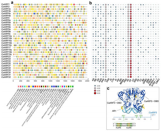
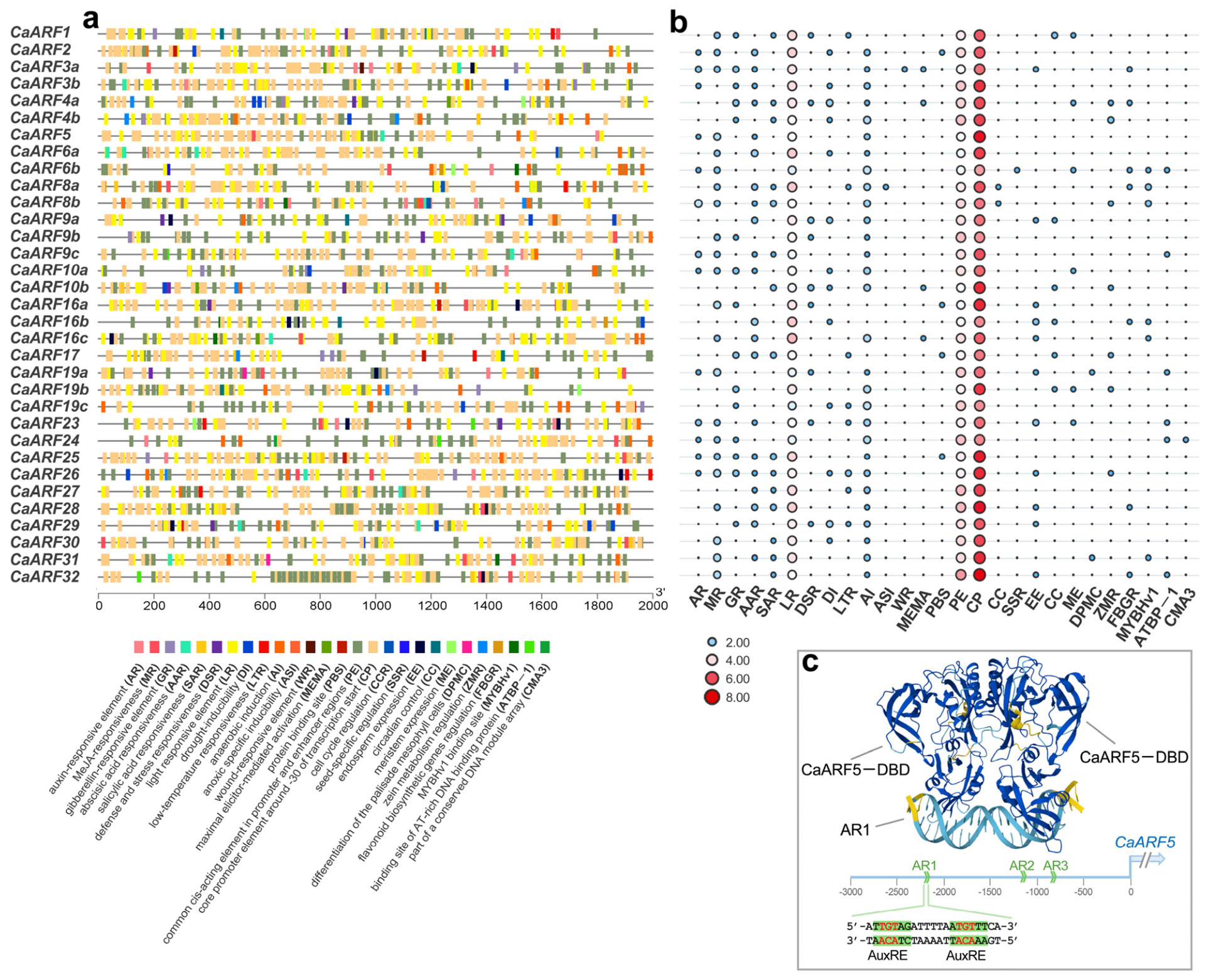
Analysis of cis-acting elements in the promoters of chickpea ARF genes revealed a complex regulatory architecture (Figure 4a,b; Supplemental Tables S8 and S9). The promoters are enriched not only with core promoter elements (e.g., TATA boxes) for precise transcriptional initiation but also with a diverse array of motifs associated with hormone response (including auxin), light signaling, and abiotic stress. This configuration suggests that CaARF genes function as integrator nodes, whose expression can be modulated by multiple internal and external signals. Notably, the promoters of several CaARF genes contain canonical Auxin Response Elements (AuxREs). Given the reported autoregulation of Arabidopsis MP/ARF5, we hypothesized a similar mechanism might exist in chickpea. We employed AlphaFold3 to predict the binding of the CaARF5 DNA-binding domain (DBD) to three potential AuxRE pairs (AR1–AR3) identified within its own promoter (Figure 4c). The structural model demonstrates high-confidence binding of the CaARF5-DBD to the AR1 motif, supporting the possibility that CaARF5 can directly regulate its own transcription. This finding positions CaARF5 not only as a key mediator of auxin signaling but also as a component subject to precise feedback regulation within the auxin response network.
Figure 4.
Analysis of cis-acting elements in the promoters of CaARFs. (a) The 2 kb upstream region from the start codon was analyzed using PlantCARE to identify promoter cis-acting elements, and the results were visualized with TBtools. Different colored boxes represent diverse promoter cis-acting elements. (b) Heatmap of promoter cis-acting element analysis for ARF genes in chickpea. AR: auxin-responsive element; MR: MeJA-responsiveness; GR: gibberellin-responsive element; AAR: abscisic acid responsiveness; SAR: salicylic acid responsiveness; DSR: defense and stress responsiveness; DI: drought-inducibility; LTR: low-temperature responsiveness; AI: anaerobic induction; ASI: anoxic-specific inducibility; WR: wound-responsive element; MEMA: maximal elicitor-mediated activation; LR: light responsive element; CCR: cell cycle regulation; SSR: seed-specific regulation; EE: endosperm expression; CC: circadian control; ME: meristem expression; DPMC: differentiation of the palisade mesophyll cells; ZMR: zein metabolism regulation; FBGR: flavonoid biosynthetic genes regulation; MYBHv1: MYBHv1 binding site; ATBP-1: binding site of AT-rich DNA binding protein; CMA3: part of a conserved DNA module array; PBS: protein binding site; PE: common cis-acting element in promoter and enhancer regions; CP: core promoter element around −30 of transcription start. (c) Predicted binding mode between the CaARF5 DBD and the AuxRE motifs in the target gene promoter. The upper panel illustrates the domain architecture of the CaARF5 protein (DBD and AR1–AR3 regulatory regions), while the lower panel depicts a 20 bp double-stranded DNA sequence containing two AuxRE motifs within the promoter region.
2.6. Expression Analysis of CaARF Genes in Different Tissues
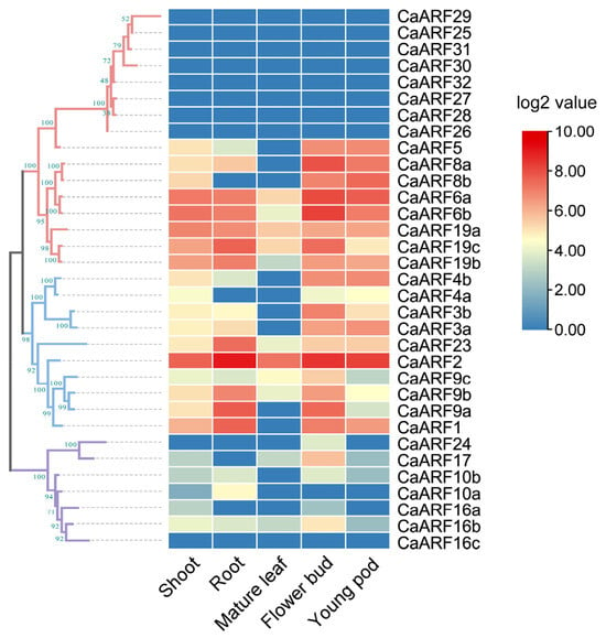
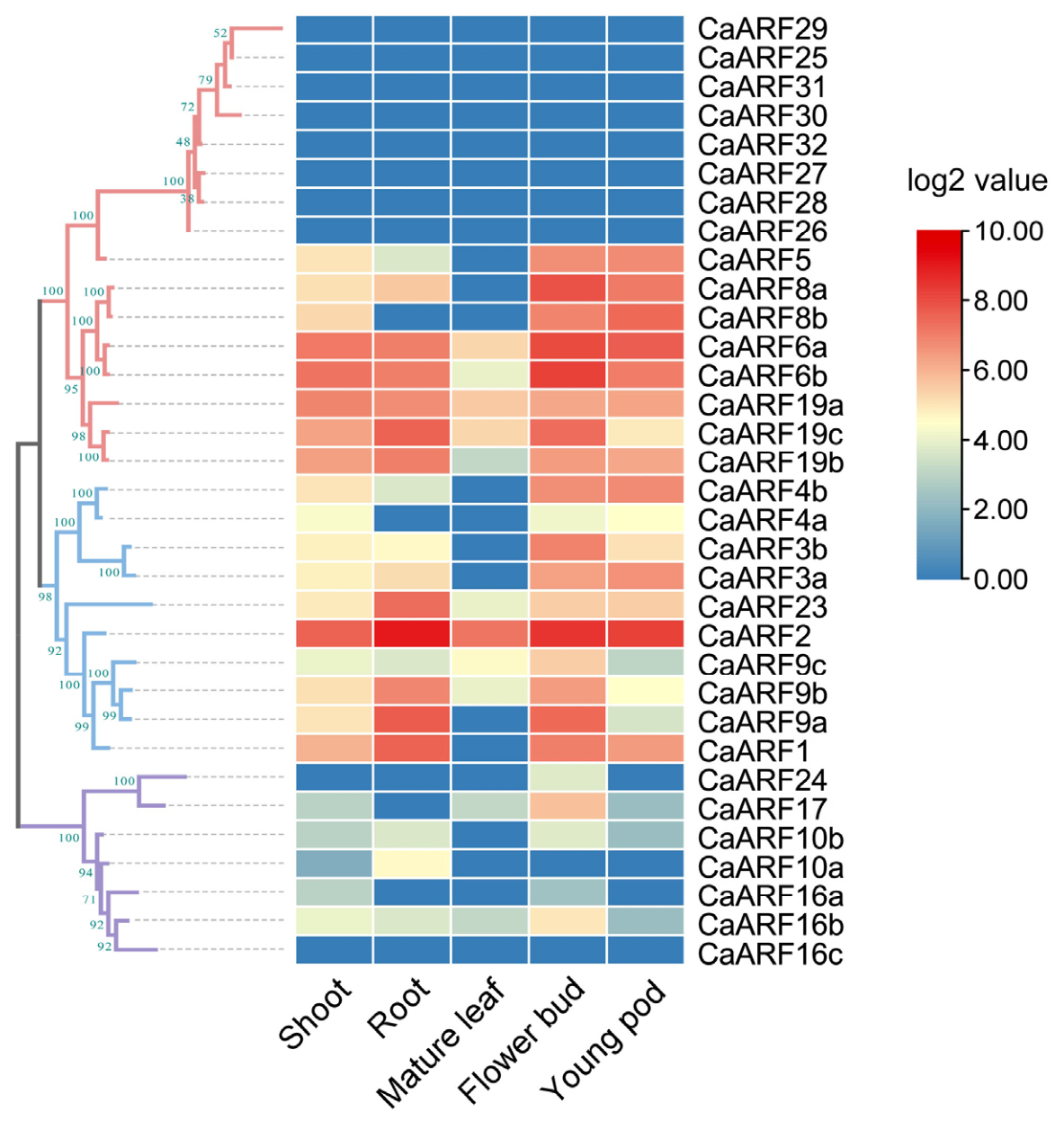
Heatmap analysis revealed distinct, subclass-specific expression profiles for the chickpea CaARF gene family across five tissues (Figure 5; Supplemental Table S10). Considerable heterogeneity was observed within the A-ARF subclass. Eight chickpea-specific A-ARF members (CaARF25-32) showed negligible expression in all tissues examined. In contrast, other A-ARF members exhibited enriched expression in young, developing tissues (shoot, flower bud, young pod) but uniformly low expression in mature leaves. Within the B-ARF subclass, most members displayed moderate, constitutive expression in young tissues, but markedly downregulated in mature leaves. A notable exception was CaARF2, which maintained high constitutive expression across all tissues. Members of the C-ARF subclass exhibited overall low or undetectable transcriptional abundance. These patterns suggest that A- and B-ARF members are likely key regulators of young organ development, while C-ARF members may be involved in distinct, possibly more subtle or condition-specific biological processes.
Figure 5.
Expression profiles of the CaARF gene family in different tissues and organs. Normalized log2 transformed values were used with hierarchical clustering represented by the color scale (0–10). Blue indicates low expression, and red indicates high expression.
2.7. qRT-PCR Validation of Leaf Development-Associated CaARF Candidates
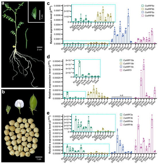
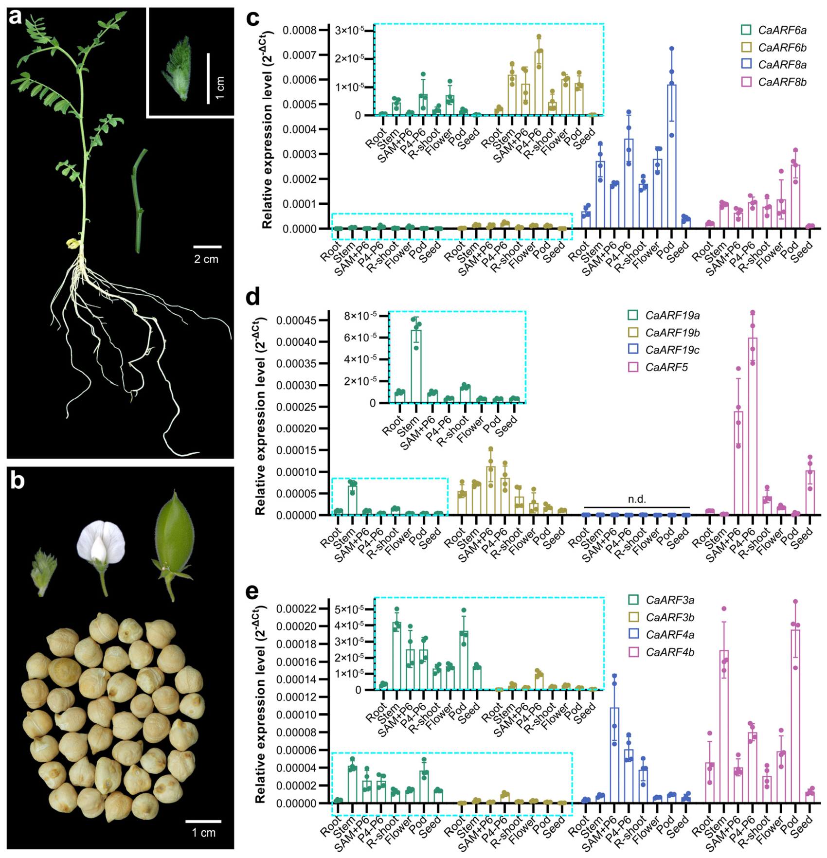
The development of the odd-pinnate compound leaf, a hallmark of chickpea, involves precise genetic regulation that remains poorly understood. Studies in model plants like Arabidopsis [31], tomato [33,36], and Medicago truncatula [34,35] have implicated specific A- and B-class ARF members in leaf morphogenesis. To identify chickpea ARF genes potentially involved in this process, we analyzed their tissue-specific expression patterns (Figure 6a,b). Initial transcriptomic data provided a broad overview of CaARF expression across multiple organs. Based on this, we selected candidate genes from the A- and B-ARF subfamilies for detailed validation using quantitative reverse transcription PCR (qRT-PCR).
Figure 6.
qRT-PCR validation of CaARF gene expression in different tissues and organs. (a) On the left is a chickpea plant at the vegetative stage; the middle shows the selected stem. Scale bar, 2 cm; the upper right corner indicates the selected vegetative shoot apex. Scale bar, 1 cm. (b) The upper panel shows, from left to right, the reproductive shoot apex, flower, and pod of chickpea; the lower panel shows seeds. Scale bar, 1 cm. (c) qRT-PCR validation of the expression levels of chickpea CaARF6a, CaARF6b, CaARF8a, and CaARF8b in different tissues. (d) qRT-PCR validation of the expression levels of chickpea CaARF19a, CaARF19b, CaARF19c, and CaARF5 in different tissues. (e) qRT-PCR validation of the expression levels of chickpea CaARF3a, CaARF3b, CaARF4a, and CaARF4b in different tissues. The data are the mean ± SD from three biological replicates. (c–e) The blue dashed boxes highlight genes with a moderate or low expression level.
Within the A-ARF subclass, we first analyzed the expression of two closely related gene pairs: CaARF6a/6b and CaARF8a/8b (Figure 6c), because (i) they are orthologs of Arabidopsis ARF6 and ARF8, two genes expressed in the petiole and vasculature of developing leaves [40]; and (ii) preliminary transcriptomic data revealed that these gene pairs show relatively high expression in young chickpea tissues (Figure 5). A clear hierarchy in overall expression abundance was observed between the pairs: CaARF8a and CaARF8b exhibited substantially higher transcript levels across all examined tissues compared to CaARF6a and CaARF6b, with CaARF6b being barely detectable. At the level of tissue specificity, all four genes showed preferential expression in young, developing tissues. Notably, CaARF8a/8b showed particularly high expression in pods.
Given the critical role of ARF5 and ARF19 orthologs in tomato leaflet initiation [33,36], we examined the expression of their chickpea counterparts: CaARF5, CaARF19a, CaARF19b, and CaARF19c (Figure 6d). CaARF5 was the most highly expressed, followed by CaARF19b. CaARF19a showed moderate expression, while CaARF19c was undetectable in all tissues tested. Their tissue-specific expression patterns were also divergent. CaARF5 was most abundant in the shoots, young leaves, and seeds. CaARF19b displayed relatively high expression in the shoots, young leaves, stem, and root, whereas CaARF19a was specifically enriched in the stem.
Within the B-ARF subclass, we focused on CaARF3a/3b and CaARF4a/4b (Figure 6e), the orthologs of Arabidopsis ARF3/ETT and ARF4, which are known to participate in leaf adaxial-abaxial polarity establishment and leaf morphogenesis in Arabidopsis, and both pairs have undergone duplication in chickpea. Overall, CaARF4a and CaARF4b were consistently expressed at higher levels than CaARF3a and CaARF3b across most tissues. Examining tissue-specific profiles revealed further functional clues: CaARF4b expression was relatively high in pods and stems, while CaARF4a peaked in the shoots and young leaves. CaARF3a maintained a ubiquitous, low-to-moderate level of expression in all tissues except roots. CaARF3b was generally low except for a notable level in leaf primordia.
The qRT-PCR expression trends for the majority of genes validated and refined the patterns observed in the RNA-seq heatmap analysis. A notable exception was CaARF19c, which showed a signal in the heatmap but was undetectable by qRT-PCR. This discrepancy likely stems from fundamental methodological differences between the two techniques as well as potential variations in the biological samples used.
2.8. Spatiotemporal Expression Dynamics of Key CaARF Genes During Compound Leaf Morphogenesis
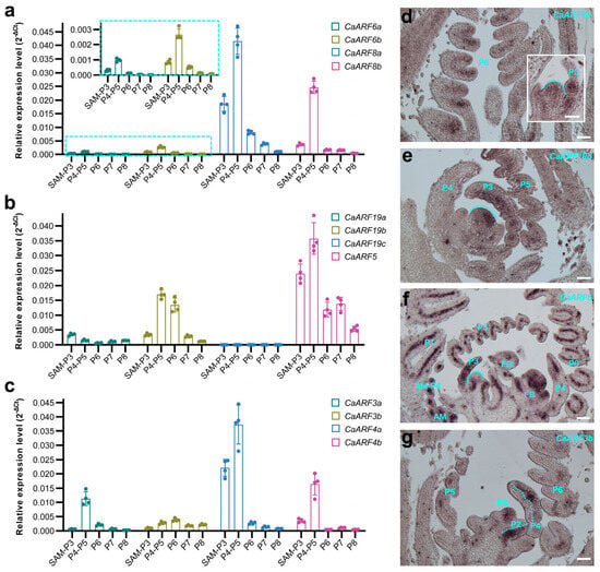
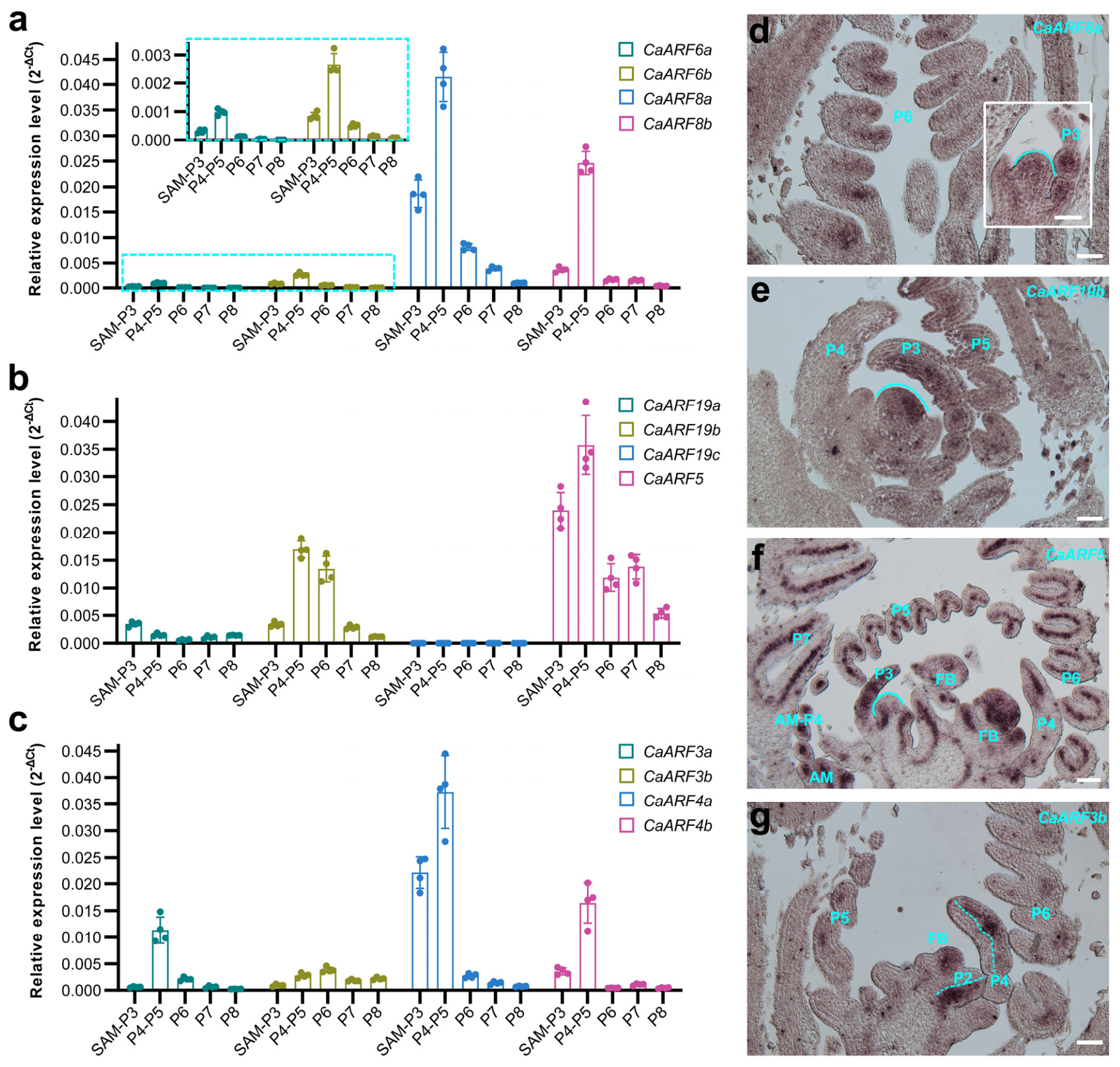
To further delineate the roles of key CaARF genes in leaf development, we analyzed their expression patterns across a developmental series of leaf primordia via qRT-PCR. Consistent with our broader tissue survey, a comparison of expression levels among orthologous pairs revealed a conserved hierarchy: CaARF8a and CaARF8b were expressed at substantially higher levels than CaARF6a and CaARF6b (Figure 7a); CaARF5 was the most highly expressed, followed by CaARF19b, with CaARF19a lower and CaARF19c nearly undetectable (Figure 7b); and CaARF4a/4b levels exceeded those of CaARF3a/3b (Figure 7c). Examining the temporal expression profile of each gene across developmental stages (SAM-P3, P4-P5, P6, P7, P8) revealed a common trend. Most genes exhibited their high transcript levels in the SAM-P3 stage, peaked during the P4-P5 leaf primordia stage, and subsequently declined as leaves underwent differentiation and maturation. Here, SAM denotes the shoot apical meristem, and P followed by a number (e.g., P3) refers to the leaf plastochron index, indicating successively older leaf primordia. This pattern indicates that these ARFs predominantly function in regulating morphogenetic events during organ initiation and patterning, rather than in maintaining the functions of mature tissues.
Figure 7.
qRT-PCR validation of CaARF gene expression in different stages. (a) qRT-PCR validation of the expression levels of chickpea CaARF6a, CaARF6b, CaARF8a, and CaARF8b in shoot apical meristems at different developmental stages. The blue dashed boxes highlight genes with a moderate or low expression level. (b) qRT-PCR validation of the expression levels of chickpea CaARF19a, CaARF19b, CaARF19c, and CaARF5 in shoot apical meristems at different developmental stages. (c) qRT-PCR validation of the expression levels of chickpea CaARF3a, CaARF3b, CaARF4a, and CaARF4b in shoot apical meristems at different developmental stages. The data are the mean ± SD from three biological replicates. (d–g) RNA in situ hybridization of CaARF8a, CaARF19b, CaARF5 and CaARF3b mRNA in wild-type leaf primordia. Similar results were obtained from three independent experiments. P, plastochron. Scale bar, 50 μm.
To complement the quantitative temporal data with spatial resolution, we performed in situ hybridization. Due to the high nucleotide sequence similarity (70–80%) among many homologous genes (e.g., CaARF3b with CaARF3a/4a/4b), we selected representative members from different ARF classes for analysis. CaARF8a transcripts were detected in the SAM and leaf primordia at various stages, showing a broad distribution throughout the P3 leaflet primordium that later became more restricted to the marginal region at P6 (Figure 7d). CaARF19b expression was observed at the SAM periphery and showed a strong signal in the adaxial domain of early leaf primordia (Figure 7e). Among all genes examined, CaARF5 exhibited the strongest hybridization signal. It was detected in leaf primordia of all stages examined, floral primordia, and axillary meristems (Figure 7f). Notably, within early leaf primordia, CaARF5 mRNA showed particularly prominent and specific accumulation in the region of provascular tissue initiation. This spatial expression pattern not only aligns with the qRT-PCR data showing high CaARF5 expression during early development but also provides direct spatial evidence supporting a core regulator for CaARF5 during leaf morphogenesis in chickpea. CaARF3b expression was detected in both leaf and floral primordia (Figure 7g). In early leaf primordia, transcripts were specifically enriched in the abaxial domain, a pattern analogous to the expression of its orthologs in Arabidopsis and Medicago truncatula, which are established regulators of abaxial cell fate and leaf patterning. Sense probe control experiments for the corresponding genes yielded no specific hybridization signals (Figure S1), confirming the specificity of the antisense probe signals.
Collectively, the distinct yet developmentally regulated spatial expression patterns of CaARF8a, CaARF19b, CaARF5, and CaARF3b—encompassing meristematic zones, specific leaf domains (adaxial, abaxial, marginal, vascular), and reproductive primordia—provide direct spatial evidence that the functions of these ARF genes are closely associated with fundamental processes in compound leaf development, including primordia initiation, polarity establishment, and tissue patterning.
2.9. In-Depth Characterization of CaARF5 Expression and Subcellular Localization of the Encoded Protein
Building on our preceding expression analyses, which highlighted CaARF5 as a prominently expressed candidate during leaf development, we hypothesized that it plays a central regulatory role. To rigorously test this hypothesis and delineate its functional context, we conducted a multi-faceted characterization of CaARF5.
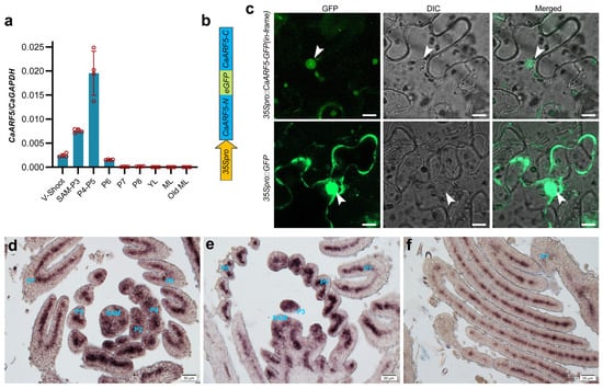
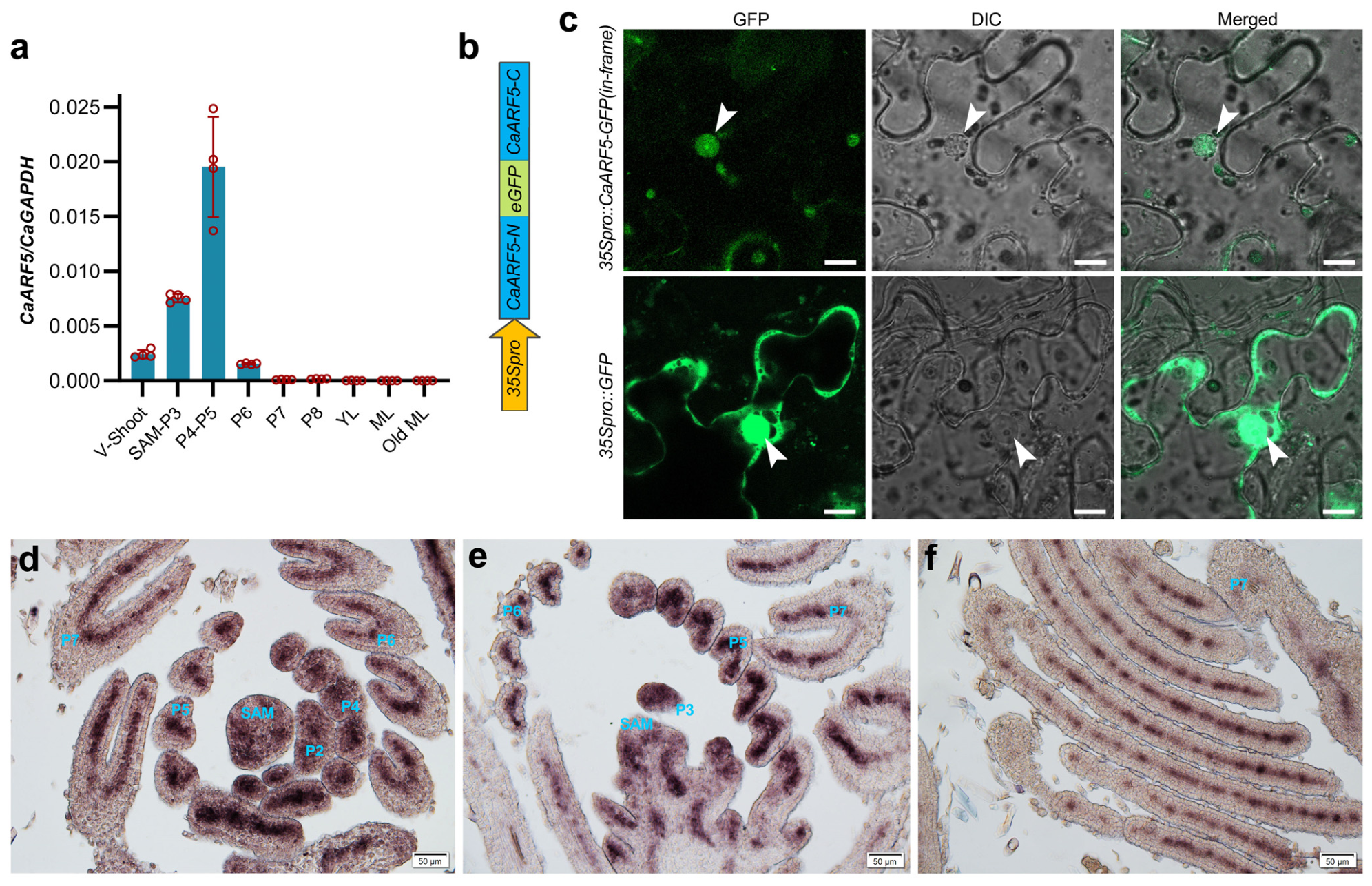
We first analyzed its transcript levels in leaf primordia at different developmental stages using qRT-PCR. The results showed that CaARF5 was highly expressed in the shoot apical meristem (SAM), with particularly significant enrichment in the early leaf developmental stages (P4–P5 leaf primordia), suggesting that it may play a regulatory role in early organogenesis, participating in axial growth and vascular development (Figure 8a). As leaf development progressed to the P6–P8 stages, its expression gradually declined and was maintained at relatively low levels in mature leaves (ML) and old mature leaves (Old ML).
Figure 8.
Spatiotemporal expression pattern analysis of the CaARF5 gene. (a) Transcription signals of CaARF5 were detected in different tissues and developing shoot apices by real-time quantitative PCR. V-shoots: vegetative shoot apical meristem; SAM: shoot apical meristem; YL: young leaf; ML: mature leaf; Old ML: old leaf. The data are the mean ± SD from three biological replicates. (b) Schematic diagram of the construction of the 35Spro::CaARF5-GFP(in-frame) fusion expression vector. (c) Subcellular localization of CaARF5 in tobacco leaf epidermal cells. Arrows point to cell nuclei. Scale bar, 10 μm. (d–f) In situ hybridization analysis of CaARF5 in the shoot apical meristem of wild-type chickpea. Longitudinal sections of the shoot apex were prepared using the paraffin sectioning method. P, plastochron. Scale bar, 50 μm.
To determine the subcellular compartment in which CaARF5 functions, a fusion construct 35Spro::CaARF5-GFP(in-frame) (Figure 8b) was transiently expressed in tobacco epidermal cells. Confocal microscopy showed that the CaARF5-GFP(in-frame) fluorescence was exclusively localized to the nucleus, as delineated in the DIC image, in contrast to the diffuse nucleus-cytoplasmic distribution of free GFP, confirming CaARF5 as a nuclear protein (Figure 8c).
Finally, we performed high-resolution RNA in situ hybridization to map the spatial distribution of CaARF5 mRNA during leaf development (Figure 8d–f). Transcripts were specifically detected in the SAM and leaf primordia. From stages P2 to P4, expression was broad and strong throughout the early compound leaf common primordium, including incipient leaflet and stipule domains. By stage P5, expression became more restricted to the central region of the leaflet primordia. As development proceeded to stages P6 and P7, hybridization signals were prominently associated with the developing vascular bundles, coinciding with vasculature differentiation. Together, these data delineate a dynamic and stage-specific expression pattern for CaARF5, underscoring its potential involvement in multiple key events during compound leaf morphogenesis, from primordia initiation to vascular patterning.
2.10. Functional Conservation Analysis of Chickpea CaARF5 in Arabidopsis
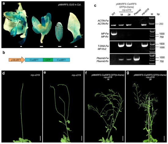
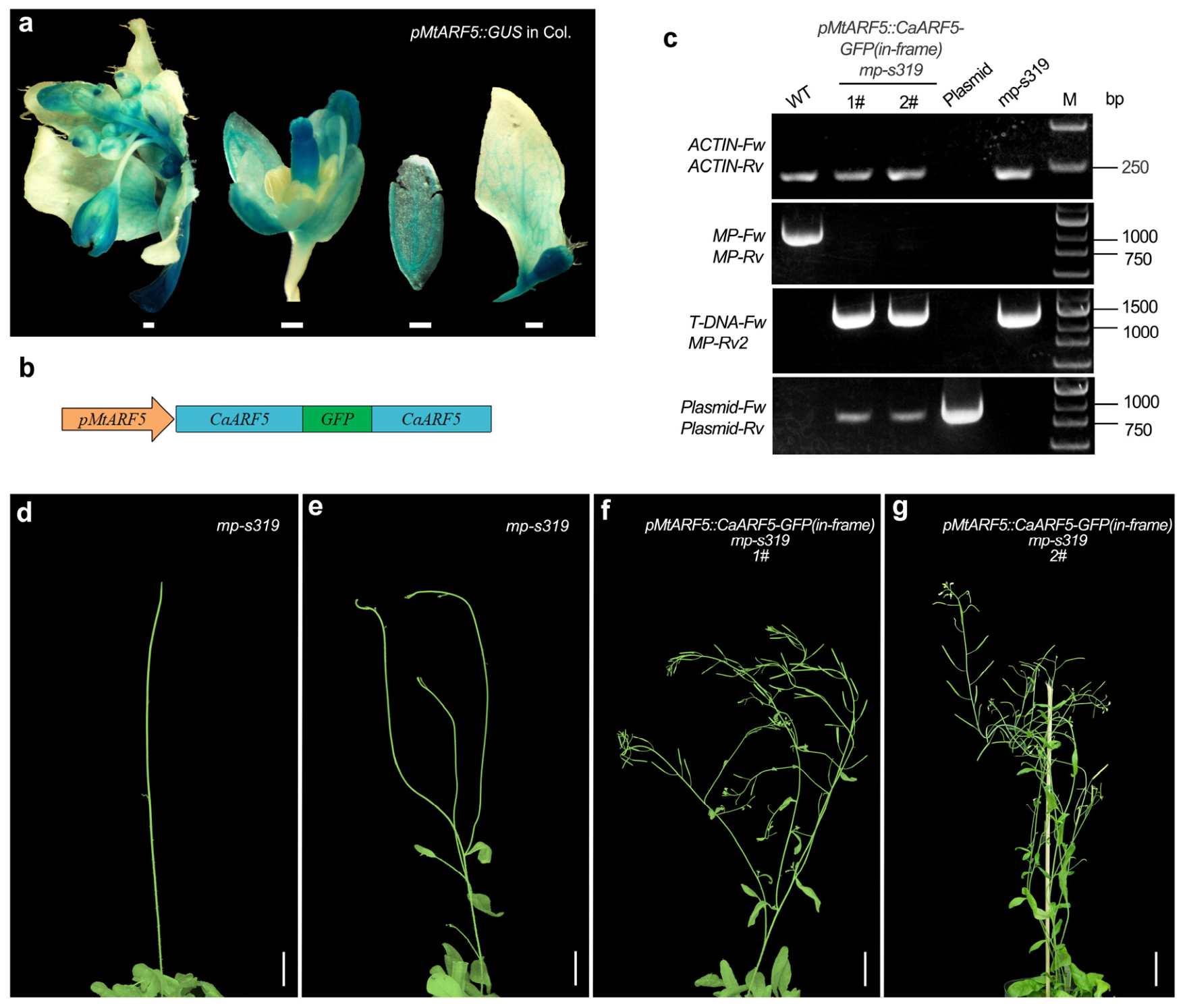
To assess the functional conservation of the chickpea CaARF5 gene, we conducted a heterologous complementation assay in Arabidopsis. We first confirmed the activity of a promoter from the Medicago truncatula ARF5 ortholog (pMtARF5), in Arabidopsis. Histochemical staining revealed that a pMtARF5::GUS reporter was specifically expressed in reproductive and vegetative tissues, including inflorescences, stems, floral buds, and developing leaves, with signal enrichment in the vascular region of leaf primordia (Figure 9a).
Figure 9.
Phenotypic traits of ectopically overexpressed CaARF5 in transgenic Arabidopsis. (a) Staining results of positive Arabidopsis wild-type Col plants transformed with pMtARF5::GUS. Scale bar, 200 μm. (b) Schematic diagram of the pMtARF5::CaARF5-GFP(in-frame) vector; (c) PCR genotyping of transgenic plants expressing pMARF5::CaARF5-GFP(in-frame) (in the mp-s319 background). M, DNA molecular weight marker (bp); wild-type (WT), the recombinant plasmid (Plasmid), and genomic DNA from the mp-s319 mutant were used as controls. (d,e) Arabidopsis mp-s319 homozygous mutant lines; Scale bar, 1 cm. (f,g) Arabidopsis transgenic lines 1# and 2#. Scale bar, 1 cm.
We then constructed a fusion vector, pMtARF5::CaARF5-GFP(in-frame) (Figure 9b), and introduced it into heterozygous mp-s319 mutants. Transgenic lines were selected by Basta resistance and validated by PCR genotyping (Figure 9c). Compared to the severe developmental defects of the mp-s319 homozygous mutant—which forms a pin-like inflorescence and is completely sterile (Figure 9d,e)—the complemented lines expressing pMtARF5::CaARF5-GFP(in-frame) developed normal inflorescences and exhibited a partial restoration of fertility (Figure 9f,g). These results demonstrate that CaARF5 can effectively rescue the reproductive defects of the Arabidopsis mp mutant, indicating a high degree of functional conservation.
3. Discussion
Chickpea is an important dual-purpose crop globally used for both food and feed, and its leaf shape directly affects photosynthetic efficiency and yield. Auxin Response Factors (ARFs) are key transcriptional regulators that interpret auxin gradients to direct developmental outcomes, including the morphogenesis of complex structures like compound leaves [41]. A previous study by Die et al. [42] first identified the ARF family in chickpea, reporting 24 members based on an earlier genome assembly (Kabuli type, CDC Frontier) and employing a nomenclature system according to chromosomal location. Our study provides substantial updates and advancements. We employed the widely accepted, phylogeny-based classification (A, B, and C clades) anchored to Arabidopsis orthologs, enabling direct functional inference and meaningful cross-species evolutionary comparisons. Furthermore, utilizing an improved, more contiguous reference genome integrated with pan-genome resources, coupled with refined Hidden Markov Model (HMM) profiles and a systematic screening strategy, we identified 33 ARF family members. The increase in the number of identified members stems from several key improvements: (1) the use of a more complete and accurately annotated genome assembly, which recovers loci missed in earlier versions; (2) the application of updated, more sensitive HMMs and adjusted significance thresholds; and (3) the adoption of inclusive verification criteria that retained putative ARFs lacking canonical domains (e.g., the C-terminal PB1 domain), with subsequent phylogenetic analysis used for validation. This approach ensures a more comprehensive and accurate catalog. Collinearity analysis indicated that segmental duplication is the primary mechanism driving the expansion of the chickpea ARF family, consistent with the expansion patterns of ARF genes in soybean and common bean [43]. Notably, the chickpea ARF genes are unevenly distributed across chromosomes, forming a local gene cluster on chromosome 4, suggesting that tandem duplication may have also played a role in the evolution of this family.
The discovery of a chickpea-specific ARF clade presents an intriguing paradox. While its members, which lack the canonical C-terminal PB1 domain and are arranged in tandem with CaARF5, evidently originated from a recent duplication event, they are consistently expressed at barely detectable levels. This minimal expression suggests these genes are unlikely to perform crucial functions in routine development, though a role in specific contexts or as subtle modulators of auxin responses remains possible. The very persistence of this low-expressing, truncated clade underscores the remarkable evolutionary constraint on the CaARF5 locus. The strong selective pressure to maintain the low copy number and precise function of the core CaARF5 gene may have tolerated the retention of these duplicates only under strict regulatory silencing, highlighting the fundamental importance of ARF5 dosage and protein structure.
ARF5 is one of the most thoroughly studied members of the ARF family. Over more than three decades of research, scientists have established that MP/ARF5 plays core regulatory roles in multiple developmental processes in Arabidopsis, including embryogenesis, vascular differentiation, leaf formation, and meristem maintenance [30]. In this study, we found that CaARF5 is highly expressed in the shoot apical meristem and young leaf primordia of chickpea, and later becoming restricted to vascular bundles, with expression gradually decreasing as leaves mature. This spatiotemporal expression pattern is highly similar to that of AtARF5 in Arabidopsis [30]. Importantly, CaARF5 was able to partially rescue the severe developmental and fertility defects of the Arabidopsis mp mutant. This underscores the profound evolutionary constraint on the molecular function of this key auxin response regulator.
Mounting evidence indicates that the precision of ARF-mediated regulation depends, in part, on expression dosage. In tomato, for instance, the promotive strength of A-ARFs such as SlMP and SlARF19 on leaflet production correlates directly with their expression levels [33]. These findings provide a relevant framework for understanding compound leaf development in chickpea. Our expression profiling of CaARF members revealed substantial variation in transcript abundance among paralogs within the same tissues, such as leaf primordia (Figure 6 and Figure 7). For example, closely related genes displayed a clear expression hierarchy: CaARF5 > CaARF19b > CaARF19a, while CaARF19c was barely detectable. Similarly, CaARF8a/8b were expressed at significantly higher levels than CaARF6a/6b, and CaARF4a/4b showed consistently stronger expression across multiple tissues compared to their paralogs CaARF3a/3b. This differential expression pattern suggests that chickpea may fine-tune the intensity, spatiotemporal specificity, and responsiveness of auxin signaling output through a combination of gene duplication, structural divergence, and differential expression regulation.
Following duplication, paralogs often undergo subfunctionalization, where ancestral functions or expression domains are partitioned [44]. The expression hierarchy within the CaARF5/19a/19b/19c clade may represent a case of subfunctionalization. In Medicago truncatula, MtARF2/MtARF3b/MtARF4a/MtARF4b are post-transcriptionally regulated via the miR390/TAS3 ta-siRNA pathway to control lateral root development and nodule formation respectively, illustrating finely partitioned functional divergence among homologous genes [45]. Therefore, the differential expression hierarchy of paralogous genes in chickpea provides a molecular basis for the precise regulation of leaf morphology (e.g., leaflet number, incision depth) and a theoretical foundation for target gene selection and expression modulation in molecular breeding.
The DBD of ARF transcription factors recognizes AuxREs in gene promoters to directly activate or repress transcription. Previous studies have shown that MP can directly bind to and activate its own expression [30]. Our promoter cis-element analysis and structural modeling indicated that the upstream regions of many CaARF genes contain multiple AuxREs that could be efficiently bound by their own DBDs. For instance, the promoter of CaARF5 itself contains an AuxRE pair predicted to be bound well by its DBD. This suggests that the expression of these ARF genes is subject to fine-tuning by their own or other ARF proteins. Therefore, our analysis provides a basis for a more comprehensive understanding of the precise spatiotemporal regulation of ARF genes during organ morphogenesis.
A central paradigm in legume compound leaf development is that LFY/FLO orthologs, such as CaLFY in chickpea, are crucial drivers of leaflet initiation. The spatiotemporal control of LFY activity, often through repression by specific factors, dictates the final leaf pattern [46]. In chickpea, the C2H2 zinc-finger protein MPL1, expressed basally, directly represses the distally enriched CaLFY, forming a complementary gradient that orchestrates the acropetal initiation of leaflets, maintaining the pinnate form. Notably, mpl1 mutants exhibit upregulated expression of several ARF genes, including CaARF5, suggesting MPL1 may also modulate the auxin pathway [4]. Studies in Arabidopsis flower development have established that the auxin response factor ARF5/MONOPTEROS directly activates LFY expression, indicating a core auxin–ARF–LFY regulatory axis [47]. Here, we found that several CaARF genes, including CaARF5, CaARF8a/8b, and CaARF4a/4b, exhibit strong and dynamic expression in early leaf primordia (P3-P5), coinciding with the critical window for patterning. This spatiotemporal overlap suggests a plausible model wherein auxin signaling, mediated by specific CaARFs, acts in concert with the MPL1-CaLFY module to jointly sculpt the compound leaf developmental blueprint.
Compound leaf development involves not only leaflet number determination but also the establishment of leaf polarity (adaxial-abaxial axis), a process in which auxin and ARFs are also key regulators. Our in situ hybridization analysis revealed the spatially specific expression of different CaARF genes in early primordia: CaARF19b transcripts were enriched in the adaxial domain, whereas CaARF3b was specifically localized to the abaxial domain. This pattern is conserved with orthologs in Arabidopsis and tomato. In Medicago, MtARF3 expression is negatively regulated by MtAGO7-mediated TAS3 ta-siRNA, and altering its level directly affects leaflet number [48]. The abaxial enrichment of CaARF3b observed here strongly suggests that a similar, small RNA-mediated post-transcriptional regulatory layer may exist in chickpea to finely calibrate ARF activity for leaflet morphogenesis.
In summary, this study establishes a foundational resource for the chickpea ARF family, delineating its evolutionary history, genomic organization, and expression landscape. We highlight both deep functional conservation, exemplified by CaARF5, and species-specific innovations, such as the PB1-lacking clade. The differential expression patterns among paralogs suggest a role for gene dosage in modulating auxin response outputs. The functional validation of CaARF5 and the in silico evidence for its potential autoregulation provide a focused entry point for future mechanistic studies. This work not only advances our understanding of auxin-mediated leaf development in legumes but also identifies specific genetic components that could be targeted for molecular breeding strategies aimed at optimizing plant architecture in chickpea and related crops.
4. Materials and Methods
4.1. Searching for ARF Genes and Data Collection
Chickpea protein sequences were retrieved from the chickpea genome database (https://www.pulsedb.org/; accessed on 22 March 2026), and AtARF protein sequences were downloaded from The Arabidopsis Information Resource database (TAIR; https://www.arabidopsis.org/; accessed on 22 March 2026). To more comprehensively identify members of the chickpea ARF family, we employed two complementary search strategies. First, we performed an initial screening using the hidden Markov model (HMM) of the ARF characteristic domain obtained from the Pfam database (http://pfam.xfam.org/; accessed on 22 March 2026), via the simple HMM search function in TBtools (v2.376). Second, we conducted a BLASTP search against the chickpea proteome using AtARF sequences as queries with default parameters. The results from both search methods were merged to identify putative chickpea ARF homologs. Subsequently, all candidate protein sequences were verified for the presence of canonical ARF domains using the NCBI Conserved Domain Database (CDD; https://www.ncbi.nlm.nih.gov/Structure/cdd/cdd.shtml; accessed on 22 March 2026). The finalized set of chickpea ARF protein sequences was analyzed using TBtools (v2.376) to calculate their length, molecular weight, and isoelectric point.
4.2. Phylogenetic Analysis
The accession numbers or IDs of ARF homologs from Arabidopsis, tomato (Solanum lycopersicum), woodland strawberry (Fragaria vesca) and soybean were retrieved via BLASTp searches from Phytozome 14 (https://phytozome-next.jgi.doe.gov/; accessed on 24 March 2026). ARF homologs from M. truncatula and chickpea were identified through BLASTp searches in the Legume Information System (https://www.legumeinfo.org/; accessed on 24 March 2026). The phylogenetic tree was constructed using the maximum likelihood method in IQ-TREE v1.6.12 (http://www.iqtree.org/; accessed on 24 March 2026), with the JTT + F + G4 model, as suggested by the IQ-TREE model test tool (BIC criterion), using 1000 ultrafast bootstrap replicates and 5000 iterations. The resulting tree was then edited using MEGA 5.0 program and manually optimized for viewing clarity.
4.3. Gene Structures and Conserved Protein Domains
The genomic and coding sequences of ARF genes were obtained from chickpea genome database. Gene structures were visualized using the Gene Structure Display Server (GSDS) 2.0 (http://gsds.cbi.pku.edu.cn/; accessed on 27 March 2026). The output image was then refined using Adobe Photoshop for color enhancement and figure annotation, involving only cosmetic optimization without any alteration to the structural data. Conserved protein domains (DBD, B3 and PB1 domains) were identified using the NCBI BLAST suite. The core prion-like domain (PrD) was predicted with the Prion-like Amino Acid Composition (PLAAC) tool (http://plaac.wi.mit.edu/, Lcore = 60, α = 1; accessed on 27 March 2026). All protein domains were visualized using IBS 2.0 (https://ibs.renlab.org/#/server; accessed on 27 March 2026).
4.4. Protein–DNA Complex Structure Prediction
The three-dimensional structures of the DNA-binding domains (DBDs) from CaARF5, CaARF19b, CaARF4a, and CaARF10a in complex with a pair of AuxRE motifs (TGTCGG) (https://alphafoldserver.com/; accessed on 28 March 2026), were predicted using AlphaFold3 with default parameters [49].
4.5. Chromosome Locations, Syntenic Analysis and Cis-Elements Analysis of CaARFs
Chromosomal distribution information of ARF genes was obtained based on the genome GFF3 file. The chromosomal locations were visualized using TBtools software (v2.376). Duplication events of CaARF genes were detected by calling the MCScanX tool through TBtools, and segmental duplications were visualized using the Advanced Circos function. The One Step MCScanX tool was employed, together with genome annotation and sequence files, to predict syntenic relationships of ARF genes among chickpea, M. truncatula, and A. thaliana. The promoter sequences spanning 2000 bp upstream of the start codon of 33 CaARF genes were submitted to the PlantCARE database (http://bioinformatics.psb.ugent.be/webtools/plantcare/html/; accessed on 30 March 2026), for cis-acting element prediction, and the physical distribution of these elements was visualized using TBtools software (v2.376).
4.6. Plant Materials
The wild-type chickpea used in this study was CDC Frontier (Kabuli type), obtained from the USDA Western Regional Plant Introduction Station (Pullman, WA, USA) via Dr. Clarice Coyne. A. thaliana accession Col. and M. truncatula cv. R108 served as the wild-type references. Seeds of the Arabidopsis mp-s319 mutant were kindly provided by Professor Zhao Zhong (University of Science and Technology of China, Hefei, China). All plants were grown in greenhouses under the following controlled conditions: a relative humidity of 50–60%, a temperature range of 24 °C during the day and 20 °C at night, a light intensity of 150 μmol/m2/s, and a 16 h light/8 h dark cycle.
4.7. RNA Extraction and Real-Time qPCR Analysis
Total RNA was isolated from samples using the RNAEx TriZol solution (GK3006, GENEray Biotech, Shanghai, China) according to the manufacturer’s protocol. First-strand cDNA was synthesized from 2 μg of total RNA using the HiScript II 1st Strand cDNA Synthesis Kit (R211, Vazyme, Nanjing, China). The resulting cDNA was diluted to a final volume of 100 μL with RNase-free water for subsequent analysis. Quantitative real-time PCR (real-time qPCR) was performed in 15 μL reaction volumes on a LightCycler 480 system (Roche, Basel, Switzerland). Each reaction contained 2.5 μL diluted cDNA, 0.45 μL of each gene-specific primer (10 μM), 7.5 μL of 2 × QuantFast Green qPCR SuperMix (M2211, Magic-bio, Hangzhou, China), and 4.1 μL of RNase-free water. All reactions were performed in triplicate. The thermal cycling conditions were as follows: 95 °C for 3 min; followed by 40 cycles of 95 °C for 10 s, 58 °C for 10 s and 72 °C for 20 s. The housekeeping gene CaGAPDH was used as internal reference for normalization. Relative gene expression levels were calculated using the 2−ΔΔCT method [50]. All primers used for real-time qPCR in this study are listed in Supplemental Table S1. The expression profiles of CaARF genes in different tissues were predicted using the CTDB website (http://223.31.159.7/ctdb/; accessed on 3 April 2026), and visualized with TBtools software (v2.376).
4.8. RNA In Situ Hybridization
In situ hybridization was performed as previously described [50,51,52], with modifications for chickpea tissues. Briefly, shoot apices were harvested from 2-week-old and 6-week-old greenhouse-grown wild-type (cv. CDC Frontier) plants. Samples were immediately fixed in FAA fixative, processed overnight using an automated tissue processor (ASP200S, Leica, Wetzlar, Germany), and embedded in paraffin with a heated embedding station (EG1150, Leica, Wetzlar, Germany).
Digoxigenin (DIG)-labelled antisense RNA probes were prepared using the DIG RNA Labeling Kit (11175025910, Roche, Basel, Switzerland). The full length coding sequences of CaARF8a, CaARF19b, CaARF5 and CaARF3b were amplified with the reverse primer (for antisense probe) or the forward primer (for sense probe) containing a T7 promoter sequence (5′-TGTAATACGACTCACTATAGGGC-3′) at its 5′end. The resulting amplicons served as templates for in vitro transcription to generate DIG-labeled RNA probes, which were subsequently hydrolyzed to fragments of 100–200 bp.
Paraffin-embedded shoot apices were sectioned into 8-μm-thick sections using a microtome (RM2235, Leica, Wetzlar, Germany). Sections were deparaffinized, rehydrated, and treated with 1 mg/L Proteinase K (03115879001, Roche, Basel, Switzerland) for 30 min at 37 °C. Following a brief post-fixation in FAA for 10 min and dehydration through an ethanol series, sections were hybridized with the DIG-labeled probes overnight at 55 °C. Post-hybridization, signals were detected by incubation with Anti-DIG-AP Fab fragments (11093274910, Roche, Basel, Switzerland) for 90 min, followed by colorimetric development with NBT/BCIP substrate (11681451001, Roche, Basel, Switzerland) for 48 h–72 h. Images were captured using a light microscope (BX63, Olympus, Tokyo, Japan). All primer sequences used for probe synthesis are listed in Supplemental Table S1.
4.9. Subcellular Localization
Subcellular localization was analyzed via transient expression in tobacco leaves. Agrobacterium tumefaciens strain EHA105 harboring the target vector was cultured overnight in a shaker at 28°C. The next day, 50 ul of the bacterial culture was added to 5 mL of fresh LB liquid medium and incubated at 28 °C with shaking until an OD600 of 1.0–1.5 was reached. The culture was centrifuged at 3900 rpm for 10 min, and the supernatant was discarded. During centrifugation, MMA buffer (10 mM MES pH 5.6, 10 mM MgCl2, 200 μM acetosyringone) was prepared. The bacterial pellet was resuspended in MMA buffer to an OD600 of 1.0 and incubated at room temperature for 1 h. Fully expanded tobacco leaves were selected, and small wounds were gently made on the abaxial surface using a needle. The bacterial suspension was infiltrated into the wounds using a 1 mL syringe without a needle. The plants were incubated in the dark at 28 °C for 36–48 h. The lower epidermal peel of the leaf was then transferred to a clean coverslip, mounted, and observed under a laser confocal microscope. GFP signals were detected under 488 nm excitation light.
4.10. GUS Staining
GUS staining was performed as previously described [53], with modifications for Arabidopsis tissues. The brief procedure is as follows. First, the samples were fixed: fresh tissues were placed in centrifuge tubes, covered with 90% acetone, and incubated on ice for 30 min, followed by rinsing twice with phosphate buffer. Next, for staining: after fixation, the samples were immersed in GUS staining solution, vacuum-infiltrated for 30 min, and incubated overnight at 37 °C. Finally, after destaining with 75% ethanol, the samples were imaged under a stereomicroscope.
4.11. Vector Construction and Plant Transformation
A GFP reporter construct was generated by in-frame fusion of GFP to the CaARF5 coding sequence between residues Ile492 and Lys493. The N- and C-terminal fragments of CaARF5 and the full-length GFP were amplified separately and assembled by overlap-extension PCR. The resulting fusion fragment was ligated into the binary vector pCAMBIA3301, creating a CaMV35S promoter-driven CaARF5-GFP(in-frame) construct. The CaMV35S promoter was then excised by SacI/AatII digestion and replaced with a PCR-amplified MtARF5 promoter fragment using homologous recombination, yielding the final ProMtARF5:CaARF5-GFP plasmid. The construct was introduced into Agrobacterium tumefaciens strain EHA105 and transformed into the Arabidopsis mp mutant via the floral dip method. T0 transgenic seeds were sown in soil, and transformants were selected by spraying with Basta. In this experiment, the active ingredient of Basta was glufosinate ammonium, which was used for screening transgenic Arabidopsis at a working concentration of 20 mg/L.
Supplementary Materials
The following supporting information can be downloaded at: https://www.mdpi.com/article/10.3390/plants15111708/s1, Figure S1: Sense probe control. In the in situ hybridization experiment, the sense probe was used as a negative control to assess signal specificity; Table S1: List of primer sequences used in this study; Table S2: CaARF family member ID and gene name; Table S3: Physicochemical properties of CaARFs gene family members in chickpea; Table S4: Conserved domains of CaARF family proteins in chickpea; Table S5: The location of CaARF in chickpea chromosomes; Table S6: Segmentally duplicated ARF gene pairs identified in the chickpea genome; Table S7: Orthologous relationships of ARF genes between chickpea and two representative plants.; Table S8: Detailed Information on the Analysis of Cis-acting Elements in Chickpea ARF Family Genes; Table S9: Statistics of cis-acting elements in chickpea ARF family genes; Table S10: Expression profiles of the CaARF family in different tissues of chickpea.
Author Contributions
L.H., J.C. and D.Z. designed research; H.F., Y.F. and L.H. performed experiments; X.Y., Y.Z., Z.H., Q.C., L.M. and J.L. participated in analyzing data; H.F. and L.H. wrote the paper. All authors have read and agreed to the published version of the manuscript.
Funding
This research was funded by the Open Fund Program of Hou Ji Laboratory in Shanxi Province (Minor Crops Bio-breeding Laboratory in Shanxi Province) (Grant No: 2025HJZ05), the grants from National Natural Science Foundation of China (Grants No: 32570980, U2102222, 32070204, and 32170839), and Yunnan Revitalization Talent Support Program (Grant No: XDYC-QNRC-79, and XDYC-QNRC-2022-0335).
Data Availability Statement
All relevant data are available within the manuscript and the Supplementary Materials.
Acknowledgments
The authors sincerely acknowledge the Institutional Center for Shared Technologies and Facilities of Xishuangbanna Tropical Botanical Garden, CAS, for providing the research facilities and academic environment necessary for this work. We also extend our gratitude to our supervisors, colleagues, and laboratory members for their guidance, encouragement, and technical assistance throughout the study.
Conflicts of Interest
The authors declare no conflicts of interest.
References
- Shahnaz, Z.; Shahnaz, A.; Ismail, A.; Manzoor, Z.; Iqbal, R.; Zia, M.A.B. Chickpea (Cicer arietinum) Genetic Diversity: Genetic Resources, Breeding Progress, and Future Prospects. Crop Pasture Sci. 2025, 76, CP24238. [Google Scholar] [CrossRef] [Scilit]
- Jukanti, A.K.; Gaur, P.M.; Gowda, C.L.L.; Chibbar, R.N. Nutritional Quality and Health Benefits of Chickpea (Cicer arietinum L.): A Review. Br. J. Nutr. 2012, 108, S11–S26. [Google Scholar] [CrossRef] [Scilit]
- Nakayama, H.; Sinha, N.R. Leaf Evolution: Integrating Phylogenetics, Developmental Dynamics, and Genetic Insights across Land Plants. New Phytol. 2025, 248, 2205–2220. [Google Scholar] [CrossRef] [Scilit]
- Liu, Y.; Yang, Y.; Wang, R.; Liu, M.; Ji, X.; He, Y.; Zhao, B.; Li, W.; Mo, X.; Zhang, X.; et al. Control of Compound Leaf Patterning by MULTI-PINNATE LEAF1 (MPL1) in Chickpea. Nat. Commun. 2023, 14, 8088. [Google Scholar] [CrossRef] [Scilit] [PubMed]
- Chandler, J.W. Auxin Response Factors. Plant Cell Environ. 2016, 39, 1014–1028. [Google Scholar] [CrossRef] [Scilit] [PubMed]
- Ulmasov, T.; Hagen, G.; Guilfoyle, T.J. ARF1, a Transcription Factor That Binds to Auxin Response Elements. Science 1997, 276, 1865–1868. [Google Scholar] [CrossRef] [Scilit]
- Boer, D.R.; Freire-Rios, A.; van den Berg, W.A.M.; Saaki, T.; Manfield, I.W.; Kepinski, S.; López-Vidrieo, I.; Franco-Zorrilla, J.M.; de Vries, S.C.; Solano, R.; et al. Structural Basis for DNA Binding Specificity by the Auxin-Dependent ARF Transcription Factors. Cell 2014, 156, 577–589. [Google Scholar] [CrossRef] [Scilit] [PubMed]
- Zemlyanskaya, E.V.; Wiebe, D.S.; Omelyanchuk, N.A.; Levitsky, V.G.; Mironova, V.V. Meta-Analysis of Transcriptome Data Identified TGTCNN Motif Variants Associated with the Response to Plant Hormone Auxin in Arabidopsis thaliana L. J. Bioinform. Comput. Biol. 2016, 14, 1641009. [Google Scholar] [CrossRef] [Scilit]
- Tiwari, S.B.; Hagen, G.; Guilfoyle, T. The Roles of Auxin Response Factor Domains in Auxin-Responsive Transcription. Plant Cell 2003, 15, 533–543. [Google Scholar] [CrossRef] [Scilit]
- Kim, Y.; Park, C.; Cha, S.; Han, M.; Ryu, K.-S.; Suh, J.-Y. Determinants of PB1 Domain Interactions in Auxin Response Factor ARF5 and Repressor IAA17. J. Mol. Biol. 2020, 432, 4011–4022. [Google Scholar] [CrossRef] [Scilit]
- Gray, W.M.; Kepinski, S.; Rouse, D.; Leyser, O.; Estelle, M. Auxin Regulates SCFTIR1-Dependent Degradation of AUX/IAA Proteins. Nature 2001, 414, 271–276. [Google Scholar] [CrossRef] [Scilit] [PubMed]
- Salehin, M.; Bagchi, R.; Estelle, M. SCFTIR1/AFB-Based Auxin Perception: Mechanism and Role in Plant Growth and Development. Plant Cell 2015, 27, 9–19. [Google Scholar] [CrossRef] [Scilit]
- Cancé, C.; Martin-Arevalillo, R.; Boubekeur, K.; Dumas, R. Auxin Response Factors Are Keys to the Many Auxin Doors. New Phytol. 2022, 235, 402–419. [Google Scholar] [CrossRef] [Scilit] [PubMed]
- Hardtke, C.S.; Berleth, T. The Arabidopsis Gene MONOPTEROS Encodes a Transcription Factor Mediating Embryo Axis Formation and Vascular Development. EMBO J. 1998, 17, 1405–1411. [Google Scholar] [CrossRef] [Scilit]
- Schlereth, A.; Möller, B.; Liu, W.; Kientz, M.; Flipse, J.; Rademacher, E.H.; Schmid, M.; Jürgens, G.; Weijers, D. MONOPTEROS Controls Embryonic Root Initiation by Regulating a Mobile Transcription Factor. Nature 2010, 464, 913–916. [Google Scholar] [CrossRef] [Scilit]
- Okushima, Y.; Fukaki, H.; Onoda, M.; Theologis, A.; Tasaka, M. ARF7 and ARF19 Regulate Lateral Root Formation via Direct Activation of LBD/ASL Genes in Arabidopsis. Plant Cell 2007, 19, 118–130. [Google Scholar] [CrossRef] [Scilit]
- Huang, K.-L.; Ma, G.-J.; Zhang, M.-L.; Xiong, H.; Wu, H.; Zhao, C.-Z.; Liu, C.-S.; Jia, H.-X.; Chen, L.; Kjorven, J.O.; et al. The ARF7 and ARF19 Transcription Factors Positively Regulate PHOSPHATE STARVATION RESPONSE1 in Arabidopsis Roots. Plant Physiol. 2018, 178, 413–427. [Google Scholar] [CrossRef] [Scilit]
- Pekker, I.; Alvarez, J.P.; Eshed, Y. Auxin Response Factors Mediate Arabidopsis Organ Asymmetry via Modulation of KANADI Activity. Plant Cell 2005, 17, 2899–2910. [Google Scholar] [CrossRef] [Scilit]
- Nagpal, P.; Ellis, C.M.; Weber, H.; Ploense, S.E.; Barkawi, L.S.; Guilfoyle, T.J.; Hagen, G.; Alonso, J.M.; Cohen, J.D.; Farmer, E.E.; et al. Auxin Response Factors ARF6 and ARF8 Promote Jasmonic Acid Production and Flower Maturation. Development 2005, 132, 4107–4118. [Google Scholar] [CrossRef] [Scilit]
- Wang, Y.; Kelley, S.; Zentella, R.; Hu, J.; Wei, H.; Wang, L.; Shabanowitz, J.; Hunt, D.F.; Sun, T. O-Fucosyltransferase SPINDLY Attenuates Auxin-Induced Fruit Growth by Inhibiting ARF6/8-Coactivator Mediator Complex Interaction in Arabidopsis. Nat. Commun. 2025, 16, 3965. [Google Scholar] [CrossRef] [Scilit] [PubMed]
- Tabata, R.; Ikezaki, M.; Fujibe, T.; Aida, M.; Tian, C.; Ueno, Y.; Yamamoto, K.T.; Machida, Y.; Nakamura, K.; Ishiguro, S. Arabidopsis AUXIN RESPONSE FACTOR6 and 8 Regulate Jasmonic Acid Biosynthesis and Floral Organ Development via Repression of Class 1 KNOX Genes. Plant Cell Physiol. 2010, 51, 164–175. [Google Scholar] [CrossRef] [Scilit] [PubMed]
- Wang, B.; Xue, J.-S.; Yu, Y.-H.; Liu, S.-Q.; Zhang, J.-X.; Yao, X.-Z.; Liu, Z.-X.; Xu, X.-F.; Yang, Z.-N. Fine Regulation of ARF17 for Anther Development and Pollen Formation. BMC Plant Biol. 2017, 17, 243. [Google Scholar] [CrossRef] [Scilit]
- Shen, C.; Wang, S.; Zhang, S.; Xu, Y.; Qian, Q.; Qi, Y.; Jiang, D.A. OsARF16, a Transcription Factor, Is Required for Auxin and Phosphate Starvation Response in Rice (Oryza sativa L.). Plant Cell Environ. 2013, 36, 607–620. [Google Scholar] [CrossRef] [Scilit] [PubMed]
- Wang, S.; Zhang, S.; Sun, C.; Xu, Y.; Chen, Y.; Yu, C.; Qian, Q.; Jiang, D.-A.; Qi, Y. Auxin Response Factor (OsARF12), a Novel Regulator for Phosphate Homeostasis in Rice (Oryza sativa). New Phytol. 2014, 201, 91–103. [Google Scholar] [CrossRef] [Scilit] [PubMed]
- Sagar, M.; Chervin, C.; Mila, I.; Hao, Y.; Roustan, J.-P.; Benichou, M.; Gibon, Y.; Biais, B.; Maury, P.; Latché, A.; et al. SlARF4, an Auxin Response Factor Involved in the Control of Sugar Metabolism during Tomato Fruit Development. Plant Physiol. 2013, 161, 1362–1374. [Google Scholar] [CrossRef] [Scilit]
- Yuan, Y.; Mei, L.; Wu, M.; Wei, W.; Shan, W.; Gong, Z.; Zhang, Q.; Yang, F.; Yan, F.; Zhang, Q.; et al. SlARF10, an Auxin Response Factor, Is Involved in Chlorophyll and Sugar Accumulation during Tomato Fruit Development. J. Exp. Bot. 2018, 69, 5507–5518. [Google Scholar] [CrossRef] [Scilit]
- Shen, C.; Yue, R.; Sun, T.; Zhang, L.; Yang, Y.; Wang, H. OsARF16, a Transcription Factor Regulating Auxin Redistribution, Is Required for Iron Deficiency Response in Rice (Oryza sativa L.). Plant Sci. 2015, 231, 148–158. [Google Scholar] [CrossRef] [Scilit]
- Kang, C.; He, S.; Zhai, H.; Li, R.; Zhao, N.; Liu, Q. A Sweetpotato Auxin Response Factor Gene (IbARF5) Is Involved in Carotenoid Biosynthesis and Salt and Drought Tolerance in Transgenic Arabidopsis. Front. Plant Sci. 2018, 9, 1307. [Google Scholar] [CrossRef] [Scilit]
- Xiao, G.; He, P.; Zhao, P.; Liu, H.; Zhang, L.; Pang, C.; Yu, J. Genome-Wide Identification of the GhARF Gene Family Reveals That GhARF2 and GhARF18 Are Involved in Cotton Fibre Cell Initiation. J. Exp. Bot. 2018, 69, 4323–4337. [Google Scholar] [CrossRef] [Scilit]
- Wójcikowska, B.; Belaidi, S.; Robert, H.S. Game of Thrones among AUXIN RESPONSE FACTORs—Over 30 Years of MONOPTEROS Research. J. Exp. Bot. 2023, 74, 6904–6921. [Google Scholar] [CrossRef] [Scilit]
- Okushima, Y.; Overvoorde, P.J.; Arima, K.; Alonso, J.M.; Chan, A.; Chang, C.; Ecker, J.R.; Hughes, B.; Lui, A.; Nguyen, D.; et al. Functional Genomic Analysis of the AUXIN RESPONSE FACTOR Gene Family Members in Arabidopsis thaliana: Unique and Overlapping Functions of ARF7 and ARF19. Plant Cell 2005, 17, 444–463. [Google Scholar] [CrossRef] [Scilit]
- Su, X.; Zhang, X.; Luo, J.; Wang, Y.; Feng, B.; Yang, Y.; Zhang, B.; Li, Y.; Zhang, H.; Yuan, W.; et al. The IAA7-ARF7-ARF19 Auxin Signaling Module Plays Diverse Roles in Arabidopsis Growth and Development. Planta 2025, 262, 12. [Google Scholar] [CrossRef] [Scilit]
- Israeli, A.; Capua, Y.; Shwartz, I.; Tal, L.; Meir, Z.; Levy, M.; Bar, M.; Efroni, I.; Ori, N. Multiple Auxin-Response Regulators Enable Stability and Variability in Leaf Development. Curr. Biol. 2019, 29, 1746–1759.e5. [Google Scholar] [CrossRef] [Scilit] [PubMed]
- Zhou, C.; Han, L.; Fu, C.; Wen, J.; Cheng, X.; Nakashima, J.; Ma, J.; Tang, Y.; Tan, Y.; Tadege, M.; et al. The Trans-Acting Short Interfering RNA3 Pathway and NO APICAL MERISTEM Antagonistically Regulate Leaf Margin Development and Lateral Organ Separation, as Revealed by Analysis of an Argonaute7/Lobed Leaflet1 Mutant in Medicago truncatula. Plant Cell 2013, 25, 4845–4862. [Google Scholar] [CrossRef] [Scilit] [PubMed]
- Peng, J.; Berbel, A.; Madueño, F.; Chen, R. AUXIN RESPONSE FACTOR3 Regulates Compound Leaf Patterning by Directly Repressing PALMATE-LIKE PENTAFOLIATA1 Expression in Medicago truncatula. Front. Plant Sci. 2017, 8, 1630. [Google Scholar] [CrossRef] [Scilit] [PubMed]
- Yifhar, T.; Pekker, I.; Peled, D.; Friedlander, G.; Pistunov, A.; Sabban, M.; Wachsman, G.; Alvarez, J.P.; Amsellem, Z.; Eshed, Y. Failure of the Tomato Trans-Acting Short Interfering RNA Program to Regulate AUXIN RESPONSE FACTOR3 and ARF4 Underlies the Wiry Leaf Syndrome. Plant Cell 2012, 24, 3575–3589. [Google Scholar] [CrossRef] [Scilit]
- Ali, S.; Wang, W.; Zhang, Z.; Xie, L.; Boer, D.R.; Khan, N. Genome-Wide Identification, Expression and Interaction Analysis of ARF and AUX/IAA Gene Family in Soybean. Front. Biosci. 2022, 27, 251. [Google Scholar] [CrossRef] [Scilit]
- Jiang, J.; Wang, Z.; Chen, Z.; Wu, Y.; Mu, M.; Nie, W.; Zhao, S.; Cui, G.; Yin, X. Identification and Evolutionary Analysis of the Auxin Response Factor (ARF) Family Based on Transcriptome Data from Caucasian Clover and Analysis of Expression Responses to Hormones. Int. J. Mol. Sci. 2023, 24, 15357. [Google Scholar] [CrossRef] [Scilit]
- Chen, F.; Zhang, J.; Ha, X.; Ma, H. Genome-Wide Identification and Expression Analysis of the Auxin-Response Factor (ARF) Gene Family in Medicago Sativa under Abiotic Stress. BMC Genom. 2023, 24, 498. [Google Scholar] [CrossRef] [Scilit]
- Schuetz, M.; Fidanza, M.; Mattsson, J. Identification of Auxin Response Factor-Encoding Genes Expressed in Distinct Phases of Leaf Vein Development and with Overlapping Functions in Leaf Formation. Plants 2019, 8, 242. [Google Scholar] [CrossRef] [Scilit]
- Li, Y.; Han, S.; Qi, Y. Advances in Structure and Function of Auxin Response Factor in Plants. J. Integr. Plant Biol. 2023, 65, 617–632. [Google Scholar] [CrossRef] [Scilit]
- Die, J.V.; Gil, J.; Millan, T. Genome-Wide Identification of the Auxin Response Factor Gene Family in Cicer Arietinum. BMC Genom. 2018, 19, 301. [Google Scholar] [CrossRef] [Scilit]
- Le, B.; Nawaz, M.A.; Rehman, H.M.; Le, T.; Yang, S.H.; Golokhvast, K.S.; Son, E.; Chung, G. Genome-Wide Characterization and Expression Pattern of Auxin Response Factor (ARF) Gene Family in Soybean and Common Bean. Genes Genom. 2016, 38, 1165–1178. [Google Scholar] [CrossRef] [Scilit]
- Hernández-García, J.; Carrillo-Carrasco, V.P.; Rienstra, J.; Tanaka, K.; de Roij, M.; Dipp-Álvarez, M.; Freire-Ríos, A.; Crespo, I.; Boer, R.; van den Berg, W.A.M.; et al. Evolutionary Origins and Functional Diversification of Auxin Response Factors. Nat. Commun. 2024, 15, 10909. [Google Scholar] [CrossRef] [Scilit] [PubMed]
- Kirolinko, C.; Hobecker, K.; Wen, J.; Mysore, K.S.; Niebel, A.; Blanco, F.A.; Zanetti, M.E. Auxin Response Factor 2 (ARF2), ARF3, and ARF4 Mediate Both Lateral Root and Nitrogen Fixing Nodule Development in Medicago truncatula. Front. Plant Sci. 2021, 12, 659061. [Google Scholar] [CrossRef] [Scilit]
- Hofer, J.; Turner, L.; Hellens, R.; Ambrose, M.; Matthews, P.; Michael, A.; Ellis, N. UNIFOLIATA Regulates Leaf and Flower Morphogenesis in Pea. Curr. Biol. 1997, 7, 581–587. [Google Scholar] [CrossRef] [Scilit] [PubMed]
- Yamaguchi, N.; Wu, M.-F.; Winter, C.M.; Wagner, D. LEAFY and Polar Auxin Transport Coordinately Regulate Arabidopsis Flower Development. Plants 2014, 3, 251–265. [Google Scholar] [CrossRef] [Scilit]
- Wang, X.; Zhang, J.; Xie, Y.; Liu, X.; Wen, L.; Wang, H.; Zhang, J.; Li, J.; Han, L.; Yu, X.; et al. LATE MERISTEM IDENTITY1 Regulates Leaf Margin Development via the Auxin Transporter Gene SMOOTH LEAF MARGIN1. Plant Physiol. 2021, 187, 218–235. [Google Scholar] [CrossRef] [Scilit]
- Abramson, J.; Adler, J.; Dunger, J.; Evans, R.; Green, T.; Pritzel, A.; Ronneberger, O.; Willmore, L.; Ballard, A.J.; Bambrick, J.; et al. Accurate Structure Prediction of Biomolecular Interactions with AlphaFold 3. Nature 2024, 630, 493–500. [Google Scholar] [CrossRef] [Scilit]
- Pfaffl, M.W. A New Mathematical Model for Relative Quantification in Real-Time RT–PCR. Nucleic Acids Res. 2001, 29, e45. [Google Scholar] [CrossRef] [Scilit]
- He, L.; Liu, Y.; Mao, Y.; Wu, X.; Zheng, X.; Zhao, W.; Mo, X.; Wang, R.; Wu, Q.; Wang, D.; et al. GRAS Transcription Factor PINNATE-LIKE PENTAFOLIATA2 Controls Compound Leaf Morphogenesis in Medicago truncatula. Plant Cell 2024, 36, 1755–1776. [Google Scholar] [CrossRef] [Scilit] [PubMed]
- Coen, E.S.; Romero, J.; Doyle, S.; Elliott, R.; Murphy, G.; Carpenter, R. Floricaula: A Homeotic Gene Required for Flower Development in Antirrhinum Majus. Cell 1990, 63, 1311–1322. [Google Scholar] [CrossRef] [Scilit] [PubMed]
- Dedow, L.K.; Oren, E.; Braybrook, S.A. Fake News Blues: A GUS Staining Protocol to Reduce False-Negative Data. Plant Direct 2022, 6, e367. [Google Scholar] [CrossRef] [Scilit] [PubMed]
Disclaimer/Publisher’s Note: The statements, opinions and data contained in all publications are solely those of the individual author(s) and contributor(s) and not of MDPI and/or the editor(s). MDPI and/or the editor(s) disclaim responsibility for any injury to people or property resulting from any ideas, methods, instructions or products referred to in the content. |
© 2026 by the authors. Licensee MDPI, Basel, Switzerland. This article is an open access article distributed under the terms and conditions of the Creative Commons Attribution (CC BY) license.