


Abstract
The blood–brain barrier (BBB), together with associated perivascular cells, forms a key component of the neurovascular unit and constitutes a highly specialized multicellular interface that tightly regulates the movement of ions, nutrients, oxygen, metabolites, and immune cells between the circulation and the central nervous system (CNS). This selective barrier is essential for maintaining CNS homeostasis, supporting neuronal function, and protecting neural tissue from circulating toxins, pathogens, and inflammatory mediators. Disruption of BBB integrity is a hallmark of numerous neurological disorders and contributes to disease initiation and progression. Among the structural components of the BBB, the endothelial glycocalyx has emerged as a critical yet underrecognized regulator of barrier function. This carbohydrate-rich layer, composed primarily of proteoglycans, glycoproteins, and glycolipids, forms an important interface between circulating blood and the brain endothelium. Beyond serving as a physical barrier, glycocalyx proteoglycans regulate vascular permeability, mechanotransduction, leukocyte adhesion, and inflammatory signaling, thereby contributing to BBB integrity and cerebrovascular homeostasis. In this review, we discuss the architecture and function of the BBB, highlight the overlooked roles of proteoglycans in maintaining BBB integrity and regulating neurovascular function, examine their contributions to BBB dysfunction in neurological diseases, and explore emerging therapeutic approaches targeting the glycocalyx to preserve or restore BBB integrity.
1. Introduction
The blood–brain barrier (BBB) is a highly specialized neurovascular interface that regulates molecular and cellular exchange between the systemic circulation and the central nervous system (CNS), thereby maintaining neural homeostasis and supporting normal brain function [1]. The highly selective properties of the BBB are conferred by the specialized phenotype of brain microvascular endothelial cells, which are characterized by complex tight junction architecture, markedly reduced rates of fluid-phase transcytosis, and the coordinated expression of selective influx and efflux transport systems that regulate molecular exchange between the blood and the CNS [2].
Structurally and functionally, the BBB is composed of a multicellular neurovascular unit consisting of brain microvascular endothelial cells, pericytes, astrocytes, and the surrounding extracellular matrix. Among the endothelial components of the BBB, the endothelial surface glycocalyx, composed primarily of proteoglycans, glycoproteins, and glycolipids, has emerged as a critical yet underrecognized regulator of barrier function [3]. This carbohydrate-rich layer lines the luminal surface of endothelial cells and constitutes an important interface between circulating blood and the BBB endothelium. Beyond acting as a physical barrier, the glycocalyx regulates vascular permeability, mechanotransduction, leukocyte-endothelial interactions, and inflammatory signaling, thereby contributing significantly to BBB integrity and cerebrovascular homeostasis.
Despite constituting an important interface between circulating blood and the brain endothelium, the molecular composition and specific functional roles of the brain endothelial glycocalyx remain incompletely understood. Furthermore, BBB dysfunction, often characterized by increased vascular permeability and the entry of circulating neurotoxic and inflammatory mediators into the brain parenchyma, is a prominent pathological feature of aging and neurodegenerative disorders. Emerging evidence suggests that alterations in the endothelial glycocalyx contribute to BBB disruption and disease progression [1]. Structural and compositional alterations in the glycocalyx, particularly the loss of heparan sulfate (HS), altered hyaluronan (HA) levels, changes in mucin-type O-glycans, and degradation of basement membrane proteoglycans, are important contributors to BBB leakage in both acute and chronic disease states. Proteoglycans are particularly important because their core proteins and glycosaminoglycan chains provide structural organization while also serving as dynamic regulators of signaling, ligand binding, mechanotransduction, and cell–cell and cell–matrix interactions. However, comprehensive reviews focusing specifically on the role of the brain endothelial glycocalyx, particularly its proteoglycan components, in regulating BBB integrity are limited.
In this review, we discuss the architecture and physiological functions of the BBB, BBB dysfunction in neurological diseases, and the molecular and cellular determinants that govern BBB integrity. Particular emphasis is placed on the major proteoglycans of the endothelial glycocalyx and their roles in maintaining BBB structure and function. We also examine emerging therapeutic strategies aimed at targeting the endothelial glycocalyx to preserve or restore BBB integrity in neurological disorders.
2. Architecture and Physiologic Function of the BBB
2.1. Structure and Function of BBB
The BBB is a multicellular vascular structure that separates the CNS from the peripheral blood circulation. The luminal-most component of the BBB is the endothelial glycocalyx, a complex meshwork of polysaccharides covering the luminal surface of endothelial cells [4]. This structure is composed of proteoglycans, glycoproteins, and glycolipids [1] and serves as the first physical and electrostatic barrier of the BBB, providing direct protection to the underlying endothelium [3]. The composition of the glycocalyx within the BBB differs from that of the glycocalyx found in other tissues. Compared with other vascular beds, the BBB glycocalyx is characterized by a more negative surface charge density and enriched expression of components such as chondroitin sulfate, heparan sulfate, phosphatidylinositol, and phosphatidylserine [3]. However, its precise molecular composition remains incompletely characterized and is likely influenced by factors such as age, disease status, and physiological conditions.
Beneath the glycocalyx lies a specialized layer of endothelial cells joined by continuous tight junctions that restrict paracellular diffusion [5]. The abluminal surface of these endothelial cells is supported by a vascular basement membrane, a specialized extracellular matrix (ECM) composed primarily of laminin, type IV collagen, fibronectin, and perlecan. At the level of the postcapillary venule, this architecture becomes more complex, with distinct endothelial and parenchymal basement membranes forming a defined perivascular space [5].
The outermost component of the BBB is formed by astrocytic end-feet, which envelop nearly the entire vascular surface [5]. Together, these cellular and extracellular components, along with specialized junctional complexes, membrane transporters, and adhesion molecules, establish a continuous and highly selective barrier throughout the CNS vasculature [5,6]. Functionally, the BBB serves as a dynamic regulatory interface between the systemic circulation and the CNS. It enables the selective transport of essential nutrients, restricts the entry of toxins and pathogens, regulates immune surveillance, and facilitates the clearance of metabolic waste products from the brain [7,8,9].
From a metabolic perspective, oxygen, as a small lipophilic solute, can freely cross the BBB via passive diffusion through the plasma membrane of endothelial cells [7], while cerebral oxygen utilization is tightly coupled to cerebral blood flow [10]. In contrast, key energy substrates, including glucose, lactate, fatty acids, and ketone bodies, require carrier-mediated transport systems to cross the BBB [10,11]. Despite the brain’s high metabolic demand, it has minimal intrinsic energy reserves, making it highly dependent on continuous nutrient transport across the BBB [12]. Moreover, cerebral glucose utilization varies with regional neuronal activity, requiring dynamic regulation of BBB transport to match regional metabolic demands [12].
Peripheral immune cells have limited infiltration into the brain compared with other tissues [7]. Under physiological conditions, peripheral immune cell entry into the CNS is tightly restricted by intact tight junctions and low basal expression of leukocyte adhesion molecules (LAMs) [7,13]. During inflammation, however, the BBB becomes dynamically responsive, with increased expression of LAMs that permits controlled leukocyte trafficking into the CNS [13].
Beyond regulating immune cell trafficking, the BBB plays a critical role in protecting the CNS by restricting the entry of potentially harmful substances while facilitating the clearance of metabolic waste. These protective functions are mediated through multiple complementary mechanisms. Metabolic enzymes expressed by the neurovascular unit degrade unwanted small water-soluble molecules that could otherwise pass through the BBB via paracellular transport [7]. Lipophilic compounds are actively exported from the brain by efflux transporters, preventing their accumulation within cerebral tissue [7]. In addition, proteins and other metabolic waste products are cleared through BBB transport mechanisms and the glymphatic–lymphatic system, thereby contributing to CNS homeostasis [14,15].
Collectively, these integrated structural and functional properties establish the BBB as a highly selective and dynamically regulated interface that is essential for maintaining CNS homeostasis. Disruption of these tightly coordinated mechanisms is increasingly recognized as a key contributor to the pathogenesis of numerous neurological disorders.
2.2. BBB Dysfunction in Disease
BBB dysfunction is a hallmark of numerous neurological disorders, including ischemic and hemorrhagic stroke, traumatic brain injury, multiple sclerosis, and neurodegenerative diseases. Despite their distinct etiologies, these conditions exhibit several common mechanisms of BBB disruption (Figure 1), including disruption of tight junctions, altered endothelial signaling, basement membrane remodeling, oxidative stress-mediated inflammation, and degradation of the endothelial glycocalyx. Together, these changes compromise BBB integrity and contribute to disease progression. The major BBB alterations associated with these CNS disorders are summarized in Table 1.
Figure 1.
Mechanisms of blood–brain barrier disruption. The top panel illustrates healthy versus diseased blood–brain barrier (BBB). In the healthy state, endothelial cells, pericytes, and astrocytic end-feet maintain BBB integrity. In the diseased state, disruption of tight junction proteins, including claudin and occludin, increases BBB permeability, leading to immune cell infiltration, red blood cell leakage, oxidative stress, and amyloid-β deposition. Panel (a) depicts the mechanism of BBB injury during ischemic stroke. Early after ischemia, BBB permeability increases in response to reactive oxygen species (ROS) and cytokines. In later stages, activation of MMP-2/9 disrupts tight junctions, leading to increased transcytosis, vascular leakage, and cerebral edema. Panel (b) illustrates amyloid-β plaque accumulation in Alzheimer’s disease. Impaired amyloid-β transport contributes to chronic BBB dysregulation through pericyte dysfunction. Amyloid-β accumulation also alters tight junction integrity, resulting in increased vascular permeability. Panel (c) shows traumatic brain injury (TBI)-mediated BBB disruption. BBB dysfunction following TBI is multifactorial but commonly involves shear stress-induced vascular injury, increased cytokine production, and activation of enzymatic cascades involving MMPs and other proteases. Panel (d) illustrates BBB dysfunction during multiple sclerosis (MS). Sialyl Lewis X (sLeX) glycan ligands on circulating leukocytes interact with selectin-family adhesion molecules (P-selectin and E-selectin) expressed on activated brain endothelial cells. These interactions mediate leukocyte tethering and rolling along the vascular wall, facilitating adhesion and transmigration across the BBB. Inflammatory cytokines further disrupt tight junctions and the basement membrane, compromising barrier integrity. Panel (e) shows the mechanism of BBB dysfunction following hemorrhagic stroke. Blood-derived products, including hemoglobin, iron, thrombin, and ROS, induce endothelial injury and activate inflammatory and proteolytic pathways that disrupt BBB integrity. Panel (f) illustrates BBB dysfunction in epilepsy. BBB disruption results in albumin extravasation and downregulation of glucose transporter 1 (GLUT-1), impairing BBB homeostasis and contributing to seizure activity.
Table 1.
BBB dysfunction in major CNS disorders.
3. Molecular and Cellular Determinants of BBB Integrity
The highly restrictive nature of the BBB is maintained by a coordinated network of intercellular junctions, transport systems, and adhesion molecules that regulate paracellular permeability, transcellular transport, and immune cell trafficking. Together, these molecular components preserve CNS homeostasis and protect the brain from circulating toxins, pathogens, and inflammatory mediators.
3.1. Tight Junction Complexes
Tight junctions (TJs) are the principal structural elements responsible for restricting paracellular diffusion across the BBB. The TJ complex consists of transmembrane proteins, cytoplasmic scaffolding proteins, and associated signaling molecules that collectively regulate junctional assembly, maintenance, and barrier permeability [40].
Claudins are the primary sealing components of TJ strands. These integral membrane proteins contain four transmembrane domains and form homophilic and heterophilic interactions that seal the paracellular space between adjacent endothelial cells [41]. Among them, claudin-5 is the most abundant tight junction protein in the CNS endothelium and is a key determinant of BBB permeability to small molecules [42]. Altered claudin-5 expression has been implicated in several neurological disorders, including Alzheimer’s disease, multiple sclerosis, and neuropsychiatric conditions [42].
Occludin, a 65 kDa integral membrane protein, was the first transmembrane TJ protein identified [43]. As a member of the tight junction-associated MARVEL protein (TAMP) family, occludin contributes to junctional organization and barrier stability. Although not essential for tight junction formation, occludin plays an important role in preserving BBB integrity under pathological conditions, particularly following ischemic injury [44]. Additional TAMP family members, including tricellulin and MARVELD3, also contribute to the regulation of macromolecular permeability across the BBB [45,46].
Junctional adhesion molecules (JAM-A, JAM-B, and JAM-C) are members of the immunoglobulin superfamily that contribute to both barrier formation and immune cell trafficking. Through homotypic contacts, JAM proteins regulate paracellular occlusion and contribute to leukocyte recruitment through interactions with integrins such as leukocyte function-associated antigen 1 (LFA-1) [47]. Notably, inflammatory stimulation induces the redistribution of JAM-A from intercellular junctions to the luminal endothelial surface, shifting its function from barrier maintenance to leukocyte adhesion [48].
The intracellular organization of tight junctions is coordinated by the zonula occludens (ZO) proteins ZO-1, ZO-2, and ZO-3. These scaffolding proteins contain PDZ domains that link transmembrane TJ proteins to the actin cytoskeleton and participate in signaling pathways that regulate barrier integrity [40,49].
3.2. Adherens Junctions
Adherens junctions (AJs) establish endothelial cell–cell adhesion and are essential for the formation and maturation of tight junctions within the BBB. Vascular endothelial cadherin (VE-cadherin) is the primary transmembrane AJ protein in brain endothelial cells and mediates calcium-dependent homophilic interactions between adjacent endothelial cells, thereby maintaining vascular integrity and barrier stability [50]. Its cytoplasmic domain interacts with p120-catenin, β-catenin, and plakoglobin, which in turn connect to the actin cytoskeleton through α-catenin and associated adaptor proteins [51].
Beyond its structural function, VE-cadherin actively regulates BBB development and maintenance. VE-cadherin signaling promotes claudin-5 expression by inhibiting Forkhead box protein O1 activity and sequestering β-catenin, thereby coordinating adherens junction formation with tight junction maturation [52]. In addition, β-catenin is a key component of the Wingless-related integration site/β-catenin (Wnt/β-catenin) signaling pathway, which is essential for CNS angiogenesis and BBB formation [53]. The critical role of VE-cadherin in vascular development is demonstrated by the embryonic lethality of VE-cadherin–deficient mice, which exhibit severe defects in vascular remodeling [50].
In addition to classical tight and adherens junction proteins, other endothelial adhesion molecules contribute to BBB stability. Platelet endothelial cell adhesion molecule-1 (PECAM-1/CD31) and CD99 play important roles in maintaining vascular stability and regulating leukocyte transmigration through homophilic interactions [41,54].
3.3. Transport Systems
Beyond restricting paracellular diffusion, the BBB employs specialized transcellular transport systems to tightly regulate the exchange of nutrients, metabolites, and other molecules between the bloodstream and the CNS. These transport mechanisms ensure the delivery of essential substrates while maintaining brain homeostasis.
The glucose transporter type 1 (GLUT1) is the principal transporter mediating glucose uptake across the BBB, thereby supporting the brain’s high metabolic demand [55]. Likewise, the large neutral amino acid transporter type 1 mediates the transport of phenylalanine and other neutral amino acids across the BBB. Additional influx transporters, including monocarboxylate transporters, organic anion transporters, and peptide transporters, facilitate the uptake of a diverse range of nutrients, signaling molecules, and pharmaceutical compounds [56].
In parallel, active efflux transporters protect the CNS by mediating the selective removal of endogenous metabolites, toxins, and xenobiotics from the brain into the circulation [57]. Among these, P-glycoprotein (P-gp/MDR1/ABCB1), a member of the ATP-binding cassette (ABC) transporter family, is one of the most important efflux transporters at the BBB and substantially limits the CNS penetration of numerous therapeutic agents [57]. P-gp transports a wide range of substrates, including chemotherapeutic drugs and amyloid-β peptides. Experimental studies have demonstrated that P-gp deficiency results in increased amyloid-β accumulation in the brain [58]. Other major ABC transporters expressed at the BBB include multidrug resistance-associated proteins (MRPs) and breast cancer resistance protein (BCRP), which function alongside P-gp to maintain BBB integrity and regulate brain exposure to endogenous and exogenous compounds [57].
3.4. Leukocyte Adhesion Molecules
Under physiological conditions, the BBB expresses low levels of leukocyte adhesion molecules, contributing to the relative immune privilege of the CNS. However, these molecules are rapidly and dynamically upregulated in response to inflammatory stimuli [59].
Key adhesion molecules include intercellular adhesion molecule-1 (ICAM-1) and vascular cell adhesion molecule-1 (VCAM-1), both members of the immunoglobulin superfamily that mediate firm leukocyte adhesion through interactions with the integrins LFA-1 and VLA-4, respectively [60]. Although their baseline expression is minimal, proinflammatory cytokines such as TNF-α and IL-1β markedly enhance their expression on brain endothelial cells. Inflammatory signaling also activates intracellular pathways that promote tight junction remodeling and junctional disassembly, increasing paracellular permeability and facilitating leukocyte transmigration across the BBB [61]. Clinically, the importance of these pathways is demonstrated by the efficacy of natalizumab, a monoclonal antibody targeting VLA-4, which reduces immune cell trafficking into the CNS in multiple sclerosis [62].
Selectins, including P-selectin and E-selectin, further regulate leukocyte recruitment by mediating the initial tethering and rolling of leukocytes along the endothelial surface, allowing subsequent activation and firm adhesion [59]. Additional adhesion molecules implicated in neuroinflammatory cell trafficking include activated leukocyte cell adhesion molecule (ALCAM) [63], melanoma cell adhesion molecule (MCAM) [64], and Ninjurin-1 [65], which collectively contribute to leukocyte recruitment and transmigration under pathological conditions.
Collectively, the molecular organization of the BBB highlights its role as a highly specialized and dynamic interface that maintains CNS homeostasis. Tight junction proteins establish a physical diffusion barrier, adherens junctions regulate cell–cell cohesion and junctional stability, transport systems control selective nutrient influx and metabolite efflux, and leukocyte adhesion molecules govern immune cell trafficking. Together, these coordinated mechanisms form the structural and functional foundation of BBB integrity and represent important therapeutic targets for modulating barrier dysfunction in neurological diseases.
4. Endothelial Glycocalyx: Structure, Composition and Role in BBB Integrity
When discussing the endothelial glycocalyx, a critical regulator of vascular permeability and endothelial function, most existing knowledge has been derived from studies of the peripheral vascular endothelium. In contrast, the composition, organization, and functional contributions of the brain endothelial glycocalyx remain comparatively poorly understood. As the luminal surface layer of brain microvascular endothelial cells, the brain endothelial glycocalyx serves as an important interface between circulating blood components and the BBB, contributing to the regulation of molecular transport and barrier permeability. The brain endothelial glycocalyx is a complex, carbohydrate-rich meshwork composed of proteoglycans, glycoproteins, and glycolipids that lines the luminal surface of brain endothelial cells (Figure 2) [1]. This dynamic, gel-like carbohydrate-rich layer functions as a protective and interactive interface that contributes to cell signaling, mechanotransduction, and vascular permeability.
Figure 2.
Structural organization of the endothelial glycocalyx at the blood–brain barrier (BBB). Schematic illustration of the luminal surface of brain microvascular endothelial cells showing the endothelial glycocalyx, a carbohydrate-rich meshwork that forms an important interface between circulating blood and the BBB. The glycocalyx is composed primarily of proteoglycans, hyaluronan, glycoproteins, and glycolipids. Heparan sulfate proteoglycans (HSPGs), including syndecans and glypicans, are associated with the endothelial cell membrane and carry unbranched, negatively charged glycosaminoglycans (GAGs), including heparan sulfate (HS) and chondroitin sulfate (CS) chains, which contribute to the structural integrity and biochemical functions of glycocalyx. Syndecans are transmembrane proteoglycans, whereas glypicans are attached to the cell surface through glycosylphosphatidylinositol (GPI) anchors. Extracellular matrix proteoglycans, including CSPGs and HSPGs, are not depicted in this schematic. Hyaluronan forms long polymeric chains that interact with transmembrane receptors such as CD44. Glycoproteins contain branched glycan chains linked to the protein backbone via asparagine residues (N-linked glycans) or serine/threonine residues (O-linked glycans). In glycolipids, glycans are covalently linked to membrane lipids including glycosphingolipids and glycerolipids. Both glycoproteins and glycolipids are frequently modified by terminal sialic acid residues, contributing to the overall negative charge of the endothelial surface. Together, these components form a dynamic endothelial glycocalyx that regulates vascular permeability, mechanotransduction, leukocyte and platelet adhesion, and the maintenance of vascular homeostasis. Beneath the endothelial cells lies the basement membrane with embedded pericytes, which provides structural support and separates the vascular endothelium from the brain parenchyma, where astrocytic end-feet closely envelop the capillary and contribute to BBB integrity and neurovascular homeostasis.
4.1. Glycosaminoglycan Structure and Function
Glycosaminoglycans (GAGs) are long, unbranched polysaccharides consisting of repeating disaccharide units that are present in virtually all mammalian tissue [66]. They are major components of the extracellular matrix, including that of the central nervous system, and play fundamental roles in numerous biological processes [67,68]. Variations in monosaccharide composition and sulfation patterns give rise to five major types of GAGs: chondroitin sulfate (CS), heparan sulfate (HS)/heparin, dermatan sulfate (DS), keratan sulfate (KS), and hyaluronan (HA).
GAG chains can be relatively large, containing approximately 50–150 repeating disaccharide units. With the exception of HA, GAG chains are synthesized within the Golgi apparatus [69], and are covalently attached to amino acid residues on core proteins to form proteoglycans (PGs). CS, HS and DS chains are generally linked to serine residues of their respective core proteins, whereas KS can be linked through serine, threonine, or asparagine residues [70,71]. HA is distinct from the other GAGs because it is neither sulfated nor covalently attached to a core protein. Instead, HA is synthesized at the plasma membrane by hyaluronan synthase enzymes, which extrude newly formed HA chains directly into the extracellular space. These HA polymers are subsequently incorporated into the glycocalyx through interactions with cell-surface receptors, such as CD44 and other HA-binding proteins [72], thereby contributing to glycocalyx organization and stability. The molecular structures and major sulfation sites of the GAGs are summarized in Table 2.
Table 2.
Disaccharide units and sulfation patterns of different glycosaminoglycans.
An important structural feature of CS, DS, HS, and KS is their modification through sulfation. The position and extent of sulfation vary among these GAGs and contribute substantially to their negative charge. These differences are not simply structural; they influence how GAGs interact with proteins and, consequently, their biological activities. Sulfated GAGs can bind to a broad range of molecules, including growth factors, cytokines, chemokines, proteases, adhesion molecules, cell-surface receptors, and microbial pathogens, including SARS-CoV-2 [68,73,74,75,76]. GAGs can thus influence the localization and availability of signaling molecules, cell adhesion and migration, the recruitment of cells to specific tissue environments, and the cellular entry of pathogens. These structure-dependent interactions also contribute to diverse biological processes including cell signaling, neuronal growth, inflammatory responses, and the maintenance of tissue architecture. Additionally, HS within the endothelial glycocalyx participates in mechanosensing and contributes to nitric oxide (NO) production in response to shear stress [77]. Alterations in GAG structure and sulfation have also been associated with several pathological processes, including cancer, cardiovascular diseases, infectious diseases, and neurodegenerative disorders. The diverse biological functions of GAGs and their associated proteoglycans have been extensively discussed in previous reviews [78,79,80,81,82,83,84].
Heparan sulfate proteoglycans (HSPGs) are major proteoglycan components of the endothelial glycocalyx [85,86,87]. In addition to proteoglycans and HA, the endothelial glycocalyx contains other glycans that are covalently attached to proteins, forming glycoproteins, or to lipids, forming glycolipids. In glycoproteins, carbohydrate chains are attached to proteins through either N-linked glycosylation (to asparagine residues) or O-linked glycosylation (to serine or threonine residues). The type of glycosylation can significantly influence glycoprotein structure and function. Collectively, these structural components, including proteoglycans, glycoproteins, glycolipids, and HA, contribute to maintaining the barrier and signaling functions of the BBB endothelial glycocalyx.
Although the brain endothelial glycocalyx serves as an important interface between the blood and brain vasculature, its molecular composition and functional contributions to BBB homeostasis continue to be actively investigated. This knowledge gap is significant because degradation of the glycocalyx can lead to BBB disruption, vascular leakage, and brain edema [88]. Moreover, BBB dysfunction represents a key pathological feature of aging and several neurodegenerative diseases and is often characterized by increased leaky vasculature, permitting the passage of neurotoxic and inflammatory factors into the brain parenchyma [5,6,89]. Importantly, Shi et al. demonstrated that BBB dysfunction observed during aging and neurodegenerative disease is associated with severe alterations in the brain endothelial glycocalyx [1]. Their findings identified a marked downregulation of mucin-type O-glycosylation, a post-translational modification initiated by the addition of N-acetylgalactosamine (GalNAc) to the hydroxyl group of serine and threonine residues, in brain endothelial cells during these pathological states. Several mucin-domain glycoproteins are highly expressed in brain endothelial cells and are key structural components of the brain endothelial glycocalyx. The study further demonstrated that reduced brain endothelial mucin-type O-glycosylation resulted in increased BBB leakiness and cerebral bleeding in murine models, highlighting the important contribution of glycocalyx-associated glycans to endothelial homeostasis and BBB integrity [1]. Together, these findings emphasize the need to better define the specific glycans and glycan conjugates that regulate BBB function under physiological and pathological conditions.
In addition to these functional roles, the brain endothelial glycocalyx exhibits structural characteristics that distinguish it from that of other vascular beds. While cerebral capillaries are categorized as continuous, similar to those found in the heart and lungs, the cerebral endothelium exhibits unique structural and functional features that reflect the specialized demands of the BBB. Studies in C57BL/6 mice have demonstrated that the endothelial glycocalyx is denser in the brain than in the heart and lungs [90]. Furthermore, brain endothelial cells possess a more negatively charged luminal surface than endothelial cells in other vascular beds [91]. This high density of anionic surface charges is a defining feature of the BBB endothelial glycocalyx and contributes to the selective regulation of molecular transport into the brain. Collectively, these findings underscore the importance of the BBB endothelial glycocalyx as an additional layer of vascular protection and a key regulator of BBB integrity and brain homeostasis. Selected interactions between specific GAG species and their protein ligands relevant to the CNS and BBB are summarized in Table 3.
Table 3.
Selected interactions of glycosaminoglycan species with protein ligands relevant to the CNS and blood–brain barrier.
4.2. Endothelial Surface Charges and BBB Permeability
The luminal surface of brain capillaries, lined by endothelial cells, is ensheathed by a basement membrane that is closely associated with astrocytic end-feet. The highly structured and negatively charged endothelial glycocalyx of the BBB contributes to barrier function by regulating the interactions, transport, and entry of macromolecules and therapeutic agents into the CNS [4]. The BBB endothelial glycocalyx, composed of proteoglycans, glycoproteins and glycolipids, exhibits a greater net negative charge compared with the endothelial surface of other vascular beds [4,91]. This anionic layer functions as an electrostatic barrier that repels negatively charged molecules, restricts their translocation, and limits the invasion of pathogens, including bacteria, viruses and associated toxins [3]. Consistent with this charge-based exclusion mechanism, negatively charged glycan-based molecules, including heparin, GAG mimetics such as pentosan polysulfate, and sulfated dextrans, exhibit limited BBB penetration [114,115]. In addition, mathematical modeling studies predict that increased negative surface charge at the BBB reduces permeability to anionic solutes while enhancing the transport of positively charged solutes [116]. Experimental strategies aimed at modulating BBB surface charge and permeability, including the use of cationic and anionic molecules as well as enzymatic degradation of the glycocalyx, have been comprehensively reviewed by Walter et al. [4]. Furthermore, soluble factors derived from type 1 astrocytes have been shown to promote tight junction formation and stimulate proteoglycan synthesis in endothelial cells, highlighting the important role of astrocytes in inducing and maintaining BBB properties [117,118].
While several reviews have addressed the overall importance of the brain endothelial glycocalyx in BBB integrity, this review focuses specifically on the proteoglycan components of the glycocalyx and their distinct contributions to BBB function. Recent single-cell transcriptomic analysis of human brain vascular cells from glioma and neighboring non-malignant brain tissue, together with a reference protein atlas of the adult mouse brain vasculature, provides additional insight into the cell-type distribution and abundance of proteoglycans within the BBB [119,120]. However, these expression patterns should be interpreted cautiously given the disease context of the human samples and because transcript abundance may not directly reflect protein abundance or localization, warranting further validation in independent studies.
5. Proteoglycans and Their Role in Regulating Molecular Transfer Across the BBB
5.1. Hyaluronan/CD44
Hyaluronan (HA) is a non-sulfated GAG that lacks a core protein and is therefore not classified as a proteoglycan. It is synthesized at the plasma membrane. It is composed of repeating units of glucuronic acid (GlcA) and N-acetylglucosamine (GlcNAc), forming large linear polysaccharides. HA is the principal ligand for CD44 and plays a critical role in the development, structural organization, and homeostasis of the brain extracellular matrix. At the BBB, HA is an important component of the endothelial glycocalyx located on the luminal side [121], while CD44 serves as its primary receptor. The importance of HA in regulating vascular dynamics at the BBB is supported by studies demonstrating that hyaluronidase-mediated degradation of HA increases BBB permeability in rats [88]. In addition to its structural role, HA signaling has been implicated in several neurological disorders, including fungal brain infections [122], multiple sclerosis [123], stroke [124], and vascular dementia [125]. Elevated levels of HA have also been detected in the cerebrospinal fluid (CSF) of individuals who attempted suicide compared with healthy controls, and these increases were associated with enhanced BBB permeability [126]. Although the source of elevated CSF HA remains unclear, potentially reflecting either peripheral leakage across a compromised BBB or increased local synthesis, these findings suggest that dysregulated HA signaling may contribute to the pathobiology of mental stress and suicidal behavior. In vitro studies further demonstrated that HA impairs the barrier integrity of brain microvascular endothelial cells through a CD44-dependent mechanism [113]. However, the precise role of HA-CD44 signaling in regulating BBB structure and function remains incompletely understood and requires further investigation.
CD44 is widely expressed across multiple mammalian cell types, including endothelial cells, neurons and astrocytes, and has been strongly implicated in regulating inflammatory responses. Since neuroinflammation is a major contributor to BBB disruption, CD44-mediated signaling appears to play an important role in preserving barrier integrity. Indeed, studies using CD44 knockout mice demonstrated increased BBB permeability compared with wild-type controls [123], highlighting a potential direct role for CD44 in maintaining BBB stability.
Shedding of glycocalyx-associated molecules is known to increase during cellular stress and vascular injury. Elevated plasma levels of soluble CD44 observed in patients with ischemic stroke, compared with healthy individuals [127], may therefore reflect endothelial glycocalyx disruption and BBB injury following neurological insult. Nevertheless, the precise mechanisms driving glycocalyx degradation during different types of CNS injury remain poorly understood.
Fluid shear stress is another critical regulator of vascular permeability, and increasing evidence indicates that blood flow-derived mechanical forces are essential for maintaining BBB integrity. Recent studies identified CD44 as an important mechanosensor that detects shear stress and promotes BBB formation. Knockdown of CD44 in cerebral endothelial cells resulted in increased BBB permeability, supporting a role for CD44 and its associated ligands, including HA, in barrier stabilization [128]. Mechanistically, CD44 mediates shear stress–responsive signaling through differential regulation of the small GTPases RhoA and Rac1. Activation of Rac1 along with suppression of RhoA stabilizes tight junctions and promotes BBB integrity. Collectively, these findings highlight the multifaceted role of HA and CD44 in regulating BBB structure and vascular dynamics. Beyond their structural contributions to the endothelial glycocalyx, HA-CD44 signaling pathways are critically involved in mechanotransduction, inflammatory regulation, and maintenance of endothelial barrier integrity. Disruption of this protective axis may therefore contribute significantly to BBB dysfunction and pathological vascular leakage in neurological diseases.
5.2. Proteoglycan 4 (PRG4)
Proteoglycan-4 (PRG4) is a large proteoglycan encoded by the PRG4 gene and is characterized by the presence of CS and KS GAG chains. Initially recognized for its role as a key boundary lubricant in articular cartilage, PRG4 has since been shown to possess significant anti-inflammatory and tissue-protective properties. PRG4 expression was minimal across major vascular and neurovascular cell populations, with low-level expression detected in astrocytes [119]. Despite its limited expression across these cell populations, emerging evidence suggests that PRG4 may contribute to the preservation of BBB integrity.
In the context of traumatic brain injury (TBI), PRG4 has been shown to reduce neuroinflammation and preserve BBB integrity [129]. Mechanistically, PRG4 exerts anti-inflammatory effects through binding to Toll-like receptors TLR2 and TLR4, thereby modulating their downstream signaling pathways [130]. In addition to suppressing TLR2/4 signaling, PRG4 competes with HA for binding to CD44, a cell-surface glycoprotein that plays a key role in inflammatory responses [131]. Through these interactions, PRG4 may attenuate the activation of TLR2/4 and CD44 signaling induced by pro-inflammatory low-molecular-weight HA fragments generated following brain injury. Consistent with these mechanisms, PRG4 has been shown to reduce monocyte infiltration into the injured brain [129]. Furthermore, PRG4 contributes to the restoration of BBB integrity after TBI by preventing the loss of the tight junction protein claudin-5, thereby exerting a neuroprotective effect following neurotrauma [129].
Beyond injury contexts, PRG4 derived from mesenchymal progenitor cells has also been implicated in promoting regeneration, repair, and homeostasis of the dura mater [132]. Collectively, these findings suggest that PRG4 may serve as an important regulator of BBB integrity and could contribute to the prevention of hemorrhagic transformation following stroke. Supporting this possibility, Yang et al. demonstrated that recombinant PRG4 alleviates neurological impairments, reduces BBB damage, and attenuates hemorrhagic transformation in a mouse model of stroke [133]. The authors further proposed that recombinant PRG4 mitigates BBB injury by suppressing neutrophil-derived MMP-9 through inhibition of the TLR2-mediated NF-κB signaling pathway, ultimately reducing post-stroke hemorrhagic transformation and neurological deficits. Together, these findings highlight PRG4 as a potential therapeutic candidate for the protection and restoration of BBB integrity in neurological disorders.
5.3. Heparan Sulfate Proteoglycans (HSPGs)
5.3.1. Agrin
In addition to cellular components such as pericytes and astrocytes, which actively support the development and maintenance of barrier properties in brain microvascular endothelial cells [134], acellular components of the ECM, including collagen, laminins, and HSPGs, are increasingly recognized as important regulators of BBB integrity. Among these, agrin, a large extracellular HSPG, accumulates extensively within the basement membrane of the brain vasculature [135]. AGRN showed prominent expression in pericytes, with detectable expression in endothelial cells and astrocytes [119]. Evidence supporting its involvement in BBB development comes from studies demonstrating agrin deposition around brain microvessels during embryonic development in both chicken and rat models [136]. Beyond its role at the brain vasculature, agrin has also been implicated in synapse formation within the CNS [137].
Accumulating evidence suggests that agrin plays a critical role in maintaining BBB integrity. Reduced agrin expression has been associated with diminished junctional protein levels in brain vessels from human glioblastoma specimens, as well as with increased BBB permeability in Alzheimer’s disease and following global cerebral ischemia [138,139,140]. These observations support a protective role for agrin in preserving cerebrovascular barrier function.
Accumulation of agrin within the basement membranes surrounding brain vessels correlates with BBB maturation. Studies in mouse brain endothelial cells have demonstrated that agrin supports barrier properties and vascular integrity by stabilizing and localizing adherens junction proteins VE-cadherin, β-catenin and ZO-1 at brain endothelial junctions [141]. This effect is likely mediated by the aggregating properties of agrin, which enable the concentration of membrane-associated molecules within specialized microdomains such as lipid rafts [142]. Notably, VE-cadherin and ZO-1 have previously been shown to localize to raft-like membrane microdomains, suggesting that these domains play an important role in clustering adherens junction and tight junction proteins [141]. Consistent with this role, both neuronal and non-neuronal agrin have been shown to stabilize the localization of the water channel protein aquaporin-4 at the membranes of astrocyte foot processes [143], further contributing to the organization and function of neurovascular unit structures.
In glioblastoma-associated vessels, agrin is absent from the basal lamina when endothelial tight junction proteins such as occludin, claudin-5, and claudin-1 are lost [138]. Conversely, when agrin is retained, occludin remains localized at tight junctions and claudin-5 is frequently preserved, whereas claudin-1 is generally absent. Notably, the loss of agrin strongly correlates with the deposition of tenascin, an ECM glycoprotein typically absent from mature brain vasculature but enriched within the basal lamina of tumor-associated vessels [138]. Despite the marked heterogeneity of glioblastoma vasculature, these findings suggest that BBB disruption is accompanied by coordinated alterations in both endothelial tight junction architecture and basement membrane composition. Although accumulating evidence supports an important role for agrin in BBB maintenance, the precise molecular mechanisms through which agrin regulates, or potentially contributes to, BBB formation and stability remain largely unknown.
5.3.2. Syndecans
The endothelial glycocalyx is increasingly recognized as an important component of the neurovascular unit that contributes to BBB homeostasis, while its disruption is associated with BBB dysfunction [144,145,146]. Among the major proteoglycan components of the glycocalyx are syndecans, a family of HSPGs widely expressed on endothelial cells and other cell types [147]. Syndecans are transmembrane proteoglycans with HS and CS chains attached to their extracellular domains and comprise four family members, syndecan-1 through syndecan-4. SDC genes showed distinct expression patterns, with SDC3 enriched in astrocytes and detectable levels in endothelial cells and pericytes, SDC2 enriched in meningeal fibroblasts and detectable levels in pericytes, and SDC4 enriched in perivascular fibroblasts with low-level expression in endothelial cells, whereas SDC1 showed no clear cell-type-specific enrichment [119]. These PGs function as important cell-surface co-receptors that regulate cell adhesion, migration, intracellular signaling, and tissue repair. Through interactions with extracellular components and growth factors such as FGF and VEGF, syndecans serve as key molecular hubs that coordinate cellular communication under both physiological and pathological conditions including cancer, inflammation, and development.
A defining feature of syndecans is their ability to undergo ectodomain shedding, releasing their extracellular domains into the surrounding environment [148,149]. Altered syndecan-1 expression or shedding may compromise glycocalyx integrity and impair endothelial barrier function [150]. Consistent with this, elevated circulating levels of syndecan-1 have been proposed as a biomarker of glycocalyx degradation [151], while syndecan-1–deficient mice exhibit significantly reduced glycocalyx thickness, highlighting its structural importance [152]. Clinically, elevated plasma syndecan-1 levels have also been observed in patients with acute ischemic stroke and may serve as a prognostic marker of vascular injury and disease severity [153].
Traumatic brain injury (TBI) is another condition strongly associated with glycocalyx disruption and BBB dysfunction. Secondary injury processes following TBI, including microvascular damage, neuroinflammation, cerebral edema, and BBB breakdown, contribute substantially to neuronal injury and poor neurological outcomes [154,155]. Syndecan-1, a major component of the endothelial glycocalyx, is shed rapidly following TBI, correlating with biomarkers of vascular hyperpermeability and cerebral edema [156,157]. Both experimental and clinical studies suggest that syndecan-1 shedding may contribute directly to BBB dysfunction, vascular leakage, and inflammatory activation, underscoring its important role in the pathophysiology of TBI-related cerebral edema [156,157]. Recent evidence also implicates syndecan-4 (SDC4) in BBB disruption following ischemic stroke. Chen et al. demonstrated that SDC4 expression progressively increased in microglia following cerebral ischemia–reperfusion injury, where SDC4 interaction with Dishevelled (Dvl) inhibited Wnt/β-catenin signaling, contributing to microglial mitochondrial dysfunction and BBB damage [158]. Notably, SDC4 knockdown restored mitochondrial function, increased the tight-junction proteins occludin and claudin-5, and protected BBB integrity, suggesting a pathological role for microglial SDC4 in ischemia-induced BBB dysfunction. Another study by Wang et al. demonstrated that SDC4 is upregulated in brain endothelial cells following ischemic injury and reduces tight-junction protein expression and BBB integrity through PKCα activation [159]. SDC4 interference reduced BBB permeability, further supporting a role for SDC4 in post-ischemic BBB dysfunction.
In addition to their structural functions, syndecans also contribute to neuroimmune communication across the BBB. The chemokine CCL2, an important mediator of neuroinflammation, has been shown to cross the intact BBB from the circulation into the brain. Using a human induced pluripotent stem cell (iPSC)-derived brain endothelial-like cell (iBEC) BBB model, Williams et al. demonstrated that CCL2 transport across the BBB is dependent on HS chains [160]. Molecular characterization further revealed that iBECs express multiple membrane-bound HSPGs, including syndecans 1–4 and glypicans 1–6, highlighting the important role of endothelial glycocalyx-associated HSPGs in regulating chemokine trafficking across the BBB [160].
5.3.3. Glypicans
Glypicans, a family of HSPGs, are key components of the endothelial glycocalyx. Unlike transmembrane proteoglycans such as syndecans, glypicans are anchored to the outer leaflet of the endothelial cell membrane via a glycosylphosphatidylinositol (GPI) lipid anchor rather than spanning the membrane. Six glypican family members (GPC1–GPC6) have been identified in mammals. Notably, glypicans were among the first astrocyte-secreted factors shown to unsilence synapses, highlighting their functional relevance in neurovascular biology [161].
Transcriptomic analyses of the human brain microvasculature indicate that glypicans 1–6 are variably expressed and generally present at lower levels than syndecans [160,162,163]. Another study showed heterogeneous cell-type distributions of GPC genes, with GPC3 and GPC4 relatively enriched in perivascular fibroblasts, GPC5 in pericytes, and GPC6 in meningeal fibroblasts. GPC1 showed low-level expression in astrocytes and pericytes, while GPC4 was also detected at low levels in endothelial cells and astrocytes. GPC2 showed sparse expression without clear cell-type-specific enrichment [119]. However, other studies have detected all glypican family members in human brain endothelium [164]. Glypican-1 has been associated with a protective role in the brain endothelium, and its loss has been linked to vascular dysfunctions during aging [165]. Williams et al. further demonstrated that human induced pluripotent stem cell (iPSC)-derived brain endothelial-like cells (iBECs), exhibit more robust detection of glypicans 1–6 than human brain microvessels (hBMVs). In addition, their study suggested that the HS chains on glypicans may facilitate the transport of the chemokine CCL2, a key regulator of neuroinflammation, across the BBB [160].
Collectively, the detection of multiple glypican family members in brain endothelium suggests that further studies are needed to delineate their distinct mechanistic roles in BBB homeostasis and integrity.
5.3.4. Perlecan
Perlecan is a large HSPG of the basement membrane that is widely expressed across multiple tissues. It is among the largest proteoglycans identified, consisting of a protein core of approximately 500 kDa that can be modified with N-terminal HS chains, each with a molecular mass of about 65 kDa [166]. The perlecan core protein is organized into several distinct structural domains, each contributing to its diverse biological functions. The HSPG2 gene, which encodes perlecan, showed prominent expression in brain endothelial cells [119,167], and is a major ECM component of the BBB [167].
At the BBB, brain endothelial cells are supported by a vascular basement membrane composed of extracellular matrix (ECM) components and surrounded by astrocytic end-feet. The brain basement membrane consists of at least five major ECM proteins, including fibronectin, laminin, collagen IV, nidogen and proteoglycans such as perlecan, which play important roles in regulating cellular behavior and mediating cell signaling [168]. While BBB research has largely focused on endothelial features, particularly tight junctions and transport systems such as ATP-binding cassette (ABC) transporters, the contribution of the vascular basement membrane during brain injury and neurological disease remains comparatively underexplored [169].
Under pathological conditions in the CNS, the composition and stability of ECM components are markedly altered, largely due to increased activity of proteolytic enzymes such as matrix metalloproteinases (MMPs), cathepsins, and related proteases [170]. Among these ECM molecules, perlecan undergoes rapid proteolytic cleavage within hours after ischemic stroke [169,171]. This degradation contributes to the breakdown of the vascular basement membrane, potentially promoting disruption of the BBB and exacerbating barrier dysfunction. However, the specific responses of individual basement membrane components to ischemic stroke remain incompletely understood.
Interestingly, although ECM proteolysis is often associated with detrimental effects on BBB integrity, it can also generate biologically active matrix fragments. One such fragment is the C-terminal domain V (DV) of perlecan, which has been suggested to exert beneficial effects in the injured brain. Increased levels of perlecan DV have been detected in both the ischemic core and the surrounding penumbral regions following experimental stroke [171], suggesting its involvement in the endogenous response to injury. Notably, sustained generation of perlecan DV following stroke has been reported, where it appears to exert neuroprotective effects and facilitate processes involved in post-stroke brain repair. Experimental studies demonstrate that administration of recombinant DV after stroke promotes neuroprotection, stimulates angiogenesis, improves motor recovery, and modulates astrocyte activation while reducing glial scar formation [169]. These findings suggest that ECM components of the BBB are not merely degraded during injury but can be actively processed into biologically active fragments that contribute to tissue repair. Consequently, bioactive ECM fragments such as perlecan DV represent an emerging area of interest in BBB biology and may offer novel therapeutic opportunities for central nervous system disorders.
In addition, studies have demonstrated that perlecan contributes to BBB repair following ischemic injury by regulating pericyte recruitment [167]. Mechanistically, the C-terminal domain V (DV) of perlecan interacts with pericytes and enhances PDGFRβ signaling through integrin α5β1 [167]. Perlecan DV has been shown to potentiate PDGF-BB–induced phosphorylation of PDGFRβ, SHP-2, and focal adhesion kinase (FAK), partly via integrin α5β1, thereby promoting pericyte migration [167]. Through this coordinated activation of PDGFRβ and integrin α5β1 pathways, perlecan appears to facilitate pericyte recruitment, ultimately supporting BBB maintenance and repair following ischemic stroke.
Perlecan has been proposed to function as an extracellular reservoir for growth factors. Several growth factors bind to perlecan including vascular endothelial growth factor (VEGF), platelet-derived growth factor (PDGF), transforming growth factor β (TGF-β), and members of the fibroblast growth factor (FGF) family [172]. Experimental evidence indicates that at the BBB, perlecan can bind FGF and facilitate FGF signaling in endothelial cells [107]. Through this mechanism, perlecan may facilitate the effects of astrocyte-derived soluble factors, such as FGF, on the endothelium, thereby supporting the maintenance of BBB integrity. Moreover, perlecan contributes to BBB function by modulating the expression of receptors such as α5β1 integrins on endothelial cells [173]. These integrins influence cellular behavior by transducing extracellular stimuli to intracellular signals, providing a physical connection between the cytoskeleton and the extracellular matrix.
5.4. Small Leucine-Rich Proteoglycans (SLRPs)
The SLRP family comprises a group of secreted ECM proteins with diverse structural and regulatory functions. These molecules are widely distributed within the ECM, where they interact with various collagen fibrils to regulate fibrillogenesis and overall matrix organization [174]. Beyond their structural functions, SLRPs modulate multiple signaling pathways through interactions with cytokines, growth factors, and cell-surface receptors, thereby contributing to tissue homeostasis and cellular communication.
5.4.1. Lumican
Although the role of lumican in the BBB remains poorly understood, emerging evidence suggests that it may contribute to endothelial barrier integrity. Bouhrira et al. investigated the effects of altered fluid flow on cerebral endothelial cells using a three-dimensional BBB model and identified lumican as a potential regulator of barrier function [175]. Transcriptomic analysis revealed that lumican was the most significantly downregulated gene in endothelial cells exposed to steady, disturbed flow. Functionally, lumican knockdown reduced barrier function under steady, fully developed flow, whereas supplementation with purified lumican in the surrounding extracellular matrix restored barrier function [175]. Interestingly, lumican knockdown did not significantly alter barrier permeability in the disturbed-flow region of the vessel. Similarly, lumican supplementation did not restore barrier function in regions exposed to disturbed flow, suggesting that additional mechanisms contribute to flow-induced barrier disruption. Lumican knockdown also did not alter RhoA activation, indicating that its effects on endothelial barrier function may occur through mechanisms independent of this shear-responsive pathway. The observed localization of lumican at the cell periphery [175], together with previous evidence that lumican can be incorporated into the glycocalyx [176], raises the possibility that lumican regulates endothelial barrier properties through interactions with cell-surface components. However, the mechanisms underlying these effects and the physiological role of lumican at the BBB remain to be established.
5.4.2. PRELP (Proline/Arginine-Rich End Leucine-Rich Repeat Protein)
Among the SLRP family, PRELP, a class II SLRP, exhibits diverse biological functions through its interactions with ECM components and complement proteins [177]. PRELP showed prominent expression in pericytes and perivascular and meningeal fibroblasts [119]. These multifunctional properties suggest a role for PRELP in maintaining ECM integrity and regulating signaling pathways essential for vascular stability and BBB function. Consistent with this notion, Davaapil et al. demonstrated that mural cell-derived PRELP preserves BBB integrity by regulating cell–cell adhesion [178]. Using a PRELP knockout mouse model, the authors showed that loss of PRELP resulted in increased neuroinflammation and impaired BBB function, as evidenced by enhanced vascular leakage of IgG and dextran in the cortex and cerebellum. Histological analysis showed reduced cell–cell integrity of the BBB, along with reduced association of pericytes and astrocytic end-feet with the vasculature. Furthermore, PRELP deficiency was associated with decreased expression of key junctional proteins including cadherin, claudin-5, and ZO-1, indicating compromised endothelial barrier integrity. Collectively, these findings identify PRELP as an important endogenous regulator of BBB homeostasis and suggest that strategies aimed at enhancing PRELP function may have therapeutic potential in neurological disorders characterized by BBB dysfunction and neuroinflammation.
Compared with other proteoglycan families, the roles of SLRPs at the BBB remain poorly characterized. Although emerging evidence supports the involvement of PRELP and lumican in BBB-associated processes, the functions of other SLRPs within the neurovascular unit have received limited investigation. Further studies are needed to determine whether additional members of the SLRP family contribute to BBB development, maintenance, or dysfunction under pathological conditions.
5.5. Neural/Glial Antigen 2 (NG2, CSPG4)
Neural/Glial Antigen 2 (NG2; also known as chondroitin sulfate proteoglycan 4 (CSPG4)) is a chondroitin sulfate proteoglycan expressed by various cell types both within and outside the CNS. Within the CNS, NG2 and platelet-derived growth factor receptor alpha (PDGFRα) are commonly expressed by oligodendrocyte precursor cells (OPCs), with expression of both markers decreasing as OPCs differentiate into mature oligodendrocytes [179]. At CNS microvessels, pericytes closely associate with endothelial cells and contribute to BBB development and maintenance of endothelial barrier function [180]. CSPG4 expression was enriched in pericytes [119], consistent with previous evidence that NG2 is expressed by microvascular pericytes and is implicated in their localization to the endothelial layer and interaction with endothelial cells [181]. During development, NG2 deficiency disrupts pericyte–endothelial association and impairs vascular basement membrane formation [182]. Following CNS injury, NG2-expressing reactive pericytes accumulate along microvessels, where they may contribute to inflammatory surveillance and support the functions of extravasated neutrophils and macrophages [183]. NG2 shedding from pericytes may also be altered under pathological conditions. Schultz et al. demonstrated that oligomer-enriched amyloid-β1–42 (Aβ1–42) increased soluble NG2 levels in cultured pericytes, an effect abolished by inhibition of MMP-9, suggesting metalloproteinase-dependent shedding of NG2. These findings provide a potential link between altered pericyte NG2 processing and the microvascular changes associated with Alzheimer’s disease [184].
Several studies have further implicated NG2-expressing cells in BBB regulation under pathological conditions. Girolamo et al. demonstrated increased recruitment of NG2-expressing OPCs to brain microvessels during experimental autoimmune encephalomyelitis (EAE), a model for multiple sclerosis associated with BBB disruption, particularly at vessels showing altered claudin-5 and occludin expression and BBB leakage [185]. In contrast, NG2-deficient mice showed reduced vessel-associated OPCs and better-preserved tight junctions and BBB integrity, supporting a role for NG2 in tight-junction regulation during BBB development and dysfunction. Li et al. demonstrated that NG2-glia contribute to diabetic BBB disruption through activation of Wnt/β-catenin signaling and increased MMP-9 secretion, leading to tight-junction protein degradation [186]. Conversely, pericyte-specific NG2 deletion revealed specific deficits in tumor vessels, including reduced pericyte coverage of endothelial cells, diminished basement membrane assembly, and increased vessel leakiness, suggesting a context-dependent role for NG2 in maintaining vascular integrity [182,187]. Together, these findings suggest that the effects of NG2 on vascular barrier integrity are highly dependent on the NG2-expressing cell type and pathological context.
6. Therapeutic Approaches Targeting the Endothelial Glycocalyx
Despite the structural complexity and remaining gaps in our understanding of the role of the brain endothelial glycocalyx in CNS diseases, it has emerged as a promising therapeutic target. Several strategies have been explored, including leveraging the glycocalyx for targeted delivery of therapeutics to the CNS, modulation of its components to preserve barrier integrity and, in certain contexts, transient disruption of the glycocalyx to enhance drug permeability.
The BBB presents a major challenge for the delivery of therapeutic agents to the CNS due to its highly selective regulation of molecular exchange between the bloodstream and brain tissue. In recent years, nanocarrier-based delivery systems have emerged as promising strategies to overcome this limitation by facilitating drug transport across the BBB via mechanisms such as transcytosis. Among these approaches, naturally derived nanovesicles, particularly exosomes, have gained considerable attention as potential biological vehicles for drug delivery [188].
Exosomes derived from C17.2 neural stem cells (NSCs) have been shown to efficiently transport protein cargo across an in vitro BBB model composed of human brain microvascular endothelial cells [189]. These vesicles are primarily internalized by endothelial cells through endocytic pathways, with HSPGs functioning as key surface receptors mediating their uptake [190]. Immunohistochemical analyses have demonstrated that the HSPG syndecan-2 is abundantly expressed on brain endothelial cells and may facilitate exosome internalization through interactions involving its heparan sulfate chains [189,191]. Furthermore, we previously demonstrated that cell-surface heparan sulfate chains can mediate exosome uptake through interactions with fibronectin present on the exosomal surface [192]. However, whether syndecan-2 on brain endothelial cells specifically mediates exosome uptake via binding to exosomal fibronectin remains to be determined. Collectively, these findings suggest that NSC-derived exosomes may represent promising nanocarriers for drug delivery across the BBB, and that HSPG-mediated pathways, including those involving syndecans, may provide potential targets for enhancing therapeutic transport into the brain.
Another potential approach for enhancing therapeutic delivery across the BBB is the use of GAG-binding peptides. Vähätupa et al. demonstrated that the vascular homing peptide CAR (CARSKNKDC), which requires HSPGs for cell binding and tissue penetration [193], selectively targeted pathological retinal neovasculature and penetrated retinal tissue in a mouse model of oxygen-induced retinopathy [194]. Importantly, CAR has been shown to induce a “bystander effect,” facilitating the tissue penetration of co-administered drugs [195]. Given that the blood-retinal barrier, similar to the BBB, restricts the entry of many therapeutic agents, these findings raise the possibility that targeting specific HSPGs or HS structural motifs may provide a novel strategy for tissue-selective therapeutic delivery across the BBB.
Stabilization of the endothelial glycocalyx has also been proposed as a potential strategy for preserving endothelial barrier integrity. For example, loss of the endothelial glycocalyx following ischemia has been associated with increased capillary permeability [196]. Therefore, preservation of glycocalyx, particularly through inhibition of the proteases responsible for its shedding, represents a promising therapeutic approach. Protease inhibitors such as antithrombin have been shown to reduce glycocalyx degradation, thereby preserving its structural integrity and supporting vascular barrier function [196]. Collectively, these findings suggest that preventing glycocalyx shedding, including through antithrombin-mediated mechanisms, may represent a potential strategy for maintaining BBB integrity.
Several additional approaches have been investigated to preserve the endothelial glycocalyx. Chappell et al. demonstrated that both hydrocortisone and antithrombin can prevent TNF-α-induced shedding of the endothelial glycocalyx [197]. Protease inhibitors such as doxycycline and zinc chelators have been shown to inhibit glycocalyx shedding by suppressing metalloproteinase activity [198,199], while volatile anesthetics such as sevoflurane have also been reported to reduce glycocalyx shedding [200]. Additional therapeutic strategies aimed at ameliorating glycocalyx dysfunction are discussed in a previous review [201]. Together, these multifaceted approaches highlight the potential of glycocalyx-preserving strategies to preserve glycocalyx integrity, reduce vascular damage and sustain endothelial barrier function.
Conversely, disruption of the endothelial glycocalyx has also been explored as a strategy to enhance BBB permeability. In vitro studies have shown that the use of transcranial direct current stimulation can disrupt the endothelial glycocalyx and increase BBB permeability [202]. In vivo studies have further demonstrated that enzymatic degradation of the endothelial glycocalyx by hyaluronidase (which degrades hyaluronan) and heparinase (which degrades the heparan sulfate chains), can enhance BBB permeability and facilitate substance transport across the BBB in mice [203]. Although this represents a potential strategy for transporting pharmaceutical agents across the BBB, it is associated with inherent risks related to increased BBB permeability. Endothelial glycocalyx degradation has been linked not only to increased BBB permeability but also to greater brain injury and the potential development of brain edema [88,203]. While there is evidence that the glycocalyx may recover after injury, any approach involving deliberate disruption of the glycocalyx must carefully consider the associated safety risks.
Nanoparticles and BBB Permeability
Nanoparticles have emerged as promising platforms for targeted drug delivery, offering the potential to enhance therapeutic efficacy while minimizing systemic side effects [204]. Numerous clinical trials are currently evaluating nanomedicines, particularly in oncology. Nanoparticles are also increasingly being investigated as delivery vehicles capable of crossing the BBB to target the CNS [205,206]. The interaction of nanoparticles with the BBB is strongly influenced by their physicochemical properties, including size, shape, functionalization, and charge, all of which collectively affect cellular uptake and permeability [4,205]. However, these properties alone may not be sufficient to ensure efficient CNS delivery. The intrinsic surface charge of brain endothelial cells plays a critical role in modulating nanoparticle permeability. Experimental evidence has shown that shifting the negative surface charge of brain endothelial cells toward a more positive state using cationic lipids or enzymatically removing terminal sialic acid residues from endothelial surface glycoproteins and glycolipids with neuraminidase enhances nanoparticle uptake [207].
In addition, strategies that exploit glycocalyx disruption have been explored to improve therapeutic delivery. Glycocalyx degradation under pathological conditions, such as ischemia and reperfusion injury, has been shown to enhance nanoparticle-endothelial interactions and facilitate nanoparticle penetration into underlying tissues [208]. These findings suggest that endothelial surface charge at the BBB is a key regulator of charged nanoparticle uptake and that modulation of this charge can influence internalization efficiency [208]. Moreover, the endothelial glycocalyx exerts complex, context-dependent effects on nanoparticle interactions. While glycocalyx degradation may enhance nanoparticle permeability, specific components of the glycocalyx, such as HSPGs, may also actively facilitate the transport of biologically derived nanoparticles, including exosomes across the BBB.
Together, these observations highlight the critical role of the glycocalyx and endothelial surface charge in regulating nanoparticle delivery to the CNS and underscore the need for further studies to elucidate these mechanisms and optimize BBB-targeted therapeutic strategies.
7. Conclusions and Future Directions
The blood–brain barrier is a highly specialized and dynamic interface that is essential for preserving central nervous system homeostasis, regulating molecular exchange, and protecting neural tissue from circulating toxins, pathogens, and inflammatory mediators. While extensive research has traditionally focused on endothelial tight junctions, transport systems, and cellular components of the neurovascular unit, growing evidence indicates that the endothelial glycocalyx represents an important, yet underrecognized, regulator of BBB integrity. As the first point of contact between circulating blood and the brain endothelium, the glycocalyx functions not only as a physical and electrostatic barrier but also serves as an important regulator of mechanotransduction, vascular permeability, leukocyte trafficking, and inflammatory signaling. Among its major constituents, HA-CD44 signaling and proteoglycans such as PRG4, agrin, syndecans, glypicans, perlecan, PRELP, and NG2 have emerged as active regulators of BBB homeostasis through distinct yet interconnected mechanisms that coordinate vascular stability, extracellular matrix organization, immune cell trafficking, and neurovascular signaling.
Despite these advances, significant knowledge gaps remain. The precise molecular composition of the brain endothelial glycocalyx, its regional heterogeneity across the cerebrovasculature, and the mechanistic roles of individual proteoglycans under physiological and pathological conditions remain poorly defined. Further studies should also prioritize the identification of glycocalyx- and proteoglycan-derived biomarkers of BBB dysfunction, the elucidation of mechanisms driving glycocalyx remodeling during neurological diseases, and the development of therapeutic strategies aimed at preserving or restoring glycocalyx integrity. In addition, exploiting glycocalyx-associated pathways for targeted CNS drug delivery represents a promising and relatively untapped area of translational research.
More broadly, future investigations of BBB biology should more fully integrate glycobiology into neurovascular research. The remarkable structural diversity of glycans, sulfation patterns, and proteoglycan-mediated signaling networks represents a sophisticated regulatory layer of BBB function that remains incompletely understood. Integrating glycobiology with vascular biology, immunology, neuroscience, bioengineering, and computational biology will likely uncover novel mechanisms underlying BBB regulation and identify new therapeutic avenues for neurological disorders.
Collectively, the evidence presented in this review supports the emerging importance of these underrecognized proteoglycan regulators and glycocalyx-associated signaling pathways in BBB development, homeostasis, remodeling, and repair. Taken together, these findings support the concept that the endothelial glycocalyx and its proteoglycan components should no longer be viewed merely as structural elements of the BBB but rather as dynamic regulators of neurovascular homeostasis, vascular signaling, immune interactions, and barrier function. As our understanding of glycocalyx biology continues to evolve, these molecules may emerge as novel biomarkers and therapeutic targets for a broad spectrum of neurological diseases characterized by BBB dysfunction. Ultimately, advancing our knowledge of the molecular organization and functional roles of endothelial proteoglycans may improve our understanding of how BBB dysfunction can be diagnosed, monitored, and therapeutically targeted, opening new avenues for precision diagnostics and therapeutics in neurological diseases.
Author Contributions
Writing—original draft preparation, R.C., J.M., H.P., K.W., D.V. and A.P. Writing—review and editing, R.C., J.M., H.P., K.W., D.V. and A.P. Supervision, A.P. All authors have read and agreed to the published version of the manuscript.
Funding
This research received no external funding.
Institutional Review Board Statement
Not applicable.
Informed Consent Statement
Not applicable.
Data Availability Statement
No new data were created or analyzed in this study. Data sharing is not applicable to this article.
Acknowledgments
The illustrations in Figure 1 were created using BioRender. During the preparation of this manuscript, the authors used Microsoft 365 Copilot for the purposes of English-language editing, including spelling, grammar, punctuation, wording refinement, and improvement of clarity and readability. The authors have reviewed and edited the output and take full responsibility for the content of this publication.
Conflicts of Interest
The authors declare no conflicts of interest.
Abbreviations
The following abbreviations are used in this manuscript:
| AD | Alzheimer’s disease |
| BBB | Blood–brain barrier |
| GAG | Glycosaminoglycans |
| GLUT1 | Glucose transporter type 1 |
| PG | Proteoglycan |
| TJ | Tight Junction |
| CNS | Central nervous system |
| CSF | Cerebrospinal fluid |
| LAM | Leukocyte adhesion molecule |
| PRG4 | Proteoglycan 4 |
| HS | Heparan sulfate |
| HA | Hyaluronan |
| CS | Chondroitin sulfate |
| KS | Keratan sulfate |
| ECM | Extracellular matrix |
| MMP | Matrix metalloproteinase |
| HSPG | Heparan sulfate proteoglycan |
| NP | Nanoparticle |
| TBI | Traumatic brain injury |
| TLR | Toll-like receptor |
| PRELP | Proline/arginine-rich end leucine-rich repeat protein |
| SLRP | Small leucine-rich repeat proteoglycan |
| VEGF | Vascular endothelial growth factor |
| PDGF | Platelet-derived growth factor |
| TGFβ | Transforming growth factor |
| FGF | Fibroblast growth factor |
| FAK | Focal adhesion kinase |
| GPI | Glycosylphosphatidylinositol |
| JAM | Junctional adhesion molecule |
| ZO | Zona Occludens |
| NG2 | Neural/Glial Antigen 2 |
| OPCs | Oligodendrocyte precursor cells |
| SDC | Syndecan |
| GPC | Glypican |
References
- Shi, S.M.; Suh, R.J.; Shon, D.J.; Garcia, F.J.; Buff, J.K.; Atkins, M.; Li, L.; Lu, N.; Sun, B.; Luo, J.; et al. Glycocalyx Dysregulation Impairs Blood-Brain Barrier in Ageing and Disease. Nature 2025, 639, 985–994. [Google Scholar] [CrossRef] [Scilit]
- Chow, B.W.; Gu, C. The Molecular Constituents of the Blood-Brain Barrier. Trends Neurosci. 2015, 38, 598–608. [Google Scholar] [CrossRef] [Scilit]
- Dancy, C.; Heintzelman, K.E.; Katt, M.E. The Glycocalyx: The Importance of Sugar Coating the Blood-Brain Barrier. Int. J. Mol. Sci. 2024, 25, 8404. [Google Scholar] [CrossRef] [Scilit]
- Walter, F.R.; Santa-Maria, A.R.; Meszaros, M.; Veszelka, S.; Der, A.; Deli, M.A. Surface Charge, Glycocalyx, and Blood-Brain Barrier Function. Tissue Barriers 2021, 9, 1904773. [Google Scholar] [CrossRef] [Scilit]
- Obermeier, B.; Daneman, R.; Ransohoff, R.M. Development, Maintenance and Disruption of the Blood-Brain Barrier. Nat. Med. 2013, 19, 1584–1596. [Google Scholar] [CrossRef] [Scilit]
- Sweeney, M.D.; Sagare, A.P.; Zlokovic, B.V. Blood-Brain Barrier Breakdown in Alzheimer Disease and Other Neurodegenerative Disorders. Nat. Rev. Neurol. 2018, 14, 133–150. [Google Scholar] [CrossRef] [Scilit]
- Alahmari, A. Blood-Brain Barrier Overview: Structural and Functional Correlation. Neural Plast. 2021, 2021, 6564585. [Google Scholar] [CrossRef] [Scilit]
- Iadecola, C.; Smith, E.E.; Anrather, J.; Gu, C.; Mishra, A.; Misra, S.; Perez-Pinzon, M.A.; Shih, A.Y.; Sorond, F.A.; van Veluw, S.J.; et al. The Neurovasculome: Key Roles in Brain Health and Cognitive Impairment: A Scientific Statement from the American Heart Association/American Stroke Association. Stroke 2023, 54, e251–e271. [Google Scholar] [CrossRef] [Scilit]
- Andjelkovic, A.V.; Situ, M.; Citalan-Madrid, A.F.; Stamatovic, S.M.; Xiang, J.; Keep, R.F. Blood-Brain Barrier Dysfunction in Normal Aging and Neurodegeneration: Mechanisms, Impact, and Treatments. Stroke 2023, 54, 661–672. [Google Scholar] [CrossRef] [Scilit]
- Simpson, I.A.; Carruthers, A.; Vannucci, S.J. Supply and Demand in Cerebral Energy Metabolism: The Role of Nutrient Transporters. J. Cereb. Blood Flow. Metab. 2007, 27, 1766–1791. [Google Scholar] [CrossRef] [Scilit]
- Shichkova, P.; Coggan, J.S.; Markram, H.; Keller, D. Brain Metabolism in Health and Neurodegeneration: The Interplay among Neurons and Astrocytes. Cells 2024, 13, 1714. [Google Scholar] [CrossRef] [Scilit]
- Ardanaz, C.G.; Ramirez, M.J.; Solas, M. Brain Metabolic Alterations in Alzheimer′s Disease. Int. J. Mol. Sci. 2022, 23, 3785. [Google Scholar] [CrossRef] [Scilit]
- Huang, X.; Hussain, B.; Chang, J. Peripheral Inflammation and Blood-Brain Barrier Disruption: Effects and Mechanisms. CNS Neurosci. Ther. 2021, 27, 36–47. [Google Scholar] [CrossRef] [Scilit]
- Zhao, L.; Tannenbaum, A.; Bakker, E.N.T.P.; Benveniste, H. Physiology of Glymphatic Solute Transport and Waste Clearance from the Brain. Physiology 2022, 37, 349–362. [Google Scholar] [CrossRef] [Scilit]
- He, Z.; Sun, J. Clearance Mechanisms of the Glymphatic/Lymphatic System in the Brain: New Therapeutic Perspectives for Cognitive Impairment. Cogn. Neurodyn 2025, 19, 111. [Google Scholar] [CrossRef] [Scilit]
- Knowland, D.; Arac, A.; Sekiguchi, K.J.; Hsu, M.; Lutz, S.E.; Perrino, J.; Steinberg, G.K.; Barres, B.A.; Nimmerjahn, A.; Agalliu, D. Stepwise Recruitment of Transcellular and Paracellular Pathways Underlies Blood-Brain Barrier Breakdown in Stroke. Neuron 2014, 82, 603–617. [Google Scholar] [CrossRef] [Scilit]
- Huang, Z.G.; Xue, D.; Preston, E.; Karbalai, H.; Buchan, A.M. Biphasic Opening of the Blood-Brain Barrier Following Transient Focal Ischemia: Effects of Hypothermia. Can. J. Neurol. Sci. 1999, 26, 298–304. [Google Scholar] [CrossRef] [Scilit]
- Kuroiwa, T.; Ting, P.; Martinez, H.; Klatzo, I. The Biphasic Opening of the Blood-Brain Barrier to Proteins Following Temporary Middle Cerebral Artery Occlusion. Acta Neuropathol. 1985, 68, 122–129. [Google Scholar] [CrossRef] [Scilit]
- Yang, R.; Chen, M.; Zheng, J.; Li, X.; Zhang, X. The Role of Heparin and Glycocalyx in Blood-Brain Barrier Dysfunction. Front. Immunol. 2021, 12, 754141. [Google Scholar] [CrossRef] [Scilit]
- Alsbrook, D.L.; Di Napoli, M.; Bhatia, K.; Biller, J.; Andalib, S.; Hinduja, A.; Rodrigues, R.; Rodriguez, M.; Sabbagh, S.Y.; Selim, M.; et al. Neuroinflammation in Acute Ischemic and Hemorrhagic Stroke. Curr. Neurol. Neurosci. Rep. 2023, 23, 407–431. [Google Scholar] [CrossRef] [Scilit]
- Alluri, H.; Shaji, C.A.; Davis, M.L.; Tharakan, B. A Mouse Controlled Cortical Impact Model of Traumatic Brain Injury for Studying Blood-Brain Barrier Dysfunctions. Methods Mol. Biol. 2018, 1717, 37–52. [Google Scholar] [CrossRef] [Scilit]
- Cash, A.; Theus, M.H. Mechanisms of Blood-Brain Barrier Dysfunction in Traumatic Brain Injury. Int. J. Mol. Sci. 2020, 21, 3344. [Google Scholar] [CrossRef] [Scilit]
- Song, J.; Zhang, X.; Buscher, K.; Wang, Y.; Wang, H.; Di Russo, J.; Li, L.; Lutke-Enking, S.; Zarbock, A.; Stadtmann, A.; et al. Endothelial Basement Membrane Laminin 511 Contributes to Endothelial Junctional Tightness and Thereby Inhibits Leukocyte Transmigration. Cell Rep. 2017, 18, 1256–1269. [Google Scholar] [CrossRef] [Scilit]
- Carrithers, M.D.; Visintin, I.; Kang, S.J.; Janeway, C.A., Jr. Differential Adhesion Molecule Requirements for Immune Surveillance and Inflammatory Recruitment. Brain 2000, 123, 1092–1101. [Google Scholar] [CrossRef] [Scilit]
- Greter, M.; Heppner, F.L.; Lemos, M.P.; Odermatt, B.M.; Goebels, N.; Laufer, T.; Noelle, R.J.; Becher, B. Dendritic Cells Permit Immune Invasion of the Cns in an Animal Model of Multiple Sclerosis. Nat. Med. 2005, 11, 328–334. [Google Scholar] [CrossRef] [Scilit]
- Miller, D.H.; Khan, O.A.; Sheremata, W.A.; Blumhardt, L.D.; Rice, G.P.; Libonati, M.A.; Willmer-Hulme, A.J.; Dalton, C.M.; Miszkiel, K.A.; O′Connor, P.W.; et al. A Controlled Trial of Natalizumab for Relapsing Multiple Sclerosis. N. Engl. J. Med. 2003, 348, 15–23. [Google Scholar] [CrossRef] [Scilit]
- Profaci, C.P.; Munji, R.N.; Pulido, R.S.; Daneman, R. The Blood-Brain Barrier in Health and Disease: Important Unanswered Questions. J. Exp. Med. 2020, 217, e20190062. [Google Scholar] [CrossRef] [Scilit]
- Balasa, R.; Barcutean, L.; Mosora, O.; Manu, D. Reviewing the Significance of Blood-Brain Barrier Disruption in Multiple Sclerosis Pathology and Treatment. Int. J. Mol. Sci. 2021, 22, 8370. [Google Scholar] [CrossRef] [Scilit]
- Lou, J.; Dayer, J.M.; Grau, G.E.; Burger, D. Direct Cell/Cell Contact with Stimulated T Lymphocytes Induces the Expression of Cell Adhesion Molecules and Cytokines by Human Brain Microvascular Endothelial Cells. Eur. J. Immunol. 1996, 26, 3107–3113. [Google Scholar] [CrossRef] [Scilit]
- Marchi, N.; Angelov, L.; Masaryk, T.; Fazio, V.; Granata, T.; Hernandez, N.; Hallene, K.; Diglaw, T.; Franic, L.; Najm, I.; et al. Seizure-Promoting Effect of Blood-Brain Barrier Disruption. Epilepsia 2007, 48, 732–742. [Google Scholar] [CrossRef] [Scilit]
- De Vivo, D.C.; Trifiletti, R.R.; Jacobson, R.I.; Ronen, G.M.; Behmand, R.A.; Harik, S.I. Defective Glucose Transport across the Blood-Brain Barrier as a Cause of Persistent Hypoglycorrhachia, Seizures, and Developmental Delay. N. Engl. J. Med. 1991, 325, 703–709. [Google Scholar] [CrossRef] [Scilit]
- Mihaly, A.; Bozoky, B. Immunohistochemical Localization of Extravasated Serum Albumin in the Hippocampus of Human Subjects with Partial and Generalized Epilepsies and Epileptiform Convulsions. Acta Neuropathol. 1984, 65, 25–34. [Google Scholar] [CrossRef] [Scilit]
- Loscher, W.; Friedman, A. Structural, Molecular, and Functional Alterations of the Blood-Brain Barrier During Epileptogenesis and Epilepsy: A Cause, Consequence, or Both? Int. J. Mol. Sci. 2020, 21, 591. [Google Scholar] [CrossRef] [Scilit]
- Oby, E.; Janigro, D. The Blood-Brain Barrier and Epilepsy. Epilepsia 2006, 47, 1761–1774. [Google Scholar] [CrossRef] [Scilit]
- Roman-Goldstein, S.; Clunie, D.A.; Stevens, J.; Hogan, R.; Monard, J.; Ramsey, F.; Neuwelt, E.A. Osmotic Blood-Brain Barrier Disruption: Ct and Radionuclide Imaging. AJNR Am. J. Neuroradiol. 1994, 15, 581–590. [Google Scholar]
- Montagne, A.; Barnes, S.R.; Sweeney, M.D.; Halliday, M.R.; Sagare, A.P.; Zhao, Z.; Toga, A.W.; Jacobs, R.E.; Liu, C.Y.; Amezcua, L.; et al. Blood-Brain Barrier Breakdown in the Aging Human Hippocampus. Neuron 2015, 85, 296–302. [Google Scholar] [CrossRef] [Scilit]
- Starr, J.M.; Farrall, A.J.; Armitage, P.; McGurn, B.; Wardlaw, J. Blood-Brain Barrier Permeability in Alzheimer′s Disease: A Case-Control Mri Study. Psychiatry Res. 2009, 171, 232–241. [Google Scholar] [CrossRef] [Scilit]
- Bell, R.D.; Zlokovic, B.V. Neurovascular Mechanisms and Blood-Brain Barrier Disorder in Alzheimer′s Disease. Acta Neuropathol. 2009, 118, 103–113. [Google Scholar] [CrossRef] [Scilit]
- Erickson, M.A.; Banks, W.A. Blood-Brain Barrier Dysfunction as a Cause and Consequence of Alzheimer′s Disease. J. Cereb. Blood Flow. Metab. 2013, 33, 1500–1513. [Google Scholar] [CrossRef] [Scilit]
- Stamatovic, S.M.; Johnson, A.M.; Keep, R.F.; Andjelkovic, A.V. Junctional Proteins of the Blood-Brain Barrier: New Insights into Function and Dysfunction. Tissue Barriers 2016, 4, e1154641. [Google Scholar] [CrossRef] [Scilit]
- Tietz, S.; Engelhardt, B. Brain Barriers: Crosstalk between Complex Tight Junctions and Adherens Junctions. J. Cell Biol. 2015, 209, 493–506. [Google Scholar] [CrossRef] [Scilit]
- Greene, C.; Hanley, N.; Campbell, M. Claudin-5: Gatekeeper of Neurological Function. Fluids Barriers CNS 2019, 16, 3. [Google Scholar] [CrossRef] [Scilit]
- Furuse, M.; Hirase, T.; Itoh, M.; Nagafuchi, A.; Yonemura, S.; Tsukita, S.; Tsukita, S. Occludin: A Novel Integral Membrane Protein Localizing at Tight Junctions. J. Cell Biol. 1993, 123, 1777–1788. [Google Scholar] [CrossRef] [Scilit]
- Sugiyama, S.; Sasaki, T.; Tanaka, H.; Yan, H.; Ikegami, T.; Kanki, H.; Nishiyama, K.; Beck, G.; Gon, Y.; Okazaki, S.; et al. The Tight Junction Protein Occludin Modulates Blood-Brain Barrier Integrity and Neurological Function after Ischemic Stroke in Mice. Sci. Rep. 2023, 13, 2892. [Google Scholar] [CrossRef] [Scilit]
- Raleigh, D.R.; Marchiando, A.M.; Zhang, Y.; Shen, L.; Sasaki, H.; Wang, Y.; Long, M.; Turner, J.R. Tight Junction-Associated Marvel Proteins Marveld3, Tricellulin, and Occludin Have Distinct but Overlapping Functions. Mol. Biol. Cell 2010, 21, 1200–1213. [Google Scholar] [CrossRef] [Scilit]
- Krug, S.M.; Amasheh, S.; Richter, J.F.; Milatz, S.; Gunzel, D.; Westphal, J.K.; Huber, O.; Schulzke, J.D.; Fromm, M. Tricellulin Forms a Barrier to Macromolecules in Tricellular Tight Junctions without Affecting Ion Permeability. Mol. Biol. Cell 2009, 20, 3713–3724. [Google Scholar] [CrossRef] [Scilit]
- Ebnet, K. Junctional Adhesion Molecules (Jams): Cell Adhesion Receptors with Pleiotropic Functions in Cell Physiology and Development. Physiol. Rev. 2017, 97, 1529–1554. [Google Scholar] [CrossRef] [Scilit]
- Stamatovic, S.M.; Sladojevic, N.; Keep, R.F.; Andjelkovic, A.V. Relocalization of Junctional Adhesion Molecule a During Inflammatory Stimulation of Brain Endothelial Cells. Mol. Cell Biol. 2012, 32, 3414–3427. [Google Scholar] [CrossRef] [Scilit]
- Hawkins, B.T.; Davis, T.P. The Blood-Brain Barrier/Neurovascular Unit in Health and Disease. Pharmacol. Rev. 2005, 57, 173–185. [Google Scholar] [CrossRef] [Scilit]
- Carmeliet, P.; Lampugnani, M.G.; Moons, L.; Breviario, F.; Compernolle, V.; Bono, F.; Balconi, G.; Spagnuolo, R.; Oosthuyse, B.; Dewerchin, M.; et al. Targeted Deficiency or Cytosolic Truncation of the Ve-Cadherin Gene in Mice Impairs Vegf-Mediated Endothelial Survival and Angiogenesis. Cell 1999, 98, 147–157. [Google Scholar] [CrossRef] [Scilit]
- Dejana, E.; Orsenigo, F.; Lampugnani, M.G. The Role of Adherens Junctions and Ve-Cadherin in the Control of Vascular Permeability. J. Cell Sci. 2008, 121, 2115–2122. [Google Scholar] [CrossRef] [Scilit]
- Taddei, A.; Giampietro, C.; Conti, A.; Orsenigo, F.; Breviario, F.; Pirazzoli, V.; Potente, M.; Daly, C.; Dimmeler, S.; Dejana, E. Endothelial Adherens Junctions Control Tight Junctions by Ve-Cadherin-Mediated Upregulation of Claudin-5. Nat. Cell Biol. 2008, 10, 923–934. [Google Scholar] [CrossRef] [Scilit]
- Liebner, S.; Corada, M.; Bangsow, T.; Babbage, J.; Taddei, A.; Czupalla, C.J.; Reis, M.; Felici, A.; Wolburg, H.; Fruttiger, M.; et al. Wnt/Beta-Catenin Signaling Controls Development of the Blood-Brain Barrier. J. Cell Biol. 2008, 183, 409–417. [Google Scholar] [CrossRef] [Scilit]
- Schenkel, A.R.; Mamdouh, Z.; Chen, X.; Liebman, R.M.; Muller, W.A. Cd99 Plays a Major Role in the Migration of Monocytes through Endothelial Junctions. Nat. Immunol. 2002, 3, 143–150. [Google Scholar] [CrossRef] [Scilit]
- Winkler, E.A.; Nishida, Y.; Sagare, A.P.; Rege, S.V.; Bell, R.D.; Perlmutter, D.; Sengillo, J.D.; Hillman, S.; Kong, P.; Nelson, A.R.; et al. Glut1 Reductions Exacerbate Alzheimer′s Disease Vasculo-Neuronal Dysfunction and Degeneration. Nat. Neurosci. 2015, 18, 521–530. [Google Scholar] [CrossRef] [Scilit]
- Abbott, N.J.; Patabendige, A.A.; Dolman, D.E.; Yusof, S.R.; Begley, D.J. Structure and Function of the Blood-Brain Barrier. Neurobiol. Dis. 2010, 37, 13–25. [Google Scholar] [CrossRef] [Scilit]
- Loscher, W.; Potschka, H. Blood-Brain Barrier Active Efflux Transporters: Atp-Binding Cassette Gene Family. NeuroRx 2005, 2, 86–98. [Google Scholar] [CrossRef] [Scilit]
- Cirrito, J.R.; Deane, R.; Fagan, A.M.; Spinner, M.L.; Parsadanian, M.; Finn, M.B.; Jiang, H.; Prior, J.L.; Sagare, A.; Bales, K.R.; et al. P-Glycoprotein Deficiency at the Blood-Brain Barrier Increases Amyloid-Beta Deposition in an Alzheimer Disease Mouse Model. J. Clin. Investig. 2005, 115, 3285–3290. [Google Scholar] [CrossRef] [Scilit]
- Ransohoff, R.M.; Engelhardt, B. The Anatomical and Cellular Basis of Immune Surveillance in the Central Nervous System. Nat. Rev. Immunol. 2012, 12, 623–635. [Google Scholar] [CrossRef] [Scilit]
- Greenwood, J.; Heasman, S.J.; Alvarez, J.I.; Prat, A.; Lyck, R.; Engelhardt, B. Review: Leucocyte-Endothelial Cell Crosstalk at the Blood-Brain Barrier: A Prerequisite for Successful Immune Cell Entry to the Brain. Neuropathol. Appl. Neurobiol. 2011, 37, 24–39. [Google Scholar] [CrossRef] [Scilit]
- Stamatovic, S.M.; Keep, R.F.; Wang, M.M.; Jankovic, I.; Andjelkovic, A.V. Caveolae-Mediated Internalization of Occludin and Claudin-5 During Ccl2-Induced Tight Junction Remodeling in Brain Endothelial Cells. J. Biol. Chem. 2009, 284, 19053–19066. [Google Scholar] [CrossRef] [Scilit]
- Polman, C.H.; O′Connor, P.W.; Havrdova, E.; Hutchinson, M.; Kappos, L.; Miller, D.H.; Phillips, J.T.; Lublin, F.D.; Giovannoni, G.; Wajgt, A.; et al. A Randomized, Placebo-Controlled Trial of Natalizumab for Relapsing Multiple Sclerosis. N. Engl. J. Med. 2006, 354, 899–910. [Google Scholar] [CrossRef] [Scilit]
- Cayrol, R.; Wosik, K.; Berard, J.L.; Dodelet-Devillers, A.; Ifergan, I.; Kebir, H.; Haqqani, A.S.; Kreymborg, K.; Krug, S.; Moumdjian, R.; et al. Activated Leukocyte Cell Adhesion Molecule Promotes Leukocyte Trafficking into the Central Nervous System. Nat. Immunol. 2008, 9, 137–145. [Google Scholar] [CrossRef] [Scilit]
- Larochelle, C.; Cayrol, R.; Kebir, H.; Alvarez, J.I.; Lecuyer, M.A.; Ifergan, I.; Viel, E.; Bourbonniere, L.; Beauseigle, D.; Terouz, S.; et al. Melanoma Cell Adhesion Molecule Identifies Encephalitogenic T Lymphocytes and Promotes Their Recruitment to the Central Nervous System. Brain 2012, 135, 2906–2924. [Google Scholar] [CrossRef] [Scilit]
- Ifergan, I.; Kebir, H.; Terouz, S.; Alvarez, J.I.; Lecuyer, M.A.; Gendron, S.; Bourbonniere, L.; Dunay, I.R.; Bouthillier, A.; Moumdjian, R.; et al. Role of Ninjurin-1 in the Migration of Myeloid Cells to Central Nervous System Inflammatory Lesions. Ann. Neurol. 2011, 70, 751–763. [Google Scholar] [CrossRef] [Scilit]
- Afratis, N.; Gialeli, C.; Nikitovic, D.; Tsegenidis, T.; Karousou, E.; Theocharis, A.D.; Pavao, M.S.; Tzanakakis, G.N.; Karamanos, N.K. Glycosaminoglycans: Key Players in Cancer Cell Biology and Treatment. FEBS J. 2012, 279, 1177–1197. [Google Scholar] [CrossRef] [Scilit]
- Kwok, J.C.; Warren, P.; Fawcett, J.W. Chondroitin Sulfate: A Key Molecule in the Brain Matrix. Int. J. Biochem. Cell Biol. 2012, 44, 582–586. [Google Scholar] [CrossRef] [Scilit]
- Vallet, S.D.; Clerc, O.; Ricard-Blum, S. Glycosaminoglycan-Protein Interactions: The First Draft of the Glycosaminoglycan Interactome. J. Histochem. Cytochem. 2021, 69, 93–104. [Google Scholar] [CrossRef] [Scilit]
- Gao, L.; Lipowsky, H.H. Composition of the Endothelial Glycocalyx and Its Relation to Its Thickness and Diffusion of Small Solutes. Microvasc. Res. 2010, 80, 394–401. [Google Scholar] [CrossRef] [Scilit]
- Rowlands, D.; Sugahara, K.; Kwok, J.C. Glycosaminoglycans and Glycomimetics in the Central Nervous System. Molecules 2015, 20, 3527–3548. [Google Scholar] [CrossRef] [Scilit]
- Pomin, V.H. Keratan Sulfate: An up-to-Date Review. Int. J. Biol. Macromol. 2015, 72, 282–289. [Google Scholar] [CrossRef] [Scilit]
- Toole, B.P. Hyaluronan and Its Binding Proteins, the Hyaladherins. Curr. Opin. Cell Biol. 1990, 2, 839–844. [Google Scholar] [CrossRef] [Scilit]
- Jinno, A.; Park, P.W. Role of Glycosaminoglycans in Infectious Disease. Methods Mol. Biol. 2015, 1229, 567–585. [Google Scholar] [CrossRef] [Scilit]
- Kearns, F.L.; Sandoval, D.R.; Casalino, L.; Clausen, T.M.; Rosenfeld, M.A.; Spliid, C.B.; Amaro, R.E.; Esko, J.D. Spike-Heparan Sulfate Interactions in SARS-Cov-2 Infection. Curr. Opin. Struct. Biol. 2022, 76, 102439. [Google Scholar] [CrossRef] [Scilit]
- Clausen, T.M.; Sandoval, D.R.; Spliid, C.B.; Pihl, J.; Perrett, H.R.; Painter, C.D.; Narayanan, A.; Majowicz, S.A.; Kwong, E.M.; McVicar, R.N.; et al. SARS-Cov-2 Infection Depends on Cellular Heparan Sulfate and Ace2. Cell 2020, 183, 1043–1057.e15. [Google Scholar] [CrossRef] [Scilit]
- Esko, J.D.; Prestegard, J.H.; Linhardt, R.J. Proteins That Bind Sulfated Glycosaminoglycans. In Essentials of Glycobiology; Varki, A., Cummings, R.D., Esko, J.D., Stanley, P., Hart, G.W., Aebi, M., Darvill, A.G., Kinoshita, T., Packer, N.H., Prestegard, J.H., et al., Eds.; Cold Spring Harbor: New York, NY, USA, 2015; pp. 493–502. [Google Scholar]
- Florian, J.A.; Kosky, J.R.; Ainslie, K.; Pang, Z.; Dull, R.O.; Tarbell, J.M. Heparan Sulfate Proteoglycan Is a Mechanosensor on Endothelial Cells. Circ. Res. 2003, 93, e136–e142. [Google Scholar] [CrossRef] [Scilit]
- Merry, C.L.R.; Lindahl, U.; Couchman, J.; Esko, J.D. Proteoglycans and Sulfated Glycosaminoglycans. In Essentials of Glycobiology; Varki, A., Cummings, R.D., Esko, J.D., Stanley, P., Hart, G.W., Aebi, M., Mohnen, D., Kinoshita, T., Packer, N.H., Prestegard, J.H., et al., Eds.; Cold Spring Harbor: New York, NY, USA, 2022; pp. 217–232. [Google Scholar]
- Casale, J.; Crane, J.S. Biochemistry, Glycosaminoglycans. In Statpearls; Treasure Island: Pinellas County, FL, USA, 2023. [Google Scholar]
- Esko, J.D.; Kimata, K.; Lindahl, U. Proteoglycans and Sulfated Glycosaminoglycans. In Essentials of Glycobiology; Varki, A., Cummings, R.D., Esko, J.D., Freeze, H.H., Stanley, P., Bertozzi, C.R., Hart, G.W., Etzler, M.E., Eds.; Cold Spring Harbor: New York, NY, USA, 2009. [Google Scholar]
- Shi, D.; Sheng, A.; Chi, L. Glycosaminoglycan-Protein Interactions and Their Roles in Human Disease. Front. Mol. Biosci. 2021, 8, 639666. [Google Scholar] [CrossRef] [Scilit]
- Wight, T.N. Versican: A Versatile Extracellular Matrix Proteoglycan in Cell Biology. Curr. Opin. Cell Biol. 2002, 14, 617–623. [Google Scholar] [CrossRef] [Scilit]
- Rauch, U.; Feng, K.; Zhou, X.H. Neurocan: A Brain Chondroitin Sulfate Proteoglycan. Cell Mol. Life Sci. 2001, 58, 1842–1856. [Google Scholar] [CrossRef] [Scilit]
- Xie, M.; Li, J.P. Heparan Sulfate Proteoglycan—A Common Receptor for Diverse Cytokines. Cell Signal 2019, 54, 115–121. [Google Scholar] [CrossRef] [Scilit]
- Suzuki, A.; Tomita, H.; Okada, H. Form Follows Function: The Endothelial Glycocalyx. Transl. Res. 2022, 247, 158–167. [Google Scholar] [CrossRef] [Scilit]
- Fu, B.M.; Tarbell, J.M. Mechano-Sensing and Transduction by Endothelial Surface Glycocalyx: Composition, Structure, and Function. Wiley Interdiscip. Rev. Syst. Biol. Med. 2013, 5, 381–390. [Google Scholar] [CrossRef] [Scilit]
- Sarrazin, S.; Lamanna, W.C.; Esko, J.D. Heparan Sulfate Proteoglycans. Cold Spring Harb. Perspect. Biol. 2011, 3, a004952. [Google Scholar] [CrossRef] [Scilit]
- Zhu, J.; Li, X.; Yin, J.; Hu, Y.; Gu, Y.; Pan, S. Glycocalyx Degradation Leads to Blood-Brain Barrier Dysfunction and Brain Edema after Asphyxia Cardiac Arrest in Rats. J. Cereb. Blood Flow. Metab. 2018, 38, 1979–1992. [Google Scholar] [CrossRef] [Scilit]
- Banks, W.A.; Reed, M.J.; Logsdon, A.F.; Rhea, E.M.; Erickson, M.A. Healthy Aging and the Blood-Brain Barrier. Nat. Aging 2021, 1, 243–254. [Google Scholar] [CrossRef] [Scilit]
- Ando, Y.; Okada, H.; Takemura, G.; Suzuki, K.; Takada, C.; Tomita, H.; Zaikokuji, R.; Hotta, Y.; Miyazaki, N.; Yano, H.; et al. Brain-Specific Ultrastructure of Capillary Endothelial Glycocalyx and Its Possible Contribution for Blood Brain Barrier. Sci. Rep. 2018, 8, 17523. [Google Scholar] [CrossRef] [Scilit]
- Ribeiro, M.M.; Domingues, M.M.; Freire, J.M.; Santos, N.C.; Castanho, M.A. Translocating the Blood-Brain Barrier Using Electrostatics. Front. Cell Neurosci. 2012, 6, 44. [Google Scholar] [CrossRef] [Scilit]
- Sugahara, K.; Mikami, T. Chondroitin/Dermatan Sulfate in the Central Nervous System. Curr. Opin. Struct. Biol. 2007, 17, 536–545. [Google Scholar] [CrossRef] [Scilit]
- Maeda, N.; Nishiwaki, T.; Shintani, T.; Hamanaka, H.; Noda, M. 6b4 Proteoglycan/Phosphacan, an Extracellular Variant of Receptor-Like Protein-Tyrosine Phosphatase Zeta/Rptpbeta, Binds Pleiotrophin/Heparin-Binding Growth-Associated Molecule (Hb-Gam). J. Biol. Chem. 1996, 271, 21446–21452. [Google Scholar] [CrossRef] [Scilit]
- Bao, X.; Mikami, T.; Yamada, S.; Faissner, A.; Muramatsu, T.; Sugahara, K. Heparin-Binding Growth Factor, Pleiotrophin, Mediates Neuritogenic Activity of Embryonic Pig Brain-Derived Chondroitin Sulfate/Dermatan Sulfate Hybrid Chains. J. Biol. Chem. 2005, 280, 9180–9191. [Google Scholar] [CrossRef] [Scilit]
- Karumbaiah, L.; Enam, S.F.; Brown, A.C.; Saxena, T.; Betancur, M.I.; Barker, T.H.; Bellamkonda, R.V. Chondroitin Sulfate Glycosaminoglycan Hydrogels Create Endogenous Niches for Neural Stem Cells. Bioconjug Chem. 2015, 26, 2336–2349. [Google Scholar] [CrossRef] [Scilit]
- Coles, C.H.; Shen, Y.; Tenney, A.P.; Siebold, C.; Sutton, G.C.; Lu, W.; Gallagher, J.T.; Jones, E.Y.; Flanagan, J.G.; Aricescu, A.R. Proteoglycan-Specific Molecular Switch for Rptpsigma Clustering and Neuronal Extension. Science 2011, 332, 484–488. [Google Scholar] [CrossRef] [Scilit]
- Djerbal, L.; Lortat-Jacob, H.; Kwok, J. Chondroitin Sulfates and Their Binding Molecules in the Central Nervous System. Glycoconj. J. 2017, 34, 363–376. [Google Scholar] [CrossRef] [Scilit]
- Fisher, D.; Xing, B.; Dill, J.; Li, H.; Hoang, H.H.; Zhao, Z.; Yang, X.L.; Bachoo, R.; Cannon, S.; Longo, F.M.; et al. Leukocyte Common Antigen-Related Phosphatase Is a Functional Receptor for Chondroitin Sulfate Proteoglycan Axon Growth Inhibitors. J. Neurosci. 2011, 31, 14051–14066. [Google Scholar] [CrossRef] [Scilit]
- Dickendesher, T.L.; Baldwin, K.T.; Mironova, Y.A.; Koriyama, Y.; Raiker, S.J.; Askew, K.L.; Wood, A.; Geoffroy, C.G.; Zheng, B.; Liepmann, C.D.; et al. Ngr1 and Ngr3 Are Receptors for Chondroitin Sulfate Proteoglycans. Nat. Neurosci. 2012, 15, 703–712. [Google Scholar] [CrossRef] [Scilit]
- Friedlander, D.R.; Milev, P.; Karthikeyan, L.; Margolis, R.K.; Margolis, R.U.; Grumet, M. The Neuronal Chondroitin Sulfate Proteoglycan Neurocan Binds to the Neural Cell Adhesion Molecules Ng-Cam/L1/Nile and N-Cam, and Inhibits Neuronal Adhesion and Neurite Outgrowth. J. Cell Biol. 1994, 125, 669–680. [Google Scholar] [CrossRef] [Scilit]
- Kantor, D.B.; Chivatakarn, O.; Peer, K.L.; Oster, S.F.; Inatani, M.; Hansen, M.J.; Flanagan, J.G.; Yamaguchi, Y.; Sretavan, D.W.; Giger, R.J.; et al. Semaphorin 5a Is a Bifunctional Axon Guidance Cue Regulated by Heparan and Chondroitin Sulfate Proteoglycans. Neuron 2004, 44, 961–975. [Google Scholar] [CrossRef] [Scilit]
- Castillo, G.M.; Lukito, W.; Wight, T.N.; Snow, A.D. The Sulfate Moieties of Glycosaminoglycans Are Critical for the Enhancement of Beta-Amyloid Protein Fibril Formation. J. Neurochem. 1999, 72, 1681–1687. [Google Scholar] [CrossRef] [Scilit]
- Williamson, T.G.; Nurcombe, V.; Beyreuther, K.; Masters, C.L.; Small, D.H. Affinity Purification of Proteoglycans That Bind to the Amyloid Protein Precursor of Alzheimer′s Disease. J. Neurochem. 1995, 65, 2201–2208. [Google Scholar] [CrossRef] [Scilit]
- Herndon, M.E.; Stipp, C.S.; Lander, A.D. Interactions of Neural Glycosaminoglycans and Proteoglycans with Protein Ligands: Assessment of Selectivity, Heterogeneity and the Participation of Core Proteins in Binding. Glycobiology 1999, 9, 143–155. [Google Scholar] [CrossRef] [Scilit]
- Maeda, N. Proteoglycans and Neuronal Migration in the Cerebral Cortex During Development and Disease. Front. Neurosci. 2015, 9, 98. [Google Scholar] [CrossRef] [Scilit]
- McCandless, E.E.; Piccio, L.; Woerner, B.M.; Schmidt, R.E.; Rubin, J.B.; Cross, A.H.; Klein, R.S. Pathological Expression of Cxcl12 at the Blood-Brain Barrier Correlates with Severity of Multiple Sclerosis. Am. J. Pathol. 2008, 172, 799–808. [Google Scholar] [CrossRef] [Scilit]
- Deguchi, Y.; Okutsu, H.; Okura, T.; Yamada, S.; Kimura, R.; Yuge, T.; Furukawa, A.; Morimoto, K.; Tachikawa, M.; Ohtsuki, S.; et al. Internalization of Basic Fibroblast Growth Factor at the Mouse Blood-Brain Barrier Involves Perlecan, a Heparan Sulfate Proteoglycan. J. Neurochem. 2002, 83, 381–389. [Google Scholar] [CrossRef] [Scilit]
- Qiao, D.; Meyer, K.; Mundhenke, C.; Drew, S.A.; Friedl, A. Heparan Sulfate Proteoglycans as Regulators of Fibroblast Growth Factor-2 Signaling in Brain Endothelial Cells. Specific Role for Glypican-1 in Glioma Angiogenesis. J. Biol. Chem. 2003, 278, 16045–16053. [Google Scholar]
- Quaranta, D.V.; Weaver, R.R.; Baumann, K.K.; Fujimoto, T.; Williams, L.M.; Kim, H.C.; Logsdon, A.F.; Omer, M.; Reed, M.J.; Banks, W.A.; et al. Transport of the Proinflammatory Chemokines C-C Motif Chemokine Ligand 2 (Mcp-1) and C-C Motif Chemokine Ligand 5 (Rantes) across the Intact Mouse Blood-Brain Barrier Is Inhibited by Heparin and Eprodisate and Increased with Systemic Inflammation. J. Pharmacol. Exp. Ther. 2023, 384, 205–223. [Google Scholar] [CrossRef] [Scilit]
- Aricescu, A.R.; McKinnell, I.W.; Halfter, W.; Stoker, A.W. Heparan Sulfate Proteoglycans Are Ligands for Receptor Protein Tyrosine Phosphatase Sigma. Mol. Cell Biol. 2002, 22, 1881–1892. [Google Scholar] [CrossRef] [Scilit]
- Riopelle, R.J.; Dow, K.E. Functional Interactions of Neuronal Heparan Sulphate Proteoglycans with Laminin. Brain Res. 1990, 525, 92–100. [Google Scholar] [CrossRef] [Scilit]
- Chan, S.J.; Esposito, E.; Hayakawa, K.; Mandaville, E.; Smith, R.A.A.; Guo, S.; Niu, W.; Wong, P.T.; Cool, S.M.; Lo, E.H.; et al. Vascular Endothelial Growth Factor 165-Binding Heparan Sulfate Promotes Functional Recovery from Cerebral Ischemia. Stroke 2020, 51, 2844–2853. [Google Scholar] [CrossRef] [Scilit]
- Al-Ahmad, A.J.; Patel, R.; Palecek, S.P.; Shusta, E.V. Hyaluronan Impairs the Barrier Integrity of Brain Microvascular Endothelial Cells through a Cd44-Dependent Pathway. J. Cereb. Blood Flow. Metab. 2019, 39, 1759–1775. [Google Scholar] [CrossRef] [Scilit]
- Leveugle, B.; Ding, W.; Laurence, F.; Dehouck, M.P.; Scanameo, A.; Cecchelli, R.; Fillit, H. Heparin Oligosaccharides That Pass the Blood-Brain Barrier Inhibit Beta-Amyloid Precursor Protein Secretion and Heparin Binding to Beta-Amyloid Peptide. J. Neurochem. 1998, 70, 736–744. [Google Scholar] [CrossRef] [Scilit]
- Sahagun, G.; Moore, S.A.; Hart, M.N. Permeability of Neutral Vs. Anionic Dextrans in Cultured Brain Microvascular Endothelium. Am. J. Physiol. 1990, 259, H162–H166. [Google Scholar] [CrossRef] [Scilit]
- Li, G.; Fu, B.M. An Electrodiffusion Model for the Blood-Brain Barrier Permeability to Charged Molecules. J. Biomech. Eng. 2011, 133, 021002. [Google Scholar] [CrossRef] [Scilit]
- Yamagata, K.; Tagami, M.; Nara, Y.; Mitani, M.; Kubota, A.; Fujino, H.; Numano, F.; Kato, T.; Yamori, Y. Astrocyte-Conditioned Medium Induces Blood-Brain Barrier Properties in Endothelial Cells. Clin. Exp. Pharmacol. Physiol. 1997, 24, 710–713. [Google Scholar] [CrossRef] [Scilit]
- Arthur, F.E.; Shivers, R.R.; Bowman, P.D. Astrocyte-Mediated Induction of Tight Junctions in Brain Capillary Endothelium: An Efficient in Vitro Model. Brain Res. 1987, 433, 155–159. [Google Scholar] [CrossRef] [Scilit]
- Xie, Y.; Yang, F.; He, L.; Huang, H.; Chao, M.; Cao, H.; Hu, Y.; Fan, Z.; Zhai, Y.; Zhao, W.; et al. Single-Cell Dissection of the Human Blood-Brain Barrier and Glioma Blood-Tumor Barrier. Neuron 2024, 112, 3089–3105 e3087. [Google Scholar] [CrossRef] [Scilit]
- He, L.; Johansson, H.J.; Vazquez-Liebanas, E.; Liu, J.; Muhl, L.; Nahar, K.; Vanlandewijck, M.; Lehtio, J.; Betsholtz, C.; Andaloussi Mae, M. A Reference Protein Atlas of the Adult Mouse Brain Vasculature. Proc. Natl. Acad. Sci. USA 2026, 123, e2614173123. [Google Scholar] [CrossRef] [Scilit]
- Weinbaum, S.; Tarbell, J.M.; Damiano, E.R. The Structure and Function of the Endothelial Glycocalyx Layer. Annu. Rev. Biomed. Eng. 2007, 9, 121–167. [Google Scholar] [CrossRef] [Scilit]
- Jong, A.; Wu, C.H.; Shackleford, G.M.; Kwon-Chung, K.J.; Chang, Y.C.; Chen, H.M.; Ouyang, Y.; Huang, S.H. Involvement of Human Cd44 During Cryptococcus Neoformans Infection of Brain Microvascular Endothelial Cells. Cell Microbiol. 2008, 10, 1313–1326. [Google Scholar] [CrossRef] [Scilit]
- Flynn, K.M.; Michaud, M.; Madri, J.A. Cd44 Deficiency Contributes to Enhanced Experimental Autoimmune Encephalomyelitis: A Role in Immune Cells and Vascular Cells of the Blood-Brain Barrier. Am. J. Pathol. 2013, 182, 1322–1336. [Google Scholar]
- Al′Qteishat, A.; Gaffney, J.; Krupinski, J.; Rubio, F.; West, D.; Kumar, S.; Kumar, P.; Mitsios, N.; Slevin, M. Changes in Hyaluronan Production and Metabolism Following Ischaemic Stroke in Man. Brain 2006, 129, 2158–2176. [Google Scholar] [CrossRef] [Scilit]
- Nagga, K.; Hansson, O.; van Westen, D.; Minthon, L.; Wennstrom, M. Increased Levels of Hyaluronic Acid in Cerebrospinal Fluid in Patients with Vascular Dementia. J. Alzheimers Dis. 2014, 42, 1435–1441. [Google Scholar] [CrossRef] [Scilit]
- Ventorp, F.; Barzilay, R.; Erhardt, S.; Samuelsson, M.; Traskman-Bendz, L.; Janelidze, S.; Weizman, A.; Offen, D.; Brundin, L. The Cd44 Ligand Hyaluronic Acid Is Elevated in the Cerebrospinal Fluid of Suicide Attempters and Is Associated with Increased Blood-Brain Barrier Permeability. J. Affect. Disord. 2016, 193, 349–354. [Google Scholar] [CrossRef] [Scilit]
- DellaValle, B.; Hasseldam, H.; Johansen, F.F.; Iversen, H.K.; Rungby, J.; Hempel, C. Multiple Soluble Components of the Glycocalyx Are Increased in Patient Plasma after Ischemic Stroke. Stroke 2019, 50, 2948–2951. [Google Scholar] [CrossRef] [Scilit]
- DeOre, B.J.; Partyka, P.P.; Fan, F.; Galie, P.A. Cd44 Mediates Shear Stress Mechanotransduction in an in Vitro Blood-Brain Barrier Model through Small Gtpases Rhoa and Rac1. FASEB J. 2022, 36, e22278. [Google Scholar] [CrossRef] [Scilit]
- Bennett, M.; Chin, A.; Lee, H.J.; Morales Cestero, E.; Strazielle, N.; Ghersi-Egea, J.F.; Threlkeld, S.W.; Schmidt, T.A.; Richendrfer, H.A.; Szmydynger-Chodobska, J.; et al. Proteoglycan 4 Reduces Neuroinflammation and Protects the Blood-Brain Barrier after Traumatic Brain Injury. J. Neurotrauma 2021, 38, 385–398. [Google Scholar] [CrossRef] [Scilit]
- Alquraini, A.; Garguilo, S.; D′Souza, G.; Zhang, L.X.; Schmidt, T.A.; Jay, G.D.; Elsaid, K.A. The Interaction of Lubricin/Proteoglycan 4 (Prg4) with Toll-Like Receptors 2 and 4: An Anti-Inflammatory Role of Prg4 in Synovial Fluid. Arthritis Res. Ther. 2015, 17, 353. [Google Scholar] [CrossRef] [Scilit]
- Al-Sharif, A.; Jamal, M.; Zhang, L.X.; Larson, K.; Schmidt, T.A.; Jay, G.D.; Elsaid, K.A. Lubricin/Proteoglycan 4 Binding to Cd44 Receptor: A Mechanism of the Suppression of Proinflammatory Cytokine-Induced Synoviocyte Proliferation by Lubricin. Arthritis Rheumatol. 2015, 67, 1503–1513. [Google Scholar] [CrossRef] [Scilit]
- Mudigonda, S.; Shah, S.; Das, N.; Corpuz, J.M.; Ninkovic, N.; Al-Jezani, N.; Underhill, T.M.; Salo, P.T.; Mitha, A.P.; Lyons, F.G.; et al. Proteoglycan 4 Is Present within the Dura Mater and Produced by Mesenchymal Progenitor Cells. Cell Tissue Res. 2022, 389, 483–499. [Google Scholar] [CrossRef] [Scilit]
- Yang, J.; Tang, N.; Dai, R.; Chen, J.; Li, Z.; Jiang, F.; Li, S.; Meng, Q. Proteoglycan 4 Attenuates Ischemic Post-Stroke Hemorrhagic Transformation and Blood-Brain Barrier Disruption. IBRO Neurosci. Rep. 2025, 19, 661–667. [Google Scholar] [CrossRef] [Scilit]
- Engelhardt, B.; Sorokin, L. The Blood-Brain and the Blood-Cerebrospinal Fluid Barriers: Function and Dysfunction. Semin. Immunopathol. 2009, 31, 497–511. [Google Scholar] [CrossRef] [Scilit]
- Kroger, S.; Mann, S. Biochemical and Functional Characterization of Basal Lamina-Bound Agrin in the Chick Central Nervous System. Eur. J. Neurosci. 1996, 8, 500–509. [Google Scholar] [CrossRef] [Scilit]
- Barber, A.J.; Lieth, E. Agrin Accumulates in the Brain Microvascular Basal Lamina During Development of the Blood-Brain Barrier. Dev. Dyn. 1997, 208, 62–74. [Google Scholar] [CrossRef] [Scilit]
- Ksiazek, I.; Burkhardt, C.; Lin, S.; Seddik, R.; Maj, M.; Bezakova, G.; Jucker, M.; Arber, S.; Caroni, P.; Sanes, J.R.; et al. Synapse Loss in Cortex of Agrin-Deficient Mice after Genetic Rescue of Perinatal Death. J. Neurosci. 2007, 27, 7183–7195. [Google Scholar] [CrossRef] [Scilit]
- Rascher, G.; Fischmann, A.; Kroger, S.; Duffner, F.; Grote, E.H.; Wolburg, H. Extracellular Matrix and the Blood-Brain Barrier in Glioblastoma Multiforme: Spatial Segregation of Tenascin and Agrin. Acta Neuropathol. 2002, 104, 85–91. [Google Scholar] [CrossRef] [Scilit]
- Berzin, T.M.; Zipser, B.D.; Rafii, M.S.; Kuo-Leblanc, V.; Yancopoulos, G.D.; Glass, D.J.; Fallon, J.R.; Stopa, E.G. Agrin and Microvascular Damage in Alzheimer′s Disease. Neurobiol. Aging 2000, 21, 349–355. [Google Scholar] [CrossRef] [Scilit]
- Baumann, E.; Preston, E.; Slinn, J.; Stanimirovic, D. Post-Ischemic Hypothermia Attenuates Loss of the Vascular Basement Membrane Proteins, Agrin and Sparc, and the Blood-Brain Barrier Disruption after Global Cerebral Ischemia. Brain Res. 2009, 1269, 185–197. [Google Scholar] [CrossRef] [Scilit]
- Steiner, E.; Enzmann, G.U.; Lyck, R.; Lin, S.; Ruegg, M.A.; Kroger, S.; Engelhardt, B. The Heparan Sulfate Proteoglycan Agrin Contributes to Barrier Properties of Mouse Brain Endothelial Cells by Stabilizing Adherens Junctions. Cell Tissue Res. 2014, 358, 465–479. [Google Scholar] [CrossRef] [Scilit]
- Khan, A.A.; Bose, C.; Yam, L.S.; Soloski, M.J.; Rupp, F. Physiological Regulation of the Immunological Synapse by Agrin. Science 2001, 292, 1681–1686. [Google Scholar] [CrossRef] [Scilit]
- Noell, S.; Fallier-Becker, P.; Beyer, C.; Kroger, S.; Mack, A.F.; Wolburg, H. Effects of Agrin on the Expression and Distribution of the Water Channel Protein Aquaporin-4 and Volume Regulation in Cultured Astrocytes. Eur. J. Neurosci. 2007, 26, 2109–2118. [Google Scholar] [CrossRef] [Scilit]
- Stanimirovic, D.B.; Friedman, A. Pathophysiology of the Neurovascular Unit: Disease Cause or Consequence? J. Cereb. Blood Flow. Metab. 2012, 32, 1207–1221. [Google Scholar] [CrossRef] [Scilit]
- van Vliet, E.A.; Aronica, E.; Gorter, J.A. Role of Blood-Brain Barrier in Temporal Lobe Epilepsy and Pharmacoresistance. Neuroscience 2014, 277, 455–473. [Google Scholar] [CrossRef] [Scilit]
- Wang, L.; Huang, X.; Kong, G.; Xu, H.; Li, J.; Hao, D.; Wang, T.; Han, S.; Han, C.; Sun, Y.; et al. Ulinastatin Attenuates Pulmonary Endothelial Glycocalyx Damage and Inhibits Endothelial Heparanase Activity in Lps-Induced Ards. Biochem. Biophys. Res. Commun. 2016, 478, 669–675. [Google Scholar] [CrossRef] [Scilit]
- Chronopoulos, A.; Thorpe, S.D.; Cortes, E.; Lachowski, D.; Rice, A.J.; Mykuliak, V.V.; Rog, T.; Lee, D.A.; Hytonen, V.P.; Del Rio Hernandez, A.E. Syndecan-4 Tunes Cell Mechanics by Activating the Kindlin-Integrin-Rhoa Pathway. Nat. Mater. 2020, 19, 669–678. [Google Scholar] [CrossRef] [Scilit]
- Purushothaman, A.; Chen, L.; Yang, Y.; Sanderson, R.D. Heparanase Stimulation of Protease Expression Implicates It as a Master Regulator of the Aggressive Tumor Phenotype in Myeloma. J. Biol. Chem. 2008, 283, 32628–32636. [Google Scholar] [CrossRef] [Scilit]
- Faria-Ramos, I.; Pocas, J.; Marques, C.; Santos-Antunes, J.; Macedo, G.; Reis, C.A.; Magalhaes, A. Heparan Sulfate Glycosaminoglycans: (Un)Expected Allies in Cancer Clinical Management. Biomolecules 2021, 11, 136. [Google Scholar] [CrossRef] [Scilit]
- Kim, H.B.; Soh, S.; Kwak, Y.L.; Bae, J.C.; Kang, S.H.; Song, J.W. High Preoperative Serum Syndecan-1, a Marker of Endothelial Glycocalyx Degradation, and Severe Acute Kidney Injury after Valvular Heart Surgery. J. Clin. Med. 2020, 9, 1803. [Google Scholar] [CrossRef] [Scilit]
- Yao, W.; Rose, J.L.; Wang, W.; Seth, S.; Jiang, H.; Taguchi, A.; Liu, J.; Yan, L.; Kapoor, A.; Hou, P.; et al. Syndecan 1 Is a Critical Mediator of Macropinocytosis in Pancreatic Cancer. Nature 2019, 568, 410–414. [Google Scholar] [CrossRef] [Scilit]
- Lu, R.; Sui, J.; Zheng, X.L. Elevated Plasma Levels of Syndecan-1 and Soluble Thrombomodulin Predict Adverse Outcomes in Thrombotic Thrombocytopenic Purpura. Blood Adv. 2020, 4, 5378–5388. [Google Scholar] [CrossRef] [Scilit]
- Zhao, F.; Wang, R.; Huang, Y.; Li, L.; Zhong, L.; Hu, Y.; Han, Z.; Fan, J.; Liu, P.; Zheng, Y.; et al. Elevated Plasma Syndecan-1 as Glycocalyx Injury Marker Predicts Unfavorable Outcomes after Rt-Pa Intravenous Thrombolysis in Acute Ischemic Stroke. Front. Pharmacol. 2022, 13, 949290. [Google Scholar] [CrossRef] [Scilit]
- Dewan, M.C.; Rattani, A.; Gupta, S.; Baticulon, R.E.; Hung, Y.C.; Punchak, M.; Agrawal, A.; Adeleye, A.O.; Shrime, M.G.; Rubiano, A.M.; et al. Estimating the Global Incidence of Traumatic Brain Injury. J. Neurosurg. 2019, 130, 1080–1097. [Google Scholar] [CrossRef] [Scilit]
- Werner, C.; Engelhard, K. Pathophysiology of Traumatic Brain Injury. Br. J. Anaesth. 2007, 99, 4–9. [Google Scholar] [CrossRef] [Scilit]
- Vieira de Almeida, M.C.; Zotin, M.C.Z.; Miranda, C.H. Systemic Endothelial Glycocalyx Shedding Mediates Vascular Hyperpermeability after Traumatic Brain Injury. Front. Neurol. 2025, 16, 1706102. [Google Scholar] [CrossRef] [Scilit]
- Gonzalez Rodriguez, E.; Cardenas, J.C.; Cox, C.S.; Kitagawa, R.S.; Stensballe, J.; Holcomb, J.B.; Johansson, P.I.; Wade, C.E. Traumatic Brain Injury Is Associated with Increased Syndecan-1 Shedding in Severely Injured Patients. Scand. J. Trauma. Resusc. Emerg. Med. 2018, 26, 102. [Google Scholar] [CrossRef] [Scilit]
- Chen, D.; Luo, D.; Wei, Y. Syndecan-4 in Microglia Mediates Ischemic Stroke-Induced Mitochondrial Dysfunction and Blood-Brain Barrier Damage by Interacting with Dishevelled. Sci. Rep. 2026, 16, 24612. [Google Scholar] [CrossRef] [Scilit]
- Wang, L.Q.; Wang, X.Y.; Fan, Z.X.; Zheng, J.S.; Ma, Y.H.; Zhou, H.J. Endothelial Hoxb9/Sdc4 Signaling Exacerbates Post-Ischemic Blood-Brain and Blood-Spinal Cord Barrier Disruption by Promoting Pkcalpha Activation. Brain Res. Bull. 2025, 229, 111441. [Google Scholar] [CrossRef] [Scilit]
- Williams, L.M.; Fujimoto, T.; Damodarasamy, M.; Weaver, R.R.; Keene, C.D.; Banks, W.A.; Reed, M.J.; Erickson, M.A. Transport of Ccl2 across an Induced Pluripotent Stem Cell-Derived in Vitro Model of the Human Blood-Brain Barrier Is Heparan Sulfate-Dependent. PLoS ONE 2025, 20, e0338780. [Google Scholar] [CrossRef] [Scilit]
- Tan, C.X.; Burrus Lane, C.J.; Eroglu, C. Role of Astrocytes in Synapse Formation and Maturation. Curr. Top. Dev. Biol. 2021, 142, 371–407. [Google Scholar] [CrossRef] [Scilit]
- Walchli, T.; Ghobrial, M.; Schwab, M.; Takada, S.; Zhong, H.; Suntharalingham, S.; Vetiska, S.; Gonzalez, D.R.; Wu, R.; Rehrauer, H.; et al. Single-Cell Atlas of the Human Brain Vasculature across Development, Adulthood and Disease. Nature 2024, 632, 603–613. [Google Scholar] [CrossRef] [Scilit]
- Song, H.W.; Foreman, K.L.; Gastfriend, B.D.; Kuo, J.S.; Palecek, S.P.; Shusta, E.V. Transcriptomic Comparison of Human and Mouse Brain Microvessels. Sci. Rep. 2020, 10, 12358. [Google Scholar] [CrossRef] [Scilit]
- Leite, D.M.; Matias, D.; Battaglia, G. The Role of Bar Proteins and the Glycocalyx in Brain Endothelium Transcytosis. Cells 2020, 9, 2685. [Google Scholar] [CrossRef] [Scilit]
- Mahmoud, M.; Mayer, M.; Cancel, L.M.; Bartosch, A.M.; Mathews, R.; Tarbell, J.M. The Glycocalyx Core Protein Glypican 1 Protects Vessel Wall Endothelial Cells from Stiffness-Mediated Dysfunction and Disease. Cardiovasc. Res. 2021, 117, 1592–1605. [Google Scholar] [CrossRef] [Scilit]
- Gubbiotti, M.A.; Neill, T.; Iozzo, R.V. A Current View of Perlecan in Physiology and Pathology: A Mosaic of Functions. Matrix Biol. 2017, 57–58, 285–298. [Google Scholar] [CrossRef] [Scilit]
- Nakamura, K.; Ikeuchi, T.; Nara, K.; Rhodes, C.S.; Zhang, P.; Chiba, Y.; Kazuno, S.; Miura, Y.; Ago, T.; Arikawa-Hirasawa, E.; et al. Perlecan Regulates Pericyte Dynamics in the Maintenance and Repair of the Blood-Brain Barrier. J. Cell Biol. 2019, 218, 3506–3525. [Google Scholar] [CrossRef] [Scilit]
- Reed, M.J.; Damodarasamy, M.; Banks, W.A. The Extracellular Matrix of the Blood-Brain Barrier: Structural and Functional Roles in Health, Aging, and Alzheimer′s Disease. Tissue Barriers 2019, 7, 1651157. [Google Scholar] [CrossRef] [Scilit]
- Roberts, J.; Kahle, M.P.; Bix, G.J. Perlecan and the Blood-Brain Barrier: Beneficial Proteolysis? Front. Pharmacol. 2012, 3, 155. [Google Scholar] [CrossRef] [Scilit]
- Fukuda, S.; Fini, C.A.; Mabuchi, T.; Koziol, J.A.; Eggleston, L.L., Jr.; del Zoppo, G.J. Focal Cerebral Ischemia Induces Active Proteases That Degrade Microvascular Matrix. Stroke 2004, 35, 998–1004. [Google Scholar] [CrossRef] [Scilit]
- Lee, B.; Clarke, D.; Al Ahmad, A.; Kahle, M.; Parham, C.; Auckland, L.; Shaw, C.; Fidanboylu, M.; Orr, A.W.; Ogunshola, O.; et al. Perlecan Domain V Is Neuroprotective and Proangiogenic Following Ischemic Stroke in Rodents. J. Clin. Investig. 2011, 121, 3005–3023. [Google Scholar] [CrossRef] [Scilit]
- Whitelock, J.M.; Melrose, J.; Iozzo, R.V. Diverse Cell Signaling Events Modulated by Perlecan. Biochemistry 2008, 47, 11174–11183. [Google Scholar] [CrossRef] [Scilit]
- Milner, R.; Hung, S.; Wang, X.; Berg, G.I.; Spatz, M.; del Zoppo, G.J. Responses of Endothelial Cell and Astrocyte Matrix-Integrin Receptors to Ischemia Mimic Those Observed in the Neurovascular Unit. Stroke 2008, 39, 191–197. [Google Scholar] [CrossRef] [Scilit]
- Chen, S.; Birk, D.E. The Regulatory Roles of Small Leucine-Rich Proteoglycans in Extracellular Matrix Assembly. FEBS J. 2013, 280, 2120–2137. [Google Scholar] [CrossRef] [Scilit]
- Bouhrira, N.; DeOre, B.J.; Tran, K.A.; Galie, P.A. Transcriptomic Analysis of a 3d Blood-Brain Barrier Model Exposed to Disturbed Fluid Flow. Fluids Barriers CNS 2022, 19, 94. [Google Scholar] [CrossRef] [Scilit]
- Friden, V.; Oveland, E.; Tenstad, O.; Ebefors, K.; Nystrom, J.; Nilsson, U.A.; Haraldsson, B. The Glomerular Endothelial Cell Coat Is Essential for Glomerular Filtration. Kidney Int. 2011, 79, 1322–1330. [Google Scholar] [CrossRef] [Scilit]
- Kosuge, H.; Nakakido, M.; de Vega, S.; Ohnuma, S.I.; Tsumoto, K. Prelp Functions Via Multiple Interactions with Intrinsically Weak Affinity Relying on Ecm Anchoring and Remodeling. Sci. Rep. 2025, 15, 24634. [Google Scholar] [CrossRef] [Scilit]
- Davaapil, H.; Hopkins, J.; Bonnin, N.; Papadaki, V.; Leung, A.; Kosuge, H.; Tashima, T.; Nakakido, M.; Sekido, R.; Tsumoto, K.; et al. Prelp Secreted from Mural Cells Protects the Function of Blood Brain Barrier through Regulation of Endothelial Cell-Cell Integrity. Front. Cell Dev. Biol. 2023, 11, 1147625. [Google Scholar] [CrossRef] [Scilit]
- Ferrara, G.; Errede, M.; Girolamo, F.; Morando, S.; Ivaldi, F.; Panini, N.; Bendotti, C.; Perris, R.; Furlan, R.; Virgintino, D.; et al. Ng2, a Common Denominator for Neuroinflammation, Blood-Brain Barrier Alteration, and Oligodendrocyte Precursor Response in Eae, Plays a Role in Dendritic Cell Activation. Acta Neuropathol. 2016, 132, 23–42. [Google Scholar] [CrossRef] [Scilit]
- Armulik, A.; Genove, G.; Mae, M.; Nisancioglu, M.H.; Wallgard, E.; Niaudet, C.; He, L.; Norlin, J.; Lindblom, P.; Strittmatter, K.; et al. Pericytes Regulate the Blood-Brain Barrier. Nature 2010, 468, 557–561. [Google Scholar] [CrossRef] [Scilit]
- Ozerdem, U.; Monosov, E.; Stallcup, W.B. Ng2 Proteoglycan Expression by Pericytes in Pathological Microvasculature. Microvasc. Res. 2002, 63, 129–134. [Google Scholar] [CrossRef] [Scilit]
- Huang, F.J.; You, W.K.; Bonaldo, P.; Seyfried, T.N.; Pasquale, E.B.; Stallcup, W.B. Pericyte Deficiencies Lead to Aberrant Tumor Vascularizaton in the Brain of the Ng2 Null Mouse. Dev. Biol. 2010, 344, 1035–1046. [Google Scholar] [CrossRef] [Scilit]
- Stark, K.; Eckart, A.; Haidari, S.; Tirniceriu, A.; Lorenz, M.; von Bruhl, M.L.; Gartner, F.; Khandoga, A.G.; Legate, K.R.; Pless, R.; et al. Capillary and Arteriolar Pericytes Attract Innate Leukocytes Exiting through Venules and ′Instruct′ Them with Pattern-Recognition and Motility Programs. Nat. Immunol. 2013, 14, 41–51. [Google Scholar] [CrossRef] [Scilit]
- Schultz, N.; Nielsen, H.M.; Minthon, L.; Wennstrom, M. Involvement of Matrix Metalloproteinase-9 in Amyloid-Beta 1-42-Induced Shedding of the Pericyte Proteoglycan Ng2. J. Neuropathol. Exp. Neurol. 2014, 73, 684–692. [Google Scholar] [CrossRef] [Scilit]
- Girolamo, F.; Errede, M.; Longo, G.; Annese, T.; Alias, C.; Ferrara, G.; Morando, S.; Trojano, M.; Kerlero de Rosbo, N.; Uccelli, A.; et al. Defining the Role of Ng2-Expressing Cells in Experimental Models of Multiple Sclerosis. A Biofunctional Analysis of the Neurovascular Unit in Wild Type and Ng2 Null Mice. PLoS ONE 2019, 14, e0213508. [Google Scholar] [CrossRef] [Scilit]
- Li, X.; Cai, Y.; Zhong, Z.; Li, M.; Huang, D.; Qiao, Z.; Zhou, H.; Zhang, Z.; Zhou, J. Ng2-Glia Cause Diabetic Blood-Brain Barrier Disruption by Secreting Mmp-9. Diabetes Metab. J. 2026, 50, 47–61. [Google Scholar] [CrossRef] [Scilit]
- Stallcup, W.B. The Ng2 Proteoglycan in Pericyte Biology. Adv. Exp. Med. Biol. 2018, 1109, 5–19. [Google Scholar] [CrossRef] [Scilit]
- Rajput, A.; Varshney, A.; Bajaj, R.; Pokharkar, V. Exosomes as New Generation Vehicles for Drug Delivery: Biomedical Applications and Future Perspectives. Molecules 2022, 27, 7289. [Google Scholar] [CrossRef] [Scilit]
- Joshi, B.S.; Zuhorn, I.S. Heparan Sulfate Proteoglycan-Mediated Dynamin-Dependent Transport of Neural Stem Cell Exosomes in an in Vitro Blood-Brain Barrier Model. Eur. J. Neurosci. 2021, 53, 706–719. [Google Scholar] [CrossRef] [Scilit]
- Christianson, H.C.; Svensson, K.J.; van Kuppevelt, T.H.; Li, J.P.; Belting, M. Cancer Cell Exosomes Depend on Cell-Surface Heparan Sulfate Proteoglycans for Their Internalization and Functional Activity. Proc. Natl. Acad. Sci. USA 2013, 110, 17380–17385. [Google Scholar] [CrossRef] [Scilit]
- Floris, S.; van den Born, J.; van der Pol, S.M.; Dijkstra, C.D.; De Vries, H.E. Heparan Sulfate Proteoglycans Modulate Monocyte Migration across Cerebral Endothelium. J. Neuropathol. Exp. Neurol. 2003, 62, 780–790. [Google Scholar] [CrossRef] [Scilit]
- Purushothaman, A.; Bandari, S.K.; Liu, J.; Mobley, J.A.; Brown, E.E.; Sanderson, R.D. Fibronectin on the Surface of Myeloma Cell-Derived Exosomes Mediates Exosome-Cell Interactions. J. Biol. Chem. 2016, 291, 1652–1663. [Google Scholar] [CrossRef] [Scilit]
- Jarvinen, T.A.; Ruoslahti, E. Molecular Changes in the Vasculature of Injured Tissues. Am. J. Pathol. 2007, 171, 702–711. [Google Scholar] [CrossRef] [Scilit]
- Vahatupa, M.; Salonen, N.; Uusitalo-Jarvinen, H.; Jarvinen, T.A.H. Selective Targeting and Tissue Penetration to the Retina by a Systemically Administered Vascular Homing Peptide in Oxygen Induced Retinopathy (Oir). Pharmaceutics 2021, 13, 1932. [Google Scholar] [CrossRef] [Scilit]
- Toba, M.; Alzoubi, A.; O′Neill, K.; Abe, K.; Urakami, T.; Komatsu, M.; Alvarez, D.; Jarvinen, T.A.; Mann, D.; Ruoslahti, E.; et al. A Novel Vascular Homing Peptide Strategy to Selectively Enhance Pulmonary Drug Efficacy in Pulmonary Arterial Hypertension. Am. J. Pathol. 2014, 184, 369–375. [Google Scholar] [CrossRef] [Scilit]
- Chappell, D.; Jacob, M.; Hofmann-Kiefer, K.; Rehm, M.; Welsch, U.; Conzen, P.; Becker, B.F. Antithrombin Reduces Shedding of the Endothelial Glycocalyx Following Ischaemia/Reperfusion. Cardiovasc. Res. 2009, 83, 388–396. [Google Scholar] [CrossRef] [Scilit]
- Chappell, D.; Hofmann-Kiefer, K.; Jacob, M.; Rehm, M.; Briegel, J.; Welsch, U.; Conzen, P.; Becker, B.F. Tnf-Alpha Induced Shedding of the Endothelial Glycocalyx Is Prevented by Hydrocortisone and Antithrombin. Basic. Res. Cardiol. 2009, 104, 78–89. [Google Scholar] [CrossRef] [Scilit]
- Lipowsky, H.H. The Endothelial Glycocalyx as a Barrier to Leukocyte Adhesion and Its Mediation by Extracellular Proteases. Ann. Biomed. Eng. 2012, 40, 840–848. [Google Scholar] [CrossRef] [Scilit]
- Lipowsky, H.H. Protease Activity and the Role of the Endothelial Glycocalyx in Inflammation. Drug Discov. Today Dis. Models 2011, 8, 57–62. [Google Scholar] [CrossRef] [Scilit]
- Chappell, D.; Heindl, B.; Jacob, M.; Annecke, T.; Chen, C.; Rehm, M.; Conzen, P.; Becker, B.F. Sevoflurane Reduces Leukocyte and Platelet Adhesion after Ischemia-Reperfusion by Protecting the Endothelial Glycocalyx. Anesthesiology 2011, 115, 483–491. [Google Scholar] [CrossRef] [Scilit]
- Kolarova, H.; Ambruzova, B.; Svihalkova Sindlerova, L.; Klinke, A.; Kubala, L. Modulation of Endothelial Glycocalyx Structure under Inflammatory Conditions. Mediat. Inflamm. 2014, 2014, 694312. [Google Scholar] [CrossRef] [Scilit]
- Xia, Y.; Li, Y.; Khalid, W.; Bikson, M.; Fu, B.M. Direct Current Stimulation Disrupts Endothelial Glycocalyx and Tight Junctions of the Blood-Brain Barrier in Vitro. Front. Cell Dev. Biol. 2021, 9, 731028. [Google Scholar] [CrossRef] [Scilit]
- Zhu, J.; Li, Z.; Ji, Z.; Wu, Y.; He, Y.; Liu, K.; Chang, Y.; Peng, Y.; Lin, Z.; Wang, S.; et al. Glycocalyx Is Critical for Blood-Brain Barrier Integrity by Suppressing Caveolin1-Dependent Endothelial Transcytosis Following Ischemic Stroke. Brain Pathol. 2022, 32, e13006. [Google Scholar] [CrossRef] [Scilit]
- Hua, S.; de Matos, M.B.C.; Metselaar, J.M.; Storm, G. Current Trends and Challenges in the Clinical Translation of Nanoparticulate Nanomedicines: Pathways for Translational Development and Commercialization. Front. Pharmacol. 2018, 9, 790. [Google Scholar] [CrossRef] [Scilit]
- Kevadiya, B.D.; Ottemann, B.M.; Thomas, M.B.; Mukadam, I.; Nigam, S.; McMillan, J.; Gorantla, S.; Bronich, T.K.; Edagwa, B.; Gendelman, H.E. Neurotheranostics as Personalized Medicines. Adv. Drug Deliv. Rev. 2019, 148, 252–289. [Google Scholar] [CrossRef] [Scilit]
- Faiz, K.; Lam, F.C.; Chen, J.; Kasper, E.M.; Salehi, F. The Emerging Applications of Nanotechnology in Neuroimaging: A Comprehensive Review. Front. Bioeng. Biotechnol. 2022, 10, 855195. [Google Scholar] [CrossRef] [Scilit]
- Meszaros, M.; Porkolab, G.; Kiss, L.; Pilbat, A.M.; Kota, Z.; Kupihar, Z.; Keri, A.; Galbacs, G.; Siklos, L.; Toth, A.; et al. Niosomes Decorated with Dual Ligands Targeting Brain Endothelial Transporters Increase Cargo Penetration across the Blood-Brain Barrier. Eur. J. Pharm. Sci. 2018, 123, 228–240. [Google Scholar] [CrossRef] [Scilit]
- Uhl, B.; Hirn, S.; Immler, R.; Mildner, K.; Mockl, L.; Sperandio, M.; Brauchle, C.; Reichel, C.A.; Zeuschner, D.; Krombach, F. The Endothelial Glycocalyx Controls Interactions of Quantum Dots with the Endothelium and Their Translocation across the Blood-Tissue Border. ACS Nano 2017, 11, 1498–1508. [Google Scholar] [CrossRef] [Scilit]
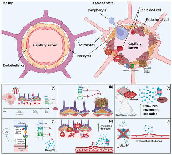
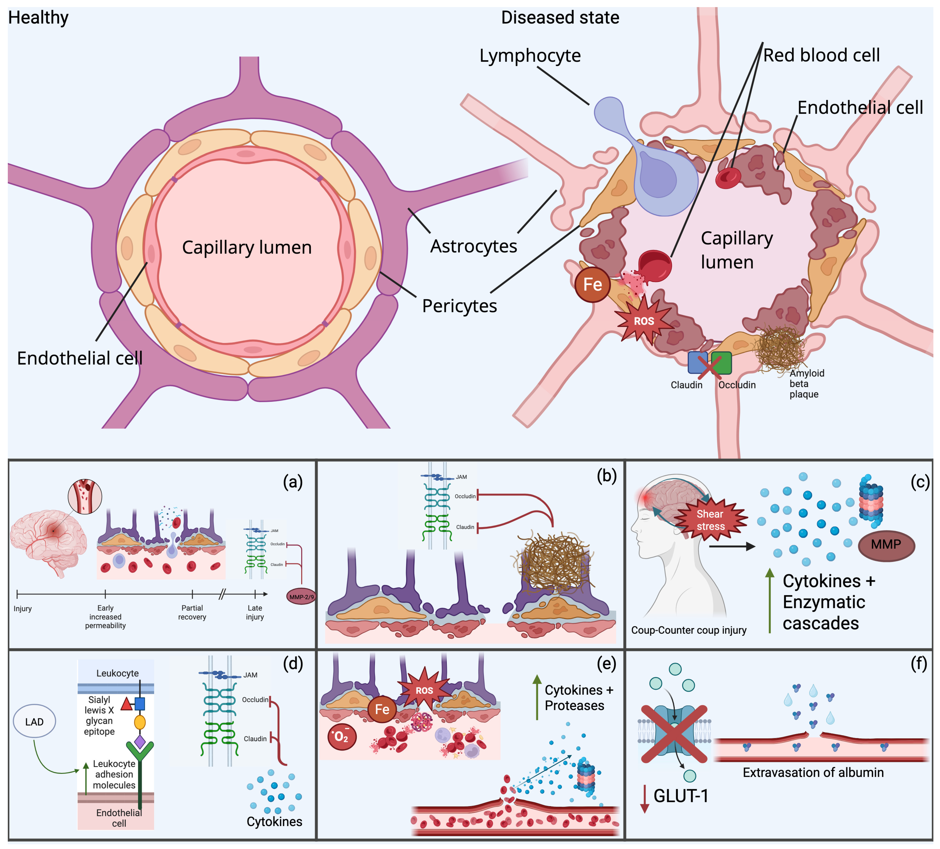
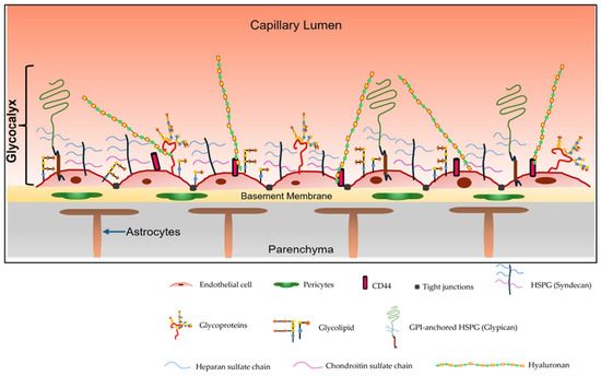
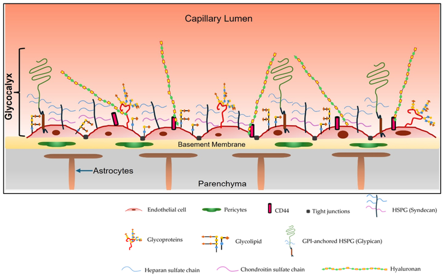

Disclaimer/Publisher’s Note: The statements, opinions and data contained in all publications are solely those of the individual author(s) and contributor(s) and not of MDPI and/or the editor(s). MDPI and/or the editor(s) disclaim responsibility for any injury to people or property resulting from any ideas, methods, instructions or products referred to in the content. |
© 2026 by the authors. Licensee MDPI, Basel, Switzerland. This article is an open access article distributed under the terms and conditions of the Creative Commons Attribution (CC BY) license.