


Abstract
Oncolytic virotherapy (OVT) is a promising anticancer strategy, owing to its capacity to lyse tumor cells selectively and remodel the tumor microenvironment (TME) to enhance antitumor efficacy. Despite the continuous introduction and investigation of newly engineered oncolytic viruses (OVs) in clinical trials, their clinical translation remains limited, with Toll-like receptor (TLR)-mediated host immune responses representing a critical influencing factor. TLRs constitute a family of structurally conserved transmembrane proteins distributed on the plasma membrane and endosomal membranes. They initiate innate immune responses by recognizing pathogen-associated molecular patterns (PAMPs) and damage-associated molecular patterns (DAMPs) and are essential for dendritic cells (DCs) maturation, playing a pivotal regulatory role across diverse tumor therapies. However, TLRs may also restrict viral replication and spread, and some studies suggest that they could, under certain conditions, act as drivers of tumor progression. Nonetheless, the role of TLRs in OVT has not been comprehensively summarized to date. In this review, we synthesize recent advances in the application of TLRs in OVT, with particular attention to their mechanisms of action and the challenges that remain to be overcome, and consider their prospects in oncology, offering insights that may inform the clinical translation of this therapeutic approach.
1. Introduction
Oncolytic virotherapy (OVT) is an emerging anticancer strategy that employs genetically modified or naturally occurring viruses to selectively replicate within and lyse tumor cells while largely sparing normal tissues. The resulting tumor-restricted oncolysis releases tumor-associated antigens and danger signals, which recruit and activate the host antitumor immune response. As early as the beginning of the 20th century, clinical observations revealed that certain cancer patients experienced spontaneous tumor regression following viral infections. This phenomenon inspired scientists to propose the concept of using viruses to treat tumors. However, progress in this field remained limited for several decades due to the lack of effective methods to control viral virulence. It was not until the 1990s, with advances in viral engineering techniques, that researchers gained the ability to precisely modify viral genomes. This breakthrough substantially enhanced the safety, targeting capability, and therapeutic efficacy of oncolytic viruses (OVs), marking a turning point in modern oncolytic virotherapy research [1]. Early studies of OVT focused primarily on the intrinsic replicative and lytic capacities of these viruses, under the assumption that the adaptive immune system would eliminate the viruses and thereby diminish therapeutic outcomes. However, accumulating evidence in recent years indicates that the immune system—particularly antitumor immune responses—plays an indispensable role in OV-mediated tumor clearance [2,3].
Toll-like receptors (TLRs), as critical pattern-recognition receptors (PRRs) of the innate immune system, play a pivotal role in OVT through their activation mechanisms. These receptors are broadly expressed on the surface of immune cells, certain tumor cells, and stromal and endothelial cells, as well as on endosomal membranes. Following OV infection and subsequent lysis of tumor cells, PAMPs such as viral nucleic acids and DAMPs such as high-mobility group box 1 (HMGB1) or heat shock protein 70 (HSP70) are released and recognized by TLRs, initiating downstream signaling cascades that drive the production of pro-inflammatory cytokines and type I interferons (IFN-I) [4]. However, TLR activation represents a double-edged sword. On one hand, it enhances antigen presentation and activates and recruits effector cells such as T cells, thereby establishing effective antitumor immunity [5]. On the other hand, an excessive IFN-I response may prematurely clear the virus, limiting its replication and spread within the tumor microenvironment (TME) and ultimately attenuating the direct oncolytic effect [6]. Moreover, studies indicate that under certain circumstances, TLR signaling may paradoxically promote tumor progression, adding further complexity to therapeutic strategies [7].
Therefore, precisely harnessing the dual nature of TLR activation is important for optimizing OVT. However, translational research bridging these two fields remains limited, leaving several key mechanistic and therapeutic questions unresolved. Consequently, this review aims at systematically elucidating the mechanistic roles of TLR signaling pathways in OVT. We focus specifically on strategies to balance immune activation and antiviral clearance within the TME through targeted modulation of TLR activity, thereby providing insights and potential directions for enhancing the clinical efficacy of OVs.
2. Characteristics of Oncolytic Viruses
OVT primarily utilizes either wild-type or genetically engineered viruses. Their design aims at targeting tumor cells specifically, leveraging abnormal gene expression or signaling pathway defects to enable selective replication, thereby achieving tumor cell killing through direct lysis and stimulation of the host’s antitumor immune response [8]. A representative strategy involves modifying the regulatory effects of viral gene products on the host cell cycle. For example, through genetic engineering, the CR2 domain—essential for binding to the retinoblastoma protein (Rb)—can be deleted from the adenovirus (AdV) E1A protein. This deletion prevents infected normal cells from entering S phase, thereby disrupting the intracellular environment required for viral replication and ensuring that the engineered virus can replicate only in tumor cells capable of proliferating independently of the E1A-Rb interaction [9].
Notably, certain viruses derived from non-human hosts, such as Vesicular Stomatitis Virus (VSV) and Newcastle Disease Virus (NDV), can also serve as effective antitumor therapeutic agents. A key advantage of these viruses is that human populations typically lack high levels of pre-existing neutralizing antibodies against them, which to some extent avoids rapid antibody-mediated clearance. Combined with their capacity for selective replication in tumor cells, they may sustain immune stimulation within the TME [10,11]. However, as non-human viruses, their long-term safety in humans, the potential risk of transmission to non-target species, and the biosafety hazards associated with viral mutation or environmental release still require evaluation through rigorous clinical trials [12].
The viral genome is the core of OVT, but the engineering logic is not uniform across virus families. Each family replicates by a fundamentally different mechanism and therefore has distinct advantages, limitations, and engineering levers. Beyond differences in mRNA synthesis defined by the Baltimore classification, viruses differ in replication site, genome size, and packaging capacity: small RNA-based OVs strongly constrain transgene insertion, whereas large DNA viruses can accommodate more complex expression cassettes. Genome integration is likewise family-dependent rather than a general property of oncolytic viruses: retroviral and lentiviral vectors can integrate, whereas the oncolytic viruses currently used in clinical practice do not rely on genome integration, so insertional mutagenesis is not a shared design concern [13,14]. Accordingly, tumor-selective replication must be engineered in a family-specific manner—for example, by targeting the E1B/Rb–p53 axis in AdV, deleting ICP34.5/ICP47 in Herpes simplex virus (HSV), attenuating thymidine kinase (TK) and vaccinia growth factor (VGF) in vaccinia virus (VACV), or exploiting defects in IFN-I signaling in NDV and VSV—making oncolytic virus engineering a family-specific optimization of selective replication, cytolysis, and immune activation [15,16].
All mature viral particles consist of at least a protein shell (the capsid) and an internal nucleic acid genome, which together form the nucleocapsid. The capsid protects the nucleic acid from degradation by external physical and chemical factors. Some viruses also possess an envelope surrounding the nucleocapsid, typically derived from the host cell membrane and embedded with virus-encoded glycoproteins (spikes). These glycoproteins specifically recognize and bind receptors on the surface of host cells, mediating viral entry [17]. In OV research, envelope glycoproteins are frequently engineered to bind tumor cells specifically. Detailed characteristics of representative oncolytic viruses are summarized in Table 1.
Table 1.
Features of selected oncolytic viruses.
3. Infection of Tumor Cells by Oncolytic Viruses
Infection of tumor cells by OVs is initiated by the binding of viral surface proteins to specific cellular receptors. Although certain entry receptors are upregulated in some tumors, OV selectivity is determined predominantly by defective IFN-I/antiviral signaling, tumor-responsive engineered promoters, deleted or attenuated viral genes, and tumor-restricted replication, with receptor abundance being only one contributory factor. For example, the CD46 molecule is upregulated in various tumors and serves as a key recognition receptor for measles virus (MV) vaccine strains and AdV serotypes of species B, for which desmoglein-2 (DSG2) is an additional protein entry receptor [25,26]. Coxsackievirus A21 (CVA21) preferentially utilizes decay-accelerating factor (DAF) and intercellular adhesion molecule 1 (ICAM-1), both highly expressed on tumor cell surfaces [27]. Additionally, AdV relies on integrin co-receptors to mediate endocytosis [28]; HSV first attaches via heparan sulfate proteoglycans (HSPGs) and subsequently triggers membrane fusion through binding of glycoprotein D (gD) to entry receptors [29]. Viral fusion mediated by enveloped viruses typically involves oligomeric transmembrane glycoproteins on the envelope, whereas non-enveloped viruses accomplish analogous functions via capsid proteins [30,31].
Receptor binding induces conformational changes in viral proteins that mediate entry. Enveloped viruses primarily enter cells through membrane fusion, a process that can occur at the plasma membrane or within endosomal membranes [32]; non-enveloped viruses often enter via endocytosis, followed by capsid reorganization within endosomes and penetration through membrane structures to release the viral genome [33]. Most RNA viruses (e.g., CVA21) replicate in the cytoplasm, whereas most DNA viruses (e.g., AdV)—with the exception of VACV—replicate within the nuclear compartment [19,20].
OVs undergo extensive replication within tumor cells, inducing cellular damage and immunogenic cell death (ICD) through mechanisms including resource depletion, spatial competition, and expression of lytic proteins [34]. However, reliance solely on the intrinsic replication capacity of wild-type viruses is often insufficient to achieve complete tumor eradication together with adequate targeting safety. To address this limitation, researchers employ genetic engineering strategies to enhance efficacy: on one hand, insertion of pro-apoptotic genes (e.g., P53, BNIP3) directly strengthens apoptotic pathways [35]; on the other hand, introduction of suicide gene systems (e.g., thymidine kinase) enables infected cells to convert non-toxic prodrugs locally into cytotoxic compounds, thereby amplifying the lytic effect [36].
Tumor cell lysis releases two critical categories of immunostimulatory signals: endogenous DAMPs are released from damaged cells, such as calreticulin (CRT), HMGB1, HSP70, and ATP; and PAMPs are generated during viral replication, including viral nucleic acids and capsid proteins [37]. Together, these molecules act to reverse the immunosuppressive state of the TME. Figure 1 illustrates the process of oncolytic virus infection and tumor cell lysis.
Figure 1.
Schematic diagram depicting the infection process of selected oncolytic viruses and the subsequent activation of innate immunity. Left: Distinct viral entry strategies into tumor cells are illustrated. Herpes simplex virus (HSV) envelope fuses with the cell membrane, releasing the nucleocapsid into the cytoplasm. The nucleocapsid is then transported along microtubules by the dynein/dynactin motor complex toward the nuclear pore, where viral DNA is injected into the nucleus. Adenovirus (AdV) enters the cell via endocytosis. Endosomal acidification triggers capsid disassembly and endosomal membrane damage, allowing viral particles to escape into the cytoplasm. Subsequently, the virus undergoes microtubule-dependent trafficking toward the nuclear pore, where viral DNA is released into the nucleus. Poliovirus (PV) binds to its receptor, inducing capsid conformational changes and viral entry. Within endosomes, viral RNA is released directly into the cytoplasm through membrane-formed tubular structures. Measles virus (MV) enters via receptor-triggered, pH-independent fusion at the plasma membrane, releasing its nucleocapsid into the cytoplasm. Note that oncolytic virus (OV) tumor selectivity is multifactorial, involving not only receptor-mediated entry but also defective interferon signaling, tumor-specific promoters, and viral gene deletions that restrict replication to cancer cells. Right: Immune activation following successful infection is shown. Upon viral genome amplification and progeny release, infected tumor cells emit danger signals, including pathogen-associated molecular patterns (PAMPs) and damage-associated molecular patterns (DAMPs). These signals activate pattern recognition receptors (PRRs) within the tumor cells, leading to the production of antiviral mediators, such as inflammatory cytokines, chemokines, and type I interferon (IFN-I). These mediators recruit and activate the host immune system, thereby initiating an antitumor immune response.
4. TLR Signaling and Its Crosstalk with Other Pattern-Recognition Pathways
4.1. Downstream Consequences of TLR Activation During Oncolytic Virotherapy
TLRs are type I transmembrane proteins expressed on the plasma membrane or endosomal membrane of a broad range of cell types, including dendritic cells (DCs), macrophages, and other leukocyte subsets, as well as tumor cells, stromal cells and endothelial cells. Their structure comprises three domains: an extracellular domain rich in leucine-rich repeats (LRRs) responsible for recognizing danger signals; a transmembrane domain that anchors the receptor; and an intracellular Toll/interleukin-1 receptor (TIR) domain that mediates activation of downstream signaling pathways [38]. To date, 10 and 12 TLRs have been identified in humans and mice, respectively, with TLR1 through TLR9 highly conserved between the two species.
TLR3, TLR7, TLR8, and TLR9 are localized to endosomal membranes and primarily recognize nucleic acid components derived from OVs or tumor cells themselves. TLR3 recognizes double-stranded RNA (dsRNA) and its synthetic analog polyinosinic:polycytidylic acid (poly(I:C)); the dsRNA generated during viral genome replication can bind and activate TLR3 [39]. TLR7 and TLR8 mainly recognize viral single-stranded RNA (ssRNA) but can also recognize self-RNA released from damaged tumor cells [40]. TLR7 binds free guanosine and ssRNA, whereas TLR8 binds free uridine and ssRNA with high affinity [41,42]. Notably, TLR8 in rodent species (e.g., mice, rats) cannot be activated by human-specific ligands, a limitation that constrains the utility of mouse models in TLR8-related drug development [43]. TLR9 recognizes unmethylated 5′-C-phosphate-G-3′ (CpG) motifs enriched in viral genomes as well as synthetic CpG oligodeoxynucleotides (CpG ODNs) [44]. Moreover, self-DNA released from tumor cells can also serve as a ligand for TLR9 [40].
Transmembrane TLRs located on the plasma membrane include TLR1, TLR2, TLR4, and TLR6, which primarily recognize lipid and protein components. Although these receptors are not the primary sensors of viral nucleic acids, they play important roles in modulating immune responses during OVT. TLR2 can form heterodimers with TLR1 or TLR6 to recognize peptidoglycan, lipoteichoic acid, and other structural components; specifically, TLR2/TLR1 and TLR2/TLR6 selectively recognize triacylated and diacylated lipopeptides, respectively [45]. Additionally, TLR2 can recognize envelope proteins from various viruses as well as host-derived DAMPs. For example, the HSV glycoprotein B (gB) activates cell-surface TLR2, triggering innate immune signaling via the myeloid differentiation primary response protein 88 (MyD88) and TNF receptor-associated factor 6 (TRAF6)-dependent pathway, ultimately leading to NF-κB activation [46]. Cytomegalovirus (CMV) glycoproteins gB and gH interact directly with cell-surface TLR2/TLR1 heterodimers during viral entry, initiating signaling cascades that culminate in NF-κB activation and inflammatory cytokine secretion [47]. The H protein of wild-type MV binds TLR2 and activates innate immune responses; however, recent studies suggest that this recognition mechanism may not apply to attenuated MV vaccine strains [48]. Furthermore, self-proteins and danger signals (e.g., heat shock proteins, HSPs) released from damaged tumor cells may also activate TLR2 [40].
TLR4 primarily recognizes bacterial lipopolysaccharide (LPS). Upon binding to lipopolysaccharide-binding protein (LBP), LPS is transferred to the cell surface with the assistance of CD14, where it interacts with the TLR4-myeloid differentiation factor 2 (MD-2) complex to activate downstream signaling [49]. HMGB1, a nuclear protein released from necrotic cells, can also act as an endogenous ligand that binds TLR4 and induces NF-κB-dependent transcription [50]. Notably, the molecular mechanism by which HMGB1 activates TLR4 appears to vary, depending heavily on the redox state of HMGB1 and the presence of co-factors. Studies suggest that this signaling axis may exhibit differential dependence on MD-2 across disease models, and the detailed molecular recognition mechanism warrants further investigation [51,52]. HSPs can bind TLR4 to activate immune responses; however, some studies suggest that HSPs and TLR4 may also contribute to the maintenance of chronic inflammation and facilitate tumor cell survival [53]. The HSP70/TLR4 axis has been proposed to establish an immunosuppressive TME by recruiting immunosuppressive cells, which may antagonize T cell responses intended to be activated by anti-PD-1 therapy, potentially contributing to therapeutic failure and unexpected disease progression [54]. Furthermore, the S100 protein family (e.g., S100A8 and S100A9) produced by stressed tumor cells has been identified as an important endogenous TLR4 ligand capable of activating and amplifying inflammatory signaling and immune cell recruitment [55]. TLR4 signals through the TIR-domain-containing adaptor inducing IFN-β (TRIF) to activate interferon regulatory factor 3 (IRF3), which induces IFN-I production.
In the TME, abundant DAMPs and PAMPs bind TLRs on antigen-presenting cells or tumor cell surfaces, triggering the recruitment of adaptor proteins via the TIR domain of TLRs to initiate signal transduction. Membrane-bound TLR2 and TLR4 first interact with the TIR-domain-containing adaptor protein (TIRAP) and recruit MyD88. Subsequently, they activate downstream TRAF6 via interleukin-1 receptor-associated kinases (IRAKs); this cascade activates the IKK–IκB pathway to enable NF-κB nuclear translocation and also triggers the MAPK pathway (e.g., p38 and JNK), ultimately promoting the transcription and release of pro-inflammatory cytokines and chemokines, including TNF-α, IL-6, IL-1β, and CXCL8/CXCL10 [38]. It should be noted that TLR4 is not confined solely to the plasma membrane; a portion of TLR4 is internalized into endosomes, where the signaling adaptor molecules switch to TIR-domain-containing adaptor molecule inducing IFN-β (TRAM) and TRIF, initiating a separate downstream pathway: one branch activates IRF3/7 via TRAF3, driving IFN-I production, while the other activates a comparatively milder, late-phase NF-κB/MAPK pathway through TRAF6, inducing cytokine production [56]. Additionally, endosomal TLR7 and TLR9 remain dependent on MyD88 and primarily activate the aforementioned signaling through rapid early-phase pathways, inducing the expression of inflammatory and co-stimulatory molecules [57]. TLR3 is generally considered the only TLR that does not rely on MyD88 and instead depends entirely on TRIF; the activation of this pathway predominantly phosphorylates IRF3 and promotes its nuclear translocation, strongly inducing IFN-I production, and can also activate NF-κB via the TRAF6 pathway, although the resulting pro-inflammatory cytokine output is typically lower than that observed with MyD88-dependent receptors. Although TLR3 canonically signals via TRIF independently of MyD88, one study in mouse bone marrow-derived macrophages showed that MyD88 partially contributes to TLR3-driven NF-κB activation and the induction of IL-6 and IFN-β following poly(I:C) stimulation, an effect weaker than that of TRIF deficiency and not shared by all cell types. These findings are consistent with a restricted, modulatory rather than general role for MyD88 in TLR3 signaling, and warrant confirmation in additional cell types [58]. The TLRs and pathways involved in activation during OVT are shown in Figure 2.
Figure 2.
Sensing and signaling pathways of Toll-like receptors (TLRs). Viral structural proteins and damage-associated molecular patterns (DAMPs) are extracellularly recognized by TLR2 and TLR4, which activate nuclear factor-κB (NF-κB) and mitogen-activated protein kinase (MAPK) signaling pathways via myeloid differentiation primary response 88 (MyD88), leading to the production of pro-inflammatory cytokines. Within endosomes, TLR7, TLR8, and TLR9 also utilize MyD88 to generate type I interferons (IFN-I) and pro-inflammatory cytokines. TIR-domain-containing adaptor-inducing interferon-β (TRIF) is employed by TLR3 and TLR4 to activate TRIF-dependent pathways, resulting in pro-inflammatory effects and antiviral responses. Additionally, TLR7 and TLR9 signal through plasmacytoid dendritic cells (pDCs)-specific pathway, the MyD88-TRAF6 (TNF receptor-associated factor 6)-IRF7 (interferon regulatory factor 7) pathway, which facilitates the induction of IFN-I. This figure was created with MedPeer (https://www.medpeer.cn/, accessed on 1 June 2026).
4.2. Intersection of TLR Signaling with Other Pattern-Recognition Pathways
Although TLRs occupy a central position in discussions regarding the antiviral and antitumor immunity mediated by OVT, they represent merely one branch of a vast network of pattern-recognition pathways. For actively replicating OVs, TLRs are limited by their subcellular localization and thus struggle to directly sense the substantial quantities of viral and tumor-derived ligands produced in the cytoplasm. For instance, NDV, VSV, reovirus (RV), MV, and CVA21 replicate in the cytoplasm and generate dsRNA, which is recognized by the RIG-I-like receptor (RLR) family. Specifically, retinoic acid-inducible gene I (RIG-I) primarily recognizes short RNA molecules with a 5′-triphosphate group (5′-ppp), whereas melanoma differentiation-associated gene 5 (MDA5) recognizes long dsRNA. These receptors transmit signals via the mitochondrial antiviral signaling protein (MAVS) [59]. Although the genomic replication of HSV and AdV occurs within the nucleus, leakage of replication intermediates and viral genomic DNA through the nuclear membrane exposes cytosolic dsDNA to cyclic GMP-AMP synthase (cGAS). cGAS recognizes cytosolic dsDNA in a sequence-agnostic manner, catalyzes the synthesis of the second messenger cGAMP, and activates Stimulator of Interferon Genes (STING), thereby activating IRF3 to drive IFN-I production and NF-κB to induce pro-inflammatory cytokines and chemokines [60]. Recent studies have identified crosstalk between the cGAS-STING and RLR pathways, revealing that many RNA virus infections cause the leakage of self-DNA, such as mitochondrial DNA, into the cytoplasm, thereby activating cGAS-STING [61]. The three principal IFN-I adapter proteins—TRIF, MAVS, and STING—ultimately converge on the TBK1-IRF3 axis. Viruses may exploit these nodes of convergence across pathways to simultaneously block multiple pathways via a single protease. For example, Seneca Valley virus (SVV, an oncolytic virus) encodes a 3C protease that cleaves multiple critical nodes in host immune signaling pathways, including TRIF, MAVS, and TANK, thereby systematically suppressing IFN-I production to achieve immune evasion [62].
Because these pathways share critical downstream molecules, experimental designs must distinguish the specific pathways contributing to the observed effects. For instance, the elevated IFN-β levels measured following poly(I:C) stimulation may result from the TLR3-TRIF pathway stimulated via the extracellular/endosomal route or from the MDA5-MAVS pathway stimulated via cytoplasmic transfection [63]. Furthermore, MyD88 serves not only as an adapter protein for TLRs but also as a key signaling molecule for IL-1R and IL-18R [64]. Studies of VSV in the murine B16 melanoma model demonstrated that MyD88 deficiency leads to a loss of oncolytic efficacy, proving that MyD88 is an essential host factor for VSV-mediated oncolysis; however, these studies could not attribute the efficacy to any specific TLR or IL-1R upstream receptor [65]. Thus, it is important to consider the possibility of pathway crosstalk when analyzing downstream gene signatures, ensuring that conclusions regarding TLR-specific involvement are supported by adequate control of upstream receptor specificity.
Notably, NF-κB activation driven by TLR signaling can also provide critical priming signals for inflammasomes, upregulating the expression of AIM2/NLRP3 inflammasome-associated molecules and thereby licensing the activation of Caspase-1 and the maturation of IL-1β and IL-18 [66]. Thus, TLR involvement often serves as an upstream priming condition for inflammasome activation, rather than an absolute prerequisite in all contexts. Additionally, protein kinase R (PKR) is another kinase activated by double-stranded RNA, which enhances cellular antiviral states by inhibiting translation initiation through the phosphorylation of eIF2α [67]. Unlike TLR pathways, PKR functions primarily as a cytoplasmic dsRNA sensor, and its antiviral effects are largely independent of TLRs. In the context of OVT, dsRNA sensors such as PKR may contribute to TLR-independent antiviral effects, although their relative contribution weights to viral replication and antitumor immunity across various OV models remain to be further quantified and evaluated [68].
5. Strategies Linking TLR Signaling to Oncolytic Virotherapy
TLR signaling occupies a dual position in OVT. On the one hand, TLR activation bridges innate and adaptive immunity, driving DCs maturation, natural killer (NK) cell activation, and CD8+ T cell priming to convert the TME toward a “hot” phenotype. On the other hand, excessive or chronic TLR activation can trigger premature viral clearance, recruit immunosuppressive myeloid populations, and in certain contexts sustain malignant progression. This tension makes TLRs both a therapeutic target and a liability. Consequently, to delineate the independent role of TLRs in OVT, we compiled research findings from the past two decades that explicitly focused on TLRs themselves, rather than merely their shared downstream signaling molecules, as summarized in Table 2.
Table 2.
Research progress in oncolytic virus therapies associated with Toll-like receptors.
We reorganize the field around five mechanism-based categories of TLR-centered intervention: natural sensing of OVs by endogenous TLRs; using non-replicating viruses or viral particles as TLR agonists; combining OVs with an exogenous TLR agonist; engineering OVs to enhance TLR signaling; and deliberately inhibiting TLR signaling to augment viral replication. These five categories are conceptualized in Figure 3.
Figure 3.
Mechanism-based framework of Toll-like receptor (TLR)-centered interventions in oncolytic virotherapy and the resulting immunological remodeling of the tumor microenvironment (TME). Left: the five categories of TLR-targeted intervention discussed in this section, comprising natural sensing of oncolytic viruses (OVs) by endogenous TLRs, the use of inactivated viruses or viral particles as TLR agonists, the combination of OVs with an exogenous TLR agonist such as poly(I:C), engineering of OVs to enhance TLR signaling, and the deliberate inhibition of TLR signaling, each converging on a central TLR signaling hub. Upper right: TLR-driven transition from an immunosuppressive “cold” TME to an inflamed, immunologically active “hot” TME. In the cold state (blue), TLR activation drives immature dendritic cells (DCs) to take up tumor antigens and mature; mature DCs migrate to prime naïve T cells, generating CD4+ and CD8+ effector T cells and NK cells that, together with M1-polarized macrophages, infiltrate the tumor bed (red) to establish effective antitumor immunity. Lower right: potential adverse consequences of dysregulated or excessive TLR activation, including interferon-stimulated gene (ISG)-mediated premature viral clearance, recruitment of immunosuppressive populations such as MDSCs, TAMs and Tregs, and other outcomes such as cytokine storm and malignant progression. Color-coded symbols for cold/hot tumor cells, NK cells, DC subsets, T cell subsets, and macrophage phenotypes are defined in the legend. This figure was created with MedPeer (https://www.medpeer.cn/, accessed on 4 October 2026).
5.1. Natural Sensing of OVs by Endogenous TLRs
When OVs productively infect tumors, PAMPs and DAMPs are sensed by endogenous TLRs expressed on tumor, stromal, endothelial, and immune cells, initiating the innate branch of the antitumor immune response. The consequence is highly cell-type-specific: whereas normal mammary tissue stem cells retain an intact IFN-I response that resists viral infection, breast cancer-initiating cells harbor defective IFN-I and TLR signaling, characterized by impaired TLR9/MyD88 trafficking and loss of TLR2, which renders them permissive to the capsid-modified adenovirus Ad5/3-Delta24 [69].
As key sentinel cells, DCs sit at the center of this sensing. Conventional DCs (cDCs) mature upon detecting danger signals via the NF-κB and MAPK pathways, releasing TNF-α and IL-12 and initiating T cell co-stimulation [93,94], whereas plasmacytoid DCs (pDCs) secrete large amounts of IFN-I upon TLR7/9 stimulation [95]. This discrimination is functionally decisive: when tumor cells are compared head-to-head, only MV-infected cells, rather than UV-irradiated ones, induce pDC maturation and robust IFN-α production [82]. Mechanistically, in pDCs, oncolytic measles is recognized by RIG-I-like receptors and TLR7, a pDC-restricted sensing combination that licenses these cells to express TNF-related apoptosis-inducing ligand (TRAIL) and kill via TRAIL–DR5 (death receptors 5) engagement [83]. The resulting IFN-I, together with DC-derived IL-12, promotes the activation and expansion of NK cells [73,96]. During this IFN-I pre-stimulation phase, NK cells display enhanced cytotoxicity against targets with low or absent MHC-I, lysing them via granzyme B and perforin and secreting IFN-γ and TNF-α, thereby establishing a positive-feedback loop that amplifies antitumor immunity [97].
Crucially, this innate sensing is not an end in itself: its terminal readout is the priming of durable adaptive immunity. Beyond chemokine-mediated recruitment of CD8+ T cells, mature DCs take up exogenous tumor antigens and, after lysosomal processing, present derived peptides on MHC-II to prime naïve CD4+ T cells toward Th1 effector cells [98,99,100,101]. Through cross-presentation, DCs also load exogenous antigens onto MHC-I and, supported by Th1-derived cytokines, prime naïve CD8+ T cells into cytotoxic T lymphocytes (CTLs) [102,103]. Two lines of evidence illustrate this relay. DCs exposed to H-1 parvovirus-induced tumor cell lysates upregulate TLR3/TLR9 together with maturation markers such as CD86 and NF-κB/TNF-α, a phenotype consistent with enhanced cross-priming [91]. Complementary to this, pDCs phagocytosing MV-infected tumor cells have been shown to cross-present NY-ESO-1 to a specific CD8+ T cell clone, confirming that OV-infected cells can license DC-mediated CD8+ responses [82]. These lymphocytes then migrate to the primary tumor, expand locally, and exert potent cytotoxic effects.
5.2. Using Inactivated Viruses or Viral Particles as TLR Agonists
Unlike strategies that rely on intratumoral viral replication and direct oncolysis, some studies utilize only specific structural components of OVs or inactivated, replication-deficient viral particles as immunotherapeutic agents. Although these preparations are incapable of expressing viral genes, they fully retain virus-derived PAMPs. Consequently, they can function as natural TLR ligands to activate both innate and adaptive immunity, thereby exerting antitumor effects while avoiding replication-associated toxicity.
Regarding the TLR2 axis, studies have shown that UV-inactivated HSV-1 (UV-HSV-1) can rapidly enhance the cytotoxicity of peripheral blood mononuclear cells (PBMCs) and purified NK cells against leukemia cells within one hour of exposure. This process involves the upregulation of CD69, degranulation, and IFN-γ secretion, accompanied by metabolic reprogramming such as glycolysis. Notably, the use of TLR2-neutralizing antibodies significantly antagonizes this enhancement of cytotoxicity [77]. Similarly, UV-inactivated HSV-2 has been shown to stimulate NK cells via the TLR2-dependent NF-κB pathway, promoting IFN-γ release and enhancing antitumor activity. This effect can be further potentiated when combined with anti-NKG2A/anti-HLA-E checkpoint antibodies [76].
In the context of the TLR3 axis, RV and its UV-inactivated form (UV-RV) were found to sensitize PBMCs and CD8+ tumor-infiltrating lymphocytes from gastric cancer patients. Both cell types were shown to upregulate IFN-γ and TRAIL via the TLR3/NF-κB pathway. Furthermore, the RV σC protein can engage tumor cell-associated TLR3, upregulating DR4/DR5 via the p38/p53 pathway, thereby enhancing the killing of gastric cancer cells by effector cells [86]. Additionally, transfecting ovarian cancer cells with RV-derived dsRNA was found to induce apoptosis by upregulating TLR3 and activating TBK1/IRF3 and NF-κB downstream of TRIF; this apoptosis could be reversed by using TLR3 inhibitors or TBK1/IKKε inhibitors [87].
Furthermore, although Cowpea Mosaic Virus (CPMV) does not replicate intracellularly or exert direct oncolytic effects, it may function as an in situ vaccine: its empty viral capsids activate TLR2/4, whereas intact virions additionally engage TLR7, thereby inducing IFN-I production and potentially eliciting stronger immunotherapeutic efficacy [104,105]. Collectively, the use of inactivated viruses or viral particles as TLR agonists balances safety with immune activation potency and can be combined with immune checkpoint inhibitors (ICIs), chemotherapy, and other treatments, laying the foundation for its use as a “replication-incompetent” adjuvant for cancer immunotherapy.
5.3. Combining OVs with an Exogenous TLR Agonist
Although OVs expose tumor antigens, OVs alone may fail to reverse the immunosuppressive TME. The resulting accumulation of regulatory T cells (Tregs), myeloid-derived suppressor cells (MDSCs), and M2 macrophages prevents effective antigen presentation and leads to insufficient T cell activation. TLR agonists can provide potent complementary danger signals, thereby promoting the secretion of IFN-I and pro-inflammatory cytokines. This drives the re-polarization of M2 macrophages into antitumor M1 macrophages, further enhancing T cell immune responses [106,107]. For instance, combining the TLR3 ligand poly(I:C) with HSV-1 in the treatment of uveal melanoma reactivates the virus-downregulated TLR3–NF-κB axis, promoting M1 polarization and thereby bolstering antitumor immunity [5]. Similarly, the combination of the TLR9 ligand CpG ODN (self-assembled with melittin into a CpGMel complex to enhance delivery) and coxsackievirus B3 in treating triple-negative breast cancer and melanoma leverages ICD to activate antigen presentation and T cell responses, suppressing primary tumors and delaying distant metastasis [90].
However, combination strategies may also yield unfavorable outcomes. While an “in situ vaccine” approach using a TLR9 ligand (CpG ODN) combined with a modified vaccinia virus expressing membrane-anchored IL-2 (vvDD-mIL2) successfully cleared injected tumors, prolonged survival, and induced abscopal effects with a favorable safety profile—dynamically remodeling the TME of untreated sites by increasing effector T cells, NK cells, and DCs infiltration—it also triggered a compensatory upregulation of Tregs, MDSCs, and tumor-associated macrophages (TAMs). Consequently, myeloid cell depletion or anti-PD-1 therapy is required to further amplify efficacy [80]. Furthermore, while sequential intratumoral administration of the TLR4 ligand LPS and VSVs cured 100% of tumor-bearing mice, switching to intravenous VSV followed by LPS administration caused a lethal cytokine storm characterized by a surge in TNF-α and IL-6 [89]. This serves as a stark warning regarding the narrow therapeutic window associated with systemic administration.
5.4. Engineering an OV to Enhance TLR Signaling
Unlike the strategy of administering TLR agonists exogenously, genetic engineering approaches involve directly inserting TLR agonist molecules or their coding sequences into the viral genome. This allows the OV to continuously release agonist signals in situ while replicating within the tumor. By employing a strategy of enriching TLR recognition sequences, the researchers of a previous study inserted 18 immunostimulatory CpG motifs into the AdV genome to construct the CpG-enriched virus Ad5D24-CpG. This significantly enhanced TLR9 recognition and NF-κB activation, boosting immunogenicity while fully preserving the safety profile of the parental virus. The virus attenuated the immunosuppressive function of MDSCs and increased intratumoral CD8+ T cell infiltration via the TLR9 pathway, an effect confirmed to be TLR9-dependent using TLR9-knockout oligonucleotides [73].
Another strategy involves engineering the virus to express endogenous TLR agonists. For instance, an AdV was engineered to express human β-defensin-2 (BD2), an endogenous danger signal recognized by TLR4. While the virus lysed tumor cells and released antigens, BD2 recruited and promoted the maturation of pDCs, inducing tumor-specific CD4+/CD8+ T cell and IFN-γ responses. This generated complete protective immunity against re-challenge and inhibited natural metastasis [71]. Similarly, engineering MV to express Helicobacter pylori neutrophil-activating protein (NAP)—a TLR2 agonist-like lipoprotein—resulted in massive recruitment and activation of neutrophils in immunologically “cold” synovial sarcomas characterized by low PD-L1 expression and low T cell infiltration. This converted “cold” tumors into “hot” ones and upregulated PD-L1, creating conditions favorable for combination therapy with ICIs [84].
In addition to introducing agonist elements, another approach to enhancement is removing the virus’s own innate immune antagonistic genes. One study proposed constructing a VACV that activates the TLR3–IRF3 pathway post-infection. By stepwise removal of viral genes that antagonize this pathway (C10L, N2L, and C6L), IRF3 phosphorylation and IFN-β expression were restored within tumor cells. This translated into stronger tumor antigen-specific T cell responses and therapeutic benefits in mouse models [79].
5.5. Inhibiting TLR Signaling to Modulate the Antiviral Response
The signaling cascade following TLR activation ultimately establishes a critical barrier that restricts viral replication and accelerates viral clearance. Based on this rationale, studies have sought to enhance viral replication and delivery efficiency by inhibiting or dampening TLR recognition.
At the tumor cell level, TLR3 is constitutively expressed in colonic epithelium. Knockdown of TLR3 significantly reduces host IFN-I triggering, thereby enhancing the replication of RV within tumors. This is accompanied by a marked upregulation of the viral σ1 protein and significantly enhanced virus-mediated inhibition of tumor growth compared to controls [6]. In terms of viral engineering, deglycosylation of surface proteins on VACV can weaken its binding to TLR2 and the subsequent activation of MyD88 signaling. This reduces the production of antiviral neutralizing antibodies, thereby enhancing systemic delivery of the virus to tumors following intravenous administration and preserving the capacity for repeated dosing [81]. Regarding delivery vehicles, the use of mesenchymal stem cells (MSCs) with silenced TLR4 or MyD88 as carriers for AdV can reduce systemic inflammation after systemic administration. This improves the homing and delivery efficiency of the vectors to tumors, thereby enhancing antitumor efficacy [75].
However, studies in TLR9-knockdown mice have revealed that TLR9 signaling also participates in the recruitment of NK cells to tumors, serving as a contributing factor to antitumor immunity [74]. Therefore, the activation or inhibition of TLR pathways presents a distinct “double-edged sword” effect. An ideal strategy requires striking a balance between enhancing viral replication and maintaining antitumor immunity, tailored to the specific virus type, tumor type, route of administration, and immune microenvironment.
6. Clinical Translation of TLR-Targeted Oncolytic Virotherapy
Although much of the evidence discussed above is preclinical, several OV platforms and TLR-targeted immunomodulators have reached the clinic, and their accumulated experience is essential for evaluating the translational potential of TLR-centred OVT, as summarized in Table 3.
Table 3.
Clinical trials of oncolytic viruses and Toll-like receptor agonists for cancer/malignancies.
6.1. Clinical Translation of Oncolytic Viruses
The clinical landscape of OVs has matured from first-in-class monotherapy approvals into a broad set of platform-specific and combination strategies that increasingly pair viral oncolysis with immune checkpoint blockade. Among the approved products, talimogene laherparepvec (T-VEC, Imlygic), a granulocyte-macrophage colony-stimulating factor (GM-CSF)-armed HSV-1, received FDA approval in 2015 for intralesional treatment of unresectable melanoma [143]. Most recently, in August 2026, the FDA granted accelerated approval to vusolimogene oderparepvec (TUDRIQEV, formerly RP1), a dual-engineered HSV-1 expressing the GALV fusogenic glycoprotein and GM-CSF, in combination with nivolumab for advanced cutaneous melanoma that had progressed on prior anti-PD-1 therapy [115].
Across platforms, single-agent activity remains modest and largely confined to local control, whereas clinically meaningful responses concentrate on intratumoral delivery of armed or engineered viruses, most often combined with checkpoint blockade. HSV-1 platforms typify this: RP1 plus nivolumab yielded an objective response rate (ORR) of 32.9% in anti-PD-1-refractory melanoma [115], and T-VEC plus pembrolizumab reached an ORR of 19.7% in PD-1-refractory advanced melanoma [117] and 30% in sarcoma [118], even though the confirmatory phase III T-VEC-pembrolizumab trial in PD-1-naïve melanoma missed its primary endpoints [120]. Comparable signals extend beyond HSV-1: coxsackievirus V937 plus pembrolizumab reached an ORR of 47% in advanced melanoma [126], and DNX-2401 plus pembrolizumab gave a 56.2% clinical-benefit rate in recurrent glioblastoma [113]. Conversely, poorly matched delivery or patient selection underdelivered—most clearly intravenous V937, which showed no additive benefit with pembrolizumab [128], and intrahepatic T-VEC, which produced consistently low response rates across liver-tumor cohorts [119].
The mechanistic rationale linking OVT to durable and systemic benefit is the conversion of immunologically “cold” tumors into “hot” ones, and the clinical data are broadly consistent with this model: benefit is greatest when oncolysis is delivered intratumorally and paired with checkpoint blockade, and regressions of distant, non-injected lesions have been documented with V937 combined with ipilimumab and with RP1-nivolumab [115,125].
OVT is generally well tolerated, with adverse events (AEs) predominantly being injection-site reactions (ISRs) and transient, low-grade flu-like symptoms, and treatment-related grade ≥ 3 events remaining low (12.7% with T-VEC plus pembrolizumab; 12.9% with RP1 plus nivolumab), indicating a favorable systemic safety margin [115,117]. Efficacy, by contrast, is shaped chiefly by disease setting and prior therapy exposure rather than by tolerability: in both trials, response rates diverged sharply between patients recurring in the adjuvant setting (ORR 40–47% with T-VEC plus pembrolizumab [117]; 50% with RP1 plus nivolumab) and those progressing in the metastatic setting (ORR 0–6.7% with T-VEC plus pembrolizumab) [115].
6.2. Clinical Translation of TLR-Targeted Immunomodulators
Single-agent activity of TLR agonists is generally limited. In a phase I study of RO7119929, an oral TLR7 agonist prodrug, one patient with hepatocellular carcinoma achieved a durable complete response, but overall clinical activity remained modest [135]. By contrast, imiquimod, an FDA-approved TLR7 agonist, has shown more consistent utility. It has long been used for precancerous cervical lesions, and neoadjuvant imiquimod appears safe and promising in early-stage oral cancer [136,144].
Most current developments therefore focus on combination regimens. TLR agonists are frequently paired with an ICI such as nivolumab, pembrolizumab, or ipilimumab, as well as being combined with targeted agents or chemotherapy according to tumor type. Bacillus Calmette–Guérin (BCG), which activates TLR2, TLR4, and TLR9 pathways, illustrates a similar pattern: although it remains a cornerstone of bladder cancer immunotherapy in trials such as NCT03022825 and NCT03528694, optimal efficacy generally requires combination with additional agents [145,146,147].
Nevertheless, several phase II/III trials have yielded disappointing results. In anti-PD-1-refractory advanced melanoma, adding intratumoral tilsotolimod, a TLR9 agonist, to ipilimumab did not significantly improve ORR, progression-free survival (PFS), or overall survival (OS) compared with ipilimumab alone, and this strategy increased toxicity, including higher rates of grade ≥ 3 AEs and new signals of serious anaphylactic reactions [141]. Similarly, in recurrent ovarian cancer, adding the TLR8 agonist motolimod to pegylated liposomal doxorubicin did not improve OS, PFS, or response rate relative to placebo, despite confirmed immune activation [138]. Notably, motolimod-treated patients who exhibited injection-site reactions showed a substantially lower risk of death and longer OS than those who did not [138].
Systemic administration is further constrained by dose-limiting toxicity (DLT), particularly cytokine release syndrome (CRS). Oral RO7119929 induced CRS in a dose-dependent manner in 81% of patients, with treatment-related AEs of any grade occurring in 91% [135]. Intravenous GSK1795091 was associated with treatment-emergent AEs in 94% of patients [133], and intravenous pixatimod caused AEs in all treated patients [142]. Common AEs included fever, chills, fatigue, and nausea, consistent with flu-like or immune-activation symptoms. By contrast, rintatolimod, a TLR3 agonist, was relatively well tolerated after intravenous administration, with no DLTs reported [132].
Local administration combined with radiotherapy has produced the most mechanistically compelling results, particularly through induction of the abscopal effect. In patients with relapsed/refractory indolent non-Hodgkin lymphoma, treatment with intratumoral SD-101, a TLR9 agonist, combined with low-dose radiotherapy and ibrutinib induced the regression of distant, untreated tumors in most patients, with a median PFS of 11 months [140]. Similarly, following low-dose radiotherapy, intratumoral injection of G100, a TLR4 agonist, led to abscopal regression in 13 of 18 evaluable patients in the 20 mg dose cohort with follicular lymphoma [134]. These findings position TLR agonists as candidate components of an “in situ vaccination” strategy, in which localized innate immune activation generates systemic, antigen-specific adaptive immunity. This rationale also supports exploring TLR modulation as part of OVs-based immunotherapy, where local innate immune stimulation may similarly help convert an immunologically “cold” TME into one more permissive to antitumor T cell responses.
7. Discussion
This discussion synthesizes the current evidence to provide concrete translational guidance, addressing three pivotal questions that determine the clinical viability of TLR-based modulation in OVT: which receptors should be targeted or inhibited for specific viral platforms, the contexts in which such manipulation proves beneficial versus detrimental, and how antiviral and antitumor immune responses can be effectively balanced.
First, the choice of the most actionable TLR agonist depends on the genome type and replication compartment of the OV, because these features determine which ligands accumulate and where. Endosomal TLR3, signaling through TRIF, is one of the most pharmacologically accessible targets for RNA OVs that generate dsRNA—such as RV, VSV, NDV and MV—making poly(I:C) and related TLR3 agonists mechanistically compatible partners for these platforms, although RIG-I/MDA5 and TLR7/8 may also contribute to RNA sensing [39]. DNA OVs (HSV, AdV, and VACV) present unmethylated CpG motifs and release replication-derived DNA, thereby engaging TLR9 and cytosolic DNA sensors, particularly the cGAS–STING axis, so CpG ODN and STING-directed adjuncts represent mechanistically plausible pairings [44,61]. Surface-expressed TLR2 and TLR4 mainly sense viral envelope glycoproteins and DAMPs (for example, HSV gB, MV H, HMGB1, HSP70, and S100A8/9) and are best exploited locally or with inactivated/particle preparations rather than as general drivers of productive replication [48,50]. Endosomal TLR7 and TLR8 sense ssRNA and are relevant targets for RNA-based platforms, particularly when systemic delivery or endosomal exposure is expected; however, the risk of systemic cytokine release should temper preclinical-to-clinical inference [41,42].
Second, it is the timing and anatomical context of TLR modulation, rather than its direction alone, that largely determines therapeutic benefit. Restraining the antiviral arm—as illustrated by TLR3 silencing to permit RV replication [6], VACV deglycosylation to attenuate TLR2 engagement [81], and TLR4/MyD88-silenced MSC carriers for AdV [75]—can enhance viral spread and oncolysis. This strategy appears most valuable during the early replication-and-dissemination window, particularly in IFN-competent or heavily infiltrated tumors where premature viral clearance might otherwise limit therapy. However, the same intervention may be detrimental when applied systemically or at later stages, since TLR9 signaling also supports NK cell recruitment and DC cross-priming, and indiscriminate inhibition could compromise the adaptive arm that underpins durable OVT efficacy [74,91]. A practical rule is thus to apply transient, local inhibition to protect the virus while antigen is being generated, and to favor TLR agonism after antigen release to preserve DC licensing.
Third, antiviral and antitumor immunity may be reconciled not by prioritizing one over the other, but by spatially and temporally separating these two phases. As shown above, route and sequence were decisive, and systemic OVs combined with TLR stimulation risked a lethal TNF-α/IL-6 cytokine storm, underscoring the safety advantage of intratumoral over systemic delivery [89]. Within this narrow window, pairing TLR-driven innate activation with OV-induced ICD and myeloid repolarization may help convert transient inflammation into durable CTL responses while limiting systemic toxicity [5,90]. This framework may help explain the limited single-agent activity of TLR agonists in the clinic, but remains largely preclinical and requires prospective validation.
To render these principles operational, we summarize them as a decision matrix (Table 4) that links viral genome type, dominant sensing pathways, tumor phenotype, intervention strategy, and the predicted translational benefit.
Table 4.
Decision matrix for TLR-targeted oncolytic virotherapy.
8. Conclusions
TLR activation in OVT functions as a genuine double-edged sword: the antiviral arm can prematurely curtail viral replication and spread, whereas the inflammatory arm licenses dendritic cells and sustains antigen-driven immunity. The balance between these opposing outcomes is shaped by the genome type and replication compartment of each viral family, which determine which ligands accumulate and where. We have organized these relationships into five mechanism-based intervention strategies, weighed them against current translational evidence, and distilled them into an actionable decision matrix. Distinguishing beneficial from detrimental TLR responses therefore remains a central challenge in harnessing innate immunity for OVT. Progress will require technologies that resolve immune responses across cellular, spatial, and temporal dimensions—single-cell multi-omics to assign TLR pathways to defined cell populations, spatial transcriptomics to recover positional information within the TME, and longitudinal sampling to define the kinetic window in which a response shifts from inflammation-sustaining to immunogenic. These approaches sharpen a practical question: Which TLR pathway, in which cell type, in which region of which tumor, and at what point during treatment, should be selectively activated or inhibited? Answering it will require converting this dichotomy into a controllable therapeutic strategy that complements rational OV engineering and checkpoint blockade. Its clinical value will depend on careful calibration of timing, dose, and route to limit systemic exposure and avoid the cytokine-driven toxicity that has constrained earlier-generation agonists, as well as reliable identification of target cell types and patient stratification based on TME heterogeneity. Future progress will hinge on reproducible biomarkers for prospective assignment of patients to TLR-pathway-dominant subgroups, standardized frameworks for serial intratumoral sampling, and trial designs that explicitly test timing, such as window-of-opportunity or adaptive scheduling studies. With these advances, TLR-targeted OVT can move from mechanistic promise toward predictable, clinically actionable practice.
Author Contributions
Conceptualization, D.Z. and J.P.; methodology, S.L. and X.H.; writing—original draft preparation, S.L.; writing—review and editing, X.H. and Z.Z.; visualization, D.Z. and J.P.; supervision, Z.Z. All authors have read and agreed to the published version of the manuscript.
Funding
This research received no external funding.
Institutional Review Board Statement
Not applicable.
Informed Consent Statement
Not applicable.
Data Availability Statement
The data used in this article were sourced from materials mentioned in the References section.
Acknowledgments
During the preparation of this manuscript, the authors used MiniMax M2.7 for the purposes of language editing and grammar check. The authors have reviewed and edited the output and take full responsibility for the content of this publication.
Conflicts of Interest
The authors declare no conflicts of interest.
References
- Bifulco, M.; Di Zazzo, E.; Napolitano, F.; Malfitano, A.M.; Portella, G. History of how viruses can fight cancer: From the miraculous healings to the approval of oncolytic viruses. Biochimie 2023, 206, 89–92. [Google Scholar] [CrossRef] [Scilit] [PubMed]
- Groeneveldt, C.; van den Ende, J.; van Montfoort, N. Preexisting immunity: Barrier or bridge to effective oncolytic virus therapy? Cytokine Growth Factor Rev. 2023, 70, 1–12. [Google Scholar] [CrossRef] [Scilit] [PubMed]
- DePeaux, K.; Delgoffe, G.M. Integrating innate and adaptive immunity in oncolytic virus therapy. Trends Cancer 2024, 10, 135–146. [Google Scholar] [CrossRef] [Scilit] [PubMed]
- Lin, Y.S.; Chang, Y.C.; Pu, T.Y.; Chuang, T.H.; Hsu, L.C. Involvement of nucleic acid-sensing toll-like receptors in human diseases and their controlling mechanisms. J. Biomed. Sci. 2025, 32, 56. [Google Scholar] [CrossRef] [Scilit] [PubMed]
- Liu, S.; Li, M.; Sun, F.; Zhang, J.; Liu, F. Enhancing the immune effect of oHSV-1 therapy through TLR3 signaling in uveal melanoma. J. Cancer Res. Clin. Oncol. 2023, 149, 901–912. [Google Scholar] [CrossRef] [Scilit] [PubMed]
- Maitra, R.; Augustine, T.; Dayan, Y.; Chandy, C.; Coffey, M.; Goel, S. Toll like receptor 3 as an immunotherapeutic target for KRAS mutated colorectal cancer. Oncotarget 2017, 8, 35138–35153. [Google Scholar] [CrossRef] [Scilit] [PubMed]
- Patidar, A.; Selvaraj, S.; Sarode, A.; Chauhan, P.; Chattopadhyay, D.; Saha, B. DAMP-TLR-cytokine axis dictates the fate of tumor. Cytokine 2018, 104, 114–123. [Google Scholar] [CrossRef] [Scilit] [PubMed]
- Lan, S.; Zhao, Z.; He, Z. Measles Virus-Based Genetic Modifications: Progress in Hematological Malignancy Treatment. OncoTargets Ther. 2025, 18, 605–615. [Google Scholar] [CrossRef] [Scilit] [PubMed]
- Ehrenfeld, M.; Segeth, F.; Mantwill, K.; Brockhaus, C.; Zhao, Y.; Ploner, C.; Kolk, A.; Gschwend, J.E.; Nawroth, R.; Holm, P.S. Targeting Cell Cycle Facilitates E1A-Independent Adenoviral Replication. J. Virol. 2023, 97, e0037023. [Google Scholar] [CrossRef] [Scilit] [PubMed]
- Zinovieva, M.; Ryapolova, A.; Karabelsky, A.; Minskaia, E. Oncolytic Vesicular Stomatitis Virus: Optimisation Strategies for Anti-Cancer Therapies. Front. Biosci. 2024, 29, 374. [Google Scholar] [CrossRef] [Scilit] [PubMed]
- Yang, H.; Tian, J.; Zhao, J.; Zhao, Y.; Zhang, G. The Application of Newcastle Disease Virus (NDV): Vaccine Vectors and Tumor Therapy. Viruses 2024, 16, 886. [Google Scholar] [CrossRef] [Scilit] [PubMed]
- de Graaf, J.F.; van Nieuwkoop, S.; Bestebroer, T.; Groeneveld, D.; van Eijck, C.H.J.; Fouchier, R.A.M.; van den Hoogen, B.G. Optimizing environmental safety and cell-killing potential of oncolytic Newcastle Disease virus with modifications of the V, F and HN genes. PLoS ONE 2022, 17, e0263707. [Google Scholar] [CrossRef] [Scilit] [PubMed]
- Kontermann, R.E.; Ungerechts, G.; Nettelbeck, D.M. Viro-antibody therapy: Engineering oncolytic viruses for genetic delivery of diverse antibody-based biotherapeutics. MAbs 2021, 13, 1982447. [Google Scholar] [CrossRef] [Scilit] [PubMed]
- Knight, S.; Collins, M.; Takeuchi, Y. Insertional mutagenesis by retroviral vectors: Current concepts and methods of analysis. Curr. Gene Ther. 2013, 13, 211–227. [Google Scholar] [CrossRef] [Scilit] [PubMed]
- Muthukutty, P.; Yoo, S.Y. Oncolytic Virus Engineering and Utilizations: Cancer Immunotherapy Perspective. Viruses 2023, 15, 1645. [Google Scholar] [CrossRef] [Scilit] [PubMed]
- Appleton, E.; Chiocca, E.A.; Ungerechts, G.; Melcher, A.; Vile, R. Oncolytic viruses as anticancer agents: Clinical progress and remaining challenges. Lancet 2025, 406, 1295–1312. [Google Scholar] [CrossRef] [Scilit] [PubMed]
- Cao, G.X.; Liu, F.X.; Meng, C.C.; Ding, C.; Dai, J.; Qiu, X.S. Virus infection and vesicle trafficking. Front. Immunol. 2025, 16, 1682139. [Google Scholar] [CrossRef] [Scilit] [PubMed]
- Aldrak, N.; Alsaab, S.; Algethami, A.; Bhere, D.; Wakimoto, H.; Shah, K.; Alomary, M.N.; Zaidan, N. Oncolytic Herpes Simplex Virus-Based Therapies for Cancer. Cells 2021, 10, 1541. [Google Scholar] [CrossRef] [Scilit] [PubMed]
- Zhao, Y.; Liu, Z.; Li, L.; Wu, J.; Zhang, H.; Zhang, H.; Lei, T.; Xu, B. Oncolytic Adenovirus: Prospects for Cancer Immunotherapy. Front. Microbiol. 2021, 12, 707290. [Google Scholar] [CrossRef] [Scilit] [PubMed]
- Xu, L.; Sun, H.; Lemoine, N.R.; Xuan, Y.; Wang, P. Oncolytic vaccinia virus and cancer immunotherapy. Front. Immunol. 2024, 14, 1324744. [Google Scholar] [CrossRef] [Scilit] [PubMed]
- Pidelaserra-Martí, G.; Engeland, C.E. Mechanisms of measles virus oncolytic immunotherapy. Cytokine Growth Factor Rev. 2020, 56, 28–38. [Google Scholar] [CrossRef] [Scilit] [PubMed]
- Sakunchotpanit, G.; Patil, M.K.; Venkatesh, K.; Rohan, T.Z.; Cheng, D.; Nambudiri, V.E. Treatment of malignant melanoma with coxsackievirus A21 (V937): An emerging oncolytic virotherapy. Exp. Dermatol. 2024, 33, e15169. [Google Scholar] [CrossRef] [Scilit] [PubMed]
- Dighe, O.R.; Korde, P.; Bisen, Y.T.; Iratwar, S.; Kesharwani, A.; Vardhan, S.; Singh, A. Emerging Recombinant Oncolytic Poliovirus Therapies Against Malignant Glioma: A Review. Cureus 2023, 15, e34028. [Google Scholar] [CrossRef] [Scilit] [PubMed]
- Kumar, V.; Giacomantonio, M.A.; Gujar, S. Role of Myeloid Cells in Oncolytic Reovirus-Based Cancer Therapy. Viruses 2021, 13, 654. [Google Scholar] [CrossRef] [Scilit] [PubMed]
- Liszewski, M.K.; Atkinson, J.P. The multiverse of CD46 and oncologic interactions. J. Clin. Investig. 2025, 135, e188355. [Google Scholar] [CrossRef] [Scilit] [PubMed]
- Bahlmann, N.A.; Tsoukas, R.L.; Erkens, S.; Wang, H.; Jönsson, F.; Aydin, M.; Naumova, E.A.; Lieber, A.; Ehrhardt, A.; Zhang, W. Properties of Adenovirus Vectors with Increased Affinity to DSG2 and the Potential Benefits of Oncolytic Approaches and Gene Therapy. Viruses 2022, 14, 1835. [Google Scholar] [CrossRef] [Scilit] [PubMed]
- Shafren, D.R.; Au, G.G.; Nguyen, T.; Newcombe, N.G.; Haley, E.S.; Beagley, L.; Johansson, E.S.; Hersey, P.; Barry, R.D. Systemic therapy of malignant human melanoma tumors by a common cold-producing enterovirus, coxsackievirus a21. Clin. Cancer Res. 2004, 10, 53–60. [Google Scholar] [CrossRef] [Scilit] [PubMed]
- Nemerow, G.R. Integrin-Targeting Strategies for Adenovirus Gene Therapy. Viruses 2024, 16, 770. [Google Scholar] [CrossRef] [Scilit] [PubMed]
- Rahangdale, R.; Tender, T.; Balireddy, S.; Goswami, K.; Pasupuleti, M.; Hariharapura, R.C. A critical review on antiviral peptides derived from viral glycoproteins and host receptors to decoy herpes simplex virus. Microb. Biotechnol. 2023, 16, 2036–2052. [Google Scholar] [CrossRef] [Scilit] [PubMed]
- Liu, Y.; Ma, C. The Biological Role of Enveloped Virus Glycosylation and Its Interactions with Antiviral Lectins. Adv. Exp. Med. Biol. 2026, 1491, 447–473. [Google Scholar] [CrossRef] [Scilit] [PubMed]
- Li, Y.; Zhang, H.; Xin, W.; Qin, T. Adenoviral Therapy for Cervical Cancer: From Targeted Modification to Immunotherapy. Anticancer Agents Med. Chem. 2025, 25, 967–977. [Google Scholar] [CrossRef] [Scilit] [PubMed]
- Mas, V.; Melero, J.A. Entry of Enveloped Viruses into Host Cells: Membrane Fusion. Subcell. Biochem. 2024, 105, 567–592. [Google Scholar] [CrossRef] [Scilit] [PubMed]
- Ferreira, T.; Kulkarni, A.; Bretscher, C.; Richter, K.; Ehrlich, M.; Marchini, A. Oncolytic H-1 Parvovirus Enters Cancer Cells through Clathrin-Mediated Endocytosis. Viruses 2020, 12, 1199. [Google Scholar] [CrossRef] [Scilit] [PubMed]
- Wu, Y.Y.; Sun, T.K.; Chen, M.S.; Munir, M.; Liu, H.J. Oncolytic viruses-modulated immunogenic cell death, apoptosis and autophagy linking to virotherapy and cancer immune response. Front. Cell Infect. Microbiol. 2023, 13, 1142172. [Google Scholar] [CrossRef] [Scilit] [PubMed]
- Lal, G.; Rajala, M.S. Combination of Oncolytic Measles Virus Armed with BNiP3, a Pro-apoptotic Gene and Paclitaxel Induces Breast Cancer Cell Death. Front. Oncol. 2019, 8, 676. [Google Scholar] [CrossRef] [Scilit] [PubMed]
- Maurer, S.; Salih, H.R.; Smirnow, I.; Lauer, U.M.; Berchtold, S. Suicide gene-armed measles vaccine virus for the treatment of AML. Int. J. Oncol. 2019, 55, 347–358. [Google Scholar] [CrossRef] [Scilit] [PubMed]
- Garg, A.D.; Agostinis, P. Cell death and immunity in cancer: From danger signals to mimicry of pathogen defense responses. Immunol. Rev. 2017, 280, 126–148. [Google Scholar] [CrossRef] [Scilit] [PubMed]
- Kawai, T.; Ikegawa, M.; Ori, D.; Akira, S. Decoding Toll-like receptors: Recent insights and perspectives in innate immunity. Immunity 2024, 57, 649–673. [Google Scholar] [CrossRef] [Scilit] [PubMed]
- Matsumoto, M.; Seya, T. TLR3: Interferon induction by double-stranded RNA including poly(I:C). Adv. Drug Deliv. Rev. 2008, 60, 805–812. [Google Scholar] [CrossRef] [Scilit] [PubMed]
- Ma, M.; Jiang, W.; Zhou, R. DAMPs and DAMP-sensing receptors in inflammation and diseases. Immunity 2024, 57, 752–771. [Google Scholar] [CrossRef] [Scilit] [PubMed]
- Zhang, Z.; Ohto, U.; Shibata, T.; Krayukhina, E.; Taoka, M.; Yamauchi, Y.; Tanji, H.; Isobe, T.; Uchiyama, S.; Miyake, K.; et al. Structural Analysis Reveals that Toll-like Receptor 7 Is a Dual Receptor for Guanosine and Single-Stranded RNA. Immunity 2016, 45, 737–748. [Google Scholar] [CrossRef] [Scilit] [PubMed]
- Tanji, H.; Ohto, U.; Shibata, T.; Taoka, M.; Yamauchi, Y.; Isobe, T.; Miyake, K.; Shimizu, T. Toll-like receptor 8 senses degradation products of single-stranded RNA. Nat. Struct. Mol. Biol. 2015, 22, 109–115. [Google Scholar] [CrossRef] [Scilit] [PubMed]
- Liu, J.; Xu, C.; Hsu, L.C.; Luo, Y.; Xiang, R.; Chuang, T.H. A five-amino-acid motif in the undefined region of the TLR8 ectodomain is required for species-specific ligand recognition. Mol. Immunol. 2010, 47, 1083–1090. [Google Scholar] [CrossRef] [Scilit] [PubMed]
- Vollmer, J.; Krieg, A.M. Immunotherapeutic applications of CpG oligodeoxynucleotide TLR9 agonists. Adv. Drug Deliv. Rev. 2009, 61, 195–204. [Google Scholar] [CrossRef] [Scilit] [PubMed]
- Colleselli, K.; Stierschneider, A.; Wiesner, C. An Update on Toll-like Receptor 2, Its Function and Dimerization in Pro- and Anti-Inflammatory Processes. Int. J. Mol. Sci. 2023, 24, 12464. [Google Scholar] [CrossRef] [Scilit] [PubMed]
- Cai, M.; Li, M.; Wang, K.; Wang, S.; Lu, Q.; Yan, J.; Mossman, K.L.; Lin, R.; Zheng, C. The herpes simplex virus 1-encoded envelope glycoprotein B activates NF-κB through the Toll-like receptor 2 and MyD88/TRAF6-dependent signaling pathway. PLoS ONE 2013, 8, e54586. [Google Scholar] [CrossRef] [Scilit] [PubMed]
- Boehme, K.W.; Guerrero, M.; Compton, T. Human cytomegalovirus envelope glycoproteins B and H are necessary for TLR2 activation in permissive cells. J. Immunol. 2006, 177, 7094–7102. [Google Scholar] [CrossRef] [Scilit] [PubMed]
- Zhou, R.; Liu, L.; Wang, Y. Viral proteins recognized by different TLRs. J. Med. Virol. 2021, 93, 6116–6123. [Google Scholar] [CrossRef] [Scilit] [PubMed]
- Luo, R.; Yao, Y.; Chen, Z.; Sun, X. An examination of the LPS-TLR4 immune response through the analysis of molecular structures and protein-protein interactions. Cell Commun. Signal. 2025, 23, 142. [Google Scholar] [CrossRef] [Scilit] [PubMed]
- Yang, H.; Wang, H.; Andersson, U. Targeting Inflammation Driven by HMGB1. Front. Immunol. 2020, 11, 484. [Google Scholar] [CrossRef] [Scilit] [PubMed]
- Yang, H.; Wang, H.; Ju, Z.; Ragab, A.A.; Lundbäck, P.; Long, W.; Valdés-Ferrer, S.I.; He, M.; Pribis, J.P.; Li, J.; et al. MD-2 is required for disulfide HMGB1-dependent TLR4 signaling. J. Exp. Med. 2015, 212, 5–14. [Google Scholar] [CrossRef] [Scilit] [PubMed]
- Mo, J.; Lu, Z.; Peng, J.; Li, X.P.; Lan, L.; Wang, H.; Peng, Y. PAG neuronal NMDARs activation mediated morphine-induced hyperalgesia by HMGB1-TLR4 dependent microglial inflammation. J. Psychiatr. Res. 2023, 164, 150–161. [Google Scholar] [CrossRef] [Scilit] [PubMed]
- Ikwegbue, P.C.; Masamba, P.; Mbatha, L.S.; Oyinloye, B.E.; Kappo, A.P. Interplay between heat shock proteins, inflammation and cancer: A potential cancer therapeutic target. Am. J. Cancer Res. 2019, 9, 242–249. [Google Scholar] [PubMed]
- Theivanthiran, B.; Yarla, N.; Haykal, T.; Nguyen, Y.V.; Cao, L.; Ferreira, M.; Holtzhausen, A.; Al-Rohil, R.; Salama, A.K.S.; Beasley, G.M.; et al. Tumor-intrinsic NLRP3-HSP70-TLR4 axis drives premetastatic niche development and hyperprogression during anti-PD-1 immunotherapy. Sci. Transl. Med. 2022, 14, eabq7019. [Google Scholar] [CrossRef] [Scilit] [PubMed]
- Mondet, J.; Chevalier, S.; Mossuz, P. Pathogenic Roles of S100A8 and S100A9 Proteins in Acute Myeloid and Lymphoid Leukemia: Clinical and Therapeutic Impacts. Molecules 2021, 26, 1323. [Google Scholar] [CrossRef] [Scilit] [PubMed]
- Schultz, T.E.; Mathmann, C.D.; Domínguez Cadena, L.C.; Muusse, T.W.; Kim, H.; Wells, J.W.; Ulett, G.C.; Hamerman, J.A.; Brooks, A.J.; Kobe, B.; et al. TLR4 endocytosis and endosomal TLR4 signaling are distinct and independent outcomes of TLR4 activation. EMBO Rep. 2025, 26, 2740–2766. [Google Scholar] [CrossRef] [Scilit] [PubMed]
- Chiang, C.Y.; Engel, A.; Opaluch, A.M.; Ramos, I.; Maestre, A.M.; Secundino, I.; De Jesus, P.D.; Nguyen, Q.T.; Welch, G.; Bonamy, G.M.; et al. Cofactors required for TLR7- and TLR9-dependent innate immune responses. Cell Host Microbe 2012, 11, 306–318. [Google Scholar] [CrossRef] [Scilit] [PubMed]
- Chai, Z.; Zhou, Y.; Yang, L.; Zhang, Y.; Hossain, S.; Hajimirzaei, S.; Qi, J.; Zhang, G.; Wei, Y.; Li, Z. MyD88 Contributes to TLR3-Mediated NF-κB Activation and Cytokine Production in Macrophages. Cells 2025, 14, 1507. [Google Scholar] [CrossRef] [Scilit] [PubMed]
- van Huizen, M.; Gack, M.U. The RIG-I-like receptor family of immune proteins. Mol. Cell 2025, 85, 3793–3806. [Google Scholar] [CrossRef] [Scilit] [PubMed]
- Zhang, Z.; Zhang, C. Regulation of cGAS-STING signalling and its diversity of cellular outcomes. Nat. Rev. Immunol. 2025, 25, 425–444. [Google Scholar] [CrossRef] [Scilit] [PubMed]
- Webb, L.G.; Fernandez-Sesma, A. RNA viruses and the cGAS-STING pathway: Reframing our understanding of innate immune sensing. Curr. Opin. Virol. 2022, 53, 101206. [Google Scholar] [CrossRef] [Scilit] [PubMed]
- Qian, S.; Fan, W.; Liu, T.; Wu, M.; Zhang, H.; Cui, X.; Zhou, Y.; Hu, J.; Wei, S.; Chen, H.; et al. Seneca Valley Virus Suppresses Host Type I Interferon Production by Targeting Adaptor Proteins MAVS, TRIF, and TANK for Cleavage. J. Virol. 2017, 91, e00823-17. [Google Scholar] [CrossRef] [Scilit] [PubMed]
- McCartney, S.; Vermi, W.; Gilfillan, S.; Cella, M.; Murphy, T.L.; Schreiber, R.D.; Murphy, K.M.; Colonna, M. Distinct and complementary functions of MDA5 and TLR3 in poly(I:C)-mediated activation of mouse NK cells. J. Exp. Med. 2009, 206, 2967–2976. [Google Scholar] [CrossRef] [Scilit] [PubMed]
- Salcedo, R.; Cataisson, C.; Hasan, U.; Yuspa, S.H.; Trinchieri, G. MyD88 and its divergent toll in carcinogenesis. Trends Immunol. 2013, 34, 379–389. [Google Scholar] [CrossRef] [Scilit] [PubMed]
- Wongthida, P.; Diaz, R.M.; Galivo, F.; Kottke, T.; Thompson, J.; Melcher, A.; Vile, R. VSV oncolytic virotherapy in the B16 model depends upon intact MyD88 signaling. Mol. Ther. 2011, 19, 150–158. [Google Scholar] [CrossRef] [Scilit] [PubMed]
- Swanson, K.V.; Deng, M.; Ting, J.P. The NLRP3 inflammasome: Molecular activation and regulation to therapeutics. Nat. Rev. Immunol. 2019, 19, 477–489. [Google Scholar] [CrossRef] [Scilit] [PubMed]
- Kim, Y.; Park, J.; Kim, S.; Kim, M.; Kang, M.G.; Kwak, C.; Kang, M.; Kim, B.; Rhee, H.W.; Kim, V.N. PKR Senses Nuclear and Mitochondrial Signals by Interacting with Endogenous Double-Stranded RNAs. Mol. Cell 2018, 71, 1051–1063.e6. [Google Scholar] [CrossRef] [Scilit] [PubMed]
- Hong, B.; Sahu, U.; Mullarkey, M.P.; Hong, E.; Pei, G.; Yan, Y.; Otani, Y.; Banasavadi-Siddegowda, Y.; Fan, H.; Zhao, Z.; et al. PKR induces TGF-β and limits oncolytic immune therapy. J. Immunother. Cancer 2023, 11, e006164. [Google Scholar] [CrossRef] [Scilit] [PubMed]
- Ahtiainen, L.; Mirantes, C.; Jahkola, T.; Escutenaire, S.; Diaconu, I.; Osterlund, P.; Kanerva, A.; Cerullo, V.; Hemminki, A. Defects in innate immunity render breast cancer initiating cells permissive to oncolytic adenovirus. PLoS ONE 2010, 5, e13859. [Google Scholar] [CrossRef] [Scilit] [PubMed]
- Moreno, R.; Fajardo, C.A.; Farrera-Sal, M.; Perisé-Barrios, A.J.; Morales-Molina, A.; Al-Zaher, A.A.; García-Castro, J.; Alemany, R. Enhanced Antitumor Efficacy of Oncolytic Adenovirus-loaded Menstrual Blood-derived Mesenchymal Stem Cells in Combination with Peripheral Blood Mononuclear Cells. Mol. Cancer Ther. 2019, 18, 127–138. [Google Scholar] [CrossRef] [Scilit] [PubMed]
- Lapteva, N.; Aldrich, M.; Rollins, L.; Ren, W.; Goltsova, T.; Chen, S.Y.; Huang, X.F. Attraction and activation of dendritic cells at the site of tumor elicits potent antitumor immunity. Mol. Ther. 2009, 17, 1626–1636. [Google Scholar] [CrossRef] [Scilit] [PubMed]
- Zang, J.; Zheng, M.H.; Cao, X.L.; Zhang, Y.Z.; Zhang, Y.F.; Gao, X.Y.; Cao, Y.; Shi, M.; Han, H.; Liang, L. Adenovirus infection promotes the formation of glioma stem cells from glioblastoma cells through the TLR9/NEAT1/STAT3 pathway. Cell Commun. Signal. 2020, 18, 135. [Google Scholar] [CrossRef] [Scilit] [PubMed]
- Cerullo, V.; Diaconu, I.; Romano, V.; Hirvinen, M.; Ugolini, M.; Escutenaire, S.; Holm, S.L.; Kipar, A.; Kanerva, A.; Hemminki, A. An oncolytic adenovirus enhanced for toll-like receptor 9 stimulation increases antitumor immune responses and tumor clearance. Mol. Ther. 2012, 20, 2076–2086. [Google Scholar] [CrossRef] [Scilit] [PubMed]
- Ono, R.; Tokuoka, S.; Tachibana, M.; Ishii, K.J.; Sakurai, F.; Mizuguchi, H. Oncolytic adenovirus serotype 35 mediated tumor growth suppression via efficient activation of antitumor immunity. J. Immunother. Cancer 2025, 13, e006558. [Google Scholar] [CrossRef] [Scilit] [PubMed]
- Morales-Molina, A.; Rodriguez-Milla, M.Á.; Gambera, S.; Cejalvo, T.; de Andrés, B.; Gaspar, M.L.; García-Castro, J. Toll-like Receptor Signaling-deficient Cells Enhance Antitumor Activity of Cell-based Immunotherapy by Increasing Tumor Homing. Cancer Res. Commun. 2023, 3, 347–360. [Google Scholar] [CrossRef] [Scilit] [PubMed]
- Wang, Y.; Jin, J.; Li, Y.; Zhou, Q.; Yao, R.; Wu, Z.; Hu, H.; Fang, Z.; Dong, S.; Cai, Q.; et al. NK cell tumor therapy modulated by UV-inactivated oncolytic herpes simplex virus type 2 and checkpoint inhibitors. Transl. Res. 2022, 240, 64–86. [Google Scholar] [CrossRef] [Scilit] [PubMed]
- Samudio, I.; Rezvani, K.; Shaim, H.; Hofs, E.; Ngom, M.; Bu, L.; Liu, G.; Lee, J.T.; Imren, S.; Lam, V.; et al. UV-inactivated HSV-1 potently activates NK cell killing of leukemic cells. Blood 2016, 127, 2575–2586. [Google Scholar] [CrossRef] [Scilit] [PubMed]
- Bollino, D.; Colunga, A.; Li, B.; Aurelian, L. ΔPK oncolytic activity includes modulation of the tumour cell milieu. J. Gen. Virol. 2016, 97, 496–508. [Google Scholar] [CrossRef] [Scilit] [PubMed]
- Riederer, S.; Fux, R.; Lehmann, M.H.; Volz, A.; Sutter, G.; Rojas, J.J. Activation of interferon regulatory factor 3 by replication-competent vaccinia viruses improves antitumor efficacy mediated by T cell responses. Mol. Ther. Oncolytics 2021, 22, 399–409. [Google Scholar] [CrossRef] [Scilit] [PubMed]
- Liu, W.; Dai, E.; Liu, Z.; Ma, C.; Guo, Z.S.; Bartlett, D.L. In Situ Therapeutic Cancer Vaccination with an Oncolytic Virus Expressing Membrane-Tethered IL-2. Mol. Ther. Oncolytics 2020, 17, 350–360. [Google Scholar] [CrossRef] [Scilit] [PubMed]
- Rojas, J.J.; Sampath, P.; Bonilla, B.; Ashley, A.; Hou, W.; Byrd, D.; Thorne, S.H. Manipulating TLR Signaling Increases the Anti-tumor T Cell Response Induced by Viral Cancer Therapies. Cell Rep. 2016, 15, 264–273. [Google Scholar] [CrossRef] [Scilit] [PubMed]
- Guillerme, J.B.; Boisgerault, N.; Roulois, D.; Ménager, J.; Combredet, C.; Tangy, F.; Fonteneau, J.F.; Gregoire, M. Measles virus vaccine-infected tumor cells induce tumor antigen cross-presentation by human plasmacytoid dendritic cells. Clin. Cancer Res. 2013, 19, 1147–1158. [Google Scholar] [CrossRef] [Scilit] [PubMed]
- Achard, C.; Guillerme, J.B.; Bruni, D.; Boisgerault, N.; Combredet, C.; Tangy, F.; Jouvenet, N.; Grégoire, M.; Fonteneau, J.F. Oncolytic measles virus induces tumor necrosis factor-related apoptosis-inducing ligand (TRAIL)-mediated cytotoxicity by human myeloid and plasmacytoid dendritic cells. Oncoimmunology 2016, 6, e1261240. [Google Scholar] [CrossRef] [Scilit] [PubMed]
- Robinson, S.I.; Clark, S.M.; Iankov, I.D.; Concilio, S.C.; Viker, K.B.; Stergiopoulos, G.M.; Siontis, B.L.; Ho, T.P.; Okuno, S.H.; Houdek, M.T.; et al. Oncolytic measles virotherapy encoding the neutrophil-activating protein is effective in synovial sarcoma. Mol. Ther. Oncol. 2025, 33, 201062. [Google Scholar] [CrossRef] [Scilit] [PubMed]
- Hsu, C.Y.; Lo, Z.Y.; Wu, Y.Y.; Huang, W.R.; Ju, T.C.; Chuang, K.P.; Lye, L.F.; Munir, M.; Liu, H.J. Oncolytic avian reovirus regulates the TLR3-IRF3-IFN-γ-JAK-STAT1 and TLR3-NF-κB-IFN-γ-JAK-STAT1 pathways inducing autophagy in murine melanoma cells. Vet. Microbiol. 2025, 307, 110624. [Google Scholar] [CrossRef] [Scilit] [PubMed]
- Wu, Y.Y.; Wu, F.H.; Chen, I.C.; Liao, T.L.; Munir, M.; Liu, H.J. Oncolytic avian reovirus-sensitized tumor infiltrating CD8+ T cells triggering immunogenic apoptosis in gastric cancer. Cell Commun. Signal. 2024, 22, 514. [Google Scholar] [CrossRef] [Scilit] [PubMed]
- An, Y.; Wang, X.; Wu, X.; Chen, L.; Yang, Y.; Lin, X.; Wang, N.; Duan, J.; Long, S.; Zhao, X. Oncolytic reovirus induces ovarian cancer cell apoptosis in a TLR3-dependent manner. Virus Res. 2021, 301, 198440. [Google Scholar] [CrossRef] [Scilit] [PubMed]
- Katayama, Y.; Tachibana, M.; Kurisu, N.; Oya, Y.; Terasawa, Y.; Goda, H.; Kobiyama, K.; Ishii, K.J.; Akira, S.; Mizuguchi, H.; et al. Oncolytic Reovirus Inhibits Immunosuppressive Activity of Myeloid-Derived Suppressor Cells in a TLR3-Dependent Manner. J. Immunol. 2018, 200, 2987–2999. [Google Scholar] [CrossRef] [Scilit] [PubMed]
- Rommelfanger, D.M.; Compte, M.; Grau, M.C.; Diaz, R.M.; Ilett, E.; Alvarez-Vallina, L.; Thompson, J.M.; Kottke, T.J.; Melcher, A.; Vile, R.G. The efficacy versus toxicity profile of combination virotherapy and TLR immunotherapy highlights the danger of administering TLR agonists to oncolytic virus-treated mice. Mol. Ther. 2013, 21, 348–357. [Google Scholar] [CrossRef] [Scilit] [PubMed]
- Bahreyni, A.; Liu, H.; Mohamud, Y.; Xue, Y.C.; Fan, Y.M.; Zhang, Y.L.; Luo, H. A combination of genetically engineered oncolytic virus and melittin-CpG for cancer viro-chemo-immunotherapy. BMC Med. 2023, 21, 193. [Google Scholar] [CrossRef] [Scilit] [PubMed]
- Sieben, M.; Schäfer, P.; Dinsart, C.; Galle, P.R.; Moehler, M. Activation of the human immune system via toll-like receptors by the oncolytic parvovirus H-1. Int. J. Cancer 2013, 132, 2548–2556. [Google Scholar] [CrossRef] [Scilit] [PubMed]
- Villanueva, B.H.A.; Zhang, J.B.; Chuang, J.P.; Chuang, K.P. Experimental adaptation of pigeon rotavirus A (pRVA) in human colorectal cancer cells reveals interferon-driven host responses and immune checkpoint modulation. New Microbes New Infect. 2026, 70, 101726. [Google Scholar] [CrossRef] [Scilit] [PubMed]
- Rescigno, M.; Martino, M.; Sutherland, C.L.; Gold, M.R.; Ricciardi-Castagnoli, P. Dendritic cell survival and maturation are regulated by different signaling pathways. J. Exp. Med. 1998, 188, 2175–2180. [Google Scholar] [CrossRef] [Scilit] [PubMed]
- Onishi, H.; Kuroki, H.; Matsumoto, K.; Baba, E.; Sasaki, N.; Kuga, H.; Tanaka, M.; Katano, M.; Morisaki, T. Monocyte-derived dendritic cells that capture dead tumor cells secrete IL-12 and TNF-alpha through IL-12/TNF-alpha/NF-kappaB autocrine loop. Cancer Immunol. Immunother. 2004, 53, 1093–1100. [Google Scholar] [CrossRef] [Scilit] [PubMed]
- Zhou, B.; Lawrence, T.; Liang, Y. The Role of Plasmacytoid Dendritic Cells in Cancers. Front. Immunol. 2021, 12, 749190. [Google Scholar] [CrossRef] [Scilit] [PubMed]
- Alli, R.S.; Khar, A. Interleukin-12 secreted by mature dendritic cells mediates activation of NK cell function. FEBS Lett. 2004, 559, 71–76. [Google Scholar] [CrossRef] [Scilit] [PubMed]
- Chester, C.; Fritsch, K.; Kohrt, H.E. Natural Killer Cell Immunomodulation: Targeting Activating, Inhibitory, and Co-stimulatory Receptor Signaling for Cancer Immunotherapy. Front. Immunol. 2015, 6, 601. [Google Scholar] [CrossRef] [Scilit] [PubMed]
- Romero, J.M.; Grünwald, B.; Jang, G.H.; Bavi, P.P.; Jhaveri, A.; Masoomian, M.; Fischer, S.E.; Zhang, A.; Denroche, R.E.; Lungu, I.M.; et al. A Four-Chemokine Signature Is Associated with a T-cell-Inflamed Phenotype in Primary and Metastatic Pancreatic Cancer. Clin. Cancer Res. 2020, 26, 1997–2010. [Google Scholar] [CrossRef] [Scilit] [PubMed]
- Guermonprez, P.; Valladeau, J.; Zitvogel, L.; Théry, C.; Amigorena, S. Antigen presentation and T cell stimulation by dendritic cells. Annu. Rev. Immunol. 2002, 20, 621–667. [Google Scholar] [CrossRef] [Scilit] [PubMed]
- Moussion, C.; Delamarre, L. Antigen cross-presentation by dendritic cells: A critical axis in cancer immunotherapy. Semin. Immunol. 2024, 71, 101848. [Google Scholar] [CrossRef] [Scilit] [PubMed]
- Buatois, V.; Baillet, M.; Bécart, S.; Mooney, N.; Leserman, L.; Machy, P. MHC class II-peptide complexes in dendritic cell lipid microdomains initiate the CD4 Th1 phenotype. J. Immunol. 2003, 171, 5812–5819. [Google Scholar] [CrossRef] [Scilit] [PubMed]
- Macri, C.; Jenika, D.; Ouslinis, C.; Mintern, J.D. Targeting dendritic cells to advance cross-presentation and vaccination outcomes. Semin. Immunol. 2023, 68, 101762. [Google Scholar] [CrossRef] [Scilit] [PubMed]
- Zha, Z.K.; Deng, C.J.; Shen, L.J.; Liu, L.Z.; Huang, Y.X.; Li, Y.H.; Lv, L.; Zhang, K.; Chen, L.S.; Chen, F.E.; et al. Perforin-2 enhances antigen-specific CTL immune response by promoting cross presentation. Cell Death Dis. 2026, 17, 485. [Google Scholar] [CrossRef] [Scilit] [PubMed]
- Mao, C.; Beiss, V.; Fields, J.; Steinmetz, N.F.; Fiering, S. Cowpea mosaic virus stimulates antitumor immunity through recognition by multiple MYD88-dependent toll-like receptors. Biomaterials 2021, 275, 120914. [Google Scholar] [CrossRef] [Scilit] [PubMed]
- Koellhoffer, E.C.; Mao, C.; Beiss, V.; Wang, L.; Fiering, S.N.; Boone, C.E.; Steinmetz, N.F. Inactivated Cowpea Mosaic Virus in Combination with OX40 Agonist Primes Potent Antitumor Immunity in a Bilateral Melanoma Mouse Model. Mol. Pharm. 2022, 19, 592–601. [Google Scholar] [CrossRef] [Scilit] [PubMed]
- Ivashkiv, L.B. IFNγ: Signalling, epigenetics and roles in immunity, metabolism, disease and cancer immunotherapy. Nat. Rev. Immunol. 2018, 18, 545–558. [Google Scholar] [CrossRef] [Scilit] [PubMed]
- Huang, X.; Li, Y.; Fu, M.; Xin, H.B. Polarizing Macrophages In Vitro. Methods Mol. Biol. 2018, 1784, 119–126. [Google Scholar] [CrossRef] [Scilit] [PubMed]
- Naing, A.; Khalil, D.; Rosen, O.; Camidge, D.R.; Lillie, T.; Ji, R.R.; Stacey, A.; Thomas, M.; Rosen, L. First-in-human clinical outcomes with NG-350A, an anti-CD40 expressing tumor-selective vector designed to remodel immunosuppressive tumor microenvironments. J. Immunother. Cancer 2024, 12, e010016. [Google Scholar] [CrossRef] [Scilit] [PubMed]
- Nyati, S.; Stricker, H.; Barton, K.N.; Li, P.; Elshaikh, M.; Ali, H.; Brown, S.L.; Hwang, C.; Peabody, J.; Freytag, S.O.; et al. A phase I clinical trial of oncolytic adenovirus mediated suicide and interleukin-12 gene therapy in patients with recurrent localized prostate adenocarcinoma. PLoS ONE 2023, 18, e0291315. [Google Scholar] [CrossRef] [Scilit] [PubMed]
- Jirovec, E.; Quixabeira, D.C.A.; Clubb, J.H.A.; Pakola, S.A.; Kudling, T.; Arias, V.; Haybout, L.; Jalkanen, K.; Alanko, T.; Monberg, T.; et al. Single intravenous administration of oncolytic adenovirus TILT-123 results in systemic tumor transduction and immune response in patients with advanced solid tumors. J. Exp. Clin. Cancer Res. 2024, 43, 297. [Google Scholar] [CrossRef] [Scilit] [PubMed]
- Bazan-Peregrino, M.; Garcia-Carbonero, R.; Laquente, B.; Álvarez, R.; Mato-Berciano, A.; Gimenez-Alejandre, M.; Morgado, S.; Rodríguez-García, A.; Maliandi, M.V.; Riesco, M.C.; et al. VCN-01 disrupts pancreatic cancer stroma and exerts antitumor effects. J. Immunother. Cancer 2021, 9, e003254. [Google Scholar] [CrossRef] [Scilit] [PubMed]
- Musher, B.L.; Rowinsky, E.K.; Smaglo, B.G.; Abidi, W.; Othman, M.; Patel, K.; Jawaid, S.; Jing, J.; Brisco, A.; Leen, A.M.; et al. LOAd703, an oncolytic virus-based immunostimulatory gene therapy, combined with chemotherapy for unresectable or metastatic pancreatic cancer (LOKON001): Results from arm 1 of a non-randomised, single-centre, phase 1/2 study. Lancet Oncol. 2024, 25, 488–500. [Google Scholar] [CrossRef] [Scilit] [PubMed]
- Nassiri, F.; Patil, V.; Yefet, L.S.; Singh, O.; Liu, J.; Dang, R.M.A.; Yamaguchi, T.N.; Daras, M.; Cloughesy, T.F.; Colman, H.; et al. Oncolytic DNX-2401 virotherapy plus pembrolizumab in recurrent glioblastoma: A phase 1/2 trial. Nat. Med. 2023, 29, 1370–1378. [Google Scholar] [CrossRef] [Scilit] [PubMed]
- Zhang, Y.; Qian, L.; Chen, K.; Gu, S.; Meng, Z.; Wang, J.; Li, Y.; Wang, P. Oncolytic adenovirus in treating malignant ascites: A phase II trial and longitudinal single-cell study. Mol. Ther. 2024, 32, 2000–2020. [Google Scholar] [CrossRef] [Scilit] [PubMed]
- Wong, M.K.; Milhem, M.M.; Sacco, J.J.; Michels, J.; In, G.K.; Muñoz Couselo, E.; Schadendorf, D.; Beasley, G.M.; Niu, J.; Chmielowski, B.; et al. RP1 Combined with Nivolumab in Advanced Anti-PD-1-Failed Melanoma (IGNYTE). J. Clin. Oncol. 2025, 43, 3589–3599. [Google Scholar] [CrossRef] [Scilit] [PubMed]
- Schwarze, J.K.; Tijtgat, J.; Awada, G.; Cras, L.; Vasaturo, A.; Bagnall, C.; Forsyth, R.; Dufait, I.; Tuyaerts, S.; Van Riet, I.; et al. Intratumoral administration of CD1c (BDCA-1)+ and CD141 (BDCA-3)+ myeloid dendritic cells in combination with talimogene laherparepvec in immune checkpoint blockade refractory advanced melanoma patients: A phase I clinical trial. J. Immunother. Cancer 2022, 10, e005141. [Google Scholar] [CrossRef] [Scilit] [PubMed]
- Robert, C.; Gastman, B.; Gogas, H.; Rutkowski, P.; Long, G.V.; Chaney, M.F.; Joshi, H.; Lin, Y.L.; Snyder, W.; Chesney, J.A. Open-label, phase II study of talimogene laherparepvec plus pembrolizumab for the treatment of advanced melanoma that progressed on prior anti-PD-1 therapy: MASTERKEY-115. Eur. J. Cancer 2024, 207, 114120. [Google Scholar] [CrossRef] [Scilit] [PubMed]
- Kelly, C.M.; Antonescu, C.R.; Bowler, T.; Munhoz, R.; Chi, P.; Dickson, M.A.; Gounder, M.M.; Keohan, M.L.; Movva, S.; Dholakia, R.; et al. Objective Response Rate Among Patients with Locally Advanced or Metastatic Sarcoma Treated with Talimogene Laherparepvec in Combination with Pembrolizumab: A Phase 2 Clinical Trial. JAMA Oncol. 2020, 6, 402–408. [Google Scholar] [CrossRef] [Scilit] [PubMed]
- Hecht, J.R.; Oberoi, A.; Garralda Cabanas, E.; Jae Chon, H.; Digklia, A.; Rottey, S.; Martin Jimenez, M.; Chaney, M.; Hippenmeyer, J.; Lawrence, T.; et al. Phase Ib/II trial of talimogene laherparepvec alone and with pembrolizumab in advanced solid tumors with liver metastases and hepatocellular carcinoma. Oncologist 2025, 30, oyae203. [Google Scholar] [CrossRef] [Scilit] [PubMed]
- Chesney, J.A.; Ribas, A.; Long, G.V.; Kirkwood, J.M.; Dummer, R.; Puzanov, I.; Hoeller, C.; Gajewski, T.F.; Gutzmer, R.; Rutkowski, P.; et al. Randomized, Double-Blind, Placebo-Controlled, Global Phase III Trial of Talimogene Laherparepvec Combined with Pembrolizumab for Advanced Melanoma. J. Clin. Oncol. 2023, 41, 528–540. [Google Scholar] [CrossRef] [Scilit] [PubMed]
- Liu, J.; Wang, X.; Li, Z.; Gao, S.; Mao, L.; Dai, J.; Li, C.; Cui, C.; Chi, Z.; Sheng, X.; et al. Neoadjuvant oncolytic virus orienx010 and toripalimab in resectable acral melanoma: A phase Ib trial. Signal Transduct. Target. Ther. 2024, 9, 318. [Google Scholar] [CrossRef] [Scilit] [PubMed]
- Ling, A.L.; Solomon, I.H.; Landivar, A.M.; Nakashima, H.; Woods, J.K.; Santos, A.; Masud, N.; Fell, G.; Mo, X.; Yilmaz, A.S.; et al. Clinical trial links oncolytic immunoactivation to survival in glioblastoma. Nature 2023, 623, 157–166. [Google Scholar] [CrossRef] [Scilit] [PubMed]
- Park, B.H.; Hwang, T.; Liu, T.C.; Sze, D.Y.; Kim, J.S.; Kwon, H.C.; Oh, S.Y.; Han, S.Y.; Yoon, J.H.; Hong, S.H.; et al. Use of a targeted oncolytic poxvirus, JX-594, in patients with refractory primary or metastatic liver cancer: A phase I trial. Lancet Oncol. 2008, 9, 533–542. [Google Scholar] [CrossRef] [Scilit] [PubMed]
- Galanis, E.; Dooley, K.E.; Keith Anderson, S.; Kurokawa, C.B.; Carrero, X.W.; Uhm, J.H.; Federspiel, M.J.; Leontovich, A.A.; Aderca, I.; Viker, K.B.; et al. Carcinoembryonic antigen-expressing oncolytic measles virus derivative in recurrent glioblastoma: A phase 1 trial. Nat. Commun. 2024, 15, 493. [Google Scholar] [CrossRef] [Scilit] [PubMed]
- Curti, B.D.; Richards, J.; Hyngstrom, J.R.; Daniels, G.A.; Faries, M.; Feun, L.; Margolin, K.A.; Hallmeyer, S.; Grose, M.; Zhang, Y.; et al. Intratumoral oncolytic virus V937 plus ipilimumab in patients with advanced melanoma: The phase 1b MITCI study. J. Immunother. Cancer 2022, 10, e005224. [Google Scholar] [CrossRef] [Scilit] [PubMed]
- Silk, A.W.; O’Day, S.J.; Kaufman, H.L.; Bryan, J.; Norrell, J.T.; Imbergamo, C.; Portal, D.; Zambrano-Acosta, E.; Palmeri, M.; Fein, S.; et al. A phase 1b single-arm trial of intratumoral oncolytic virus V937 in combination with pembrolizumab in patients with advanced melanoma: Results from the CAPRA study. Cancer Immunol. Immunother. 2023, 72, 1405–1415. [Google Scholar] [CrossRef] [Scilit] [PubMed]
- Dummer, R.; Robert, C.; Scolyer, R.A.; Taube, J.M.; Tetzlaff, M.T.; Menzies, A.M.; Hill, A.; Grob, J.J.; Portnoy, D.C.; Lebbe, C.; et al. Neoadjuvant anti-PD-1 alone or in combination with anti-TIGIT or an oncolytic virus in resectable stage IIIB-D melanoma: A phase 1/2 trial. Nat. Med. 2025, 31, 144–151. [Google Scholar] [CrossRef] [Scilit] [PubMed]
- Rudin, C.M.; Pandha, H.S.; Zibelman, M.; Akerley, W.L.; Harrington, K.J.; Day, D.; Hill, A.G.; O’Day, S.J.; Clay, T.D.; Wright, G.M.; et al. Phase 1, open-label, dose-escalation study on the safety, pharmacokinetics, and preliminary efficacy of intravenous Coxsackievirus A21 (V937), with or without pembrolizumab, in patients with advanced solid tumors. J. Immunother. Cancer 2023, 11, e005007. [Google Scholar] [CrossRef] [Scilit] [PubMed]
- Desjardins, A.; Gromeier, M.; Herndon, J.E., 2nd; Beaubier, N.; Bolognesi, D.P.; Friedman, A.H.; Friedman, H.S.; McSherry, F.; Muscat, A.M.; Nair, S.; et al. Recurrent Glioblastoma Treated with Recombinant Poliovirus. N. Engl. J. Med. 2018, 379, 150–161. [Google Scholar] [CrossRef] [Scilit] [PubMed]
- Cook, J.; Peng, K.W.; Witzig, T.E.; Broski, S.M.; Villasboas, J.C.; Paludo, J.; Patnaik, M.; Rajkumar, V.; Dispenzieri, A.; Leung, N.; et al. Clinical activity of single-dose systemic oncolytic VSV virotherapy in patients with relapsed refractory T-cell lymphoma. Blood Adv. 2022, 6, 3268–3279. [Google Scholar] [CrossRef] [Scilit] [PubMed]
- Davar, D.; Carneiro, B.A.; Dy, G.K.; Sheth, S.; Borad, M.J.; Harrington, K.J.; Patel, S.P.; Galanis, E.; Samson, A.; Agrawal, S.; et al. Phase I study of a recombinant attenuated oncolytic virus, MEDI5395 (NDV-GM-CSF), administered systemically in combination with durvalumab in patients with advanced solid tumors. J. Immunother. Cancer 2024, 12, e009336. [Google Scholar] [CrossRef] [Scilit] [PubMed]
- Gandhi, S.; Slomba, R.T.; Janes, C.; Fitzpatrick, V.; Miller, J.; Attwood, K.; Ioannou, G.; Ozbey, S.; De Souza, I.; Roudko, V.; et al. Systemic chemokine-modulatory regimen combined with neoadjuvant chemotherapy in patients with triple-negative breast cancer. J. Immunother. Cancer 2024, 12, e010058. [Google Scholar] [CrossRef] [Scilit] [PubMed]
- Steeghs, N.; Hansen, A.R.; Hanna, G.J.; Garralda, E.; Park, H.; Strauss, J.; Adam, M.; Campbell, G.; Carver, J.; Easton, R.; et al. Manufacturing-dependent change in biological activity of the TLR4 agonist GSK1795091 and implications for lipid A analog development. Clin. Transl. Sci. 2022, 15, 2625–2639. [Google Scholar] [CrossRef] [Scilit] [PubMed]
- Halwani, A.S.; Panizo, C.; Isufi, I.; Herrera, A.F.; Okada, C.Y.; Cull, E.H.; Kis, B.; Chaves, J.M.; Bartlett, N.L.; Ai, W.; et al. Phase 1/2 study of intratumoral G100 (TLR4 agonist) with or without pembrolizumab in follicular lymphoma. Leuk. Lymphoma 2022, 63, 821–833. [Google Scholar] [CrossRef] [Scilit] [PubMed]
- Yoo, C.; Fabregat-Franco, C.; Lin, C.C.; Qvortrup, C.; Oh, D.Y.; Yau, T.; Kim, H.D.; Castet, F.; Ponz Sarvise, M.; Hsu, C.; et al. First-in-human phase 1 study of RO7119929, an oral TLR7 agonist prodrug, in patients with advanced primary or metastatic liver cancers. J. Immunother. Cancer 2026, 14, e012783. [Google Scholar] [CrossRef] [Scilit] [PubMed]
- Yoon, A.J.; Carvajal, R.D.; Graboyes, E.M.; Kaczmar, J.M.; Albergotti, W.G.; Kejner, A.E.; Troob, S.H.; Philipone, E.; Anoma, J.S.; Armeson, K.E.; et al. Pilot clinical trial of neoadjuvant toll-like receptor 7 agonist (Imiquimod) immunotherapy in early-stage oral squamous cell carcinoma. Front. Immunol. 2025, 16, 1530262. [Google Scholar] [CrossRef] [Scilit] [PubMed]
- Ferris, R.L.; Saba, N.F.; Gitlitz, B.J.; Haddad, R.; Sukari, A.; Neupane, P.; Morris, J.C.; Misiukiewicz, K.; Bauman, J.E.; Fenton, M.; et al. Effect of Adding Motolimod to Standard Combination Chemotherapy and Cetuximab Treatment of Patients with Squamous Cell Carcinoma of the Head and Neck: The Active8 Randomized Clinical Trial. JAMA Oncol. 2018, 4, 1583–1588. [Google Scholar] [CrossRef] [Scilit] [PubMed]
- Monk, B.J.; Brady, M.F.; Aghajanian, C.; Lankes, H.A.; Rizack, T.; Leach, J.; Fowler, J.M.; Higgins, R.; Hanjani, P.; Morgan, M.; et al. A phase 2, randomized, double-blind, placebo- controlled study of chemo-immunotherapy combination using motolimod with pegylated liposomal doxorubicin in recurrent or persistent ovarian cancer: A Gynecologic Oncology Group partners study. Ann. Oncol. 2017, 28, 996–1004. [Google Scholar] [CrossRef] [Scilit] [PubMed]
- Davar, D.; Morrison, R.M.; Dzutsev, A.K.; Karunamurthy, A.; Chauvin, J.M.; Amatore, F.; Deutsch, J.S.; Das Neves, R.X.; Rodrigues, R.R.; McCulloch, J.A.; et al. Neoadjuvant vidutolimod and nivolumab in high-risk resectable melanoma: A prospective phase II trial. Cancer Cell 2024, 42, 1898–1918.e12. [Google Scholar] [CrossRef] [Scilit] [PubMed]
- Shree, T.; Haebe, S.; Czerwinski, D.K.; Eckhert, E.; Day, G.; Sathe, A.; Grimes, S.; Frank, M.J.; Maeda, L.S.; Alizadeh, A.A.; et al. A clinical trial of therapeutic vaccination in lymphoma with serial tumor sampling and single-cell analysis. Blood Adv. 2024, 8, 130–142. [Google Scholar] [CrossRef] [Scilit] [PubMed]
- Diab, A.; Ascierto, P.A.; Maio, M.; Abdel-Wahab, R.; Negrier, S.; Mortier, L.; Arenberger, P.; Dalle, S.; Krajsova, I.; de la Cruz, L.; et al. Randomized, Open-Label, Phase III Study of Tilsotolimod in Combination with Ipilimumab Versus Ipilimumab Alone in Patients with Advanced Refractory Melanoma (ILLUMINATE-301). J. Clin. Oncol. 2025, 43, 1800–1809. [Google Scholar] [CrossRef] [Scilit] [PubMed]
- Lemech, C.; Dredge, K.; Bampton, D.; Hammond, E.; Clouston, A.; Waterhouse, N.J.; Stanley, A.C.; Leveque-El Mouttie, L.; Chojnowski, G.M.; Haydon, A.; et al. Phase Ib open-label, multicenter study of pixatimod, an activator of TLR9, in combination with nivolumab in subjects with microsatellite-stable metastatic colorectal cancer, metastatic pancreatic ductal adenocarcinoma and other solid tumors. J. Immunother. Cancer 2023, 11, e006136. [Google Scholar] [CrossRef] [Scilit] [PubMed]
- Andtbacka, R.H.; Kaufman, H.L.; Collichio, F.; Amatruda, T.; Senzer, N.; Chesney, J.; Delman, K.A.; Spitler, L.E.; Puzanov, I.; Agarwala, S.S.; et al. Talimogene Laherparepvec Improves Durable Response Rate in Patients with Advanced Melanoma. J. Clin. Oncol. 2015, 33, 2780–2788. [Google Scholar] [CrossRef] [Scilit] [PubMed]
- Sheth, S.S.; Oh, J.E.; Bellone, S.; Siegel, E.R.; Greenman, M.; Mutlu, L.; McNamara, B.; Pathy, S.; Clark, M.; Azodi, M.; et al. Randomized Phase II Trial of Imiquimod with or without 9-Valent HPV Vaccine versus Observation in Patients with High-grade Pre-neoplastic Cervical Lesions (NCT02864147). Clin. Cancer Res. 2024, 30, 1768–1777. [Google Scholar] [CrossRef] [Scilit] [PubMed]
- van Puffelen, J.H.; Keating, S.T.; Oosterwijk, E.; van der Heijden, A.G.; Netea, M.G.; Joosten, L.A.B.; Vermeulen, S.H. Trained immunity as a molecular mechanism for BCG immunotherapy in bladder cancer. Nat. Rev. Urol. 2020, 17, 513–525. [Google Scholar] [CrossRef] [Scilit] [PubMed]
- Chang, S.S.; Chamie, K.; Kramolowsky, E.; Gonzalgo, M.L.; Agarwal, P.K.; Bassett, J.C.; Bjurlin, M.; Cher, M.L.; Clark, W.; Cowan, B.E.; et al. Prolonged Progression-Free Survival, Disease-Free Survival, and Cystectomy Avoidance with IL-15 Receptor Lymphocyte-Stimulating Agent NAI Plus Bacillus Calmette-Guérin in Bacillus Calmette-Guérin-Unresponsive Papillary-Only Nonmuscle-Invasive Bladder Cancer. J. Urol. 2026, 215, 44–56. [Google Scholar] [CrossRef] [Scilit] [PubMed]
- De Santis, M.; Palou Redorta, J.; Nishiyama, H.; Krawczyński, M.; Seyitkuliev, A.; Novikov, A.; Guerrero-Ramos, F.; Zukov, R.; Kato, M.; Kawahara, T.; et al. Durvalumab in combination with BCG for BCG-naive, high-risk, non-muscle-invasive bladder cancer (POTOMAC): Final analysis of a randomised, open-label, phase 3 trial. Lancet 2025, 406, 2221–2234. [Google Scholar] [CrossRef] [Scilit] [PubMed]
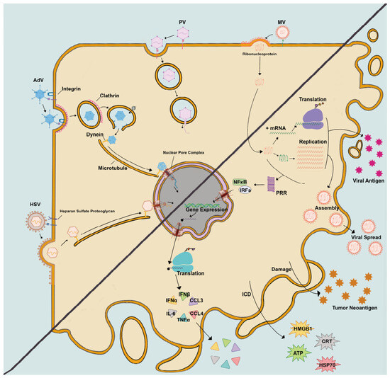
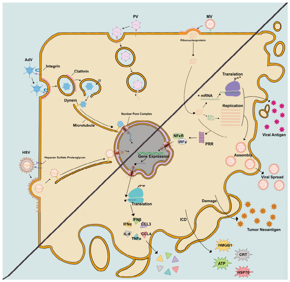
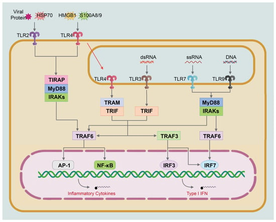
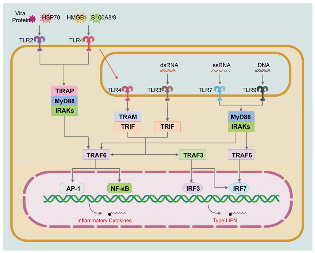
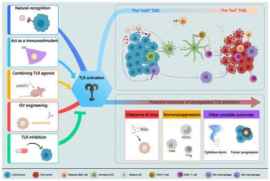
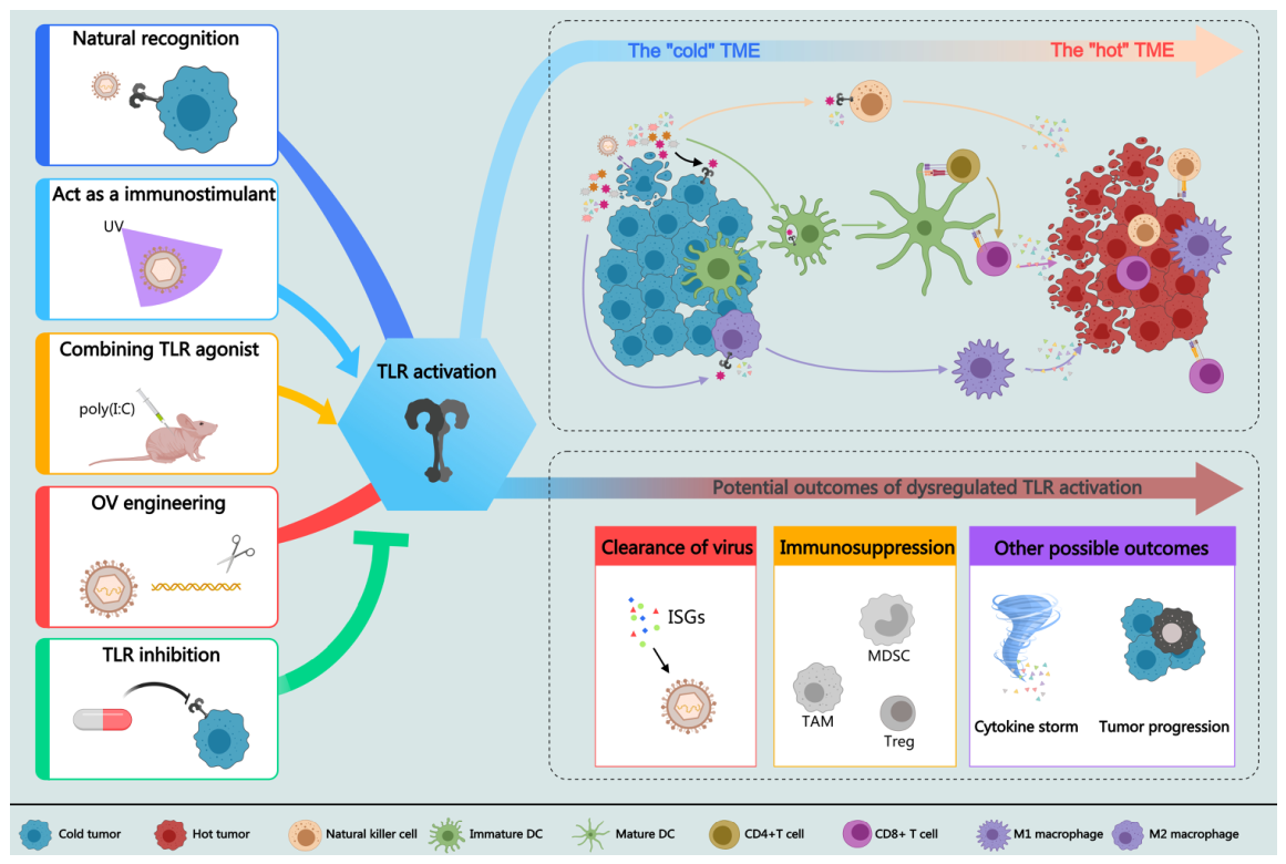

Disclaimer/Publisher’s Note: The statements, opinions and data contained in all publications are solely those of the individual author(s) and contributor(s) and not of MDPI and/or the editor(s). MDPI and/or the editor(s) disclaim responsibility for any injury to people or property resulting from any ideas, methods, instructions or products referred to in the content. |
© 2026 by the authors. Licensee MDPI, Basel, Switzerland. This article is an open access article distributed under the terms and conditions of the Creative Commons Attribution (CC BY) license.