


Abstract
Mediterranean Lamiaceae species are important sources of bioactive compounds with potential applications in food, pharmaceutical, and cosmetic industries. In the present study, controlled salinity was evaluated as a biotechnological elicitation strategy to enhance the accumulation of bioactive metabolites in two species, Lavandula viridis and Thymus lotocephalus, cultured in vitro. Morphological, biochemical and metabolic responses of shoots grown for seven weeks in culture medium containing four NaCl concentrations (0, 25, 50, or 75 mM) were assessed. Mild salinity (25 mM NaCl) stimulated shoot growth in both species, whereas higher salt concentrations progressively reduced growth. Salinity induced oxidative stress, as indicated by elevated malondialdehyde (MDA) levels at 25 mM NaCl in both species, while hydrogen peroxide (H2O2) exhibited contrasting patterns, decreasing with increasing salinity in L. viridis but peaking at 50 mM NaCl in T. lotocephalus. Salt stress caused reductions in photosynthetic pigments, phenolic compounds, antioxidant capacity (DPPH, FRAP, ABTS, and ORAC), and overall metabolic performance in L. viridis, indicating limited tolerance to salinity. Conversely, T. lotocephalus displayed greater metabolic plasticity, characterized by increased levels of phenolic compounds, particularly rosmarinic acid and its derivatives, and higher antioxidant activity of the extracts, as well as the accumulation of proline and soluble sugars. These findings demonstrate marked species-specific differences in salinity responses and identify controlled salinity as an effective elicitation strategy to sustainably produce high-value bioactive compounds in T. lotocephalus.
1. Introduction
Climate change is one of the most pressing environmental challenges of the 21st century, posing significant threats to plant biodiversity and ecosystem resilience [1]. Rising global temperatures, erratic precipitation patterns, increasing soil salinity, and more frequent extreme weather events expose plants to multiple abiotic stresses that adversely affect growth, development, and survival [2]. These environmental constraints disrupt key physiological and metabolic processes, including water uptake, nutrient acquisition, and photosynthesis, ultimately reducing plant productivity, destabilizing crop yields, and accelerating biodiversity loss [3]. Endemic and rare species are particularly vulnerable, as their restricted geographic distribution and limited adaptive capacity increase their extinction risk under rapidly changing environmental conditions [4].
Among abiotic stresses, soil salinity is a primary factor limiting agricultural productivity worldwide. It is estimated that salinization could affect more than 50% of arable land by 2050 [5]. Soil salinity results from the excessive accumulation of soluble salts, mainly Na+, Cl‒, SO42‒, Mg2+ and Ca2+ [6]. It may originate from natural processes (primary salinization) prevalent in arid and semi-arid regions due to low rainfall, high evapotranspiration, and capillary rise of saline groundwater, or from anthropogenic activities (secondary salinization), such as irrigation with poor-quality water and excessive fertilizer application [5,7]. Furthermore, salinity can be classified as neutral (e.g., NaCl) or alkaline (e.g., Na2CO3 or K2CO3), the latter being associated with elevated soil pH that further aggravates plant and microbial stress [8].
Salinity alters soil physicochemical properties by reducing porosity, aeration, and hydraulic conductivity, thereby restricting water availability and nutrient uptake [9]. The resulting stress primarily involves two interconnected mechanisms: (i) osmotic stress, caused by reduced soil water potential, which limits water absorption, and (ii) ionic toxicity resulting from excessive accumulation of Na+ and Cl−, leading to membrane destabilization and cellular damage [7]. To cope with these constraints, plants undergo a range of morphological, anatomical, physiological, and biochemical adaptations. A primary response includes increasing root-to-shoot biomass ratio, which optimizes water acquisition while restricting the translocation of toxic ions to photosynthetically active tissues [10].
Excessive accumulation of Na+ and Cl‒ also promotes the overproduction of reactive oxygen species (ROS), triggering oxidative stress and lipid peroxidation [9,11]. Although severe oxidative stress is detrimental, moderate abiotic stress can act as an elicitor, stimulating the biosynthesis of secondary metabolites, including phenolic compounds and terpenoids, which function as non-enzymatic antioxidants to aid in ROS detoxification [6]. This stress-induced metabolic reprogramming has attracted considerable attention because many of these compounds possess antioxidant, anti-inflammatory, antimicrobial, and other bioactive properties of high value for the pharmaceutical, cosmetic, and food industries [6].
Plant tissue culture provides a powerful platform for investigating plant responses to abiotic stress while offering a sustainable approach for producing high-value secondary metabolites. Compared with conventional cultivation, in vitro systems enable the production of bioactive compounds under controlled and aseptic conditions, independent of seasonal or geographical constraints, and reduces harvesting pressure on wild populations of threatened species [12,13]. Moreover, these systems allow the application of elicitation strategies and metabolic engineering approaches to enhance or even induce the biosynthesis of target metabolites that are absent or present only in trace amounts in naturally grown plants [13].
The Lamiaceae is one of the most economically significant families of medicinal and aromatic plants, renowned for producing essential oils and phenolic compounds with widespread industrial applications [6]. Many Mediterranean Lamiaceae species are particularly vulnerable to climate change due to their ecological specialization and restricted distribution [14]. Among them, Lavandula viridis L’Hér and Thymus lotocephalus G. López and R. Morales are endemic to the Iberian Peninsula and the Algarve region (southern Portugal), respectively. Both species are rich sources of rosmarinic acid (RA), and previous studies have demonstrated that their in vitro cultures accumulate higher concentrations of this phenolic compound than micropropagated plants grown in ex vitro conditions [15]. Despite being relatively understudied, both species have attracted increasing scientific interest owing to their diverse biological properties, including the antioxidant, antimicrobial, antifungal, antiprotozoal, nematocidal, and inhibitory effects of enzymes associated with diabetes, neurodegenerative diseases, and skin ageing [12].
Against this background, understanding the mechanisms underlying plant responses to salinity is essential both for developing climate-resilient crops and for optimizing the production of valuable secondary metabolites through controlled elicitation. Therefore, this study aimed, for the first time, to evaluate the morphological and biochemical responses of L. viridis and T. lotocephalus in vitro shoots to salinity. In addition, the impact of salinity on the phenolic profile, determined by Ultra-High-Performance Liquid Chromatography coupled to High-Resolution Mass Spectrometry (UHPLC-HRMS-MS), and on the antioxidant activity of green-solvent extracts was comprehensively investigated.
2. Materials and Methods
2.1. Plant Material and Salinity Treatments
In vitro shoots of L. viridis and T. lotocephalus, established from multiple genotypes collected from natural populations and maintained under standard in vitro culture procedures according to Dias et al. [16] and Coelho et al. [17], respectively, were used in this study. Previously optimized multiplication media were used throughout the experiment for each species to ensure species-specific normal shoot development. Accordingly, L. viridis shoots were cultured on Murashige and Skoog (MS) medium [18] supplemented with 0.2 mg L−1 6-benzyladenine (BA), whereas T. lotocephalus shoots were cultured on hormone-free MS medium. Media contained 2% (w/v) sucrose and 0.8% (w/v) agar. To evaluate the effects of salinity, sodium chloride (NaCl) was added to the basal medium at final concentrations of 25, 50, or 75 mM. These selected concentrations were based on the reported salinity sensitivity of Lamiaceae species and on previous studies indicating that salinity levels ranging from 25 to 100 mM are sufficient to induce physiological and biochemical stress responses while maintaining plant viability [19,20,21,22]. Higher concentrations (100 and 150 mM) were also tested in preliminary assays but proved highly phytotoxic and were therefore excluded from subsequent experiments. Shoots cultured on the corresponding multiplication media without NaCl served as the control treatment (CT). Culture media (80 mL) were dispensed into 250 mL Erlenmeyer flasks and sterilized at 121 °C for 20 min. Seven shoots were cultured per flask, and twelve flasks were tested for each treatment. Cultures were maintained at 25 ± 1 °C under a 16 h light/8 h dark photoperiod provided by cool-white fluorescent lamps (40 μmol m−2 s−1). After seven weeks of culture, morphological parameters were recorded. Plant material for biochemical analyses was immediately frozen in liquid nitrogen and stored until analysis, whereas material intended for extract preparation was oven-dried at 40 °C to constant weight.
2.2. Shoot Growth Evaluation
Seven shoots were initially inoculated per culture flask. After seven weeks of culture, four shoots were randomly selected from each flask for growth evaluation. The number of shoots produced by each selected shoot and the length of the longest shoot were recorded. The same four shoots were pooled for fresh weight determination, generating one fresh biomass value per flask. Dry weight was subsequently determined after oven-drying the pooled shoots at 40 °C until constant weight.
2.3. Determination of Chlorophyll and Carotenoid Contents
Photosynthetic pigments were quantified according to the method of Lichtenthaler [23]. Fresh plant material (25 mg) was homogenized in 4 mL of pure acetone and centrifuged. The absorbance of the supernatant was measured at 470, 644.8, and 661.6 nm using a T70+ UV/Vis spectrophotometer (PG Instruments Ltd., Leicestershire, UK). The contents of chlorophyll a, chlorophyll b, total chlorophyll, and carotenoids and the chlorophyll a/b and total chlorophyll/carotenoid ratios were calculated for each salinity treatment. The results were expressed as milligrams per gram of fresh weight (mg g−1FW).
2.4. Determination of Hydrogen Peroxide and Lipid Peroxidation
Hydrogen peroxide (H2O2) and lipid peroxidation were determined according to Mansinhos et al. [24]. Approximately 100 mg of fresh plant material was homogenized in 1 mL of 0.1% (w/v) trichloroacetic acid (TCA) and centrifuged. The resulting supernatant was used for both analyses. For H2O2 determination, 0.2 mL of supernatant was mixed with 0.2 mL of 10 mM potassium phosphate buffer and 0.4 mL of 1 M potassium iodide, and absorbance was measured at 390 nm after 30 min using a SynergyTM HTX MultiMode Microplate Reader (BioTek Instruments, Inc., Winooski, VT, USA). H2O2 concentration was determined from a calibration curve prepared with H2O2 standards and expressed as H2O2 equivalents per gram of fresh weight (µmolH2O2 g−1FW). Lipid peroxidation was estimated by measuring thiobarbituric acid-reactive substances (TBARSs) such as malondialdehyde (MDA). Briefly, 0.5 mL of supernatant was mixed with 0.5 mL of either a reaction solution containing 0.5% (w/v) TBA and 20% (w/v) TCA or a blank solution containing 20% (w/v) TCA. Samples were incubated at 95 °C for 30 min, rapidly cooled on ice, and absorbance was recorded at 440, 532, and 600 nm. MDA content was expressed as MDA equivalents per gram of fresh weight (nmolMDA g−1FW).
2.5. Determination of Soluble Sugars and Proline
Soluble sugar content was determined according to Abid et al. [25]. Fresh plant material (25 mg) was homogenized in 2 mL of 80% (v/v) ethanol and incubated in a water bath at 80 °C for 30 min. After centrifugation, 100 μL of supernatant was mixed with an equal volume of distilled water, followed by the addition of 200 μL of 9% (w/v) phenol and 1 mL of concentrated sulfuric acid. After incubation for 30 min, absorbance was measured at 490 nm using the microplate reader. The results were expressed in milligrams of glucose equivalents per gram of fresh weight (mgGLU g−1FW). Proline content was determined according to Mansinhos et al. [24]. Fresh plant material (25 mg) was subjected to three successive extractions with 0.5 mL of 80% (v/v) ethanol at 80 °C for 30 min each. The combined extracts were incubated with 1% (w/v) ninhydrin prepared in 60% (v/v) acetic acid for 1 h at 100 °C. After cooling, 1 mL of toluene was added, and the absorbance of the organic phase was measured at 520 nm. The results were expressed as micrograms of proline equivalents per gram of fresh weight (µgPRO g−1FW).
2.6. Extraction, Profiling, and Antioxidant Capacity Evaluation of Phenolic Compounds
2.6.1. Combined NADES and Ultrasound-Assisted Extraction (NADES-UAE)
Plant material was oven-dried at 40 °C to constant weight and ground to a particle size of <2 mm. Phenolic compounds were extracted using a natural deep eutectic solvent (NADES) composed of choline chloride and lactic acid (1:2 molar ratio) containing 30% (w/w) distilled water. This solvent was selected based on its demonstrated efficiency for extracting phenolic compounds from Lamiaceae species and the recognized advantages of NADESs as environmentally friendly extraction media [15,24,26]. Extractions were performed at a plant-to-solvent ratio of 2.5:100 (w/v) using an ultrasound-assisted extraction (UAE) bath (Elmasonic S 100 H, 220–240 V, 550 W; Elma Hans Schmidbauer GmbH & Co. KG, Singen, Germany). Samples were sonicated at a frequency of 37 kHz (sweep function) for 30 min at 50 °C. The extracts were subsequently filtered and stored at −20 °C until further analysis.
2.6.2. Phenolic Profiling by HPLC-HRMS
The phenolic composition of the extracts was characterized by an HPLC-HRMS-MS system equipped with a Vanquish Neo UHPLC system (Thermo Scientific, San José, CA, USA) and coupled to an Orbitrap Exploris 240 high-resolution mass spectrometer (Thermo Scientific, San José, CA, USA). For chromatographic separation, a Hypersil GOLD column (100 × 1.0 mm i.d., 1.9 µ Thermo Scientific) was used and thermostated at 40 °C. The autosampler temperature was kept at 10 °C. The mobile phase consisted of 0.1% aqueous formic acid solution (solvent A) and can/deionized water (80, 20, v/v) acidified with 0.1% formic acid (solvent B) with the followed gradient programme: 0–2 min, 5% B; 2–18 min, 5–95%; 18–22 min, 95% B. Then, the column was re-equilibrated to the initial conditions; the flow rate was 0.05 mL/min. The Orbitrap Exploris 240 operated at a resolution of 180,000 and was fitted with a Heated Electrospray Ionization Probe (H-ESI) in negative ionization mode for the determination of secondary metabolites (from 70 to 1000 m/z). H-ESI parameters were as follows: −2.5 kV spray voltage, 20 arbitrary units (AU) of sheath gas (N2), 10 AU of auxiliary gas (N2), ion transfer tube temperature at 325 °C, and vaporizer temperature at 120° C. The instrument was operated in Full Scan and Data-Dependent MS2 modes.
Data acquisition and processing were performed using Xcalibur software (version 4.6, Thermo Fisher Scientific, San Jose, CA, USA). Quality control (QC) samples, prepared by pooling equal aliquots of representative extracts, were periodically injected throughout the analytical sequence to monitor instrumental stability and analytical reproducibility, maintaining metabolite variability below 20%. Compound identification was based on the analysis of authentic standards, considering retention times and accurate masses. When standards were unavailable, tentative identification was achieved by comparing the measured exact mass of the molecular ion with theoretical values and consulting metabolite databases, including PubChem, Phenol-Explorer, Metlin, and ChemSpider. Metabolite annotation followed the MSI reporting criteria proposed by Sumner et al. [27]. Information regarding molecular formulae, exact masses, mass accuracy (Δppm), retention times (RT), and MSI identification levels is provided in Table S1. Validation parameters used for phenolic quantification, including linearity ranges, slopes, intercepts, coefficients of determination (R2), limits of detection (LOD), and limits of quantification (LOQ), are presented in Table S2. LOD and LOQ values ranged from 0.02 to 49.61 µg L−1 and from 0.06 to 150.35 µg L−1, respectively. Quantitative results were expressed as µg g−1DW.
2.6.3. Antioxidant Capacity Assessment
The antioxidant capacity of the green extracts was assessed using the procedure outlined by Mansinhos et al. [24], which combines four complementary assays. The ferric reducing antioxidant power (FRAP) assay is based on a single-electron transfer mechanism, whereas the oxygen radical absorbance capacity (ORAC) assay measures hydrogen atom transfer. The ABTS [2,2′-azino-bis(3-ethylbenzothiazoline-6-sulfonic acid)] and DPPH (2,2-diphenyl-1-picrylhydrazyl) assays involve both reaction mechanisms. The results are reported as milligrams of ascorbic acid equivalents per gram of dry weight (mgAAE g−1DW) for the FRAP assay, and as milligrams of Trolox (6-hydroxy-2,5,7,8-tetramethylchroman-2-carboxylic acid) equivalents per gram of dry weight (mgTE g−1DW) for the ABTS, DPPH, and ORAC assays.
2.7. Statistical Analysis
Data are presented as the mean ± standard error (SE). Growth parameters were obtained from five independent flasks per treatment (n = 5), with each flask considered an experimental unit. For each flask, the mean shoot number and mean shoot length were calculated from the four randomly selected shoots and used for statistical analyses. Fresh and dry biomass were determined from the pooled four selected shoots from each flask, generating one biomass value per flask. All biochemical analyses were performed using three independent biological replicates per treatment (n = 3). Each biological replicate consisted of plant material pooled from independent culture flasks within the same treatment. Prior to statistical analysis, the assumptions of normality and homogeneity of variances were assessed using the Shapiro–Wilk and Levene’s tests, respectively. Statistical differences were assessed through one-way analysis of variance (ANOVA) followed by Duncan’s new multiple range test (p < 0.05). For variables in which the assumption of homogeneity of variances was not met, Welch’s ANOVA was additionally performed to assess the robustness of the results. The robust analyses confirmed the same pattern of statistical significance as the conventional ANOVA. For multivariate analysis, OriginPro software (version 2024, OriginLab Corporation, Northampton, MA, USA) was employed using the mean values of each treatment. Variables were standardized using z-score normalization prior to heatmap and hierarchical cluster analysis (HCA). Compounds that were not detected or presented concentrations below the LOQ were excluded from the multivariate dataset. Hierarchical clustering was performed using Euclidean distance and the Group Average linkage method.
3. Results and Discussion
As each species was maintained on its own previously optimized multiplication medium, the following discussion focuses on species-specific responses to salinity.
3.1. Growth Parameters
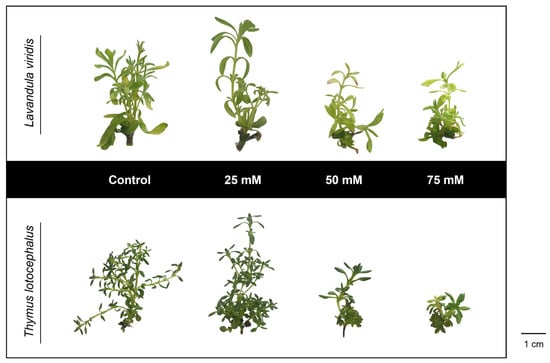
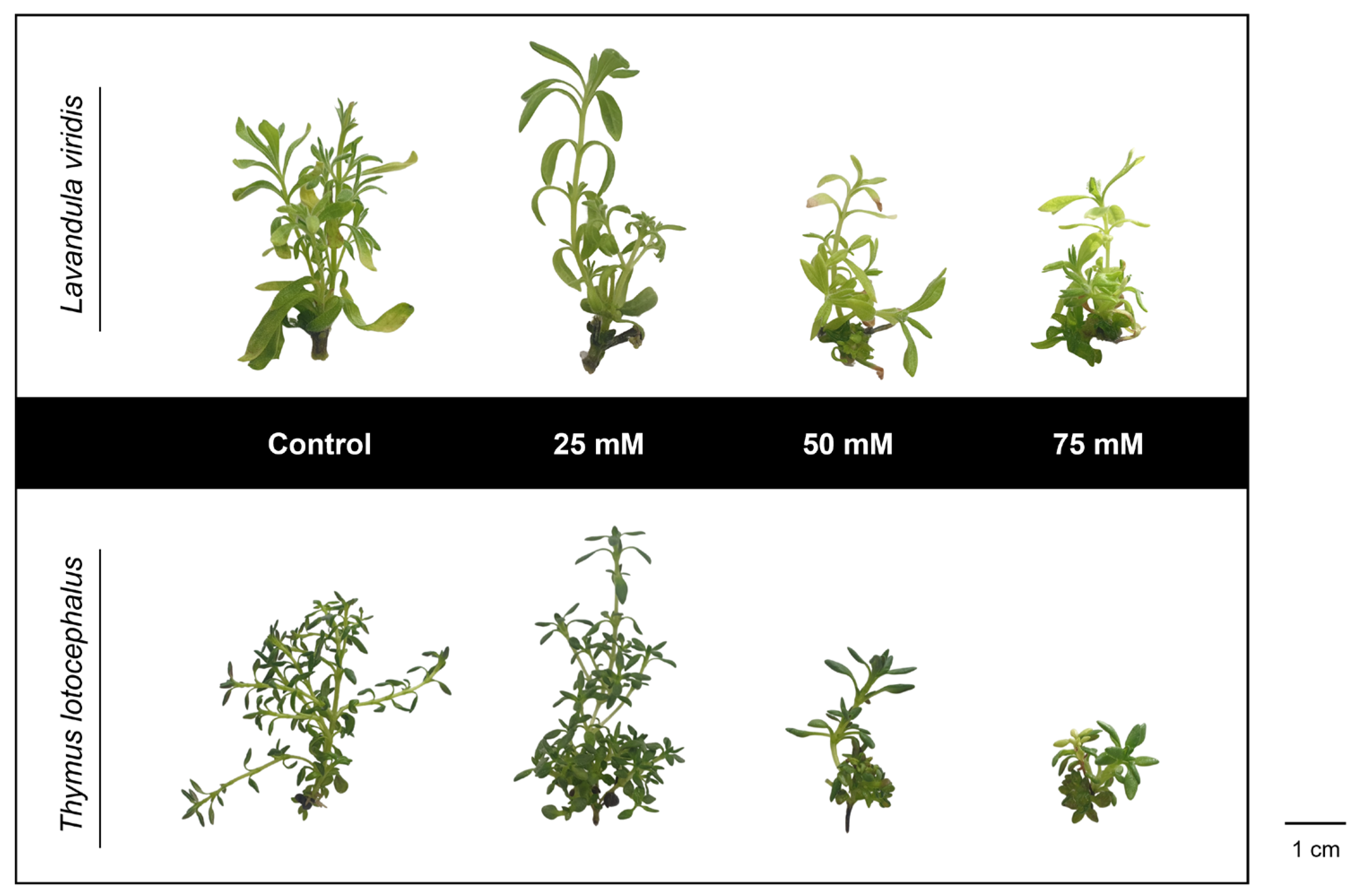
The effects of NaCl on the growth of L. viridis and T. lotocephalus shoots cultured under control conditions and in media containing 25, 50, or 75 mM NaCl are presented in Table 1 and Figure 1.
Table 1.
Shoot growth parameters of Lavandula viridis and Thymus lotocephalus cultured on control medium (CT; 0 mM NaCl) and media with 25, 50, or 75 mM NaCl.
Figure 1.
Shoots of Lavandula viridis and Thymus lotocephalus cultured on control medium (CT; 0 mM NaCl) and media with 25, 50, or 75 mM NaCl. The scale bar corresponds to 1 cm.
In both species, increasing NaCl concentration generally reduced growth performance, a response widely reported in Lamiaceae species, in which salt stress adversely affects morphological, physiological, and biochemical processes [20,28]. Nevertheless, mild salinity (25 mM NaCl) was associated with slight improvements in specific growth traits, including increased length of the longest shoots in L. viridis and increased shoot number and fresh biomass in T. lotocephalus. Similar responses to moderate salinity have previously been described in related species such as Salvia coccinea, where low salt concentrations promoted plant growth [29].
The number of shoots, shoot length, fresh weight, and dry weight markedly declined in L. viridis at 50 and 75 mM NaCl, demonstrating the pronounced sensitivity of this species to salinity. This response agrees with observations in Lavandula angustifolia, in which NaCl concentrations above 50 mM significantly reduced plant growth and induced visible symptoms of stress, including chlorosis and necrosis [19]. In contrast, Lavandula multifida has been reported to tolerate NaCl levels of up to 60 mM without significant reductions in biomass accumulation [30]. The highest NaCl levels (50 and 75 mM) also significantly reduced shoot development and biomass production in T. lotocephalus. Similar growth inhibition has been reported for Thymus vulgaris and Thymus daenensis, in which 60 mM NaCl or higher levels markedly decreased dry matter accumulation [20]. Conversely, Thymus pannonicus exhibited only minor growth reductions under the same salinity level, highlighting the considerable interspecific variability in salt tolerance within the genus [20].
Growth inhibition is one of the earliest plant responses to salinity stress. The osmotic imbalance caused by elevated salt concentrations rapidly decreases cellular turgor, restricting cell expansion and shoot growth. As stress persists, plant metabolism is progressively reprogrammed, shifting resource allocation from growth and primary metabolism towards protective and defence mechanisms, including osmotic adjustment and antioxidant responses [1]. This inhibitory pattern aligns with our previous findings under polyethylene glycol (PEG)-induced osmotic stress, where increasing PEG concentrations progressively reduced T. lotocephalus shoot length [31]. Likewise, nutrient deprivation significantly impaired the growth of both T. lotocephalus and L. viridis, with nitrogen deficiency exerting the strongest inhibitory effect on shoot development and biomass accumulation [12].
Overall, these findings demonstrate that both species are sensitive to salinity stress, although T. lotocephalus exhibited a greater capacity to tolerate moderate salt concentrations. The responses observed at 25 mM NaCl suggest that mild salinity may act as an effective elicitor, whereas higher concentrations progressively compromise shoot growth and development.
3.2. Photosynthetic Pigments
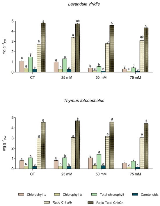
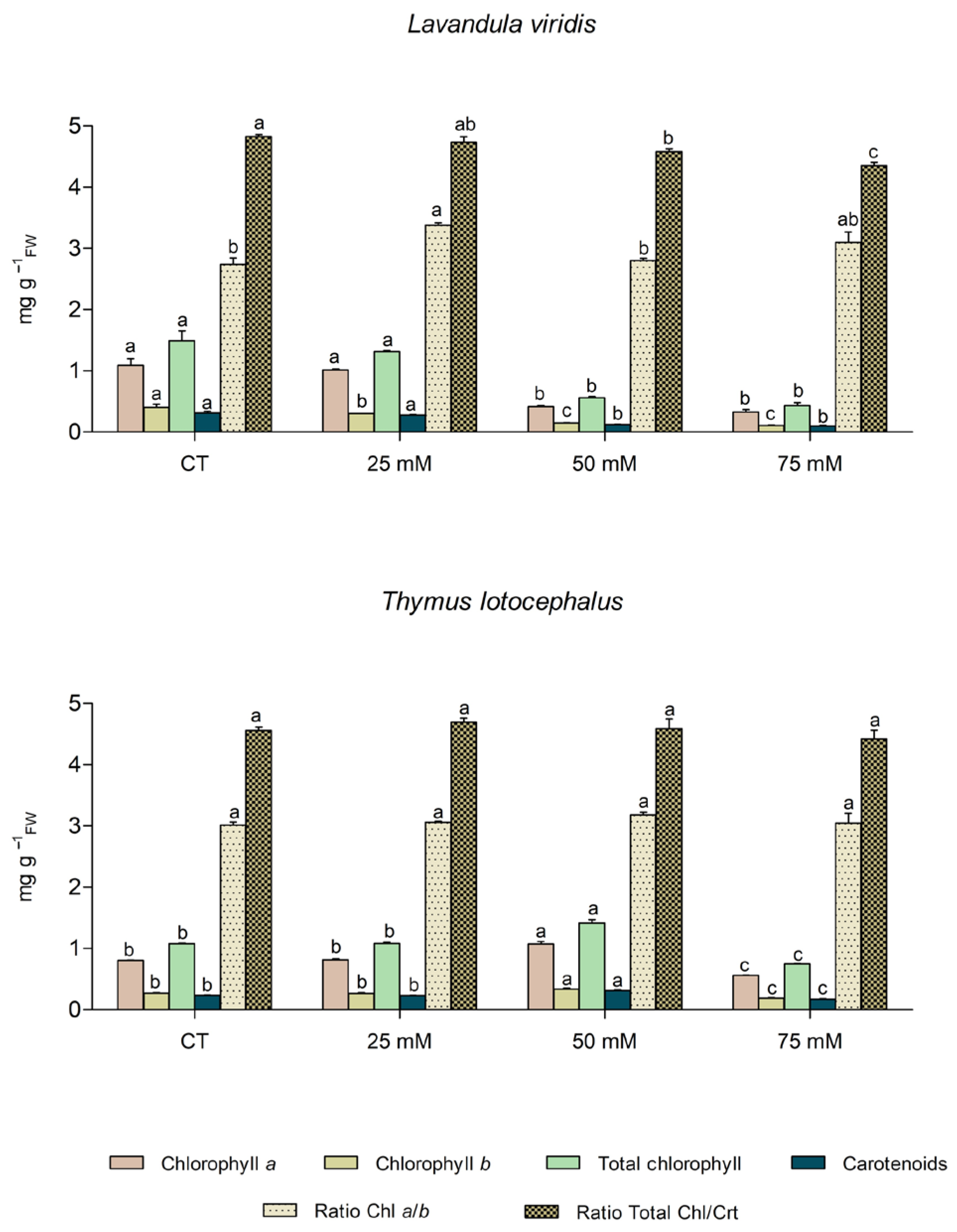
Salinity impairs water and nutrient uptake, leading to ionic imbalance, osmotic stress, oxidative damage, and the disruption of essential metabolic processes. These pressures reduce cellular turgor and can damage the photosynthetic apparatus, ultimately inducing chlorosis and accelerating leaf senescence [32]. Figure 2 presents the effects of NaCl on the photosynthetic pigment contents and pigment ratios of L. viridis and T. lotocephalus. As chlorophyll and carotenoid contents are highly sensitive to environmental stress, they serve as reliable biochemical indicators of plant physiological status and salinity tolerance [33].
Figure 2.
Chlorophyll a, chlorophyll b, total chlorophyll, and carotenoid contents, chlorophyll (Chl) a/b ratio, and total chlorophyll/carotenoid (Chl/Crt) ratio in Lavandula viridis and Thymus lotocephalus cultured on control medium (CT; 0 mM NaCl) and media with 25, 50, or 75 mM NaCl. Values are expressed as mean ± standard error (SE) and expressed as mg g-1 fresh weight (FW), except for Chl a/b and Chl/Crt ratios, which are dimensionless. Different letters indicate significant differences among NaCl treatments within each species according to Duncan’s multiple range test (p < 0.05).
In L. viridis, chlorophyll a, total chlorophyll and carotenoid contents remained relatively stable at 25 mM NaCl but declined significantly at 50 and 75 mM. This salinity sensitivity is consistent with findings on L. angustifolia, where NaCl concentrations above 50 mM reduced chlorophyll a and b contents by up to 73% [21]. More recently, Miri et al. [28] demonstrated that pigment response is genotype-dependent, since L. angustifolia subsp. angustifolia exhibits greater pigment loss than subsp. officinalis. The progressive decline in chlorophyll b and in the total chlorophyll/carotenoid ratio observed in L. viridis suggests enhanced chlorophyll degradation under severe salinity, likely associated with chlorophyllase activation and thylakoid membrane disruption [28].
By contrast, T. lotocephalus maintained stable pigment levels at 25 mM NaCl and even showed increased chlorophyll and carotenoid contents at 50 mM. A marked reduction occurred only at 75 mM NaCl. This response diverges from the salt sensitivity reported for T. vulgaris and T. daenensis, where 60–90 mM NaCl significantly reduced chlorophyll a, chlorophyll b, and total chlorophyll contents [20]. Instead, T. lotocephalus behaves similarly to the salt-tolerant T. pannonicus, which maintains pigment stability under 60 mM NaCl [34]. The increase in photosynthetic pigments under moderate salinity may represent an adaptive strategy to maximize light harvesting and maintain the energetic requirements of stress defence mechanisms. Supporting this, Li et al. [35] found that moderate salinity upregulates photosystem II (PSII)-stabilizing genes (LaPSB28, LaPSBS, and LaPSBR) in lavender, preserving pigments and maintaining electron transport efficiency.
Previous studies showed that photosynthetic pigments of both species were also affected by other abiotic stresses. Under in vitro drought simulation conditions, 7% PEG reduced chlorophyll and carotenoid contents by more than 50% in T. lotocephalus [31]. Likewise, nitrogen and iron deficiencies caused reductions in pigment levels in both species, reflecting the fundamental roles of nitrogen as a structural component of chlorophyll molecules and iron as an essential cofactor in photosynthetic electron transport [12]. Thermal stress also significantly impaired pigment stability. Both low (15 °C) and high (30 °C) temperatures also reduced pigment contents, with heat stress causing the most severe declines (65–81%), probably as a consequence of thylakoid membrane disruption and protein degradation induced by excessive ROS production [15].
Pigment ratios further highlight the distinct responses of the two species to salinity under their respective optimized in vitro culture medium. Both the chlorophyll a/b and total chlorophyll/carotenoid ratios remained relatively stable across treatments in T. lotocephalus, indicating efficient pigment homeostasis and preservation of the photosynthetic apparatus (Figure 2). In contrast, L. viridis exhibited a progressive decrease in the total chlorophyll/carotenoid ratio, reaching its lowest value at 75 mM NaCl. This trend indicates faster degradation of chlorophyll than carotenoids, reflecting increasing oxidative stress and a greater dependence on carotenoid-mediated photoprotection to detoxify ROS [29]. The lowest value observed at 75 mM NaCl suggests that severe salinity compromises pigment homeostasis, likely by causing chloroplast damage and/or triggering a defensive adjustment, with carotenoids playing a relatively more important role in protecting against oxidative stress [10].
The chlorophyll a/b ratio provides additional insight into photosynthetic acclimation, as it reflects changes in PSII antenna size and the relative abundance of accessory pigments. An increase in this ratio generally indicates a loss of chlorophyll b and contraction of the light-harvesting antenna, which minimizes photodamage under stress conditions [24]. In L. viridis, the highest chlorophyll a/b ratio was recorded at 25 mM NaCl, suggesting an early acclimation response to mild salinity through antenna size adjustment before the onset of severe pigment degradation at higher salt concentrations. Although the ratio decreased at 50 mM NaCl, the relatively high values observed at 75 mM suggest that chlorophyll b remained more susceptible to salinity-induced degradation than chlorophyll a. This is consistent with progressive adjustments of the photosynthetic antenna complex under severe stress conditions. Comparable changes in chlorophyll/carotenoid and chlorophyll a/b ratios have been reported in S. coccinea [29] and Lavandula officinalis [36,37], reinforcing the role of these pigment ratios as sensitive indicators of salinity-induced stress in Lamiaceae species.
3.3. Oxidative Stress Markers
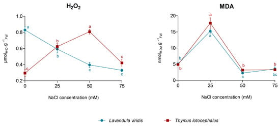
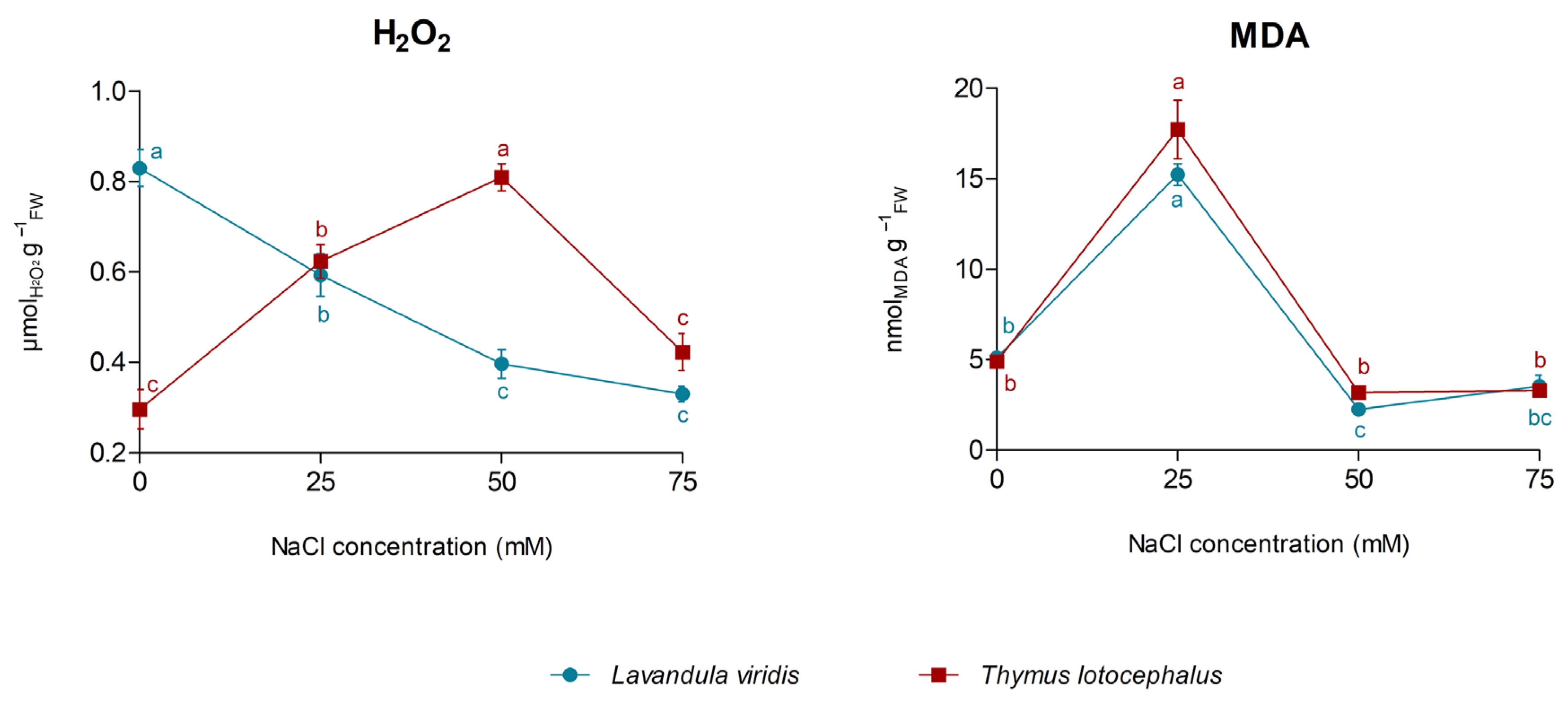
To assess the salinity-induced oxidative stress and cellular damage in L. viridis and T. lotocephalus, H2O2, a major ROS, and MDA, a widely used marker of lipid peroxidation, were quantified under the different NaCl treatments. As shown in Figure 3, the two species exhibited opposing H2O2 accumulation patterns.
Figure 3.
Hydrogen peroxide (H2O2) and malondialdehyde (MDA) contents in Lavandula viridis and Thymus lotocephalus cultured on control medium (CT; 0 mM NaCl) and media with 25, 50, or 75 mM NaCl. Values are presented as mean ± standard error (SE). Different letters indicate significant differences among NaCl treatments within each species according to Duncan’s multiple range test (p < 0.05).
In L. viridis, H2O2 levels progressively and significantly declined with increasing NaCl concentration, reaching reductions of approximately 52% and 60% (relative to the control) at 50 and 75 mM NaCl, respectively. This response diverges from several Lamiaceae species, such as L. angustifolia subsp. officinalis [28], Dracocephalum kotschyi [22] and Rosmarinus officinalis [38], which typically accumulate H2O2 under salinity. Conversely, T. lotocephalus showed a transient oxidative response, with H2O2 content increasing significantly at 25 mM NaCl and reaching a maximum at 50 mM (~174% above control), before declining to near-control levels at 75 mM NaCl. This contrasting pattern between the two species indicates a dual role of H2O2. While high levels are often associated with damage, the moderate increase observed in T. lotocephalus could indicate its capacity to act as a key signalling molecule rather than a purely toxic stress byproduct [39].
Lipid peroxidation patterns were similar in both species. Mild salinity (25 mM NaCl) induced pronounced MDA accumulation, reaching 15.24 nmol g−1FW in L. viridis and 17.72 nmol g−1FW in T. lotocephalus. These results indicate that the initial exposure to salinity caused substantial membrane damage and oxidative stress, reflecting the high sensitivity typically observed in glycophytes during the early stages of salt stress [10]. This transient MDA behaviour has also been reported in other Lamiaceae species, including Mentha piperita [40] and Ocimum basilicum [41]. Interestingly, at 50 and 75 mM NaCl, MDA levels returned to, or even fell below, those of the control.
The simultaneous decline in H2O2 and MDA observed at 75 mM NaCl suggests that both species underwent substantial metabolic adjustments in response to prolonged or severe salinity. Rather than signalling genuine stress tolerance, this pattern likely reflects a complex physiological acclimation involving diminished ROS generation paired with enhanced detoxification. At this salinity level, the marked decline in photosynthetic pigments (Figure 2) suggests compromised chloroplast function, which may have restricted photosynthetic electron transport and consequently limited ROS production. Simultaneously, non-enzymatic antioxidant systems, like carotenoids and phenolic compounds, may have contributed to ROS scavenging and the protection of cellular structures from oxidative damage [42,43]. This response is consistent with the growth–defence trade-off hypothesis, which proposes that, under severe environmental stress, plants reallocate metabolic resources from growth and primary metabolism towards protective and defence mechanisms [22]. This is supported by the substantial reductions in shoot growth and biomass observed at 75 mM NaCl (Figure 1 and Table 1), suggesting that resource allocation was shifted from growth to stress acclimation.
3.4. Osmotic Regulation
To investigate the osmotic adjustment mechanisms induced by salinity in L. viridis and T. lotocephalus, the accumulation of two major compatible solutes, proline and soluble sugars, was evaluated. Proline is one of the most effective osmoprotectants in plants, mitigating stress by acting as an intracellular osmolyte, ROS scavenger, molecular chaperone, and stabilizer of proteins and cellular membranes under dehydration conditions [10]. Soluble sugars also assist in osmotic balancing, acting both as compatible solutes and essential signalling molecules that regulate stress perception, carbon metabolism, and the maintenance of cellular homeostasis under abiotic constraints [24].
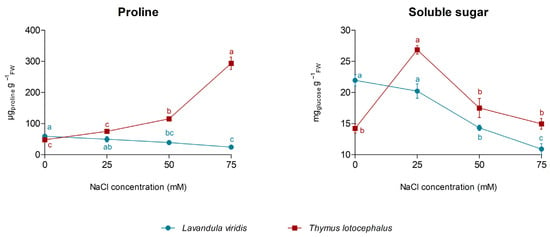
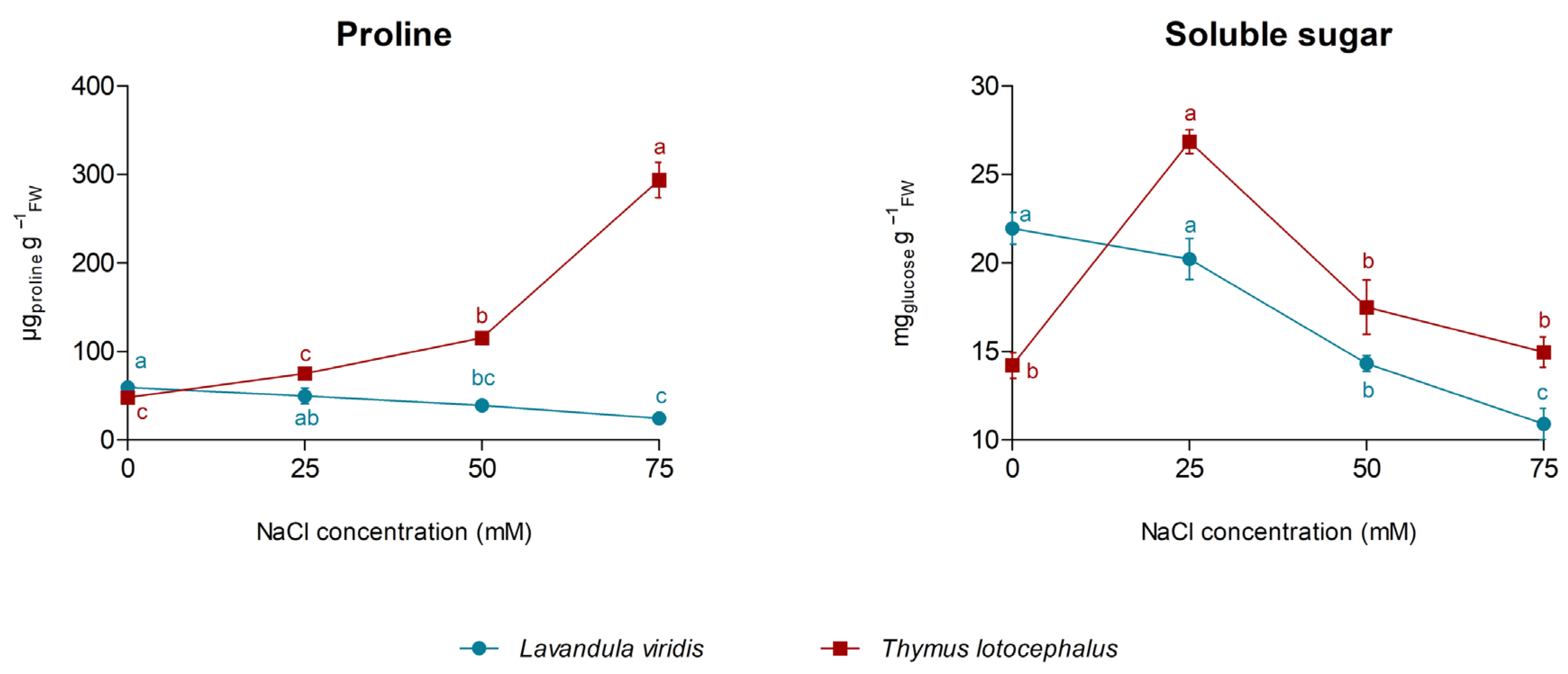
As shown in Figure 4, the two Lamiaceae species exhibited markedly different osmoregulatory responses to salinity. Proline content increased progressively up to 50 mM NaCl in T. lotocephalus, followed by a sharp rise at 75 mM, reaching 293.8 µg g−1FW (~6-fold higher than the control). This pronounced accumulation is in accordance with previous studies on this species, in which proline also increased substantially under PEG-induced drought [31] and temperature stress (15 and 30 °C) [15]. In T. vulgaris, proline accumulation also constitutes an important component of the adaptive response to prolonged salinity [44]. Nevertheless, the magnitude of this response depends on stress duration. For example, T. vulgaris exposed to 100 mM NaCl showed an initial reduction in proline after two weeks, followed by a threefold increase after four weeks of treatment [39], highlighting the dynamic nature of proline metabolism during stress acclimation. In contrast, in L. viridis, proline levels decreased progressively as NaCl concentration increased, indicating a fundamentally different adaptive strategy. A similar decline has been described in L. multifida, where reduced proline levels were attributed to enhanced proline catabolism, providing energy and reducing power to sustain metabolic maintenance under severe salinity [30]. Although this mechanism was not investigated in the present study, it may partially explain the response observed in L. viridis. These behaviours contrast with that reported for L. angustifolia, which exhibits a dose-dependent accumulation of proline under salt stress [28].
Figure 4.
Proline and soluble sugar contents in Lavandula viridis and Thymus lotocephalus cultured on control medium (CT; 0 mM NaCl) and media supplemented with 25, 50, or 75 mM NaCl. Values are presented as mean ± standard error (SE). Different letters indicate significant differences among NaCl treatments within each species according to Duncan’s multiple range test (p < 0.05).
The soluble sugar profile further emphasized the contrasting strategies between both species. In L. viridis, soluble sugars declined progressively with increasing salinity, indicating impaired osmotic adjustment. The simultaneous depletion of both proline and soluble sugars suggests that severe salinity compromised carbon assimilation and reduced the availability of compatible solutes required to maintain cellular osmotic balance. This response is consistent with the marked reduction in photosynthetic pigments (Figure 2) and growth (Figure 1 and Table 1), suggesting an overall metabolic limitation under high salinity. Consequently, the observed reduction in ROS and lipid peroxidation at the highest NaCl concentration is likely associated with alternative protective mechanisms rather than effective osmotic adjustment [10]. Conversely, T. lotocephalus accumulated soluble sugars under mild salinity (25 mM NaCl), a similar response to that previously observed under low-temperature stress (15 °C) [15]. At higher salinity levels (50 and 75 mM NaCl), however, soluble sugar concentrations declined to values close to those of the control, whereas proline accumulation increased markedly. This complementary behaviour suggests a coordinated osmotic response in which soluble sugars contribute to the initial acclimation to moderate stress, while proline becomes the predominant compatible solute under more severe salinity.
Overall, these findings indicate distinct osmoregulatory responses to salinity in the two species. Under their respective culture conditions, T. lotocephalus showed coordinated accumulation of compatible solutes, whereas L. viridis exhibited marked depletion of these compounds under increasing salinity. These observations suggest differences in their adaptive responses to salinity but should not be interpreted as direct quantitative comparisons between species.
3.5. Regulation of Secondary Metabolism
3.5.1. Phenolic Composition
Salinity is a major abiotic stress that profoundly affects plant metabolism by triggering the reprogramming of secondary metabolic pathways as part of the adaptive defence response [6]. In the present study, L. viridis and T. lotocephalus exhibited divergent metabolic shifts to increasing concentrations of NaCl, highlighting species-specific strategies for coping with osmotic and ionic stress. Both phenolic profiles were dominated by phenolic acids, particularly RA, which accounted for 38% and 43% of the total phenolic content in control cultures of L. viridis and T. lotocephalus, respectively. This predominance confirms the central role of the RA biosynthetic pathway in the stress physiology of these Mediterranean Lamiaceae species. In total, 26 phenolic compounds were identified in L. viridis extracts, of which 20 were quantified, whereas 31 compounds were identified and 27 were quantified in T. lotocephalus (Table 2). Since phytochemical analyses were performed using extracts prepared from a fixed amount of dried plant material, the reported values reflect metabolite concentrations in the analyzed samples rather than total metabolite yields per culture. Therefore, the results should be interpreted as changes in metabolite accumulation patterns under salinity stress and not as estimates of overall metabolite productivity.
Table 2.
Qualitative and quantitative (µg g −1DW, mean ± SE) analysis by UHPLC-HRMS-MS of the phenolic profile of extracts from Lavandula viridis and Thymus lotocephalus cultured on control medium (CT; 0 mM NaCl) and media with 25, 50, or 75 mM NaCl.
In L. viridis, the total phenolic content decreased progressively with increasing salinity (from 6710.82 µg g−1DW in the control to 4637.13 µg g−1DW at 75 mM NaCl). This response suggests that severe salinity progressively impaired secondary metabolism, a pattern previously reported in other Lamiaceae species, including L. angustifolia [21], Lavandula coronopifolia [45], Salvia macrosiphon [46], and Thymus serpyllum [47]. The reduction in phenolic accumulation is likely associated with the inhibition of carbon assimilation and photosynthetic performance observed under severe salinity, which limits the availability of ATP, reducing power, and carbon skeletons required for phenylpropanoid biosynthesis [22].
Despite the overall decline in total phenolics, phenolic acids remained the dominant metabolite class in L. viridis. RA was consistently the most abundant compound, although its concentration decreased from 3099.98 µg g−1DW in the control to 2298.92 µg g−1DW at 75 mM NaCl (Table 2). Responses of RA to salinity have been shown to be species- and genotype-dependent. For example, Scagel et al. [48] reported that salinity increased RA accumulation in Ocimum basilicum ‘Sweet Broadleaf’ but the opposite response occurred the ‘Siam Queen’ cultivar. A similar decline was observed for the major salvianolic acid isomers, particularly salvianolic acid B isomer III, whose concentration decreased ~43% under the highest salinity treatment. In contrast, some simple phenolic acids, notably ferulic acid, increased markedly, exhibiting a 181% increase at 75 mM NaCl, while caffeic acid showed a more modest rise. The accumulation of these precursor compounds may indicate the partial reorganization of phenylpropanoid metabolism, reflecting either reduced conversion into more complex derivatives or an increased requirement for these molecules as direct antioxidants under stress conditions [49].
Conversely, T. lotocephalus displayed a distinct metabolic response, accumulating phenolics under salinity. Total phenolic content increased by ~13% at 25 mM NaCl (5570.79 µg g−1DW), remained relatively stable at 50 mM, and declined only slightly at 75 mM NaCl (Table 2). This pattern indicates that mild and moderate salinity promoted the accumulation of phenolic compounds, whereas only severe stress approached the physiological tolerance threshold of the species.
RA remained the predominant phenolic compound in T. lotocephalus, but unlike L. viridis, its concentration increased under mild salinity, reaching 3193.34 µg g−1DW at 25 mM NaCl and remaining high at 50 mM. Given the well-established antioxidant, anti-inflammatory, antimicrobial, and antitumoral properties of RA, this accumulation represents a relevant result [50]. Enhanced RA accumulation likely contributes to mitigating oxidative damage caused by salinity-induced ROS, reinforcing its central role in T. lotocephalus’s adaptive response. The salinity-induced stimulation of RA has also been reported in T. vulgaris and T. daenensis, where accumulation increased by up to 32% under saline conditions [20]. Likewise, D. kotschyi exhibited increased RA accumulation up to 75 mM NaCl, followed by a decline at higher salinity levels [22].
Beyond RA itself, T. lotocephalus showed a pronounced increase in several RA-derived metabolites. Total salvianolic acids increased by ~44%, from 923.28 µg g−1DW in the control to 1324.75 µg g−1DW at 75 mM NaCl (Table 2), largely driven by the strong accumulation of salvianolic acid B isomer III, whose concentration more than tripled under severe salinity. Similarly, yunnaneic acid F, salviaflaside isomers and methylrosmarinic acid isomer I increased by 55%, 50–62% and 75%, respectively, under mild salinity. Since these compounds arise from the oxidative coupling and structural diversification of RA [51], their increased accumulation under saline conditions is consistent with enhanced flux through RA-related phenolic metabolic pathways. This metabolic profile is also consistent with the growth–defence trade-off hypothesis, where carbon and energy may be preferentially allocated to secondary metabolism rather than biomass accumulation under stress conditions [22]. From a chemotaxonomic perspective, the predominance of RA and its derivatives in both species is noteworthy. These compounds are characteristic of the Lamiaceae family and are frequently associated with high antioxidant capacity [6,51]. Their abundance further supports the importance of phenolic acid metabolism in the response of these species to salinity-induced oxidative stress.
Flavonoids, although recognized as effective antioxidants that complement the action of phenolic acids [52], showed minimal responses to salinity in both species. Luteolin-7-O-glucuronide was the predominant flavonoid in both species, although its concentration was substantially higher in T. lotocephalus. Its content progressively declined with increasing NaCl concentration, decreasing by ~25% at 75 mM. A similar pattern was observed for naringenin in T. lotocephalus. In L. viridis, total flavonoid content remained relatively stable throughout the treatments, suggesting that flavonoid accumulation was only marginally affected by salinity. The main exception was apigenin, whose concentration increased approximately fourfold at 50 mM NaCl (Table 2).
The divergent metabolic responses observed between the two species possibly reflect differences in stress perception, phenylpropanoid pathway regulation, and metabolic plasticity [49]. Under the culture conditions used in this study, T. lotocephalus exhibited a coordinated accumulation of RA and its derivatives, together with increased H2O2 levels at 50 mM NaCl. The positive association between these responses is consistent with the involvement of redox-related signalling in the acclimation process, although the present data do not establish a causal relationship. In contrast, L. viridis showed progressive decreases in both H2O2 and phenolic compounds with increasing salinity, indicating a reduced capacity to maintain these responses under the experimental conditions. Whereas T. lotocephalus accumulated higher levels of RA and its derivatives under mild and moderate stress, L. viridis exhibited a progressive decline in these metabolites, which may be associated with reduced carbon availability and impaired photosynthetic performance under saline conditions [53]. These findings are fully consistent with the physiological responses described in the previous sections and highlight distinct acclimation patterns in the two species under their respective optimized culture conditions.
From an applied perspective, these results show that controlled salinity may represent a useful elicitation approach to enhance the accumulation of high-value phenolic compounds in T. lotocephalus cultures. A similar metabolic response was previously observed under iron deficiency, where RA accumulation increased by ~34% [12]. Likewise, under temperature stress, T. lotocephalus maintained a stable antioxidant system [15]. In contrast, osmotic stress induced by PEG reduced both growth and phenolic accumulation in T. lotocephalus [31].
3.5.2. Antioxidant Activity of Extracts
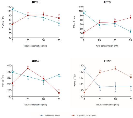
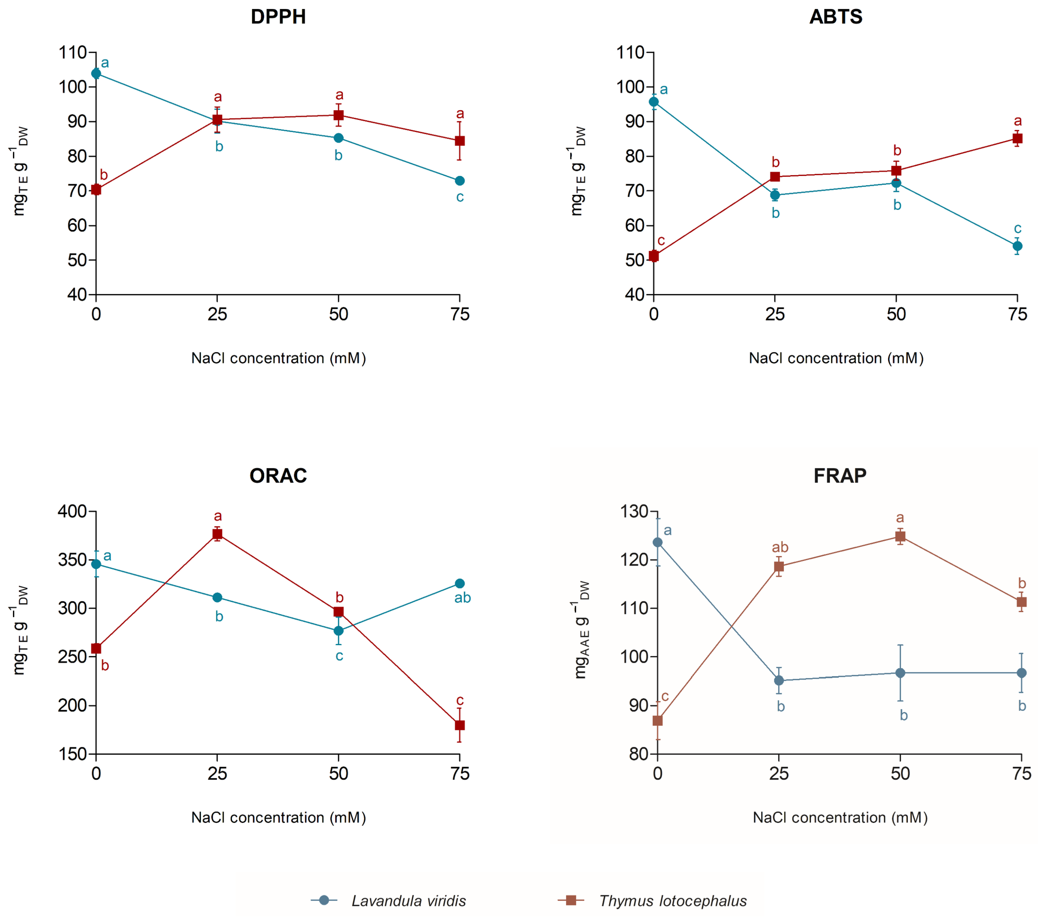
The antioxidant capacity of L. viridis and T. lotocephalus extracts obtained from cultures grown under salinity stress closely reflected the contrasting metabolic responses described in the previous section (Figure 5). Overall, the antioxidant assays demonstrated that salinity progressively reduced the antioxidant capacity of L. viridis extracts, whereas T. lotocephalus extracts maintained or exhibited higher antioxidant capacity under mild to moderate salinity, highlighting contrasting responses to salt stress.
Figure 5.
Antioxidant capacity determined by DPPH, ABTS, FRAP, and ORAC assays in Lavandula viridis and Thymus lotocephalus cultured on control medium (CT; 0 mM NaCl) and media with 25, 50, or 75 mM NaCl. Different letters indicate significant differences among NaCl treatments within each species according to Duncan’s multiple range test (p < 0.05).
In L. viridis, DPPH and ABTS radical scavenging activities declined progressively with increasing NaCl concentration, indicating a gradual loss of antioxidant capacity. This reduction closely paralleled the decrease in total phenolic content, RA and salvianolic acid derivatives, suggesting that the lower antioxidant activity was associated with reduced accumulation of phenolic compounds. Similar results have been reported for L. angustifolia, where salinity levels above 50 mM significantly reduced antioxidant capacity, measured by DPPH, FRAP, and ABTS assays [21]. Likewise, the sharp reduction in FRAP values observed at 25 mM NaCl indicates that the reducing capacity of L. viridis was rapidly compromised under salt stress conditions.
In contrast, T. lotocephalus exhibited a distinct antioxidant response. DPPH and FRAP values increased under mild and moderate salinity (25 and 50 mM), while ABTS activity continued to increase throughout the salinity gradient, reaching its highest value at 75 mM NaCl. These results indicate that moderate salinity acted as an effective elicitor, being associated with higher extract antioxidant capacity and increased accumulation of phenolic compounds. This behaviour is in agreement with the enhanced accumulation of RA, salvianolic acids, and related phenolic derivatives observed under the same conditions and is consistent with reports for other salt-tolerant Lamiaceae species, including T. vulgaris and T. daenensis, in which antioxidant capacity increased under saline conditions [20]. The maintenance of the high antioxidant capacity of extracts of T. lotocephalus under mild to moderate salinity was accompanied by increased accumulation of phenolic compounds, including RA and salvianolic acid derivatives.
The ORAC assay exhibited a pattern distinct from the other antioxidant methods. At 75 mM NaCl, L. viridis showed partial recovery in ORAC values, whereas T. lotocephalus exhibited a marked decline. Since ORAC reflects hydrogen atom transfer (HAT) reactions, whereas FRAP is based on single-electron transfer (SET) and DPPH and ABTS involve both mechanisms, these contrasting responses likely indicate changes in the composition of the antioxidant pool rather than its overall abundance alone. In L. viridis, the partial recovery may indicate the relative contribution of specific hydrogen atom-donating compounds that remain active despite the overall reduction in phenolic content. Conversely, the decline in ORAC activity observed in T. lotocephalus at the highest salinity level suggests that the most severe treatment affected the contribution of some HAT-active antioxidants, despite the maintenance of high levels of phenolic compounds and strong antioxidant performance measured by the other assays.
Overall, the antioxidant results were consistent with the biochemical and metabolic patterns described throughout this study. L. viridis showed progressive metabolic impairment under salinity stress, characterized by reduced photosynthetic pigments, depletion of compatible solutes, the suppression of phenolic metabolism, and declining antioxidant capacity, whereas T. lotocephalus maintained coordinated biochemical and metabolic responses, including osmotic adjustment, increased accumulation of RA and its derivatives, and high antioxidant activity of the extract under the respective culture conditions. The association between phenolic accumulation and extract antioxidant capacity suggests an important contribution of phenolic acids, particularly RA and salvianolic derivatives, to the antioxidant properties of T. lotocephalus under salinity stress.
3.6. Multivariate Analysis
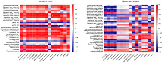
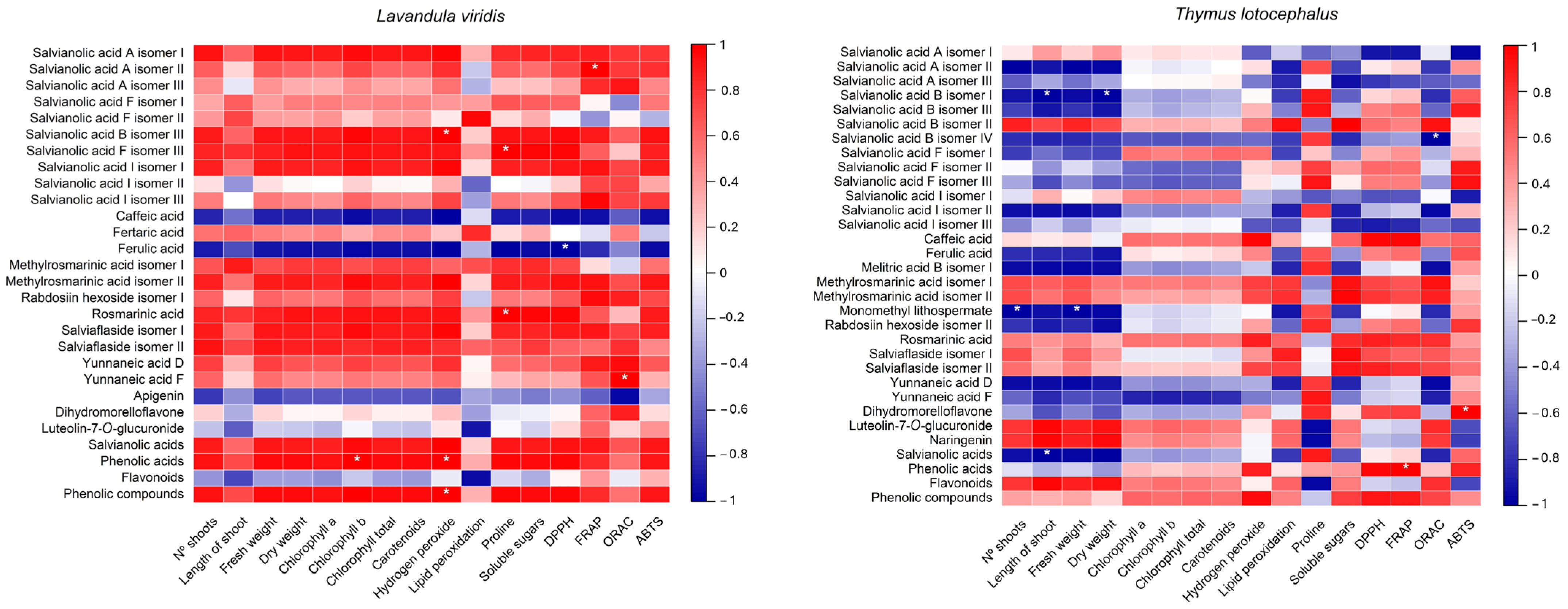
Multivariate analyses including the heatmap (Figure 6) and hierarchical cluster analysis (Figure 7), integrating all morphological, biochemical, and metabolic variables, further highlighted the distinct response patterns displayed by L. viridis and T. lotocephalus in response to salinity.
Figure 6.
A heatmap showing the Pearson correlation coefficients between growth parameters, photosynthetic pigments, oxidative stress markers, osmolytes, antioxidant capacity, and individual and grouped phenolic compounds in Lavandula viridis and Thymus lotocephalus cultured on control medium (CT; 0 mM NaCl) and media with 25, 50, or 75 mM NaCl. Positive correlations are shown in red and negative correlations in blue, with colour intensity proportional to the correlation coefficient (−1 to +1). Asterisks (*) indicate statistically significant correlations (p ≤ 0.01). Phenolic compounds are identified according to their UHPLC–HRMS-MS characterization, and grouped variables include total salvianolic acids, total flavonoids, total phenolic acids, and total phenolic compounds.
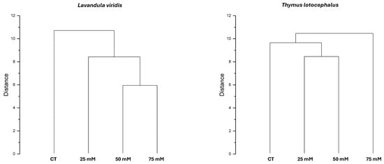
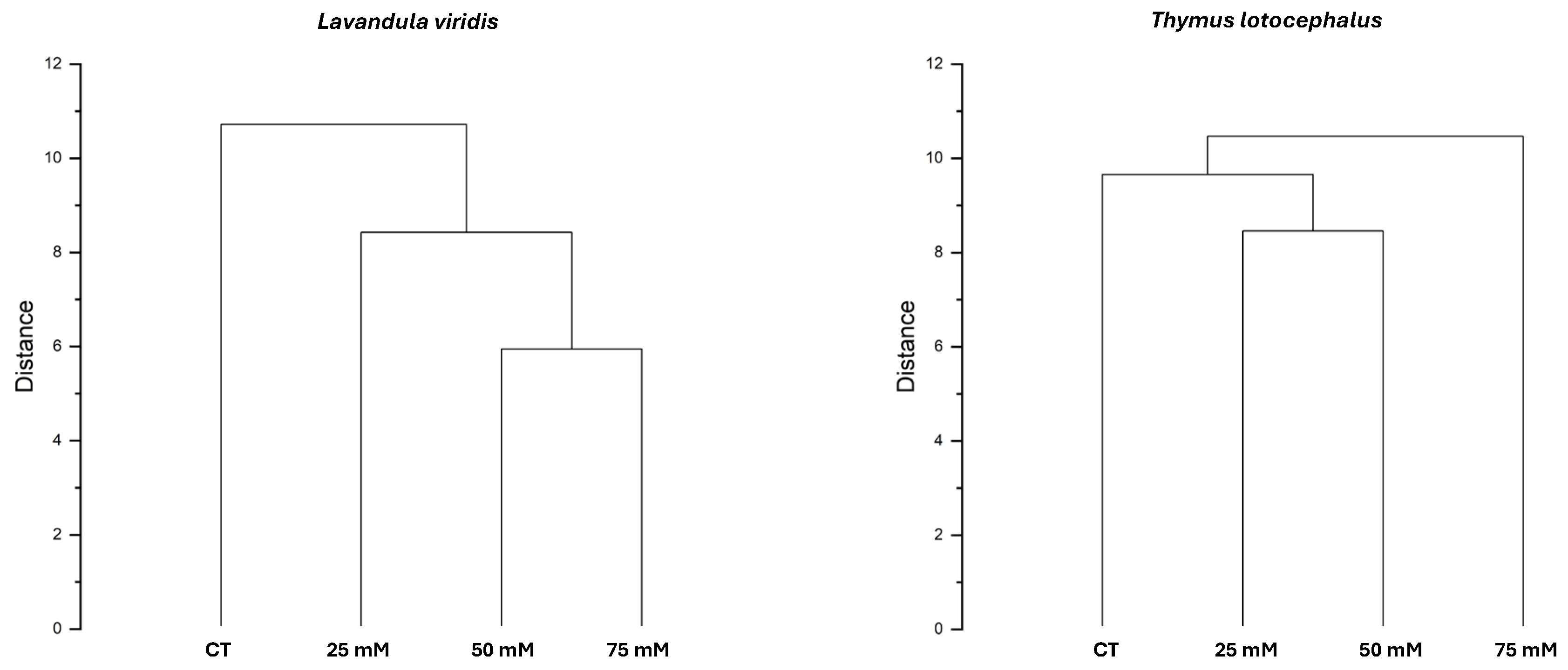
Figure 7.
Dendrograms of the hierarchical cluster analysis (HCA) based on Euclidean distance for Lavandula viridis and Thymus lotocephalus cultured on control medium (CT; 0 mM NaCl) and media with 25, 50, or 75 mM NaCl. The vertical axis represents the linkage distance between clusters.
In T. lotocephalus, the heatmap revealed a more heterogeneous correlation network, reflecting its greater metabolic plasticity. RA and several related metabolites, including salviaflaside and methylrosmarinic acid isomers, were positively associated with antioxidant activity, whereas other compounds showed negative correlations with growth- and pigment-related variables. This pattern suggests that different groups of phenolic compounds exhibited distinct responses to increasing salinity. The negative associations observed for salvianolic acids, ferulic acid, monomethyl lithospermate, rabdosiin hexoside isomer I, and yunnaneic acids D and F are consistent with a metabolic reallocation from primary growth towards the synthesis of protective secondary metabolites under saline conditions. In contrast, flavonoids were positively correlated with growth-related traits, indicating that their accumulation was associated with the maintenance of physiological performance rather than representing an additional metabolic cost. Following z-score standardization, hierarchical cluster analysis revealed that the 25 and 50 mM NaCl treatments were the most similar, clustering together at the shortest distance. This cluster subsequently grouped with the control treatment, whereas the 75 mM NaCl treatment remained separated until the final clustering stage, indicating a markedly different response under severe salinity conditions. The proximity between the 25 and 50 mM treatments suggests the occurrence of a common metabolic adjustment to mild and moderate salt stress, reflected in coordinated changes in growth, biochemical parameters, and antioxidant responses. In contrast, the separation of the 75 mM treatment indicates that severe salinity induced a broader shift in the physiological and metabolic profile of T. lotocephalus, reflecting a more pronounced stress response.
In L. viridis, the heatmap revealed a relatively coordinated correlation pattern between RA, its derivatives, and antioxidant capacity, indicating that phenolic acids were closely associated with the extract’s antioxidant responses observed under salt stress (Figure 6). Most phenolic compounds exhibited positive correlations with growth- and pigment-related parameters, suggesting that higher levels of phenolic compounds were associated with improved physiological performance. Conversely, the decline in these metabolites at higher salinity levels coincided with reduced biomass production, pigment degradation, and lower antioxidant activity, reinforcing the overall metabolic impairment previously described. A contrasting clustering pattern was observed in L. viridis. The 50 and 75 mM NaCl treatments clustered together at the shortest distance, indicating a high degree of similarity in their overall physiological and metabolic responses under moderate to severe salinity. In contrast, the control and 25 mM NaCl treatments were more distinct and joined the cluster at later stages. The close association between the 50 and 75 mM treatments suggests that salinity levels above 25 mM induced marked changes in growth, biochemical, and metabolic parameters. Furthermore, the clear separation of these treatments from the control indicates that L. viridis responded differently once a critical salinity threshold was exceeded. This is in agreement with the reductions in growth and photosynthetic performance observed throughout the study.
Overall, the multivariate analyses corroborate the morphological, biochemical, and metabolic findings, revealing distinct response patterns to salinity in the two species. T. lotocephalus exhibited coordinated changes in metabolite accumulation, antioxidant capacity, and growth-related parameters across the salinity treatments, whereas L. viridis showed a progressive decline in several physiological and biochemical parameters with increasing salinity.
4. Conclusions
This study demonstrates that T. lotocephalus and L. viridis exhibit markedly divergent physiological and metabolic responses to salinity under their respective optimized in vitro culture conditions. The results indicate that the ability to maintain redox homeostasis was associated with different acclimation patterns in the two species. Under the experimental conditions tested, T. lotocephalus displayed a great metabolic plasticity, characterized by efficient osmotic adjustment, enhanced accumulation of proline, RA, salvianolic acids, and related phenolic derivatives, and sustained antioxidant capacity under mild to moderate salinity. In contrast, L. viridis showed progressive reductions in growth, photosynthetic pigments, phenolic metabolism, and antioxidant activity as salinity increased, indicating a comparatively lower tolerance to NaCl concentrations above 25 mM.
RA and its derivatives emerged as key metabolites associated with salinity tolerance, showing strong relationships with antioxidant capacity and supporting their central role in protecting plants against oxidative stress. Multivariate analyses further highlighted distinct metabolic response patterns in the two species, with coordinated metabolic adjustments in T. lotocephalus and progressive metabolic impairment in L. viridis.
Overall, this work provides new insights into the biochemical and metabolic responses of two Mediterranean endemic Lamiaceae species to salinity. From a biotechnological perspective, these findings demonstrate that controlled salinity can be successfully exploited as an elicitation strategy for enhancing the accumulation of valuable phenolic compounds with antioxidant capacity in T. lotocephalus in vitro cultures. This approach offers a sustainable and environmentally friendly alternative for increasing the production of bioactive metabolites of pharmaceutical, nutraceutical, cosmetic, and food interest, while contributing to the conservation and valorization of Mediterranean endemic plant species.
Supplementary Materials
The following supporting information can be downloaded at https://www.mdpi.com/article/10.3390/biom16081178/s1: Table S1. HPLC-HRMS data of identified phenolic compounds in Lavandula viridis and Thymus lotocephalus extracts; Table S2. Summary of HPLC-HRMS criterion for quantification of phenolic compounds in Lavandula viridis and Thymus lotocephalus extracts.
Author Contributions
Conceptualization, I.M., S.G. and A.R.; methodology, I.M., S.G., R.R.-S. and G.P.-C.; software, I.M., R.R.-S. and G.P.-C.; validation, I.M., S.G., R.R.-S., G.P.-C. and A.R.; formal analysis, I.M., S.G., R.R.-S. and G.P.-C.; investigation, I.M., S.G., R.R.-S., G.P.-C. and A.R.; resources, S.G. and A.R.; data curation, I.M., S.G. and R.R.-S.; writing—original draft preparation, I.M.; writing—review and editing, S.G., R.R.-S., G.P.-C. and A.R.; visualization, I.M. and S.G.; supervision, S.G. and A.R.; project administration, S.G. and A.R.; funding acquisition, S.G. and A.R. All authors have read and agreed to the published version of the manuscript.
Funding
This research was financed by National Funds through the FCT-Foundation for Science and Technology under the Projects MED (https://doi.org/10.54499/UID/05183/2025) and CHANGE (https://doi.org/10.54499/LA/P/0121/2020). Sandra Gonçalves (CEECINST/00052/2021) acknowledges the financial support from the FCT. Raquel Rodríguez Solana was supported by the grant RYC2022-036888-I, funded by MCIU/AEI/10.13039/501100011033 and the FSE+.
Institutional Review Board Statement
Not applicable.
Informed Consent Statement
Not applicable.
Data Availability Statement
The raw data supporting the conclusions of this article will be made available by the authors on request.
Conflicts of Interest
The authors declare no conflicts of interest. The funders had no role in the design of the study; in the collection, analyses, or interpretation of data; in the writing of the manuscript; or in the decision to publish the results.
Abbreviations
The following abbreviations are used in this manuscript:
| NaCl | Sodium chloride |
| H2O2 | Hydrogen peroxide |
| MDA | Malondialdehyde |
| ROS | Reactive Oxygen Species |
| NADES | Natural Deep Eutectic Solvent |
| UAE | Ultrasound-Assisted Extraction |
| RA | Rosmarinic acid |
| UHPLC-HRMS-MS | Ultra-High-Performance Liquid Chromatography coupled with High-Resolution Mass Spectrometry |
References
- Szekely-Varga, Z.; González-Orenga, S.; Cantor, M.; Jucan, D.; Boscaiu, M.; Vicente, O. Effects of Drought and Salinity on Two Commercial Varieties of Lavandula angustifolia Mill. Plants 2020, 9, 637. [Google Scholar] [CrossRef] [Scilit] [PubMed]
- Muhammad, M.; Waheed, A.; Wahab, A.; Majeed, M.; Nazim, M.; Liu, Y.-H.; Li, L.; Li, W.-J. Soil Salinity and Drought Tolerance: An Evaluation of Plant Growth, Productivity, Microbial Diversity, and Amelioration Strategies. Plant Stress 2024, 11, 100319. [Google Scholar] [CrossRef] [Scilit]
- Jing, Z.; Liu, N.; Zhang, Z.; Hou, X. Research Progress on Plant Responses to Stress Combinations in the Context of Climate Change. Plants 2024, 13, 469. [Google Scholar] [CrossRef] [Scilit] [PubMed]
- Kougioumoutzis, K.; Papanikolaou, A.; Kokkoris, I.P.; Strid, A.; Dimopoulos, P.; Panitsa, M. Climate Change Impacts and Extinction Risk Assessment of Nepeta Representatives (Lamiaceae) in Greece. Sustainability 2022, 14, 4269. [Google Scholar] [CrossRef] [Scilit]
- Singh, M.; Singh, S.K.; Sharma, J.G.; Giri, B. Insights into the Multifaceted Roles of Soil Microbes in Mitigating Abiotic Stress in Crop Plants: A Review. Environ. Exp. Bot. 2024, 228, 106010. [Google Scholar] [CrossRef] [Scilit]
- Mansinhos, I.; Gonçalves, S.; Romano, A. How Climate Change-Related Abiotic Factors Affect the Production of Industrial Valuable Compounds in Lamiaceae Plant Species: A Review. Front. Plant Sci. 2024, 15, 1370810. [Google Scholar] [CrossRef] [Scilit] [PubMed]
- Butcher, K.; Wick, A.F.; DeSutter, T.; Chatterjee, A.; Harmon, J. Soil Salinity: A Threat to Global Food Security. Agron. J. 2016, 108, 2189–2200. [Google Scholar] [CrossRef] [Scilit]
- Ondrasek, G.; Rathod, S.; Manohara, K.K.; Gireesh, C.; Anantha, M.S.; Sakhare, A.S.; Parmar, B.; Yadav, B.K.; Bandumula, N.; Raihan, F.; et al. Salt Stress in Plants and Mitigation Approaches. Plants 2022, 11, 717. [Google Scholar] [CrossRef] [Scilit] [PubMed]
- Mahajan, M.; Kuiry, R.; Pal, P.K. Understanding the Consequence of Environmental Stress for Accumulation of Secondary Metabolites in Medicinal and Aromatic Plants. J. Appl. Res. Med. Aromat. Plants 2020, 18, 100255. [Google Scholar] [CrossRef] [Scilit]
- Acosta-Motos, J.R.; Ortuño, M.F.; Bernal-Vicente, A.; Diaz-Vivancos, P.; Sanchez-Blanco, M.J.; Hernandez, J.A. Plant Responses to Salt Stress: Adaptive Mechanisms. Agronomy 2017, 7, 18. [Google Scholar] [CrossRef] [Scilit]
- Tokarz, B.; Wójtowicz, T.; Makowski, W.; Jędrzejczyk, R.J.; Tokarz, K.M. What Is the Difference between the Response of Grass Pea (Lathyrus sativus L.) to Salinity and Drought Stress?—A Physiological Study. Agronomy 2020, 10, 833. [Google Scholar] [CrossRef] [Scilit]
- Mansinhos, I.; Gonçalves, S.; Rodríguez-Solana, R.; Pereira-Caro, G.; Moreno-Rojas, J.M.; Romano, A. Nutrient Deficiency-Induced Stress Improves Skincare Effects and Phytochemical Content of Green Extracts from Lamiaceae In Vitro Cultures. Horticulturae 2024, 10, 947. [Google Scholar] [CrossRef] [Scilit]
- Wijerathna-Yapa, A.; Hiti-Bandaralage, J.; Pathirana, R. Harnessing Metabolites from Plant Cell Tissue and Organ Culture for Sustainable Biotechnology. Plant Cell Tissue Organ Cult. 2025, 162, 55. [Google Scholar] [CrossRef] [Scilit]
- Tomou, E.-M.; Skaltsa, H.; Economou, G.; Trichopoulou, A. Sustainable Diets & Medicinal Aromatic Plants in Greece: Perspectives towards Climate Change. Food Chem. 2022, 374, 131767. [Google Scholar] [CrossRef] [Scilit] [PubMed]
- Mansinhos, I.; Gonçalves, S.; Rodríguez-Solana, R.; Ordóñez-Díaz, J.L.; Moreno-Rojas, J.M.; Romano, A. Impact of Temperature on Phenolic and Osmolyte Contents in in Vitro Cultures and Micropropagated Plants of Two Mediterranean Plant Species, Lavandula viridis and Thymus lotocephalus. Plants 2022, 11, 3516. [Google Scholar] [CrossRef] [Scilit] [PubMed]
- Dias, M.C.; Almeida, R.; Romano, A. Rapid Clonal Multiplication of Lavandula viridis L’Hér through in Vitro Axillary Shoot Proliferation. Plant Cell Tissue Organ Cult. 2002, 68, 99–102. [Google Scholar] [CrossRef] [Scilit]
- Coelho, N.; Goncalves, S.; Elena Gonzalez-Benito, M.; Romano, A. Establishment of an in Vitro Propagation Protocol for Thymus lotocephalus, a Rare Aromatic Species of the Algarve (Portugal). Plant Growth Regul. 2012, 66, 69–74. [Google Scholar] [CrossRef] [Scilit]
- Murashige, T.; Skoog, F. A Revised Medium for Rapid Growth and Bio Assays with Tobacco Tissue Cultures. Physiol. Plant. 1962, 15, 473–497. [Google Scholar] [CrossRef] [Scilit]
- Paraskevopoulou, A.T.; Karantzi, A.K.; Liakopoulos, G.; Londra, P.A.; Bertsouklis, K. The Effect of Salinity on the Growth of Lavender Species. Water 2020, 12, 618. [Google Scholar] [CrossRef] [Scilit]
- Bistgani, Z.E.; Hashemi, M.; DaCosta, M.; Craker, L.; Maggi, F.; Morshedloo, M.R. Effect of Salinity Stress on the Physiological Characteristics, Phenolic Compounds and Antioxidant Activity of Thymus vulgaris L. and Thymus daenensis Celak. Ind. Crops Prod. 2019, 135, 311–320. [Google Scholar] [CrossRef] [Scilit]
- Chrysargyris, A.; Michailidi, E.; Tzortzakis, N. Physiological and Biochemical Responses of Lavandula angustifolia to Salinity Under Mineral Foliar Application. Front. Plant Sci. 2018, 9, 489. [Google Scholar] [CrossRef] [Scilit] [PubMed]
- Vafadar, F.; Amooaghaie, R.; Ehsanzadeh, P.; Ghanadian, M. Salinity Stress Alters Ion Homeostasis, Antioxidant Activities and the Production of Rosmarinic Acid, Luteolin and Apigenin in Dracocephalum kotschyi Boiss. Biologia 2020, 75, 2147–2158. [Google Scholar] [CrossRef] [Scilit]
- Lichtenthaler, H.K. Chlorophylls and Carotenoids: Pigments of Photosynthetic Biomembranes. In Methods in Enzymology; Plant Cell Membranes; Academic Press: Cambridge, MA, USA, 1987; Volume 148, pp. 350–382. [Google Scholar] [CrossRef] [Scilit]
- Mansinhos, I.; Gonçalves, S.; Rodríguez-Solana, R.; Pérez-Santín, E.; Fernández-Marín, M.I.; Cantos-Villar, E.; Romano, A. Chitosan-Induced Modulation of Secondary Metabolism and Stress Tolerance in Salvia rosmarinus under Combined Drought and Heat Stress. J. Sci. Food Agric. 2026, 106, 5989–6001. [Google Scholar] [CrossRef] [Scilit] [PubMed]
- Abid, G.; Ouertani, R.N.; Muhovski, Y.; Jebara, S.H.; Hidri, Y.; Ghouili, E.; Abdelkarim, S.; Chaieb, O.; Souissi, F.; Zribi, F.; et al. Variation in Antioxidant Metabolism of Faba Bean (Vicia faba) under Drought Stress Induced by Polyethylene Glycol Reveals Biochemical Markers Associated with Antioxidant Defense. Plant Biosyst. Int. J. Deal. All Asp. Plant Biol. 2021, 155, 797–806. [Google Scholar] [CrossRef] [Scilit]
- Mansinhos, I.; Gonçalves, S.; Rodríguez-Solana, R.; Ordóñez-Díaz, J.L.; Moreno-Rojas, J.M.; Romano, A. Ultrasonic-Assisted Extraction and Natural Deep Eutectic Solvents Combination: A Green Strategy to Improve the Recovery of Phenolic Compounds from Lavandula pedunculata Subsp. Lusitanica (Chaytor) Franco. Antioxidants 2021, 10, 582. [Google Scholar] [CrossRef] [Scilit] [PubMed]
- Sumner, L.W.; Amberg, A.; Barrett, D.; Beale, M.H.; Beger, R.; Daykin, C.A.; Fan, T.W.-M.; Fiehn, O.; Goodacre, R.; Griffin, J.L.; et al. Proposed Minimum Reporting Standards for Chemical Analysis Chemical Analysis Working Group (CAWG) Metabolomics Standards Initiative (MSI). Metabolomics 2007, 3, 211–221. [Google Scholar] [CrossRef] [Scilit] [PubMed]
- Miri, A.; Rezayian, M.; Niknam, V. Exogenous Curcumin Alleviates Salt Stress in Lavender Plants: A Physiological and Biochemical Investigation. BMC Plant Biol. 2025, 25, 1637. [Google Scholar] [CrossRef] [Scilit] [PubMed]
- Grzeszczuk, M.; Salachna, P.; Meller, E. Changes in Photosynthetic Pigments, Total Phenolic Content, and Antioxidant Activity of Salvia coccinea Buc’hoz Ex Etl. Induced by Exogenous Salicylic Acid and Soil Salinity. Molecules 2018, 23, 1296. [Google Scholar] [CrossRef] [Scilit] [PubMed]
- García-Caparrós, P.; Llanderal, A.; Pestana, M.; Correia, P.J.; Lao, M.T. Lavandula multifida Response to Salinity: Growth, Nutrient Uptake, and Physiological Changes. J. Plant Nutr. Soil Sci. 2017, 180, 96–104. [Google Scholar] [CrossRef] [Scilit]
- Mansinhos, I.; Gonçalves, S.; Rodríguez-Solana, R.; Duarte, H.; Ordóñez-Díaz, J.L.; Moreno-Rojas, J.M.; Romano, A. Response of Thymus lotocephalus In Vitro Cultures to Drought Stress and Role of Green Extracts in Cosmetics. Antioxidants 2022, 11, 1475. [Google Scholar] [CrossRef] [Scilit] [PubMed]
- Zamljen, T.; Veberic, R.; Grohar, M.C. Impact of Salinity and Harvest Date on the Yield, Phenolic and Volatile Contents of Thyme (Thymus vulgaris L.), Coriander (Coriandrum sativum L.) and Dill (Anethum graveolens L.). Sci. Hortic. 2025, 352, 114445. [Google Scholar] [CrossRef] [Scilit]
- Bahcesular, B.; Yildirim, E.D.; Karaçocuk, M.; Kulak, M.; Karaman, S. Seed Priming with Melatonin Effects on Growth, Essential Oil Compounds and Antioxidant Activity of Basil (Ocimum basilicum L.) under Salinity Stress. Ind. Crops Prod. 2020, 146, 112165. [Google Scholar] [CrossRef] [Scilit]
- Etri, K.; Gosztola, B.; Végvári, G.; Ficzek, G.; Radácsi, P.; Simon, G.; Pluhár, Z. Unravelling the Impact of Drought and Salt Stresses on Thymus pannonicus: Morpho-Physiological and Biochemical Insights. Plant Stress 2024, 13, 100557. [Google Scholar] [CrossRef] [Scilit]
- Li, L.; Liu, Y.; Jia, Y.; Yuan, Z. Investigation into the Mechanisms of Photosynthetic Regulation and Adaptation under Salt Stress in Lavender. Plant Physiol. Biochem. 2025, 219, 109376. [Google Scholar] [CrossRef] [Scilit] [PubMed]
- Korkmaz, E.; Çiçek, N. Investigation of The Alleviating Effect of Liquid Seaweed Fertilizer on Lavandula officinalis under Salt Stress. Environ. Monit. Assess. 2024, 196, 187. [Google Scholar] [CrossRef] [Scilit] [PubMed]
- Öztürk, G.; Çiçek, N.; Yücedağ, C. Harnessing Sheep Wool Fertilizer to Enhance Lavandula officinalis Mill. Resilience to Salt Stress. J. Soil Sci. Plant Nutr. 2025, 25, 4828–4839. [Google Scholar] [CrossRef] [Scilit]
- El-Esawi, M.A.; Elansary, H.O.; El-Shanhorey, N.A.; Abdel-Hamid, A.M.E.; Ali, H.M.; Elshikh, M.S. Salicylic Acid-Regulated Antioxidant Mechanisms and Gene Expression Enhance Rosemary Performance under Saline Conditions. Front. Physiol. 2017, 8, 716. [Google Scholar] [CrossRef] [Scilit] [PubMed]
- Zrig, A.; Ferreira, J.F.S.; Hamouda, F.; Tounekti, T.; Selim, S.; Al Jaouni, S.; Khemira, H.; Abdelgawad, H. The Impact of Foliar Fertilizers on Growth and Biochemical Responses of Thymus vulgaris to Salinity Stress. Arid. Land Res. Manag. 2019, 33, 297–320. [Google Scholar] [CrossRef] [Scilit]
- Cappellari, L.d.R.; Chiappero, J.; Palermo, T.B.; Giordano, W.; Banchio, E. Volatile Organic Compounds from Rhizobacteria Increase the Biosynthesis of Secondary Metabolites and Improve the Antioxidant Status in Mentha piperita L. Grown under Salt Stress. Agronomy 2020, 10, 1094. [Google Scholar] [CrossRef] [Scilit]
- Masoudniaragh, A.; Oraei, M.; Gohari, G.; Akbari, A.; Faramarzi, A. Using Halloysite Nanotubes as Carrier for Proline to Alleviate Salt Stress Effects in Sweet Basil (Ocimum basilicum L.). Sci. Hortic. 2021, 285, 110202. [Google Scholar] [CrossRef] [Scilit]
- Chaves, M.M.; Flexas, J.; Pinheiro, C. Photosynthesis under Drought and Salt Stress: Regulation Mechanisms from Whole Plant to Cell. Ann. Bot. 2009, 103, 551–560. [Google Scholar] [CrossRef] [Scilit] [PubMed]
- Havaux, M. Carotenoid Oxidation Products as Stress Signals in Plants. Plant J. 2014, 79, 597–606. [Google Scholar] [CrossRef] [Scilit] [PubMed]
- Salehi-Arjmand, H.; Akhoundi, M.; Akramian, M.; Khadivi, A.; Tunç, Y. Time Course of Physiological and Biochemical Responses of Thymus vulgaris to Short-Term Salinity Stress in Hydroponics. Sci. Rep. 2025, 15, 16272. [Google Scholar] [CrossRef] [Scilit] [PubMed]
- Debbagh-Nour, H.; El Mouttaqi, A.; Lazaar, K.; Mnaouer, I.; Malki, S.; Ducousso, M.; Boukcim, H.; Hirich, A. Water Deficit and Salinity Stresses Modulate Growth, Physiology, and Phytochemical Composition of Lavandula coronopifolia Poir. Grown in Pots under Controlled Conditions. Front. Plant Sci. 2026, 16, 1719460. [Google Scholar] [CrossRef] [Scilit] [PubMed]
- Valifard, M.; Mohsenzadeh, S.; Kholdebarin, B. Salinity Effects on Phenolic Content and Antioxidant Activity of Salvia macrosiphon. Iran. J. Sci. Technol. Trans. Sci. 2017, 41, 295–300. [Google Scholar] [CrossRef] [Scilit]
- Koźmińska, A.; Hanus-Fajerska, E.; Halecki, W.; Ciarkowska, K. Beet Molasses Enhance Salinity Tolerance in Thymus serpyllum—A Study under Greenhouse Condition. Plants 2021, 10, 1819. [Google Scholar] [CrossRef] [Scilit] [PubMed]
- Scagel, C.F.; Lee, J.; Mitchell, J.N. Salinity from NaCl Changes the Nutrient and Polyphenolic Composition of Basil Leaves. Ind. Crops Prod. 2019, 127, 119–128. [Google Scholar] [CrossRef] [Scilit]
- Sharma, A.; Shahzad, B.; Rehman, A.; Bhardwaj, R.; Landi, M.; Zheng, B. Response of Phenylpropanoid Pathway and the Role of Polyphenols in Plants under Abiotic Stress. Molecules 2019, 24, 2452. [Google Scholar] [CrossRef] [Scilit] [PubMed]
- Kowalczyk, A.; Tuberoso, C.I.G.; Jerković, I. The Role of Rosmarinic Acid in Cancer Prevention and Therapy: Mechanisms of Antioxidant and Anticancer Activity. Antioxidants 2024, 13, 1313. [Google Scholar] [CrossRef] [Scilit] [PubMed]
- Bulgakov, V.P.; Inyushkina, Y.V.; Fedoreyev, S.A. Rosmarinic Acid and Its Derivatives: Biotechnology and Applications. Crit. Rev. Biotechnol. 2012, 32, 203–217. [Google Scholar] [CrossRef] [Scilit] [PubMed]
- Agati, G.; Azzarello, E.; Pollastri, S.; Tattini, M. Flavonoids as Antioxidants in Plants: Location and Functional Significance. Plant Sci. 2012, 196, 67–76. [Google Scholar] [CrossRef] [Scilit] [PubMed]
- Trócsányi, E.; György, Z.; Zámboriné-Németh, É. New Insights into Rosmarinic Acid Biosynthesis Based on Molecular Studies. Curr. Plant Biol. 2020, 23, 100162. [Google Scholar] [CrossRef] [Scilit]

Disclaimer/Publisher’s Note: The statements, opinions and data contained in all publications are solely those of the individual author(s) and contributor(s) and not of MDPI and/or the editor(s). MDPI and/or the editor(s) disclaim responsibility for any injury to people or property resulting from any ideas, methods, instructions or products referred to in the content. |
© 2026 by the authors. Licensee MDPI, Basel, Switzerland. This article is an open access article distributed under the terms and conditions of the Creative Commons Attribution (CC BY) license.