


Abstract
N-methyl-D-aspartate (NMDA) receptors (NMDARs) are Ca2+-permeable ionotropic glutamate receptors in the brain that have critical roles in learning, memory, neural development, and synaptic plasticity. NMDARs are heterotetrameric Ca2+ channels, which open upon binding to the neurotransmitters, glutamate and glycine. Channel opening causes Ca2+ influx that activates a range of Ca2+-dependent cellular processes, including activation of Ca2+-dependent enzymes, which mediates various forms of synaptic plasticity. Prolonged channel opening elevates the intracellular Ca2+ level to a cytotoxic concentration. To maintain Ca2+ homeostasis, NMDAR channel activity is finely regulated by α-actinin (ACTN), which promotes channel opening by reducing the closed time, and by calmodulin (CaM), which promotes Ca2+-dependent channel desensitization (CDD). Defects in the regulation of NMDAR function are associated with a spectrum of neurological diseases. In this review, we integrate cryo-EM structures of NMDARs, NMR structures of the NMDAR cytosolic C0 domain of the GluN1 and GluN2A subunits bound to Ca2+-bound CaM (Ca2+-CaM), and various structures of α-actinin-1 (ACTN1) to construct structural models of NMDAR in the open channel state bound to Ca2+-free ACTN1 and the agonist-bound, desensitized channel state bound to Ca2+-CaM. These structural models provide insights into the Ca2+-dependent conformational changes that promote CDD.
1. Overview of NMDA Receptors
N-methyl-D-aspartate (NMDA) receptors (NMDARs) are ligand-gated and Ca2+-permeable ion channels expressed throughout the central nervous system and are most often localized at the postsynaptic membrane in the brain. The activation of NMDARs results in excitatory currents that underlie neural development, synaptic plasticity, learning, and memory [1,2,3,4,5,6]. Mutations in NMDARs cause severe neuropsychiatric disorders and have been implicated in chronic pain syndromes and neurodegenerative diseases [7]. NMDARs assemble as heterotetrameric ion channels (Figure 1) composed of two obligatory GluN1 subunits that bind glycine (or D-serine) and two subunits that can be any combination of glutamate-binding GluN2 or glycine-binding GluN3 subunits. RNA splicing produces eight distinct GluN1 [8], four GluN2(A-D), and two GluN3(A-B) subunits [9]. The four GluN2 subtypes are differentially expressed in nervous tissue [10,11]. The NMDAR subunits interact with numerous auxiliary binding proteins [12,13,14] that are necessary for proper trafficking, targeting, turnover, and regulation of NMDARs. In this review, we will focus on NMDAR interactions with the intracellular proteins, calmodulin (CaM) and α-actinin-1 (ACTN1).
Figure 1.
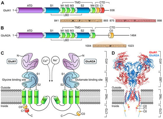
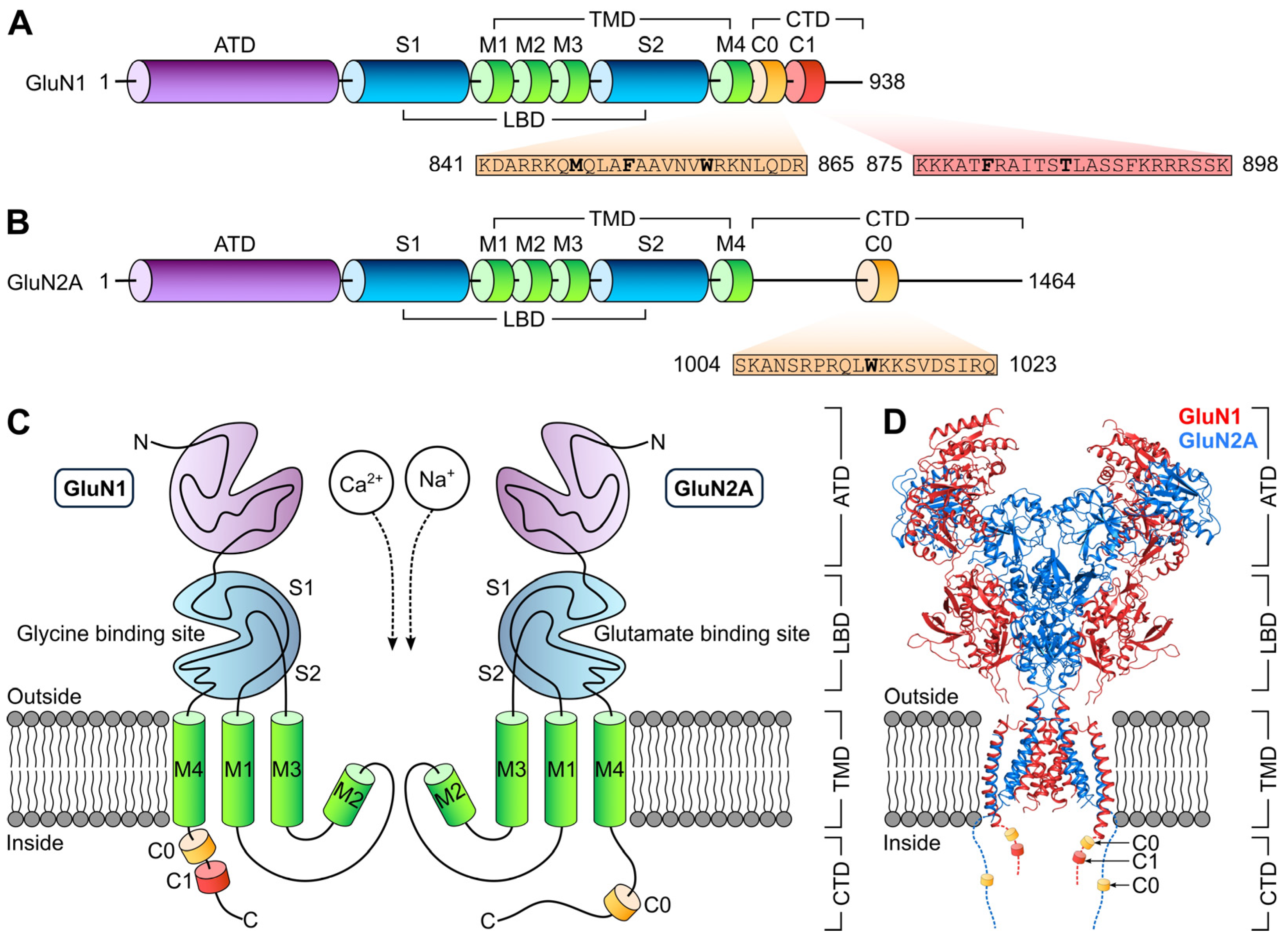
Domain organization in the NMDA receptor. Linear representations of (A) GluN1 and (B) GluN2A domain architecture. Each subunit consists of four domains: the extracellular amino-terminal domain (ATD) and ligand binding domain (LBD) where glycine and glutamate bind to the GluN1 and GluN2 subunits, respectively, the transmembrane domain (TMD), and the intracellular carboxy-terminal domain (CTD). The two CaM binding sites (C0 and C1) in GluN1 and the single CaM binding site (C0) in GluN2A are indicated by boxes. Critical residues involved in CaM interactions are highlighted in bold. (C) Schematic representation of the assembly and modular organization of the NMDA receptor. Dashed arrows indicate the influx of Ca2+ and Na+. (D) Cryo-EM structure of the NMDA receptor (PDB ID: 6IRA [15]). GluN1 and GluN2A subunits are colored red and blue, respectively. The CTD was truncated and not determined in the experimental structures. Intracellular CaM binding sites (GluN1-C0, GluN1-C1, and GluN2A-C0) are represented as cylinder and labeled.
NMDAR subunits contain a modular domain architecture (Figure 1A–C), including an extracellular amino-terminal domain (ATD, purple in Figure 1A–C), ligand binding domain (LBD, cyan in Figure 1A–C), a central transmembrane domain (TMD, green in Figure 1A–C), and an intracellular carboxy-terminal domain (CTD, yellow and red in Figure 1A–C) [16,17]. The LBD harbors binding sites for the obligatory co-agonists, glutamate and glycine. The TMD consists of three transmembrane helices (M1, M3, and M4) and a re-entry loop that includes a short M2 helix (Figure 1C). The CTD contains helical segments (called C0 and C1 in Figure 1A–C) that bind to CaM [12,18,19] and ACTN [20,21,22]. Activation of NMDARs upon binding of agonist glutamate and co-agonist glycine opens the channels and causes an increase in intracellular Ca2+ concentration, thus initiating a wide range of Ca2+-dependent downstream signaling pathways.
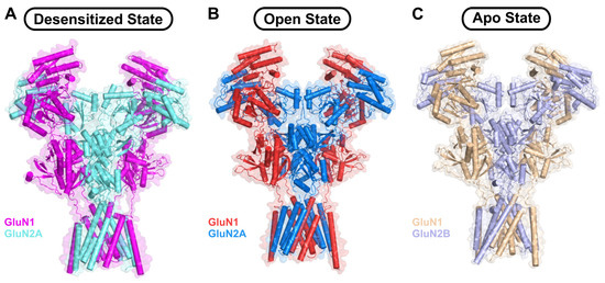
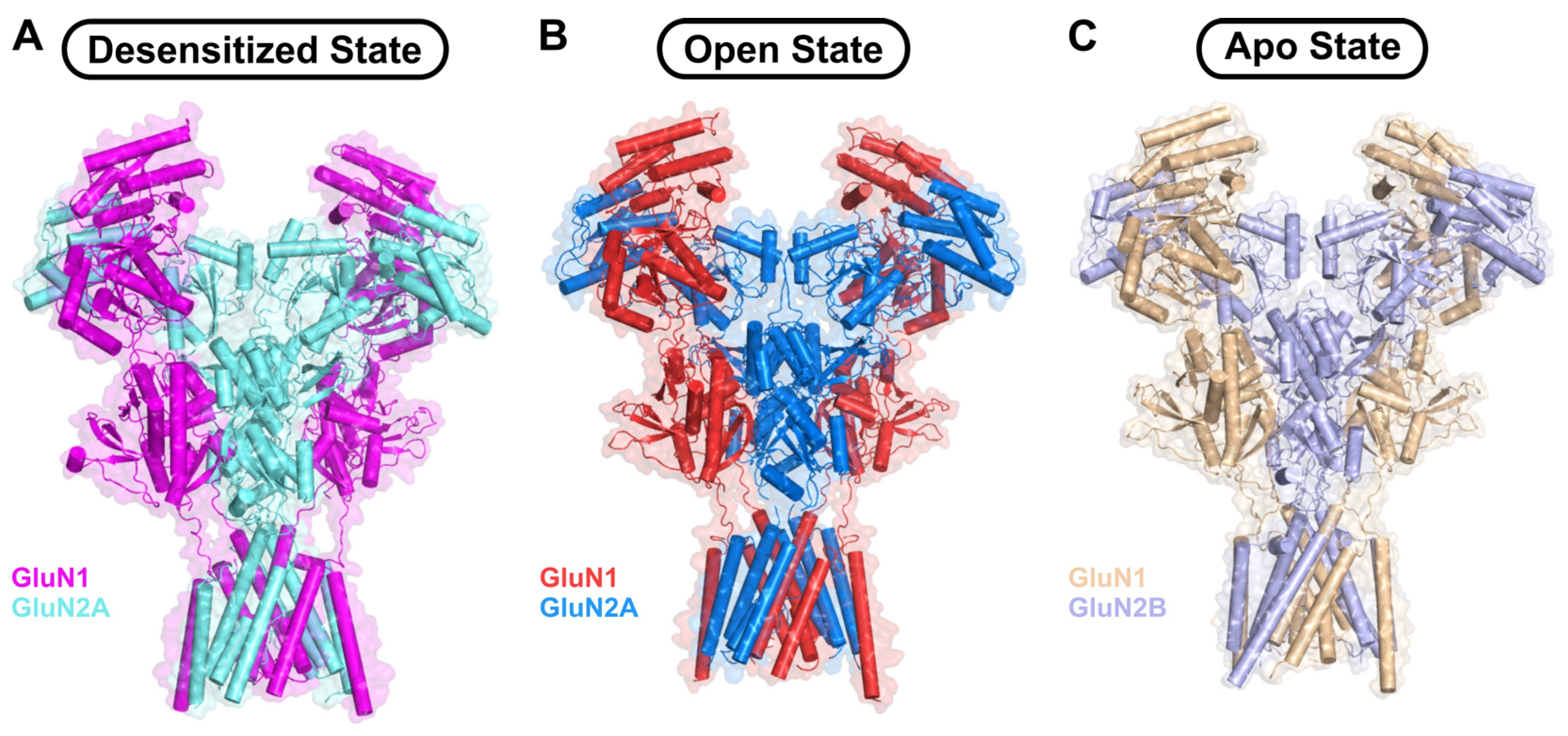
Three-dimensional structures of NMDARs (Figure 1D and Figure 2A–C) have been determined by x-ray crystallography [16,17,23,24] and cryo-electron microscopy (cryo-EM) [25,26,27,28,29]. The structures show detailed inter-subunit interactions between the ATD and LBD and between the LBD and TMD. A recent cryo-EM structure of the native GluN1-GluN2A NMDAR represents the heterotetrameric channel in a ligand-bound desensitized state (Figure 2A) in which glutamate and glycine are bound to the LBD and the channel pore is closed [28]. Other cryo-EM structures captured the agonist-bound NMDAR in the open state (Figure 2B) with the channel pore fully open [25,30]. A cryo-EM structure is also known for GluN1-GluN2B NMDAR in the apo state (Figure 2C), in which no ligand is bound to the LBD and the channel pore is closed [31]. The structure of the CTD is missing in all known structures, because the NMDAR samples in the structural studies either lacked the CTD or the CTD was dynamically disordered in the native NMDAR structures [28]. Therefore, the dynamical conformational changes in the CTD that underlie channel modulation by CaM and ACTN are not resolved by x-ray crystallography or cryo-EM. In this review, we use computational modeling to predict structural models of the NMDAR with a structured CTD bound to Ca2+-CaM or ACTN1 in the desensitized or open channel states, respectively (see Section 5).
Figure 2.
Structures of the NMDARs in different conformational states. Cryo-EM structures of GluN1-GluN2A NMDA receptor in the ligand-bound (A) desensitized state (PDB ID: 9UNN [28]), (B) open state (PDB ID: 7EOS [30]), and (C) GluN1-GluN2B NMDAR in apo conformation (PDB ID: 9ARG [31]).
2. Ca2+-Dependent Desensitization of NMDARs by CaM
The large and rapid Ca2+ influx through NMDARs can be cytotoxic if channel opening persists and is not regulated. Therefore, NMDARs are negatively regulated by intracellular Ca2+ elevation through a process known as either Ca2+-dependent inactivation (CDI) or as Ca2+-dependent desensitization (CDD), which is the term used in this review [32,33,34,35,36]. The term “desensitization” is more accurate than “inactivation” because the Ca2+-dependent inactivation of NMDAR activity is saturable and manifests as a fast-macroscopic desensitization [37]. CDD of NMDARs requires direct binding of CaM to the intracellular region of GluN1 [37,38] and GluN2A [12]. Previous studies revealed two intracellular CaM binding sites in GluN1: residues 841–865 (called GluN1-C0, Figure 1A,C,D) [38] and residues 875–898 (called GluN1-C1, Figure 1A,C,D) [19]. CaM binding to GluN1-C0 was demonstrated previously to be essential for CDD [33,38] and was suggested to promote dimerization of GluN1 [39]. By contrast, the GluN1-C1 site likely does not contribute to CDD [38] but rather it controls GluN1 binding to the cytoskeleton and its trafficking [19,40]. A single CaM binding site is also located in GluN2A (residues 1004–1023, called GluN2A-C0, Figure 1B–D) [18]. A crystal structure of CaM bound to the GluN1-C1 has been reported [19], along with two recent NMR structures of Ca2+-CaM bound separately to GluN1-C0 and GluN2A-C0 [12]. Mutations in NMDARs or CaM that cause defects in the Ca2+-dependent regulation of NMDAR channel function are associated with a spectrum of neurological diseases and neuropsychiatric disorders [41,42,43,44]. Elucidating NMDAR interactions with CaM may help improve our understanding of Ca2+-dependent channel regulation and provide a basis for treating neuronal diseases.
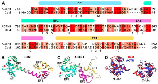
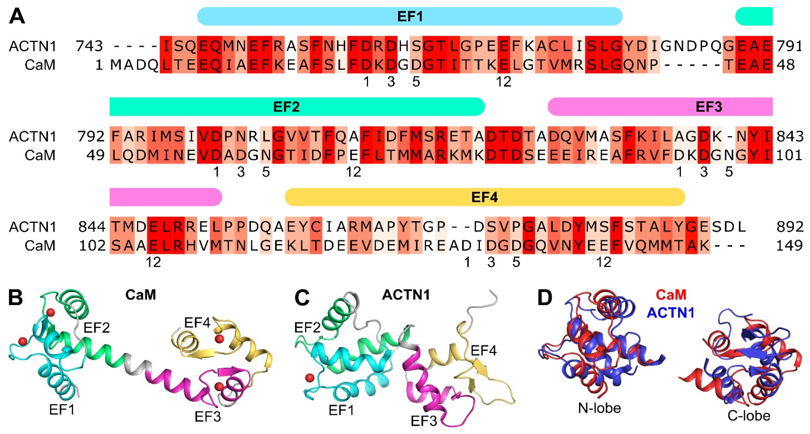
CaM is a 16.7 kDa Ca2+ sensor protein that contains four EF-hand Ca2+ binding motifs (EF1 in cyan, EF2 in green, EF3 in magenta, and EF4 in yellow; Figure 3A,B). The 12-residue Ca2+-binding loop in each EF-hand contains conserved acidic residues at loop positions 1, 3, 5 and 12 (labeled underneath the sequence in Figure 3A) whose carboxylate side chains chelate the bound Ca2+. The four EF-hands are grouped into two domains: EF1 and EF2 interact structurally to form the N-lobe (residues 5–77 in Figure 3B), whereas EF3 and EF4 combine to form the C-lobe (residues 82–102 in Figure 3B). The binding of Ca2+ to each EF-hand promotes a conformational change, which causes the exposure of a hydrophobic groove in each lobe that binds to hydrophobic sites (typically aliphatic helices) in target binding proteins [45]. The recent NMR structure of Ca2+-CaM bound to the GluN1-C0 peptide [12] reveals that both CaM lobes bind to opposite sides of the GluN1-C0 helix. Key hydrophobic residues in GluN1 (M848 and F852) structurally contact the hydrophobic groove in the CaM C-lobe, whereas GluN1 residues (V855 and W858) contact the hydrophobic groove in the CaM N-lobe. The NMR structure of Ca2+-CaM bound to the GluN2A-C0 peptide [12] reveals that only the CaM C-lobe binds to the GluN2A-C0 helix in which GluN2A residues (W1014 and V1018) structurally contact the hydrophobic groove in the CaM C-lobe. The GluN1 point mutations (F852E and W858E) and GluN2A mutation (W1014E) each disrupt electrophysiologically measured CDD [12], suggesting CaM interaction with these residues contributes to CDD.
Figure 3.
Sequence and structural comparison of CaM and ACTN1. (A) Amino acid sequence alignment of CaM and ACTN1. Sequence conservation is indicated by a red gradient, where dark red signifies highly conserved residues and light red represents areas of low conservation. Crystal structures of (B) Ca2+-CaM (PDB ID: 1CLL [46]) and (C) Ca2+-bound ACTN1 (PDB ID: 2N8Y [47]). The Ca2+ bound to EF1 in ACTN1 has a dissociation constant of ~40 μM [47] and is probably not physiologically relevant. The EF-hand motifs are shaded in color (EF1: cyan, EF2: green, EF3: magenta, and EF4: yellow). The four EF-hands are grouped into two domains: N-lobe (EF1 and EF2) and C-lobe (EF3 and EF4). Each EF-hand contains a 12-residue Ca2+-binding loop with conserved acidic residues marked at the 1, 3, 5, and 12 positions that chelate the bound Ca2+. Ca2+ ions are represented in red spheres. (D) Structural comparison of the N-lobe and C-lobe of CaM (red) and ACTN1 (blue).
The Ca2+-free form of CaM (called apo-CaM) has been suggested to bind to GluN1 [32,33] and may play a role in NMDAR channel modulation. Surprisingly, apo-CaM binding to the GluN1-C0 or GluN2A-C0 peptide could not be detected by NMR titrations or by isothermal titration calorimetry (ITC) [12]. Therefore, in this review, we prefer to not speculate about a functional role for apo-CaM binding to NMDAR and will not present a model of the basal state. The apo-CaM binding to the GluN1-C0 peptide detected by fluorescence polarization studies [32] might be an artifact of the attached fluorescent probe in these studies, because the GluN1-C0 peptide (without a fluorescent tag) did not exhibit any binding to apo-CaM in either the NMR or ITC studies. The lack of any detectable heat signal in the ITC titration with apo-CaM might indicate a zero-enthalpy change (ΔH = 0) for apo-CaM binding to the GluN1-C0 peptide rather than zero binding. The lack of any NMR spectral change in the NMR titration with apo-CaM might be explained if the GluN1-C0 peptide binding does not cause a detectable structural change in apo-CaM. Future studies on EF-hand mutants of CaM (called CaM12, CaM34 and CaM1234 as described by [48]) that abolish Ca2+ binding to the particular EF-hands are needed to more rigorously probe the functional effects of Ca2+ binding. In particular, the CaM1234 mutant (that disables Ca2+ binding to all four EF-hands) should be studied to assess whether apo-CaM can pre-associate with NMDAR under basal conditions and whether apo-CaM binding is physiologically relevant. Alternatively, a half-calcified form of CaM (Ca2+ bound to EF3 and EF4 and not bound to EF1 and EF2) might pre-associate with NMDAR under resting Ca2+ conditions ([Ca2+] = 100 nM). Since the Ca2+-bound CaM C-lobe binds to the GluN1-C0 peptide with a dissociation constant of 100 nM [12], this implies that the CaM C-lobe bound to the C0 peptide should have an apparent Ca2+ affinity in the nanomolar range, suggesting that a significant fraction of the CaM C-lobe bound to NMDAR may have Ca2+ bound under basal conditions. This is analogous to the recent finding that L-type voltage-gated Ca2+ channels (CaV1.2) are likely pre-associated with half-calcified CaM rather than apo-CaM [48].
3. GluN2 Subtype-Specific Ca2+-Dependent Desensitization of NMDARs
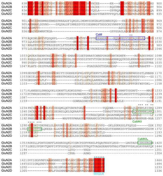
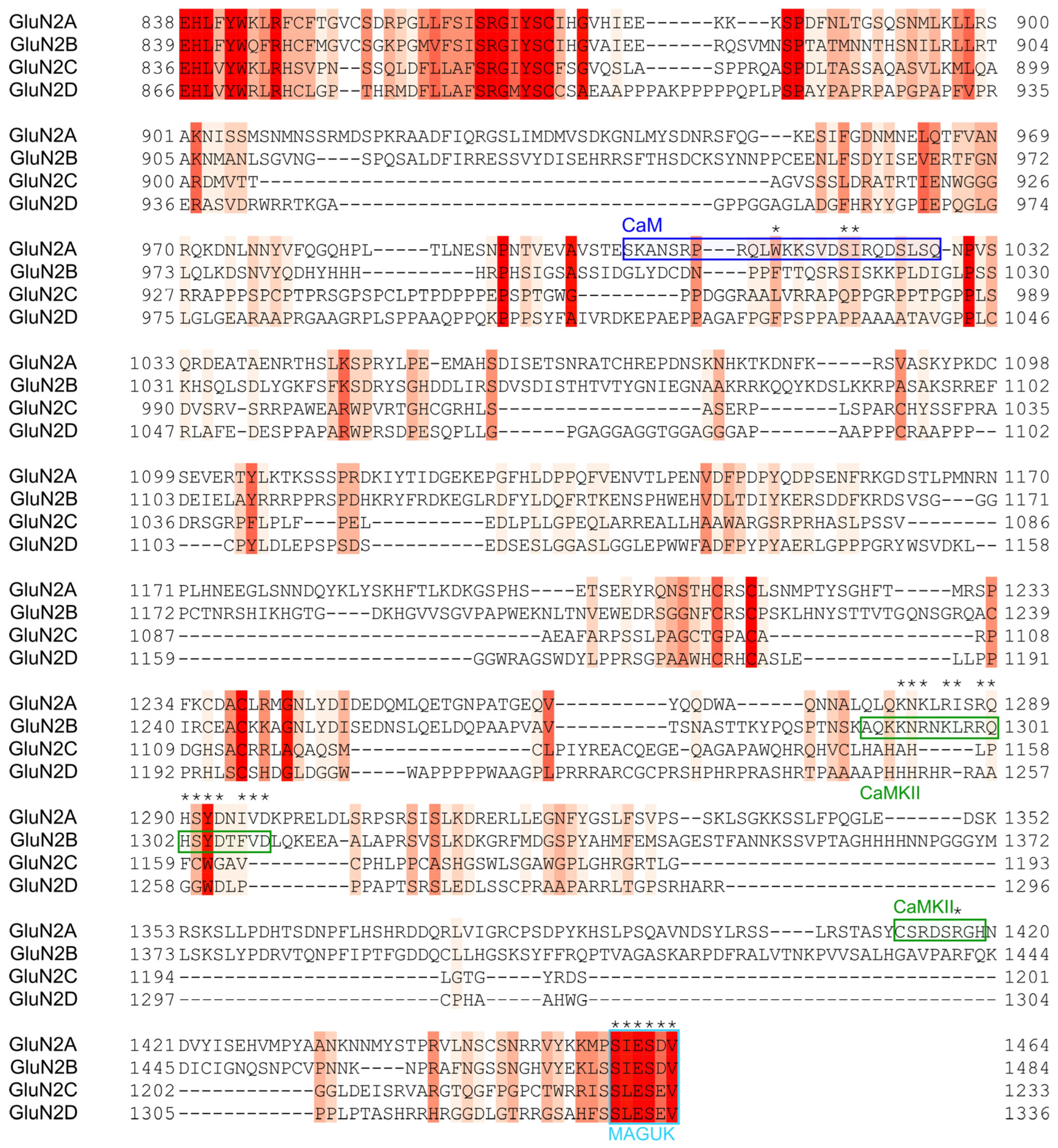
The magnitude of CDD in NMDARs differs markedly among NMDARs containing distinct GluN2 subtypes [34]. GluN2A-containing receptors (NMDAR heterotetramer with two GluN1 and two GluN2A, called GluN1/2A) exhibit the most pronounced desensitization, whereas GluN1/2B displays intermediate desensitization and GluN1/2C and GluN1/2D exhibit the weakest desensitization [49,50]. These differences are thought to arise from subtype-specific variations in channel gating, open probability, and allosteric coupling that govern receptor activation and desensitization. Although the four GluN2 subtypes differ in length, sequence alignments reveal that the ATD, LBD, and TMD are highly conserved (>50% sequence identity), in contrast to CTD (<35% identity). Sequence alignment of the human GluN2A–D CTDs further reveals that GluN2A and GluN2B share approximately 34% sequence identity, whereas GluN2C and GluN2D share approximately 30% sequence identity, highlighting the substantial sequence divergence within this regulatory domain (Figure 4). This divergence in the GluN2 CTDs enables interactions with numerous regulatory proteins, including CaM, Ca2+-CaM-dependent protein kinase II (CaMKII), protein kinase A (PKA), protein kinase C (PKC), casein kinase (CK), protein tyrosine kinases (Fyn and Src), and membrane-associated guanylate kinases (MAGUKs), and these interactions play critical roles in regulating NMDAR function [51]. Although multiple mechanisms contribute to subtype-specific gating and desensitization, this review focuses on the CaM-dependent CDD in NMDARs containing different GluN2 subtypes A–D. While the primary Ca2+-CaM binding site responsible for CDD is located in the C0 region of the GluN1 and GluN2A subunits [12], no well-established, functional CaM binding site has been identified in GluN2B, GluN2C, or GluN2D. Nevertheless, sequence alignment of the GluN2 CTD reveals that the GluN2A C0 region shares moderate sequence similarity with GluN2B (Figure 4). Notably, three critical CaM-binding residues identified in GluN2A (W1014, S1020, and I1021) are conserved in GluN2B (Figure 4, blue box, asterisks). This sequence conservation suggests a potential CaM-binding site in GluN2B that may contribute to the CDD observed in GluN1/2B [50]. Future experimental validation is required to determine whether CaM binding to this putative CaM-binding site in GluN2B is essential for CDD observed for GluN1/2B. In contrast, GluN2C and GluN2D exhibit low sequence conservation in the CaM-binding region and are predicted to not bind to CaM. In particular, the critical CaM-binding residues in GluN2A (W1014, S1020, and I1021) are not conserved in GluN2C and GluN2D. We propose that the relatively weak CDD observed in GluN1/2C and GluN1/2D may be caused by a lack of CaM binding to either GluN2C or GluN2D.
Figure 4.
Sequence alignment of human GluN2A-D CTD. Sequence conservation is shown as a red color gradient, with dark red representing highly conserved residues and light red indicating low sequence conservation. Key interaction sites are highlighted with colored boxes: the CaM-binding site is shown in blue, the CaMKII-binding site in green, and the MAGUK-binding site in cyan. Conserved residues within the binding sites are highlighted with asterisks.
4. NMDAR Channel Activation by ACTN1
The cytoskeletal proteins, ACTN1 and ACTN2 (100 kDa), both contain an N-terminal actin binding domain, four spectrin coiled-coil repeats, and C-terminal EF-hand domain [52]. The EF-hand domain of ACTN1 (Figure 3A,C) was shown previously to bind to GluN1-C0 [20,22]. Also, ACTN2 binding to NMDAR has been suggested to increase channel open probability [21,53]. The EF-hand domain in ACTN1 has mutations to conserved acidic residues in the EF-hand Ca2+-binding loops (see S763 in EF1, L804 and A811 in EF2, A837 in EF3, and S872, P874, and S881 in EF4; see Figure 3A), which prevent Ca2+ binding in the physiological range. Therefore, ACTN1 binds weakly to Ca2+ in the high micromolar range and is thought to not function as a physiological Ca2+ sensor [47,54,55,56]. Previous studies have shown that, under Ca2+-free conditions, ACTN1 and ACTN2 both compete with CaM for binding to GluN1-C0 and prevent NMDAR channel desensitization (see Displacement Model below), whereas elevated intracellular Ca2+ levels promote CaM binding, which displaces ACTN from the same binding site [22,57]. A similar competitive binding mechanism has been observed in the CaV1.2 L-type voltage-gated Ca2+ channel, where the Ca2+-free EF-hand domain of ACTN1 competes with CaM for binding to the IQ motif, and ACTN1 binding to the IQ causes an increase in CaV1.2 channel open probability [55]. In this review, we suggest that the Ca2+-free EF-hand domain of ACTN1 binding to GluN1-C0 may stabilize the NMDAR channel in the ligand-bound open state at low cytosolic Ca2+ levels, in contrast to Ca2+-CaM binding to GluN1-C0 and GluN2A-C0 that stabilizes NMDAR in the ligand-bound desensitized state [12]. In essence, we suggest that ACTN1 and CaM work together to cause Ca2+-dependent NMDAR channel regulation: ACTN1 binding to NMDAR causes channel activation at low Ca2+ levels in contrast to Ca2+-CaM binding that displaces ACTN1 and causes channel desensitization at high cytosolic Ca2+ levels.
In this review, we focused on the structural modeling of ACTN1 binding to NMDAR, because a recent NMR study has characterized the structure of ACTN1 bound to the GluN1-C0 peptide [20]. Also, a crystal structure is known for ACTN1 [52], and NMR structures are known for the metal-free EF-hand domain of ACTN1 [20,47,55] and ACTN2 [54]. Importantly, the affinity of the GluN1-C0 peptide is tenfold higher for ACTN1 compared to ACTN2 [22]. The ACTN1 EF-hand region is comprised of separate N-lobe and C-lobe domains that are structurally similar to those of CaM (Figure 3D). The ACTN1 C-lobe was shown to interact structurally with the IQ motif in CaV1.2 [55] and GluN1-C0 [20]. These ACTN1 structures will be combined with the recent cryo-EM structure of NMDAR in the open channel state [28] to construct a structural model of ACTN1 bound to NMDAR as described below. We believe that ACTN1 and ACTN2 are structurally similar based on the high sequence similarity for the residues that contact GluN1-C0. However, it is important to point out that the physiology of NMDAR regulation is better known for ACTN2 rather than ACTN1, and it is possible that ACTN1 and ACTN2 may have different roles in regulating channel function. Future electrophysiology studies are needed to better characterize NMDAR channel regulation by ACTN1.
5. Displacement Mechanism of NMDAR Channel Modulation
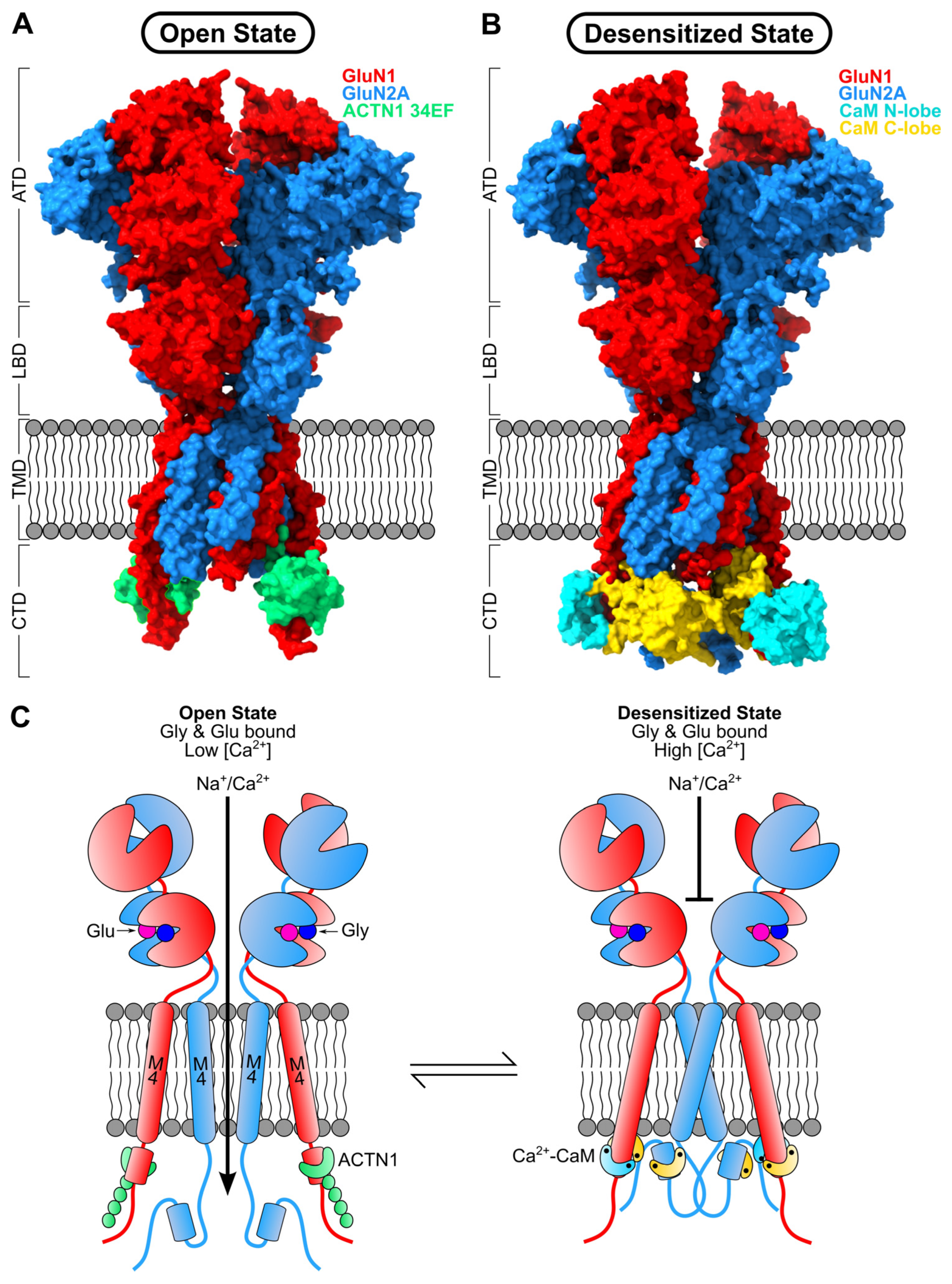
We propose a “displacement” mechanism for CDD in which ACTN1 binding to GluN1-C0 (in the open channel state) is displaced by Ca2+-CaM binding, which promotes channel desensitization (Figure 5). At low cytosolic Ca2+ levels, we suggest that ACTN1 binding to GluN1-C0 stabilizes the channel open state. By contrast, at high Ca2+ levels, Ca2+-CaM displaces ACTN1 and binds to both GluN1-C0 and GluN2A-C0 to stabilize the desensitized channel state. A structural model of the NMDAR desensitized channel tetramer bound to Ca2+-CaM was constructed using AlphaFold3 [58] (Figure 5B,C right panel). In this model, each GluN1 M4 helix extends continuously into the C0 helix, thus forming one long continuous helix that connects M4 and C0. The NMR structure of Ca2+-CaM bound to GluN1-C0 [12] was superimposed onto each GluN1 C0 helix in the tetramer model. NMR structures of Ca2+-CaM bound to GluN2A-C0 [12] were manually docked into the NMDAR tetramer (Figure 5B). Our model of the desensitized channel tetramer suggests that Ca2+-CaM binds to helical structures in GluN1-C0 and GluN2A-C0 that come together to form a concentric ring-like structure located underneath the channel pore, which may serve to allosterically stabilize the desensitized channel and facilitate closure of the pore (see cyan and yellow region in Figure 5B). At low Ca2+ levels, the competitive binding of ACTN1 displaces CaM from GluN1-C0, and Ca2+-free CaM dissociates from GluN2A-C0 (Figure 5A). This removal of CaM from NMDAR and subsequent binding of ACTN1 is predicted to destabilize the ring-like concentric arrangement of the GluN1-C0 and GluN2A-C0 helices underneath the channel pore. The Ca2+-sensitive dismantling of the concentric complex of GluN1-C0 and GluN2A-C0 is suggested here to facilitate a conformational change in the M4 helix of both GluN1 and GluN2A that promotes channel opening at low Ca2+ levels (see black arrow in Figure 5C). A structural model of the NMDAR open channel tetramer bound to the ACTN1 C-lobe (Figure 5A,C left panel) was generated using AlphaFold3 [58]. Our model of the channel open state suggests that the C-lobe of ACTN1 binds to the helical structure of GluN1-C0 as suggested recently by [20], and ACTN1 is not bound to GluN2A-C0. As a result, GluN2A-C0 becomes unstructured and no longer interacts with GluN1-C0, which dissociates the concentric complex of GluN1-C0/GluN2A-C0 and facilitates channel opening (Figure 5C). ACTN1 also serves to bridge NMDARs to the cytoskeleton, which enhances the cell surface expression of NMDARs [22,59]. This bridging of NMDARs to the cytoskeleton is mediated by ACTN1 in which its N-terminal domain (actin binding domain) connects to the actin cytoskeleton, while the other end of ACTN1 (C-terminal EF-hand region) binds to GluN1. ACTN1 also bridges CaV1.2 to the cytoskeleton and enhances the CaV1.2 cell surface expression in the brain and heart [55]. Future studies are needed to experimentally determine atomic-level structures of the NMDAR bound to both Ca2+-CaM and ACTN1 to more rigorously test the Ca2+-dependent conformational changes predicted by our model (Figure 5C).
Our model suggests that Ca2+ binding to CaM is essential for promoting CDD (Figure 5C). Thus, mutations in CaM that weaken or disable Ca2+ binding are expected to disrupt CDD, which could enable unregulated Ca2+ influx and possibly cause neuronal hyperexcitability. Calmodulinopathies are a class of diseases in which de novo mutations in CaM (D94A, D96G, N98S, E105A, D130A, D132G, D134H, and E141G) impair Ca2+ binding to the third or fourth EF-hands and promote fatal cardiac arrhythmias caused by impaired Ca2+-dependent inactivation of various Ca2+ channels (ryanodine receptor 2, RyR2 [60,61] and CaV1.2 [62]). In particular, CaM mutations that disrupt inactivation of RyR2 cause a phenotype known as catecholaminergic polymorphic ventricular tachycardia (CPVT) [63], whereas the CaM mutations that disrupt CDI of CaV1.2 cause a distinct phenotype known as long-QT syndrome (LQTS) [64]. Calmodulinopathies also cause epileptic seizures [65,66] and cognitive deficits [67]. The role of NMDARs in neuronal excitability implies that dysregulation of CDD may contribute to these pathologies. Indeed, weakened CaM binding to GluN1 has been observed in epileptic disorders [68]. Future studies are needed to explore whether any of the known CaM mutations (D94A, D96G, N98S, E105A, D130A, D132G, D134H, and E141G) might also disrupt CDD in NMDARs and whether this dysregulation of NMDARs might contribute to calmodulinopathies.
Our model suggests how CaM binding to NMDAR (Figure 5C, right panel) might control the sensitivity of the NMDAR channel blocker, memantine, which is clinically approved for treating Alzheimer’s Disease [69] and shows promise for treating Parkinson’s Disease [70]. A recent study revealed that inhibition of NMDARs by memantine increases with increasing intracellular Ca2+ concentration [71], and a separate study demonstrated that memantine binds to NMDAR (GluN1/2A) with about 3-fold higher affinity when the intracellular Ca2+ concentration is raised to ~5.0 μM in the presence of overexpressed and recombinant wildtype CaM but not in the presence of CaM1234 [72]. These results indicate that Ca2+-CaM binding to NMDAR is essential for achieving its high-affinity binding to memantine. The results also suggest that memantine selectively binds to the desensitized channel state bound to Ca2+-CaM (Figure 5C, right panel). We suggest that Ca2+-CaM binding to C0 may allosterically stabilize the memantine binding site in the channel pore as observed for GluN1/2B [73]. The preferential binding of memantine to the desensitized channel bound to Ca2+-CaM selectively inhibits a subpopulation of NMDARs that promote excitotoxicity, which may explain why memantine has such high clinical safety [71]. Future studies are needed to determine the cryo-EM structure of GluN1/2A bound to memantine and to elucidate how memantine can selectively stabilize the desensitized channel bound to Ca2+-CaM.
Figure 5.
Hypothetical structural mechanism of NMDAR channel regulation by ACTN1 and CaM. (A) Surface representation of the NMDAR tetramer model (GluN1 in red and GluN2A in blue) in the open channel state bound to two ACTN1 molecules (green). This model was constructed using AlphaFold3 [58] and contained two GluN1 subunits (residues K25–R865), two GluN2A subunits (residues L34–Y842), and two ACTN1 C-lobes (residues D822–L892), followed by energy minimization under vacuum conditions using MD simulations as described previously [74]. (B) Surface representation of the NMDAR tetramer model (GluN1 in red and GluN2A in blue) in the desensitized channel state bound to four Ca2+-CaM molecules (N-lobe in cyan and C-lobe in yellow). The CaM binding stoichiometry of 4 CaM bound per tetramer is assumed in the modeling and is based on previous binding studies [12] that determined each C0 peptide binds to one CaM molecule, which suggests that 4 CaM bind per tetramer because each tetramer (GluN1/2A) has four C0 sites. The structural model of the NMDAR tetramer featuring structured C0 sites was generated using AlphaFold3 [58]. The details of the modeling methods were described recently by [12]. The tetramer model contains two GluN1 subunits (residues K25–R865) and two GluN2A subunits (residues L34–Q1023). NMR structures of Ca2+-CaM bound to GluN1-C0 were superimposed onto the GluN1-C0 sites within the NMDAR tetramer model. NMR structures of the Ca2+-CaM C-lobe bound to GluN2A-C0 were manually docked into the NMDAR tetramer. The NMDAR–CaM complex was energy-minimized using molecular dynamics (MD) simulations. Atomic coordinates of the structural models will be made available upon request. (C) Schematic summary of conformational changes of NMDAR that occur during the transition from the agonist-bound open channel state (left panel) to the Ca2+-induced and agonist-bound desensitized state (right panel). Under low Ca2+ concentrations, GluN1-C0 is anchored to ACTN1 C-lobe (green), which increases the channel open probability of the NMDAR. The vertical arrow indicates Ca2+ influx through the open channel. Under high Ca2+ levels, four Ca2+-CaM molecules (N-lobe in cyan; C-lobe in yellow) bind to the C0 domains of GluN1 and GluN2A, leading to channel desensitization. The vertical bar indicates the channel pore is closed. Bound agonists (Glu and Gly) are represented as magenta and blue spheres.
6. Conclusions
NMDARs are Ca2+-permeable ionotropic glutamate receptors that serve as important Ca2+ channels in the brain and have critical roles in neural development, synaptic plasticity, learning, and memory. Channel opening causes Ca2+ influx that activates a range of Ca2+-dependent cellular processes. NMDAR channel activity is finely regulated by α-actinin, which promotes channel opening, and by CaM, which mediates Ca2+-dependent channel desensitization (CDD). We propose a “displacement” mechanism for CDD in which ACTN1 binding to NMDAR stabilizes the channel open state under basal conditions (at low cytosolic Ca2+ levels). At high Ca2+ levels (following neuronal stimulation), ACTN1 is displaced by Ca2+-CaM binding to NMDAR, which stabilizes the desensitized channel state. Our model (Figure 5C) suggests that Ca2+-CaM binding to NMDAR promotes the formation of helical structures in GluN1-C0 and GluN2A-C0 that come together to form a concentric ring-like complex located underneath the channel pore that may serve to allosterically stabilize the desensitized channel and facilitate closure of the pore.
Author Contributions
Conceptualization, A.B. and J.B.A.; methodology, A.B.; software, A.B.; validation, A.B.; formal analysis, A.B.; writing—original draft preparation, J.B.A.; writing—review and editing, A.B., J.W.H. and J.B.A.; visualization, A.B.; supervision, J.B.A.; project administration, J.B.A.; funding acquisition, J.B.A. and J.W.H. All authors have read and agreed to the published version of the manuscript.
Funding
This research was funded by the National Institutes of Health, grant numbers R01-EY012347 to J.B.A. and R01-AG055357 to J.W.H.
Institutional Review Board Statement
Not applicable.
Informed Consent Statement
Not applicable.
Data Availability Statement
Atomic coordinates of the structures in Figure 2 and Figure 3 are openly available in the Protein Data Bank (https://www.rcsb.org). Atomic coordinates of the structural models in Figure 5 will be made available from the authors upon request.
Conflicts of Interest
The authors declare no conflicts of interest. The funders had no role in the design of the study; in the collection, analyses, or interpretation of data; in the writing of the manuscript; or in the decision to publish the results.
Abbreviations
The following abbreviations are used in this manuscript:
| ACTN | α-actinin |
| ACTN1 | α-actinin-1 |
| ACTN2 | α-actinin-2 |
| CaM | calmodulin |
| CDD | Ca2+-dependent desensitization |
| ATD | Amino-terminal domain |
| LBD | Ligand binding domain |
| TMD | Transmembrane domain |
| CTD | Carboxy-terminal domain |
| NMDAR | N-methyl D-aspartate receptor |
References
- Bliss, J.V.P.; Collingridge, G.L. A synaptic model of memory: Long-term potentiation in the hippocampus. Nature 1993, 361, 31–39. [Google Scholar] [CrossRef] [Scilit] [PubMed]
- Chittajallu, R.; Alford, S.; Collingridge, G.L. Ca2+ and synaptic plasticity. Cell Calcium 1998, 24, 377–385. [Google Scholar] [CrossRef] [Scilit] [PubMed]
- Iacobucci, G.J.; Popescu, G.K. NMDA receptors: Linking physiological output to biophysical operation. Nat. Rev. Neurosci. 2017, 18, 236–249. [Google Scholar] [CrossRef] [Scilit] [PubMed]
- Paoletti, P.; Bellone, C.; Zhou, Q. NMDA receptor subunit diversity: Impact on receptor properties, synaptic plasticity and disease. Nat. Rev. Neurosci. 2013, 14, 383–400. [Google Scholar] [CrossRef] [Scilit] [PubMed]
- Tang, Y.P.; Shimizu, E.; Dube, G.R.; Rampon, C.; Kerchner, G.A.; Zhuo, M.; Liu, G.; Tsien, J.Z. Genetic enhancement of learning and memory in mice. Nature 1999, 401, 63–69. [Google Scholar] [CrossRef] [Scilit] [PubMed]
- Zucker, R.S. Calcium and activity-dependent synaptic plasticity. Curr. Opin. Neurobiol. 1999, 9, 305–313. [Google Scholar] [CrossRef] [Scilit] [PubMed]
- Dupuis, J.P.; Nicole, O.; Groc, L. nMdA receptor functions in health and disease: Old actor, new dimensions. Neuron 2023, 111, 2312–2348. [Google Scholar] [CrossRef] [Scilit] [PubMed]
- Herbrechter, R.; Hube, N.; Reiner, A. Splicing and editing of ionotropic glutamate receptors: A comprehensive analysis based on human RNA-seq data. Cell Mol. Life Sci. 2021, 78, 5605–5630. [Google Scholar] [CrossRef] [Scilit] [PubMed]
- Cull-Candy, S.G.; Leszkiewicz, D.N. Role of distinct NMDA receptor subtypes at central synapses. Sci. STKE 2004, 2004, 16. [Google Scholar] [CrossRef] [Scilit] [PubMed]
- Benveniste, M.; Mayer, M.L. Kinetic analysis of antagonist action at N-methyl-D-aspartic acid receptors. Two binding sites each for glutamate and glycine. Biophys. J. 1991, 59, 560–573. [Google Scholar] [CrossRef] [Scilit] [PubMed]
- Monyer, H.; Sprengel, R.; Schoepfer, R.; Herb, A.; Higuchi, M.; Lomeli, H.; Burnashev, N.; Sakmann, B.; Seeburg, P.H. Heteromeric NMDA Receptors: Molecular and Functional Distinction of Subtypes. Science 1992, 256, 1217–1221. [Google Scholar] [CrossRef] [Scilit] [PubMed]
- Bej, A.; Erickson-Oberg, M.Q.; Nigam, A.; Yu, I.; Hell, J.W.; Johnson, J.W.; Ames, J.B. Structural basis and functional analysis of NMDA receptor regulation by calmodulin. J. Biol. Chem. 2026, 302, 111131. [Google Scholar] [CrossRef] [Scilit] [PubMed]
- Frank, R.A.; Grant, S.G. Supramolecular organization of NMDA receptors and the postsynaptic density. Curr. Opin. Neurobiol. 2017, 45, 139–147. [Google Scholar] [CrossRef] [Scilit] [PubMed]
- Gardoni, F.; Di Luca, M. Protein-protein interactions at the NMDA receptor complex: From synaptic retention to synaptonuclear protein messengers. Neuropharmacology 2021, 190, 108551. [Google Scholar] [CrossRef] [Scilit] [PubMed]
- Zhang, J.B.; Chang, S.L.; Xu, P.; Miao, M.; Wu, H.; Zhang, Y.; Zhang, T.; Wang, H.; Zhang, J.; Xie, C.; et al. Structural Basis of the Proton Sensitivity of Human GluN1-GluN2A NMDA Receptors. Cell Rep. 2018, 25, 3582–3590. [Google Scholar] [CrossRef] [Scilit] [PubMed]
- Karakas, E.; Furukawa, H. Crystal structure of a heterotetrameric NMDA receptor ion channel. Science 2014, 344, 992–997. [Google Scholar] [CrossRef] [Scilit] [PubMed]
- Lee, C.H.; Lu, W.; Michel, J.C.; Goehring, A.; Du, J.; Song, X.; Gouaux, E. NMDA receptor structures reveal subunit arrangement and pore architecture. Nature 2014, 511, 191–197. [Google Scholar] [CrossRef] [Scilit] [PubMed]
- Bajaj, G.; Hau, A.M.; Hsu, P.; Gafken, P.R.; Schimerlik, M.I.; Ishmael, J.E. Identification of an atypical calcium-dependent calmodulin binding site on the C-terminal domain of GluN2A. Biochem. Biophys. Res. Commun. 2014, 444, 588–594. [Google Scholar] [CrossRef] [Scilit] [PubMed]
- Ataman, Z.A.; Gakhar, L.; Sorensen, B.R.; Hell, J.W.; Shea, M.A. The NMDA receptor NR1 C1 region bound to calmodulin: Structural insights into functional differences between homologous domains. Structure 2007, 15, 1603–1617. [Google Scholar] [CrossRef] [Scilit] [PubMed]
- Bej, A.; Hell, J.W.; Ames, J.B. Chemical shift assignments of the alpha-actinin C-terminal EF-hand domain bound to a cytosolic C0 domain of GluN1 (residues 841–865) from the NMDA receptor. Biomol. NMR Assign. 2024, 18, 239–244. [Google Scholar] [CrossRef] [Scilit] [PubMed]
- Krupp, J.J.; Vissel, B.; Thomas, C.G.; Heinemann, S.F.; Westbrook, G.L. Interactions of calmodulin and alpha-actinin with the NR1 subunit modulate Ca2+-dependent inactivation of NMDA receptors. J. Neurosci. 1999, 19, 1165–1178. [Google Scholar] [CrossRef] [Scilit] [PubMed]
- Merrill, M.A.; Malik, Z.; Akyol, Z.; Bartos, J.A.; Leonard, A.S.; Hudmon, A.; Shea, M.A.; Hell, J.W. Displacement of alpha-actinin from the NMDA receptor NR1 C0 domain By Ca2+/calmodulin promotes CaMKII binding. Biochemistry 2007, 46, 8485–8497. [Google Scholar] [CrossRef] [Scilit] [PubMed]
- Yao, Y.; Belcher, J.; Berger, A.J.; Mayer, M.L.; Lau, A.Y. Conformational Analysis of NMDA Receptor GluN1, GluN2, and GluN3 Ligand-Binding Domains Reveals Subtype-Specific Characteristics. Structure 2013, 21, 1788–1799. [Google Scholar] [CrossRef] [Scilit] [PubMed]
- Tajima, N.; Karakas, E.; Grant, T.; Simorowski, N.; Diaz-Avalos, R.; Grigorieff, N.; Furukawa, H. Activation of NMDA receptors and the mechanism of inhibition by ifenprodil. Nature 2016, 534, 63–68. [Google Scholar] [CrossRef] [Scilit] [PubMed]
- Abbott, J.A.; Kim, J.; Liu, B.; Popescu, G.K.; Gouaux, E.; Jalali, F. Cryo-EM snapshots of NMDA receptor activation illuminate sequential rearrangements. Sci. Adv. 2025, 11, eadx4647. [Google Scholar] [CrossRef] [Scilit] [PubMed]
- Chou, T.H.; Tajima, N.; Romero-Hernandez, A.; Furukawa, H. Structural Basis of Functional Transitions in Mammalian NMDA Receptors. Cell 2020, 182, 357–371.e13. [Google Scholar] [CrossRef] [Scilit] [PubMed]
- Kim, J.; Jalali, F.; Jones, B.E.; Westbrook, G.L.; Gouaux, E. Cryo-EM of autoantibody-bound NMDA receptors reveals antigenic hotspots in an active immunization model of anti-NMDAR encephalitis. Sci. Adv. 2026, 12, 4249. [Google Scholar] [CrossRef] [Scilit] [PubMed]
- Xu, R.; Jiang, Q.; Xu, H.; Zhang, L.; Hu, X.; Lu, Z.; Deng, H.; Xiong, H.; Zhang, S.; Chen, Z.; et al. Conformational diversity and fully opening mechanism of native NMDA receptor. Nature 2026, 652, 1405–1414. [Google Scholar] [CrossRef] [Scilit] [PubMed]
- Zhang, M.; Feng, J.; Xie, C.; Song, N.; Jin, C.; Wang, J.; Zhao, Q.; Zhang, L.; Wang, B.; Sun, Y.; et al. Assembly and architecture of endogenous NMDA receptors in adult cerebral cortex and hippocampus. Cell 2025, 188, 1198–1207.e13. [Google Scholar] [CrossRef] [Scilit] [PubMed]
- Wang, H.; Lv, S.; Stroebel, D.; Zhang, J.; Pan, Y.; Huang, X.; Zhang, X.; Paoletti, P.; Zhu, S. Gating mechanism and a modulatory niche of human GluN1-GluN2A NMDA receptors. Neuron 2021, 109, 2443–2456.e5. [Google Scholar] [CrossRef] [Scilit] [PubMed]
- Chou, T.H.; Epstein, M.; Fritzemeier, R.G.; Akins, N.S.; Paladugu, S.; Ullman, E.Z.; Liotta, D.C.; Traynelis, S.F.; Furukawa, H. Molecular mechanism of ligand gating and opening of NMDA receptor. Nature 2024, 632, 209–217. [Google Scholar] [CrossRef] [Scilit] [PubMed]
- Akyol, Z.; Bartos, J.A.; Merrill, M.A.; Faga, L.A.; Jaren, O.R.; Shea, M.A.; Hell, J.W. Apo-calmodulin binds with its C-terminal domain to the N-methyl-D-aspartate receptor NR1 C0 region. J. Biol. Chem. 2004, 279, 2166–2175. [Google Scholar] [CrossRef] [Scilit] [PubMed]
- Ehlers, M.D.; Zhang, S.; Bernhadt, J.P.; Huganir, R.L. Inactivation of NMDA receptors by direct interaction of calmodulin with the NR1 subunit. Cell 1996, 84, 745–755. [Google Scholar] [CrossRef] [Scilit] [PubMed]
- Krupp, J.J.; Vissel, B.; Heinemann, S.F.; Westbrook, G.L. Calcium-dependent inactivation of recombinant N-methyl-D-aspartate receptors is NR2 subunit specific. Mol. Pharmacol. 1996, 50, 1680–1688. [Google Scholar] [CrossRef] [Scilit]
- Rosenmund, C.; Feltz, A.; Westbrook, G.L. Calcium-dependent inactivation of synaptic NMDA receptors in hippocampal neurons. J. Neurophysiol. 1995, 73, 427–430. [Google Scholar] [CrossRef] [Scilit] [PubMed]
- Umemiya, M.; Chen, N.; Raymond, L.A.; Murphy, T.H. A Calcium-Dependent Feedback Mechanism Participates in Shaping Single NMDA Miniature EPSCs. J. Neurosci. 2001, 21, 1–9. [Google Scholar] [CrossRef] [Scilit] [PubMed]
- Iacobucci, G.J.; Popescu, G.K. Resident Calmodulin Primes NMDA Receptors for Ca2+-Dependent Inactivation. Biophys. J. 2017, 113, 2236–2248. [Google Scholar] [CrossRef] [Scilit] [PubMed]
- Zhang, S.; Ehlers, M.D.; Bernhardt, J.P.; Su, C.-T.; Huganir, R.L. Calmodulin Mediates Calcium-Dependent Inactivation of N-Methyl-D-Aspartate Receptors. Neuron 1998, 21, 443–453. [Google Scholar] [CrossRef] [Scilit] [PubMed]
- Wang, C.; Wang, H.G.; Xie, H.; Pitt, G.S. Ca2+/CaM controls Ca2+-dependent inactivation of NMDA receptors by dimerizing the NR1 C termini. J. Neurosci. 2008, 28, 1865–1870. [Google Scholar] [CrossRef] [Scilit] [PubMed]
- Ehlers, M.D.; Tingley, W.G.; Huganir, R.L. Regulated Subcellular Distribution of the NR1 Subunit of the NMDA Receptor. Science 1995, 269, 1734–1737. [Google Scholar] [CrossRef] [Scilit] [PubMed]
- Balu, D.T. The NMDA Receptor and Schizophrenia: From Pathophysiology to Treatment. Neuropsychopharmacology 2016, 76, 351–382. [Google Scholar]
- Bourgeron, T. From the genetic architecture to synaptic plasticity in autism spectrum disorder. Nat. Rev. Neurosci. 2015, 16, 551–563. [Google Scholar] [CrossRef] [Scilit] [PubMed]
- Hardingham, G.E.; Bading, H. Synaptic versus extrasynaptic NMDA receptor signalling: Implications for neurodegenerative disorders. Nat. Rev. Neurosci. 2010, 11, 682–696. [Google Scholar] [CrossRef] [Scilit] [PubMed]
- Soto, D.; Altafaj, X.; Sindreu, C.; Bayes, A. Glutamate receptor mutations in psychiatric and neurodevelopmental disorders. Commun. Integr. 2014, 7, e27887. [Google Scholar] [CrossRef] [Scilit] [PubMed]
- Vetter, S.W.; Leclerc, E. Novel aspects of calmodulin target recognition and activation. Eur. J. Biochem. 2003, 270, 404–414. [Google Scholar] [CrossRef] [Scilit] [PubMed]
- Chattopadhyaya, R.; Meador, W.E.; Means, A.R.; Quiocho, F.A. Calmodulin structure refined at 1.7 Å resolution. J. Mol. Biol. 1992, 228, 1177–1192. [Google Scholar] [CrossRef] [Scilit] [PubMed]
- Drmota Prebil, S.; Slapsak, U.; Pavsic, M.; Ilc, G.; Puz, V.; de Almeida Ribeiro, E.; Anrather, D.; Hartl, M.; Backman, L.; Plavec, J.; et al. Structure and calcium-binding studies of calmodulin-like domain of human non-muscle alpha-actinin-1. Sci. Rep. 2016, 6, 27383. [Google Scholar] [CrossRef] [Scilit] [PubMed]
- Bartels, P.; Salveson, I.; Coleman, A.M.; Anderson, D.E.; Jeng, G.; Estrada-Tobar, Z.M.; Mimi Man, K.N.; Yu, Q.; Kuzmenkina, E.; Nieves-Cintron, M.; et al. Half-calcified calmodulin promotes basal activity and inactivation of the L-type calcium channel CaV1.2. J. Biol. Chem. 2022, 298, 102701. [Google Scholar] [CrossRef] [Scilit] [PubMed]
- Bleier, J.; Furtado de Mendonca, P.R.; Habrian, C.H.; Stanley, C.; Vyklicky, V.; Isacoff, E.Y. Subtype-specific conformational landscape of NMDA receptor gating. Cell Rep. 2024, 43, 114634. [Google Scholar] [CrossRef] [Scilit] [PubMed]
- Iacobucci, G.J.; Popescu, G.K. Ca2+-Dependent Inactivation of GluN2A and GluN2B NMDA Receptors Occurs by a Common Kinetic Mechanism. Biophys. J. 2020, 118, 798–812. [Google Scholar] [CrossRef] [Scilit] [PubMed]
- Sanz-Clemente, A.; Nicoll, R.A.; Roche, K.W. Diversity in NMDA receptor composition: Many regulators, many consequences. Neuroscientist 2013, 19, 62–75. [Google Scholar] [PubMed]
- Ribeiro Ede, A., Jr.; Pinotsis, N.; Ghisleni, A.; Salmazo, A.; Konarev, P.V.; Kostan, J.; Sjoblom, B.; Schreiner, C.; Polyansky, A.A.; Gkougkoulia, E.A.; et al. The structure and regulation of human muscle alpha-actinin. Cell 2014, 159, 1447–1460. [Google Scholar] [CrossRef] [Scilit] [PubMed]
- Rycroft, B.K.; Gibb, A.J. Regulation of single NMDA receptor channel activity by alpha-actinin and calmodulin in rat hippocampal granule cells. J. Physiol. 2004, 557, 795–808. [Google Scholar] [CrossRef] [Scilit] [PubMed]
- Atkinson, R.A.; Joseph, C.; Kelly, G.; Muskett, F.W.; Frenkiel, T.A.; Nietlispach, D.; Pastore, A. Ca2+-independent binding of an EF-hand domain to a novel motif in the alpha-actinin-titin complex. Nat. Struct. Biol. 2001, 8, 853–857. [Google Scholar] [CrossRef] [Scilit] [PubMed]
- Turner, M.; Anderson, D.E.; Nieves-Cintron, M.; Bartels, P.; Coleman, A.M.; Yarov, V.; Bers, D.M.; Navedo, M.F.; Horne, M.C.; Ames, J.B.; et al. a-Actinin-1 promotes gating of the L-type Ca2+ Channel CaV1.2. EMBO J. 2020, 39, e102622. [Google Scholar] [CrossRef] [Scilit] [PubMed]
- Backman, L. Calcium affinity of human α-actinin 1. Peer J. 2015, 3, e944. [Google Scholar] [PubMed]
- Wyszynski, M.; Lin, J.; Rao, A.; Nigh, E.; Beggs, A.H.; Craig, A.M.; Sheng, M. Competitive binding of alpha-actinin and calmodulin to the NMDA receptor. Nature 1997, 385, 439–442. [Google Scholar] [CrossRef] [Scilit] [PubMed]
- Abramson, J.; Adler, J.; Dunger, J.; Evans, R.; Green, T.; Pritzel, A.; Ronneberger, O.; Willmore, L.; Ballard, A.J.; Bambrick, J.; et al. Accurate structure prediction of biomolecular interactions with AlphaFold 3. Nature 2024, 630, 493–500. [Google Scholar] [CrossRef] [Scilit] [PubMed]
- Shaw, J.E.; Koleske, A.J. Functional interactions of ion channels with the actin cytoskeleton: Does coupling to dynamic actin regulate NMDA receptors? J. Physiol. 2021, 599, 431–441. [Google Scholar] [CrossRef] [Scilit] [PubMed]
- Crotti, L.; Spazzolini, C.; Nyegaard, M.; Overgaard, M.T.; Kotta, M.C.; Dagradi, F.; Sala, L.; Aiba, T.; Ayers, M.D.; Baban, A.; et al. Clinical presentation of calmodulin mutations: The International Calmodulinopathy Registry. Eur. Heart J. 2023, 44, 3357–3370. [Google Scholar] [CrossRef] [Scilit] [PubMed]
- Crotti, L.; Spazzolini, C.; Tester, D.J.; Ghidoni, A.; Baruteau, A.E.; Beckmann, B.M.; Behr, E.R.; Bennett, J.S.; Bezzina, C.R.; Bhuiyan, Z.A.; et al. Calmodulin mutations and life-threatening cardiac arrhythmias: Insights from the International Calmodulinopathy Registry. Eur. Heart J. 2019, 40, 2964–2975. [Google Scholar] [CrossRef] [Scilit] [PubMed]
- Wang, K.; Holt, C.; Lu, J.; Brohus, M.; Larsen, K.; Overgaard, M.; Wimmer, R.; Van Petegem, F. Arrhythmia mutations in calmodulin cause conformational changes that affect interactions with the cardiac voltage-gated calcium channel. Proc. Natl. Acad. Sci. USA 2018, 115, E10556–E10565. [Google Scholar] [CrossRef] [Scilit] [PubMed]
- Nyegaard, M.; Overgaard, M.T.; Sondergaard, M.T.; Vranas, M.; Behr, E.R.; Hildebrandt, L.L.; Lund, J.; Hedley, P.L.; Camm, A.J.; Wettrell, G.; et al. Mutations in calmodulin cause ventricular tachycardia and sudden cardiac death. Am. J. Hum. Genet. 2012, 91, 703–712. [Google Scholar] [CrossRef] [Scilit] [PubMed]
- Crotti, L.; Johnson, C.N.; Graf, E.; De Ferrari, G.M.; Cuneo, B.F.; Ovadia, M.; Papagiannis, J.; Feldkamp, M.D.; Rathi, S.G.; Kunic, J.D.; et al. Calmodulin mutations associated with recurrent cardiac arrest in infants. Circulation 2013, 127, 1009–1017. [Google Scholar] [CrossRef] [Scilit] [PubMed]
- Behere, S.P.; Shubkin, C.D.; Weindling, S.N. Recent advances in the understanding and management of long QT syndrome. Curr. Opin. Pediatr. 2014, 26, 727–733. [Google Scholar] [CrossRef] [Scilit] [PubMed]
- Chen, L.S.; Spoonamore, K. Is long QT syndrome a cardioneurologic or neurocardiologic disorder? Heart Rhythm 2013, 10, 1884–1885. [Google Scholar] [CrossRef] [Scilit] [PubMed]
- Bader, P.L.; Faizi, M.; Kim, L.H.; Owen, S.F.; Tadross, M.R.; Alfa, R.W.; Bett, G.C.; Tsien, R.W.; Rasmusson, R.L.; Shamloo, M. Mouse model of Timothy syndrome recapitulates triad of autistic traits. Proc. Natl. Acad. Sci. USA 2011, 108, 15432–15437. [Google Scholar] [CrossRef] [Scilit] [PubMed]
- Mikuni, N.; Nishiyama, K.; Babb, T.; Ying, Z.; Najm, I.; Okamoto, T.; Luders, H.; Wylie, C. Decreased calmodulin-NR1 co-assembly as a mechanism for focal epilepsy in cortical dysplasia. Neuroreport 1999, 10, 1609–1612. [Google Scholar] [CrossRef] [Scilit] [PubMed]
- Johnson, J.W.; Glasgow, N.G.; Povysheva, N.V. Recent insights into the mode of action of memantine and ketamine. Curr. Opin. Pharmacol. 2015, 20, 54–63. [Google Scholar] [CrossRef] [Scilit] [PubMed]
- Olivares, D.; Deshpande, V.K.; Shi, Y.; Lahiri, D.K.; Greig, N.H.; Rogers, J.T.; Huang, X. N-Methyl D-Aspartate (NMDA) Receptor Antagonists and Memantine Treatment for Alzheimer’s Disease, Vascular Dementia and Parkinson’s Disease. Curr. Alzheimer Res. 2012, 9, 746–758. [Google Scholar]
- Phillips, M.B.; Povysheva, N.V.; Neureiter, E.G.; Nigam, A.; Harnett-Scott, K.A.; Hell, J.W.; Johnson, J.W. State-specific inhibition of NMDA receptors by memantine provides insight into NMDAR channel blocker tolerability. Sci. Adv. 2026, 12, eaec3154. [Google Scholar] [CrossRef] [Scilit] [PubMed]
- Weaver, M.G.; Abbott, J.A.; Popescu, G.K. Distinct allosteric paths mediate a Ca2+-dependent increase in NMDA receptor sensitivity to open-channel blockers. Biophys. J. 2026; in press.
- Chou, T.H.; Epstein, M.; Michalski, K.; Fine, E.; Biggin, P.C.; Furukawa, H. Structural insights into binding of therapeutic channel blockers in NMDA receptors. Nat. Struct. Mol. Biol. 2022, 29, 507–518. [Google Scholar] [CrossRef] [Scilit] [PubMed]
- Cudia, D.L.; Ahoulou, E.O.; Bej, A.; Janssen, A.N.; Scholten, A.; Koch, K.W.; Ames, J.B. NMR Structure of Retinal Guanylate Cyclase Activating Protein 5 (GCAP5) with R22A Mutation That Abolishes Dimerization and Enhances Cyclase Activation. Biochemistry 2024, 63, 1246–1256. [Google Scholar] [CrossRef] [Scilit] [PubMed]
Disclaimer/Publisher’s Note: The statements, opinions and data contained in all publications are solely those of the individual author(s) and contributor(s) and not of MDPI and/or the editor(s). MDPI and/or the editor(s) disclaim responsibility for any injury to people or property resulting from any ideas, methods, instructions or products referred to in the content. |
© 2026 by the authors. Licensee MDPI, Basel, Switzerland. This article is an open access article distributed under the terms and conditions of the Creative Commons Attribution (CC BY) license.