


Abstract
Oxidation of RNA has gained interest from the community due, in part, to a link in the progression/development of disease as well as other biological processes such as apoptosis, ageing, hibernation, and signalling, amongst others. Different types of RNA with varying functions and size have been shown to be oxidized in vivo, including ribosomal RNA (rRNA), transfer RNA (tRNA), microRNA (miRNA), messenger RNA (mRNA), and mitochondrial RNA (mtRNA). This process occurs from reactions between reactive oxygen species (ROS) and all biopolymers, including RNA, from endogenous as well as exogenous sources. As a consequence, mechanisms that handle oxidized RNA are important, and enzymatic degradation is the most commonly studied process to date. This review focuses on the ribonucleases that have been shown to play a role in the degradation of oxidized RNA. While emphasis is placed on, arguably, the most common oxidatively generated chemical modification, 8-oxo-7,8-dihydroguanosine (8-oxoG), the products that arise from the oxidation of other canonical nucleosides as well as naturally occurring modifications are also discussed in the context of RNA oxidation. Processing of oxidized RNA via its enzymatic degradation is likely the main route, but a potential role of other proteins involved in the handling of oxidized RNA is hypothesized, e.g., helicases, export proteins, and extracellular environments. We postulate that this is an area with great potential for discovery.
1. Introduction
All biopolymers are constantly exposed to different types of stress and oftentimes undergo constant modification as a result of (or in response to) the same. Meanwhile, those occurring from reactions with reactive oxygen species (ROS) result in chemically modified molecules with important biological implications. Cellular metabolism produces ROS that are in constant flux until equilibrium is achieved [1]; however, an imbalance in these processes can lead to increased concentrations of ROS, known as oxidative stress, and with a direct impact on all kingdoms of life, including plants [2], bacteria [3], and mammals [4]. The oxidation of biomolecules occurs in important cellular constituents such as lipids, DNA, and proteins, all of which are ubiquitous and much better understood than the analogous processes in RNA. This is perhaps due to the transient nature of RNA and its inherited shorter half-life of ~60 s—20 min [5,6] compared to the other biopolymers, with some exceptions of other persistent RNA [7]. This is also coupled with the long-known mechanisms that exist for its processing/degradation [8,9]. However, the last decade has seen an increased interest and research focus on RNA oxidation [10], where its importance spans past its role in various biological mechanisms, including progression and development of disease (see Supplementary Materials, Section S1, for a comprehensive list detailing a link between RNA oxidation and such processes). RNA is also exposed to reactive nitrogen species and other alkylating agents that can ‘damage’ this biopolymer; however, those are not discussed here. This review summarizes the available literature about oxidation reactions of canonical nucleobases (more commonly studied) as well as those occurring on natural RNA modifications. Then, a transition is made to describe how oxidized RNA (containing 8-oxoG) may be handled via interactions with ribonucleases and other RNA binding proteins.
2. Nucleobase Oxidation
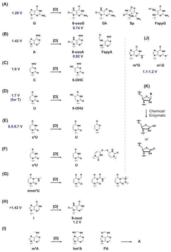
Examples of ROS include hydrogen peroxide (H2O2), hydroxyl radical (•OH), carbonate radical anion (CO3•−), superoxide (O2•−), singlet oxygen (1O2), ozone (O3), hypochlorous acid (HOCl), peroxyl radical (ROO•), and alkoxyl radical (RO•) [11]. Some of these species can be naturally handled by enzymes such as superoxide dismutases and reductases (2O2•− + 2H+ + e−→ H2O2 or 2O2•− + 2H+ → H2O2 + O2) [12], or glutathione peroxidases and catalase (2H2O2 → 2H2O + O2) [13,14], while other ROS, e.g., •OH [15,16], 1O2 [17], or CO3•− [18,19], may not be captured by biological constituents and can react at diffusion controlled rates with biomolecules within their vicinity. The topic of other antioxidant enzymes and their relationship with various processes was recently reviewed [20]. Importantly, ROS are found in almost every organelle within the cell [21], and can also be generated from exogenous sources, e.g., exposure to ionizing or UV radiation [22]. A consequence of their reactivity is that ROS can induce changes in the structure of all nucleobases of DNA and RNA [23], with guanine (G) as a preferred target amongst the canonical nucleosides given its lower oxidation potential [24,25]. In fact, four oxidized nucleosides were first identified to be involved in oxidoreduction processes in yeast RNA, specifically 8-oxo-7,8-dihydropurines 8-oxoG and 8-oxoA, and 5-hydroxypyrimidines 5-OHU and 5-OHC (Scheme 1A–D) [26,27]. The oxidation of guanosine can lead to a variety of oxidatively generated modifications, and 8-oxo-7,8-dihydroguanosine (8-oxoG, or its 2′-deoxy homologue—Scheme 1A) is generally used as a biomarker, due to its established presence in DNA and RNA [28], and can be a modulator of biosystems broadly [29]. Structurally, oxidation at the C8 position changes the H-bonding pattern of the nucleobase, which can affect its base pairing ability [30], thus potentially impacting RNA–protein interactions.
Scheme 1.
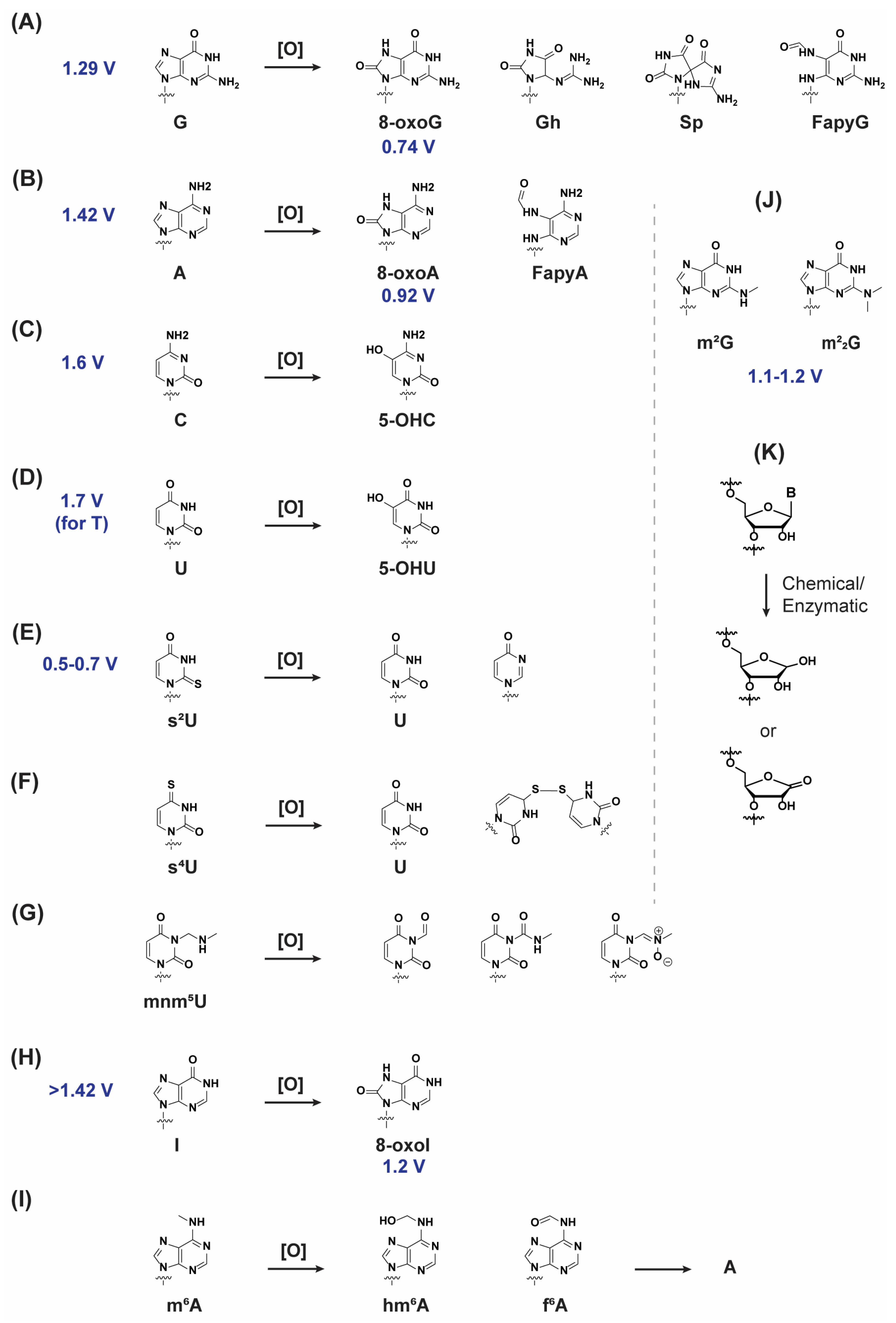
Structure of the nucleobases and their corresponding oxidation products. Oxidation potentials are coloured in blue and represented as Volts (V), reported versus Normal Hydrogen Electrode (NHE). Nucleobase code: (A) G = Guanine, 8-oxoG = 8-Oxoguanine, Gh = 5-guanidinohydantoin, Sp = spiroiminodihydantoin, FapyG = Formamidopyrimidineguanine; (B) A = Adenine, 8-oxoA = 8-Oxoadenine, FapyA = Formamidoadenine; (C) C = Cytosine, 5-OHC = 5-Hydroxycytosine; (D) U = Uracil, 5-OHU = 5-hydroxyuracil; (E) s2U = 2-Thiouracil; (F) s4U = 4-Thiouracil; (G) mnm5U = 5-Methylaminomethyluracil; (H) I = inosine, 8-oxoI = 8-Oxoinosine; (I) m6A = 6-Methyladenine, hm6A = 6-hydroxymethyladenine, f6A = 6-formyladenine; (J) m2G = 2-Methylguanine, m22G = 2,2-dimethylguanine; (K) abasic site formation.
Although other canonical nucleobases are less prone to oxidatively induced modification, these can also undergo the corresponding reactions, recently reviewed in the context of DNA [31], to give a range of different oxidative modifications at the nucleobase. It is reasonable to expect similar reactivity within strands of RNA. The oxidation of adenosine can also occur, where the reactivity of the purine ring is similar to that of guanosine and results in the formation of 8-oxo-7,8-dihydroadenosine (8-oxoA—Scheme 1B) as one of the main products (others can also be formed, such as Fapy•A, [32]). This has prompted various studies that focused on the development of antibodies [33], computational methodology to identify or evolve RNA–protein interactions [34,35], altered H-bonding interactions between helicases and oxidized nucleotides [36], as well as its impact on the structure of duplex regions within RNA [37]. It has also been incorporated and studied on a model of rRNA [38], and was found in patients with late-stage Alzheimer’s disease [39]. Its presence and repair on DNA have been thoroughly studied and are not discussed herein. Importantly, some of the reported reactivity, such as the formation of interstrand cross-links in DNA [40], may be potentially useful to detect or explore its role within RNA. The oxidation of pyrimidine rings (Scheme 1C,D) has been reported in the context of DNA, and their biochemical/biophysical properties have not been explored within RNA, with the exception of some experiments carried out on models of mRNA [41]. In addition, the natural occurrence of 5-hydroxylated pyrimidines has been established on rRNA [38,42], as well as their biogenesis [43]. This review does not focus on naturally occurring oxidative processes such as those observed in the transformation from 5-methylcytosine to 5-hydroxymethylcytosine (m5C → hm5C—[44,45]) or analogous processes involved in editing [46].
It is also worth mentioning that there are over 140 naturally occurring RNA modifications, and not much is known about their oxidation [47]. Examples include those of 2-methylguanosine (m2G) and 2,2-dimethylguanosine (m22G, Scheme 1J), which have been shown to have a lower oxidation potential than G [48], with the former characterized in tRNA [49]. 2-Thiouridine (Scheme 1E,F) displays a lower oxidation potential than its canonical analogue and can also be oxidized by cytochrome c [50,51]. A related derivative, 5-methylaminomethyl-2-thiouridine (mnm5s2U, Scheme 1G), is more vulnerable to oxidative reactivity [52], while 5-methoxycarbonylmethyl-2-thiouridine (mcm5s2U) in eukaryotic cells is also prone to oxidation [53]. Similarly, 4-thiouridine (s4U) can undergo oxidation in vitro, which has been characterized within isolates of tRNA [54,55], with the oxidation of mnm5U also observed (Scheme 1G) [56]. Generally, thio-containing nucleosides possess distinct chemical and photophysical properties [57,58]. On the other hand, the oxidation of inosine is not likely to be a factor of biological relevance, given its higher oxidation potential (Scheme 1H) [59]. Oxidation reactions have been shown to have distinct reactivity for pseudouridine (Ψ) with potential for sequencing technologies [60], suggesting that their oxidation is possible and can lead to interesting biological properties within functional RNA, or alter rRNA modification profiles [61]. Another modification that can undergo oxidation is 6-methyladenosine (m6A), enzymatically or by reaction with ROS, which can result in its conversion to adenosine (Scheme 1I) [62,63].
Thus, the reactivity of different RNA modifications with ROS can potentially lead to other oxidatively modified nucleobases with distinct reactivity and biological implications. One example is the impact that nucleobase modification has on the stability of the glycosidic bond, where both oxidation and methylation can decrease bond strength [64,65,66] and have an impact on important processes such as the formation of abasic sites (Scheme 1K) [67]. In this regard, it is important to note that the chemical stability of RNA containing abasic sites is higher than that observed on DNA [68], and that such nucleobase-free modifications can occur as a consequence of oxidative modification on RNA [69]. These have also been observed in RNA oxidation catalyzed by cytochrome-c [70].
3. RNA Oxidation and 8-OxoG
Different types of RNA have been shown to be oxidized in vivo [71], including coding and non-coding RNAs, short (<200-nt) and long (>200-nt). Some examples include those involving microRNA (miRNA—[72,73,74,75]), ribosomal RNA (rRNA—[38,76]) that can lead to strand breaks [77], messenger RNA (mRNA—[41,78,79,80]), mRNA in plants [81], or mitochondrial RNA (mtRNA—[82]), as well as those in processes involved with viral RNA (vRNA—[83,84]). Most of the published studies use 8-oxoG as a biomarker (see Supplementary Materials, Section S2 for more about the detection and quantification of this oxidatively generated modification). In fact, the importance of oxidatively modified RNA and its presence in many instances has prompted the development of methods to effectively sequence for oxidatively modified nucleosides within RNA [48,85,86]. The detection and quantification of ROS and their products in biological systems is challenging, and has prompted the development of guidelines for their accurate characterization in such experiments [87]. In addition, recent reviews have addressed different aspects regarding RNA damage [88].
3.1. Handling of Oxidized RNA
As a consequence of structural differences (induced by reactions between ROS and nucleobases) and the biological implications arising from the presence of oxidatively generated lesions within RNA, it is reasonable to expect cellular processes to be in place to cope with such species. For example, folding mechanisms that reduce the amount of available guanine exposure to solvent, associated with increased susceptibility to react with ROS, have been identified [89]. The compartmentalization of oxidized RNA has also been shown as a potential coping mechanism, as well as for other type of RNA damage, perhaps for their eventual degradation [90,91]. The recruitment of oxidized RNA to stress granules along with their corresponding sorting and processing have also been proposed as a mechanism of action, with the direct involvement of various proteins, e.g., adenosine deaminase action on RNA (ADAR1), Staufen double-stranded RNA protein (STAU1), and Tudor staphylococcal nuclease (Tudor-SN) [92]. In addition, biochemical pathways to evade the incorporation of oxidized nucleotides (present in the nucleotide pool), through their hydrolysis to the corresponding nucleosides have been identified. These involve an Arabidopsis Nudix hydrolase (AtNUDX1) in plants [93], or guanylate kinases MutT and NUDT5 in bacterial and mammalian systems [94,95]. The importance of the efficiency of these processes is highlighted by the fact that some RNA polymerases can effectively incorporate 8-oxoGTP and exhibit distinct reactivity with 8-oxoATP [96]. On the other hand, inducing an excess of 8-oxoG nucleotides has been shown to lead to β-cell dysfunction [97], so it is plausible that their enzymatic hydrolysis is impacted and adverse reactions/processes to the cellular health are triggered. For the remainder of this review, we place focus on the ribonucleases that are known to be involved in the processing of oxidized RNA, and propose pathways that could be important in the handling of oxidatively modified RNA. The word oxidative ‘damage’ (a common term in DNA) is not used herein, as the possibility that oxidized RNA may function to trigger signalling pathways is plausible and potentially important. A review about RNA surveillance has been published and discusses some relevant RNA–protein interactions [98].
RNA quality control mechanisms are in place to target aberrant RNAs for their degradation [99], a function that is carried out by specific ribonucleases which can cleave strands of RNA in a processive or distributive manner [100] via cleavage of the phosphodiester bond within its backbone. Ribonucleases are also involved in RNA processing, including its degradation, and mRNA or noncoding-RNA turnover [101]. A distinction between RNA processing and RNA degradation can be inferred from their formed stable structures, bound proteins, and/or presence of base/sugar modifications. Of note are mechanisms that have evolved to deal with defective mRNAs, such as nonsense-mediated decay (NMD), which refers to the process of targeting aberrant mRNAs for decapping and degradation, or no-go decay (NGD), which refers to endonucleolytic cleavage of mRNA with strong pauses in elongation [102], and staufen mediated decay (SMD) which targets double-stranded RNA [103]. In this regard, the oxidation of NAT8L mRNA has been shown to reduce protein synthesis [79], displaying >1000 fold decreased peptide bond formation when 8-oxoG is present within mRNA [41,104]. In addition, mRNA containing 8-oxoG has also been shown to affect translational fidelity [78]. For a review about the impact of other modifications on mRNA see [105]. At this point, it is also important to note that the functions of ribonucleases go well beyond those of solely RNA degradation and with a rich history in the community [106,107]; however, those are not discussed in this review. For examples about ribonucleases involved in other processes see Supplementary Materials, Section S3 and Table S1.
3.2. Interactions Between Oxidized RNA and Ribonucleases or RNA-Binding Proteins
With regard to ribonuclease involvement in the handling of oxidatively modified RNA, turnover of oxidized RNA has been shown in epithelial cells, where a portion of 8-oxoG in RNA is cleared following the removal of oxidative stress, providing evidence of RNA degradation [108], as also observed on HeLa cells [109]. Furthermore, the formation of 8-oxoG has been shown to coincide with a decrease in rRNA [110,111]. An association between oxidative stress and tRNA cleavage has also been observed [112]. Simms et al. discussed chemical modifications, some of which arise from oxidative stress and touch upon mRNA, tRNA, and rRNA oxidation, as well as cellular handling of damaged RNA [113,114]. As delineated within Table 1 (top), the three ribonucleases that have been reported to be involved in the enzymatic cleavage of oxidized RNA are Xrn-1, ISG20, and PNPase. It is important to note that PNPase is part of a larger body composed of proteins with various functions, namely the degradosome, although it can also act independently [115] or in association with other binding proteins such as Hfq and poly(A) polymerase I [116], as well as helicase RhIB [117]. In addition the exosome is another multi-subunit complex essential for RNA processing [118,119]; and other ribonucleases with various roles, including activity under stress, are mentioned in Table 1 (bottom).
Table 1.
Ribonucleases reported to be involved in the hydrolysis/phosphorolysis/degradation of oxidized RNA (containing 8-oxoG) (top), and ribonucleases within the processing bodies that may also handle these species (bottom).
Some of the handling of oxidized mRNA has been shown to involve the ribosome, displaying decreased peptydil transfer rates [104]. In this case, Xrn-1 was shown to be responsible for its degradation, and subject to NGD in a coordinated mechanism with Dom34, which is known to expedite RNA degradation [132,133] by binding to the A-site of the ribosome, and recruiting the exosome. In fact, Dom34 can form a surveillance complex with RNase L and act in the recognition of exogenous mRNA for its subsequent degradation, as a potential antiviral defence mechanism [134]. The combination of Xrn-1 and Dom-34 seems to be important as Xrn-1 has been shown to stall upon encountering 8-oxoG within strands of RNA, in the absence of Dom34 [135]; however, more work is needed to establish the exact interactions between oxidized RNA and these proteins. Importantly, exoribonuclease family Xrn1/Xrn2 carries out enzymatic hydrolysis of RNA within the nucleus [136]. Another protein of interest is the AU-rich element/poly(U)-binding/degradation factor AUF1 (also called HNRNPD, since it is a heterogeneous nuclear ribonucleoprotein) which has been shown to participate in the decay of oxidized mRNA [137,138]; however, the specific ribonuclease has not been determined. In this regard, AUF1 is an RNA binding protein known to be involved in RNA folding [139] and with functions associated with mRNA turnover. Other proteins that bind with lower affinity to oxidized RNA include HNRNPC and PNP. Amongst these, polynucleotide phosphorylase (PNPase and its human homologue PNPT1) is of particular interest, as it has been shown to display preferential binding to oxidized RNA, specifically RNA containing 8-oxoG [140,141]. This was reviewed recently [142]; the S1 domain plays an important role in the binding and handling of RNA containing 8-oxoG in the cytoplasm and mitochondria [143]. PNPase and DEAD box RNA helicase (DR_0335/Rh1B) are involved in targeting RNAs containing 8-oxoG and grant resistance to oxidative stress in Deinococcus radiodurans [144]. Interestingly, as previously described with Xrn-1, PNPase (expressed from E. coli) has also been shown to stall upon encountering 8-oxoG within strands of RNA. In this case, Cryo-EM structures displayed interactions between residues in the active site of PNPase and 8-oxoG that are responsible for the stalling behaviour [145]. Therefore, other factors might be involved that allow for the processing of the oxidized substrates. For example, the role of the three main bacterial ribonucleases (RNase II, RNase R, PNPase) has been studied, with the conclusion that they have different effects on cellular motility and different RNA targets [146]. It is also important to note that PNPase is a subunit of the degradosome, a multiprotein complex, and is necessary for oxidative stress under certain conditions [147]. In addition, the degradosome also contains other ribonucleases such as RNase E, an endonuclease that complements the exonuclease activity of PNPase [148], although this relationship is yet to be explored in the context of handling oxidized RNA. Contrary to the observed stalling using bacterial PNPase (E. coli), human mitochondrial PNPase was recently shown to have evolved with distinct structural features that allow for efficient degradation of RNA containing 8-oxoG [149]. This brings into question the role of PNPase within different organisms and possibly distinct mechanisms that handle oxidized RNA.
Another ribonuclease that was found to be involved in the degradation of oxidized RNA is ISG20 (also known as HEM45) [124,125]. A link between the pathophysiology of acute kidney injury and oxidation of RNA was established by measuring the upregulation of ISG20 in kidneys of AKI model mice. Furthermore, levels of oxidized RNA were reduced on models overexpressing ISG20, with the conclusion that ISG20 takes an active role in the degradation of oxidatively modified RNA. While this exonuclease is commonly associated with an antiviral response [150], recent reports have studied orthologs with a role in ribosomal biogenesis [151], which suggests that the activity of this exonuclease is broader than our current knowledge.
Other than Xrn-1, PNPase, and ISG20, there are no reports that have specifically tested for the degradation of oxidized RNA, which brings into question the potential role of other protein complexes, such as the exosome, to perform these processing functions. For example, the nuclear-specific subunit Rrp4 (also called Dis3), found within the exosome, has the capability to direct up to three RNA substrates for degradation [152]. On this note, it is worth noting that the exosomes from different organisms can vary in structure and function [153], and thus distinct mechanisms could be at play. Another important factor is the role of RNA-binding proteins (RBPs) that can form complexes with oxidized RNA, a topic that was recently reviewed [154] and is briefly summarized within the Supporting Materials (Section S4).
The role of helicases is also worth considering since these are in part responsible for facilitating RNA degradation, amongst other functions, and are present within the RNA degradation complexes [155,156,157,158]. There are no available reports that test the impact of oxidative stress (or presence of oxidatively generated modifications) in the context with helicases; however, at least one helicase has been found to be associated with RNA containing 8-oxoG [143], although other experiments displayed independence between PNPase degradation of oxidized RNA and the activity of helicase RhIB [141]. In addition, the presence of modifications that alter duplex stability is known to affect helicase activity [159], which has prompted a view of their role as protective agents against oxidative stress [160]. Another aspect of potential relevance is the relationship between miRNA oxidation and proteins associated with miR biogenesis [161], as well as with siRNA [162]. Amongst the studies that have reported on oxidized miRNA ([72,73,74,75,163] all containing 8-oxoG), the stage at which miRNA is oxidized has not been established (nuclear pri-miRNA, cytoplasmic pre-miRNA or mature miRNA). This brings into question the potential for altered RNA–protein interactions in miRNA biogenesis involving processing (DROSHA, DGCR8, spliceosome and AGO2, DICER) and/or export (Exportin 1 or 5, RanGTP) [164], following RNA oxidation.
It is also important to note that the landscape of RNA–enzyme interactions is known to vary significantly in the presence/absence of oxidative stress [165], which indicates that distinct mechanisms are in place to handle oxidized RNA. This includes the potential role of extracellular vesicles to clear the cells of oxidized RNA, with a known role with other types of RNA [166], although this is yet to be studied. In this regard, extracellular ribonucleases such as those in the RNase A and T families could play an important role; these are known to hydrolyze/process stress-induced tRNAs, specifically from oxidative stress [167]. Therefore, while the reactivity between these RNases and RNA containing 8-oxoG has been shown to vary (RNase T1 does not cleave at 8-oxoG sites, while RNase A does, in some sequence contexts [168]), it is possible that the degradation of extracellular RNAs by these ribonucleases is effective. In addition to degradation, apoptosis [169] and autophagy [170] may play a role in the clearance of cells or cellular materials that accumulate oxidized RNA products.
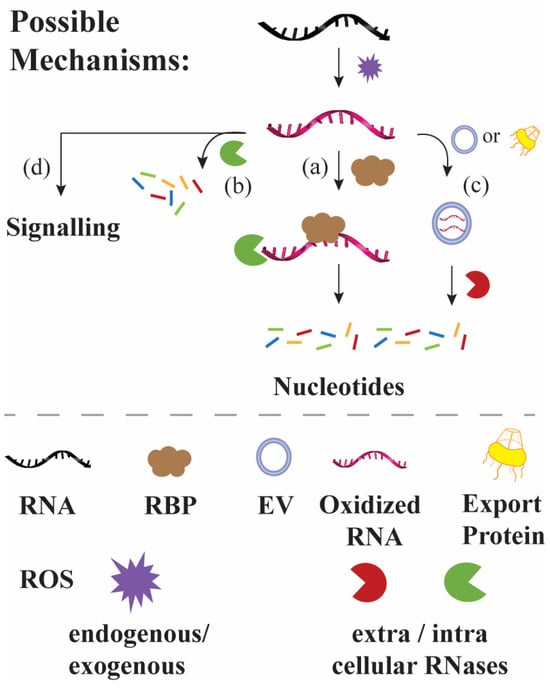
Another point of consideration is the potential repair of oxidized RNA, where enzymes involved in DNA repair have been shown to be involved in RNA processing; this relationship and other data suggesting a possible repair have been recently reviewed [171]. Our group has found that ribonucleases exhibit different reactivity towards the enzymatic cleavage of oxidized RNA [135,145,168], and it is worth noting that other groups have also used altered reactivity of RNA containing 8-oxoG with other enzymes [75], as well as with naturally occurring modifications; e.g., pseudouridine can also alter the reactivity of nucleases towards RNA containing this modification [172]. Thus, a full picture that describes the handling of oxidized RA within different cellular environments is yet to be fully elucidated. As depicted in Figure 1, mechanisms that can be involved in the handling of oxidized RNA include (a) specific recognition of oxidized RNA followed by degradation, (b) direct enzymatic hydrolysis, (c) export to extracellular environment and subsequent degradation, (d) repair or (e) triggering of a cellular signal.
Figure 1.
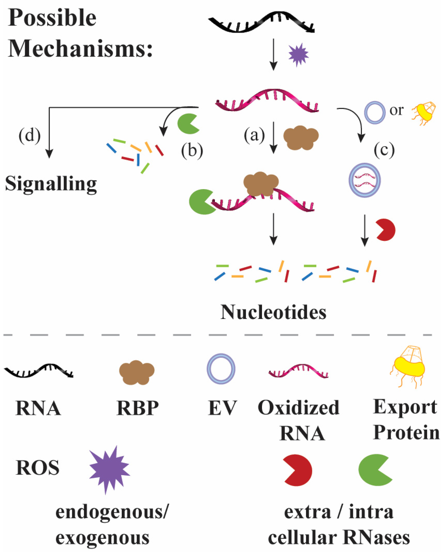
Simplified picture illustrating known and potential intra- and extracellular paths in charge of handling oxidized RNA (left) and mechanisms mentioned in this review that led to the degradation of oxidized RNA, some which have been reported ((a), (b), (d)) and others that were hypothesized herein (c). (a) Specific recognition of oxidized RNA by binding proteins; (b) enzymatic degradation of oxidized RNA by ribonucleases; (c) intra-to-extracellular export; and (d) signalling mechanisms.
4. Conclusions
RNA is a highly diverse biopolymer that is involved in a plethora of important biological functions, which can be orchestrated as a function of their sequence. Therefore, alteration of the nucleobase(s) by oxidative modification may lead to dysfunction, and/or trigger a signal that results in the handling of the oxidized RNA, or lead to apoptosis, among other outcomes. One of the most common intracellular mechanisms involves the enzymatic hydrolysis of the oxidatively modified RNA by ribonucleases, which include Xrn-1, ISG-20, and PNPase. How these oxidized RNAs are recognized remains an area to be further studied; however, there are a number of studies that have found proteins with high affinity towards these species. More research and information are needed to create a clear picture of how oxidized RNA is handled, as well as its biological implications.
Supplementary Materials
The following supporting information can be downloaded at: https://www.mdpi.com/article/10.3390/biom16040564/s1, Supporting Materials Sections S1–S4 and Table S1. Proteins known to bind to and/or react with oxidized RNA, where provided references focus on the RNA binding domain corresponding to each protein.
Author Contributions
D.G.-F. provided background information, identified literature, and was involved in the writing process. M.J.E.R. identified critical literature, generated all of the images, and wrote the manuscript. All authors have read and agreed to the published version of the manuscript.
Funding
M.J.E.R. acknowledges support from NIGMS, via 2R15GM132816-02. The work was also supported by a Teacher-Scholar Award (M.J.E.R.), TH-21-028, from the Camille & Henry Dreyfus Foundation.
Institutional Review Board Statement
Not applicable.
Informed Consent Statement
Not applicable.
Data Availability Statement
No new data were created or analyzed in this study.
Conflicts of Interest
The authors declare no conflicts of interest.
Abbreviations
The following abbreviations are used in this manuscript (alphabetical order):
| m2G | 2-Methylguanosine |
| m22G | 2,2-Dimethylguanosine |
| S4U | 4-Thiouridine |
| hm5C | 5-Hydroxymethylcytosine |
| mcm5s2U | 5-Methoxycarbonylmethyl-2-thiouridine |
| mnm5s2U | 5-Methylaminomethyl-2-thiouridine |
| m5C | 5-Methylcytosine |
| m6A | 6-Methyladenosine |
| 8-oxoA | 8-Oxo-7,8-dihydroadenosine |
| 8-oxoATP | 8-Oxo-7,8-dihydroadenosine triphosphate |
| 8-oxoG | 8-Oxo-7,8-dihydroguanosine |
| 8-oxoGTP | 8-Oxo-7,8-dihydroguanosine triphosphate |
| ADAR1 | Adenosine deaminase action on RNA |
| RO• | Alkoxyl radical |
| APE1 | Apurinic/apyrimidinic endonuclease 1 |
| AtNUDX1 | Arabidopsis Nudix hydrolase |
| AUF1 | AU-rich element/poly(U)-binding/degradation factor |
| CTD | C-terminal domain |
| CO3•− | Carbonate radical anion |
| CSD | Cold-shock domain |
| DR_0335/Rh1B | DEAD box RNA helicase |
| DEDD | Death Effector Domain containing DNA binding protein |
| dsRNA | Double stranded RNA |
| DOM34 | Duplicate of multilocus |
| Xrn | Exoribonuclease |
| exRNA | Extracellular RNA |
| gRNA | Guide RNA |
| HNRNP | Heterogeneous nuclear ribonucleoprotein |
| H2O2 | Hydrogen peroxide |
| •OH | Hydroxyl radical |
| HOCl | Hypochlorous acid |
| ISG20 | Interferon-stimulated gene 20 |
| IGF2BP | Insulin-line growth factor 2 mRNA binding protein |
| KH | Kinase homology |
| mRNA | Messenger RNA |
| miRNA | microRNA |
| mtRNA | Mitochondrial RNA |
| NTD | N-terminal domain |
| NAT8L | N-acetyl aspartate transferase 8 like |
| NBS | Nijmegen breakage syndrome |
| NGD | No-go decay |
| NMD | Nonsense mediated decay |
| NUDT5 | Nudix Hydrolase 5 |
| O3 | Ozone |
| ROO• | Peroxyl radical |
| PCB or PCBP | Poly(rC)-binding protein |
| PNPase | Polyribonucleotide Phosphorylase |
| PNP | Purine Nucleoside Phosphorylase |
| PNPT1 | Polyribonucleotide nucleotidyltransferase 1 |
| ROS | Reactive oxygen species |
| RNase | Ribonuclease |
| RNP | Ribonucleoprotein |
| rRNA | Ribosomal RNA |
| Rrp | Ribosomal RNA-processing protein |
| RBPs | RNA-binding proteins |
| RhlB | RNA Helicase B |
| RRM | RNA recognition motif |
| ssRNA | Single Stranded RNA |
| 1O2 | Singlet oxygen |
| SMD | Staufen mediated decay |
| sRNA | Small RNA |
| siRNA | Small interfering RNA |
| STAU1 | Staufen double-stranded RNA protein |
| O2•− | Superoxide |
| tRNA | Transfer RNA |
| Tudor-SN | Tudor staphylococcal nuclease |
| tiRNA | tRNA-derived stress-induced small RNA |
| vRNA | Viral RNA |
| YB-1 | Y-box binding protein |
References
- Lennicke, C.; Cochemé, H.M. Redox metabolism: ROS as specific molecular regulators of cell signaling and function. Mol. Cell 2021, 81, 3691–3707. [Google Scholar] [CrossRef] [Scilit]
- Tripathy, B.C.; Oelmüller, R. Reactive oxygen species generation and signaling in plants. Plant Signal. Behav. 2012, 12, 1621–1633. [Google Scholar] [CrossRef] [Scilit]
- Fasnacht, M.; Polacek, N. Oxidative stress in bacteria and the central dogma of molecular biology. Front. Mol. Biosci. 2021, 8, 671037. [Google Scholar] [CrossRef] [Scilit]
- Durand, D.; Collin, A.; Merlot, E.; Baéza, E.; Guilloteau, L.A.; Le Floc’h, N.; Thomas, A.; Fontagné-Dicharry, S.; Gondret, F. Review: Implication of redox imbalance in animal health and performance at critical periods, insights from different farm species. Animal 2022, 16, 100543. [Google Scholar] [CrossRef] [Scilit] [PubMed]
- Emory, S.A.; Bouvet, P.; Belasco, J.G. A 5′-terminal stem-loop structure can stabilize mRNA in Escherichia coli. Genes Dev. 1992, 6, 135–148. [Google Scholar] [CrossRef] [Scilit] [PubMed]
- Selinger, D.W.; Saxena, R.M.; Cheung, K.J.; Church, G.M.; Rosenow, C. Global RNA half-life analysis in Escherichia coli reveals positional patterns of transcript degradation. Genome Res. 2003, 13, 216–223. [Google Scholar] [CrossRef] [Scilit] [PubMed]
- Zocher, S.; McCloskey, A.; Karasinsky, A.; Schulte, R.; Friedrich, U.; Lesche, M.; Rund, N.; Gage, F.H.; Hetzer, M.W.; Toda, T. Lifelong persistence of nuclear RNAs in the mouse brain. Science 2024, 384, 53–59. [Google Scholar] [CrossRef] [Scilit]
- Houseley, J.; Tollervey, D. The many pathways of RNA degradation. Cell 2009, 136, 763–776. [Google Scholar] [CrossRef] [Scilit]
- Hia, F.; Takeuchi, O. The role of ribonucleases in RNA damage, inactivation and degradation. In RNA Damage and Repair; Kotta-Loizou, I., Ed.; Springer: Cham, Switzerland, 2021. [Google Scholar] [CrossRef] [Scilit]
- Poulsen, H.E.; Specht, E.; Broedbaek, K.; Henriksen, T.; Ellervik, C.; Mandrup-Poulsen, T.; Tonnesen, M.; Nielsen, P.E.; Andersen, H.U.; Weimann, A. RNA modifications by oxidation: A novel disease mechanism? Free Rad. Biol. Med. 2012, 52, 1353–1361. [Google Scholar] [CrossRef] [Scilit]
- Sies, H.; Jones, D.P. Reactive oxygen species (ROS) as pleiotropic physiological signaling agents. Nat. Rev. Mol. Cell Biol. 2020, 21, 363–383. [Google Scholar] [CrossRef] [Scilit]
- Sheng, Y.; Abreu, I.A.; Cabelli, D.E.; Maroney, M.J.; Miller, A.-F.; Teixeira, M.; Valentine, J.S. Superoxide dismutases and superoxide reductases. Chem. Rev. 2014, 114, 3854–3918. [Google Scholar] [CrossRef] [Scilit] [PubMed]
- Handy, D.E.; Loscalzo, J. The role of glutathione peroxidase-1 in health and disease. Free Rad. Biol. Med. 2022, 188, 146–161. [Google Scholar] [CrossRef] [Scilit]
- Glorieux, C.; Calderon, P.B. Catalase, a remarkable enzyme: Targeting the oldest antioxidant enzyme to find new cancer treatment approach. Biol. Chem. 2017, 398, 1095–1108. [Google Scholar] [CrossRef] [Scilit]
- Gligorovski, S.; Strekowski, R.; Barbati, S.; Vione, D. Environmental implications of hydroxyl radicals (•OH). Chem. Rev. 2015, 115, 13051–13092. [Google Scholar] [CrossRef] [Scilit]
- Buxton, G.V.; Greenstock, C.L.; Helman, W.P.; Ross, A.B. Critical review of rate constants for reactions of hydrated electrons, hydrogen atoms and hydroxyl radicals (•OH/•O−) in aqueous solution. J. Phys. Chem. Ref. Data 1988, 17, 513–886. [Google Scholar] [CrossRef] [Scilit]
- Ravanat, J.-L.; Dumont, E. Reactivity of singlet oxygen with DNA, an update. Photochem. Photobiol. 2022, 98, 564–571. [Google Scholar] [CrossRef] [Scilit]
- Koppenol, W.H.; Serrano-Luginbuehl, S.; Nauser, T.; Kissner, R. Thinking outside the cage: A new hypothesis that accounts for variable yields of radicals from the reaction of CO2 and ONOO−. Chem. Res. Toxicol. 2020, 33, 1516–1527. [Google Scholar] [CrossRef] [Scilit]
- Dingman, J.C.; Fleming, A.M.; Burrows, C.J. RNA oxidative damage by the iron-Fenton reaction is influenced by bicarbonate concentration and ligand strength. Chem. Res. Toxicol. 2025, 38, 2019–2025. [Google Scholar] [CrossRef] [Scilit]
- Solc, V.; Preto, O.; Karhanek, M.; Freund, F.; Griko, Y.; Loftus, D.J.; Ohayon, M.M. RNA-DNA differences: Mechanisms, oxidative stress, transcriptional fidelity, and health implications. Antioxidants 2025, 14, 544. [Google Scholar] [CrossRef] [Scilit]
- Sun, Y.; Lu, Y.; Saredy, J.; Wang, X.; Drummer, C., IV; Shao, Y.; Saaoud, F.; Xu, K.; Liu, M.; Yang, W.Y.; et al. ROS systems are a new integrated network for sensing homeostasis and alarming stresses in organelle metabolic processes. Redox Biol. 2020, 37, 101696. [Google Scholar] [CrossRef] [Scilit] [PubMed]
- Ravanat, J.-L.; Douki, T. UV and ionizing radiations induced DNA damage, differences and similarities. Rad. Phys. Chem. 2016, 128, 92–102. [Google Scholar] [CrossRef] [Scilit]
- Cadet, J.; Douki, T.; Ravanat, J.-L. Oxidatively generated base damage to cellular DNA. Free Rad. Biol. Med. 2010, 49, 9–21. [Google Scholar] [CrossRef] [Scilit]
- Steenken, S.; Jovanovic, S.V. How easily oxidizable is DNA? One-electron reduction potentials of adenosine and guanosine radicals in aqueous solutions. J. Am. Chem. Soc. 1997, 119, 617–618. [Google Scholar] [CrossRef] [Scilit]
- Nilov, D.I.; Komarov, D.Y.; Panov, M.S.; Karabaeva, K.E.; Mereshchenko, A.S.; Tarnovsky, A.N.; Wilson, R.M. Oxidation of adenosine and inosine: The chemistry of 8-oxo-7,8-dihydropurines, purine, iminoquinones, and purine quinones as observed by ultrafast spectroscopy. J. Am. Chem. Soc. 2013, 135, 3423–3438. [Google Scholar] [CrossRef] [Scilit]
- Yanagawa, H.; Ogawa, Y.; Ueno, M. Redox ribonucleosides. Isolation and characterization of 5-hydroxyuridine, 8-hydroxyguanosine, and 8-hydroxyadenosine from Torula yeast RNA. J. Biol. Chem. 1992, 267, 13320–13326. [Google Scholar] [CrossRef] [Scilit] [PubMed]
- Yanagawa, H.; Ogawa, Y.; Ueno, M.; Sasaki, K.; Sato, T. A novel minimum ribozyme with oxidoreduction activity. Biochemistry 1990, 29, 10585–10589. [Google Scholar] [CrossRef] [Scilit]
- Neeley, W.L.; Essigmann, J.M. Mechanisms of formation, genotoxicity, and mutation of guanine oxidation products. Chem. Res. Toxicol. 2006, 19, 491–505. [Google Scholar] [CrossRef] [Scilit]
- Tanaka, M.; Chock, P.B. Oxidative modifications of RNA and its potential roles in biosystem. Front. Mol. Biosci. 2021, 8, 685331. [Google Scholar] [CrossRef] [Scilit]
- Choi, Y.J.; Gibala, K.; Ayele, T.; Van Deventer, K.; Resendiz, M.J.E. Biophysical Properties, Thermal Stability, and Functional Impact of 8-oxo-7,8-dihydroguanine on Oligonucleotides of RNA. A Study of Duplex, Hairpins, and the Aptamer for preQ1 as Models. Nucleic Acids Res. 2017, 45, 2099–2111. [Google Scholar] [CrossRef] [Scilit] [PubMed]
- Andrés, C.M.C.; Lastra, J.M.P.; Juan, C.A.; Plou, F.J.; Pérez-Lebeña, E. Chemical insights into oxidative and nitrative modifications of DNA. Int. J. Mol. Sci. 2023, 24, 15240. [Google Scholar] [CrossRef] [Scilit]
- Greenberg, M.M. The formamidopyrimidines: Purine lesions formed in competition with 8-oxopurines from oxidative stress. Acc. Chem. Res. 2011, 45, 588–597. [Google Scholar] [CrossRef] [Scilit] [PubMed]
- Tew, D.J.; Hebert, J.M.; Schmier, B.J. Discovery and properties of a monoclonal antibody targeting 8-oxoA, and oxidized adenine lesion in DNA and RNA. Redox Biol. 2023, 62, 102658. [Google Scholar] [CrossRef] [Scilit] [PubMed]
- Orr, A.A.; Gonzalez-Rivera, J.C.; Wilson, M.; Bhikha, P.R.; Wang, D.; Contreras, L.M.; Tamamis, P. A high-throughput and rapid computational method for screening of NRA post-transcriptional modifications that can be recognized by target proteins. Methods 2018, 143, 34–47. [Google Scholar] [CrossRef] [Scilit]
- Gonzalez-Rivera, J.C.; Orr, A.A.; Engels, S.M.; Jakubowski, J.M.; Sherman, M.W.; O’Connor, K.N.; Matteson, T.; Woodcock, B.C.; Contreras, L.M.; Tamamis, P. Computational evolution of an RNA-binding protein towards enhanced oxidized-RNA binding. Comp. Struct. Biotechnol. J. 2020, 18, 137–152. [Google Scholar] [CrossRef] [Scilit]
- Strohmeier, J.; Hertel, I.; Diedeichsen, U.; Rudolph, M.G.; Klostermeier, D. Changing nucleotide specificity of the DEAD-box helicase Hera abrogates communication between the Q-motif and the P-loop. Biol. Chem. 2011, 392, 357–369. [Google Scholar] [CrossRef] [Scilit]
- Chauca-Diaz, A.M.; Choi, Y.J.; Resendiz, M.J.E. Biophysical properties and thermal stability of oligonucleotides of RNA containing 7,8-dihydro-8-hydroxyadenosine. Biopolymers 2014, 103, 167–174. [Google Scholar] [CrossRef] [Scilit]
- Willi, J.; Küpfer, P.; Evéquoz, D.; Fernandez, G.; Katz, A.; Leumann, C.; Polacek, N. Oxidative stress damages rRNA inside the ribosome and differentially affects the catalytic center. Nucleic Acids Res. 2018, 46, 1947–1957. [Google Scholar] [CrossRef] [Scilit]
- Weidner, A.M.; Bradley, M.A.; Beckett, T.L.; Niedowicz, D.M.; Dowling, A.L.S.; Matveev, S.V.; LeVine, H.; Lovell, M.A.; Murphy, M.P. RNA oxidation adducts 8-OHG and 8-OHA change with Aβ42 levels in late-stage Alzheimer’s disease. PLoS ONE 2011, 6, e24930. [Google Scholar] [CrossRef] [Scilit]
- Rozelle, A.L.; Cheun, Y.; Vilas, C.K.; Koag, M.-C.; Lee, S. DNA Interstrand cross-links induced by the major oxidative adenine lesion 7,8-dihydro-8-oxoadenine. Nat. Commun. 2021, 12, 1897. [Google Scholar] [CrossRef] [Scilit] [PubMed]
- Calabretta, A.; Küpfer, P.A.; Leumann, C.J. The effect of RNA base lesions on mRNA translation. Nucleic Acids Res. 2015, 43, 4713–4720. [Google Scholar] [CrossRef] [Scilit]
- Kimura, S.; Sakai, Y.; Ishiguro, K.; Suzuki, T. Biogenesis and iron-dependency of ribosomal RNA hydroxylation. Nucleic Acids Res. 2017, 45, 12974–12986. [Google Scholar] [CrossRef] [Scilit]
- Lauhon, C.T. Identification and characterization of genes required for 5-hydroxyuridine synthesis in Bacillus subtilis and Escherichia coli tRNA. J. Bacteriol. 2019, 201, e00433-19. [Google Scholar] [CrossRef] [Scilit]
- Fu, L.; Guerrero, C.R.; Zhong, N.; Amato, N.J.; Liu, Y.; Liu, S.; Cai, Q.; Ji, D.; Jin, S.-G.; Niedenhofer, L.J.; et al. Tet-mediated formation of 5-hydroxymethylcytosine in RNA. J. Am. Chem. Soc. 2014, 136, 11582–11585. [Google Scholar] [CrossRef] [Scilit]
- Huber, S.M.; van Delft, P.; Mendil, L.; Bachman, M.; Smollett, K.; Werner, F.; Miska, E.A.; Balasubramanian, S. Formation and abundance of 5-hydroxymethylcytosine in RNA. ChemBioChem 2015, 16, 752–755. [Google Scholar] [CrossRef] [Scilit]
- Jia, G.; Fu, Y.; Zhao, X.; Dai, Q.; Zheng, G.; Yang, Y.; Yi, C.; Lindahl, T.; Pan, T.; Yang, Y.-G.; et al. N6-methyladenosine in nuclear RNA is a major substrate of the obesity-associated FTO. Nat. Chem. Biol. 2011, 7, 885–887. [Google Scholar] [CrossRef] [Scilit]
- McCown, P.J.; Ruszkowska, A.; Kunkler, C.N.; Breger, K.; Hulewicz, J.P.; Wang, M.C.; Springer, N.A.; Brown, J.A. Naturally occurring modified ribonucleosides. WIREs RNA 2020, 11, e1595. [Google Scholar] [CrossRef] [Scilit] [PubMed]
- Chung, K.C.D.; Mahdavi-Amiri, Y.; Korfmann, C.; Hili, R. PhOxi-Seq: Single-nucleotide resolution sequencing of N2-methylation at guanosine in RNA by photoredox catalysis. J. Am. Chem. Soc. 2022, 144, 5723–5727. [Google Scholar] [CrossRef] [Scilit]
- Helbock, H.J.; Thompson, J.; Yeo, H.; Ames, B.N. N2-methyl-8-oxoguanine: A tRNA urinary metabolite-role of xanthine oxidase. Free Rad. Biol. Med. 1996, 20, 475–481. [Google Scholar] [CrossRef] [Scilit]
- Sierant, M.; Kulik, K.; Sochacka, E.; Szewcyzk, R.; Sobczak, M.; Nawrot, B. Citochrome c catalyzes the hydrogen peroxide-assisted oxidative desulfuration of 2-thiouridines in transfer RNAs. ChemBioChem 2018, 19, 687–695. [Google Scholar] [CrossRef] [Scilit] [PubMed]
- Skotnicki, K.; Taras-Goslinska, K.; Janik, I.; Bobrowski, K. Radiation induced one-electron oxidation of 2-thiouracil in aqueous solutions. Molecules 2019, 24, 4402. [Google Scholar] [CrossRef] [Scilit]
- Estevez, M.; Valesyan, S.; Jora, M.; Limbach, P.A.; Addepalli, B. Oxidative damage to RNA is altered by the presence of interacting proteins or modified nucleosides. Front. Mol. Biosci. 2021, 8, 697149. [Google Scholar] [CrossRef] [Scilit]
- Sierant, M.; Szewczyk, R.; Dziergowska, A.; Krolewska-Golinska, K.; Szczupak, P.; Bernat, P.; Nawrot, B. Studies on the oxidative damage of the wobble 5-methylcarboxymethyl-2-thiouridine in the tRNA of eukaryotic cells with disturbed homeostasis of the antioxidant system. Int. J. Mol. Sci. 2024, 25, 12336. [Google Scholar] [CrossRef] [Scilit] [PubMed]
- Bessler, L.; Groβ, J.; Kampf, C.J.; Opatz, T.; Helm, M. Reversible oxidative dimerization of 4-thiouridines in tRNA isolates. RSC Chem. Biol. 2024, 5, 216. [Google Scholar] [CrossRef] [Scilit]
- Durán, N.; Marcucci, M.C.; De Mello, M.P.; Faljoni-Alario, A. Enzymatically generated electronically excited molecules induce transformation of 4-thiouridine to uridine. Biochem. Biophys. Res. Commun. 1983, 117, 923–929. [Google Scholar] [CrossRef] [Scilit]
- Zhou, Q.; Ngoc, B.T.V.; Lesczynska, G.; Stigliani, J.-L.; Pratviel, G. Oxidation of 5-methylaminomethyl uridine (mnm5U) by oxone leads to aldonitrone derivatives. Biomolecules 2018, 8, 145. [Google Scholar] [CrossRef] [Scilit]
- Ashwood, B.; Pollum, M.; Crespo-Hernández, C.E. Photochemical and photodynamical properties of sulphur-substituted nucleic acid bases. Photochem. Photobiol. 2019, 95, 33–58. [Google Scholar] [CrossRef] [Scilit]
- Ohashi, S.; Nakamura, M.; Acharyya, S.; Inagaki, M.; Abe, N.; Kimura, Y.; Hashiya, F.; Abe, H. Development and comparison of 4-thiouridine to cytidine base conversion reaction. ACS Omega 2024, 9, 9300–9308. [Google Scholar] [CrossRef] [Scilit]
- Glennon, M.M.; Skinner, A.; Krutsinger, M.; Resendiz, M.J.E. Translesion synthesis by AMV, HIV, and MMLV reverse transcriptases using RNA templates containing inosine, guanosine, and their 8-oxo-7,8-dihydropurine derivatives. PLoS ONE 2020, 15, e0235102. [Google Scholar] [CrossRef] [Scilit]
- Fleming, A.M.; Xiao, S.; Burrows, C.J. Pseudouridine and N1-methylpseudouridine display pH-independent reaction rates with bisulfite yielding ribose adducts. Org. Lett. 2022, 24, 6182–6185. [Google Scholar] [CrossRef] [Scilit] [PubMed]
- Akiyama, Y.; Ivanov, P. Oxidative stress, transfer RNA metabolism, and protein synthesis. Antioxid. Redox Signal. 2024, 40, 715–735. [Google Scholar] [CrossRef] [Scilit] [PubMed]
- Fu, Y.; Jia, G.; Pang, X.; Wang, R.N.; Wang, X.; Li, C.J.; Smemo, S.; Dai, Q.; Bailey, K.A.; Nobrega, M.A.; et al. FTO-mediated formation of N6-hydroxymethyladenosine and N6-formyladenosine in mammalian RNA. Nat. Commun. 2013, 4, 1798. [Google Scholar] [CrossRef] [Scilit]
- Wu, J.; Xiao, H.; Wang, T.; Hong, T.; Fu, B.; Bai, D.; He, Z.; Peng, Z.; Peng, S.; Xing, X.; et al. N6-Hydroperoxymethyladenosine: A new intermediate of chemical oxidation of N6-methyladenosine mediated by bicarbonate-activated hydrogen peroxide. Chem. Sci. 2015, 6, 3013. [Google Scholar] [CrossRef] [Scilit]
- Lenz, S.A.P.; Kellie, J.L.; Wetmore, S.D. Glycosidic bond cleavage in DNA nucleosides: Effect of nucleobase damage and activation on the mechanism and barrier. J. Phys. Chem. B 2015, 119, 15601–15612. [Google Scholar] [CrossRef] [Scilit]
- Lenz, S.A.P.; Kohout, J.D.; Wetmore, S.D. Hydrolytic glycosidic bond cleavage in RNA nucleosides: Effects of the 2′-hydroxy group and acid-base catalysis. J. Phys. Chem. B 2016, 120, 12795–12806. [Google Scholar] [CrossRef] [Scilit]
- Deveraux, Z.J.; Zhu, Y.; Rodgers, M.T. Relative glycosidic bond stabilities of naturally occurring methylguanosines: 7-methylation is intrinsically activating. Eur. J. Mass Spectrom. 2019, 25, 16–29. [Google Scholar] [CrossRef] [Scilit]
- Prashar, T.; De La Selle, F.; Hudak, K. Abasic RNA: Its formation and potential role in cellular stress response. RNA Biol. 2023, 20, 348–358. [Google Scholar] [CrossRef] [Scilit]
- Küpfer, P.A.; Leumann, C.J. The chemical stability of abasic RNA compared to abasic DNA. Nucleic Acids Res. 2007, 35, 58–68. [Google Scholar] [CrossRef] [Scilit]
- Tanaka, M.; Song, H.; Küpfer, P.A.; Leumann, C.J.; Sonntag, W.E. An assay for RNA oxidation induced abasic sites using the aldehyde reactive probe. Free. Radic. Res. 2010, 45, 237–247. [Google Scholar] [CrossRef] [Scilit] [PubMed]
- Tanaka, M.; Jaruga, P.; Küpfer, P.A.; Leumann, C.J.; Dizdaroglu, M.; Sonntag, W.E.; Chock, P.B. RNA oxidation catalyzed by cytochrome c leads to its depurination and cross-linking, which may facilitate cytochrome c release from mitochondria. Free Rad. Biol. Med. 2012, 53, 854–862. [Google Scholar] [CrossRef] [Scilit] [PubMed]
- Liu, M.; Gong, X.; Alluri, R.K.; Wu, J.; Sablo, T.; Li, Z. Characterization of RNA damage under oxidative stress in Escherichia coli. Biol. Chem. 2012, 393, 123–132. [Google Scholar] [CrossRef] [Scilit]
- Seok, H.; Lee, H.; Lee, S.; Ahan, S.H.; Lee, H.-S.; Kim, G.-W.D.; Peak, J.; Park, J.; Cho, Y.K.; Jeong, Y.; et al. Position-specific oxidation of miR-1 encodes cardiac hypertrophy. Nature 2020, 584, 279–285. [Google Scholar] [CrossRef] [Scilit] [PubMed]
- Wang, J.-X.; Gao, J.; Ding, S.-L.; Wang, K.; Jiao, J.-Q.; Wang, Y.; Sun, T.; Zhou, L.-Y.; Long, B.; Zhang, X.-J.; et al. Oxidative modification of miR-184 enables it to target Bcl-xL and Bcl-W. Mol. Cell 2015, 59, 50–61. [Google Scholar] [CrossRef] [Scilit]
- Chang, W.; Xiao, D.; Fang, X.; Wang, J. Oxidative modification of miR-30c promotes cardiac fibroblast proliferation via CDKN2C mismatch. Sci. Rep. 2024, 14, 13085. [Google Scholar] [CrossRef] [Scilit] [PubMed]
- Ye, X.; Li, Z.; Ye, S.; Liang, X.; Bao, C.; He, M.; Wang, H.; Xia, L.; Cao, X. Accurate identification of 8-oxoguanine in RNA with single-nucleotide resolution using ligase-dependent qPCR. Org. Biomol. Chem. 2024, 22, 5629–5635. [Google Scholar] [CrossRef] [Scilit]
- Yan, L.L.; Simms, C.L.; McLoughlin, F.; Vierstra, R.D.; Zaher, H.S. Oxidation and alkylation stresses activate ribosome-quality control. Nat. Commun. 2019, 10, 5611. [Google Scholar] [CrossRef] [Scilit]
- Zinskie, J.A.; Ghosh, A.; Trainor, B.M.; Shedlovskiy, D.; Pestov, D.G.; Shcherbik, N. Iron-dependent cleavage of ribosomal RNA during oxidative stress in the yeast Saccharomyces cerevisiae. J. Biol. Chem. 2018, 293, 14237–14248. [Google Scholar] [CrossRef] [Scilit]
- Tanaka, M.; Chock, P.B.; Stadtman, E.R. Oxidized messenger RNA induces translational errors. Proc. Natl. Acad. Sci. USA 2007, 104, 66–71. [Google Scholar] [CrossRef] [Scilit] [PubMed]
- Kharel, P.; Singhal, N.K.; Mahendran, T.; West, N.; Croos, B.; Rana, J.; Smith, L.; Freeman, E.; Chattopadhyay, A.; McDonough, J.; et al. NAT8L mRNA oxidation is linked to neurodegeneration in multiple sclerosis. Cell Chem. Biol. 2023, 30, 308–320. [Google Scholar] [CrossRef] [Scilit]
- Chang, Y.; Kong, Q.; Shan, X.; Tian, G.; Ilieva, H.; Cleveland, D.W.; Rothstein, J.D.; Borchelt, D.R.; Wong, P.C.; Lin, C.-L.G. Messenger RNA oxidation occurs early in disease pathogenesis and promotes motor neuron degeneration in ALS. PLoS ONE 2008, 3, e2849. [Google Scholar] [CrossRef] [Scilit]
- Bazin, J.; Langlade, N.; Vincourt, P.; Arribat, S.; Balzergue, S.; El-Maarouf-Bouteau, H.; Bailly, C. Targeted mRNA oxidation regulates sunflower seed dormancy alleviation during dry after-ripening. Plant Cell 2011, 23, 2196–2208. [Google Scholar] [CrossRef] [Scilit]
- Gu, L.; Kwong, J.M.; Caprioli, J.; Piri, N. DNA and RNA oxidative damage in the retina is associated with ganglion cell mitochondria. Sci. Rep. 2022, 12, 8705. [Google Scholar] [CrossRef] [Scilit]
- Pan, L.; Wang, K.; Hao, W.; Xue, Y.; Zheng, Y.; Basu, R.S.; Hazra, T.K.; Islam, A.; Hosakote, Y.; Tian, B.; et al. 8-Oxoguanine DNA glycosylase1 conceals oxidized guanine in nucleoprotein-associated RNA of respiratory syncytial virus. PLoS Pathog. 2024, 20, e1012616. [Google Scholar] [CrossRef] [Scilit]
- Akagawa, M.; Sugasawa, K.; Ura, K.; Sassa, A. Impact of an oxidative RNA lesion on in vitro replication catalyzed by SARS-CoV-2 RNA-dependent RNA polymerase. J. Biol. Chem. 2025, 310, 108512. [Google Scholar] [CrossRef] [Scilit]
- Burroughs, M.R.; Sweet, P.J.; Contreras, L.M. Optimized chemical labeling method for isolation of 8-oxoG-modified RNA, ChLoRox-Seq, identifies mRNAs enriched in oxidation and transcriptome-wide distribution biases of oxidation events post environmental stress. RNA Biol. 2024, 21, 132–148. [Google Scholar] [CrossRef] [Scilit]
- Weber, M.; Raorane, K.; Grampp, C.J.; Bourguignon, V.; Kilz, L.-M.; Glänzer, D.; Marchand, V.; Kreutz, C.; Motorin, Y.; Helm, M. Mapping of HOCl-oxidized RNA identifies abasic sites as major damage and oxidation product of oxo8G. Nat. Commun. 2025, 16, 10251. [Google Scholar] [CrossRef] [Scilit]
- Murhpy, M.P.; Bayir, H.; Belousov, V.; Chang, C.J.; Davies, K.J.A.; Davies, M.J.; Dick, T.P.; Finkel, T.; Forman, H.J.; Janssen-Heininger, Y.; et al. Guidelines for measuring reactive oxygen species and oxidative damage in cells and in vivo. Nat. Metab. 2022, 4, 651–662. [Google Scholar] [CrossRef] [Scilit]
- Cordes, J.; Zhao, S.; Engel, C.M.; Stingele, J. Cellular responses to RNA damage. Cell 2005, 188, 885–900. [Google Scholar] [CrossRef] [Scilit] [PubMed]
- Hosseini, M.; Roy, P.; Sissler, M.; Zirbel, C.L.; Westhof, E.; Leontis, N. How to fold and protect mitochondrial ribosomal RNA with fewer guanines. Nucleic Acids Res. 2018, 46, 10946–10968. [Google Scholar] [CrossRef] [Scilit]
- Zhan, Y.; Dhaliwal, J.S.; Adjibade, P.; Uniacke, J.; Mazroui, R.; Zerges, W. Localized control of oxidized RNA. J. Cell Sci. 2015, 128, 4210–4219. [Google Scholar] [CrossRef] [Scilit] [PubMed]
- Zhou, Y.; Panhale, A.; Shvedunova, M.; Balan, M.; Gomez-Auli, A.; Holz, H.; Seyfferth, J.; Helmstädter, M.; Kayser, S.; Zhao, Y.; et al. RNA damage compartmentalization by DHX9 stress granules. Cell 2024, 187, 1701–1718. [Google Scholar] [CrossRef] [Scilit]
- Alluri, R.K.; Li, Z.; McCrae, K.R. Stress granule-mediated oxidized RNA decay in P-body: Hypothetical role of ADAR1, Tudor-SN, and STAU1. Front. Mol. Biosci. 2021, 8, 672988. [Google Scholar] [CrossRef] [Scilit]
- Yoshimura, K.; Ogawa, T.; Ueda, Y.; Shigeoka, S. AtNUDX1, an 8-oxo-7,8-dihydro-2′-deoxyguanosine 5′-triphosphate pyrophosphohydrolase, is responsible for eliminating oxidized nucleotides in Arabidopsis. Plant Cell Physiol. 2007, 48, 1438–1449. [Google Scholar] [CrossRef] [Scilit] [PubMed]
- Koh, E.; Cohen, D.; Brandis, A.; Fluhr, R. Attenuation of cytosolic translation by RNA oxidation is involved in singlet oxygen-mediated transcriptomic responses. Plant Cell Environ. 2021, 44, 3597–3615. [Google Scholar] [CrossRef] [Scilit] [PubMed]
- Sekiguchi, T.; Ito, R.; Hayakawa, H.; Sekiguchi, M. Elimination and utilization of oxidized guanine nucleotides in the synthesis of RNA and its precursors. J. Biol. Chem. 2013, 288, 8128–8135. [Google Scholar] [CrossRef] [Scilit] [PubMed]
- Mäkinen, J.J.; Rosenqvist, P.; Virta, P.; Metsä-Ketelä, M.; Belogurov, G.A. Probing the nucleobase selectivity of RNA polymerases with dual-coding substrates. J. Biol. Chem. 2024, 300, 107755. [Google Scholar] [CrossRef] [Scilit]
- Sileikaite-Morvaközi, I.; Morozova, K.; Brazhe, N.A.; Sosnovtseva, O.; Desler, C.; Mandrup-Poulsen, T.; Davies, M.J.; Hawkins, C.L. Oxidized guanosines induce mitochondrial dysfunction and loss of viability in β-cells. Redox Biochem. Chem. 2024, 8, 100022. [Google Scholar] [CrossRef] [Scilit]
- Wurtmann, E.J.; Wolin, S.L. RNA under attack: Cellular handling of RNA damage. Crit. Rev. Biochem. Mol. Biol. 2009, 44, 34–49. [Google Scholar] [CrossRef] [Scilit]
- Li, Z.; Malla, S.; Shin, B.; Li, J.M. Battle against RNA oxidation: Molecular mechanisms for reducing oxidized RNA to protect cells. WIREs RNA 2014, 5, 335–346. [Google Scholar] [CrossRef] [Scilit]
- Yang, W. Nucleases: Diversity of structure, function and mechanism. Q. Rev. Biophys. 2011, 44, 1–93. [Google Scholar] [CrossRef] [Scilit]
- Miglierina, E.; Ordanoska, D.; Le Noir, S.; Laffleur, B. RNA processing mechanisms contribute to genome organization and stability in B cells. Oncogene 2024, 43, 615–623. [Google Scholar] [CrossRef] [Scilit]
- Isken, O.; Maquat, L.E. Quality control of eukaryotic mRNA: Safeguarding cells from abnormal mRNA function. Genes Dev. 2007, 21, 1833–1856. [Google Scholar] [CrossRef] [Scilit] [PubMed]
- Kim, Y.K.; Maquat, L.E. UPFront and center in RNA decay: UPF1 in nonsense-mediated mRNA decay and beyond. RNA 2019, 25, 407–422. [Google Scholar] [CrossRef] [Scilit] [PubMed]
- Simms, C.L.; Hudson, B.H.; Mosior, J.W.; Rangwala, A.S.; Zaher, H.S. An active role for the ribosome in determining the fate of oxidized mRNA. Cell Rep. 2014, 9, 1256–1264. [Google Scholar] [CrossRef] [Scilit] [PubMed]
- Franco, M.K.; Koutmou, K.S. Chemical modifications to mRNA nucleobases impact translation elongation and termination. Biophys. Chem. 2022, 285, 106780. [Google Scholar] [CrossRef] [Scilit]
- Elder, J.J.H.; Papadopoulos, R.; Hayne, C.K.; Stanley, R.E. The making and breaking of rRNAs by ribonucleases. Trends Genet. 2024, 40, 511–525. [Google Scholar] [CrossRef] [Scilit]
- Bechhofer, D.H.; Deutscher, M.P. Bacterial ribonucleases and their roles in RNA metabolism. Crit. Rev. Biochem. Mol. Biol. 2019, 54, 242–300. [Google Scholar] [CrossRef] [Scilit]
- Hofer, T.; Badouard, C.; Bajak, E.; Ravanat, J.-L.; Mattsson, A.; Cotgreave, I.A. Hydrogen peroxide causes greater oxidation in cellular RNA than in DNA. Biol. Chem. 2005, 386, 333–337. [Google Scholar] [CrossRef] [Scilit]
- Wu, J.; Li, Z. Human polynucleotide phosphorylase reduces oxidative RNA damage and protects HeLa cell against oxidative stress. Biochem. Biophys. Res. Commun. 2008, 372, 288–292. [Google Scholar] [CrossRef] [Scilit]
- Martinet, W.; De Meyer, G.R.Y.; Herman, A.G.; Kockx, M.M. Reactive oxygen species induce RNA damage in human atherosclerosis. Eur.J. Clin. Investig. 2004, 34, 323–327. [Google Scholar] [CrossRef] [Scilit]
- Ding, Q.; Markesbery, W.R.; Cecarini, V.; Keller, J.N. Decreased RNA, and increased RNA oxidation, in ribosomes from early Alzheimer’s disease. Neurochem. Res. 2006, 31, 705–710. [Google Scholar] [CrossRef] [Scilit]
- Thompson, D.M.; Lu, C.; Green, P.J.; Parker, R. tRNA cleavage is a conserved response to oxidative stress in eukaryotes. RNA 2008, 14, 2095–2103. [Google Scholar] [CrossRef] [Scilit]
- Simms, C.L.; Zaher, H.S. Quality or chemically damaged RNA. Cell. Mol. Life Sci. 2016, 73, 3639–3653. [Google Scholar] [CrossRef] [Scilit]
- Yan, L.L.; Zaher, H.S. How do cells cope with RNA damage and its consequences? J. Biol. Chem. 2019, 41, 15158–15171. [Google Scholar] [CrossRef] [Scilit]
- Andrade, J.M.; Arraiano, C.M. PNPase is a key player in the regulation of small RNAs that control the expression of outer membrane proteins. RNA 2008, 14, 543–551. [Google Scholar] [CrossRef] [Scilit] [PubMed]
- Mohanty, B.K.; Maples, V.F.; Kushner, S.R. The Sm-like protein Hfq regulates polyadenylation dependent mRNA decay in Escherichia coli. Mol. Microbiol. 2004, 54, 905–920. [Google Scholar] [CrossRef] [Scilit] [PubMed]
- Tseng, Y.; Chiou, N.; Gogiraju, R.; Lin-Chao, S. The Protein Interaction of RNA Helicase B (RhlB) and Polynucleotide Phosphorylase (PNPase) Contributes to the Homeostatic Control of Cysteine in Escherichia coli. J. Biol. Chem. 2015, 290, 29953–29963. [Google Scholar] [CrossRef] [Scilit] [PubMed]
- Kilchert, C.; Wittman, S.; Vasiljeva, L. The regulation and functions of the nuclear RNA exosome complex. Nat. Rev. Mol. Cell Biol. 2016, 17, 227–239. [Google Scholar] [CrossRef] [Scilit]
- Sikorska, N.; Zuber, H.; Gobert, A.; Lange, H.; Gagliardi, D. RNA degradation by the plant RNA exosome involves both phosphorolytic and hydrolytic activities. Nat. Commun. 2017, 8, 2162. [Google Scholar] [CrossRef] [Scilit]
- Lima, W.F.; De Hoyos, C.L.; Liang, X.-h.; Crooke, S.T. RNA cleavage products generated by antisense oligonucleotides and siRNAs processed by the RNA surveillance machinery. Nucleic Acids Res. 2016, 44, 3351–3363. [Google Scholar] [CrossRef] [Scilit]
- Li, Y.; Yamane, D.; Lemon, S.M. Dissecting the roles of the 5′ exoribonucleases Xrn1 and Wrn2 in restricting hepatitis C virus replication. J. Virol. 2015, 89, 4857–4865. [Google Scholar] [CrossRef] [Scilit]
- Sarkar, D.; Park, E.S.; Emdad, L.; Randolph, A.; Valerie, K.; Fisher, P.B. Defining the domains of human polynucleotide phosphorylase (hPNPase OLD-35) mediating cellular senescence. Mol. Cell. Biol. 2005, 25, 7333–7343. [Google Scholar] [CrossRef] [Scilit]
- Nurmohamed, S.; Vincent, H.A.; Titman, C.M.; Chandran, V.; Pears, M.R.; Du, D.; Griffin, J.L.; Callaghan, A.J.; Luisi, B.F. Polynucleotide phosphorylase activity may be modulated by metabolites in Escherichia coli. J. Biol. Chem. 2011, 286, 14315–14323. [Google Scholar] [CrossRef] [Scilit]
- Jia, M.; Li, L.; Chem, R.; Du, J.; Qiao, Z.; Zhou, D.; Liu, M.; Wang, X.; Wu, J.; Xie, Y.; et al. Targeting RNA oxidation by ISG20-mediated degradation is a potential therapeutic strategy for acute kidney injury. Mol. Ther. 2023, 31, 3034–3051. [Google Scholar] [CrossRef] [Scilit] [PubMed]
- Nguyen, L.H.; Espert, L.; Mechti, N.; Wilson, D.M., III. The human interferon- and estrogen-regulated ISG20/HEM45 gene product degrades single-stranded RNA and DNA in vitro. Biochemistry 2001, 40, 7174–7179. [Google Scholar] [CrossRef] [Scilit]
- Mackie, G.A. RNase E: At the interface of bacterial RNA processing and decay. Nat. Rev. Microbiol. 2013, 11, 45–57. [Google Scholar] [CrossRef] [Scilit]
- Bandyra, K.J.; Luisi, B.F. RNase E and the high-fidelity orchestration of RNA metabolism. Microbiol. Spectr. 2018, 6, RWR-0008-2017. [Google Scholar] [CrossRef] [Scilit]
- Kimura, S.; Waldor, M.K. The RNA degradosome promotes tRNA quality control through clearance of hypomodified tRNA. Proc. Natl. Acad. Sci. USA 2019, 116, 1394–1403. [Google Scholar] [CrossRef] [Scilit]
- Sulthana, S.; Deutscher, M.P. Multiple exoribonucleases catalyse maturation of the 3′ terminus of 16S ribosomal RNA (rRNA). J. Biol. Chem. 2013, 288, 12574–12579. [Google Scholar] [CrossRef] [Scilit] [PubMed]
- Hossain, S.T.; Malhotra, A.; Deutscher, M.P. How RNase R degrades structures RNA. J. Biol. Chem. 2016, 291, 7877–7887. [Google Scholar] [CrossRef] [Scilit] [PubMed]
- Tejada-Arranz, A.; Matos, R.G.; Quentin, Y.; Bouilloux-Lafont, M.; Galtier, E.; Briolat, V.; Kornobis, E.; Douché, T.; Matondo, M.; Arraiano, C.M.; et al. RNase R is associated in a functional complex with the RhpA DEAD-box RNA helicase in Helicobacter pylori. Nucleic Acids Res. 2021, 49, 5249–5264. [Google Scholar] [CrossRef] [Scilit]
- Saito, S.; Hosoda, N.; Hoshino, S.-I. The Hbs1-Dom34 protein complex functions in non-stop mRNA decay in mammalian cells. J. Biol. Chem. 2013, 288, 17832–17843. [Google Scholar] [CrossRef] [Scilit] [PubMed]
- Tsuboi, T.; Kuroha, K.; Kudo, K.; Makino, S.; Inoue, E.; Kashima, I.; Inada, T. Dom34:HBs1 plays a general role in quality-control systems by dissociation of a stalled ribosome at the 3′ end of aberrant mRNA. Mol. Cell 2012, 46, 518–529. [Google Scholar] [CrossRef] [Scilit] [PubMed]
- Nogimori, T.; Nishiura, K.; Kawashima, S.; Nagai, T.; Oishi, Y.; Hosoda, N.; Imataka, H.; Kitamura, Y.; Kitade, Y.; Hoshino, S.-I. Dom34 mediates targeting of exogenous RNA in the antiviral OAS/RNase L pathway. Nucleic Acids Res. 2019, 47, 432–449. [Google Scholar] [CrossRef] [Scilit]
- Phillips, C.N.; Schowe, S.; Langeberg, C.J.; Siddique, N.; Chapman, E.G.; Resendiz, M.J.E. Processing of RNA containing 8-Oxo-7,8-dihydroguanosine (8-oxoG) by the exoribonuclease Xrn-1. Front. Mol. Biosci. 2021, 8, 780315. [Google Scholar] [CrossRef] [Scilit]
- Doma, M.K.; Parker, R. RNA Quality control in eukaryotes. Cell 2007, 131, 660–668. [Google Scholar] [CrossRef] [Scilit]
- Ishii, T.; Sekiguchi, M. Two ways of escaping from oxidative RNA damage: Selective degradation and cell death. DNA Repair 2019, 81, 102666. [Google Scholar] [CrossRef] [Scilit]
- Ishii, T.; Hayakawa, H.; Sekiguchi, T.; Adachi, N.; Sekiguchi, M. Role of Auf1 in elimination of oxidatively damaged messenger RNA in human cells. Free Rad. Biol. Med. 2015, 79, 109–116. [Google Scholar] [CrossRef] [Scilit]
- Lee, N.C.; Tilley, H.H.; Acle, G.A.; McGinnis, P.J.; Wilson, G.M. Unstructured protein domains stabilize RNA binding and mediate RNA folding by AUF1. J. Biol. Chem. 2025, 301, 108442. [Google Scholar] [CrossRef] [Scilit]
- Hayakawa, H.; Kuwano, M.; Sekiguchi, M. Specific binding of 8-oxoguanine-containing RNA to poplynucleotide phosphorylase protein. Biochemistry 2001, 40, 9977–9982. [Google Scholar] [CrossRef] [Scilit]
- Wu, J.; Jiang, Z.; Liu, M.; Gong, X.; Wu, S.; Burns, C.M.; Li, Z. Polynucleotide phosphorylase protects Escherichia coli against oxidative stress. Biochemistry 2009, 48, 2012–2020. [Google Scholar] [CrossRef] [Scilit]
- Malla, S.; Kwakye, A. RNA oxidation: Role of polynucleotide phosphorylase in the quality control of oxidized RNA. J. Life Sci. 2021, 3, 43–60. [Google Scholar] [CrossRef] [Scilit]
- Malla, S.; Li, Z. Functions of conserved domains of human polynucleotide phosphorylase on RNA oxidation. Insights Biomed. Res. 2019, 3, 62–67. [Google Scholar] [CrossRef] [Scilit]
- Han, R.; Jiang, J.; Fang, J.; Contreras, L.M. PNPase and RhIB interact and reduce the cellular availability of oxidized RNA in Deinococcus radiodurans. Microbiol. Spectr. 2022, 10, e0214022. [Google Scholar] [CrossRef] [Scilit]
- Miller, L.G.; Kim, W.; Schowe, S.; Taylor, K.; Han, R.; Jain, V.; Park, R.; Sherman, M.; Fang, J.; Ramirez, H.; et al. Selective 8-oxoG stalling occurs in the catalytic core of polynucleotide phosphorylase (PNPase) during degradation. Proc. Natl. Acad. Sci. USA 2024, 121, e2317865121. [Google Scholar] [CrossRef] [Scilit]
- Pobre, V.; Arraiano, C. Next generation sequencing analysis reveals that the ribonucleases RNase II, RNase R and PNPase affect bacterial motility and biofilm formation in E. coli. BMC Genom. 2015, 16, 72. [Google Scholar] [CrossRef] [Scilit] [PubMed]
- Henry, A.; Shanks, J.; van Hoof, A.; Rosenzweig, J.A. The Yersinia pseudotuberculosis degradosome is required for oxidative stress, while its PNPase subunit plays a degradosome-independent role in cold growth. FEMS Microbiol. Lett. 2012, 336, 139–147. [Google Scholar] [CrossRef] [Scilit] [PubMed]
- Collins, M.J.; Tomares, D.T.; Nandana, V.; Schrader, J.M.; Childers, W.S. RNase E biomolecular condensates stimulate PNPase activity. Sci. Rep. 2023, 13, 12937. [Google Scholar] [CrossRef] [Scilit]
- Unseld, O.; Das, H.; Hällberg, B.M. Loop-mediated regulation and base flipping drive RNA cleavage by human mitochondrial PNPase. Nucleic Acids Res. 2025, 53, GKAF1296. [Google Scholar] [CrossRef] [Scilit]
- Deymier, S.; Louvat, C.; Fiorini, F.; Cimarelli, A. ISG20: An enigmatic antiviral RNase targeting multiple viruses. FEBS Open Bio 2022, 12, 1096–1111. [Google Scholar] [CrossRef] [Scilit] [PubMed]
- Ma, Y.; Wang, J.; He, X.; Liu, Y.; Zhen, S.; An, L.; Yang, Q.; Niu, F.; Wang, H.; An, B.; et al. Molecular mechanism of human ISG20L2 for the ITS1 cleavage in the processing of 18S precursor ribosomal RNA. Nucleic Acids Res. 2024, 52, 1878–1895. [Google Scholar] [CrossRef] [Scilit]
- Cvetkovic, M.A.; Wurm, J.P.; Audin, M.J.; Schütz, S.; Sprangers, R. The Rrp4-exosome complex recruits and channels substrate RNA by a unique mechanism. Nat. Chem. Biol. 2017, 13, 522–528. [Google Scholar] [CrossRef] [Scilit]
- Roppelt, V.; Klug, G.; Evguenieva-Hackenberg, E. The evolutionarily conserved subunits Rrp4 and Csl4 confer different substrate specificities to the archaeal exosome. FEBS Lett. 2010, 584, 2931–2936. [Google Scholar] [CrossRef] [Scilit]
- Taylor, K.E.; Miller, L.G.; Contreras, L.M. RNA-binding proteins that preferentially interact with 8-oxoG-modified RNAs: Our current understanding. Biochem. Soc. Trans. 2024, 52, 111–122. [Google Scholar] [CrossRef] [Scilit]
- Sloan, K.E.; Bohnsack, M.T. Unravelling the mechanisms of RNA helicase regulation. Trends Biochem. Sci. 2018, 43, 237–250. [Google Scholar] [CrossRef] [Scilit] [PubMed]
- Lang, N.; Jagtap, P.K.A.; Hennig, J. Regulation and mechanisms of action of RNA helicases. RNA Biol. 2024, 21, 24–38. [Google Scholar] [CrossRef] [Scilit] [PubMed]
- Khemici, V.; Linder, P. RNA helicases in RNA decay. Biochem. Soc. Trans. 2018, 46, 163–172. [Google Scholar] [CrossRef] [Scilit]
- Ingle, S.; Chhabra, S.; Laspina, D.; Salvo, E.; Liu, B.; Bechhofer, D.H. Polynucleotide phosphorylase and RNA helicase ChsA cooperate in Bacillus subtilis mRNA decay. RNA Biol. 2021, 18, 1692–1701. [Google Scholar] [CrossRef] [Scilit]
- Mao, Y.; Dong, L.; Liu, X.-M.; Guo, J.; Ma, H.; Shen, B.; Qian, S.-B. m6A in mRNA coding regions promotes translation via the RNA helicase-containing YTHDC2. Nat. Commun. 2019, 10, 5332. [Google Scholar] [CrossRef] [Scilit]
- Hussain, A.; Ray, M.K. DEAD box RNA helicases protect Antarctic pseudomonas syringae Lz4W against oxidative stress. Infect. Genet. Evol. 2022, 106, 105382. [Google Scholar] [CrossRef] [Scilit]
- Ha, M.; Kim, V.N. Regulation of microRNA biogenesis. Nat. Rev. Mol. Cell Biol. 2014, 15, 509–524. [Google Scholar] [CrossRef] [Scilit]
- Aderounmu, A.M.; Maus-Conn, J.; Consalvo, C.D.; Shen, P.S.; Bass, B.L. Biochemical and structural basis of dicer helicase function unveiled by resurrecting ancient proteins. Proc. Natl. Acad. Sci. USA 2025, 122, e2500825122. [Google Scholar] [CrossRef] [Scilit]
- Eom, S.; Peak, J.; Park, J.; Ahn, S.H.; Cho, Y.K.; Jeong, Y.; Lee, H.-S.; Lee, J.; Ignatova, E.; Lee, S.E.; et al. Widespread 8-oxoguanine modifications of miRNA seeds differentially regulate redox-dependent cancer development. Nat. Cell Biol. 2023, 25, 1369–1383. [Google Scholar] [CrossRef] [Scilit] [PubMed]
- O’Brien, J.; Hayder, H.; Zayed, Y.; Peng, C. Overview of microRNA biogenesis, mechanisms of actions, and circulation. Front. Endocrinol. 2018, 9, 402. [Google Scholar] [CrossRef] [Scilit] [PubMed]
- Matia-González, A.M.; Jabre, I.; Laing, E.E.; Gerber, A.P. Oxidative stress induces coordinated remodelling of RNA-enzyme interactions. iScience 2021, 24, 102753. [Google Scholar] [CrossRef] [Scilit]
- Dellar, E.T.; Hill, C.; Melling, G.E.; Carter, D.R.F.; Baena-Lopez, L.A. Unpacking extracellular vesicles: RNA cargo loading and function. J. Extracel. Biol. 2022, 1, e40. [Google Scholar] [CrossRef] [Scilit]
- Sanadgol, N.; König, L.; Drino, A.; Jovic, M.; Schaefer, M.R. Experimental paradigms revisited: Oxidative stress-induced tRNA fragmentation does not correlate with stress granule formation but is associated with delayed cell death. Nucleic Acids Res. 2022, 50, 6919–6937. [Google Scholar] [CrossRef] [Scilit] [PubMed]
- Herbert, C.; Dzowo, Y.K.; Urban, A.; Kiggins, C.N.; Resendiz, M.J.E. Reactivity and specificity of RNase T1, RNase A, and RNase H towards oligonucleotides of RNA containing 8-oxo-7,8-dihydroguanosine. Biochemistry 2018, 57, 2971–2983. [Google Scholar] [CrossRef] [Scilit]
- Kannan, K.; Jain, S.K. Oxidative stress and apoptosis. Pathophysiology 2000, 7, 153–163. [Google Scholar] [CrossRef] [Scilit]
- Chen, Y.; Azad, M.B.; Gibson, S.B. Superoxide is the major reactive oxygen species regulating autophagy. Cell Death Differ. 2009, 16, 1040–1052. [Google Scholar] [CrossRef] [Scilit]
- Olatunji, M.; Liu, Y. RNA damage and its implications in genome stability. DNA Repair 2025, 147, 103821. [Google Scholar] [CrossRef] [Scilit]
- Gutierrez, C.S.; Silkenath, B.; Kojasoy, V.; Pich, J.A.; Lim, D.C.; Raines, R.T. Pseudouridine residues as substrates for serum ribonucleases. RNA 2025, 31, 1542–1556. [Google Scholar] [CrossRef] [Scilit] [PubMed]

Disclaimer/Publisher’s Note: The statements, opinions and data contained in all publications are solely those of the individual author(s) and contributor(s) and not of MDPI and/or the editor(s). MDPI and/or the editor(s) disclaim responsibility for any injury to people or property resulting from any ideas, methods, instructions or products referred to in the content. |
© 2026 by the authors. Licensee MDPI, Basel, Switzerland. This article is an open access article distributed under the terms and conditions of the Creative Commons Attribution (CC BY) license.