


Abstract
Cancer immunotherapies are shaping oncologic treatments by harnessing the immune system to recognize and eliminate malignant cells. Central to this approach are immunotherapies involving programmed cell death protein-1 (PD-1), PD-L1 (programmed cell death protein ligand 1), and cytotoxic T-Lymphocyte-associated antigen 4 (CTLA-4), where tumors exploit these biomarkers to halt antitumor responses. With emerging checkpoint targets such as lymphocyte-activation gene 3 (LAG-3), T-cell immunoglobulin and mucin domain-containing protein 3 (TIM-3), and T cell immunoreceptor with Ig and ITIM domains (TIGIT) highly expressed on exhausted T cells in pancreatic ductal adenocarcinoma (PDAC), contributing to its immune-evasive, “cold” tumor microenvironment, immuno-oncology has progressed significantly by targeting them. The significant ways to enhance clinical treatments of cancer include CAR-T (chimeric antigen receptor T-cell therapy), which utilizes genetically engineered T cells and interacts with tumor specific antigen in a major histocompatibility complex (MHC) independent manner, leading to various cytotoxic effects. Cancer vaccines aim to induce tumor suppression and even “memory” to respond to cancer. Understanding the roles of primary resistance and other resistance mechanisms, involving tumor microenvironments, can lead to better application of current therapies in pharmacology. Monoclonal antibodies, bi-specific T-cell engagers, and antibody–drug conjugates can help shape personalized and precision therapy in PDAC. This narrative review discusses these aspects in relation to PDAC followed by a focus on the importance of the integration of emerging checkpoints with multiple drug therapy.
1. Introduction
Pancreatic ductal adenocarcinoma (PDAC) is the most common and aggressive form of pancreatic cancer, making up over 90% of cases. It originates in ductal cells, the cells lining pancreatic ducts. Because it spreads rapidly and rarely causes symptoms in early stages, it is often diagnosed at an advanced, inoperable stage, resulting in a poor prognosis. Pancreatic cancer is one of the most aggressive and lethal malignancies, and more than 80% of affected individuals are diagnosed at an advanced stage, with a 5-year survival rate of 11%, mainly due to its late detection [1,2]. The Surveillance, Epidemiology, and End Results reported the 5-year relative survival as 13.7% (2016–2022) (https://seer.cancer.gov/statfacts/html/pancreas.html, accessed on 11 November 2026). PDAC is driven by the sequential accumulation of specific genetic mutations and epigenetic alterations in the pancreatic epithelium. The molecular progression frequently originates in precancerous lesions like pancreatic intraepithelial neoplasias (PanINs), resulting in unchecked cellular proliferation, dense stromal desmoplasia, and an immunosuppressive tumor microenvironment (TME) (Figure 1). Oncogenic KRAS mutations, present in approximately 90% of PDAC cases, perpetually activate downstream signaling pathways that inhibit apoptosis and repress differentiation regulators, thereby reinforcing therapeutic resistance. Activating mutations in the KRAS gene (most commonly G12D, G12V, and G12R) serve as the primary initiating event in tumorigenesis. Mutated KRAS locks the protein in an active state, causing persistent activation of downstream signaling involving mitogen-activated protein kinase (MAPK) and phosphoinositide 3-kinase (PI3K), driving rapid cell growth and survival [3] (Figure 1).
Figure 1.
Pathogenesis and molecular mechanisms in the development of pancreatic ductal adenocarcinoma (PDAC). PDAC development is driven by sequential genetic mutations, most notably early activating KRAS mutations followed by alterations in tumor suppressor genes like CDKN2A, TP53, and SMAD4. Th2 cytokines play a complex, predominantly tumor-promoting role in PDAC development. Oncogenic KRAS collaborates with infiltrating Th2 cells early in tumorigenesis. IL-4 and IL-13 signal through receptors like IL-4Rα/IL-13Rα1 to activate the JAK1-STAT6 pathway in cancer cells. Activated STAT6 directly upregulates c-Myc, which drives glycolysis and provides continuous growth and survival signals to neoplastic cells. Th2 cytokines promote the polarization of tumor-associated macrophages (TAMs) into an immunosuppressive M2 phenotype, helping cancer cells evade immune surveillance. ↑ indicates increase expression Created in BioRender. Rai, V. (2026) https://BioRender.com/gqiwhg8.
PDAC treatment requires a multidisciplinary approach tailored to the cancer stage and tumor resectability. Primary options include surgery (for resectable tumors; stage I and II), combination chemotherapy and radiation therapy (borderline resectable and locally advanced), and specific chemotherapy and targeted treatments or immunotherapies (advanced and metastasized) based on specific genetic mutations. PDAC has been considered resistant to immunotherapy due to its characteristically low tumor mutational burden (TMB), which yields fewer classic mutation-derived neoantigens. However, initial results of a recent clinical trials with individualized mRNA neoantigen vaccines (such as autogene cevumeran) have successfully generated long-lived, functional T-cell responses and improved recurrence-free survival in 16 resected patients [4,5]. Cancer immunotherapies are transforming oncologic treatment by harnessing the immune system to recognize and eliminate malignant cells. Central to this paradigm are therapies targeting programmed cell death protein-1 (PD-1), its ligand PD-L1, and cytotoxic T-lymphocyte-associated antigen 4 (CTLA-4), the pathways exploited by tumors to suppress antitumor immune responses. However, these single-agent immune checkpoint inhibitors (ICIs) have largely failed to demonstrate objective clinical responses in PDAC due to a uniquely dense, immunosuppressive TME. PDAC remains among the most lethal and therapeutically resistant malignancies because of its aggressive biology, early systemic metastasis, and marked resistance to conventional therapies [6,7]. Thus, there is a need for novel targets and better therapeutic approaches for PDAC with improved clinical outcomes.
Emerging checkpoint targets, including lymphocyte-activation gene 3 (LAG-3), T-cell immunoglobulin and mucin domain-containing protein 3 (TIM-3), and T cell immunoreceptor with Ig and ITIM domains (TIGIT), are highly expressed on exhausted T cells in PDAC and represent promising investigational avenues [7]. Advanced immunotherapeutic modalities such as CAR-T cell therapy, cancer vaccines, antibody–drug conjugates, and bispecific antibodies provide additional strategies to potentiate antitumor responses. Given the consistent failure of single-agent therapies to overcome PDAC’s complex defenses, current research increasingly focuses on multi-modal combination approaches that concurrently disrupt stromal barriers, inhibit oncogenic pathways, and reestablish effective immune surveillance. There are multiple reviews on PDAC immunotherapy, resistance, or barriers in the literature. This review discusses the existing immunotherapies and resistance to them in PDAC followed by a stress on targeting multiple targets using multidrug therapy with preclinical and clinical trial evidence, which has been reviewed scarcely in the literature.
2. Therapeutic Resistance and Immune Targets in PDAC
PDAC remains one of the most lethal malignancies in oncology, characterized by an exceptionally aggressive biology, early systemic metastasis, and a profound resistance to conventional therapies. A primary driver of this therapeutic resistance is the tumor’s unique desmoplastic reaction, which forms a dense, fibrotic extracellular matrix (ECM) stroma that can constitute up to 80% of the total tumor mass (Figure 2). This dense physical barrier creates high interstitial fluid pressure that collapses local blood vessels, physically preventing adequate concentrations of systemic chemotherapeutic agents from penetrating the tumor core, while inducing severe hypoxia that stabilizes survival-promoting transcription factors like hypoxia-inducible factor 1 alpha (HIF-1α) [8]. Furthermore, this hostile environment creates an immunologically cold microenvironment; the fibrotic stroma physically traps cytotoxic CD8 + T cells outside the tumor nests, while the interior is actively populated by immunosuppressive myeloid-derived suppressor cells (MDSCs) and regulatory T cells (Tregs) that secrete deactivating cytokines to neutralize host immune surveillance [9] (Figure 2).
Figure 2.
Fibrous stroma and immune targets in pancreatic ductal adenocarcinoma (PDAC). Emerging therapeutic and investigational immune targets in PDAC focus on reprogramming the tumor microenvironment (TME) and overcoming immune evasion. IL-6 & TGF-β are pro-tumorigenic cytokines secreted by cancer-associated fibroblasts (CAFs) and myeloid cells that drive T-cell exclusion and stromal fibrosis. Targeting pathways like IL-6 and TGF-β aims to reduce T-cell exclusion and block signals that recruit immunosuppressive cells. CXCL12/CXCR4 axis mediate T-cell entrapment and exclusion within the stroma. Inhibiting the CXCL12/CXCR4 axis helps disrupt immunosuppressive trafficking and supports effector T-cell entry into the tumor. Targeting CD40, PD1/PD-L1, and CTLA-4 are common strategies but with limited clinical efficacy. Emerging targets include checkpoint molecules like TIGIT, TIM-3, B7-H4, and LAG3. → indicates activation,  indicates inhibition, dotted arrows indicate predicted. Created in BioRender. Rai, V. (2026) https://BioRender.com/zo65a9y.
On a molecular level, therapeutic evasion is further sustained by constitutive hyperactivation of downstream signaling pathways and compensatory checkpoint mechanisms. The oncogenic KRAS mutation continuously drives the mitogen-activated protein kinase (MAPK)/extracellular signal-regulated kinases (ERK), and phosphoinositide 3-kinase (PI3K)/protein kinase B (Akt)/mammalian target of rapamycin (mTOR) cascades to override drug-induced apoptotic signals, while actively silencing master differentiation regulators like GATA6 to transition tumor cells into a highly aggressive, treatment-resistant mesenchymal state [8]. When single-agent targeted therapies or immunotherapies targeting PD1/PD-L1 and CTLA-4 attempt to breach these defenses, PDAC cells rapidly upregulate a redundant network of parallel co-inhibitory checkpoint pathways—such as LAG-3, VISTA, B7-H4, and TIGIT—to preserve their immunosuppressive shield and induce functional T-cell exhaustion [10,11] (Figure 2). Because standard cytotoxic regimens yield minor survival benefits at the cost of high systemic toxicity, there is a critical need for novel, multi-modal precision therapies. Future management must move away from single-agent silver bullets toward combination strategies that simultaneously dismantle the physical stromal wall, inhibit underlying oncogenic drivers, and utilize dual checkpoint blockades to safely restore durable immune surveillance [12]. Before delving into the upcoming multitargeted therapies, it is important to discuss the available immunotherapies, their limitations, and issues in PDAC treatment.
3. PDAC Immunotherapies
3.1. Chimeric Antigen Receptor T-Cell (CAR-T)
CAR-T cell therapy is a revolutionary form of adoptive cell immunotherapy that involves genetically engineering a patient’s own T lymphocytes to express synthetic receptors capable of directly recognizing and killing cancer cells. Unlike traditional T-cell activation, which requires the immune system to identify antigens presented by specific molecules on the cell surface (MHC), CAR-T cells are designed with an extracellular binding domain, often a single-chain variable fragment (scFv), that bypasses MHC restriction entirely, allowing them to target surface proteins with high precision [13]. Once reinfused, these drugs utilize intracellular signaling domains, such as CD3-zeta combined with co-stimulatory domains (e.g., 4-1BB or CD28), to trigger robust cytotoxic degranulation and proliferation. While this technology has fundamentally transformed the treatment landscape for hematological cancers, its efficacy in PDAC has been profoundly throttled by severe antigen heterogeneity, an immunosuppressive TME (chronic stimulation upregulates exhaustion markers and transcription factors like ID3 and SOX4- T cell exhaustion), and the physical exclusion of engineered cells by the dense, fibrotic desmoplastic stroma [13,14] (Figure 2).
To overcome these PDAC-specific physical and immunological barriers, recent advancements have pivoted toward using alternative cell platforms that possess innate chemotactic capabilities. In a landmark study, Li et al. [13] developed an allogeneic immunotherapy by engineering hematopoietic stem cells to mass-produce Natural Killer T (NKT) cells equipped with a chimeric antigen receptor targeting mesothelin—a cell-surface glycoprotein that is highly overexpressed in PDAC cases. Unlike traditional autologous CAR-T cells, which frequently become trapped in the peritumoral stroma, these CAR-NKT cells exploit their natural chemokine receptors (such as C-X-C motif chemokine receptor 3 (CXCR3) and C-C motif chemokine receptor 2 (CCR2)) to actively traffic along chemokine gradients deep into the dense, immunologically cold microenvironment of primary pancreatic tumors and their metastatic niches in the liver and lungs [13]. Furthermore, this platform counteracts the cellular defenses of PDAC by attacking on multiple functional fronts. Beyond the direct scFv-mediated lysis of mesothelin-expressing pancreatic cancer cells, the innate CD1d-restricted activation of the NKT cell backbones allows them to selectively target and eliminate the protective layer of immunosuppressive tumor-associated macrophages (TAMs) and MDSCs that typically deactivate conventional T cells [13]. Preclinical modeling demonstrated that this multi-pronged infiltration and clearance resulted in a significant reduction in tumor volume and extended overall survival. Crucially, because these cells were differentiated from allogeneic stem cells rather than harvested from individual patient blood, the preclinical study projected that CAR-NKT cells could be manufactured at a commercial scale for approximately 5000 dollars per dose, potentially resolving the prohibitive financial and temporal barriers associated with personalized autologous cellular therapies in rapidly progressing PDAC [13].
The current clinical targets of CAR-T cell therapy in PDAC are mesothelin (heavily overexpressed in roughly 80–85% of PDAC and associated with poorer prognosis), prostate stem cell antigen (PSCA, overexpressed in roughly 50% of metastatic PDAC), Claudin18.2, carcinoembryonic antigen (CEA), human epidermal growth factor receptor 2 (HER2), and MUC16, and MUC1/MUC1CD [14,15,16]. Emerging engineering solutions to overcome the limited clinical efficacy of CAR-T cells include armoured and edited CAR-T cells co-expressing or secreting cytokines (like IL-12 or IL-18) or utilizing CRISPR-Cas9 to knock out inhibitory genes like PD-1 or exhaustion drivers (ID3/SOX4) [14,17], pairing CAR-T with agents like ibrutinib to modulate T-cell differentiation or combining with checkpoint inhibitors to restore effector function [18], and incorporating enzymatic remodeling (heparanase/hyaluronidase) or fibroblast activation protein (FAP)-targeted designs to dismantle matrix density [16].
3.2. Cancer Vaccines
Cancer vaccines are immunotherapies designed to stimulate the patient’s immune system to recognize and attack tumor cells by targeting tumor-associated or tumor-specific antigens (TAA or TSA). These can be preventive, such as HPV vaccines, or therapeutic, which are used to treat existing cancers by inducing durable, tumor-specific T-cell responses. While these vaccines aim for high precision with minimal toxicity, they are increasingly combined with immune checkpoint inhibitors to overcome the defense mechanisms of solid tumors [19].
A landmark example of this integrated approach is the phase I clinical trial that applied these principles to the specific challenges of pancreatic cancer [20]. The study utilized a pooled synthetic long peptide vaccine designed to target the six most common KRAS mutations (G12V, G12A, G12R, G12C, G12D, and G13D) that drive nearly 90% of pancreatic cancers. To enhance the immune system’s ability to penetrate the cold tumor environment—where the cancer is normally hidden from the immune system. The vaccine was administered in combination with dual checkpoint blockade therapy (Nivolumab and Ipilimumab). The results of this study [20] demonstrated that the treatment was safe, with 92% of patients (11 out of 12) generating a significant increase in T-cell responses and 83% exhibiting a response specifically tailored to their own tumor’s mutation. Notably, the study found that the vaccine successfully activated a diverse range of Th1 CD4+ and CD8+ memory T cells, and in some cases, induced “cross-reactive” immunity that recognized multiple KRAS mutations, suggesting a robust and durable defense against potential cancer recurrence.
A recent clinical trial, NCT04161755, is investigating the clinical efficacy and safety of autogene cevumeran (BNT122, RO7198457; targeting neoantigens), a therapeutic mRNA cancer vaccine with atezolizumab and modified mFOLFIRINOX, and the initial results from a small group of 16 resected PDAC patients showed that vaccines can meaningfully stimulate the immune system in some patients with pancreatic cancer, and these patients continue to do well years after vaccination. Another clinical trial, NCT04853017, a Phase 1 AMPLIFY-201 study, is evaluating the amphiphile-based peptide vaccine ELI-002 2P (targeting frequent KRAS mutations) in 20 resected stage I-IV PDAC patients with minimal residual disease and initial results have shown safety in patients with no grade 3+ events attributable to the vaccine [21].
3.3. Immune Checkpoint Inhibitors
Immune checkpoint inhibitors (ICIs) are a class of monoclonal antibodies designed to overcome the evasive mechanisms employed by malignancies to bypass immune surveillance. In a healthy physiological state, immune checkpoints—such as PD-1, PD-L1, and CTLA-4—function as critical regulatory brakes that prevent overactivation of the immune system and protect against autoimmunity. However, many tumors exploit these pathways by overexpressing checkpoint proteins, effectively signaling T cells to remain inactive despite the presence of malignant growth. By binding to these receptors or their ligands, ICIs disrupt this inhibitory signaling, thereby restoring the capacity of the host’s cytotoxic T cells to recognize, infiltrate, and eliminate tumor cells [22].
3.3.1. PD-1/PD-L1
One potential method for stopping the progression of PDAC would be to block the immunosuppressive effects of tumor cells. Tumor cells expressing PD-L1 ligands, by binding to PD-1 on immune cells, can inhibit the immune response to tumors via suppression of immune cells like T and B cells [23]. ICIs that target this pathway, like Pembrolizumab and Durvalumab, have been highly successful in treating other cancers but have unfortunately shown little success in treating PDAC specifically. This is partially explained by evidence indicating that PD-L1 is expressed in only a fraction of PDAC cases; the PD-1/PD-L1 pathway may not be the main mechanism used by PDAC cells to promote immunosuppression [24].
3.3.2. CTLA-4
Targeting CTLA-4 in PDAC has been a significant area of research, primarily using antibodies like Ipilimumab and Tremelimumab to unlock the immune system’s ability to attack cancer cells. However, unlike melanoma, CTLA-4 inhibition as a monotherapy has largely failed to produce significant objective responses in PDAC with immunosuppressive TME and low tumor immunogenicity, requiring combination strategies to overcome the tumor’s highly immunosuppressive environment [25,26]. This limited clinical efficacy stems from the fact that blocking a single checkpoint pathway is insufficient to overcome the simultaneous physical barriers and regulatory cell networks characteristic of pancreatic tumors [27,28]. The other causes may be the high numbers of Tregs that express CTLA-4 and suppress conventional T cell activation via CD80/CD86 competition, the presence of effector T cells outside the tumor core and trapped in peritumoral regions, and the presence of fewer neoantigens preventing robust immune recognition [29]. Consequently, contemporary research has pivoted away from standalone agents toward multi-modal combination strategies; recent high-dimensional single-cell profiling demonstrates that combining CTLA-4 inhibitors with anti-PD-1 agents or cellular vaccines can successfully deplete local regulatory T cells and stimulate the long-term immune memory required for a durable clinical response [9].
4. Limitations of ICIs and Resistance to Immunotherapy
Certain cancers negatively regulate the immune system to suppress responses to malignancies. A primary example of this is the interaction between the PD-1 receptor on immune cells and the PD-L1 protein on cancer cells, which signals the immune system to decrease its attack. ICIs aim to block these specific interactions to restore the body’s natural ability to identify and destroy cancer; however, this can be affected in pancreatic cancer. The limited efficacy of standalone ICIs in pancreatic cancer is primarily due to the tumor’s ability to remain immunologically “cold,” or undetectable to the immune system. According to Orlandi et al. [8], these inhibitors are only effective in the small subset of patients with microsatellite instability-high (MSI-H) or deficient mismatch repair (dMMR) status, where high genetic mutation rates provide clear targets for T cells. For most patients, the tumor lacks these mutations and is further protected by dense physical stroma, a fibrous tissue wall that creates high internal pressure and blocks immune cell infiltration. For most of the patients whose tumors lack these hypermutated profiles, monotherapy completely fails to breach the microenvironment’s defenses. Single-agent therapy cannot simultaneously provide a missing antigenic target, penetrate the high physical pressures of the fibrous stroma, and neutralize the dense network of local immunosuppressive signals. Because blocking a single pathway leaves these parallel defenses intact, monotherapy remains clinically insufficient for most of the unresectable cases [8].
Furthermore, the tumor microenvironment is saturated with immunosuppressive signals, including Tregs, MDSCs, and TAMs, actively blocking the anti-tumor immune response that deactivates any immune cells that manage to penetrate the physical barrier [30,31]. Further, thick extracellular matrix containing dense collagen and hyaluronic acid act as a physical barrier occurred recently restricts effector T-cell infiltration and limits drug delivery [32]. Furthermore, as mentioned above, secretion of inhibitory cytokines and chemokines (like GM-CSF and CXCL1) that sustain immune exclusion and dampen response rates may also contribute to limited efficacy of ICIs in PDAC. Because single-agent therapy cannot simultaneously provide a target, bypass the stroma, and neutralize these chemical defenses, it remains insufficient for most unresectable cases [31].
4.1. Immune-Related Adverse Events (irAEs)
An important aspect of ICIs in PDAC treatment that must be addressed is the occurrence of Immune-Related Adverse Events (irAEs). ICIs may lead to immune system overactivation. Clinical data demonstrate that the toxicity profile varies significantly depending on the specific pathway targeted; CTLA-4 inhibitors typically induce more frequent and severe high-grade toxicities—predominantly colitis and hypophysitis—whereas PD-1/PD-L1 inhibitors are more commonly associated with pneumonitis, thyroiditis, and cutaneous reactions [33]. When these agents are combined to treat advanced melanoma (Nivolumab with Ipilimumab), the risk of severe, Grade 3 or 4 toxicities escalates dramatically, rising from roughly 10–20% with monotherapy to over 55% in dual-blockade regimens [34].
Within PDAC clinical trials, this risk is compounded by a heightened incidence of treatment-induced pancreatitis and localized pancreatic autoimmunity, a phenomenon that introduces unique diagnostic challenges when distinguishing between therapeutic side effects and primary disease progression [35]. Specifically, a comprehensive meta-analysis of over 41,000 patients demonstrated that ICI therapy significantly elevates the risk of pancreatic irAEs, with all-grade incidences of amylase and lipase elevations reaching 2.57% and 2.78%, respectively, and clinical pancreatitis occurring in approximately 0.93% of cases [36]. Crucially, this analysis revealed that PD-1 inhibitors present a significantly higher risk of pancreatic toxicities compared to PD-L1 inhibitors, and that dual-checkpoint blockade drastically exacerbates these risks relative to single-agent therapies [36]. Because early-phase trials evaluating dual ICI combinations in pancreatic malignancies have frequently been constrained by these severe, dose-limiting toxicities, mapping the precise kinetics of organ-specific irAEs remains a critical priority. Consequently, identifying predictive biomarkers for immunotherapy-induced pancreatic toxicity represents a key future direction necessary to optimize the safety and viability of combination checkpoint blockade in PDAC patients [9].
4.2. Primary vs. Acquired Resistance
The clinical failure of immunotherapy in patients with, for example, PDAC is commonly categorized into primary and acquired resistance. Primary resistance refers to a lack of initial clinical response in patients receiving immunotherapy [37]. This phenomenon is particularly pronounced in PDAC, where objective response rates to ICIs remain below 10%. PDAC exhibits strong tumor-intrinsic and microenvironmental resistance mechanisms, which collectively contribute to its poor responsiveness to immunotherapy [38]. Mechanistically, this lack of response is often partially attributed to adaptive immune resistance, a process in which tumor cells are recognized by the immune system but evade destruction by exploiting physiologic immune checkpoint pathways to suppress T-cell cytotoxicity [37,39]. In PDAC, multiple non-redundant immune checkpoints, including PD-1, LAG-3, and CTLA-4, are upregulated, leading to T-cell dysfunction and exhaustion [40].
In contrast, acquired resistance occurs when a tumor initially responds to immunotherapy but later becomes insensitive, leading to disease progression despite continued treatment [37]. In PDAC, acquired resistance is less commonly observed in clinical settings, largely because of the high prevalence of primary resistance and the limited number of patients who initially respond to therapy. However, emerging evidence from preclinical models suggests that acquired resistance is mediated by distinct molecular and epigenetic mechanisms that differ from those underlying primary resistance [41]. Recent studies have identified reversible repression of the transcription factor Irf6 as one such mechanism [42]. This repression enables tumor cells to evade T-cell-mediated cytotoxicity without requiring genetic alterations such as neoantigen loss. Additionally, this form of epigenetic plasticity allows a subset of tumor cells to survive the initial immune response, ultimately contributing to tumor persistence and progression [43]. Together, these findings highlight the dynamic and evolving interaction between the immune system and tumor cells, underscoring PDAC’s adaptive capacity to develop resistance over time [43]. While primary and acquired resistance describe the clinical patterns of immunotherapy failure in PDAC, these outcomes are ultimately driven by tumor-intrinsic mechanisms that regulate immune recognition, antigen presentation, and cellular survival.
4.3. Tumor-Intrinsic Resistance Mechanisms
Tumor-intrinsic resistance in PDAC is driven by a complex interplay of genetic, epigenetic, and metabolic alterations that are inherent to malignant cells [44]. A central determinant of this resistance is the near-universal presence of oncogenic KRAS mutations, which orchestrate a profoundly immunosuppressive tumor phenotype [38]. One representative mechanism by which KRAS promotes immune evasion involves the upregulation of protein disulfide isomerase A6 (PDIA6), which inhibits PERK-dependent immunogenic cell death (ICD) [38]. By suppressing ICD, PDAC cells fail to release damage-associated molecular patterns (DAMPs) and TAAs necessary for effective antigen presentation. This limitation reduces activation of cytotoxic T-cell responses and renders tumor cells functionally invisible to the adaptive immune system [45].
Beyond oncogenic signaling, intrinsic resistance is further reinforced by alterations in tumor antigenicity and immune recognition. PDAC tumors are characterized by a relatively low tumor mutational burden (TMB), which contributes to reduced neoantigen availability and diminished immunogenicity [40]. Notably, even in the presence of homologous recombination DNA damage repair (hr-DDR) mutations, which would be expected to increase TMB, PDAC tumors paradoxically exhibit reduced CD8+ T-cell infiltration and decreased T-cell receptor (TCR) clonal diversity [38]. This observation suggests that increased mutational burden alone is insufficient to elicit an effective antitumor immune response in PDAC. This may be due to concurrent immunosuppressive signaling pathways and stromal exclusion mechanisms that limit immune cell access and activation.
Metabolic reprogramming represents an additional critical axis of tumor-intrinsic resistance. Under immune pressure, PDAC cells preferentially shift toward CPT1A-dependent fatty acid oxidation as a primary energy source [46]. This metabolic adaptation supports tumor cell survival under T-cell-mediated stress and contributes to resistance to immune-mediated cytotoxicity. Furthermore, the metabolically competitive tumor microenvironment, characterized by nutrient depletion and hypoxia, may further impair T-cell function and reinforce immune evasion. Collectively, these intrinsic alterations, spanning oncogenic signaling, antigen presentation, and metabolic flexibility, establish a multifaceted resistance program that enables PDAC cells to evade immune detection and destruction [41]. Although tumor-intrinsic alterations play a central role in immune evasion, they do not act in isolation, as the surrounding tumor microenvironment further reinforces resistance through complex cellular and structural interactions.
4.4. Immune-Mediated Resistance
The response of PDAC to immunotherapy is profoundly limited by immune-mediated resistance mechanisms within the TME [39]. A defining feature of the PDAC TME is the abundance of immunosuppressive myeloid populations, including MDSCs and TAMs, which play a central role in inhibiting effective antitumor immunity [40]. These cells suppress T-cell functions by secreting immunosuppressive cytokines, such as TGF-β and IL-10, and by competing for essential metabolites required for T-cell activation and proliferation [38]. As a result, even when T cells are recruited to the tumor site, their effector function is significantly impaired.
Importantly, the immunosuppressive myeloid compartment in PDAC demonstrates a high degree of adaptability. Therapeutic strategies designed to enhance T-cell infiltration can trigger compensatory remodeling of the myeloid landscape, including the expansion of highly suppressive CD206-high macrophage subsets, which can rapidly attenuate therapeutic responses [38]. This dynamic reprogramming has been observed in clinical settings, such as trials combining CXCR4 antagonism with PD-1 blockade, in which initial immune activation is offset by secondary immunosuppressive mechanisms.
In addition to myeloid-mediated suppression, cancer-associated fibroblasts (CAFs) represent a critical structural and biochemical barrier to effective immune infiltration. CAFs constitute a major component of the PDAC stroma and contribute to immune resistance through both ECM remodeling and the secretion of immunomodulatory factors [38,47]. Inflammatory CAF subsets, including those driven by MyD88 signaling, promote the formation of a dense and fibrotic stromal architecture that physically restricts T-cell penetration into the tumor core [38,48]. In parallel, CAF-derived signaling further reinforces an immunosuppressive milieu that limits T-cell activation and function.
Together, these coordinated interactions between myeloid cells and fibroblast populations establish an immune-excluded or immune-desert tumor phenotype, in which effector T cells are either prevented from entering the tumor or rendered functionally inactive upon arrival [43]. This highly organized and adaptive microenvironment represents a major barrier to the efficacy of T cell–based immunotherapies in PDAC. Given the multifaceted and cooperative nature of resistance mechanisms in both tumor cells and the tumor microenvironment, effective therapeutic strategies must be designed to target these pathways in a coordinated, integrated manner.
4.5. Strategies to Overcome Resistance
Given the multifactorial nature of resistance in PDAC, therapeutic strategies have increasingly shifted away from single-agent immune checkpoint blockade toward rationally designed combination approaches that target multiple resistance mechanisms simultaneously [38]. These strategies can be broadly conceptualized into several complementary domains, including remodeling of the tumor microenvironment, reprogramming of immunosuppressive myeloid populations, targeting of alternative immune checkpoints, and enhancement of tumor immunogenicity [45].
Efforts to remodel TME have focused on disrupting stromal and fibroblast-driven barriers that limit immune cell infiltration. Preclinical studies suggest that targeting pathways involved in CAF activation, such as MyD88 signaling, may reduce stromal density and improve T-cell access to tumor cells [38]. In parallel, strategies aimed at reprogramming the myeloid compartment have shown potential to restore antitumor immunity. Inhibition of colony-stimulating factor 1 receptor (CSF1R) can reduce the number of suppressive macrophages; however, emerging evidence indicates that effective myeloid targeting may require simultaneous inhibition of multiple pathways, including C-C chemokine receptor (CCR)2, CCR5, and C-X-C motif chemokine receptor (CXCR)2, to prevent compensatory immunosuppressive responses [40].
Beyond conventional ICIs such as PD-1 and CTLA-4, additional immune regulatory pathways have been identified as contributors to therapeutic resistance. Members of the TAM receptor family, including TYRO3, AXL, and MerTK, have emerged as important modulators of immune evasion and resistance to checkpoint blockade [40,49]. TYRO3 has been implicated in limiting ferroptosis, a form of immunogenic cell death, thereby promoting tumor survival during immune activation. Preclinical studies demonstrate that genetic or pharmacologic inhibition of TAM receptors can enhance responses to PD-1–targeted therapies and, in some models, lead to substantial tumor regression [40]. Similarly, targeting alternative inhibitory molecules such as PSGL-1 has shown synergistic effects when combined with PD-1 blockade, resulting in improved tumor clearance in preclinical PDAC models [38].
Strategies to enhance tumor immunogenicity represent an additional avenue for overcoming resistance. Targeting oncogenic pathways that suppress immune activation, including KRAS-driven signaling networks, may promote immunogenic cell death and facilitate the development of de novo antitumor immune responses [38]. Early-phase clinical studies are now evaluating combination regimens designed to activate both innate and adaptive immunity. For example, the combination of the DPP/FAP inhibitor BXCL701 with pembrolizumab has demonstrated measurable clinical activity in patients with microsatellite-stable PDAC, a population traditionally resistant to immunotherapy [38]. These approaches suggest that coordinated targeting of multiple resistance mechanisms may be required to achieve meaningful and durable responses.
Collectively, these emerging strategies reflect a shift toward integrated and biomarker-driven therapeutic approaches aimed at converting PDAC from an immunologically refractory tumor into one that is more susceptible to immune-mediated eradication. While many of these approaches remain under active investigation, they highlight the potential of combination immunotherapy to overcome the complex, adaptive resistance landscape characteristic of PDAC [43].
Because monotherapy often fails, contemporary research has shifted toward multi-modal strategies designed to actively sensitize the “cold” pancreatic tumor microenvironment to immune attack. A landmark example is the study by Sidiropoulos et al. [9] which utilized high-dimensional single-cell imaging to evaluate the distinct immunological impacts of anti-PD-1 and anti-CTLA-4 therapies when combined with a granulocyte-macrophage colony-stimulating factor (GM-CSF) secreting vaccine. The research revealed a clear division of labor between the two ICIs: anti-PD-1 primarily enhanced the immediate cytotoxic activity and effector function of T cells within the tumor, while anti-CTLA-4 was essential for the expansion of specialized follicular helper T cells and the development of long-term immune memory [9]. Notably, the study found that only the dual blockade of both pathways was sufficient to significantly deplete the regulatory T cells (Tregs) that maintain the immunologically shielded environment of pancreatic tumors [9]. This underscores that a multi-targeted approach is mandatory to achieve both a potent initial cytotoxic attack and a durable, long-term defense against cancer recurrence. Table 1 summarizes prominent strategies to counteract resistance.
Table 1.
Recent Targeted and Multi-Modal Strategies in PDAC.
5. Emerging Checkpoint Targets, Immunotherapies, and Clinical Trials
Beyond classic axis blockade, alternative immunosuppressive and co-stimulatory pathways are being actively investigated to overcome the treatment-resistant microenvironment of PDAC. Among these alternative inhibitory receptors, Lymphocyte Activation Gene 3 (LAG-3) plays a critical role in mediating T-cell exhaustion and poor clinical prognosis. Expressed primarily on activated T cells, LAG-3 signaling contributes directly to diminished cytotoxic activation, early disease recurrence, and the expanded activity of local regulatory T (Treg) cells [11]. To exploit this axis, bispecific antibodies targeting both the PD-1 and LAG-3 pathways simultaneously have been engineered to prevent compensatory resistance mechanisms [58]. However, therapeutic targeting of this pathway remains in its early clinical stages, underscored by the fact that the first regulatory approval for a LAG-3 inhibitor occurred in March 2022, for unresectable or metastatic melanoma, with the combination of nivolumab and relatlimab [59].
T-cell immunoreceptor with Ig and ITIM domains (TIGIT) is another target for combination checkpoint blockade. TIGIT binds to its primary ligand, CD155, with a significantly higher affinity than the activating co-stimulatory receptor CD226 (DNAM-1). This blocks CD226 from interacting with CD155 and contributes to T-cell and natural killer (NK) cell cytotoxicity suppression [10]. While anti-TIGIT monoclonal antibodies, such as tiragolumab, vibostolimab, or domvanalimab, initially demonstrated negligible clinical efficacy when deployed as standalone monotherapies, they have increasingly become a focal point of combination strategies designed to dismantle the complex microenvironment of PDAC [10].
Checkpoint inhibitors that target T-cell immunoglobulin and mucin domain-containing protein 3 (TIM-3) have become increasingly researched in PDAC treatment. TIM-3 is an inhibitory checkpoint receptor expressed on a multitude of immune cells encompassing both innate and adaptive immune cells like T-cells, NK cells, and APCs. It is frequently co-expressed with PD-1, LAG-3, and TIGIT on tumor-infiltrating lymphocytes (TILs), marking a state of deep or terminal T-cell exhaustion in TME [60]. TIM-3 has also notably shown the highest transcriptomic expression in pancreatic cancer compared to other cancer types. Clinical trials targeting different cancer types using TIM-3 inhibitors have been limited, and even more so for pancreatic cancer. Suppressing TIM-3 remove an inhibitory checkpoint, which can restore the function of effector T cells and natural killer cells to increase IFNγ to improve immunity against tumors [61]. This underscores the need for more research on TIM-3 and its role in PDAC cell survival, as well as its potential use in combination therapies that could more effectively treat PDAC [62].
In addition to LAG-3 and TIGIT, several other novel immune checkpoints and metabolic switches have emerged as vital therapeutic targets in PDAC, including VISTA (V-domain Ig suppressor of T-cell activation), B7-H4, and indoleamine 2,3-dioxygenase (IDO). VISTA operates as a unique co-inhibitory molecule that is heavily upregulated on tumor-infiltrating myeloid cells within pancreatic malignancies; it functions independently of the PD-1 axis to suppress T-cell proliferation and activation and cytokine production, making it a critical driver of the cold tumor phenotype. Compensatory upregulation of VISTA often occurs after PD-1/PD-L1 inhibition, making it a key resistance mechanism. VISTA also drives PDAC progression by modulating TAM polarity [63].
Similarly, B7-H4 is frequently overexpressed on the surface of PDAC tumor cells to directly inhibit T-cell immune responses. B7-H4 is frequently upregulated on PDAC tumor cells (ranging from 60.8% to 88% of cases across studies), far exceeding the positivity rates of classical checkpoints like PD-L1 [64]. As a co-inhibitory member of the B7 family, B7-H4 directly suppresses T-cell proliferation, reduces cytokine generation, impairs cytotoxic activity, and inversely correlates with CD8+ T-cell infiltration [65]. B7-H4 offers a distinct pathway from PD-L1, serving as a potential biomarker and target for non-responsive tumors. Beyond immunity, B7-H4 acts independently to promote tumor growth, migration, and invasion by activating mitogenic signaling pathways like Akt and Erk1/2 [65]. Elevated B7-H4 expression is closely associated with advanced TNM stage, lymph node or distant liver metastases, and shorter overall survival [66].
Intracellular enzyme IDO catabolizes tryptophan into kynurenine, depleting an essential amino acid required for T-cell survival, starving T-cells of essential nutrients, inducing regulatory T-cell (Treg) differentiation, and creating a locally immunosuppressive metabolic zone. IDO drives metabolic immunosuppression, effectively shutting down localized anti-tumor immune responses. IDO has emerged as a distinct, multi-functional therapeutic target. While traditionally recognized as a metabolic immune checkpoint that converts immunologically cold tumors into immune-privileged sites, recent discoveries show that IDO1 also actively reprograms cancer cell metabolism and behavior [67,68,69]. Tryptophan depletion starves and halts the proliferation of cytotoxic T cells and NK cells. Simultaneously, the accumulation of kynurenine activates the aryl hydrocarbon receptor (AhR), driving the recruitment of immunosuppressive Tregs and MDSCs. Tryptophan breakdown provides formate to support purine nucleotide synthesis, fueling rapid tumor cell proliferation and aiding crosstalk with neighboring pancreatic stellate cells. IDO1-induced tryptophan deficiency triggers glucose transporter 1 (GLUT1) translocation to the cell membrane. This drives aerobic glycolysis and protects PDAC cells from apoptosis [67,68,69,70].
Therapeutic strategies have also expanded to include agonist antibodies targeting co-stimulatory receptors, most notably 4-1BB (CD137) and CD40, alongside the inducible T-cell co-stimulator (ICOS). Rather than removing a molecular brake, activating 4-1BB on cytotoxic T cells and NK cells provides a powerful survival signal that enhances long-term immunological memory and effector function. Simultaneously, targeting CD40—a tumor necrosis factor (TNF) receptor superfamily member expressed on antigen-presenting dendritic cells and macrophages—serves to license these cells, drastically enhancing their ability to present tumor neoantigens and effectively remodeling the dense, fibrotic PDAC stroma to allow T-cell infiltration. CD40 activation repolarizes suppressive macrophages and helps degrade dense tumor fibrosis (desmoplasia) that blocks immune cells from entering the core [55,71]. 4-1BB is an inducible co-stimulatory receptor found on activated T-cells and natural killer (NK) cells. Signaling through 4-1BB enhances survival, division, and interferon-gamma production in cytotoxic CD8+ T-cells, acting downstream to amplify the immune response kicked off by CD40 activation [72].
Targeting the ICOS pathway is emerging as a novel immunomodulatory strategy to help overcome resistance in PDAC. ICOS (CD278) is a CD28-superfamily costimulatory receptor expressed on activated T cells, promoting their survival, proliferation, and effector functions [73]. Studies show that ICOS+ T cells are present in regional tumor-draining lymph nodes and within human PDAC stroma, where infiltrating T cells can correlate with prolonged disease-free survival [11]. While ICOS signaling supports antitumor effector T cells (such as Th1-polarized CD4+ and CD8+ cells), it is also active on immunosuppressive Tregs within the dense TME [74].
The early-phase clinical trials evaluating single-agent inhibitors against targets in pancreatic malignancies have largely failed to demonstrate significant objective response rates. This clinical failure is primarily attributed to the high degree of compensatory checkpoint redundancy utilized by PDAC cells; when a single alternative pathway is blocked, the tumor microenvironment rapidly upregulates parallel co-inhibitory receptors effectively maintaining its immunosuppressive shield [75]. These trial failures underscore that knocking down an emerging checkpoint is only effective if it is integrated into a multi-modal regimen capable of simultaneously disrupting the tumor’s physical defenses and parallel signaling networks [75]. Ultimately, balancing the inhibition of novel pathways like VISTA and IDO with the strategic activation of co-stimulatory cascades like CD40 represents the next frontier in customizing multi-modal immunotherapy for pancreatic cancer.
Another way to overcome resistance may be cancer vaccines (personalized mRNA and neoantigen-targeted vaccines) to prime patient-specific T-cell responses against tumor-specific antigens [76] engineered cellular treatments (adoptive cell therapy) like CAR-T, TCR-T, and CAR-NK cells targeting tumor markers such as mesothelin, CEA, and claudin 18.2, alongside strategies to improve their trafficking through fibrotic stroma [77], myeloid-targeted agents targeting suppressive myeloid populations (such as CSF-1R and CXCR2 inhibitors) when paired with checkpoint blockade to dismantle tumor-promoting cell barriers, and combining immune checkpoint inhibitors (like PD-1/PD-L1 blockers) with chemotherapy, focal adhesion kinase (FAK) inhibitors, or STING agonists for stromal modulation to help overcome physical and chemical resistance [76]. Further, combinations with chemotherapy (like Gemcitabine), c-Myc inhibitors, or TNFR2/CCL5 pathway blockades to convert the tumor microenvironment into an immunologically active state were investigated for their effects on stromal modulation, and the results suggest that anti-TNFR2 and anti-PD-L1 combination therapy may eradicate tumors by inhibiting growth, relieving tumor immunosuppression, and generating robust memory recall [78].
Dual-targeting agents such as GEN1042 (DuoBody-CD40x4-1BB), a conditional, agonistic bispecific antibody developed by Genmab and BioNTech, targeting 4–1BB and CD40, have advanced into Phase 1/2 clinical trials (NCT04083599) evaluating safety and efficacy in solid tumors including PDAC. The initial results showed lower-grade transaminase elevations, with a few episodes of liver toxicity and hepatotoxicity [79]. Table 2 summarizes the ongoing clinical trials for combination therapies in PDAC.
Table 2.
Planned, ongoing and completed clinical trials on immunotherapy/combination therapy for PDAC.
6. Future Directions
Several interesting directions for PDAC immunotherapy research arise from its evolving nature [43,45]. For example, it has become increasingly clear that reversible epigenetic modifications can contribute to resistance mechanisms in PDAC, offering a source of novel therapeutic approaches [43,84]. Results indicated that even after Irf6 immunosuppression, tumor cells can still express neoantigens and survive immune attack, suggesting that epigenetic modulators can restore sensitivity to immunotherapy [43]. Second, a shared neoantigen could be used to develop scalable, off-the-shelf vaccine strategies that promote adaptive immune responses in immunologically cold tumors [85]. Third, the combination of oncogene-targeted therapy and immunotherapy, such as KRAS G12C inhibitors with immune checkpoint blockade, is a logical next step in the multi-node targeting strategy and targets both tumor intrinsic alterations and immune evasion mechanisms [4,44]. Fourth, strategies integrating multiple drugs with lncRNAs could enable personalized drug combination therapies based on real-time immune resistance signatures [86,87]. Lastly, the intervention of the recently described mechanism of transient cell-in-cell formation, which promotes relapse and resistance, may offer another way to prevent acquired resistance [84].
Another important aspect which should be discussed is the use of biomarkers for patient selection for immunotherapy. Analysis of genes, proteins, and other molecular substances in tumor tissue or bodily fluids is important to guide personalized treatment selection, deciding treatment strategies, and follow up. In cancers, serum biomarkers, such as circulating tumor cells, circulating tumor DNA (ctDNA), proteins, DNA methylation markers, RNA biomarkers including microRNAs, small interfering RNA (siRNA), piwi-interacting RNA (piRNA), small nuclear RNA (snRNA), small nucleolar RNA (snoRNA), YRNA, circular RNA, pseudogene RNA, and telomerase RNA found in blood samples, play a vital role in precision oncology by guiding non-invasive patient selection, treatment matching, and real-time monitoring [88,89]. In PDAC, patient selection relies on a combination of serum, genomic, and transcriptomic biomarkers to guide early detection, risk stratification, and targeted therapy decisions. CA19-9 is the only FDA-cleared and guideline-recommended serological marker for monitoring recurrence, therapy response, and resectability. However, it has limited sensitivity of roughly 79–81% and a specificity of 82–90% in patients experiencing symptoms. Because early-stage tumors often do not elevate levels significantly, it has a very low positive predictive value in asymptomatic populations [90]. Another issue is falsely negative results in Lewis-negative individuals in ~5–10% of the population because they lack fucosyltransferase enzyme required to synthesize the antigen. These individuals will show undetectable or falsely normal CA 19-9 levels even when advanced pancreatic or biliary cancer is present [91]. CEA (carcinoembryonic antigen) used as a complementary serum test, though it possesses lower sensitivity than CA 19-9 [92]. Multiplex proteomics and machine learning signatures are being developed to outperform CA 19-9 in early-stage (Stage I/II) and high-risk surveillance cohorts [93].
The actionable genomic and molecular biomarkers for patient selection are germline/somatic BRCA1, BRCA2, and PALB2 mutations indicating deficiency in homologous recombination repair. Patients with these alterations are sensitive to PARP inhibitors (such as olaparib) as maintenance therapy after platinum-based chemotherapy [94]. Microsatellite instability-high/mismatch repair deficiency guides the use of immune checkpoint inhibitors like pembrolizumab. Tumors with KRAS G12C mutations are eligible for targeted KRAS G12C inhibitors. NTRK and NRG1 gene fusions guide tissue-agnostic or targeted fusion-directed therapies [94]. Identification of germline/single-nucleotide polymorphisms (SNPs) are emerging non-invasive genetic signatures (e.g., variants near CD44 or CHI3L2) that help estimate postoperative survival and assist in selecting candidates for aggressive neoadjuvant protocols [95]. Transcriptomic profiling classifies PDAC into distinct molecular subtypes associated with differential responses to first-line regimens (such as FOLFIRINOX vs. gemcitabine plus nab-paclitaxel). Low GATA6 expression typically correlates with the aggressive basal-like phenotype, whereas high expression tracks with the classical subtype. hENT1 (human equilibrative nucleoside transporter 1) has been studied as a predictor of gemcitabine efficacy, though not yet universally mandated in routine clinical practice [96]. Because single markers like CA19-9 lack sufficient sensitivity and fail in Lewis-antigen-negative patients, emerging strategies combine genomic, transcriptomic, liquid biopsy, and clinical data to guide every stage of care. The research should focus shifting from empiric, one-size-fits-all chemotherapy toward multi-omic, dynamic, and AI-driven biomarker stratification.
7. Conclusions
The focus of the research for PDAC immunotherapy should be to rationally designed multi-node combination therapy. The future of PDAC immunity looks promising, with the use of personalized, biomarker-driven combinations of therapies targeting tumor-intrinsic drivers, reprogramming the suppressive tumor microenvironment, tuning tumor immunogenicity, and blocking compensatory escape pathways. Advances in new technologies, such as shared neoantigen vaccines and epigenetic modulators will drive PDAC toward becoming a model for precision immuno-oncology. The era of multi-node targeted therapy, the advancement of biomarkers, and the rational design of trials mark a new era in which the immune system can be effectively harnessed against even the most resistant tumors.
Author Contributions
Conceptualization, V.R. and A.A.; writing—original draft preparation, V.R., A.A., H.R., S.M. and O.B.; writing—review and editing, V.R.; visualization, V.R. All authors have read and agreed to the published version of the manuscript.
Funding
This research received no external funding.
Institutional Review Board Statement
Not applicable.
Informed Consent Statement
Not applicable.
Data Availability Statement
No new data were created or analyzed in this study. Data sharing is not applicable to this article.
Conflicts of Interest
The authors declare no conflicts of interest.
Abbreviations
The following abbreviations are used in this manuscript:
| Akt | Protein kinase B |
| ATRA | All-Trans Retinoic Acid |
| CAF | Cancer associated fibroblasts |
| CD40 | Cluster of differentiation 40 (TNF Receptor Superfamily Member 5) |
| CEA | Carcinoembryonic antigen |
| CIK | Cytokine-induced killer |
| CXCR3 | C-X-C motif chemokine receptor 3 |
| CCR2 | C-C motif chemokine receptor 2 |
| CXCL1 | C-X-C motif chemokine ligand 1 |
| CAR-T | Chimeric antigen receptor T-cell |
| CRC | Colorectal carcinoma |
| CTLA-4 | Cytotoxic T-Lymphocyte-associated antigen 4 |
| DNA | Deoxyribonucleic acid |
| dMMR | Deficient mismatch repair |
| DC | Dendritic cell |
| DAMPs | Damage associated molecular proteins |
| ECM | Extracellular matrix |
| ERK | Extracellular signal-regulated kinases |
| FAP | Fibroblast activation protein |
| FMT | Fecal microbiota transplantation |
| GI | Gastrointestinal |
| GM-CSF | Granulocyte-macrophage colony-stimulating factor |
| G/GEJC | Gastric/gastroesophageal junction adenocarcinoma |
| HCC | Hepatocellular carcinoma |
| HIF-1α | Hypoxia-inducible factor 1 alpha |
| HER2 | Human epidermal growth factor receptor 2 |
| ICIs | Immune checkpoint inhibitors |
| ICOS | Inducible T-cell Co-stimulator |
| IDO | Indoleamine 2,3-dioxygenase |
| ICD | Intracellular Co-stimulatory Domain) |
| IL | Interleukin |
| irAEs | Immune-Related Adverse Events |
| JAK | Janus Kinases |
| LAG-3 | Lymphocyte-activation gene 3 |
| MAPK | Mitogen-activated protein kinase |
| mOS | Median overall survival |
| mTOR | Mammalian target of rapamycin |
| MDSCs | Myeloid-derived suppressor cells |
| MHC | Major histocompatibility complex |
| MSI | Microsatellite instability |
| MSS | Microsatellite stable |
| mPFS | Median progression-free survival |
| NSCLC | Non-small cell lung cancer |
| NKT | Natural Killer T cells |
| NALIRIFOX | Nanoliposomal irinotecan, 5-fluorouracil/leucovorin, and oxaliplatin |
| ORR | Objective response rate |
| PDAC | Pancreatic ductal adenocarcinoma |
| PD-1 | Programmed cell death protein-1 |
| PD-L1 | Programmed cell death protein ligand 1 |
| PI3K | Phosphoinositide 3-kinase |
| PRMT5 | Protein Arginine Methyltransferase 5 |
| PSCA | Prostate Stem Cell Antigen |
| RTKs | Receptor tyrosine kinases |
| SBRT | Stereotactic Body Radiation Therapy |
| scFv | Single-Chain Variable Fragment |
| STAT3 | Signal Transducers and Activators of Transcription 3. |
| SCCHN | Squamous cell carcinoma of the head and neck |
| TAMs | Tumor-associated macrophages |
| TIM-3 | T-cell immunoglobulin and mucin domain-containing protein 3 |
| TAA | Tumor-associated antigen |
| TCR | T cell receptor |
| TSA | Tumor-specific antigens |
| TIGIT | T cell immunoreceptor with Ig and ITIM domains |
| TIME | Tumor immune microenvironment |
| TMB | Tumor mutational burden |
| TNBC | Triple negative breast cancer |
| Tregs | T regulatory cells |
| VISTA | V-domain Ig-containing suppressor of T-cell activation |
References
- Sarantis, P.; Koustas, E.; Papadimitropoulou, A.; Papavassiliou, A.G.; Karamouzis, M.V. Pancreatic ductal adenocarcinoma: Treatment hurdles, tumor microenvironment and immunotherapy. World J. Gastrointest. Oncol. 2020, 12, 173–181. [Google Scholar] [CrossRef] [Scilit] [PubMed]
- Mack, S.; Koessler, T.; Bichard, P.; Frossard, J.-L. Pancreatic cancer: Epidemiology, risk factors, and prevention. Onco 2025, 5, 37. [Google Scholar] [CrossRef] [Scilit]
- Rozengurt, E.; Eibl, G. Pancreatic cancer: Molecular pathogenesis and emerging therapeutic strategies. Signal Transduct. Target. Ther. 2026, 11, 6. [Google Scholar] [CrossRef] [Scilit] [PubMed]
- Singh, G.; Kutcher, D.; Lally, R.; Rai, V. Targeting Neoantigens in Pancreatic Ductal Adenocarcinoma. Cancers 2024, 16, 2101. [Google Scholar] [CrossRef] [Scilit] [PubMed]
- Sethna, Z.; Guasp, P.; Reiche, C.; Milighetti, M.; Ceglia, N.; Patterson, E.; Lihm, J.; Payne, G.; Lyudovyk, O.; Rojas, L.A.; et al. RNA neoantigen vaccines prime long-lived CD8+ T cells in pancreatic cancer. Nature 2025, 639, 1042–1051. [Google Scholar] [CrossRef] [Scilit] [PubMed]
- Henriksen, A.; Dyhl-Polk, A.; Chen, I.; Nielsen, D. Checkpoint inhibitors in pancreatic cancer. Cancer Treat. Rev. 2019, 78, 17–30. [Google Scholar] [CrossRef] [Scilit] [PubMed]
- Nagaraju, G.P.; Nellipudi, H.; Ganji, C.; Kuppala, V.; Ganji, S.P.; Kumari, S.; Farran, B.; Srilatha, M.; Guenter, R.; El-Rayes, B.F. Pancreatic ductal adenocarcinoma: Integrating molecular insights for targeted interventions. Signal Transduct. Target. Ther. 2026, 11, 165. [Google Scholar] [CrossRef] [Scilit] [PubMed]
- Orlandi, E.; Guasconi, M.; Romboli, A.; Giuffrida, M.; Toscani, I.; Anselmi, E.; Porzio, R.; Madaro, S.; Vecchia, S.; Citterio, C. State of the Art of Immune Checkpoint Inhibitors in Unresectable Pancreatic Cancer: A Comprehensive Systematic Review. Int. J. Mol. Sci. 2025, 26, 2620. [Google Scholar] [CrossRef] [Scilit] [PubMed]
- Sidiropoulos, D.N.; Zhang, Z.; Durham, J.N.; Charmsaz, S.; Gross, N.E.; Lee, J.W.; Sun, Y.; Perikala, S.; Tandurella, J.A.; Lvovs, D.; et al. Cytometric Atlas of Combination Immunotherapy in Pancreatic Cancer: Blood-Based Signatures Reveal Vaccine and Checkpoint Inhibitor Responses. Cancer Immunol. Res. 2026, 14, 399–410. [Google Scholar] [CrossRef] [Scilit] [PubMed]
- Cui, H.; Hamad, M.; Elkord, E. TIGIT in cancer: From mechanism of action to promising immunotherapeutic strategies. Cell Death Dis. 2025, 16, 664. [Google Scholar] [CrossRef] [Scilit] [PubMed]
- Seifert, L.; Plesca, I.; Muller, L.; Sommer, U.; Heiduk, M.; von Renesse, J.; Digomann, D.; Gluck, J.; Klimova, A.; Weitz, J.; et al. LAG-3-Expressing Tumor-Infiltrating T Cells Are Associated with Reduced Disease-Free Survival in Pancreatic Cancer. Cancers 2021, 13, 1297. [Google Scholar] [CrossRef] [Scilit] [PubMed]
- Pearce, H.; Croft, W.; Nicol, S.M.; Margielewska-Davies, S.; Powell, R.; Cornall, R.; Davis, S.J.; Marcon, F.; Pugh, M.R.; Fennell, E.; et al. Tissue-Resident Memory T Cells in Pancreatic Ductal Adenocarcinoma Coexpress PD-1 and TIGIT and Functional Inhibition Is Reversible by Dual Antibody Blockade. Cancer Immunol. Res. 2023, 11, 435–449. [Google Scholar] [CrossRef] [Scilit] [PubMed]
- Li, Y.R.; Shen, X.; Zhu, E.; Li, Z.; Huang, J.; Le, T.; Tran, C.; Radu, C.G.; Yang, L. Targeting orthotopic and metastatic pancreatic cancer with allogeneic stem cell-engineered mesothelin-redirected CAR-NKT cells. Proc. Natl. Acad. Sci. USA 2025, 122, e2517786122. [Google Scholar] [CrossRef] [Scilit] [PubMed]
- Shen, S.; Ruan, Z.; Jiang, B.; Qiu, W.; Zhang, F.; Shu, R. CAR-T cell therapy for pancreatic cancer: Translating emerging targets and dual-targeting strategies from solid tumors. Front. Immunol. 2026, 17, 1764452. [Google Scholar] [CrossRef] [Scilit] [PubMed]
- Lin, H.K.; Blake, D.A.; Liu, T.; Freeman, R.; Lesinski, G.B.; Yang, L.; Rafiq, S. Muc16CD is a novel CAR T cell target antigen for the treatment of pancreatic cancer. Mol. Ther. Oncol. 2024, 32, 200868. [Google Scholar] [CrossRef] [Scilit] [PubMed]
- Guirguis, D.A.; Hasan, F.; Morris, N.; Alabd, A.; Tawfik, P.M.S.; Pramanik, K.; Pandey, M.K. Tumor tough, therapy smarter: Rethinking CAR-T for pancreatic cancer. Semin. Oncol. 2025, 52, 152411. [Google Scholar] [CrossRef] [Scilit] [PubMed]
- Aznar, M.A.; Good, C.R.; Barber-Rotenberg, J.S.; Agarwal, S.; Wilson, W.; Watts, A.; Zhang, Z.; Gonzales, D.; Donahue, G.; Hwang, W.T.; et al. Clinical and molecular dissection of CAR T cell resistance in pancreatic cancer. Cell Rep. Med. 2025, 6, 102301. [Google Scholar] [CrossRef] [Scilit] [PubMed]
- Escobar, G.; Maus, M.V. Optimizing CAR T cell therapy in pancreatic cancer through clinically actionable combinations. Mol. Ther. Oncol. 2026, 34, 201231. [Google Scholar] [CrossRef] [Scilit] [PubMed]
- Lei, W.; Zhou, K.; Lei, Y.; Li, Q.; Zhu, H. Cancer vaccines: Platforms and current progress. Mol. Biomed. 2025, 6, 3. [Google Scholar] [CrossRef] [Scilit] [PubMed]
- Huff, A.L.; Haldar, S.D.; Girgis, A.A.; Wang, H.H.; Danilova, L.; Heumann, T.; Berg, M.; Wang, Y.; Andaloori, L.; Hernandez, A.; et al. Mutant KRAS vaccine with dual checkpoint blockade in resected pancreatic cancer: A phase I trial. Nat. Commun. 2026, 17, 1538. [Google Scholar] [CrossRef] [Scilit] [PubMed]
- Wainberg, Z.A.; Weekes, C.D.; Furqan, M.; Kasi, P.M.; Devoe, C.E.; Leal, A.D.; Chung, V.; Perry, J.R.; Kheoh, T.; McNeil, L.K.; et al. Lymph node-targeted, mKRAS-specific amphiphile vaccine in pancreatic and colorectal cancer: Phase 1 AMPLIFY-201 trial final results. Nat. Med. 2025, 31, 3648–3653. [Google Scholar] [CrossRef] [Scilit] [PubMed]
- Tan, S.; Day, D.; Nicholls, S.J.; Segelov, E. Immune Checkpoint Inhibitor Therapy in Oncology: Current Uses and Future Directions: JACC: CardioOncology State-of-the-Art Review. JACC CardioOncol 2022, 4, 579–597. [Google Scholar] [CrossRef] [Scilit] [PubMed]
- Guan, J.; Zhang, J.; Zhang, X.; Yuan, Z.; Cheng, J.; Chen, B. Efficacy and safety of PD-1/PD-L1 immune checkpoint inhibitors in treating non-Hodgkin lymphoma: A systematic review and meta-analysis of clinical trials. Medicine 2022, 101, e32333. [Google Scholar] [CrossRef] [Scilit] [PubMed]
- Daunke, T.; Beckinger, S.; Rahn, S.; Kruger, S.; Heckl, S.; Schafer, H.; Wesch, D.; Pilarsky, C.; Eckstein, M.; Hartmann, A.; et al. Expression and role of the immune checkpoint regulator PD-L1 in the tumor-stroma interplay of pancreatic ductal adenocarcinoma. Front. Immunol. 2023, 14, 1157397. [Google Scholar] [CrossRef] [Scilit] [PubMed]
- Kabacaoglu, D.; Ciecielski, K.J.; Ruess, D.A.; Algul, H. Immune Checkpoint Inhibition for Pancreatic Ductal Adenocarcinoma: Current Limitations and Future Options. Front. Immunol. 2018, 9, 1878. [Google Scholar] [CrossRef] [Scilit] [PubMed]
- Ni, R.; Hu, Z.; Tao, R. Advances of immune-checkpoint inhibition of CTLA-4 in pancreatic cancer. Biomed. Pharmacother. 2024, 179, 117430. [Google Scholar] [CrossRef] [Scilit] [PubMed]
- Topalian, S.L.; Drake, C.G.; Pardoll, D.M. Immune checkpoint blockade: A common denominator approach to cancer therapy. Cancer Cell 2015, 27, 450–461. [Google Scholar] [CrossRef] [Scilit] [PubMed]
- Diana, A.; Wang, L.M.; D’Costa, Z.; Allen, P.; Azad, A.; Silva, M.A.; Soonawalla, Z.; Liu, S.; McKenna, W.G.; Muschel, R.J.; et al. Prognostic value, localization and correlation of PD-1/PD-L1, CD8 and FOXP3 with the desmoplastic stroma in pancreatic ductal adenocarcinoma. Oncotarget 2016, 7, 40992–41004. [Google Scholar] [CrossRef] [Scilit] [PubMed]
- Bengsch, F.; Knoblock, D.M.; Liu, A.; McAllister, F.; Beatty, G.L. CTLA-4/CD80 pathway regulates T cell infiltration into pancreatic cancer. Cancer Immunol. Immunother. 2017, 66, 1609–1617. [Google Scholar] [CrossRef] [Scilit] [PubMed]
- Li, H.B.; Yang, Z.H.; Guo, Q.Q. Immune checkpoint inhibition for pancreatic ductal adenocarcinoma: Limitations and prospects: A systematic review. Cell Commun. Signal 2021, 19, 117. [Google Scholar] [CrossRef] [Scilit] [PubMed]
- Akhuba, L.; Tigai, Z.; Shek, D. Major hurdles of immune-checkpoint inhibitors in pancreatic ductal adenocarcinoma. Cancer Drug Resist. 2023, 6, 327–331. [Google Scholar] [CrossRef] [Scilit] [PubMed]
- Ju, Y.; Xu, D.; Liao, M.M.; Sun, Y.; Bao, W.D.; Yao, F.; Ma, L. Barriers and opportunities in pancreatic cancer immunotherapy. npj Precis. Oncol. 2024, 8, 199. [Google Scholar] [CrossRef] [Scilit] [PubMed]
- Martins, F.; Sofiya, L.; Sykiotis, G.P.; Lamine, F.; Maillard, M.; Fraga, M.; Shabafrouz, K.; Ribi, C.; Cairoli, A.; Guex-Crosier, Y.; et al. Adverse effects of immune-checkpoint inhibitors: Epidemiology, management and surveillance. Nat. Rev. Clin. Oncol. 2019, 16, 563–580. [Google Scholar] [CrossRef] [Scilit] [PubMed]
- Wolchok, J.D.; Chiarion-Sileni, V.; Gonzalez, R.; Rutkowski, P.; Grob, J.J.; Cowey, C.L.; Lao, C.D.; Wagstaff, J.; Schadendorf, D.; Ferrucci, P.F.; et al. Overall Survival with Combined Nivolumab and Ipilimumab in Advanced Melanoma. N. Engl. J. Med. 2017, 377, 1345–1356. [Google Scholar] [CrossRef] [Scilit] [PubMed]
- Postow, M.A.; Sidlow, R.; Hellmann, M.D. Immune-related adverse events associated with immune checkpoint blockade. N. Engl. J. Med. 2018, 378, 158–168. [Google Scholar] [CrossRef] [Scilit] [PubMed]
- Zhao, Z.; Zhang, W.; Pang, L.; Zeng, L.; Liu, S.; Liu, J. Pancreatic adverse events of immune checkpoint inhibitors therapy for solid cancer patients: A systematic review and meta-analysis. Front. Immunol. 2023, 14, 1166299. [Google Scholar] [CrossRef] [Scilit] [PubMed]
- Sharma, P.; Hu-Lieskovan, S.; Wargo, J.A.; Ribas, A. Primary, Adaptive, and Acquired Resistance to Cancer Immunotherapy. Cell 2017, 168, 707–723. [Google Scholar] [CrossRef] [Scilit] [PubMed]
- Hilmi, M.; Delaye, M.; Muzzolini, M.; Nicolle, R.; Cros, J.; Hammel, P.; Cardot-Ruffino, V.; Neuzillet, C. The immunological landscape in pancreatic ductal adenocarcinoma and overcoming resistance to immunotherapy. Lancet Gastroenterol. Hepatol. 2023, 8, 1129–1142. [Google Scholar] [CrossRef] [Scilit] [PubMed]
- Vesely, M.D.; Zhang, T.; Chen, L. Resistance Mechanisms to Anti-PD Cancer Immunotherapy. Annu. Rev. Immunol. 2022, 40, 45–74. [Google Scholar] [CrossRef] [Scilit] [PubMed]
- Alsaafeen, B.H.; Ali, B.R.; Elkord, E. Resistance mechanisms to immune checkpoint inhibitors: Updated insights. Mol. Cancer 2025, 24, 20. [Google Scholar] [CrossRef] [Scilit] [PubMed]
- Bashash, D.; Zandi, Z.; Kashani, B.; Pourbagheri-Sigaroodi, A.; Salari, S.; Ghaffari, S.H. Resistance to immunotherapy in human malignancies: Mechanisms, research progresses, challenges, and opportunities. J. Cell Physiol. 2022, 237, 346–372. [Google Scholar] [CrossRef] [Scilit] [PubMed]
- Kim, I.K.; Diamond, M.S.; Yuan, S.; Kemp, S.B.; Kahn, B.M.; Li, Q.; Lin, J.H.; Li, J.; Norgard, R.J.; Thomas, S.K.; et al. Plasticity-induced repression of Irf6 underlies acquired resistance to cancer immunotherapy in pancreatic ductal adenocarcinoma. Nat. Commun. 2024, 15, 1532. [Google Scholar] [CrossRef] [Scilit] [PubMed]
- Said, S.S.; Ibrahim, W.N. Cancer Resistance to Immunotherapy: Comprehensive Insights with Future Perspectives. Pharmaceutics 2023, 15, 1143. [Google Scholar] [CrossRef] [Scilit] [PubMed]
- Hargadon, K.M. Genetic dysregulation of immunologic and oncogenic signaling pathways associated with tumor-intrinsic immune resistance: A molecular basis for combination targeted therapy-immunotherapy for cancer. Cell Mol. Life Sci. 2023, 80, 40. [Google Scholar] [CrossRef] [Scilit] [PubMed]
- Garg, P.; Malhotra, J.; Kulkarni, P.; Horne, D.; Salgia, R.; Singhal, S.S. Emerging therapeutic strategies to overcome drug resistance in cancer cells. Cancers 2024, 16, 2478. [Google Scholar] [CrossRef] [Scilit] [PubMed]
- Liu, Z.; Liu, W.; Wang, W.; Ma, Y.; Wang, Y.; Drum, D.L.; Cai, J.; Blevins, H.; Lee, E.; Shah, S.; et al. CPT1A-mediated fatty acid oxidation confers cancer cell resistance to immune-mediated cytolytic killing. Proc. Natl. Acad. Sci. USA 2023, 120, e2302878120. [Google Scholar] [CrossRef] [Scilit] [PubMed]
- Pires, A.; Burnell, S.; Gallimore, A. Exploiting ECM remodelling to promote immune-mediated tumour destruction. Curr. Opin. Immunol. 2022, 74, 32–38. [Google Scholar] [CrossRef] [Scilit] [PubMed]
- Zhang, T.; Jia, Y.; Yu, Y.; Zhang, B.; Xu, F.; Guo, H. Targeting the tumor biophysical microenvironment to reduce resistance to immunotherapy. Adv. Drug Deliv. Rev. 2022, 186, 114319. [Google Scholar] [CrossRef] [Scilit] [PubMed]
- Wang, J.; Xia, G.; Meng, B.; Guan, X. Tumor-intrinsic kinase: A promising target for overcoming resistance to immune checkpoint blockade therapies. Mol. Ther. 2024, 32, 3209–3210. [Google Scholar] [CrossRef] [Scilit] [PubMed]
- Ma, Z.; Zhou, M.; Shen, Q.; Zhou, J. RAS(ON) Therapies on the Horizon to Address KRAS Resistance: Highlight on a Phase III Clinical Candidate Daraxonrasib (RMC-6236). J. Med. Chem. 2025, 68, 12287–12292. [Google Scholar] [CrossRef] [Scilit] [PubMed]
- Aronchik, I.; Kar, S.; Zhuang, Y.; Ahler, E.; Lai, L.P.; Seshadri, V.; Yang, Y.C.; Bulle, A.; Menard, M.; Nayak, B.; et al. Acquired resistance to the RAS(ON) multi-selective inhibitor daraxonrasib guides rational combination therapy strategies in pancreatic cancer. Nat. Med. 2026, 32, 2865–2877. [Google Scholar] [CrossRef] [Scilit] [PubMed]
- Schneider, C.; Spielmann, V.; Braun, C.J.; Schneider, G. PRMT5 inhibitors: Therapeutic potential in pancreatic cancer. Transl. Oncol. 2025, 55, 102366. [Google Scholar] [CrossRef] [Scilit] [PubMed]
- Rodon, J.; Johnson, M.L.; George, B.; Shah, P.A.; Arbour, K.C. MTAP Deletion in Oncogenesis: A Synthetic Lethality Scenario. Cancer Res. 2026, 86, 1558–1569. [Google Scholar] [CrossRef] [Scilit] [PubMed]
- Chu, X.; Tian, W.; Wang, Z.; Zhang, J.; Zhou, R. Co-inhibition of TIGIT and PD-1/PD-L1 in Cancer Immunotherapy: Mechanisms and Clinical Trials. Mol. Cancer 2023, 22, 93. [Google Scholar] [CrossRef] [Scilit] [PubMed]
- McVey, J.C.; Beatty, G.L. Facts and Hopes of CD40 Agonists in Cancer Immunotherapy. Clin. Cancer Res. 2025, 31, 2079–2087. [Google Scholar] [CrossRef] [Scilit] [PubMed]
- Gulhati, P.; Schalck, A.; Jiang, S.; Shang, X.; Wu, C.J.; Hou, P.; Ruiz, S.H.; Soto, L.S.; Parra, E.; Ying, H.; et al. Targeting T cell checkpoints 41BB and LAG3 and myeloid cell CXCR1/CXCR2 results in antitumor immunity and durable response in pancreatic cancer. Nat. Cancer 2023, 4, 62–80. [Google Scholar] [CrossRef] [Scilit] [PubMed]
- Datta, J.; Dai, X.; Bianchi, A.; De Castro Silva, I.; Mehra, S.; Garrido, V.T.; Lamichhane, P.; Singh, S.P.; Zhou, Z.; Dosch, A.R.; et al. Combined MEK and STAT3 Inhibition Uncovers Stromal Plasticity by Enriching for Cancer-Associated Fibroblasts with Mesenchymal Stem Cell-Like Features to Overcome Immunotherapy Resistance in Pancreatic Cancer. Gastroenterology 2022, 163, 1593–1612. [Google Scholar] [CrossRef] [Scilit] [PubMed]
- Cebada, J.; Flores, A.; Bandala, C.; Lizaliturri-Flores, I.; Villa-Ruano, N.; Perez-Santos, M. Bispecific anti-PD-1/LAG-3 antibodies for treatment of advanced or metastatic solid tumors: A patent evaluation of US2018326054. Expert. Opin. Ther. Pat. 2020, 30, 487–494. [Google Scholar] [CrossRef] [Scilit] [PubMed]
- Paik, J. Nivolumab Plus Relatlimab: First Approval. Drugs 2022, 82, 925–931. [Google Scholar] [CrossRef] [Scilit] [PubMed]
- Yan, Z.; Wang, C.; Wu, J.; Wang, J.; Ma, T. TIM-3 teams up with PD-1 in cancer immunotherapy: Mechanisms and perspectives. Mol. Biomed. 2025, 6, 27. [Google Scholar] [CrossRef] [Scilit] [PubMed]
- Das, M.; Zhu, C.; Kuchroo, V.K. Tim-3 and its role in regulating anti-tumor immunity. Immunol. Rev. 2017, 276, 97–111. [Google Scholar] [CrossRef] [Scilit] [PubMed]
- Lim, J.; Kurzrock, R.; Nishizaki, D.; Miyashita, H.; Adashek, J.J.; Lee, S.; Pabla, S.; Nesline, M.; Conroy, J.M.; DePietro, P.; et al. Pan-cancer analysis of TIM-3 transcriptomic expression reveals high levels in pancreatic cancer and interpatient heterogeneity. Cancer Med. 2024, 13, e6844. [Google Scholar] [CrossRef] [Scilit] [PubMed]
- Shin, S.K.; Kim, G.; Park, S.M.; Seo, E.B.; Ye, S.K.; Kang, G.H.; Jung, K.; Shin, H.M.; Kim, H.R.; Lee, D.S. VISTA drives pancreatic tumor progression through modulation of the tumor-associated macrophage polarity. Nat. Commun. 2026, 17, 4582. [Google Scholar] [CrossRef] [Scilit] [PubMed]
- Dawidowicz, M.; Kot, A.; Mielcarska, S.; Psykala, K.; Kula, A.; Waniczek, D.; Swietochowska, E. B7H4 Role in Solid Cancers: A Review of the Literature. Cancers 2024, 16, 2519. [Google Scholar] [CrossRef] [Scilit] [PubMed]
- Chen, X.; Li, J.; Chen, Y.; Que, Z.; Du, J.; Zhang, J. B7 Family Members in Pancreatic Ductal Adenocarcinoma: Attractive Targets for Cancer Immunotherapy. Int. J. Mol. Sci. 2022, 23, 15005. [Google Scholar] [CrossRef] [Scilit] [PubMed]
- Chen, X.; Tao, L.; Yuan, C.; Xiu, D. The prognostic value of B7-H4 in pancreatic cancer: Systematic review and meta-analysis. Medicine 2018, 97, e0088. [Google Scholar] [CrossRef] [Scilit] [PubMed]
- Anu, R.I.; Shiu, K.K.; Khan, K.H. The immunomodulatory role of IDO1-Kynurenine-NAD+ pathway in switching cold tumor microenvironment in PDAC. Front. Oncol. 2023, 13, 1142838. [Google Scholar] [CrossRef] [Scilit] [PubMed]
- Newman, A.C.; Falcone, M.; Huerta Uribe, A.; Zhang, T.; Athineos, D.; Pietzke, M.; Vazquez, A.; Blyth, K.; Maddocks, O.D.K. Immune-regulated IDO1-dependent tryptophan metabolism is source of one-carbon units for pancreatic cancer and stellate cells. Mol. Cell 2021, 81, 2290–2302 e2297. [Google Scholar] [CrossRef] [Scilit] [PubMed]
- Hue, J.J.; Graor, H.J.; Zarei, M.; Katayama, E.S.; Ji, K.; Hajihassani, O.; Loftus, A.W.; Vaziri-Gohar, A.; Winter, J.M. IDO1 Is a Therapeutic Target for Pancreatic Cancer-Associated Depression. Mol. Cancer Ther. 2022, 21, 1810–1822. [Google Scholar] [CrossRef] [Scilit] [PubMed]
- Liang, H.; Zhan, J.; Chen, Y.; Xing, Z.; He, Z.N.T.; Liu, Y.; Li, X.; Chen, Y.; Li, Z.; Kuang, C.; et al. Tryptophan deficiency induced by indoleamine 2,3-dioxygenase 1 results in glucose transporter 1-dependent promotion of aerobic glycolysis in pancreatic cancer. MedComm 2024, 5, e555. [Google Scholar] [CrossRef] [Scilit] [PubMed]
- Kucukcelebi, S.; Diepen, A.E.V.; Vos-Geelen, J.; van Eijck, C.H.J.; Montfoort, N.V.; Eijck, C. CD40 Agonism in Pancreatic Ductal Adenocarcinoma: Expression, Biology, and Therapeutic Targeting. Cancers 2026, 18, 1743. [Google Scholar] [CrossRef] [Scilit] [PubMed]
- Salek-Ardakani, S.; Zajonc, D.M.; Croft, M. Agonism of 4-1BB for immune therapy: A perspective on possibilities and complications. Front. Immunol. 2023, 14, 1228486. [Google Scholar] [CrossRef] [Scilit] [PubMed]
- Argenziano, M.; Monge, C.; Scomparin, A.; Trotta, F.; Boscaro, V.; Stoppa, I.; Dianzani, U.; Pizzimenti, S.; Cavalli, R.; Dianzani, C. Gemcitabine-loaded ICOS-Fc decorated nanosponges: A new chemo immunotherapy combination against pancreatic cancer. Int. J. Pharm. 2025, 682, 125869. [Google Scholar] [CrossRef] [Scilit] [PubMed]
- Panneton, V.; Chang, J.; Witalis, M.; Li, J.; Suh, W.K. Inducible T-cell co-stimulator: Signaling mechanisms in T follicular helper cells and beyond. Immunol. Rev. 2019, 291, 91–103. [Google Scholar] [CrossRef] [Scilit] [PubMed]
- Cheng, X.; Xu, M.; Wei, J.; Cao, C. Cancer drug response and resistance: Molecular mechanisms and combating strategies. Signal Transduct. Target. Ther. 2026, 11, 307. [Google Scholar] [CrossRef] [Scilit] [PubMed]
- Kali, S.K.; Ramesh, R.P.G.; Alam, A.; Fernandez-Cabezudo, M.J.; Al-Awadhi, A.M.; Kishore, U.; Al-Ramadi, B.K. Rethinking PDAC: From immunological silence to therapeutic opportunity. Front. Immunol. 2026, 17, 1909191. [Google Scholar] [CrossRef] [Scilit] [PubMed]
- Sherpally, D.; Manne, A. Advancing Immunotherapy in Pancreatic Cancer: A Brief Review of Emerging Adoptive Cell Therapies. Cancers 2025, 17, 589. [Google Scholar] [CrossRef] [Scilit] [PubMed]
- Zhang, X.; Lao, M.; Xu, J.; Duan, Y.; Yang, H.; Li, M.; Ying, H.; He, L.; Sun, K.; Guo, C.; et al. Combination cancer immunotherapy targeting TNFR2 and PD-1/PD-L1 signaling reduces immunosuppressive effects in the microenvironment of pancreatic tumors. J. Immunother. Cancer 2022, 10, e003982. [Google Scholar] [CrossRef] [Scilit] [PubMed]
- Dual-Targeting Approach for CD40 and 4-1BB. Cancer Discov. 2022, 12, 9–10. [Google Scholar] [CrossRef] [Scilit] [PubMed]
- Le, D.T.; Folprecht, G.; Varghese, A.M.; Gutierrez, M.; Noel, M.; Trikalinos, N.A.; Chen, E.; Dayyani, F.; Davis, S.L.; Ma, W.W.; et al. Phase 1b/2 study of BMS-813160, a CCR2/5 dual antagonist, in combination with chemotherapy or nivolumab in patients with advanced pancreatic or colorectal cancer. J. Immunother. Cancer 2026, 14, e011284. [Google Scholar] [CrossRef] [Scilit] [PubMed]
- Wainberg, Z.A.; Link, J.M.; Premji, A.; Zheng, S.; Srienc, M.; Hammons, M.; Kim, S.E.; Li, L.; Liu, Z.; Tsvetkova, O.; et al. Neoadjuvant modified FOLFIRINOX plus nivolumab in borderline-resectable pancreatic ductal adenocarcinoma: A pilot phase 1 trial. Nat. Commun. 2026, 17, 2232. [Google Scholar] [CrossRef] [Scilit] [PubMed]
- Italiano, A.; Santa Gadea, O.S.; Papadopoulos, K.; Naing, A.; Gomez-Roca, C.; Mita, M.; Stein, M.; Rybicka, I.; van der Velden, M.; Zhang, P. 176P Phase I study of INCB106385 alone or in combination with immunotherapy in advanced solid tumors. Immuno-Oncol. Technol. 2022, 16, 100288. [Google Scholar] [CrossRef] [Scilit]
- Ko, A.H.; Kim, K.P.; Siveke, J.T.; Lopez, C.D.; Lacy, J.; O’Reilly, E.M.; Macarulla, T.; Manji, G.A.; Lee, J.; Ajani, J.; et al. Atezolizumab Plus PEGPH20 Versus Chemotherapy in Advanced Pancreatic Ductal Adenocarcinoma and Gastric Cancer: MORPHEUS Phase Ib/II Umbrella Randomized Study Platform. Oncologist 2023, 28, 553-e472. [Google Scholar] [CrossRef] [Scilit] [PubMed]
- Gutwillig, A.; Santana-Magal, N.; Farhat-Younis, L.; Rasoulouniriana, D.; Madi, A.; Luxenburg, C.; Cohen, J.; Padmanabhan, K.; Shomron, N.; Shapira, G.; et al. Transient cell-in-cell formation underlies tumor relapse and resistance to immunotherapy. eLife 2022, 11, e80315. [Google Scholar] [CrossRef] [Scilit] [PubMed]
- Goloudina, A.; Le Chevalier, F.; Authie, P.; Charneau, P.; Majlessi, L. Shared neoantigens for cancer immunotherapy. Mol. Ther. Oncol. 2025, 33, 200978. [Google Scholar] [CrossRef] [Scilit] [PubMed]
- Wei, L.; Sun, J.; Wang, X.; Huang, Y.; Huang, L.; Han, L.; Zheng, Y.; Xu, Y.; Zhang, N.; Yang, M. Noncoding RNAs: An emerging modulator of drug resistance in pancreatic cancer. Front. Cell Dev. Biol. 2023, 11, 1226639. [Google Scholar] [CrossRef] [Scilit] [PubMed]
- Li, D.; Weng, S.; Zeng, K.; Xu, H.; Wang, W.; Shi, J.; Chen, J.; Chen, C. Long non-coding RNAs and tyrosine kinase-mediated drug resistance in pancreatic cancer. Gene 2024, 895, 148007. [Google Scholar] [CrossRef] [Scilit] [PubMed]
- Passaro, A.; Al Bakir, M.; Hamilton, E.G.; Diehn, M.; Andre, F.; Roy-Chowdhuri, S.; Mountzios, G.; Wistuba, I.I.; Swanton, C.; Peters, S. Cancer biomarkers: Emerging trends and clinical implications for personalized treatment. Cell 2024, 187, 1617–1635. [Google Scholar] [CrossRef] [Scilit] [PubMed]
- Juthani, R.; Manne, A. Blood-based biomarkers in pancreatic ductal adenocarcinoma: Developments over the last decade and what holds for the future- a review. Front. Oncol. 2025, 15, 1555963. [Google Scholar] [CrossRef] [Scilit] [PubMed]
- Ballehaninna, U.K.; Chamberlain, R.S. The clinical utility of serum CA 19-9 in the diagnosis, prognosis and management of pancreatic adenocarcinoma: An evidence based appraisal. J. Gastrointest. Oncol. 2012, 3, 105–119. [Google Scholar] [CrossRef] [Scilit] [PubMed]
- Yeh, C.M.; Yu, C.C.; Lee, A.F.; Cheng, S.M.; Hsu, C.W.; Chiang, N.J.; Hou, Y.C.; Tsai, H.J.; Chen, S.H.; Yen, C.J.; et al. A New CA19-9 Cutoff Value Identifies Lewis Antigen Status and Refines Prognostic Stratification in PDAC. Clin. Cancer Res. 2026, 32, 2722–2731. [Google Scholar] [CrossRef] [Scilit] [PubMed]
- Xing, H.; Wang, J.; Wang, Y.; Tong, M.; Hu, H.; Huang, C.; Li, D. Diagnostic Value of CA 19-9 and Carcinoembryonic Antigen for Pancreatic Cancer: A Meta-Analysis. Gastroenterol. Res. Pr. 2018, 2018, 8704751. [Google Scholar] [CrossRef] [Scilit] [PubMed]
- Athanasiou, A.; Kureshi, N.; Wittig, A.; Sterner, M.; Huber, R.; Palma, N.A.; King, T.; Schiess, R. Biomarker Discovery for Early Detection of Pancreatic Ductal Adenocarcinoma (PDAC) Using Multiplex Proteomics Technology. J. Proteome Res. 2025, 24, 315–322. [Google Scholar] [CrossRef] [Scilit] [PubMed]
- Brozos-Vazquez, E.; Toledano-Fonseca, M.; Costa-Fraga, N.; Garcia-Ortiz, M.V.; Diaz-Lagares, A.; Rodriguez-Ariza, A.; Aranda, E.; Lopez-Lopez, R. Pancreatic cancer biomarkers: A pathway to advance in personalized treatment selection. Cancer Treat. Rev. 2024, 125, 102719. [Google Scholar] [CrossRef] [Scilit] [PubMed]
- Dimitrakopoulos, C.; Vrugt, B.; Flury, R.; Schraml, P.; Knippschild, U.; Wild, P.; Hoerstrup, S.; Henne-Bruns, D.; Wuerl, P.; Graf, R.; et al. Identification and Validation of a Biomarker Signature in Patients with Resectable Pancreatic Cancer via Genome-Wide Screening for Functional Genetic Variants. JAMA Surg. 2019, 154, e190484. [Google Scholar] [CrossRef] [Scilit] [PubMed]
- Muddasani, A.; Abdelnoor, A.; Manne, A. Genomic Biomarkers for First-Line Treatment Selection in Metastatic Pancreatic Ductal Adenocarcinoma: A Narrative Review. Cancers 2026, 18, 1664. [Google Scholar] [CrossRef] [Scilit] [PubMed]
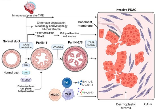
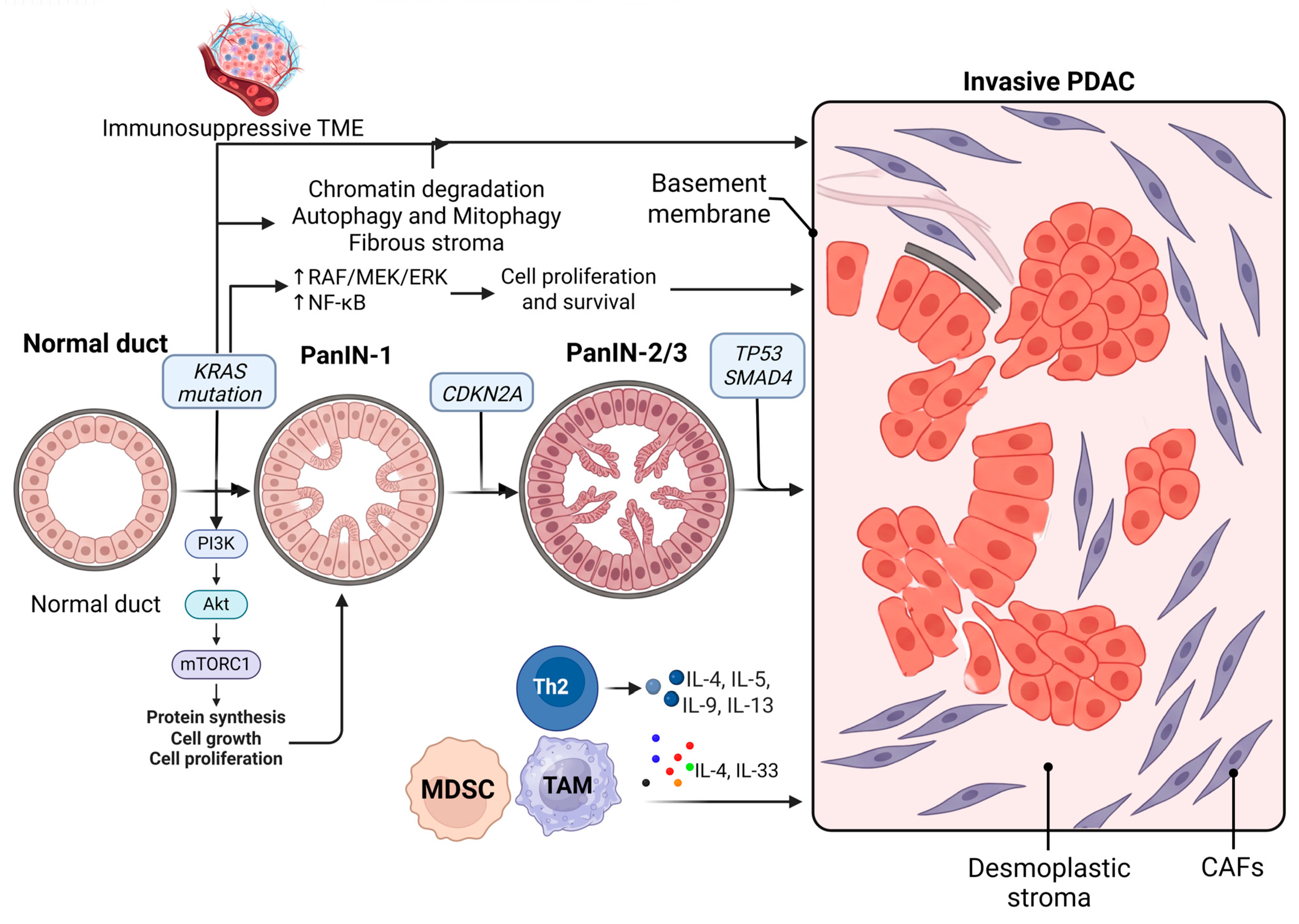
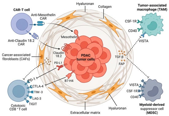
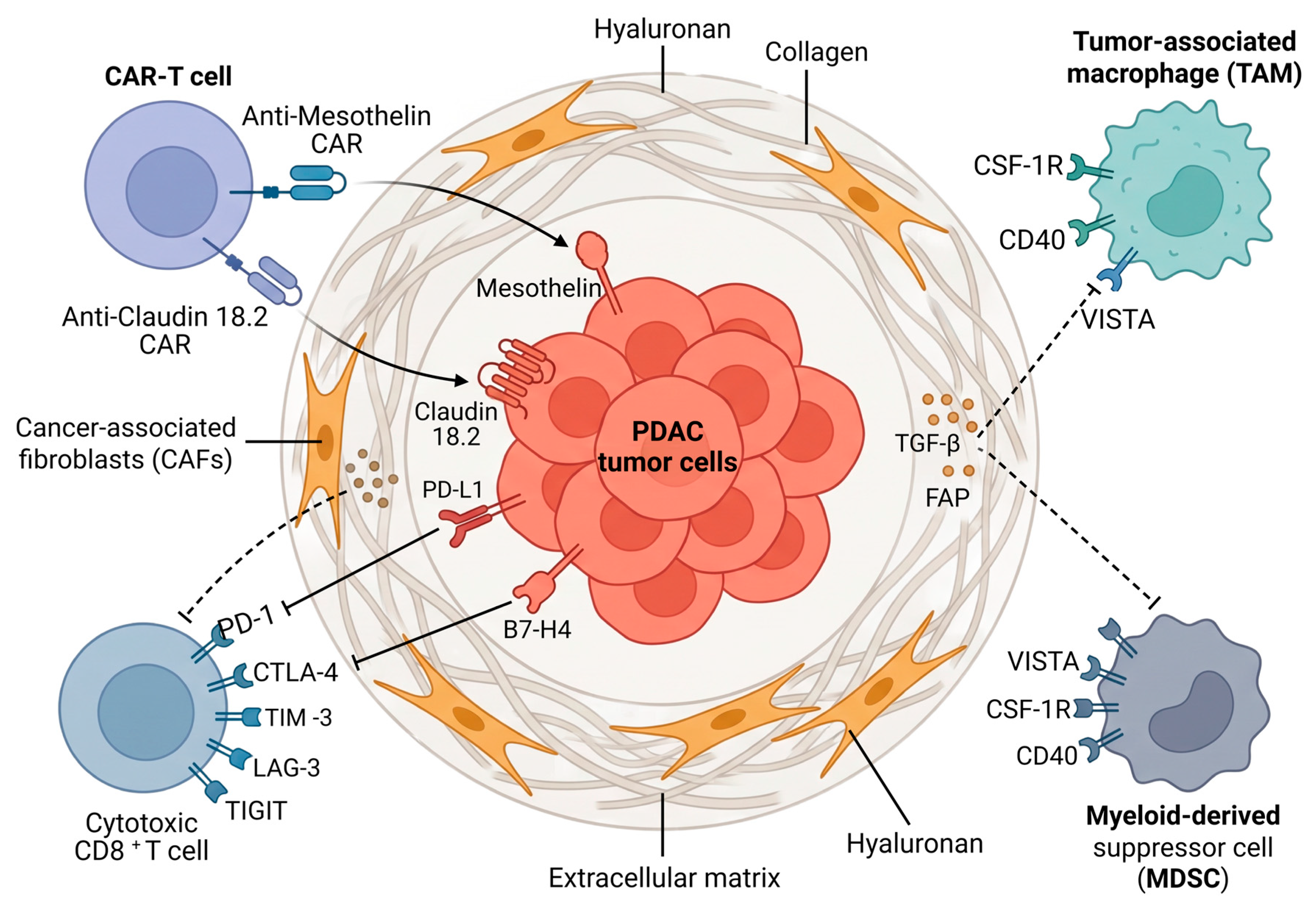

Disclaimer/Publisher’s Note: The statements, opinions and data contained in all publications are solely those of the individual author(s) and contributor(s) and not of MDPI and/or the editor(s). MDPI and/or the editor(s) disclaim responsibility for any injury to people or property resulting from any ideas, methods, instructions or products referred to in the content. |
© 2026 by the authors. Licensee MDPI, Basel, Switzerland. This article is an open access article distributed under the terms and conditions of the Creative Commons Attribution (CC BY) license.