


Abstract
Background: Fatty liver hemorrhagic syndrome (FLHS) is a nutritional-metabolic disorder of laying hens with pathological features analogous to human non-alcoholic fatty liver disease (NAFLD). Aspirin eugenol ester (AEE) has lipid-lowering, anti-inflammatory, and antioxidant activities; however, its protective effects and the underlying molecular mechanisms against FLHS remain unclear. Methods: In this study, we established an in vivo FLHS model in laying hens using a high-energy low-protein diet and an in vitro steatosis model induced by free fatty acids in LMH cells, to explore the hepatoprotective function of AEE. Results: AEE alleviated hepatic lipid accumulation and histopathological lesions, and decreased lipid levels in serum and liver tissue. AEE downregulated the expression of key lipogenic enzymes and upregulated the expression of genes responsible for fatty acid β-oxidation. RNA-seq analysis indicated that the AMPK signaling pathway and autophagy were tightly linked to the hepatoprotective effects of AEE. Treatment with the autophagy activator rapamycin recapitulated the protective effects of AEE, whereas the autophagy inhibitor chloroquine abrogated these beneficial effects. Furthermore, the AMPK inhibitor dorsomorphin partially reversed AEE-mediated liver protection, while the AMPK activator AICAR reproduced the protective effects of AEE. Conclusions: These results indicate that AEE alleviates FLHS in laying hens at least partly through modulation of fatty acid metabolism, likely associated with AMPK/mTOR-mediated autophagy. This work provides mechanistic insights for AEE as a candidate agent for preventing diet-triggered hepatic steatosis.
1. Introduction
Fatty liver hemorrhagic syndrome (FLHS), a typical diet-triggered metabolic disease in laying hens that shares high pathological similarity with non-alcoholic fatty liver disease (NAFLD) in humans, is a common nutritional and metabolic disorder pathologically characterized by excessive accumulation of hepatic and abdominal fat, hepatic steatosis, hepatic rupture, inflammatory infiltration and hemorrhage [1,2]. FLHS severely impairs laying hen health and animal welfare, and brings enormous economic losses to the commercial poultry industry [3]. The pathogenesis of FLHS is multifactorial, involving dietary composition, restricted activity, genetics, environmental stress, and endocrine disturbance, making its prevention and intervention difficult [4,5,6]. Among these contributors, excessive energy intake is the dominant predisposing factor, and FLHS is readily established via feeding a high-energy diet [7,8]. Dysregulated lipid metabolism, excessive oxidative stress, and chronic inflammation collectively accelerate disease progression in FLHS [9].
Accumulating evidence indicates that impaired autophagy participates in the initiation and progression of FLHS, as well as other hepatic steatosis disorders [10,11]. Autophagy is an evolutionarily conserved catabolic process that eliminates damaged organelles and aggregated proteins through lysosomal degradation for nutrient recycling [12]. Extensive research has demonstrated that autophagy regulates cellular lipid homeostasis [13,14]. Autophagy activation exerts a lipid-lowering effect by modulating lipid metabolism-related protein expression, whereas autophagy deficiency exacerbates lipid deposition [15,16]. Restoration of autophagic flux can attenuate or impede the progression of fatty liver disease [17]. Reportedly, autophagy disorders leading to hepatocyte damage and metabolic stress are critical contributors to FLHS in laying hens [18]. Aspirin eugenol ester (AEE) is a novel medicinal compound with multiple beneficial effects, including anti-inflammatory, antioxidant and lipid-lowering activities [19,20,21]. Previous studies reported that AEE ameliorates high-fat diet-induced hepatic steatosis and liver injury by regulating fatty acid metabolism and bile acid homeostasis [22,23,24]. Nevertheless, the specific protective effects and underlying molecular mechanisms of AEE against FLHS in laying hens remain unclear. Hence, exploring whether AEE plays a protective role in FLHS by regulating autophagy is important.
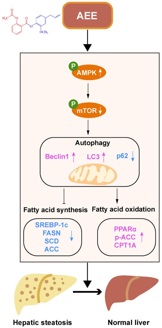
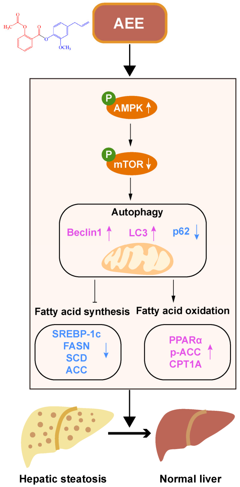
AMP-activated protein kinase (AMPK) is a core sensor and regulator of intracellular energy balance and lipid metabolism [25]. Its downstream signaling effectors, including mammalian target of rapamycin complex 1 (mTORC1) and serine/threonine-protein kinase ULK, are closely related to autophagosome formation and lysosomal acidification [26]. Activation of the AMPK-mTOR signaling cascade can trigger autophagy, alleviate hepatic steatosis, and enhance fatty acid oxidation [27]. Hepatic-specific AMPK activation has been shown to attenuate hepatic steatosis in FLHS-affected laying hens, supporting its promising therapeutic potential for FLHS intervention [28]. Although AEE has been reported to suppress FLHS-induced oxidative stress and inflammatory responses in laying hens [29], it remains unclear whether AEE protects against FLHS through activating the AMPK-mTOR pathway to regulate autophagy. The proposed AMPK/mTOR-autophagy signaling cascade by which AEE mitigates FLHS is illustrated schematically in Figure 1.
Figure 1.
Proposed mechanism underlying the protective effect of AEE against FLHS. AEE may modulate the AMPK/mTOR pathway and alter autophagy-related protein markers, and this action potentially contributes to the regulation of hepatic lipid metabolism and alleviation of FLHS in laying hens. The figure was created using Adobe Illustrator 2020. Black arrows represent the flow of the pathway; white/pink upward arrows indicate up-regulation and white/blue downward arrows indicate down-regulation.
Hence, this study aimed to investigate the regulatory effects of AEE on hepatic lipid metabolism disorders induced by a high-energy and low-protein (HELP) diet in laying hens. Furthermore, we explored the molecular mechanism whereby AEE improves lipid homeostasis by activating AMPK-mediated autophagy using oleic acid- and palmitic acid-treated chicken LMH cells. This work provides mechanistic experimental support for applying AEE as a nutritional preventive strategy against FLHS in laying hens, and also offers a reference for understanding dietary-induced hepatic steatosis relevant to human NAFLD research.
2. Materials and Methods
2.1. Reagents
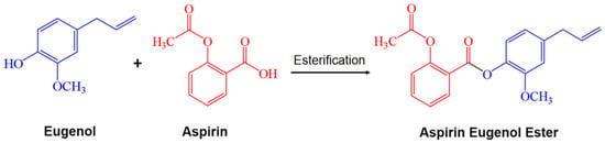
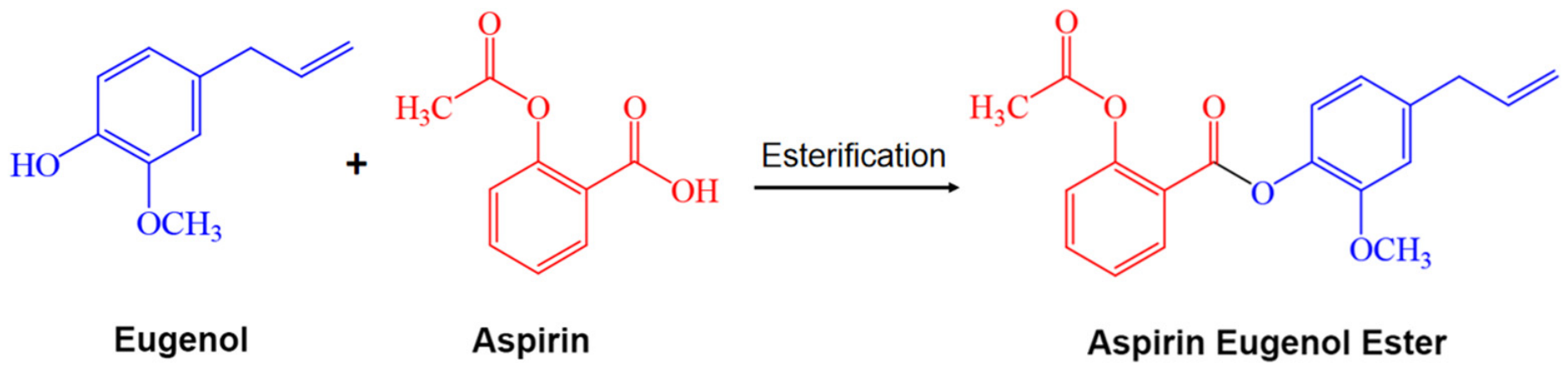
Aspirin eugenol ester (AEE) was synthesized by the Lanzhou Institute of Husbandry and Pharmaceutical Sciences of CAAS (Figure 2) [19]. Choline chloride was purchased from Beijing Zhongtai Hongfeng Technology Co., Ltd. (Beijing, China). LMH cells were purchased from Shanghai Zhong Qiao Xin Zhou Biotechnology Co., Ltd. (Shanghai, China). 0.05% Trypsin-EDTA (25200072) and DMEM (6125298) basal medium were purchased from Gibco (Grand Island, NY, USA). Fetal Bovine Serum (AUS-01E-02) was purchased from Cell-Box (Changsha, China). BCA Protein Assay Kit (PC0020), Trizol® (R1100), RIPA buffer (high) (R0010), SDS-PAGE loading buffer, 5× (with DTT) (P1040), and 100 IU/mL penicillin and 100 μg/mL streptomycin were purchased from Solarbio (Beijing, China). PrimeScript™ RT Master Mix (RR036A) and TB Green® Premix Ex Taq™ II FAST qPCR (CN830A) were purchased from Takara (Nanjing, China). Cell Counting Kit-8 (C6050) was purchased from NCM Biotech (Suzhou, China). Oleic acid (O1008-25g) and palmitic acid (8005081000) were purchased from Sigma-Aldrich (St. Louis, MO, USA). AICAR (HY-13417), Dorsomorphin (HY-13418), Rapamycin (HY-10219), and Chloroquine (HY-17589A) were purchased from Med Chem Express (Shanghai, China). PVDF membrane (ISEQ00010) was purchased from Merck-Millipore (Billerica, MA, USA). DAPI (C1002) was purchased from Beyotime (Nanjing, China). The antibodies for SREBP-1c (14088-1-AP), PPARα (66826-1-lg), FASN (10624-2-AP), ACC (21923-1-AP), p-ACC (29119-1-AP), CPT1A (15184-1-AP), Beclin1 (11306-1-AP), p62 (18420-1-AP), AMPK (10929-2-AP), p-AMPK (80209-6-RR), mTOR (20657-1-AP), p-mTOR (28881-1-AP) and β-actin (20536-1-AP) were purchased from Proteintech (Wuhan, China); the antibody for LC3 (ab192890) was purchased from abcam (Cambridge, UK). The goat anti rabbit/mouse secondary (SA00001-2)/(SA00002-1) antibody was obtained from Proteintech (Wuhan, China). The LMH cell line (Cat. No. ZQ1108) was purchased from Shanghai Zhong Qiao Xin Zhou Biotechnology (Shanghai, China).
Figure 2.
Synthesis of the aspirin eugenol ester.
2.2. Animal Experiments
All animal procedures were performed in accordance with the Guidelines for the Care and Use of Laboratory Animals in China and were approved by the Institutional Animal Care and Use Committee of the Lanzhou Institute of Husbandry and Pharmaceutical Sciences, Chinese Academy of Agricultural Sciences (Approval No. 2023-1016, approved on 30 July 2023). A total of seventy-two healthy 170-day-old Hy-Line Brown laying hens were obtained from a commercial farm. Hens with similar body weight and laying rate were selected prior to grouping. The layers were acclimatized for 1 week with ad libitum access to food and water. The housing conditions were maintained at a controlled temperature of approximately 20–22 °C and a 16/8 h light/dark cycle. Feed intake data for laying hens in this trial have been reported in our previous study [30]; therefore, these data were not included in the present manuscript. After that, the hens were randomly assigned to six groups with 12 hens in each group: normal control diet group (Control), HELP diet treatment group, HELP diet plus 25 mg/kg BW AEE treatment group, HELP diet plus 50 mg/kg BW AEE treatment group, HELP diet plus 100 mg/kg BW AEE treatment group, and HELP diet supplemented with 2 g/kg diet choline chloride (CC) treatment group. AEE and choline chloride were mixed into HELP diets and fed continuously. Choline chloride, a classic lipotropic agent that promotes hepatic TG export via phosphatidylcholine-dependent VLDL assembly and secretion, was used as the positive control [31]. The 2 g/kg dose was adopted from our previous study [29,30]. Previous studies have confirmed that the optimal dose of AEE in rats is 54 mg/kg [21], which was converted to laying hens using the body surface area method, yielding a reference dose of 50 mg/kg. Therefore, the doses of AEE used in this study were 25, 50, and 100 mg/kg (low, medium, and high doses, respectively). This in vivo dose gradient was consistent with that used in our previous study [29,30]. The basal diet was formulated according to the recommendations of the National Research Council [32]. The FLHS model was induced using a high-energy, low-protein (HELP) diet based on a previously described formulation [33], with slight modifications. The compositions of the basal and HELP diets are provided in Table S1.
2.3. Sample Collection and Tissue Index Calculation
At the end of the experiment, all laying hens were fasted for 12 h before euthanasia. Prior to exsanguination, hens were anesthetized with sodium pentobarbital to ensure unconsciousness. Euthanasia was performed via jugular vein exsanguination under gentle manual restraint to minimize stress. Individual final body weight (BW) was recorded prior to tissue sampling. Blood samples were harvested from the brachial vein, allowed to clot overnight at 4 °C, and then centrifuged at 3000 g for 10 min. The serum was stored at −80 °C for subsequent analysis. Following euthanasia, the entire liver and abdominal adipose tissue were carefully excised. Abdominal adipose tissue was defined as the collective fat tissues surrounding the gizzard and intestines as well as those distributed throughout the abdominal cavity. The wet weights of the liver and total abdominal fat were immediately determined using an electronic balance. Liver tissues were divided into two portions: one was frozen and stored at −80 °C and the other was fixed in 4% paraformaldehyde for subsequent analyses.
2.4. Cell Culture and Treatments
LMH cells were cultured in DMEM supplemented with 10% fetal bovine serum, 100 U/mL penicillin, and 100 μg/mL streptomycin. Cultures were incubated at 37 °C in a humidified atmosphere containing 5% CO2. LMH cells in the logarithmic growth phase were used for experiments. The number of biological replicates was arranged according to the requirements of different functional assays, which accounts for the difference in replicate numbers between in vitro and in vivo experiments. AEE was dissolved in dimethyl sulfoxide (DMSO) at a final concentration of ≤0.1% (v/v) in culture medium. The control group received an equivalent volume of DMSO (0.1% v/v) as solvent control to exclude any potential solvent effects.
To induce cellular steatosis, cells were exposed to 0.5 mM free fatty acids (FFAs) for 24 h. The FFAs were prepared as a mixture of oleic acid and palmitic acid (2:1, molar ratio) complexed with fatty acid-free BSA.
To assess the protective effects of AEE, cells were pretreated with AEE (16, 32, or 64 μM) for 24 h prior to FFA exposure. The AEE concentrations of 16, 32, and 64 μM used in this cellular assay were adopted from our previous study [29]. For the inhibition studies, the cells were incubated in the presence or absence of AICAR (AMPK activator, 0.1 mM), Dorsomorphin (AMPK inhibitor, 10 μM), Rapamycin (autophagy activator, 100 nM), and Chloroquine (autophagy inhibitor, 50 μM) for 1 h before FFA treatment for 24 h.
2.5. Cell Viability Assay
Cell viability was evaluated using a CCK-8. Briefly, LMH cells were seeded in 96-well plates at a density of 1 × 10³ cells per well and incubated for 24 h. The culture medium was then replaced with fresh medium containing various concentrations of FFAs and/or AEE for an additional 24 h. After treatment, the medium was discarded, and the cells were washed twice with PBS. Subsequently, 90 μL of fresh medium and 10 μL of CCK-8 solution were added to each well, followed by incubation at 37 °C for 1 h. The absorbance was measured at 450 nm using a microplate reader (Thermo Fisher Scientific, Shanghai, China).
2.6. Biochemical Analysis
Serum concentrations of total cholesterol (TC), triglyceride (TG), high-density lipoprotein cholesterol (HDL), low-density lipoprotein cholesterol (LDL), alanine aminotransferase (ALT), and aspartate aminotransferase (AST) were measured using an automatic biochemical analyzer (XL-640, Erba, Mannheim, Germany) according to the manufacturer’s protocols.
Hepatic TC, TG, HDL, LDL, and FFA levels were detected using assay kits from Solarbio Science & Technology Co., Ltd. (Beijing, China). Cellular TC, TG, HDL, LDL, and AST levels were assessed using corresponding kits from the Nanjing Jiancheng Bioengineering Institute (Nanjing, China).
2.7. Hematoxylin-Eosin (H&E) Staining
For histological evaluation, liver tissues were fixed in 4% paraformaldehyde for 24 h and subsequently embedded in paraffin. Sections of 5 μm thickness were prepared and stained with H&E as previously described [34]. The stained sections were imaged under a light microscope.
2.8. Oil Red O and Nile Red Staining
To assess hepatic steatosis, Oil Red O staining was performed on frozen liver sections and cultured cells, as previously described [35]. Briefly, frozen liver sections were equilibrated at room temperature for 10 min and rinsed with distilled water. Cultured cells were washed twice with PBS, fixed with 4% paraformaldehyde for 20 min, and then rinsed twice with distilled water. Both liver and cell samples were immersed in 60% isopropanol for 30 s, followed by staining with Oil Red O working solution for 20 min. Excess dye was removed with 60% isopropanol. Nuclei were counterstained with hematoxylin for 2 min and washed under running distilled water for 10 min to develop the blue color. Finally, samples were rinsed with distilled water, mounted with glycerin gelatin, and photographed using an optical microscope. Nile Red staining was performed according to a previous report [36].
2.9. Immunohistochemistry
Liver tissues were fixed, dehydrated, cleared, and embedded in paraffin according to standard histological procedures. Paraffin sections (5 μm) were deparaffinized, rehydrated through a graded ethanol series, and subjected to antigen retrieval. Endogenous peroxidase activity was blocked with 3% hydrogen peroxide, followed by incubation with normal serum for 30 min at room temperature to reduce nonspecific binding. The sections were then incubated overnight at 4 °C with primary antibodies against FASN, CPT1A, LC3, and Beclin1 (1:150 dilution). After washing, the sections were incubated with horseradish peroxidase (HRP)-conjugated secondary antibodies for 30 min at room temperature. Immunoreactive signals were visualized using a DAB substrate kit, and nuclei were counterstained with hematoxylin. Finally, the sections were dehydrated, cleared, mounted, and examined under a light microscope.
2.10. Immunofluorescence Staining
For tissue sections, liver tissue sections were deparaffinized. For cell samples, cultured cells were washed three times with PBS and fixed with 4% paraformaldehyde for 20 min. Both tissue sections and cultured cells were then permeabilized with 0.3% Triton X-100 for 15 min and blocked with 5% BSA for 1 h at room temperature. Subsequently, the samples were incubated overnight at 4 °C with primary antibodies against LC3 and p62 (1:100 dilution). After washing, the samples were incubated with Alexa Fluor 488-conjugated goat anti-rabbit IgG secondary antibodies at 37 °C for 45 min. Nuclei were counterstained with DAPI, and images were captured using a fluorescence microscope.
2.11. Quantitative Real-Time PCR
Quantitative real-time PCR (qRT-PCR) was conducted according to a previously reported protocol [37]. Briefly, total RNA was extracted from liver tissues using TRIzol reagent. RNA concentration and purity were assessed spectrophotometrically. Samples with an A260/A280 ratio of 1.8–2.0 and an A260/A230 ratio greater than 2.0 were considered acceptable for further analysis. cDNA was synthesized using the PrimeScript RT Reagent Kit following the manufacturer’s instructions. qRT-PCR was carried out using SYBR Green PCR Master Mix on an ABI 7500 Real-Time PCR system (Applied Biosystems, Foster City, CA, USA). Primers for SREBP-1c, PPARα, FASN, ACC, SCD1, CPT1A, p62, Beclin1, LC3B1, LC3B2, and the reference gene β-actin were designed using Primer Premier 5 software. The Primer sequences are listed in Table S2. The relative mRNA expression levels of target genes were normalized to β-actin, and calculated by the 2−ΔΔCt method [38].
2.12. Western Blot
Total protein was extracted from laying hen liver tissues and LMH cells using RIPA lysis buffer supplemented with 1% protease/phosphatase inhibitor cocktail. The protein concentrations were determined with a BCA Protein Assay Kit. Equal amounts of protein were separated by SDS-PAGE and transferred onto PVDF membranes. After blocking, the membranes were incubated overnight at 4 °C with primary antibodies specifically recognizing SREBP-1c, PPARα, FASN, ACC, p-ACC, CPT1A, Beclin1, p62, AMPK, p-AMPK, mTOR, p-mTOR and β-actin. Subsequently, the membranes were incubated with horseradish peroxidase (HRP)-conjugated secondary antibodies for 1 h at room temperature. Immunoreactive bands were visualized using an enhanced chemiluminescence (ECL) detection system and imaged with an Amersham Imager 600 (GE Healthcare Bio-Sciences, Uppsala, Sweden). β-actin was used as the loading control for normalization. The protein abundance was analyzed using the ImageJ software (version 1.53, National Institutes of Health, Bethesda, MD, USA).
2.13. RNA Sequencing and Bioinformatics Analyses
Total RNA was extracted from liver tissues using TRIzol reagent (Invitrogen, Life Technologies, Carlsbad, CA, USA). Briefly, frozen liver samples were ground in grinding tubes (60 Hz, 60 s). TRIzol (1 mL) was added on ice, followed by secondary wet grinding (55 Hz, 30 s) and 5 min room-temperature incubation. After centrifugation (12,000 rpm, 5 min), the supernatant was collected, mixed with 0.2 mL chloroform, vortexed, and incubated. Following centrifugation (12,000 rpm, 15 min, 4 °C), the aqueous phase was transferred, combined with isopropanol, and incubated for 10 min. RNA was pelleted (12,000 rpm, 10 min, 4 °C), washed with 75% ethanol, and re-centrifuged. Ethanol was removed, and the pellet was dissolved in RNase-free water; vacuum drying was avoided to prevent poor RNA solubility. RNA concentration and purity were assessed using a NanoDrop spectrophotometer (Thermo Scientific NanoDrop One, Thermo Fisher Scientific, Waltham, MA, USA). RNA integrity was evaluated by agarose gel electrophoresis and the Agilent 2100 Bioanalyzer System (Agilent Technologies, Santa Clara, CA, USA); samples with RIN > 7.0 were retained for subsequent library construction.
Sequencing libraries were constructed using 3 μg of high-quality total RNA per sample with the TruSeq RNA Sample Preparation Kit (Illumina, San Diego, CA, USA), following the manufacturer’s standard protocol. Briefly, mRNA was enriched from total RNA via poly-T oligo-attached magnetic beads and fragmented into short fragments under elevated temperature. First-strand cDNA was synthesized with random hexamer primers and SuperScript II reverse transcriptase, and second-strand cDNA was subsequently synthesized. Purified cDNA fragments underwent end repair, 3′-end adenylation, and ligation to Illumina sequencing adapters. Target cDNA fragments ranging from 400 to 500 bp were size-selected using the AMPure XP system (Beckman Coulter, Brea, CA, USA), followed by PCR amplification. The quality of final libraries was validated on the Agilent 2100 Bioanalyzer. Paired-end sequencing (2 × 150 bp) was carried out on an Illumina NovaSeq 6000 platform at Shanghai Personal Biotechnology Co., Ltd., China.
Raw FASTQ sequencing reads were preprocessed with cutadapt to trim adapter sequences, remove reads containing ambiguous bases, and filter low-quality reads (Phred quality score Q < 20). The resulting clean reads were mapped to the chicken reference genome using HISAT2 (v2.1.0). Gene expression abundance was quantified with HTSeq (v0.9.1) and normalized to FPKM (Fragments Per Kilobase of transcript per Million mapped reads) and TPM (Transcripts Per Million).
Differentially expressed genes (DEGs) were screened using the DESeq2 R package (v1.38.3) based on three independent biological replicates for each experimental group. Genes with an adjusted p value (Benjamini-Hochberg false discovery rate) < 0.05 and |log2 fold change| > 1 were defined as significantly differentially expressed. Functional enrichment analyses, including Gene Ontology (GO) and Kyoto Encyclopedia of Genes and Genomes (KEGG) pathway enrichment of DEGs, were implemented using the clusterProfiler R package (v4.6.0). Enriched terms and pathways with p < 0.05 were considered statistically significant.
2.14. Statistical Analysis
All data are presented as the mean ± standard error of mean (SEM). One-way analysis of variance (ANOVA) was performed to compare differences among multiple groups. Duncan’s multiple comparison test was applied for post hoc pairwise comparisons. All statistical analyses were conducted using SPSS software (version 22.0; IBM, Armonk, NY, USA). All graphical presentations were created using GraphPad Prism (version 8.0; GraphPad Software, San Diego, CA, USA). A p value less than 0.05 was considered statistically significant.
3. Results
3.1. AEE Alleviates Liver Injury, Hepatic Steatosis, and Dyslipidemia in HELP Diet-Fed Laying Hens
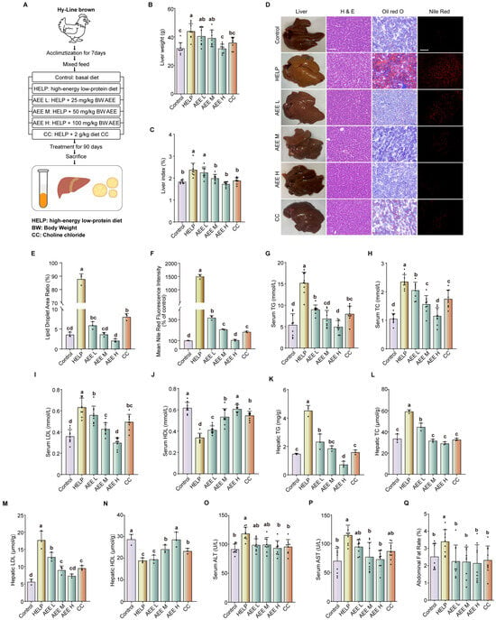
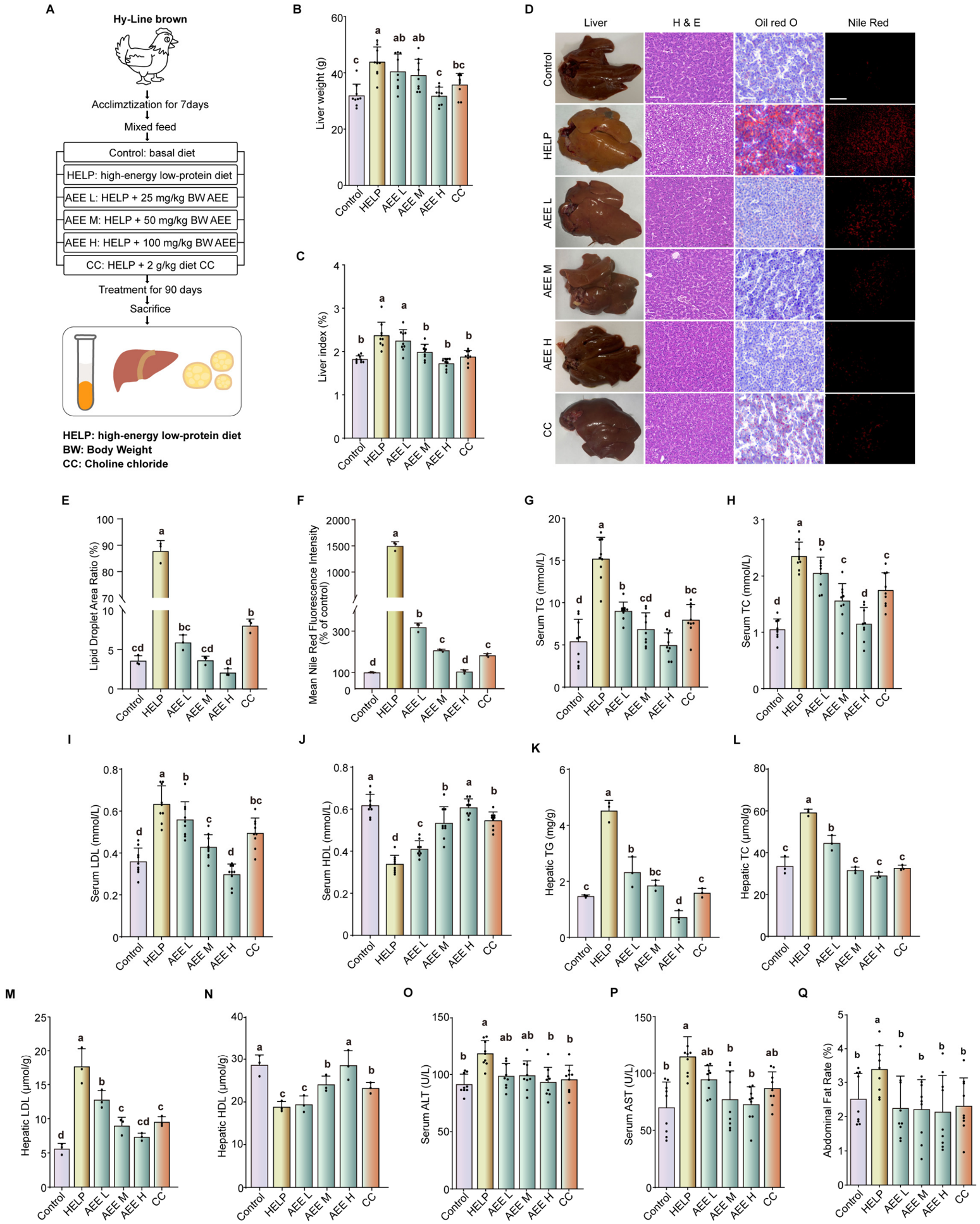
High-dose AEE and CC treatments reduced liver weight and liver index in laying hens fed a HELP diet (Figure 3B,C, p < 0.05). Gross morphological observation revealed that livers from the HELP group appeared enlarged, yellowish, and greasy compared with those of the control group, alterations alleviated by both AEE and CC treatment (Figure 3D). Consistently, H&E, Oil Red O, and Nile Red staining demonstrated that AEE and CC administration reduced hepatocellular vacuolar degeneration and lipid droplet accumulation in HELP-fed laying hens (Figure 3D–F). Serum lipid profiling revealed dyslipidemia in the HELP group, characterized by elevated TG, TC, LDL and reduced HDL, all of which were reversed by AEE and CC treatment (Figure 3G–J, p < 0.05). Hepatic lipid content showed a similar pattern (Figure 3K–N, p < 0.05). Moreover, both AEE and CC treatment reduced serum ALT and AST activities, and abdominal fat index in HELP-fed laying hens (Figure 3O–Q, p < 0.05). These results demonstrate that AEE effectively alleviates HELP diet-induced FLHS in laying hens.
Figure 3.
AEE alleviates liver injury, hepatic steatosis, and dyslipidemia in laying hens with FLHS. (A) Experimental schedule. AEE was administered at doses of 25, 50 and 100 mg/kg body weight (BW); choline chloride (CC) was supplemented to the diet at 2 g/kg diet. (B) Liver weight. (C) Liver index. (D) Representative photograph of liver morphology, and liver sections stained with H&E (400×), Oil Red O (400×), and Nile Red (200×). (E) Quantitative analysis of Oil Red O staining. (F) Quantification of Nile Red fluorescence intensity. (G) Serum triglyceride (TG) content. (H) Serum total cholesterol (TC) content. (I) Serum low-density lipoprotein cholesterol (LDL) content. (J) Serum high-density lipoprotein cholesterol (HDL) content. (K) Hepatic triglyceride (TG) content. (L) Hepatic total cholesterol (TC) content. (M) Hepatic low-density lipoprotein cholesterol (LDL) content. (N) Hepatic high-density lipoprotein cholesterol (HDL) content. (O) Serum ALT activity. (P) Serum AST activity. (Q) Abdominal fat index. Data are presented as mean ± SEM. n = 9 per group. Different lowercase letters indicate statistically significant differences (p < 0.05).
3.2. AEE Regulates Hepatic Fatty Acid Metabolism in Laying Hens Fed a HELP Diet
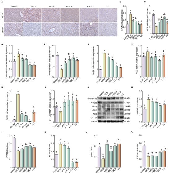
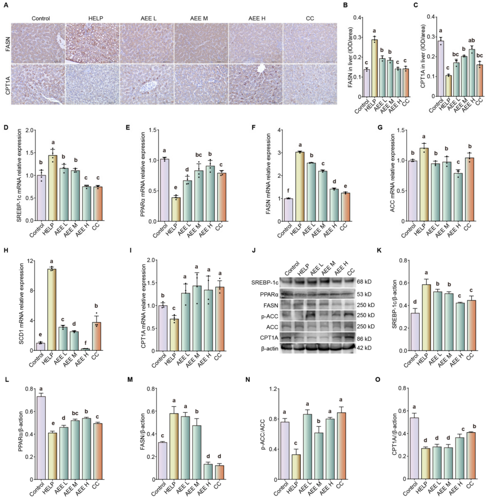
To further elucidate the protective effects of AEE against FLHS, the expression of key regulators involved in hepatic fatty acid metabolism was examined. Immunohistochemistry revealed that the HELP group exhibited stronger immunoreactivity for FASN and weaker CPT1A expression. Both AEE and CC treatment reversed these alterations (Figure 4A–C, p < 0.05). qRT-PCR results showed that AEE and CC treatment downregulated the expression of SREBP-1c, FASN, ACC, and SCD1, and upregulated the expression of PPARα and CPT1A in HELP-induced laying hens (Figure 4D–I, p < 0.05). Western blot results confirmed that the protein expression patterns were consistent with the transcriptional changes above (Figure 4J–O, p < 0.05). The original uncropped Western blot images are shown in Figure S1. These results indicate that AEE regulates hepatic fatty acid metabolism to restore metabolic homeostasis in laying hens with FLHS.
Figure 4.
AEE suppresses hepatic fatty acid synthesis and promotes fatty acid β-oxidation in HELP diet-fed laying hens. (A) Immunohistochemical staining of FASN and CPT1A in the liver (400×). (B,C) Quantitative analysis of immunohistochemical staining. (D–I) Relative mRNA levels of SREBP-1c, PPARα, FASN, ACC, SCD1, and CPT1A in livers. (J–O) Relative protein expression of SREBP-1c, PPARα, FASN, p-ACC, ACC, and CPT1A. Data are presented as mean ± SEM. n = 3–4 per group. Different lowercase letters indicate statistically significant differences (p < 0.05). The original uncropped Western blot images are shown in Figure S1.
3.3. AEE Attenuated FFA-Induced Steatosis in LMH Cells
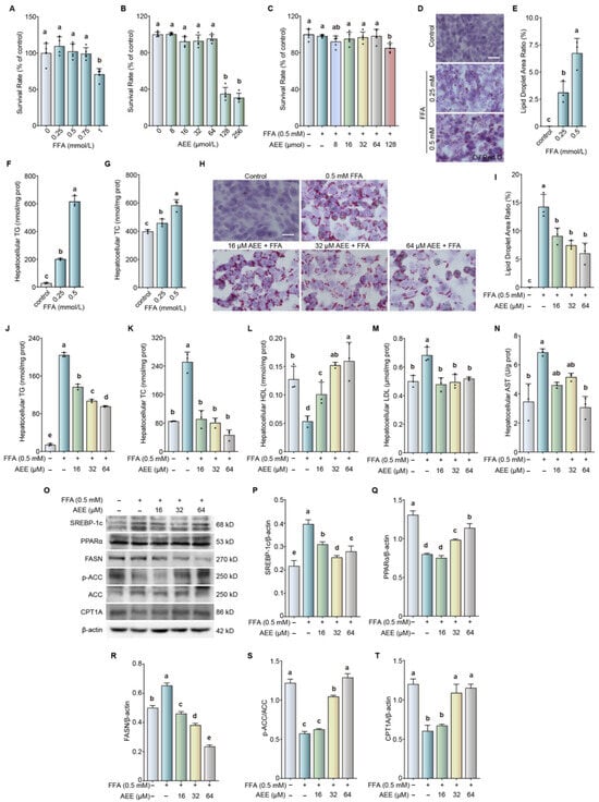
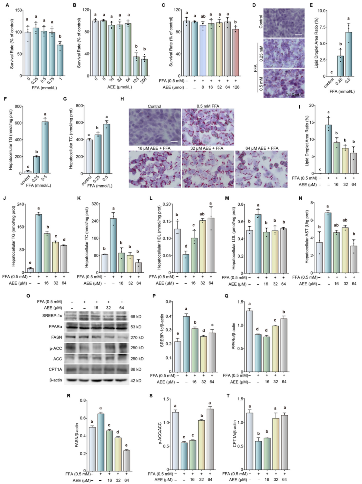
To further confirm the hepatoprotective effects of AEE observed in vivo, a cellular model of steatosis was induced in LMH cells by FFA exposure. The cytotoxicity of AEE and FFA was first evaluated. AEE (>64 μmol/L) or FFA (1 mmol/L) alone significantly reduced cell viability (Figure 5A,B, p < 0.05). Combined treatment with 16, 32, 64 μmol/L AEE and 0.5 mmol/L FFA did not affect cell viability (Figure 5C). Therefore, subsequent experiments were performed using 16–64 μmol/L AEE with 0.5 mmol/L FFA. Exposure to 0.25 or 0.5 mmol/L FFA induced steatosis in a concentration-dependent manner, as evidenced by increased lipid droplet content and elevated TG and TC levels (Figure 5D–G, p < 0.05). AEE reduced intracellular lipid droplet content, decreased TG, TC, and LDL levels and AST activity, and increased HDL levels in FFA-induced LMH cells (Figure 5H–N, p < 0.05). Western blot analysis revealed that AEE inhibited the expression of SREBP-1c and FASN, promoted the expression of PPARα and CPT1A, and enhanced ACC phosphorylation in FFA-induced LMH cells (Figure 5O–T, p < 0.05). These results indicate that AEE regulates fatty acid metabolism and alleviates FFA-induced steatosis in LMH cells.
Figure 5.
AEE alleviates hepatic steatosis by regulating fatty acid metabolism in FFA-treated LMH cells. (A–C) Cytotoxicity assessment. (D) Representative photomicrographs of LMH cells stained with Oil Red O (400×). (E) Quantitative analysis of Oil Red O staining. (F) Hepatocellular TG level. (G) Hepatocellular TC level. (H) Representative photomicrographs of LMH cells stained with Oil Red O (400×). (I) Quantitative analysis of Oil Red O staining. (J) Hepatocellular TG content. (K) Hepatocellular TC content. (L) Hepatocellular HDL content. (M) Hepatocellular LDL content. (N) Hepatocellular AST activity. (O–T) Relative protein expression of SREBP-1c, PPARα, FASN, p-ACC, ACC, and CPT1A. Data are presented as mean ± SEM. n = 3–4 per group. Different lowercase letters indicate statistically significant differences (p < 0.05) The original uncropped Western blot images are shown in Figure S2.
3.4. Transcriptomic Analyses of the Protective Mechanisms of AEE Against FLHS in Laying Hens
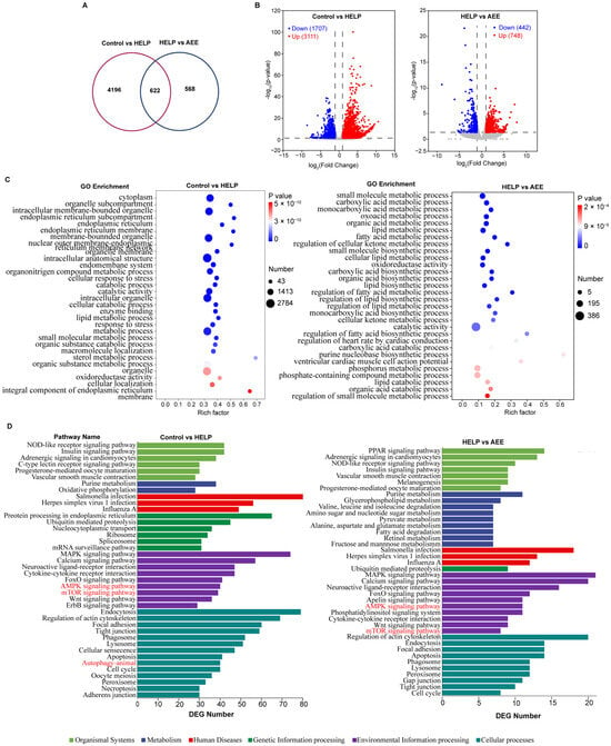
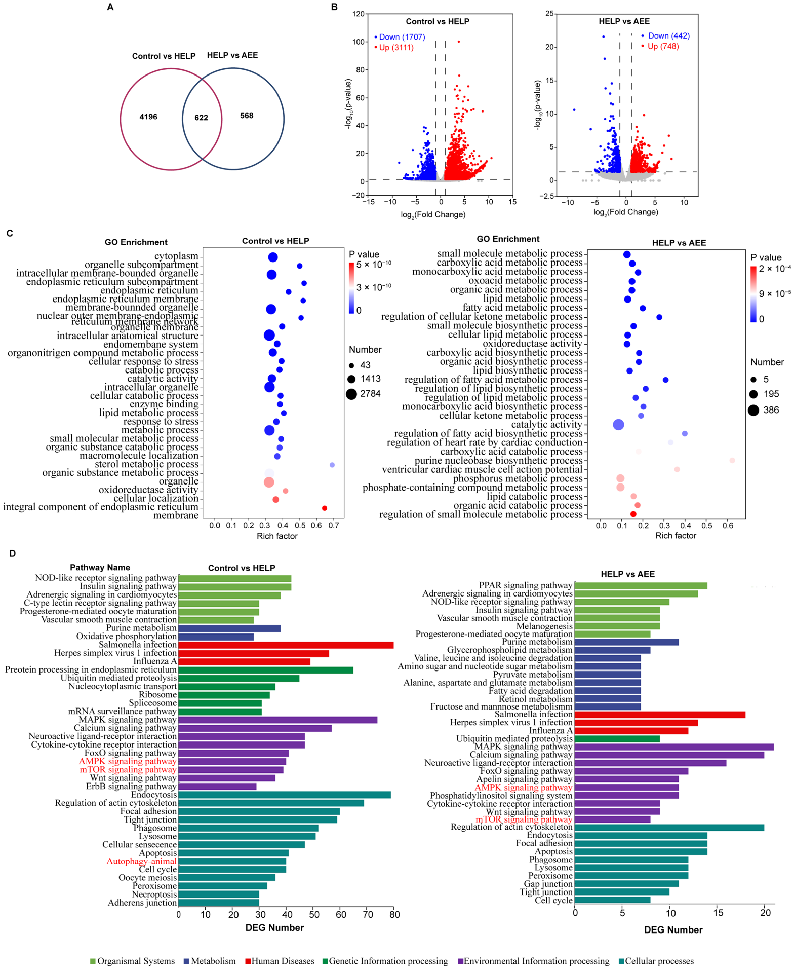
To elucidate the molecular mechanism by which AEE protects against FLHS, transcriptomic analyses were performed on liver tissues from control, HELP (FLHS model), and AEE-treated hens. A total of 4818 differentially expressed genes (DEGs) were identified between the HELP and control group, and 1190 DEGs between the AEE and HELP group (Figure 6A,B), indicating substantial transcriptomic modulation by AEE. GO enrichment analysis showed that DEGs were mainly involved in cellular catabolic processes, lipid and fatty acid metabolic processes (Figure 6C). KEGG pathway analysis identified the AMPK/mTOR and autophagy pathways (Figure 6D), which are critically involved in lipid metabolism. The omics data suggest that AEE may alleviate FLHS by modulating hepatic lipid metabolism through the AMPK/mTOR and autophagy signaling pathways.
Figure 6.
Transcriptomic profiling reveals potential mechanisms of AEE against FLHS in laying hens. (A) Venn diagram of DEGs. (B) Volcano plots of DEGs. Gray dots represent non-significant genes; the vertical dashed lines mark the fold-change threshold (|log2FC| ≥ 1), and the horizontal dashed line marks the significance threshold (−log10 p-value). Red and blue dots denote up-regulated and down-regulated genes, respectively. (C) GO enrichment analysis of the DEGs. (D) KEGG pathway enrichment analysis of DEGs.
3.5. AEE Ameliorates FFA-Induced Steatosis via Activating Autophagy in LMH Cells
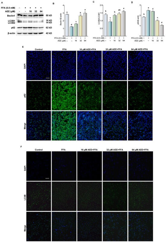
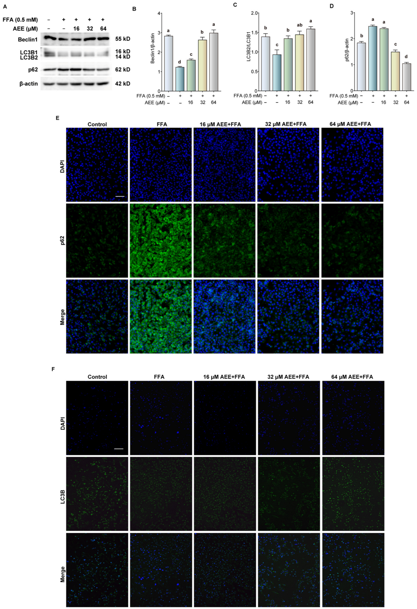
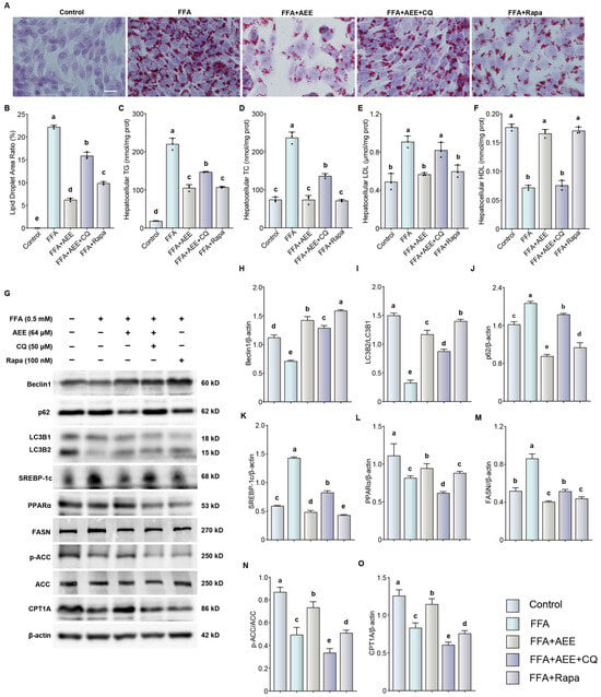
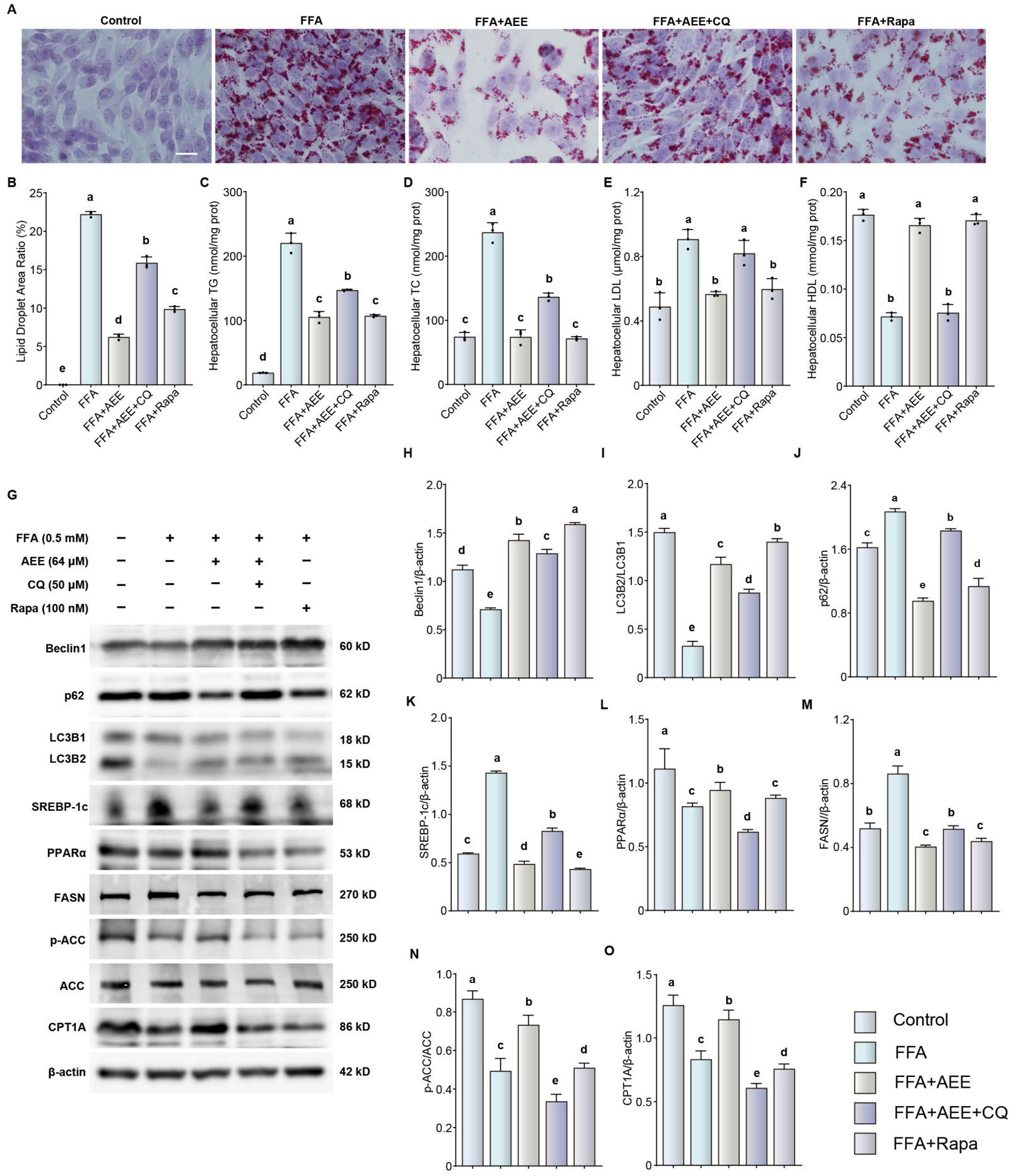
To validate the transcriptomic findings, we further examined changes in the autophagy pathway in FFA-induced LMH cells. Numerous studies have shown that autophagy plays a key role in maintaining cellular lipid balance [13,14]. AEE concentration-dependently increased the protein expression of Beclin1 and LC3B2 and decreased p62 expression (Figure 7A–D, p < 0.05). Immunofluorescence staining of LC3B and p62 further confirmed these results (Figure 7E,F). Furthermore, the autophagy inhibitor CQ partially blocked the inhibitory effects of AEE on lipid droplet content, TG, TC, and LDL, as well as its promoting effect on HDL (Figure 8A–F, p < 0.05). CQ also partially reversed the inhibitory effects of AEE on the expression of p62, SREBP-1c, and FASN, as well as its promoting effects on the expression of Beclin1, LC3B2, PPARα, and CPT1A, and on the phosphorylation level of ACC (Figure 8G–O, p < 0.05). Notably, Rapa treatment reduced FFA-induced lipid droplet deposition, lowered TG, TC and LDL-C, and increased HDL-C levels (Figure 8A–F, p < 0.05). At the protein level, Rapa upregulated Beclin1 and LC3B-II, suppressed p62, SREBP-1c and FASN, and enhanced the expression of PPARα, CPT1A and p-ACC (Figure 8G–O, p < 0.05). Collectively, these results suggest that the hepatoprotective effect of AEE against FFA-induced steatosis in LMH cells is likely linked to autophagy activation and subsequent modulation of lipid metabolism.
Figure 7.
AEE enhances autophagy in FFA-treated LMH cells. (A–D) Relative protein expression of Beclin1, LC3B, and p62. (E,F) Representative immunofluorescence staining for p62 and LC3B (400×). Data are presented as mean ± SEM. n = 3 per group. Different lowercase letters indicate statistically significant differences (p < 0.05) The original uncropped Western blot images are shown in Figure S3.
Figure 8.
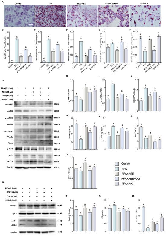
AEE alleviates hepatic steatosis by activating autophagy to regulate fatty acid metabolism in FFA-treated LMH cells. (A) Representative photomicrographs of LMH cells stained with Oil Red O (400×). (B) Quantitative analysis of Oil Red O staining. (C–F) Hepatocellular TG, TC, LDL, and HDL content. (G–O) Relative protein expression of Beclin1, p62, LC3B, SREBP-1c, PPARα, FASN, p-ACC, ACC, and CPT1A. Data are presented as mean ± SEM. n = 3 per group. Different lowercase letters indicate statistically significant differences (p < 0.05). The original uncropped Western blot images are shown in Figure S4.
3.6. AEE Ameliorates FFA-Induced Steatosis in LMH Cells Possibly Through the AMPK/mTOR-Autophagy Axis
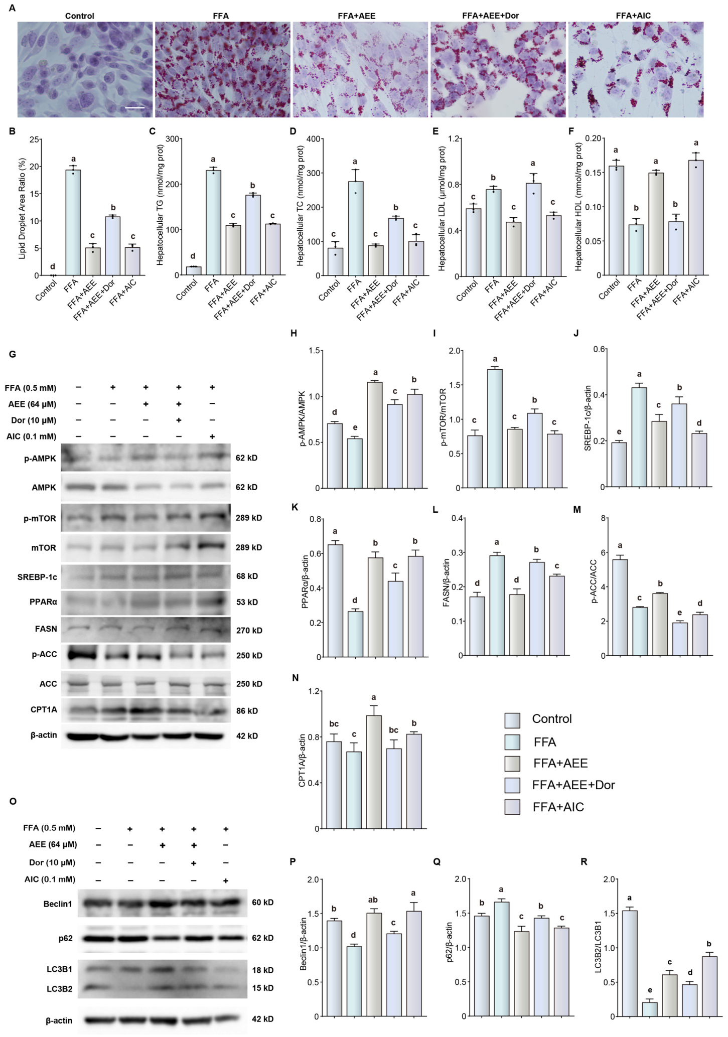
AMPK/mTOR signaling functions as a central metabolic sensor that regulates cellular energy homeostasis and autophagy [25,26]. The AMPK inhibitor Dor partially blocked the inhibitory effects of AEE on lipid droplet content, TG, TC, and LDL, as well as its promoting effect on HDL (Figure 9A–F, p < 0.05). Dor partially blocked the promoting effect of AEE on AMPK phosphorylation and its inhibitory effect on mTOR phosphorylation (Figure 9G–I, p < 0.05). Moreover, Dor reversed the inhibitory effects of AEE on the expression of SREBP-1c and FASN, as well as its promoting effects on the expression of PPARα and CPT1A and on ACC phosphorylation (Figure 9G,J–N, p < 0.05). Dor also partially blocked the promoting effects of AEE on Beclin1 and LC3B2 expression, as well as its inhibitory effect on p62 expression (Figure 9O–R, p < 0.05). Consistent with the effects of AEE, the AMPK activator AIC reduced lipid droplet accumulation, lowered cellular TG, TC and LDL levels, increased HDL content (Figure 9A–F, p < 0.05), elevated p-AMPK abundance, suppressed p-mTOR and lipogenic proteins (SREBP-1c, FASN), upregulated PPARα, CPT1A and p-ACC, and induced autophagy by increasing Beclin1 and LC3B2 while decreasing p62 (Figure 9G–R, p < 0.05). These findings suggest that the anti-steatotic effect of AEE is likely associated with activation of the AMPK/mTOR-mediated autophagy.
Figure 9.
AEE alleviates hepatic steatosis by regulating fatty acid metabolism via AMPK/mTOR-mediated autophagy in FFA-treated LMH cells. (A) Representative photomicrographs of LMH cells stained with Oil Red O (400×). (B) Quantitative analysis of Oil Red O staining. (C–F) Hepatocellular TG, TC, LDL, and HDL content. (G–N) Relative protein expression of p-AMPK, AMPK, p-mTOR, mTOR, SREBP-1c, PPARα, FASN, p-ACC, ACC, and CPT1A. (O–R) Western blotting analyses of Beclin1, p62, and LC3B. Data are presented as mean ± SEM. n = 3 per group. Different lowercase letters indicate statistically significant differences (p < 0.05). The original uncropped Western blot images are shown in Figure S5.
3.7. AEE Promotes AMPK/mTOR Pathway and Autophagy in HELP Diet-Fed Laying Hens
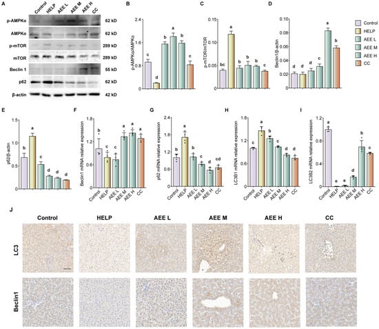
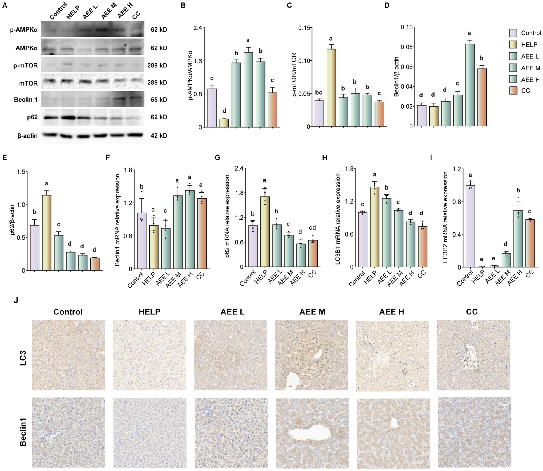
We examined the effects of AEE on AMPK/mTOR signaling and autophagy in vivo. In HELP diet-fed laying hens, Western blot results showed that AEE promoted AMPK phosphorylation and inhibited mTOR phosphorylation (Figure 10A–C, p < 0.05). Additionally, AEE increased Beclin1 protein expression and decreased p62 expression (Figure 10A,D,E, p < 0.05). qRT-PCR results demonstrated that AEE concentration-dependently increased the mRNA expression of Beclin1 and LC3B2, while decreasing the mRNA expression of p62 and LC3B1 (Figure 10F–I, p < 0.05). Immunohistochemical staining for Beclin1 and LC3B further confirmed the above results (Figure 10J).
Figure 10.
AEE activates AMPK and enhances autophagy in the liver of HELP diet-fed laying hens. (A–E) Relative protein expression of p-AMPK, AMPK, p-mTOR, mTOR, Beclin1, and p62. (F–I) Relative mRNA levels of Beclin1, p62, LC3B1, and LC3B2 in livers. (J) Representative photomicrographs of liver sections stained by immunohistochemistry (400×). Data are presented as mean ± SEM. n = 3–4 per group. Different lowercase letters indicate statistically significant differences (p < 0.05). The original uncropped Western blot images are shown in Figure S6.
4. Discussion
Fatty liver hemorrhagic syndrome (FLHS) is a prevalent nutritional and metabolic disorder in high-producing laying hens, primarily resulting from nutrient imbalances that disrupt hepatic lipid homeostasis [39]. To date, no effective pharmacological intervention has been widely adopted for FLHS prevention or treatment. Therefore, elucidating the molecular mechanisms of FLHS and identifying potential therapeutic agents are of considerable importance to the poultry industry.
The HELP diet has been widely validated as a reliable method for establishing an FLHS model in laying hens, effectively recapitulating key pathophysiological features, including hepatic lipid deposition, metabolic dysregulation, and declined production performance [40,41]. The results of this study showed that laying hens fed a HELP diet exhibited decreased production performance and increased serum and hepatic lipid contents, which was consistent with previous reports [38,39], indicating the successful establishment of the FLHS model in laying hens. Notably, AEE improved production performance, reduced the levels of TG, TC, LDL, and FFA in both serum and liver, increased HDL levels, and alleviated histopathological damage in laying hens fed with HELP diet. Studies have shown that AEE reduces lipid content in animals with HFD-induced hyperlipidemia [23,42]. Notably, the biological activity of AEE is closely linked to its chemical structure. Eugenol, the main bioactive component of clove oil, exerts anti-inflammatory, antioxidant and lipid-regulating effects [43,44]. As an ester conjugate of aspirin and eugenol, AEE depends on the eugenol skeleton for its hepatoprotective activity. Substitution with eugenol analogues may alter its bioactivity, and relevant structure–activity relationships will be explored in future studies. Thus, our findings support the notion that AEE alleviates hepatic lipid accumulation in laying hens fed a HELP diet.
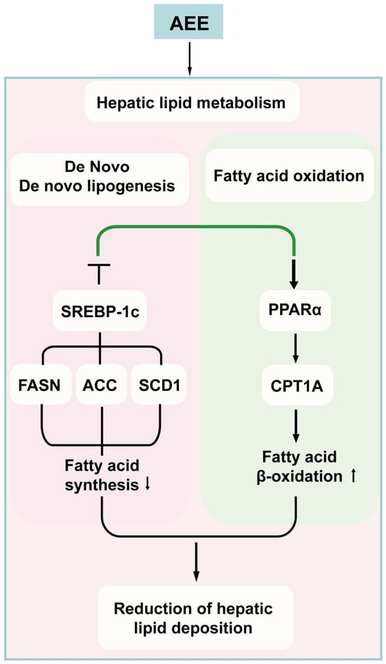
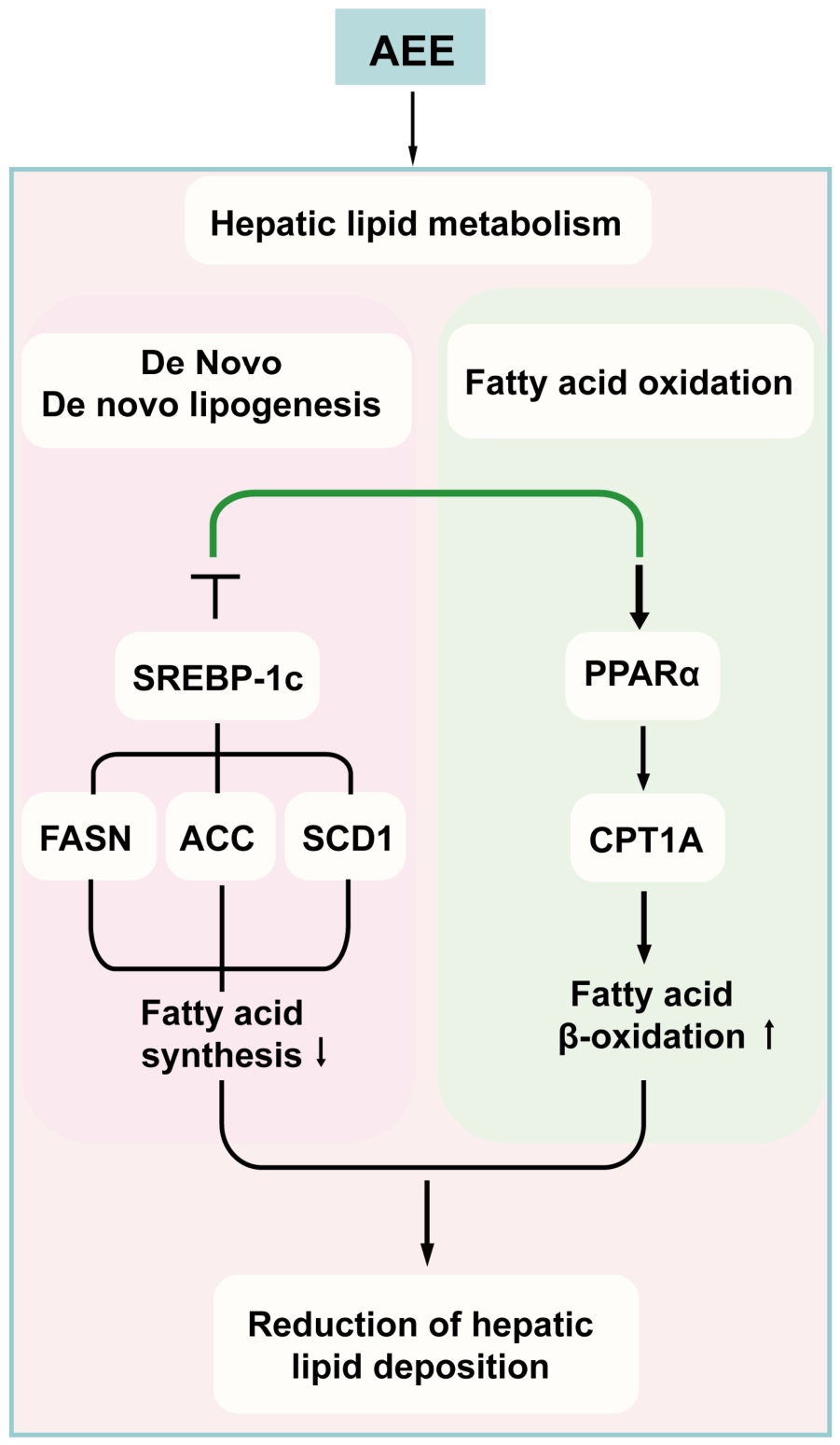
The poultry liver serves as the central organ for lipid metabolism, including fatty acid uptake, synthesis, and oxidation. Abnormal expression of genes involved in these processes leads to FLHS [45]. SREBP-1c is a key transcriptional regulator of de novo lipogenesis, activating downstream targets such as FASN and ACC [46]. The HELP diet has been shown to upregulate SREBP-1c, ACC, SCD1, and FASN in laying hens [9]. Conversely, PPARα, a ligand-activated nuclear receptor, promotes fatty acid β-oxidation by upregulating enzymes such as CPT1A, thereby enhancing lipid catabolism and reducing hepatic lipid deposition [47]. In agreement with these mechanisms, the present results showed that AEE reversed the upregulation of lipogenic genes (SREBP-1c, FASN, ACC, SCD1) and the downregulation of fatty acid oxidation-related genes (PPARα, CPT1A) in both the in vivo and in vitro model groups. These findings indicate that AEE alleviates HELP-induced FLHS by promoting fatty acid oxidation and suppressing hepatic lipogenesis. The regulatory pattern of lipogenic and fatty acid oxidative genes is shown in Figure 11.
Figure 11.
Schematic illustration showing that AEE inhibits hepatic de novo lipogenesis and promotes fatty acid β-oxidation to alleviate lipid deposition in laying hens with FLHS. The figure was created using Adobe Illustrator 2020. Black arrows indicate the direction of the pathway; the green line indicates the regulatory effect of AEE, with the flat (T-bar) end denoting inhibition and the arrowhead denoting activation. “↑” and “↓” indicate up- and down-regulation of the corresponding process, respectively.
Transcriptomic results indicated that the protective effect of AEE against FLHS in laying hens was associated with autophagy. Autophagy is a critical physiological process that maintains cellular balance and hepatic lipid homeostasis, and its impairment is closely associated with excessive lipid accumulation and metabolic dysregulation [18]. Accumulating evidence indicates that autophagy is suppressed in the livers of high-fat diet-fed mice [48]. Herein, the HELP diet caused downregulation of Beclin1 and LC3B2 and upregulation of p62 in the liver of laying hens, indicating that HELP diet induced liver autophagy impairment; meanwhile, FFA stimulation induced similar changes in LMH cells. Importantly, we found that AEE treatment restored autophagic flux. These results suggested that inhibited autophagy led to lipid metabolism disorders, which may exacerbate cell and tissue damage, but AEE treatment reverses these alterations, thereby protecting against FLHS in laying hens. Moreover, pharmacological inhibition of autophagy with chloroquine attenuated the lipid-lowering effects of AEE, confirming that autophagy activation contributes to the hepatoprotective effects of AEE. Our findings support previous evidence that sodium butyrate and osteocalcin alleviate lipid metabolism disorder by activating autophagy in LMH cells [49,50], indicating that AEE may regulate lipid metabolism by activating autophagy, thereby alleviating HELP diet-induced FLHS.
AMPK/mTOR signaling plays a pivotal role in maintaining hepatic energy balance and lipid metabolism, and its dysregulation has been closely linked to the pathogenesis of fatty liver diseases [51,52]. Moreover, under metabolic stress, activated AMPK suppresses mTOR, a central regulator of cell growth and autophagy [53]. Therefore, the interaction between AMPK and mTOR is crucial for regulating cellular autophagy [54]. The results of this study suggested that AEE may suppress the expression of fatty acid synthesis-related enzymes and promotes the expression of fatty acid oxidation enzymes by activating AMPK-mTOR, thereby increasing autophagy and alleviating FLHS in laying hens. It has been demonstrated that AMPK-mTOR is involved in lipid metabolism, leading to reduced fatty liver formation in mice and laying hens [55,56,57]. Metformin exhibits a similar effect by promoting autophagy through AMPK activation; meanwhile, previous studies have shown that metformin inhibits lipid metabolism disorders through autophagy activation in laying hens [58]. These findings suggest that the hepatoprotective effect of AEE against FLHS in laying hens is, at least in part, likely associated with modulation of lipid metabolism via the AMPK/autophagy axis. Of note, the present study is distinct from our previous report concerning AEE and FLHS. The earlier work mainly focused on the effects of AEE on basic FLHS phenotypes, inflammation and oxidative stress, while the current study further explores the underlying molecular mechanism via in vivo and in vitro assays targeting the AMPK/mTOR-autophagy pathway. No biological samples or raw data were shared between the two studies, and all data presented herein were newly generated. In addition, this study mainly investigated the protective effect of AEE under FFA-induced steatotic conditions. Future studies are warranted to evaluate the influence of AEE on untreated LMH cells without FFA challenge.
5. Conclusions
In summary, AEE exerts protective effects against HELP diet-induced FLHS in laying hens. AEE alleviates liver injury and suppresses lipid accumulation. Furthermore, AEE downregulates the expression of SREBP-1c, FASN, and ACC, while upregulating PPARα and CPT1A, thereby inhibiting fatty acid synthesis, promoting fatty acid oxidation, and ameliorating hepatic steatosis. Additionally, AEE may modulate hepatic fatty acid metabolism in association with AMPK-mediated autophagy. Nevertheless, the upstream molecular events through which AEE activates AMPK remain unvalidated in this study. As such, the exact molecular mechanisms through which AEE exerts its effects and its therapeutic efficacy in practical production warrant further investigation.
Supplementary Materials
The following supporting information can be downloaded at: https://www.mdpi.com/article/10.3390/biom16111666/s1, Table S1: Diet composition and nutrient levels (air-dry basis)%; Table S2: Primers used for qRT-PCR; Figures S1–S6: Original Western Blot figures.
Author Contributions
Conceptualization, J.L. and W.G.; Writing—original draft, W.G. and K.Y.; Data curation, W.G., K.Y. and L.B.; Supervision, Y.Y. and X.L.; Funding acquisition, W.G. and J.L.; Methodology, W.G., X.X. and S.L.; Project administration, W.G., K.Y. and J.L.; Software, K.Y., Z.L. and Z.Q.; Validation, W.G. and D.L.; Writing—review and editing, W.G. and J.L. All authors have read and agreed to the published version of the manuscript.
Funding
This study was supported by the National Natural Science Foundation of China (Grant No. 32402950), the Science and Technology Program of Gansu Province (Grant No. 25ZDNA003 and 25JRRA456), the Lanzhou Youth Science and Technology Talent Innovation Project (Grant No. 2024-QN-54), the Central Public-interest Scientific Institution Basal Research Fund (No. 1610322026007).
Institutional Review Board Statement
The animal study protocol was approved by the Animal Care and Use Committee of the Lanzhou Institute of Husbandry and Pharmaceutical Sciences, Chinese Academy of Agricultural Sciences (Approval No. 2023-1016, approved on 30 July 2023).
Informed Consent Statement
Not applicable.
Data Availability Statement
The datasets analyzed during the current study are available from the corresponding author upon reasonable request.
Conflicts of Interest
The authors declare no conflicts of interest.
Abbreviations
The following abbreviations are used in this manuscript:
| AEE | Aspirin eugenol ester |
| FLHS | Fatty liver hemorrhagic syndrome |
| HELP | high-energy low-protein |
| FFA | free fatty acid |
| NAFLD | Nonalcoholic fatty liver disease |
| TG | Triglycerides |
| TC | Total cholesterol |
| LDL | Low-density lipoprotein cholesterol |
| HDL | High-density lipoprotein cholesterol |
| ALT | Alanine aminotransferase |
| AST | Aspartate aminotransferase |
| AMPK | AMP-activated protein kinase |
| H&E | hematoxylin and eosin |
| CCK-8 | Cell Counting Kit-8 |
| mTOR | Mechanistic target of rapamycin |
| FASN | Fatty acid synthetase |
| ACC | Acetyl-CoA carboxylase |
| SCD | Stearoyl-CoA desaturase |
| CPT1A | Carnitine palmitoyl transferase IA |
| SREBP-1c | Sterol regulatory element-binding protein 1c |
| PPARα | Peroxisome proliferator-activated receptor protein α |
References
- Wang, W.; Pan, T.; Guo, C.; Wu, B.; Ye, L.; Zeng, H.; Zhao, J.; Chen, Q.; Pan, J.; Song, H. LncRNA LOC107053670 Functions as a ceRNA of miR-143-3p to Promote Triglyceride Accumulation in the Fatty Liver Syndrome of Laying Hens by Regulating SelenoF. J. Agric. Food Chem. 2026, 74, 7282–7299. [Google Scholar] [CrossRef] [Scilit] [PubMed]
- Ma, T.; Li, M.; Liu, Y.; Chen, Y.; Guan, Z.; Liu, T.; Qin, D.; Xu, J. A Multi-Omics Study Reveals the Active Components and Therapeutic Mechanism of Erhuang Quzhi Formula for Non-Alcoholic Fatty Liver Disease. Nutrients 2025, 17, 3849. [Google Scholar] [CrossRef] [Scilit] [PubMed]
- Chu, Y.; Zhao, J.; Zhao, Y.; Li, Z.; Yang, S.; Chen, N.; Liu, Y.; Zhang, J.; Zhou, L.; Chen, X. Multi-Omics Reveal the Effects and Regulatory Mechanism of Dietary Magnolol Supplementation on Production Performance of Post-Peak Laying Hens. J. Agric. Food Chem. 2025, 73, 4027–4041. [Google Scholar] [CrossRef] [Scilit] [PubMed]
- Yang, F.; Ruan, J.; Wang, T.; Luo, J.; Cao, H.; Song, Y.; Huang, J.; Hu, G. Improving effect of dietary soybean phospholipids supplement on hepatic and serum indexes relevant to fatty liver hemorrhagic syndrome in laying hens. Anim. Sci. J. 2017, 88, 1860–1869. [Google Scholar] [CrossRef] [Scilit] [PubMed]
- Rozenboim, I.; Mahato, J.; Cohen, N.A.; Tirosh, O. Low protein and high-energy diet: A possible natural cause of fatty liver hemorrhagic syndrome in caged White Leghorn laying hens. Poult. Sci. 2016, 95, 612–621. [Google Scholar] [CrossRef] [Scilit] [PubMed]
- San, J.; Hu, J.; Pang, H.; Zuo, W.; Su, N.; Guo, Z.; Wu, G.; Yang, J. Taurine protects against the fatty liver hemorrhagic syndrome in laying hens through the regulation of mitochondrial homeostasis. Int. J. Mol. Sci. 2023, 24, 10360. [Google Scholar] [CrossRef] [Scilit] [PubMed]
- Wang, Y.; Gong, P.; Zhao, S.; He, F.; Ma, H. (−)-Epicatechin Protects against Fatty Liver Hemorrhagic Syndrome in Laying Hens by Integrating Multitarget Mechanisms via ERα Signaling. J. Agric. Food. Chem. 2026, 74, 18051–18071. [Google Scholar] [CrossRef] [Scilit] [PubMed]
- Peng, G.; Huang, E.; Ruan, J.; Huang, L.; Liang, H.; Wei, Q.; Xie, X.; Zeng, Q.; Huang, J. Effects of a high energy and low protein diet on hepatic and plasma characteristics and Cidea and Cidec mRNA expression in liver and adipose tissue of laying hens with fatty liver hemorrhagic syndrome. Anim. Sci. J. 2019, 90, 247–254. [Google Scholar] [CrossRef] [Scilit] [PubMed]
- Miao, S.; Mu, T.; Li, R.; Li, Y.; Zhao, W.; Li, J.; Dong, X.; Zou, X. Coated sodium butyrate ameliorates high-energy and low-protein diet induced hepatic dysfunction via modulating mitochondrial dynamics, autophagy and apoptosis in laying hens. J. Anim. Sci. Biotechnol. 2024, 15, 15. [Google Scholar] [CrossRef] [Scilit] [PubMed]
- Fromenty, B.; Roden, M. Mitochondrial alterations in fatty liver diseases. J. Hepatol. 2023, 78, 415–429. [Google Scholar] [CrossRef] [Scilit] [PubMed]
- Shini, A.; Shini, S.; Bryden, W.L. Fatty liver haemorrhagic syndrome occurrence in laying hens: Impact of production system. Avian. Pathol. 2019, 48, 25–34. [Google Scholar] [CrossRef] [Scilit] [PubMed]
- Yorimitsu, T.; Klionsky, D.J. Autophagy: Molecular machinery for self-eating. Cell Death Differ. 2005, 12, 1542–1552. [Google Scholar] [CrossRef] [Scilit] [PubMed]
- Singh, R.; Kaushik, S.; Wang, Y.; Xiang, Y.; Novak, I.; Komatsu, M.; Tanaka, K.; Cuervo, A.M.; Czaja, M.J. Autophagy regulates lipid metabolism. Nature 2009, 458, 1131–1135. [Google Scholar] [CrossRef] [Scilit] [PubMed]
- Palikaras, K.; Lionaki, E.; Tavernarakis, N. Balancing mitochondrial biogenesis and mitophagy to maintain energy metabolism homeostasis. Cell Death Differ. 2015, 22, 1399–1401. [Google Scholar] [CrossRef] [Scilit] [PubMed]
- Sureshbabu, A.; Ryter, S.W.; Choi, M.E. Oxidative stress and autophagy: Crucial modulators of kidney injury. Redox Biol. 2015, 4, 208–214. [Google Scholar] [CrossRef] [Scilit] [PubMed]
- Riillo, C.; Parafati, M.; Crupi, F.; Fotschki, B.; Ragusa, M.; Di Vito, A.; Mignogna, C.; Mollace, V.; Janda, E. Bergamot Polyphenols Reduce Hepatic Lipogenesis While Boosting Autophagy and Short-Chain Fatty Acid Production in a Murine “Cafeteria” Model of MASLD. Nutrients 2025, 17, 3684. [Google Scholar] [CrossRef] [Scilit] [PubMed]
- Czaja, M.J. Function of autophagy in nonalcoholic fatty liver disease. Dig. Dis. Sci. 2016, 61, 1304–1313. [Google Scholar] [CrossRef] [Scilit] [PubMed]
- Deretic, V. Autophagy in inflammation, infection, and immunometabolism. Immunity 2021, 54, 437–453. [Google Scholar] [CrossRef] [Scilit] [PubMed]
- Li, J.; Yu, Y.; Wang, Q.; Zhang, J.; Yang, Y.; Li, B.; Zhou, X.; Niu, J.; Wei, X.; Liu, X.; et al. Synthesis of aspirin eugenol ester and its biological activity. Med. Chem. Res. 2012, 21, 995–999. [Google Scholar] [CrossRef] [Scilit]
- Guo, C.; Zhang, Y.; Bai, D.; Zhen, W.; Ma, P.; Wang, Z.; Zhao, X.; Ma, X.; Xie, X.; Ito, K.; et al. Aspirin Eugenol Ester Alleviates Energy Metabolism Disorders by Reducing Oxidative Damage and Inflammation in the Livers of Broilers Under High-Stocking-Density Stress. Int. J. Mol. Sci. 2025, 26, 1877. [Google Scholar] [CrossRef] [Scilit] [PubMed]
- Karam, I.; Ma, N.; Liu, X.; Kong, X.; Zhao, X.; Yang, Y.; Li, J. Lowering effects of aspirin eugenol ester on blood lipids in rats with high fat diet. Lipids Health Dis. 2016, 15, 196. [Google Scholar] [CrossRef] [Scilit] [PubMed]
- Lu, X.; Liu, X.; Li, S.; Qin, Z.; Bai, L.; Ge, W.; Li, J.; Yang, Y. Untargeted lipidomics and metagenomics reveal the mechanism of aspirin eugenol ester relieving hyperlipidemia in ApoE−/− mice. Front. Nutr. 2022, 9, 1030528. [Google Scholar] [CrossRef] [Scilit] [PubMed]
- Ma, N.; Liu, X.; Kong, X.; Li, S.; Jiao, Z.; Qin, Z.; Dong, P.; Yang, Y.; Li, J. Feces and liver tissue metabonomics studies on the regulatory effect of aspirin eugenol eater in hyperlipidemic rats. Lipids Health Dis. 2017, 16, 240. [Google Scholar] [CrossRef] [Scilit] [PubMed]
- Zhang, Z.; Tao, Q.; Feng, J.; Yu, Q.; Fan, L.; Wang, Z.; Ge, W.; Li, J.; Yang, Y. Aspirin Eugenol Ester Ameliorates HFD-Induced NAFLD in Mice via the Modulation of Bile Acid Metabolism. Int. J. Mol. Sci. 2025, 26, 7044. [Google Scholar] [CrossRef] [Scilit] [PubMed]
- Malik, N.; Ferreira, B.I.; Hollstein, P.E.; Curtis, S.D.; Trefts, E.; Weiser Novak, S.; Yu, J.; Gilson, R.; Hellberg, K.; Fang, L.; et al. Induction of lysosomal and mitochondrial biogenesis by AMPK phosphorylation of FNIP1. Science 2023, 380, eabj5559. [Google Scholar] [CrossRef] [Scilit] [PubMed]
- Lambelet, M.; Terra, L.F.; Fukaya, M.; Meyerovich, K.; Labriola, L.; Cardozo, A.K.; Allagnat, F. Dysfunctional autophagy fol lowing exposure to pro-inflammatory cytokines contributes to pancreatic β-cell apoptosis. Cell Death Dis. 2018, 9, 96. [Google Scholar] [CrossRef] [Scilit] [PubMed]
- Yan, L.; Zhang, S.; Luo, G.; Cheng, B.; Zhang, C.; Wang, Y.; Qiu, X.; Zhou, X.; Wang, Q.; Song, X.; et al. Schisandrin B mitigates hepatic steatosis and promotes fatty acid oxidation by inducing autophagy through AMPK/mTOR signaling pathway. Metab. Clin. Exp. 2022, 131, 155200. [Google Scholar] [CrossRef] [Scilit] [PubMed]
- Zhang, K.; Shi, Y.; Huang, C.; Huang, C.; Xu, P.; Zhou, C.; Liu, P.; Hu, R.; Zhang, Y.; Li, G.; et al. Activation of AMP-activated protein kinase signaling pathway ameliorates steatosis in laying hen hepatocytes. Poult. Sci. 2021, 100, 100805. [Google Scholar] [CrossRef] [Scilit] [PubMed]
- Ge, W.; Yan, K.; Yang, Y.; Liu, X.; Xu, X.; Li, S.; Bai, L.; Qin, Z.; Li, Z.; Lu, D.; et al. Aspirin Eugenol Ester Ameliorates Fatty Liver Hemorrhagic Syndrome in Laying Hens by Reducing Oxidative Stress and Inflammation. Int. J. Mol. Sci. 2026, 27, 4811. [Google Scholar] [CrossRef] [Scilit] [PubMed]
- Yan, K.; Xu, X.; Qin, Z.; Bai, L.; Li, Z.; Yang, Y.; Liu, X.; Li, S.; Ge, W.; Li, J. Preventive Effect of Aspirin Eugenol Ester on Fatty Liver Hemorrhagic Syndrome in Laying Hens. Acta Vet. Zootech. Sin. 2025, 56, 2968–2977. (In Chinese) [Google Scholar]
- Yaqoob, M.; Qi, Y.; Hou, J.; Zhe, L.; Zhu, X.; Wu, P.; Li, Z.; Wang, M.; Li, Y.; Yue, M. Coated cysteamine and choline chloride could be potential feed additives to mitigate the harmful effects of fatty liver hemorrhagic syndrome in laying hens caused by high-energy low-protein diet. Poult. Sci. 2024, 103, 104296. [Google Scholar] [CrossRef] [Scilit] [PubMed]
- National Research Council; Board on Agriculturen; Subcommittee on Poultry Nutrition. Nutrient Requirements of Poultry: Ninth Revised Edition; National Academies Press: Washington, DC, USA, 1994. [Google Scholar]
- Xing, Y.; Huang, B.; Cui, Z.; Zhang, Q.; Ma, H. Dioscin improves fatty liver hemorrhagic syndrome by promoting ERα-AMPK mediated mitophagy in laying hens. Phytomedicine 2024, 135, 156056. [Google Scholar] [CrossRef] [Scilit] [PubMed]
- Lai, C.S.; Ho, M.H.; Tsai, M.L.; Li, S.; Badmaev, V.; Ho, C.T.; Pan, M.H. Suppression of adipogenesis and obesity in high-fat induced mouse model by hydroxylated polymethoxyflavones. J. Agric. Food Chem. 2013, 61, 10320–10328. [Google Scholar] [CrossRef] [Scilit] [PubMed]
- Jin, L.; Fang, W.; Li, B.; Shi, G.; Li, X.; Yang, Y.; Yang, J.; Zhang, Z.; Ning, G. Inhibitory effect of andrographolide in 3T3-L1 adipocytes differentiation through the PPARγ pathway. Mol. Cell. Endocrinol. 2012, 358, 81–87. [Google Scholar] [CrossRef] [Scilit] [PubMed]
- Herms, A.; Bosch, M.; Reddy, B.J.; Schieber, N.L.; Fajardo, A.; Rupérez, C.; Vidal, A.F.; Ferguson, C.; Rentero, C.; Tebar, F.; et al. AMPK activation promotes lipid droplet dispersion on detyrosinated microtubules to increase mitochondrial fatty acid oxidation. Nat. Commun. 2015, 6, 7176. [Google Scholar] [CrossRef] [Scilit] [PubMed]
- Chang, T.; Chiang, H.; Lai, Y.; Huang, Y.; Huang, H.; Liang, Y.; Liu, H.; Huang, C. Helminthostachys zeylanica alleviates hepatic steatosis and insulin resistance in diet-induced obese mice. BMC Complement. Altern. Med. 2019, 19, 368. [Google Scholar] [CrossRef] [Scilit] [PubMed]
- Zhu, Y.; Zhang, X.; Du, P.; Wang, Z.; Luo, P.; Huang, Y.; Liu, Z.; Zhang, H.; Chen, W. Dietary herbaceous mixture supplementation reduced hepatic lipid deposition and improved hepatic health status in post-peak laying hens. Poult. Sci. 2022, 101, 101870. [Google Scholar] [CrossRef] [Scilit] [PubMed]
- Hamid, H.; Zhang, J.; Li, W.; Liu, C.; Li, M.; Zhao, L.; Ji, C.; Ma, Q. Interactions between the cecal microbiota and non-alcoholic steatohepatitis using laying hens as the model. Poult. Sci. 2019, 98, 2509–2521. [Google Scholar] [CrossRef] [Scilit] [PubMed]
- Yang, X.; Li, D.; Zhang, M.; Feng, Y.; Jin, X.; Liu, D.; Guo, Y.; Hu, Y. Ginkgo biloba extract alleviates fatty liver hemorrhagic syndrome in laying hens via reshaping gut microbiota. J. Anim. Sci. Biotechnol. 2023, 14, 97. [Google Scholar] [CrossRef] [Scilit] [PubMed]
- Cao, P.; Chen, J.; Zeng, C.; Hu, Y.; Yuan, J.; Guo, X.; Cao, H.; Zhang, C.; Zhuang, Y.; Hu, G. SIRT3-mediated mitochondrial fatty acid oxidation protects against hepatic lipid deposition in fatty liver hemorrhagic syndrome. Poult. Sci. 2026, 105, 106901. [Google Scholar] [CrossRef] [Scilit] [PubMed]
- Karam, I.; Ma, N.; Liu, X.; Li, S.; Kong, X.; Li, J.; Yang, Y. Regulation effect of Aspirin Eugenol Ester on blood lipids in Wistar rats with hyperlipidemia. BMC Vet. Res. 2015, 11, 217. [Google Scholar] [CrossRef] [Scilit] [PubMed]
- Li, H.; Yuan, W.; Tian, Y.; Tian, F.; Wang, Y.; Sun, X.; Gong, Y. Eugenol alleviated nonalcoholic fatty liver disease in rat via a gut-brain-liver axis involving glucagon-like peptide-1. Arch. Biochem. Biophys. 2022, 725, 109269. [Google Scholar] [CrossRef] [Scilit] [PubMed]
- Zari, A.T.; Zari, T.A.; Hakeem, K.R. Anticancer Properties of Eugenol: A Review. Molecules 2021, 26, 7407. [Google Scholar] [CrossRef] [Scilit] [PubMed]
- Zhuang, Y.; Xing, C.; Cao, H.; Zhang, C.; Luo, J.; Guo, X.; Hu, G. Insulin resistance and metabonomics analysis of fatty liver haemorrhagic syndrome in laying hens induced by a high-energy low-protein diet. Sci. Rep. 2019, 9, 10141. [Google Scholar] [CrossRef] [Scilit] [PubMed]
- Wang, H.; Peng, H.; Chien, Y.; Chen, Y.; Lu, N.; Yang, S. Effects of Fish Oil on Lipid Metabolism and Its Molecular Biological Regulators in Chronic Ethanol-Fed Rats. Nutrients 2018, 10, 802. [Google Scholar] [CrossRef] [Scilit] [PubMed]
- Chen, W.; Shi, Y.; Li, G.; Huang, C.; Zhuang, Y.; Shu, B.; Cao, X.; Li, Z.; Hu, G.; Liu, P.; et al. Preparation of the peroxisome proliferator-activated receptor α polyclonal antibody: Its application in fatty liver hemorrhagic syndrome. Int. J. Biol. Macromol. 2021, 182, 179–186. [Google Scholar] [CrossRef] [Scilit] [PubMed]
- He, A.; Chen, X.; Tan, M.; Chen, Y.; Lu, D.; Zhang, X.; Dean, J.M.; Razani, B.; Lodhi, I.J. Acetyl-CoA Derived from Hepatic Peroxisomal β-Oxidation Inhibits Autophagy and Promotes Steatosis via mTORC1 Activation. Mol. Cell 2020, 79, 30–42.e4. [Google Scholar] [CrossRef] [Scilit] [PubMed]
- Wu, X.; Zou, X.; Zhang, M.; Hu, H.; Wei, X.; Jin, M.; Cheng, H.; Jiang, S. Osteocalcin prevents insulin resistance, hepatic inflame mation, and activates autophagy associated with high-fat diet-induced fatty liver hemorrhagic syndrome in aged laying hens. Poult. Sci. 2021, 100, 73–83. [Google Scholar] [CrossRef] [Scilit] [PubMed]
- Miao, S.; Xu, M.; Liu, F.; Dong, X.; Zou, X.; Zeng, T.; Lu, L. Sodium butyrate attenuates lipid metabolism disorder via improving mitochondrial function and activating autophagy in LMH cells. Poult. Sci. 2025, 104, 105476. [Google Scholar] [CrossRef] [Scilit] [PubMed]
- Viollet, B.; Foretz, M.; Guigas, B.; Horman, S.; Dentin, R.; Bertrand, L.; Hue, L.; Andreelli, F. Activation of AMP-activated protein kinase in the liver: A new strategy for the management of metabolic hepatic disorders. J. Physiol. 2016, 574, 41–53. [Google Scholar]
- Garcia, D.; Hellberg, K.; Chaix, A.; Wallace, M.; Herzig, S.; Badur, M.G.; Lin, T.; Shokhirev, M.N.; Pinto, A.F.M.; Ross, D.S.; et al. Genetic Liver-Specific AMPK Activation Protects against Diet-Induced Obesity and NAFLD. Cell Rep. 2019, 26, 192–208.e6. [Google Scholar] [CrossRef] [Scilit] [PubMed]
- Marcondes-de-Castro, I.A.; Reis-Barbosa, P.H.; Marinho, T.S.; Aguila, M.B.; Mandarim-de-Lacerda, C.A. AMPK/mTOR path way significance in healthy liver and non-alcoholic fatty liver disease and its progression. J. Gastroenterol. Hepatol. 2023, 38, 1868–1876. [Google Scholar] [CrossRef] [Scilit] [PubMed]
- Hwang, H.Y.; Shim, J.S.; Kim, D.; Kwon, H.J. Antidepressant drug sertraline modulates AMPK-MTOR signaling-mediated autophagy via targeting mitochondrial VDAC1 protein. Autophagy 2021, 17, 2783–2799. [Google Scholar] [CrossRef] [Scilit] [PubMed]
- Ding, J.; Liu, J.; Chen, J.; Cheng, X.; Cao, H.; Guo, X.; Hu, G.; Zhuang, Y. Sodium butyrate alleviates free fatty acid-induced steatosis in primary chicken hepatocytes via the AMPK/PPARα pathway. Poult. Sci. 2024, 103, 103482. [Google Scholar] [CrossRef] [Scilit] [PubMed]
- Li, L.; Wang, Y.; Wang, H.; Yang, Y.; Ma, H. Protective effects of genistein on the production performance and lipid metabolism disorders in laying hens with fatty liver hemorrhagic syndrome by activation of the GPER-AMPK signaling pathways. J. Anim. Sci. 2023, 101, skad197. [Google Scholar] [CrossRef] [Scilit] [PubMed]
- Shen, B.; Wang, Y.; Cheng, J.; Peng, Y.; Zhang, Q.; Li, Z.; Zhao, L.; Deng, X.; Feng, H. Pterostilbene alleviated NAFLD via AMPK/mTOR signaling pathways and autophagy by promoting Nrf2. Phytomedicine 2023, 109, 154561. [Google Scholar] [CrossRef] [Scilit] [PubMed]
- Wu, H.; Ding, J.; Li, S.; Lin, J.; Jiang, R.; Lin, C.; Dai, L.; Xie, C.; Lin, D.; Xu, H.; et al. Metformin promotes the survival of random-pattern skin flaps by inducing autophagy via the AMPK-mTOR-TFEB signaling pathway. Int. J. Biol. Sci. 2019, 15, 325–340. [Google Scholar] [CrossRef] [Scilit] [PubMed]
Disclaimer/Publisher’s Note: The statements, opinions and data contained in all publications are solely those of the individual author(s) and contributor(s) and not of MDPI and/or the editor(s). MDPI and/or the editor(s) disclaim responsibility for any injury to people or property resulting from any ideas, methods, instructions or products referred to in the content. |
© 2026 by the authors. Licensee MDPI, Basel, Switzerland. This article is an open access article distributed under the terms and conditions of the Creative Commons Attribution (CC BY) license.