


Abstract
Venomous spines are widespread defensive structures in fishes, yet the anatomical organization, molecular composition, and cellular basis of venom systems in many catfishes remain poorly characterized. Here, we combined morphology, proteomics, FISH, scRNA-seq, sequence comparison, and in vivo puncture to study L. longirostris spines. Histology revealed a spine architecture with longitudinal grooves, epidermal glands, a basement layer, and internal canals. DIA-MS identified a spine-enriched toxin repertoire in which CTα-L accounted for 95.34% of toxin abundance and was among the top proteins. Based on its sequence characteristics and close relationship to CTα-L homologs from bagrid catfishes, we designated this protein Bagridae toxin-1 (BGT-1). Spatial and single-cell analyses localized CTα-L-family transcripts to glandular and basement-layer compartments, with enrichment in several specialized cellular populations, including proliferating endothelial cells, immature epithelial progenitor cells, and junctional vascular smooth muscle cells. Sequence and structural comparisons revealed that BGT-1 retains conserved features of fish cytolytic toxins while exhibiting a distinctive multidomain architecture containing SNTX-like, Stonustoxin-associated, thioredoxin-like, FNIII, and tandem galectin domains. Structural modeling further identified a predicted pore-compatible architecture within the conserved toxin-associated core and provided a basis for a putative working model of BGT-1-mediated cytolysis. Finally, puncture with active spines caused rapid hemorrhage, edema, myofiber injury, inflammatory infiltration, and increased TUNEL-positive cells in vivo, whereas heat-inactivated spines produced substantially milder tissue damage. Together, these findings link specialized anatomy, a predominant toxin-associated protein, its cellular localization, molecular architecture, and in vivo activity in this venom system, providing a framework for comparative venom studies across catfishes.
1. Introduction
Defensive adaptations in aquatic organisms have evolved under persistent predation pressure and have generated remarkable morphological and biochemical innovations [1]. Among these adaptations, venom systems are particularly intriguing because they integrate specialized anatomical structures with bioactive molecules to rapidly alter the physiology of interacting organisms [2,3]. In fishes, venom has evolved repeatedly across distantly related lineages, resulting in considerable diversity in both venom-delivery structures and toxin repertoires [4,5,6]. Current estimates suggest that nearly 3000 fish species possess venom-associated structures, highlighting fish venoms as a substantial and evolutionarily diverse component of the broader venom landscape [5,6]. Unlike the extensively characterized venoms of snakes and other terrestrial venomous animals, however, many fish venom systems remain incompletely resolved at the anatomical, molecular, and cellular levels [2].
Catfishes (Siluriformes) represent an important group for understanding the evolution and organization of fish venom systems. Many catfishes possess prominent dorsal and pectoral fin spines that are anatomically associated with venom glands, forming specialized structures capable of delivering bioactive secretions during mechanical injury [4,7]. Venom-associated spines exhibit considerable morphological diversity, and longitudinal grooves associated with glandular tissues are commonly considered characteristic features of venom delivery in siluriform fishes [8]. In the Chinese yellow catfish (Pelteobagrus fulvidraco), for example, well-developed venom glands occur at the bases of the dorsal and pectoral spines, and spine injuries can produce pronounced local pain, edema, hemorrhage, and other pathological manifestations [9]. Molecular studies have further revealed that catfish venoms comprise diverse protein families, including veficolin-like proteins, CRISPs, metalloproteinases, natterin-like toxins, and additional candidate toxin families [10]. Studies of Plotosus lineatus and related catfishes have additionally demonstrated that individual venom-associated proteins can exhibit substantial cytotoxic or cardiotoxic activities [11]. Nevertheless, toxin repertoires vary considerably among catfish lineages, and the functions of many candidate components remain unresolved [10,12].
An important unresolved question is therefore how the specialized anatomy of venomous catfish spines is connected to their molecular and cellular organization. Previous studies have established the presence and diversity of venom glands and toxins, but the spatial relationship between spine-associated structures and toxin-producing compartments remains incompletely understood [7,12]. Likewise, although transcriptomic and proteomic approaches have expanded the catalog of candidate fish toxins, relatively little is known about the specific cellular populations in which individual toxin genes are expressed or how their expression is organized within the spine-associated tissue microenvironment [12]. This gap is particularly relevant for cytolytic toxins, which represent important components of fish venoms but can differ substantially in sequence, domain organization, and biological activity among evolutionary lineages [12,13]. Resolving these relationships requires an integrated approach that connects venom anatomy with protein composition and cellular localization rather than examining these features independently.
The Chinese longsnout catfish, Leiocassis longirostris, is a representative member of Bagridae and an ecologically and economically important freshwater catfish distributed in East Asia [14,15]. Although genomic resources are available for this species [14], its venomous spine system remains poorly characterized at the molecular and cellular levels. Existing information provides limited insight into the organization of its spine-associated glands, the composition of its venom-associated proteins, and the cellular populations associated with toxin expression [10,16]. This makes L. longirostris a useful system for investigating how a specialized venomous spine is organized across multiple biological scales and for comparing toxin architecture among siluriform and other venomous fishes. Here, we integrated morphological, proteomic, spatial transcript localization by FISH, single-cell, comparative sequence, structural, and in vivo analyses to characterize the venomous spine system of L. longirostris, with the aim of defining its molecular and cellular architecture, identifying its principal toxin component, and providing a functional framework for understanding its defensive mechanism.
2. Materials and Methods
2.1. Reagents
Urea, SDS, triethylammonium bicarbonate (TEAB), iodoacetamide, and MS-222 (tricaine methanesulfonate) were purchased from Sigma-Aldrich (St. Louis, MO, USA). Tris(2-carboxyethyl)phosphine (TCEP; PierceTM Premium Grade) was from Thermo Fisher Scientific (Waltham, MA, USA). Formic acid and acetonitrile (OptimaTM LC/MS Grade) were from Fisher Chemical (Thermo Fisher Scientific, Waltham, MA, USA). Sequencing-grade trypsin was from Promega (Madison, WI, USA). Dulbecco’s phosphate-buffered saline (DPBS; 1×, without calcium and magnesium) was from Gibco (Thermo Fisher Scientific, Waltham, MA, USA), and 4% paraformaldehyde (PFA) was from Sigma-Aldrich (St. Louis, MO, USA). The cOmpleteTM protease inhibitor cocktail was from Roche (Basel, Switzerland). OSTEOSOFT® mild decalcifier, Safranin O, Fast Green FCF, Alcian Blue, and Periodic Acid–Schiff (PAS) staining reagents were from Sigma-Aldrich (St. Louis, MO, USA). FISH reagents, including hybridization buffer, BioDewax and Clear solution, Recombinant Proteinase K, DAPI, and anti-fade mounting medium, were from Servicebio (Wuhan, China). Single-cell RNA sequencing reagents were from MGI Tech (Shenzhen, China) unless otherwise stated. All other chemicals were of analytical grade or higher and were obtained from standard commercial suppliers. All solutions were prepared with ultrapure water (Milli-Q, Millipore, Burlington, MA, USA) unless otherwise indicated.
2.2. Animal Husbandry and Sampling
Leiocassis longirostris juveniles were obtained from broodstock maintained at the experimental aquaculture facility of the Fisheries Research Institute, Sichuan Academy of Agricultural Sciences (Chengdu, China). Fish were reared in a recirculating freshwater system under controlled environmental conditions. Key water quality parameters were monitored and maintained as follows: temperature, 25 ± 2 °C; pH, 7.5 ± 0.3; dissolved oxygen, ≥ 6 mg/L; and total ammonia nitrogen, < 0.05 mg/L. Fish were fed a commercial diet (Zhonglian Hechuang, Chengdu, China) twice daily until they reached 5–6 cm in total length. The diet contained approximately 42% crude protein, 8% crude lipid, 12% carbohydrate, 10% ash, and 8% moisture, with the remainder consisting mainly of fiber and additives. Both sexes were used. No obvious external sexual dimorphism was observed at this juvenile stage, and sex was therefore not separated in the downstream assays.
For sampling, fish were anesthetized with MS-222 (tricaine methanesulfonate; Sigma-Aldrich, St. Louis, MO, USA) at 100 mg/L, buffered with sodium bicarbonate to pH 7.0–7.4. Anesthesia was maintained until opercular movement ceased, after which fish were sampled [17]. Tissues were immediately processed post-sampling: samples for histology were fixed in 4% paraformaldehyde (PFA); samples for molecular and proteomic analyses were flash-frozen in liquid nitrogen and stored at –80 °C; and tissues intended for single-cell RNA sequencing were placed in ice-cold Dulbecco’s phosphate-buffered saline (DPBS; 1×, without calcium and magnesium) and processed immediately for enzymatic dissociation [18]. All animal procedures were conducted in compliance with institutional ethical guidelines and were approved by the Animal Care and Use Committees of the Sichuan Academy of Agricultural Sciences and Sichuan Agricultural University (Approval #20250408001A for L. longirostris, dated 8 April 2025).
2.3. Skeletal Preparation and Clearing of Bone Specimens
To visualize the skeletal architecture of the spines, specimens (n = 3) were prepared using standard fish skeletal clearing and staining methods. Briefly, euthanized fish were rinsed with PBS and subjected to short-term heating (90–100 °C for ~5 min) to soften connective tissues. Under a stereomicroscope, the dorsal and pectoral fin spines were carefully dissected along their longitudinal axis using micro-scissors and forceps, preserving their structural integrity. When necessary, hydrogen peroxide was applied for bleaching to achieve tissue transparency. Cleared skeletal elements were air-dried, systematically arranged according to their anatomical orientation, photographed, and documented.
2.4. Histological Study
Histological procedures were performed on spine tissues (n = 5) according to standard protocols [19]. Pectoral and dorsal fin tissues containing spines were fixed in 4% PFA, decalcified, and embedded in paraffin following conventional protocols (dehydration through graded ethanol series, clearing in xylene, and infiltration with paraffin). Serial sections of 5 μm thickness were cut using a Leica RM2016 microtome (Leica Microsystems, Wetzlar, Germany). Adjacent sections were stained with Hematoxylin and Eosin (H&E; Sigma-Aldrich, St. Louis, MO, USA) for general morphology, Safranin O–Fast Green (Sigma-Aldrich, St. Louis, MO, USA) for mineralized bone and cartilaginous matrix visualization, and Alcian Blue–Periodic Acid–Schiff (AB-PAS; Sigma-Aldrich, St. Louis, MO, USA) for simultaneous detection of acidic and neutral mucosubstances in secretory cells. Safranin O–Fast Green was chosen to distinguish mineralized and cartilaginous skeletal components, rather than Masson’s trichrome, which is better suited for general connective tissue. Stained sections were imaged under a Nikon Eclipse Ci light microscope (Nikon, Tokyo, Japan), and high-resolution images were captured with a Nikon DS-U3 camera (Nikon, Tokyo, Japan) for subsequent qualitative and quantitative analysis.
2.5. Proteomic Sample Preparation and DIA-MS Analysis
Fin tissue, skin mucus, and intestinal samples (the latter serving as a control) were collected for proteomic analysis (n = 3) [20]. All samples were processed in a frozen state. Briefly, tissues were transferred to MP disruption tubes (MP Biomedicals, Santa Ana, CA, USA) containing protein lysis buffer (8 M urea, 1% SDS, and 1× Halt protease inhibitor cocktail; Thermo Fisher Scientific, Waltham, MA, USA) and homogenized using a high-throughput tissue homogenizer (3 cycles of 180 s each). Subsequently, non-contact low-temperature sonication was applied for 30 min using a SCIENTZ08-IIIC sonicator (Scientz, Ningbo, China). Lysates were clarified by centrifugation at 14,000× g for 15 min at 8 °C using an Eppendorf 5910 Ri centrifuge (Eppendorf, Hamburg, Germany), and the supernatant was collected. Protein concentration was determined using a PierceTM BCA Protein Assay Kit (Thermo Fisher Scientific, Waltham, MA, USA) following the manufacturer’s protocol. Protein quality was assessed by SDS-PAGE using 10% Mini-PROTEAN® TGXTM precast gels (Bio-Rad, Hercules, CA, USA) under reducing conditions (5% β-mercaptoethanol, 95 °C for 5 min) at 120 V for 90 min. For digestion, 100 µg of protein per sample was reduced with 10 mM tris(2-carboxyethyl)phosphine (TCEP) in 100 mM triethylammonium bicarbonate (TEAB) at 37 °C for 60 min, followed by alkylation with 40 mM iodoacetamide in the dark at room temperature for 40 min. Proteins were precipitated with pre-chilled acetone (acetone:sample, 6:1 v/v) at −20 °C for 4 h. The pellet was collected by centrifugation (10,000× g, 20 min), reconstituted in 100 µL of 100 mM TEAB, and digested with sequencing-grade trypsin (Promega, Madison, WI, USA) at an enzyme-to-protein ratio of 1:50 (w/w) at 37 °C overnight. The resulting peptides were desalted using HLB cartridges (Waters, Milford, MA, USA), vacuum-dried, and quantified by NanoDrop One spectrophotometer (Thermo Fisher Scientific, Waltham, MA, USA).
For data-independent acquisition (DIA) analysis [21], peptides were separated on a Vanquish Neo UHPLC system (Thermo Fisher Scientific, Waltham, MA, USA) using a uPAC High Throughput column (75 µm × 55 cm, Thermo Fisher Scientific, Waltham, MA, USA). Mobile phase A was 0.1% formic acid in water (with 2% acetonitrile) and mobile phase B was 0.1% formic acid in 80% acetonitrile, with an 8 min gradient. Eluted peptides were analyzed on an Orbitrap Astral mass spectrometer (Thermo Fisher Scientific, Waltham, MA, USA) in positive ion mode with a source voltage of 1.5 kV. Full MS scans were acquired from m/z 100 to 1700. Raw DIA data were processed using SpectronautTM 19 (Biognosys AG, Schlieren, Switzerland) against a custom protein database derived from the L. longirostris reference genome (NCBI BioProject PRJNA692071) [14]. Search parameters included: trypsin/P digestion with up to 2 missed cleavages; peptide length 7–52; variable modifications of oxidation (M) and protein N-terminal acetylation; fixed modification of carbamidomethyl (C); protein FDR ≤ 0.01 and peptide FDR ≤ 0.01; peptide confidence ≥ 99%; and XIC width ≤ 75 ppm. Protein quantification was performed using the MaxLFQ algorithm [22]. For cross-sample comparison, MaxLFQ values were normalized within each sample by dividing each protein’s MaxLFQ value by the sum of MaxLFQ values of all quantified proteins in that sample (total-sum normalization), yielding normalized protein abundances. To ensure that quantitative comparisons, toxin-composition analysis, and top-15 ranking were performed on a consistent basis, protein groups sharing the same KO name/functional annotation were then summed into a single collapsed entry for all proteins, including the CTα-L-related groups. The resulting collapsed normalized abundances were used for subsequent relative abundance comparisons across samples. The relative toxin proportions between pectoral fin spine and mucus samples were compared using a two-sided t-test. All identified proteins were functionally annotated by matching against the Uniprot, GO, KEGG, EggNOG, and Pfam databases, and subcellular localization was predicted [20]. A parallel untargeted metabolomic analysis was performed on 6 spine tissue samples to assess whether small-molecule metabolites constitute major venom components, as described in Supplementary Methods S1.
2.6. Full-Length Cloning of Toxin Genes
Total RNA was extracted from spine-associated venom gland tissue (pooled from n = 5 fish) using the Trizol method (Invitrogen, Thermo Fisher Scientific, Waltham, MA, USA), followed by reverse transcription with the PrimeScriptTM II 1st Strand cDNA Synthesis Kit (Takara Bio, Shiga, Japan) using a mixture of Oligo dT and Random 6 mers primers. Gene-specific primers (F: ATGGATACCAAAATCATTGAGATGGCAGCCC; R: TCAAACATTGGCAGGAATCTTTGCTTTTATG) were designed based on proteomic-derived peptide sequences. PCR amplification was performed using primeSTAR high-fidelity DNA polymerase (Takara Bio, Shiga, Japan) on a BY-96J PCR system (Bioer Tech., Hangzhou, China) under the following cycling conditions: initial denaturation at 98 °C for 10 s; 10 cycles of 98 °C for 10 s, annealing from 60 °C to 50 °C (decreasing 1 °C per cycle) for 5 s, and extension at 72 °C for 40 s; followed by 26 cycles of 98 °C for 10 s, 58 °C for 5 s, and 72 °C for 40 s; with a final extension at 72 °C for 5 min. Amplified products were analyzed by electrophoresis using a PowerEase 90 W electrophoresis system (Life Technologies, Carlsbad, CA, USA) with high-purity low-EEO agarose (Biosharp, Hefei, China), visualized on a Chemiscop 6000 gel imaging system (ClinX, Shanghai, China), and purified with a universal DNA purification kit (TIANGEN, Beijing, China). Purified PCR products were cloned into the pMD19-T vector (Takara Bio, Shiga, Japan), and positive clones were confirmed by Sanger sequencing (Sangon Biotech, Shanghai, China). Electrophoresis was performed on 1.0% high-purity low-EEO agarose gels at 120 V for 30 min using a PowerEase 90 W system (Life Technologies, Carlsbad, CA, USA).
2.7. Fluorescence in Situ Hybridization (FISH) for Toxin Gene Localization
Spatial localization of CTα-L-family transcripts was performed using SweAMI (Signal Amplification via Exchange and Multi-round Imaging) FISH on paraffin-embedded spine tissue sections (n = 5) [23]. Briefly, tissue was fixed in animal in situ hybridization fixative (Servicebio, Wuhan, China; Cat. No. G1113) for >12 h at 4 °C, dehydrated through an ethanol series, cleared in xylene, and embedded in paraffin (58–60 °C, Servicebio). Sections (4 µm thick) were cut using a Leica RM2016 microtome (Leica Microsystems, Wetzlar, Germany), mounted on adhesion slides (Servicebio, Wuhan, China; Cat. No. G6012-1), and baked at 62 °C for 2 h. Deparaffinization was performed with BioDewax and Clear solution (Servicebio, Wuhan, China; Cat. No. G1128) and an ethanol gradient. Antigen retrieval was conducted at 90 °C for 40 min, followed by digestion with Recombinant Proteinase K (20 µg/mL, Servicebio, Wuhan, China; Cat. No. G1234) at 40 °C for 5 min. Sections were pre-hybridized at 37 °C for 1 h, then hybridized overnight at 40 °C with a CTα-L-family probe set (500 nM) designed against the cloned BGT-1 sequence, in hybridization buffer containing 35% formamide (Servicebio, Wuhan, China). The probe set comprised five target probes (Table 1). Because the probe set was designed against the cloned BGT-1 sequence, it detects CTα-L-family transcripts rather than BGT-1-exclusive transcripts; given the potential high sequence similarity among CTα-L-family members, FISH signals were interpreted at the family level and do not establish BGT-1-specific localization. Post-hybridization washes included 2×, 1×, and 0.5× saline-sodium citrate (SSC) at 37 °C. Signal amplification was achieved through sequential hybridization with specific branch probes and Cy3-labeled signal probes (1:39 dilution; Table 1) at 42 °C for 3 h, followed by stringent SSC washes. A scrambled probe and a no-probe control were used as negative controls. Nuclei were counterstained with DAPI (Servicebio, Wuhan, China; Cat. No. G1012), and sections were mounted with anti-fade mounting medium (Servicebio, Wuhan, China; Cat. No. G1401). Imaging was performed on a Nikon Eclipse Ci upright fluorescence microscope equipped with a Nikon DS-U3 camera (Nikon, Tokyo, Japan), using specific filter sets for DAPI (excitation 330–380 nm, emission 420 nm) and Cy3 (excitation 510–560 nm, emission 590 nm). All steps were conducted under RNase-free conditions using DEPC-treated reagents.
Table 1.
Sequences and conditions of FISH probes used in this study.
2.8. scRNA-Seq and Analysis
Single-cell suspensions were prepared from spine-associated tissue (pooled from n = 5 fish) [24]. Because tissue from all five fish was pooled before dissociation, the dataset represents a single pooled sample rather than five independent biological replicates. Briefly, tissue was minced on ice and dissociated using 0.25% Trypsin (Thermo Fisher Scientific, Waltham, MA, USA) and 10 µg/mL DNase I (Sigma-Aldrich, St. Louis, MO, USA) in PBS containing 5% fetal bovine serum (FBS) at 37 °C with shaking (50 rpm) for approximately 40 min, with collection intervals of 20 min to optimize yield. The cell suspension was filtered through a 40 µm strainer (Corning, Corning, NY, USA), red blood cells were lysed using red blood cell lysis buffer (Sigma-Aldrich, St. Louis, MO, USA) according to the manufacturer’s protocol, and viable cells (≥85%, assessed by acridine orange/propidium iodide (AO/PI) staining on a Countstar Rigel S2; Countstar, Shanghai, China) were washed with DPBS containing 0.4% FBS. A single scRNA-seq library was constructed from the pooled cell suspension using the DNBelab C TaiM4 V3.0 platform (MGI Tech, Shenzhen, China) following the manufacturer’s protocol, based on Gel Beads-in-Emulsion (GEM) technology where each cell is paired with a uniquely barcoded bead for mRNA capture and reverse transcription [25,26]. Libraries were quantified using a Bioanalyzer 2100 (Agilent, Santa Clara, CA, USA) and Qubit HS assay (Thermo Fisher Scientific, Waltham, MA, USA) and sequenced on a DNBSeq platform (MGI Tech, Shenzhen, China) with 2 × 100 bp paired-end chemistry. Raw sequencing data were processed using the dnbc4tools pipeline (v2.1.2) to generate a gene-barcode matrix. Cells were retained if they had ≥500 UMIs, ≥200 detected genes, and ≤10% mitochondrial UMIs; genes detected in <3 cells were excluded. Because mitochondrial features were not annotated/represented in the reference used, the mitochondrial fraction was reported as 0.00% and the >10% mitochondrial-UMI criterion was not applicable and removed 0 cells; this value should not be interpreted as biological absence of mitochondria. Putative doublets were identified and removed using scDblFinder v1.8.0 with the expected doublet rate set according to the manufacturer’s loading recommendation. After filtering, 6313 cells and 20,660 genes were retained. The matrix was then analyzed in Seurat (v4.1.1) for quality control, normalization, PCA, and graph-based clustering (Louvain algorithm) at resolution 0.5, using the top 2000 highly variable genes and 20 principal components. Cell types within the fin spine tissue were annotated based on teleost homologs of known vertebrate markers (Table 2) [27] and semi-supervised annotation using SCINA 2.8 [28]; SCINA was run with the marker set in Table 2, max_iter = 100, convergence = 0.99, and default other parameters. Finally, expression patterns of toxin-related genes identified from prior proteomic and transcriptomic analyses were visualized and quantified across the annotated cell clusters to identify toxin-secreting cell populations. As all cells were derived from a single pooled suspension of five fish, cell-level comparisons across clusters are descriptive within this pooled dataset and do not constitute independent biological replication; no population-level or inter-individual inference was performed.
Table 2.
Marker genes used for annotating cell types identified by scRNA-seq.
2.9. Envenomation Assay and Histopathological Evaluation
Adult male C57BL/6 mice (6–8 weeks old, 20–25 g) were obtained from Vitalriver (Chengdu, China) and housed under a 12 h light/dark cycle with ad libitum access to food and water. Mice were anesthetized with 2% isoflurane (RWD Life Science, Shenzhen, China) in oxygen at a flow rate of 1 L/min for induction and maintained at 1.5–2% isoflurane during the procedure. The puncture site was shaved and disinfected with 75% ethanol. Fresh dorsal and pectoral spines were collected from L. longirostris and rinsed three times with sterile PBS (Gibco, Thermo Fisher Scientific, Waltham, MA, USA) to remove residual mucus, blood, and debris before use. Active spines were used directly, whereas heat-inactivated spines were prepared by heating at 100 °C for 10 min and then cooled to room temperature. Heat-inactivated spines served as the negative control to distinguish venom-mediated effects from mechanical injury. Mice were randomly assigned to three groups (n = 4 per group): Control (untreated), active toxin spine injury (AT + SI) and toxin-inactivated spine injury (TID + SI, negative control). A single spine was inserted perpendicularly into the exposed musculus quadriceps femoris or dorsal skeletal muscle to a depth of approximately 2–3 mm and left in place for 5 s before withdrawal. Each mouse received one puncture. At 30 min post-puncture, macroscopic alterations at the puncture site were photographed. Mice were then euthanized by an overdose of isoflurane followed by cervical dislocation, and muscle and overlying skin tissues were collected.
Tissues were fixed in 4% paraformaldehyde (PFA; Sigma-Aldrich, St. Louis, MO, USA) at 4 °C for 24 h, dehydrated through a graded ethanol series, cleared in xylene, and embedded in paraffin. Serial sections (5 μm) were cut using a Leica RM2016 microtome (Leica Microsystems, Wetzlar, Germany) and mounted on adhesive slides. For histopathological evaluation, sections were stained with Hematoxylin and Eosin (H&E; Sigma-Aldrich, St. Louis, MO, USA) according to standard protocols. Images were captured under a Nikon Eclipse Ci microscope (Nikon, Tokyo, Japan) equipped with a Nikon DS-U3 camera (Nikon, Tokyo, Japan) at 100× and 400× magnification.
Apoptosis was assessed by terminal deoxynucleotidyl transferase dUTP nick end labeling (TUNEL) staining using a TUNEL BrightGreen Apoptosis Detection Kit (Vazyme, Nanjing, China) according to the manufacturer’s instructions. Briefly, sections were deparaffinized, rehydrated, permeabilized with proteinase K (20 μg/mL) at 37 °C for 20 min, and incubated with TUNEL reaction mixture at 37 °C for 1 h in the dark. Nuclei were counterstained with DAPI (Servicebio, Wuhan, China; Cat. No. G1012), and sections were mounted with anti-fade mounting medium (Servicebio, Wuhan, China; Cat. No. G1401). Images were acquired using the same microscope system with filter sets for DAPI (excitation 330–380 nm, emission 420 nm) and FITC (excitation 465–495 nm, emission 515–555 nm). TUNEL-positive cells were quantified using AIpathwell® 2.9 (Servicebio, Wuhan, China) with a default fluorescence intensity threshold of 30, according to the manufacturer’s instructions [29]. The histopathological parameters evaluated included hemorrhage, edema, myofiber necrosis, inflammatory cell infiltration, and connective tissue disruption. These changes were scored semi-quantitatively on a 6-point scale by blinded observers as follows: 0, no lesion; 1–2, mild; 3–4, moderate; 5–6, severe (diffuse lesion) [30]. The AT + SI group was compared with the TID + SI group and the control group. It should be noted that this assay evaluates the biological activity of the intact venomous spine rather than an extracted venom preparation; therefore, the observed tissue injury cannot be attributed solely to BGT-1. Future studies using extracted venom, rather than intact spines, are required to directly establish the cytotoxic activity of BGT-1. All mouse procedures were approved by the Animal Care and Use Committee of Sichuan Agricultural University (Ethical Approval #SYXK2025-0628, dated August 19, 2025).
2.10. Comparative and Structural Analyses
Sequences were aligned with Clustal Omega (version 2.10; EMBL-EBI, Hinxton, UK). Phylogenetic trees were reconstructed using IQ-TREE (version 2.10) with 1000 ultrafast bootstrap replicates. Secondary structure and hydropathy were predicted with PSIPRED/NetSurfP-3.0 and Kyte–Doolittle. Domains were annotated with InterPro (version 2.10). The BGT-1 monomer structure was predicted with AlphaFold 3 using the full-length sequence as a single chain. AlphaFold 3 does not require a separate monomer/multimer mode; a single input sequence yields a monomer prediction, and per-atom pLDDT was used as the local confidence metric. Default settings were used, including the default number of recycles and default template/database search parameters. No experimental template was provided; homologs were used only for comparison. Structural alignments of BGT-1 with homologous fish toxin monomers were performed in PyMOL (version 3.1.6; Schrödinger, New York, NY, USA) using the align command with default parameters (cutoff = 2.0 Å, cycles = 5, BLOSUM62 matrix, transform = 1). Oligomeric assemblies of the SNTX core were generated from the aligned AlphaFold 3 monomer models by symmetry application in PyMOL, following the standard approach of building assemblies from monomeric predictions by structural alignment and symmetry duplication. The pTM/ipTM values shown for these assemblies are reported only as approximate compatibility indicators and were not derived from AlphaFold 3 multimer predictions; their low values indicate that the oligomeric states are hypothetical and not confidently determined. AlphaFold 3 and PyMOL models are computational predictions and should be interpreted as hypotheses rather than experimentally determined structures. The models require experimental validation by crystallography, NMR, or mutational analysis.
2.11. Statistical Analysis
For all quantitative experiments, the biological replicate number (n) is indicated in the corresponding figure legends or method descriptions. Data are presented as mean ± standard deviation (SD) or standard error of the mean (SEM), as appropriate. Group comparisons were performed using non-parametric tests: the Kruskal–Wallis test for multiple groups, followed by the Wilcoxon rank-sum test for pairwise comparisons when applicable, except for TUNEL-positive cell quantification, which was analyzed using independent samples Welch’s t-test. Significance levels are denoted as follows: * p < 0.05, ** p < 0.01, and *** p < 0.001. In morphological and histological analyses, descriptive statistics were applied. For proteomic data, differential protein expression was analyzed using SpectronautTM 19 (Biognosys, Schlieren, Switzerland) with MaxLFQ quantification. Single-cell RNA-seq data were processed and clustered in Seurat (v4.1.1); differentially expressed genes (DEGs) between clusters were identified using the Wilcoxon rank-sum test with thresholds of |log2FC| > 0.25 and adjusted p < 0.05. Functional enrichment analysis of GO terms and KEGG pathways was performed using Goatools (version 2.11) and Python SciPy (version 2.11), respectively, with Bonferroni-corrected p ≤ 0.05.
3. Results
3.1. A Specialized Spine–Gland Architecture Characterizes the Venom Apparatus of L. Longirostris
The venomous spines of L. longirostris are located on the dorsal and pectoral fins and exhibit a distinctive combination of morphological and internal structural features (Figure 1A,B). Both pectoral and dorsal spines displayed a sharply conical shaft with a serrated posterior margin (Serr), features consistent with mechanical penetration and anchoring during spine contact (Figure S1). The left and right pectoral spines were mirror-symmetrical in shape (Figure 1A). A prominent longitudinal groove was observed along the surface of the spines, extending from the proximal region associated with glandular tissue toward the distal spine tip (Figure 1A,B). The spine tip was sharply pointed, with a curvature intermediate between that of a 0.45 mm hypodermic needle (SN) and larger hand embroidery needles (HEN), providing a morphological basis for efficient tissue penetration (Figure 1C,D). To characterize the internal organization of the spine, decalcified sections were examined using Safranin O–Fast Green staining (Figure 1E–H). Safranin O–Fast Green was used to visualize mineralized compact bone tissue (MCBT) and partially mineralized bone tissue (PMBT) containing Haversian canals (HC) and chondrocyte lacunae (CL), as well as cartilaginous matrix components. The spine contained a well-organized skeletal framework comprising a central canal (CC), proximal opening of the central canal (PO), medial foramen (MF), and multiple condyles associated with articulation and spine locking, including the lateral condyle (LC), medial condyle (MC), superior condyle (SC), and inferior condyle (IC) (Figure 1E–H). Cross-sections of the spine clearly revealed the presence of an internal central canal (CC) within the spine body (SB) and pectoral spine bone (PSB) (Figure 1F,H,J). We next examined the soft tissues surrounding the spine to determine whether the longitudinal groove was anatomically associated with specialized secretory structures (Figure 1I–M). Histological examination showed that the spine was enclosed by an epidermal layer (epi) containing epithelial cells (Epc), mucous cells (Glt), and alarm cells (Ala) (Figure 1I,J). Tubular glandular structures were distributed within the epidermis and consisted predominantly of an outer layer of columnar epithelial cells (Col.Epc) and an inner endothelial cell layer (Endo) (Figure 1K,L). Longitudinal sections further showed that these tubular structures extended into the basement layer (BM) located between the epidermis and the underlying spine (Figure 1M). Alcian Blue–Periodic Acid–Schiff (AB-PAS) staining was used to detect acidic and neutral mucosubstances, distinguishing mucous cells (Glt), alarm cells (Ala), and glandular structures within the epidermis and revealing a heterogeneous secretory compartment surrounding the spine (Figure 1N). The basement layer was mainly composed of endothelial cells (Endo) and melanocytes (Mel), and in some regions extended toward the Haversian canals (HC) (Figure 1N). Nerve fibers (NF) and connective tissues (CT) were observed around duct-like structures within the central and Haversian canals, and some canal-associated structures contained erythrocytes (RBC) and leukocytes (WBC), consistent with the presence of vascular components (BV) (Figure 1P,Q). Together, these observations reveal a close anatomical association between the grooved spine, epidermal glandular structures, basement layer, and internal canal system, defining a specialized spine-associated tissue architecture in L. longirostris (Figure 1). This anatomical organization provides a structural context for investigating the molecular components and cellular populations associated with the venom apparatus.
Figure 1.
Morphological and histological architecture of the venomous spines in L. longirostris. (A) Lateral and posterior views of the left and right pectoral spines (PS). (B) Lateral and posterior views of the dorsal spine (DS). (C) Comparison of spine tip morphology with a hypodermic syringe needle (SN) and a hand embroidery needle (HEN). (D) Quantitative comparison of point curvature between the spine tip and needle tips using the Kappa plugin in ImageJ bundled with 64-bit Java 8. (E,F) Longitudinal and cross-sectional views of the pectoral spine stained with Safranin O–Fast Green. Dashed lines in (E,F) indicate the position of the spine body (SB). (G,H) Higher-magnification longitudinal and cross-sectional views of the pectoral spine (Safranin O–Fast Green), showing mineralized compact bone tissue (MCBT), partially mineralized bone tissue (PMBT), Haversian canals (HC), and chondrocyte lacunae (CL). (I) Cross-section of the pectoral spine stained with H&E. (J,K) Enlarged cross-sectional views of the epidermal region (H&E). Dashed lines in (K) outline the glandular structures (Gland). (L) Enlarged cross-section of the epidermis (Safranin O–Fast Green). Dashed lines in (L) outline the glandular structures (Gland). (M) Longitudinal section of the epidermis. (N) Epidermal details stained with Alcian Blue–PAS. Dashed lines in (M) indicate the frontal view of the glandular structures (Gland). (O) Histology of the basement layer (H&E). (P) Histology of the central canal (H&E). (Q) Overview of Haversian canal structure (H&E). Abbreviations: Serr, serrations; epi, epidermis; SB, spine body; PSB, pectoral spine bone; CC, central canal; PO, proximal opening of the central canal; MF, medial foramen; LC, lateral condyle; MC, medial condyle; SC, superior condyle; IC, inferior condyle; SN, syringe needle; HEN, hand embroidery needle; PS, pectoral spine; DS, dorsal spine; SR, soft rays; PSLM, pectoral spine locking mechanism; HC, Haversian canal; CL, chondrocyte lacuna; MCBT, mineralized compact bone tissue; PMBT, partially mineralized bone tissue; Endo, endothelial cell; Glt, goblet cell; Ala, alarm cell; Epc, epithelial cell; BV, blood vessel; Mel, melanocyte; BM, basement layer; RBC, red blood cell; WBC, white blood cell; NF, nerve fiber; CT, connective tissue; Sq.Epc, squamous epithelial cell; Col.Epc, columnar epithelial cell; Ost, osteocyte. Scale bars are indicated in all photographic panels.
3.2. Active Spine Envenomation Induces Rapid Local Tissue Injury In Vivo
To determine whether the venom-associated activity of the spine produces measurable biological effects in vivo, we established a murine puncture model using active and heat-inactivated spines (Figure 2A). Within 30 min after puncture with an active spine, animals exhibited pronounced local pathological changes, including hemorrhage and edema in the underlying muscle tissue (Figure 2A). Histological examination revealed extensive disruption of muscle architecture in tissues exposed to active spines (Figure 2B,C). In the AT + SI group, muscle fibers were widely disrupted, with strongly eosinophilic cytoplasm (deep pink), loss of striations, and sarcoplasmic dissolution appearing as unstructured flocculent or granular material. Muscle fibers were swollen with blurred boundaries, and in some areas myofibrils were completely disintegrated. Infiltrating cells were distributed around necrotic muscle fibers, indicating an acute inflammatory response. Small round, uniformly eosinophilic bodies (erythrocytes) were scattered in the interstitium, suggesting vascular injury and hemorrhage. The interstitial space was markedly widened, with clear blank areas between muscle fibers and a loose structure. In contrast, tissues exposed to heat-inactivated spines exhibited substantially milder pathological changes, with relatively preserved muscle architecture and limited inflammatory responses (Figure 2B). The marked difference between active and heat-inactivated spine treatments indicates that the observed tissue injury cannot be attributed solely to the mechanical effect of spine penetration. To quantify these differences, histopathological scoring of infiltration, hemorrhage, edema, and cell death was performed (Figure 2E). Compared with the negative control, both heat-inactivated and active spines significantly increased infiltration, hemorrhage, and edema, with no significant difference between the two groups. However, the cell death score in the active spine group was significantly higher than in the heat-inactivated group, and both were significantly higher than the negative control, suggesting that the toxin effect beyond mechanical injury primarily leads to cell death. TUNEL staining further revealed a significant increase in TUNEL-positive cells following exposure to active spines compared with the heat-inactivated control (Figure 2D,F). Quantification of TUNEL-positive cells showed that the AT + SI group had markedly more positive cells than the TID + SI group, which in turn had more than the negative control, although the difference between TID + SI and control was not statistically significant (Figure 2F). These results demonstrate that active spine exposure produces rapid local tissue injury accompanied by cellular death, whereas heat-inactivated spines cause substantially less severe pathological changes. The rapid appearance of hemorrhage, edema, necrosis, inflammatory infiltration, and increased TUNEL-positive cells is consistent with a heat-labile cytotoxic activity associated with the spine tissue. Because the present in vivo experiment used intact active spines rather than purified venom components, these findings demonstrate the biological activity of the venomous spine but do not identify the specific molecule(s) responsible for the observed pathology. These in vivo observations establish that the spine-associated tissue delivers a heat-labile activity capable of rapid local tissue injury, providing a functional phenotype for the molecular and cellular characterization described below.
Figure 2.
Histopathological assessment of mouse tissue following spine envenomation. (A) Macroscopic alterations in muscle tissue 30 min post-puncture. (B) Histopathological alterations in muscle sections (H&E) 30 min post-puncture. Arrowheads indicate myofiber necrosis. (C) Higher-magnification view of local pathology in the AT + SI group. Arrows indicate inflammatory cell infiltration, asterisks indicate edema in the interstitium between muscle fibers, white arrowheads indicate hemorrhage, and black arrowheads indicate myofiber necrosis. (D) TUNEL staining showing cell death in envenomed muscle tissue. (E) Histopathological scores for different groups (n = 4). (F) Quantification of TUNEL-positive cells in different groups (n = 4), analyzed using Alpathwell® (Servicebio, Wuhan, China) [29]. The numbers of captured cells per sample were 1421 ± 744.71 in the control group, 796.75 ± 52.15 in the TID + SI group, and 2198.5 ± 525.63 in the AT + SI group. Differences were analyzed using independent samples Welch’s t-test; * p < 0.05. Control: untreated (blank control); TID + SI, heat-inactivated intact spine injury (negative control); AT + SI, intact active spine injury. Dashed boxes and corresponding solid arrows indicate the direction of spine insertion. This assay evaluates intact spine envenomation and does not isolate the effect of a single purified toxin.
3.3. Proteomic Profiling Identifies Ctα-L as the Predominant Toxin-Associated Protein in the Spine
Having established the specialized anatomical organization of the spine-associated tissue, we next characterized its molecular composition to identify proteins associated with the venom apparatus (Figure 3A,B). A parallel metabolomic analysis was used to exclude small-molecule metabolites as the major venom components (Supplementary Methods); no known toxin-like small-molecule metabolite was detected (Table S1). DIA-MS then identified 10,184 non-redundant proteins (peptide length and protein molecular weight distributions for quality control are shown in Figure S2), of which 10,034 proteins were consistently annotated across the spine samples (Figure 3A,B). Functional annotation identified a subset of putative toxin-associated proteins accounting for approximately 1.53% of the summed MaxLFQ protein abundance, rather than of the total number of identified proteins (Figure 3C). These proteins included cytolytic toxin-alpha-like protein (CTα-L), Ly-6/neurotoxin-like proteins, three-finger toxins, Scorpine-like peptide Ev37, dermonecrotic toxin, and other toxin-associated proteins (Figure 3D). Proteins were classified as toxin-associated based on homology to curated toxin families in UniProt, Pfam, and Tox-Prot, with manual exclusion of non-toxin homologues; the complete list and classification criteria are provided in Supplementary Table S2. Among the annotated toxin-associated proteins, CTα-L was by far the predominant component, accounting for 95.34% of the detected toxin protein abundance (Figure 3D). CTα-L was also among the most abundant proteins in the spine proteome, ranking third among the top 15 proteins detected in the pectoral spine extract (Figure 3E,F), following KRT1 and ACTC1. To determine whether this toxin enrichment was specific to the spine-associated tissue, we compared the relative abundance of CTα-L/BGT-1 among three mucus-producing compartments: spine/fin tissue, skin mucus, and intestine (Figure 3G). CTα-L/BGT-1 was significantly more abundant in spine/fin tissue than in skin mucus (p = 0.0162), whereas it was not detected in intestine (ND). We then analyzed the toxin-type composition of skin mucus (Figure 3H) and its top 15 most abundant proteins (Figure 3I). CTα-L was detected in skin mucus and ranked eighth among all proteins identified in this tissue (Figure 3I). The toxin composition of skin mucus differed markedly from that of spine tissue, although CTα-L remained the most abundant toxin-associated protein in skin mucus, showing that CTα-L distribution was not fully restricted to spine-associated compartments. The high abundance of CTα-L in the spine toxin repertoire and its homology to Bagridae cytolytic toxins indicate that CTα-L is the major toxin-associated protein in the L. longirostris spine proteome. We therefore designated the L. longirostris CTα-L protein as Bagridae toxin-1 (BGT-1) for subsequent spatial, cellular, and structural analyses.
Figure 3.
Proteomic identification and characterization of spine-associated venom proteins. (A) Functional annotation of the spine proteome. Abbreviations: GO, Gene Ontology; KEGG, Kyoto Encyclopedia of Genes and Genomes; EggNOG, evolutionary genealogy of genes: Non-supervised Orthologous Groups; Pfam, protein family database; UniProt, Universal Protein Resource. (B) Venn diagram showing protein overlap among different tissue types. F1–F3 indicate three independent fin/spine tissue samples. (C) Relative abundance of toxin-related proteins in the spine proteome. (D) Composition and abundance of distinct toxin types. (E) Overall protein abundance profile of pectoral fin extracts. (F) Top 15 most abundant proteins in the pectoral fin proteome. Before ranking, protein groups sharing the same KO name/functional annotation were summed into a single collapsed entry for all proteins. The CTα-L bar therefore represents the CTα-L family. (G) Abundance levels of CTα-L-family proteins among spine/fin tissue, intestine and skin mucus. Values are relative abundance (normalization, mean ± SD); ND, not detected. (H) Toxin type composition in skin mucus. (I) Top 15 most abundant proteins in skin mucus.
3.4. Spatial and Single-Cell Analyses Localize CTα-L-Family Transcripts to Specialized Spine-Associated Cell Populations
Having identified BGT-1 as the predominant toxin-associated protein in the spine proteome, we next investigated its spatial distribution and cellular expression pattern within the spine-associated tissue (Figure 4A–D). Fluorescence in situ hybridization using probes targeting CTα-L-family revealed strong and discrete signals within the secretory lobules of the spine-associated glands and within the basement layer (Figure 4A,B). The spatial distribution of CTα-L-family-positive signals was detected in both the glandular regions surrounding the spine grooves and the basal compartment, consistent with the histological observation that the papillary glands extend from the basal structure (Figure 4A–D). A scrambled probe and a no-probe control were used as negative controls, and no specific signal was observed. Higher-resolution imaging further localized CTα-L-family signals to the glandular structures and endothelial-cell-associated regions within the basement layer, whereas only minimal signals were detected in the surrounding epidermal and connective tissues (Figure 4C,D). These observations indicate that CTα-L-family transcript expression is spatially restricted within the spine-associated tissue rather than being broadly distributed across the surrounding epidermis (Figure 4A–D).
Figure 4.
Spatial localization and single-cell expression profiling of CTα-L-family transcripts. (A) FISH showing CTα-L-family mRNA distribution in spine tissue. (B) H&E-stained histological reference image of the spine epidermis. (C) Enlarged view of glandular FISH signals in the epidermis. (D) FISH signals in the basement layer region. (E) Dot plot showing expression patterns of key marker genes across clusters; dot size indicates the percentage of expressing cells. (F) Cell cluster proportions. (G) UMAP projection following sub-clustering analysis. (H) CTα-L-family expression scores across single-cell clusters. (I) Bubble plot showing expression patterns of CTα-L-related unigenes in single-cell data. Arrowheads indicate glandular structures; dashed lines outline glandular regions. Scale bars are indicated in all photographic panels.
To further resolve the cellular populations associated with CTα-L-family expression, we analyzed the single-cell transcriptomic dataset generated from spine tissue. Single-cell RNA sequencing yielded 5591 high-quality cells that were resolved into 18 transcriptionally distinct clusters (Table S3; Figure S3). Marker-based annotation identified major cell populations including epithelial cells, vascular smooth muscle cells, T cells, B cells, endothelial cells, dendritic cells, fibroblasts, and macrophages, among other populations (Figures S3 and S4). Epithelial cells and vascular smooth muscle cells constituted the predominant cellular populations (Figure 4E,F), while sub-clustering further resolved specialized states including cytoskeleton-rich vascular smooth muscle cells and immature epithelial progenitor cells (Figure 4G and Figure S5). Mapping CTα-L-family transcripts onto the single-cell atlas revealed a non-uniform expression pattern, with enrichment observed in proliferating endothelial cells, immature epithelial progenitor cells, and junctional vascular smooth muscle cells (Figure 4H). Although glandular secretion is typically attributed to epithelial cells, the enrichment in proliferating endothelial cells may reflect the vascularized microenvironment of the spine or the limited resolution of single-cell clustering, and some endothelial cells may share transcriptional features with secretory cells. Among the eight CTα-L-related unigenes detected in the single-cell dataset, most displayed similar expression patterns across these major CTα-L-associated populations (Figure 4I). In contrast, the unigene Llo-GLEAN-10002593 showed a predominant expression pattern in T-cell clusters, indicating heterogeneity among the CTα-L-related transcripts (Figure 4I). The spatial localization detected by FISH and the cell-type-associated expression patterns identified by scRNA-seq independently place CTα-L-family transcripts within specialized cellular compartments of the spine-associated tissue. Together, these data establish a spatially organized pattern of CTα-L expression within the glandular and basement-layer compartments of the spine, with CTα-L-family transcripts enriched in several specialized cellular populations. Although transcript detection does not establish protein synthesis, the high abundance of CTα-L-family transcripts in the spine proteome and its localization to specialized cell populations support its biological relevance. The close anatomical relationship between these CTα-L-family-associated regions and the spine canal system provides a spatial framework for further investigating how CTα-L-family is organized within the venom apparatus.
3.5. Comparative Analyses Reveal a Distinctive Modular Architecture of BGT-1
We next examined the evolutionary relationships of BGT-1 with homologous cytolytic toxins from other fishes (Figure 5A). Phylogenetic analysis placed the L. longirostris BGT-1 sequence within a clade of homologous cytolytic toxins from siluriform fishes and showed close relationships with CTα-L sequences from the bagrid catfishes P. fulvidraco and Tachysurus vachellii (Figure 5A). BGT-1 was also homologous to the α- and β-subunits of verrucotoxin, while remaining distinct from corresponding toxins in more distantly related fish lineages (Figure 5A). Having established the predominant abundance and spatial expression of BGT-1, we next characterized its sequence and structural organization and compared it with homologous cytolytic toxins from other fishes. Full-length cloning of the BGT-1 transcript yielded a 2930 bp sequence containing the entire coding sequence and untranslated regions, with an open reading frame of 2913 bp encoding a 971-amino-acid protein (Figure S6 and Figure 5B). Secondary-structure prediction revealed a combination of α-helical and β-sheet elements, with a relatively α-helix-rich N-terminal region and a β-sheet-enriched C-terminal region (Figure 5B). Hydropathy analysis identified both hydrophobic and hydrophilic regions distributed throughout the protein sequence, including regions with predicted membrane-interaction properties (Figure 5C). Domain analysis identified a conserved SNTX-like toxin core, a Stonustoxin-associated helical region, a thioredoxin-like module, a fibronectin type III (FNIII) domain, and two C-terminal galactoside-binding lectin (galectin) domains (Figure 5D). The overall domain composition and protein length of BGT-1 were similar to those of the corresponding CTα-L from P. fulvidraco (Figure 5D). In comparison with CTα-L from the southern catfish Silurus meridionalis, BGT-1 contained a substantially longer SNTX-like toxin core region (Figure 5D). Comparison with the sciaenid cytolytic toxins verrucotoxin subunit beta-like (Vrtβ-L) and verrucotoxin alpha (Vrtα) further revealed differences in accessory-domain composition, with FNIII and two galectin domains present in BGT-1 but absent from the compared verrucotoxin proteins (Figure 5D). Structural alignment showed that regions associated with the predicted membrane-interaction properties were relatively conserved among BGT-1 and its homologous toxins, whereas several accessory regions displayed greater sequence and structural divergence (Figure 5E). These comparisons indicate that BGT-1 retains structural features shared with homologous fish cytolytic toxins while exhibiting a distinctive combination of conserved and accessory domains.
Figure 5.
Comparative and structural analyses of BGT-1. (A) Maximum-likelihood phylogenetic tree of BGT-1 homologs. (B) Secondary structure prediction of L. longirostris BGT-1 using PSIPRED and NetSurfP-3.0. (C) Hydropathy plot generated using the Kyte–Doolittle method. (D) Domain architecture analysis based on InterPro. (E) Structural alignments of the BGT-1 monomer (AlphaFold 3) with homologous fish toxin monomers, superposed in PyMOL with default align parameters. Models are computational predictions, not experimental structures. Accession numbers: P. fulvidraco CTα-L (XP_027035129.2), Silurus meridionalis Vrtβ-L (KAF7689451.1), Synanceia verrucosa Vrtα (CAA69254.1), Synanceia verrucosa NeoVrtβ (A0ZSK4.1). Abbreviations: Vrtβ-L, verrucotoxin subunit beta-like; Vrtα, verrucotoxin alpha. SNTX, MACPF/CDC-like (MACPF/CDC-lk); SNTX-Hel, Stonustoxin helical; Trx-lk, thioredoxin-like SNTX; FnIII, fibronectin type III; Gal-BD, galactoside-binding lectin (galectin).
3.6. Structural Modeling Supports a Putative Mechanism of BGT-1-Mediated Cytolysis
The predicted three-dimensional model of the BGT-1 monomer revealed a compact multidomain architecture in which the conserved toxin-associated region is structurally integrated with the surrounding accessory modules (Figure 6A). The predicted structure places the SNTX-like toxin-associated core within the central region of the protein, whereas the FNIII and tandem galectin domains occupy more peripheral positions within the multidomain architecture. The conserved core displays a β-barrel-like configuration that is structurally compatible with membrane pore formation, consistent with the cytolytic properties reported for related fish toxin families. The adjacent Stonustoxin-associated helical region may contribute to the organization or stabilization of the membrane-associated toxin complex, although this functional contribution cannot be determined from the structural model alone. Similarly, the FNIII and galectin domains may provide additional molecular surfaces for interactions with extracellular or cell-surface components, but their specific binding partners and functional contributions remain to be experimentally established. The thioredoxin-like module may also contribute to the overall structural organization of BGT-1, although no direct catalytic or host-interaction function can be assigned from the present structural analysis. On the basis of these structural relationships, we propose a working model in which the accessory domains could contribute to target-cell association or membrane-proximal positioning, while the conserved toxin core provides the principal pore-forming activity (Figure 6B). In this model, accessory-domain-mediated interactions would precede engagement of the conserved cytolytic core with the target-cell membrane, potentially facilitating subsequent membrane insertion and pore formation (Figure 6C). This putative sequence of events provides a possible structural explanation for how the multidomain organization of BGT-1 could be integrated with its cytolytic activity, but direct biochemical experiments will be required to determine whether the individual domains perform these proposed functions (Figure 6C). Thus, the present structural data support a working “anchor–localize–intoxicate” model for BGT-1, rather than establishing this sequence of molecular events experimentally. Together, these findings define a specialized spine-associated venom system in L. longirostris, identify BGT-1 as its predominant toxin-associated protein, localize CTα-L-family transcripts to specialized cellular compartments, reveal a distinctive multidomain architecture, and demonstrate rapid local tissue injury following exposure to the active spine.
Figure 6.
Structural modeling supports a putative cytolytic mechanism of BGT-1. (A) Monomeric three-dimensional structure prediction generated by AlphaFold 3.0. The green matrix in the bottom-left quadrant represents the AlphaFold-predicted Expected Position Error (Å). The three-dimensional representations in the upper right quadrant are shown separately and enlarged. (B) AlphaFold 3 multimer prediction of the proposed SNTX-core assembly. BGT-1 SNTX-core chains were input as separate chains; pTM/ipTM were taken directly from the AlphaFold 3 summary output. PyMOL was used only for visualization. These are prediction confidence scores, not experimental validation; low values indicate low confidence in the oligomeric state. (C) Hypothetical pore-forming model of BGT-1 and the proposed membrane-insertion mechanism. This model is hypothetical and requires experimental validation by NMR, crystallography, or mutant analysis.
4. Discussion
The venomous spines of catfishes represent one of the most evolutionarily successful defensive adaptations among teleosts, yet their molecular and cellular underpinnings have remained largely obscure [5,6]. Here, by integrating morphology, proteomics, single-cell transcriptomics, and in vivo functional assays, we have dissected the venom system of L. longirostris at an unprecedented resolution. Our findings identify BGT-1 as the predominant toxin-associated protein in the spine proteome and a candidate cytolytic effector, and reveal a modular domain architecture that suggests a more complex mechanism of action than previously appreciated for fish venoms. This integrated framework allows us to revisit several fundamental questions regarding the evolution, assembly, and function of catfish venom systems.
A central finding of our study is the striking anatomical and molecular specialization of spine-associated tissue. The close association between the grooved spine, the overlying secretory glandular lobules, and the underlying vascularized canal system illustrates a highly integrated venom delivery apparatus. This architecture, which is broadly conserved across siluriforms, exemplifies how venom systems can evolve from pre-existing epidermal glandular structures by co-opting them into a spatially confined and mechanically coupled defensive organ [12]. The presence of a prominent longitudinal groove, coupled with a sharp and curved spine tip, suggests an evolutionary optimization for both penetration and efficient venom channeling upon tissue compression, a feature that likely enhances the rapid onset of local symptoms observed clinically [31]. Our proteomic data further reinforce this specialization: the spine tissue harbors a distinct toxin-enriched protein repertoire that is markedly different from that of skin mucus, indicating that toxin production is not merely a byproduct of general epidermal secretion but a tightly regulated, tissue-specific process. Among this repertoire, the overwhelming abundance of BGT-1—accounting for over 95% of toxin-related protein abundance—identifies BGT-1 as the predominant toxin-associated protein and the leading candidate effector; this abundance does not by itself establish that BGT-1 is the primary effector of cytotoxicity. This streamlined composition, dominated by a single potent toxin, contrasts with the more complex cocktails found in many snake or arthropod venoms and may reflect an evolutionary strategy favoring potency and rapid action over functional diversification [32].
The structural dissection of BGT-1 provides a structural framework for generating hypotheses about its potential mechanism of action and its evolutionary relationship within the broader fish toxin landscape. Phylogenetically, BGT-1 clusters within a clade of siluriform cytolytic toxins, yet its domain architecture sets it apart from homologs such as the sciaenid verrucotoxins. Notably, the presence of accessory FNIII and tandem galectin domains, which are absent in verrucotoxin, suggests a functional elaboration beyond simple pore formation [33]. While the conserved MACPF/CDC-like core is predicted to mediate membrane insertion and oligomerization—a hallmark of the perforin superfamily [34]—the accessory domains likely confer additional capabilities. FNIII domains are well-established mediators of protein–protein and protein-carbohydrate interactions, often involved in cell adhesion and receptor binding. Galectins, on the other hand, are carbohydrate-binding proteins that can recognize specific glycoconjugates on cell surfaces, modulating cell signaling and adhesion [35]. The combination of these modules within a single toxin leads us to propose a working “anchor–localize–intoxicate” model: the accessory domains may first recognize and bind to specific target-cell glycoproteins or glycolipids, thereby concentrating the toxin on the appropriate membrane surface; this could be followed by membrane insertion and pore formation by the MACPF/CDC core, leading to rapid cytolysis. This model is hypothetical and requires experimental validation. If experimentally supported, this multi-step, targeted mechanism would represent a significant functional advancement over simple, non-selective pore-forming toxins, potentially allowing BGT-1 to exert its cytotoxic effects with greater efficiency and specificity. Whether this domain architecture reflects an adaptation to particular prey or predator cell types, or whether it represents a general strategy to enhance toxin potency, remains an intriguing question for future structure–function studies.
The cellular origin of CTα-L-family transcripts, as resolved by single-cell transcriptomics and spatial localization, adds another layer of complexity to our understanding of venom production. CTα-L/BGT-1-family transcripts detected by the probe set were not ubiquitously expressed but were confined to specialized secretory epithelial cells and, notably, to a subset of proliferating endothelial cells within the basement layer. Because the probe set may recognize closely related CTα-L transcripts, this localization is interpreted at the CTα-L/BGT-1-family level. This restricted expression pattern indicates that venom production is a highly specialized cellular function, rather than a generalized stress response or a consequence of tissue damage. The involvement of endothelial cells in toxin expression is particularly intriguing, as it suggests a potential autocrine or paracrine role for BGT-1 within the vascularized microenvironment of the spine, or perhaps a dual function in both venom delivery and local tissue remodeling. The detection of multiple CTα-L/BGT-1-related unigenes with distinct expression patterns across different cell populations further hints at a degree of functional heterogeneity among venom-producing cells, possibly reflecting different stages of toxin synthesis, processing, or secretion. However, the relationship between these unigenes and the cloned BGT-1 sequence requires further resolution. This cellular-level specialization underscores the sophistication of the venom gland as a true secretory organ, paralleling the complex cellular hierarchies observed in the venom glands of snakes and cone snails.
The in vivo envenomation model provides a direct functional validation of the pathological potency of the intact venomous spine. The significantly higher cell death induced by active spines compared with heat-inactivated spines demonstrates that, beyond the mechanical tissue injury that drives hemorrhage, edema, and infiltration, a heat-labile venom-mediated component contributes to local cytotoxicity and apoptosis. The severity and rapidity of these effects are consistent with the cytolytic activity predicted by BGT-1’s MACPF/CDC core and are in line with clinical reports of catfish stings, which often feature intense local pain and tissue necrosis [9,31]. However, because intact spines rather than purified BGT-1 were used, these data do not establish that BGT-1 is solely or primarily responsible for the observed pathology. This “high-pain, rapid-local-damage” strategy is evolutionarily advantageous for a defensive venom, as it effectively deters predators by inflicting immediate and memorable tissue injury, thereby increasing the prey’s chance of escape [12]. Unlike the lethal neurotoxins found in some snake venoms, the cytotoxic action of BGT-1 aligns with a defensive, rather than predatory, function—a distinction that is ecologically relevant and supports the view that fish venoms have evolved primarily as a defense against gape-limited predators.
Collectively, our multi-scale analysis positions BGT-1 as a candidate cytolytic toxin and a paradigm for understanding modular venom protein architecture. The integration of targeting modules (FNIII and galectin) with an effector module (MACPF/CDC) represents a versatile evolutionary blueprint that could be harnessed for biomedical applications, such as designing targeted cytolytic agents for cancer therapy or developing novel antimicrobial peptides [36]. Furthermore, the cellular and spatial resolution provided by our single-cell and FISH analyses establishes a foundation for future investigations into the regulatory networks governing venom gene expression and secretion. Future work should focus on generating recombinant BGT-1 isoforms and a polyclonal antiserum recognizing BGT-1, which will enable inhibition experiments to further characterize the damage involved and complement the existing experimental data. The present study thus transforms our understanding of catfish venom from a simple cytotoxic mixture to a sophisticated, cell-specialized, and mechanically integrated system, and opens new avenues for exploring the evolutionary convergence and divergence of venom strategies across the animal kingdom.
5. Conclusions
In this study, we integrated morphological, proteomic, spatial, single-cell, structural, and in vivo analyses to characterize the venomous spine system of L. longirostris. We identified CTα-L as the predominant toxin-associated protein in the spine, designated it Bagridae toxin-1 (BGT-1), and localized CTα-L-family transcripts to glandular and basal compartments and to specialized cell populations. Structural modeling revealed a multidomain architecture with a conserved pore-compatible core, and active spines induced rapid local tissue injury in vivo. However, because intact spines rather than purified or recombinant BGT-1 were used, direct attribution of cytotoxicity to BGT-1 requires further validation; these findings nonetheless provide a multi-scale framework for comparative studies of catfish venom systems.
Supplementary Materials
The following supporting information can be downloaded at https://www.mdpi.com/article/10.3390/biom16111587/s1. Methods S1. Untargeted metabolomic profiling of spine tissue. Table S1. Metabolite annotation table for pectoral fin spine tissue of L. longirostris. Table S2: Identification, classification, and annotation of toxin-associated proteins in the venomous spine proteome of L. longirostris. Table S3: Summary of single-cell RNA sequencing quality metrics for L. longirostris pectoral fin spine tissue. Figure S1: Overview of skeletal articulation in the dorsal and pectoral spines of L. longirostris. Figure S2: Analysis of peptide length and protein molecular weight distribution for proteomics quality control. Figure S3: Marker gene expression across the 18 cell clusters. Figure S4: UMAP visualization of annotated cell clusters. Figure S5: Top 10 marker genes for each subcluster identified by UMAP analysis. Figure S6. Electrophoretic analysis of the full-length BGT-1 PCR product.
Author Contributions
Y.F.: Writing—Original Draft/Review and Editing, Methodology, Investigation, Funding acquisition; S.L. (Shuai Liu): Investigation, Formal analysis, Validation; S.L. (Senyue Liu): Investigation, Formal analysis, Visualization; J.Z.: Resources, Methodology, Formal analysis; C.M.: Investigation, Writing—review and editing; H.Z.: Investigation, Methodology; H.L.: Investigation, Methodology; Q.L.: Writing—review and editing, Funding acquisition, Methodology; Y.D.: Writing—review and editing, Conceptualization, Funding acquisition, Supervision. All authors have read and agreed to the published version of the manuscript.
Funding
This study was supported by the Sichuan Provincial Natural Science Foundation (2025ZNSFSC1081), Technical Innovation and R&D Project of Chengdu Key Research and Development Support Program (2026-YF05-00539-SN), and the “10+9” Autonomous Innovation and Expansion Plan of the Sichuan Academy of Agricultural Sciences (“10+9” ZZCX-65). The authors would like to express their gratitude to the funding agency.
Institutional Review Board Statement
All animal procedures were conducted in compliance with institutional ethical guidelines. Fish procedures were approved by the Animal Care and Use Committee of the Fisheries Research Institute, Sichuan Academy of Agricultural Sciences (Approval #20250408001A, dated 8 April 2025). Mouse procedures were approved by the Animal Care and Use Committee of Sichuan Agricultural University (Approval #SYXK2025-0628, dated 19 August 2025).
Informed Consent Statement
Not applicable.
Data Availability Statement
The original contributions presented in this study are included in the article/Supplementary Material. The cloned BGT-1 sequence has been deposited in NCBI GenBank under accession number QB220309. Proteomics and single-cell transcriptomics are available from the CNCB databases under accession numbers OMIX016161 (OMIX) and CRA041262 (GSA). Reviewers can access the data directly using this link.
Acknowledgments
The authors acknowledge the use of DeepSeek during manuscript preparation and revision to assist with language editing, clarity, readability and refinement of selected passages.
Conflicts of Interest
All authors read and approved the final manuscript. The authors declare that the research was conducted in the absence of any commercial or financial relationships that could be construed as potential conflicts of interest.
Abbreviations
The following abbreviations are used in this manuscript: ACTB_G1: actin beta, gene 1; ACTC1: actin alpha cardiac muscle 1; ACTA2: actin alpha 2, smooth muscle; AGR2: anterior gradient 2; AGR3: anterior gradient 3; AHNAK: AHNAK nucleoprotein; ANG/RNASE5: angiogenin, ribonuclease A family member 5; APOA1: apolipoprotein A1; APOA4: apolipoprotein A4; BIRC5: baculoviral IAP repeat containing 5; CAPN1: calpain 1; CCL3: C-C motif chemokine ligand 3; CCL4: C-C motif chemokine ligand 4; CCL18L: C-C motif chemokine ligand 18 like; CCR9/CDw199: C-C chemokine receptor 9; CD3E: CD3e molecule; CD79A/IGA: CD79a molecule, immunoglobulin-associated alpha; CD79B/IGB: CD79b molecule, immunoglobulin-associated beta; CDH1/CDHE/CD324: cadherin 1; CDH5/CD144: cadherin 5; CDK1/CDC2: cyclin dependent kinase 1; CENPF: centromere protein F; CLDN: claudin; CLU: clusterin; COL1A/COL1α/col1a: collagen type I alpha; COL6α: collagen type VI alpha; CSF1R/FMS/CD115: colony stimulating factor 1 receptor; CSTA β: cystatin A beta; CSTA_B: cystatin B; CTα-L: cytolytic toxin-alpha-like; DCN: decorin; DES: desmin; DHDDS/RER2/SRT1: dehydrodolichyl diphosphate synthase; DUT/dut: deoxyuridine triphosphatase; EPCAM/TACSTD1/CD326: epithelial cell adhesion molecule; ESLEC: E-selectin; FABP3: fatty acid binding protein 3; FLT1/VEGFR1: Fms related receptor tyrosine kinase 1; FLT3/FLK2/CD135: Fms related receptor tyrosine kinase 3; FSTL1: follistatin like 1; GJB3L: gap junction protein beta 3 like; GP1BB/CD42c: glycoprotein Ib platelet subunit beta; H2A: histone H2A; HBA: hemoglobin subunit alpha; HBE: hemoglobin subunit epsilon; HCAR2_3/GPR109: hydroxycarboxylic acid receptor 2/3; HNMT: histamine N-methyltransferase; HMOX1: heme oxygenase 1; IL1B: interleukin 1 beta; IL1R2/CD121B: interleukin 1 receptor type 2; IL8: interleukin 8; KCNJ8/KIR6.1: potassium inwardly rectifying channel subfamily J member 8; KRT1/krt1: keratin 1; KRT2: keratin 2; LAMB3: laminin subunit beta 3; LOC132853799: uncharacterized LOC132853799; LUM: lumican; MFAP4: microfibril associated protein 4; MKI67: marker of proliferation Ki-67; MMP13: matrix metallopeptidase 13; MUC2L: mucin 2 like; MYLPF: myosin light chain, phosphorylatable, fast skeletal muscle; PAX5: paired box 5; PDE10: phosphodiesterase 10; PDGFRA/CD140A: platelet derived growth factor receptor alpha; PECAM1/CD31: platelet and endothelial cell adhesion molecule 1; PFN: profilin; PKNOX1: PBX/knotted 1 homeobox 1; PLG: plasminogen; PVALB: parvalbumin; RBL: retinoblastoma-like protein; RGS: regulator of G protein signaling; S100A4: S100 calcium binding protein A4; S100A10: S100 calcium binding protein A10; TAGLN: transgelin; TCF7/TCF-1: transcription factor 7, T-cell specific; TOP2: DNA topoisomerase II; TUBB: tubulin beta; UBE2C/UBC11: ubiquitin conjugating enzyme E2 C; VIM: vimentin; VWF: von Willebrand factor; thyA/TYMS: thymidylate synthetase.
References
- Kikuchi, D.W.; Allen, W.L.; Arbuckle, K.; Aubier, T.G.; Briolat, E.S.; Burdfield-Steel, E.R.; Cheney, K.L.; Daňková, K.; Elias, M.; Hämäläinen, L. The evolution and ecology of multiple antipredator defences. J. Evol. Biol. 2023, 36, 975–991. [Google Scholar] [CrossRef] [Scilit] [PubMed]
- Zancolli, G.; Casewell, N. Venom Systems as Models for Studying the Origin and Regulation of Evolutionary Novelties. Mol. Biol. Evol. 2020, 37, 2777–2790. [Google Scholar] [CrossRef] [Scilit] [PubMed]
- Harris, R.J. The piscine arsenal: An updated review of venomous fishes. Rev. Fish Biol. Fish. 2024, 34, 539–574. [Google Scholar] [CrossRef] [Scilit]
- Wright, J.J. Evolutionary History of Venom Glands in the Siluriformes. In Evolution of Venomous Animals and Their Toxins; Gopalakrishnakone, P., Malhotra, A., Eds.; Springer: Dordrecht, The Netherlands, 2015; pp. 1–19. [Google Scholar]
- Annona, G.; Tarallo, A.; Locatello, L.; De Luca, P.; Modica, M.V. Morphology of spines and milking practices in venomous fishes. Discov. Anim. 2025, 2, 53. [Google Scholar] [CrossRef] [Scilit]
- Brighton Ndandala, C.; Mustapha, U.F.; Wang, Y.; Assan, D.; Zhao, G.; Huang, C.; Mkuye, R.; Huang, H.; Li, G.; Chen, H. The perspective of fish venom: An overview of the physiology, evolution, molecular and genetics. Front. Mar. Sci. 2023, 10, 1085669. [Google Scholar] [CrossRef] [Scilit]
- Egge, J.J.D.; Simons, A.M. Evolution of venom delivery structures in madtom catfishes (Siluriformes: Ictaluridae). Biol. J. Linn. Soc. 2010, 102, 115–129. [Google Scholar] [CrossRef] [Scilit]
- Carvalho, T.I.; Klaczko, J.; Slobodian, V. Pectoral-fin glands and delivery apparatus in the catfish genus Brachyrhamdia Myers, 1927 (Siluriformes: Heptapteridae). Papéis Avulsos Zool. 2021, 61, e20216174. [Google Scholar] [CrossRef] [Scilit]
- Xiaoyu, F.; Jin, X.; Bangyuan, W.; Yu, Z.; Yonghui, D.; Fubin, Z. Structure of venomous spines of Pelteobagrus fulvidraco. J. Fish. China 2024, 48, 069112. [Google Scholar]
- Xie, B.; Li, X.; Lin, Z.; Ruan, Z.; Wang, M.; Liu, J.; Tong, T.; Li, J.; Huang, Y.; Wen, B.; et al. Prediction of Toxin Genes from Chinese Yellow Catfish Based on Transcriptomic and Proteomic Sequencing. Int. J. Mol. Sci. 2016, 17, 556. [Google Scholar] [CrossRef] [Scilit] [PubMed]
- Tamura, S.; Yamakawa, M.; Shiomi, K. Purification, characterization and cDNA cloning of two natterin-like toxins from the skin secretion of oriental catfish Plotosus lineatus. Toxicon 2011, 58, 430–438. [Google Scholar] [CrossRef] [Scilit] [PubMed]
- Wright, J.J. Diversity, phylogenetic distribution, and origins of venomous catfishes. BMC Evol. Biol. 2009, 9, 282. [Google Scholar] [CrossRef] [Scilit] [PubMed]
- Stuart, T.; Satija, R. Integrative single-cell analysis. Nat. Rev. Genet. 2019, 20, 257–272. [Google Scholar] [CrossRef] [Scilit] [PubMed]
- He, W.P.; Zhou, J.; Li, Z.; Jing, T.S.; Li, C.H.; Yang, Y.J.; Xiang, M.B.; Zhou, C.W.; Lv, G.J.; Xu, H.Y.; et al. Chromosome-level genome assembly of the Chinese longsnout catfish Leiocassis longirostris. Zool. Res. 2021, 42, 417–422. [Google Scholar] [CrossRef] [Scilit] [PubMed]
- Zhang, L.; Mou, C.; Zhou, J.; Ye, H.; Wei, Z.; Ke, H.; Huang, Z.; Duan, Y.; Zhao, Z.; Zhao, H. Genetic diversity of Chinese longsnout catfish (Leiocassis longirostris) in four farmed populations based on 20 new microsatellite DNA markers. Diversity 2022, 14, 654. [Google Scholar] [CrossRef] [Scilit]
- Wang, Z.; Zhou, J.; Ye, Y.; Wei, Q.; Wu, Q. Genetic Structure and Low-genetic Diversity Suggesting the Necessity for Conservation of the Chinese Longsnout Catfish, Leiocassis longirostris (Pisces: Bagriidae). Environ. Biol. 2006, 75, 455–463. [Google Scholar] [CrossRef] [Scilit]
- Kramer, A.G.; Bern, L.R.; Ferguson, L.T.; Palillo, M.B.; Malbrue, R.A.; White, M.E.; Kendall, G.C.; Arsuaga-Zorrilla, C.B.; Fehrenbach, L.A. Evaluation of Tricaine Methanesulfonate Concentrations for Flow Anesthesia in Zebrafish (Danio rerio). Zebrafish 2025, 22, 99–106. [Google Scholar] [CrossRef] [Scilit] [PubMed]
- Marsh, S.E.; Walker, A.J.; Kamath, T.; Dissing-Olesen, L.; Hammond, T.R.; de Soysa, T.Y.; Young, A.M.; Murphy, S.; Abdulraouf, A.; Nadaf, N. Dissection of artifactual and confounding glial signatures by single-cell sequencing of mouse and human brain. Nat. Neurosci. 2022, 25, 306–316. [Google Scholar] [CrossRef] [Scilit] [PubMed]
- Wang, K.; Huang, X.; Du, Z. Aquatic Animal Pathological Diagnosis Techniques; China Agriculture Press: Beijing, China, 2021. (In Chinese) [Google Scholar]
- Krasny, L.; Huang, P.H. Data-independent acquisition mass spectrometry (DIA-MS) for proteomic applications in oncology. Mol. Omics 2021, 17, 29–42. [Google Scholar] [CrossRef] [Scilit] [PubMed]
- Martinez-Val, A.; Bekker-Jensen, D.B.; Hogrebe, A.; Olsen, J.V. Data Processing and Analysis for DIA-Based Phosphoproteomics Using Spectronaut. Methods Mol. Biol. 2021, 2361, 95–107. [Google Scholar] [CrossRef] [Scilit] [PubMed]
- Cox, J.; Hein, M.Y.; Luber, C.A.; Paron, I.; Nagaraj, N.; Mann, M. Accurate proteome-wide label-free quantification by delayed normalization and maximal peptide ratio extraction, termed MaxLFQ. Mol. Cell Proteom. 2014, 13, 2513–2526. [Google Scholar] [CrossRef] [Scilit] [PubMed]
- Kishi, J.Y.; Lapan, S.W.; Beliveau, B.J.; West, E.R.; Zhu, A.; Sasaki, H.M.; Saka, S.K.; Wang, Y.; Cepko, C.L.; Yin, P. SABER amplifies FISH: Enhanced multiplexed imaging of RNA and DNA in cells and tissues. Nat. Methods 2019, 16, 533–544. [Google Scholar] [CrossRef] [Scilit] [PubMed]
- Zhu, Q.; Zhao, X.; Zhang, Y.; Li, Y.; Liu, S.; Han, J.; Sun, Z.; Wang, C.; Deng, D.; Wang, S. Single cell multi-omics reveal intra-cell-line heterogeneity across human cancer cell lines. Nat. Commun. 2023, 14, 8170. [Google Scholar] [CrossRef] [Scilit] [PubMed]
- Ma, W.; Ma, P.; Yuan, Y.; Hu, D.; Li, K.; Cai, P.; Liu, C. Protocol for conducting a single-cell sequencing assay for transposase-accessible chromatin analysis. STAR Protoc. 2025, 6, 103960. [Google Scholar] [CrossRef] [Scilit] [PubMed]
- Liu, F.; Sun, X.; Wei, C.; Ji, L.; Song, Y.; Yang, C.; Wang, Y.; Liu, X.; Wang, D.; Kang, J. Single-cell mitochondrial sequencing reveals low-frequency mitochondrial mutations in naturally aging mice. Aging Cell 2024, 23, e14242. [Google Scholar] [CrossRef] [Scilit]
- Hu, C.; Li, T.; Xu, Y.; Zhang, X.; Li, F.; Bai, J.; Chen, J.; Jiang, W.; Yang, K.; Ou, Q. CellMarker 2.0: An updated database of manually curated cell markers in human/mouse and web tools based on scRNA-seq data. Nucleic Acids Res. 2023, 51, D870–D876. [Google Scholar] [CrossRef] [Scilit] [PubMed]
- Zhang, Z.; Luo, D.; Zhong, X.; Choi, J.H.; Ma, Y.; Wang, S.; Mahrt, E.; Guo, W.; Stawiski, E.W.; Modrusan, Z.; et al. SCINA: A Semi-Supervised Subtyping Algorithm of Single Cells and Bulk Samples. Genes 2019, 10, 531. [Google Scholar] [CrossRef] [Scilit] [PubMed]
- Chen, H.; Shi, J.; Tang, C.; Xu, J.; Li, B.; Wang, J.; Zhou, Y.; Yang, Y.; Yang, H.; Huang, Q.; et al. CHIKV infection drives shifts in the gastrointestinal microbiome and metabolites in rhesus monkeys. Microbiome 2024, 12, 161. [Google Scholar] [CrossRef] [Scilit] [PubMed]
- Huang, X.; Feng, Y.; Duan, H.; Zhao, L.; Yang, C.; Geng, Y.; Ouyang, P.; Chen, D.; Yin, L.; Yang, S. Evaluation of pathology and environmental variables contributing to hepatopancreatic necrosis syndrome of Chinese mitten crab, Eriocheir sinensis. Ecotox. Environ. Safe. 2021, 215, 112157. [Google Scholar] [CrossRef] [Scilit]
- Harris, R.J.; Jenner, R.A. Evolutionary Ecology of Fish Venom: Adaptations and Consequences of Evolving a Venom System. Toxins 2019, 11, 60. [Google Scholar] [CrossRef] [Scilit] [PubMed]
- Campos, F.V.; Fiorotti, H.B.; Coitinho, J.B.; Figueiredo, S.G. Fish cytolysins in all their complexity. Toxins 2021, 13, 877. [Google Scholar] [CrossRef] [Scilit] [PubMed]
- Zhang, Z.; Li, Q.; Li, H.; Wei, S.; Yu, W.; Peng, Z.; Wei, F.; Zhou, W. Integrative multi-omics analysis reveals the contribution of neoVTX genes to venom diversity of Synanceia verrucosa. BMC Genom. 2024, 25, 1210. [Google Scholar] [CrossRef] [Scilit] [PubMed]
- Ellisdon, A.M.; Reboul, C.F.; Panjikar, S.; Huynh, K.; Oellig, C.A.; Winter, K.L.; Dunstone, M.A.; Hodgson, W.C.; Seymour, J.; Dearden, P.K.; et al. Stonefish toxin defines an ancient branch of the perforin-like superfamily. Proc. Natl. Acad. Sci. USA 2015, 112, 15360–15365. [Google Scholar] [CrossRef] [Scilit] [PubMed]
- Zhang, N.; Liu, Q.; Wang, D.; Wang, X.; Pan, Z.; Han, B.; He, G. Multifaceted roles of Galectins: From carbohydrate binding to targeted cancer therapy. Biomark. Res. 2025, 13, 49. [Google Scholar] [CrossRef] [Scilit] [PubMed]
- Johnstone, B.A.; Joseph, R.; Christie, M.P.; Morton, C.J.; McGuiness, C.; Walsh, J.C.; Böcking, T.; Tweten, R.K.; Parker, M.W. Cholesterol-dependent cytolysins: The outstanding questions. IUBMB Life 2022, 74, 1169–1179. [Google Scholar] [CrossRef] [Scilit] [PubMed]
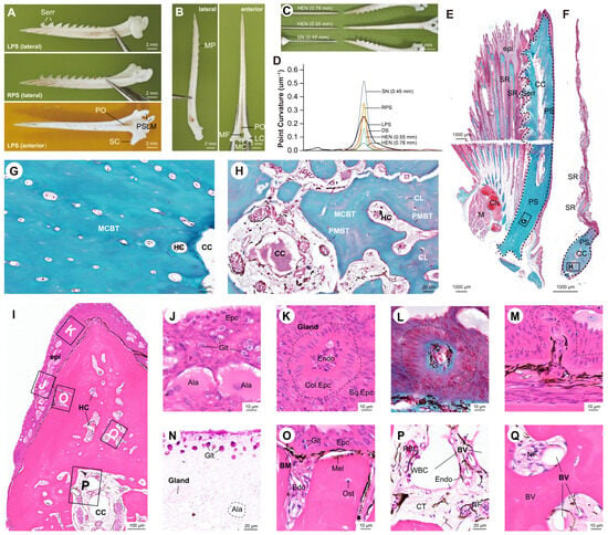
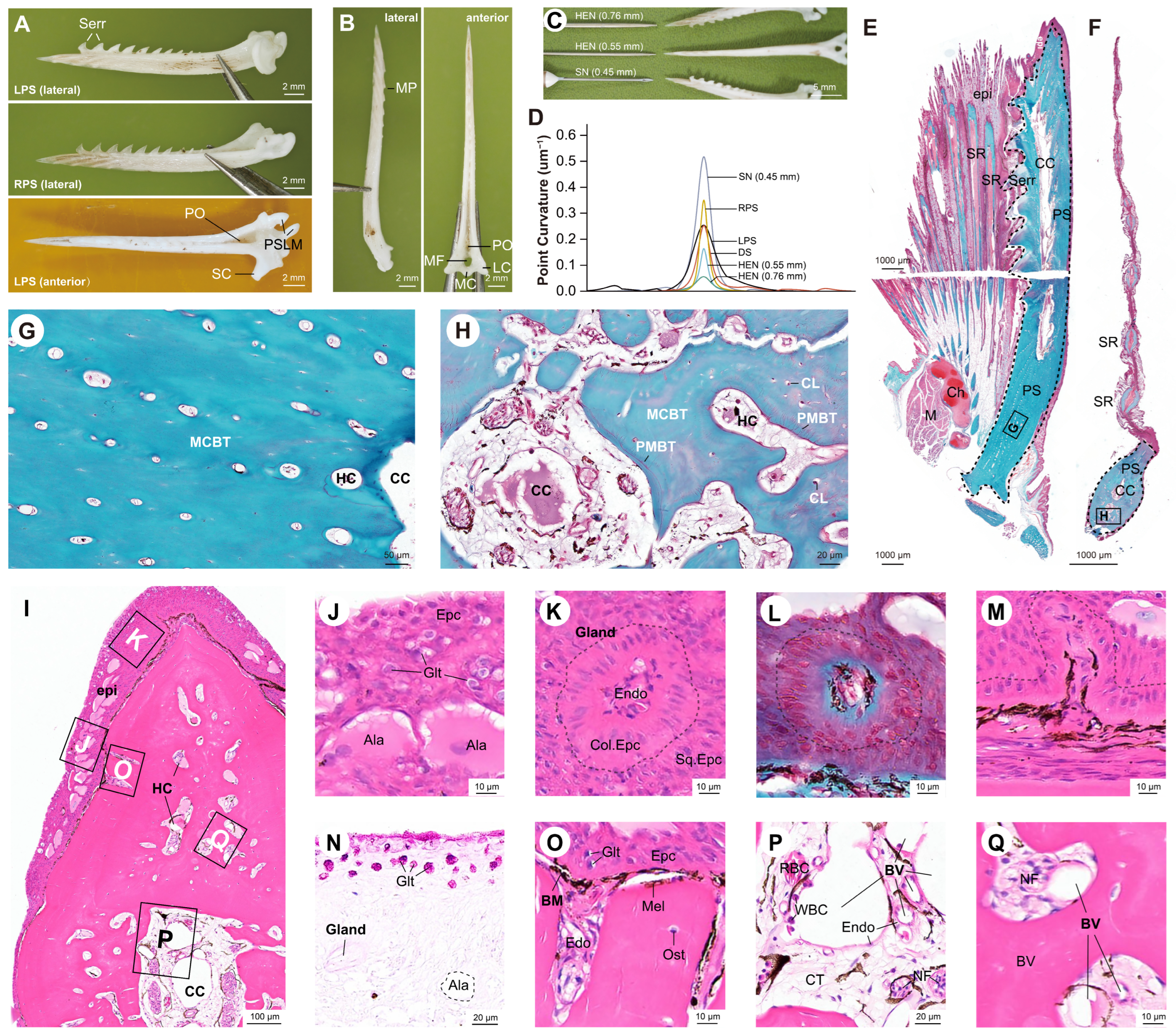
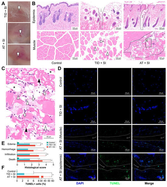
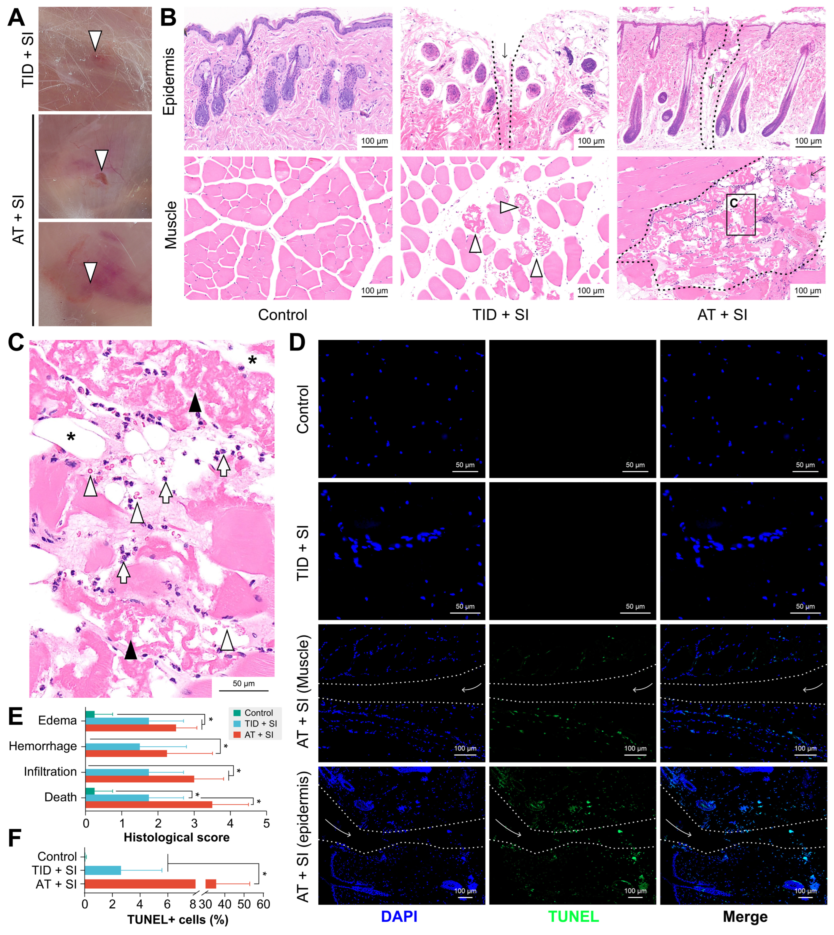
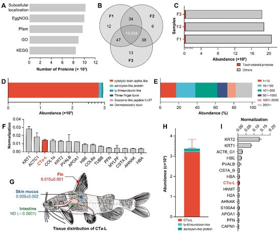
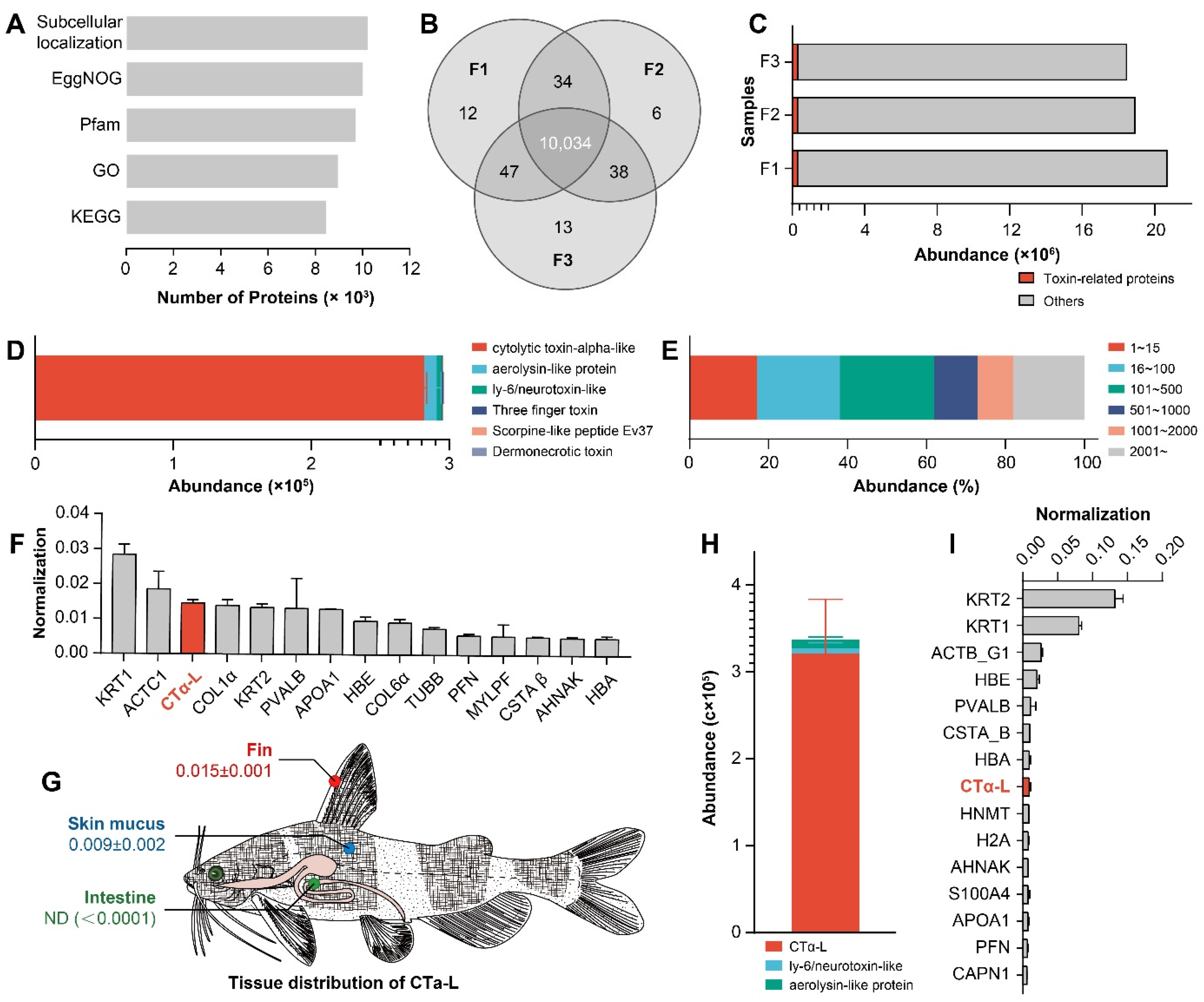
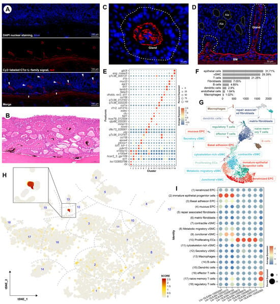
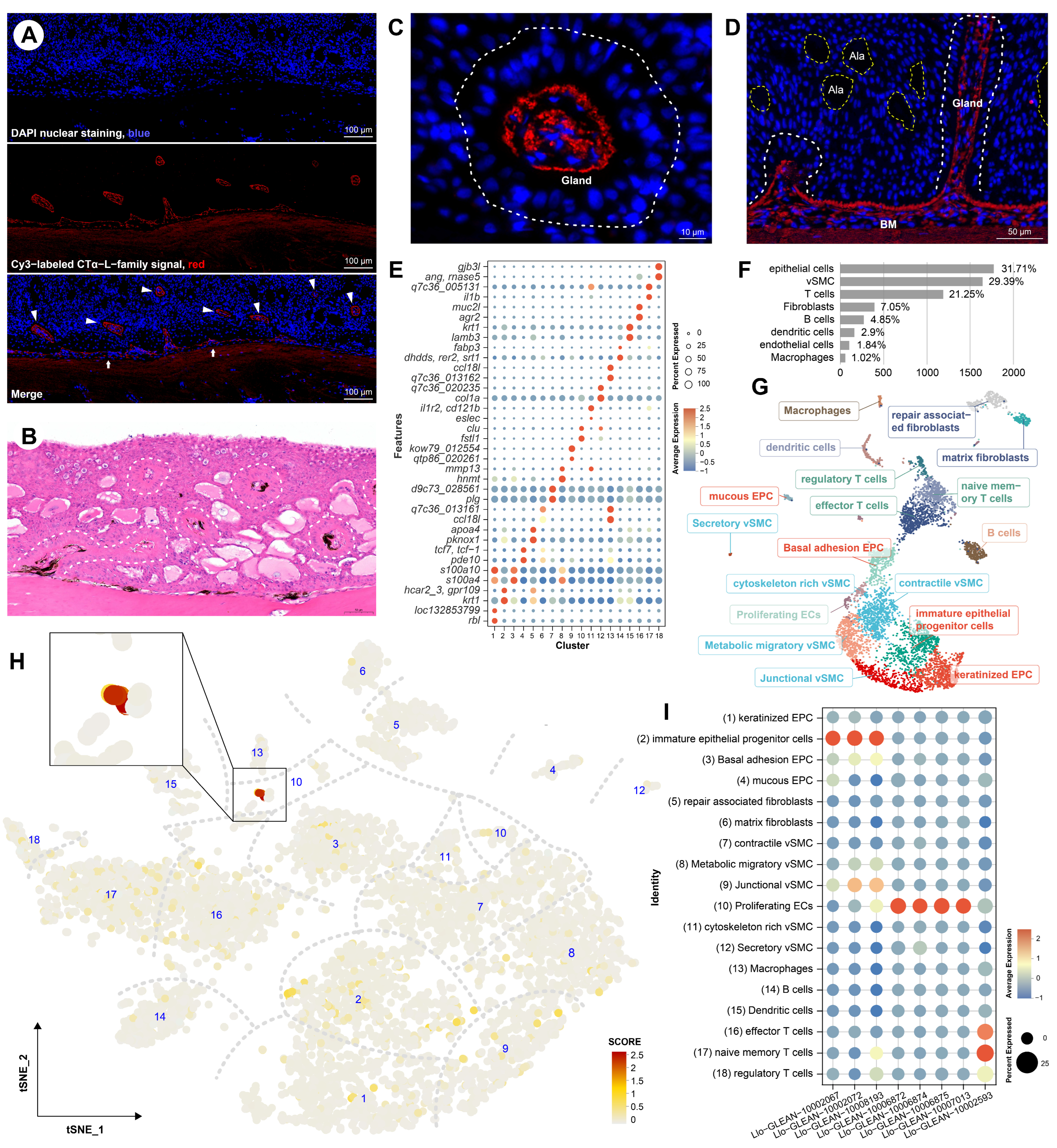
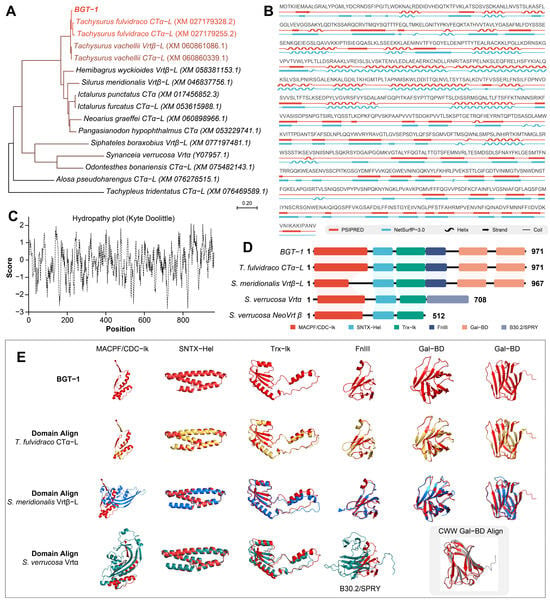
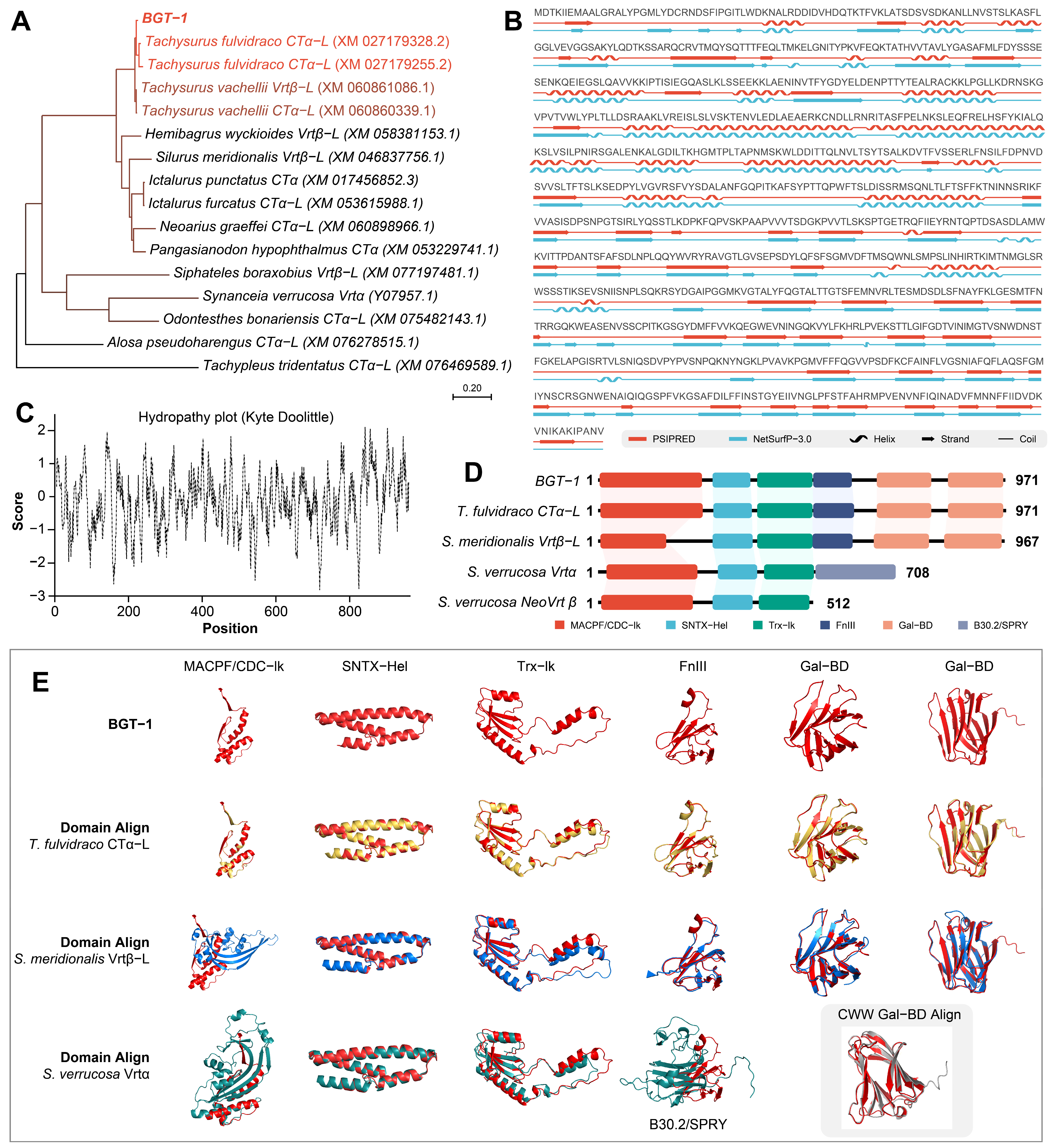
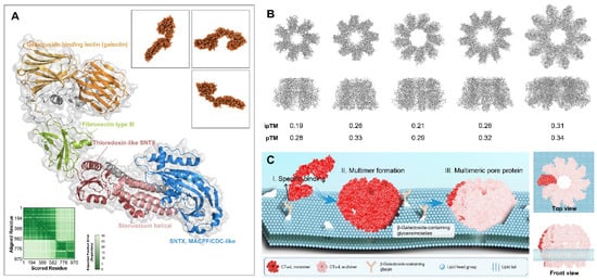
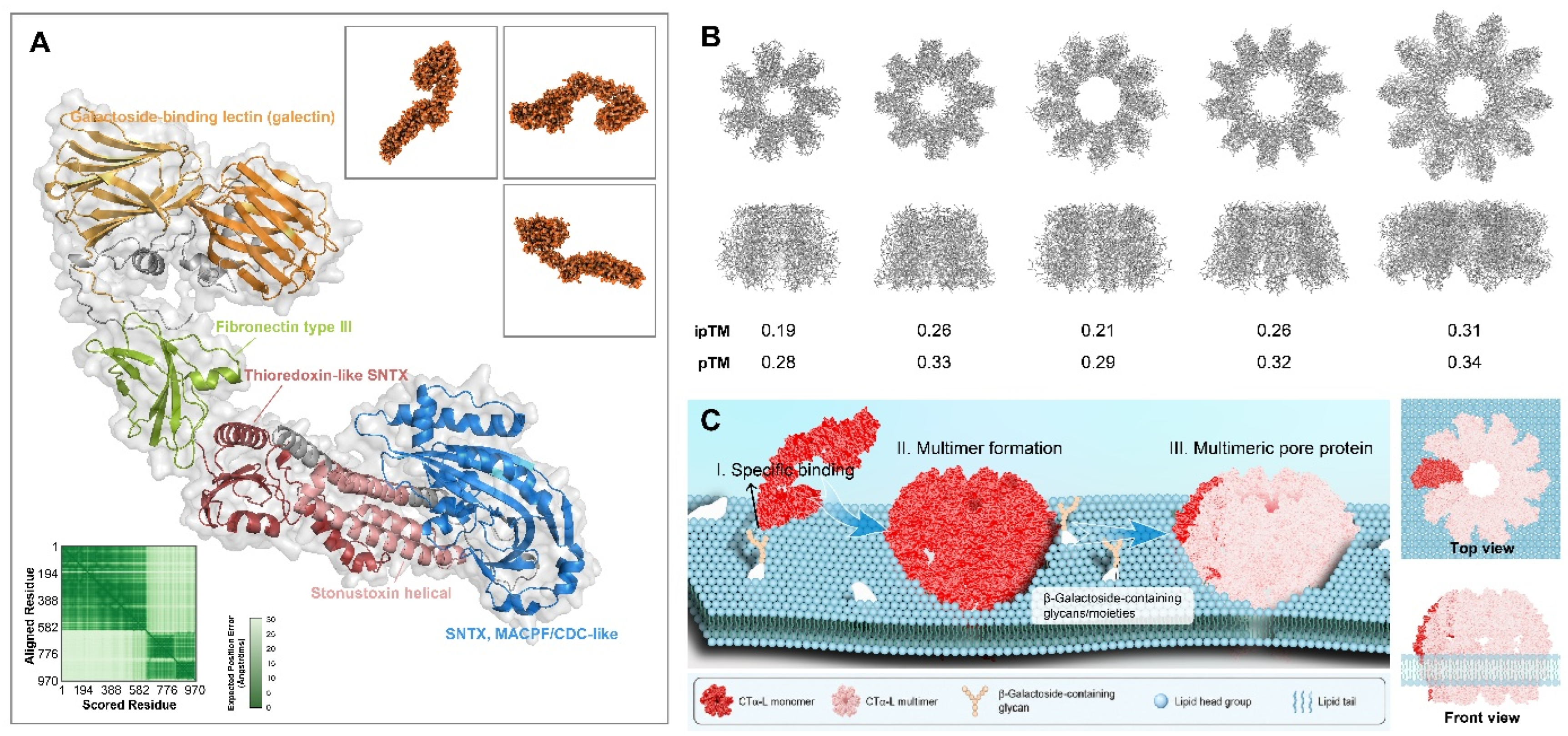

Disclaimer/Publisher’s Note: The statements, opinions and data contained in all publications are solely those of the individual author(s) and contributor(s) and not of MDPI and/or the editor(s). MDPI and/or the editor(s) disclaim responsibility for any injury to people or property resulting from any ideas, methods, instructions or products referred to in the content. |
© 2026 by the authors. Licensee MDPI, Basel, Switzerland. This article is an open access article distributed under the terms and conditions of the Creative Commons Attribution (CC BY) license.