


Abstract
Cardiovascular diseases develop through an interconnected continuum that extends from cardiometabolic risk factors and vascular dysfunction to myocardial ischemic injury, pathological remodeling, and heart failure. Panax ginseng and its bioactive constituents have shown cardiovascular protective effects in various experimental models. However, the molecular mechanisms shared across different stages of the cardiovascular continuum remain insufficiently integrated. This review critically synthesizes current evidence on Panax ginseng, ginseng-derived constituents, and ginseng-containing preparations across major stages of cardiovascular disease. Particular emphasis is placed on convergent molecular networks involving metabolic and mitochondrial homeostasis, oxidative stress and inflammation, autophagy and mitophagy, regulated cell death, calcium handling, vascular and fibrotic remodeling, and gut microbiota–host interactions. Several target-oriented mechanisms, including peroxisome proliferator activated receptor (PPAR) γ, ryanodine receptor 2, histone acetyltransferase P300/dihydrolipoamide dehydrogenase/pyruvate dehydrogenase, Fas-associated death domain/PPARα, and sarcoplasmic/endoplasmic reticulum Ca2+-ATPase 2A-associated regulation, are highlighted and distinguished from pathway-level or phenotypic evidence. Despite extensive preclinical data, direct target engagement, causal validation, pharmacokinetic characterization, preparation standardization, and clinical evidence remain limited. Integrating target-level validation with standardized ginseng interventions and clinically relevant models may provide a stronger molecular basis for evaluating the cardiovascular potential of ginseng.
1. Introduction
With the global aging population and the growing prevalence of unhealthy lifestyles, the incidence of cardiovascular diseases (CVDs) is constantly rising and has become a major cause of death and disability [1]. CVDs are defined as a series of events called the cardiovascular continuum first proposed by Victor J. Dzau. This concept describes a sequence of interrelated events that begins with risk factors, such as obesity, dyslipidemia and diabetes, which collectively constitute metabolic syndrome, then it gradually progresses to atherosclerosis, coronary artery disease, myocardial ischemia, coronary thrombosis, myocardial infarction, arrhythmia, remodeling, ventricular enlargement, heart failure, and ultimately leads to death [2]. Pharmacological interruption of the cardiovascular continuum effectively reduces cardiac-related mortality and is therefore of profound clinical importance.
Panax ginseng C. A. Mey (Ginseng, Renshen), a representative traditional Chinese medicine, has been used for centuries to improve lipid profiles, enhance cardiac function, and reduce hypertension for preventing and treating cardiovascular conditions [3]. Clinically, ginseng-containing formulas have been used as adjunctive therapies for heart failure [4] and coronary heart disease [5], while experimental evidence further supports their roles in alleviating myocardial hypertrophy [6], fibrosis [7], ischemia/reperfusion injury [8], and pathological cardiac remodeling [9]. Modern pharmacological research has confirmed that ginseng contains numerous active constituents that exert cardiovascular protective effects, including ginsenosides, polysaccharides, peptides, and other small molecules [10], which may act through multiple mechanisms such as anti-inflammatory, anti-oxidant, anti-apoptotic, anti-fibrotic, and metabolic regulatory effects [11,12]. Through multiple pathways and targets, ginseng and its bioactive constituents may modulate pathological processes at multiple stages of the cardiovascular continuum, ranging from cardiometabolic risk and vascular dysfunction to myocardial ischemic injury, pathological remodeling, and heart failure. Previous reviews have summarized the cardiovascular pharmacological effects and therapeutic applications of ginseng and ginsenosides [3], highlighted their protective mechanisms, particularly those involving energy metabolism and adenosine monophosphate-activated protein kinase (AMPK)/peroxisome proliferator activated receptor (PPAR) signaling [10] and evaluated preclinical and clinical evidence, including safety and toxicity considerations [11]. Other reviews have largely focused on individual cardiovascular conditions, such as cardiac ischemia/reperfusion injury [13] or heart failure [14]. However, the molecular mechanisms that converge across different stages of the cardiovascular continuum, as well as the strength of evidence supporting specific molecular targets, remain insufficiently integrated.
Using the cardiovascular continuum as a conceptual framework, this review critically synthesizes current evidence on the cardioprotective actions of ginseng, ginseng-derived constituents, and ginseng-containing preparations across major stages of cardiovascular disease. Rather than merely cataloguing disease-specific effects, we focus on shared mechanistic networks involving metabolic and mitochondrial homeostasis, oxidative stress and inflammation, regulated cell death, autophagy and mitophagy, vascular homeostasis, and pathological remodeling. This review could provide a basis for its rational clinical application and future research (Figure 1). The interventions discussed in this review encompass ginseng extracts, isolated ginsenosides, ginseng-derived polysaccharides, microbial metabolites of ginsenosides, synthetic ginsenoside derivatives, and multiherbal formulations containing ginseng, these interventions differ in composition, pharmacokinetic properties, and potential biological effects. Therefore, their findings are discussed according to the specific intervention investigated, and the effects observed with isolated compounds or multiherbal formulations are not automatically attributed to ginseng extracts.
Figure 1.
Overview of the cardiovascular continuum and the major cardioprotective mechanisms of ginseng and its related interventions. The upper panel illustrates the progression of the cardiovascular continuum from cardiometabolic risk factors and atherosclerosis to myocardial ischemic injury, pathological cardiac remodeling, and heart failure. The middle panel summarizes the major mechanistic networks associated with the cardiovascular effects of ginseng, including antioxidative and immunomodulatory actions, regulation of mitochondrial function and autophagy, modulation of regulated cell death, promotion of angiogenesis, and gut microbiota regulation. The lower panel presents representative ginseng-derived constituents and selected ginseng-containing multi-herbal formulations discussed in this review. The arrow indicates the progression of the cardiovascular continuum. The colors are used solely for visual differentiation and do not convey any specific biological or clinical significance. This figure was created by the authors based on a synthesis of the existing literature.
2. Cardiometabolic Risk Factors
Cardiometabolic risk factors, including obesity, insulin resistance, dyslipidemia, hypertension, and hyperglycemia, represent the early stages of the cardiovascular continuum and collectively contribute to subsequent vascular and myocardial injury. These metabolic abnormalities are closely interconnected through the disturbances in energy homeostasis, chronic low-grade inflammation, oxidative stress, endothelial dysfunction, and impaired glucose and lipid metabolism. Accumulating preclinical evidence suggests that Panax ginseng and its bioactive constituents may modulate several of these pathological processes, supporting their potential roles in the early cardiometabolic stages of cardiovascular disease.
2.1. Obesity and Insulin Resistance
Obesity and insulin resistance are closely interconnected cardiometabolic abnormalities that substantially increase cardiovascular risk. Obesity results from a chronic imbalance of energy homeostasis, in which calorie intake exceeds calorie expenditure. It is accompanied by adipose tissue dysfunction and dysregulated inflammatory adipocytokine secretion, which contribute to cardiovascular structural and functional alterations and broader metabolic abnormalities [15]. Insulin resistance is a central pathological feature of metabolic syndrome and contributes to impaired glucose utilization, compensatory hyperinsulinemia, and subsequent cardiometabolic dysfunction [16,17]. Increasing preclinical evidence suggests that ginseng and its bioactive constituents may modulate these abnormalities through coordinated effects on adipocyte function, energy metabolism, insulin signaling, and mitochondrial homeostasis. In addition to improving glucose and lipid metabolism, ginseng-derived constituents may alleviate insulin resistance through modulation of insulin signaling and mitochondrial quality control. For example, the malonyl-ginsenosides from Panax ginseng activate the insulin receptor substrate-1/phosphatidylinositol3-kinase (PI3K)/protein kinase B (AKT) and AMPK signal pathways to improve glucose and lipid metabolism and insulin resistance [18]. Compound K (CK) also attenuates skeletal-muscle insulin resistance by triggering dynamin-related protein 1 (Drp1)/PINK1-dependent mitophagic activity [19]. These findings suggest that restoration of insulin signaling and mitochondrial homeostasis may contribute to the metabolic effects of ginseng-derived constituents, although the direct molecular targets underlying these effects remain to be clarified.
2.1.1. Promotion of Adipocyte Browning
The browning of white adipose tissue, namely the conversion of white adipocytes into beige or brown-like adipocytes, is critical in counteracting obesity [20]. Rb1 facilitates the browning of white adipocytes, whereas Rb2 enhances brown adipose activity and induces white adipose tissue browning, accompanied by AMPK activation and increased expression of thermogenic and mitochondrial genes [21,22]. Ginsenoside F1 has also been reported to promote adipocyte browning [23]. Collectively, these findings suggest that the enhancement of thermogenic adipocyte activity may represent one mechanism by which ginseng-derived constituents improve obesity-related metabolic dysfunction. However, the available evidence remains predominantly preclinical, and whether these effects translate into clinically meaningful improvements in cardiometabolic risk remains unclear. A similar browning-promoting effect has also been reported for Shengmai San, a multi-herbal formula containing ginseng, although this effect cannot be attributed specifically to ginseng itself [24].
2.1.2. Regulation of the Gut Microbiota
The gut microbiota plays a key causal role in the development of diet-induced obesity [25]. Pretreatment with ginseng polysaccharides has been shown to regulate fecal microbiota and hepatic lysine degradation, thereby attenuating obesity and liver lipid accumulation [26]. A novel polysaccharide, a homogeneous ginseng polysaccharide, increases the expression of tight junction proteins and regulates the gut microbiota thereby reducing body weight and alleviating hepatic lipid accumulation and inflammatory injury [27]. In addition, (20R)-panaxadiol may ameliorate obesity, at least in part, by modulating the gut microbiota [28]. Ginsenosides have also been reported to improve high-fat diet (HFD)-induced hepatic lipid disorders via gut microbiota modulation and cholesterol signaling regulation [29]. These findings suggest that microbiota-mediated metabolic regulation may contribute to the anti-obesity effects of ginseng-derived constituents, although the specific microbial metabolites and host targets responsible for these effects remain incompletely defined.
2.1.3. Inhibition of Adipogenesis and Lipogenesis
Another important mechanism underlying the anti-obesity effects of ginseng is the inhibition of adipocyte differentiation and lipid accumulation. Adipogenesis, the process by which adipocyte precursor cells undergo proliferation and differentiation, is a fundamental mechanism underlying adipose tissue expansion [30]. Several ginsenosides have been reported to suppress these processes, with PPARγ and CCAAT/enhancer binding protein alpha (C/EBPα) emerging as recurrent regulatory nodes. Rf and F2 inhibit adipocyte differentiation through PPARγ-related mechanisms, whereas Rc, Rg3, the Rg5/Rk1 mixture, and Rh2 reduce adipogenesis by downregulating PPARγ and/or C/EBPα signaling [31,32,33,34,35,36]. Another study indicates that Rg3 inhibits signal transducer and activator of transcription 5 (STAT5) phosphorylation and suppresses PPARγ expression in HFD models, leading to downregulation of lipogenic proteins [37]. AMPK-dependent regulation also appears to contribute to these effects, as F2, Rg1, Rg2, and CK have been reported to inhibit adipogenesis or lipid accumulation through AMPK-associated signaling, while Rd acts through the cyclic adenosine monophosphate (cAMP)/protein kinase A (PKA) pathway [38,39,40,41,42]. These findings indicate that suppression of adipocyte differentiation and lipid storage is a recurring metabolic action of ginseng-derived constituents; however, most studies rely on cellular or diet-induced obesity models, and direct molecular target validation remains limited.
2.1.4. Improvement of Glucose and Lipid Metabolism
Obesity is closely associated with type 2 diabetes mellitus (T2DM), and insulin resistance represents a major pathogenic link between these two conditions [43]. Several ginsenosides have been reported to improve these abnormalities through distinct metabolic signaling pathways. Rb2 enhances glucose metabolism by activating AKT signaling while suppressing tumor necrosis factor-alpha (TNF-α)-induced mitogen-activated protein kinase (MAPK)/nuclear factor kappa B (NF-κB) activation [44], whereas Rg1 improves obesity-associated glucose disturbances through AMPK activation [45]. Rb1 also contributes to glycolipid metabolic regulation by upregulating PPARγ and aquaporin 7 (AQP7) expression [46]. In addition, ginsenoside Ro promotes glucagon-like peptide-1 secretion and increases energy expenditure through G protein–coupled bile acid receptor 5 (TGR5) activation [47]. Furthermore, (20R)-panaxadiol promotes the expression of thermogenic proteins through β2-adrenergic receptor-mediated the phosphorylation of cyclic AMP response element binding protein (CREB), leading to anti-obesity effects [48]. Collectively, these findings suggest that ginseng-derived constituents may improve obesity-related metabolic dysfunction through coordinated regulation of glucose utilization, lipid metabolism, and energy expenditure. However, most available evidence remains preclinical, and the relative contribution of these pathways to clinically meaningful metabolic improvement requires further validation (Table 1).
Table 1.
Summary of ginseng-related interventions and their effects on obesity.
2.2. Dyslipidemia
Dyslipidemia, commonly characterized by elevated triglycerides, reduced high-density lipoprotein cholesterol, and increased small dense low-density lipoprotein particles, is a major cardiometabolic risk factor and a key component of metabolic syndrome [49,50]. Current evidence suggests that Panax ginseng may improve lipid metabolism by reducing lipid accumulation, enhancing fatty acid oxidation, and regulating cholesterol and triglyceride metabolism [51]. These lipid-modulating effects may help limit the progression from metabolic dysfunction to atherosclerotic vascular injury, although the specific molecular targets and clinical relevance remain insufficiently defined.
2.3. Hypertension
Hypertension is a major cardiometabolic risk factor that interacts closely with obesity, insulin resistance, and dyslipidemia to accelerate cardiovascular injury [52]. Ginseng may contribute to blood pressure regulation by improving endothelial function, enhancing nitric oxide bioavailability, reducing oxidative stress, and modulating vascular inflammation. These vascular effects may help preserve endothelial homeostasis and limit the progression from cardiometabolic dysfunction to structural vascular damage [53]. However, the molecular targets responsible for these antihypertensive effects and their clinical relevance remain insufficiently defined.
3. Diabetic Cardiomyopathy
Diabetic cardiomyopathy represents an important cardiometabolic complication of diabetes, characterized by structural and functional myocardial abnormalities that develop independently of overt coronary artery disease, hypertension, or significant valvular disease [54]. The mechanisms underlying the decline in cardiac function in diabetic cardiomyopathy include increased fatty acids and cytokines, to adapt the diabetic hyperglycemic environment [55]. Ginseng has shown considerable potential in the treatment of diabetic cardiomyopathy, with several ginsenosides acting through distinct molecular pathways (Figure 2). Rb1 attenuates oxidative injury and abnormal ryanodine receptor 2 (RyR2) phosphorylation linking ginseng-derived constituents to the regulation of myocardial calcium homeostasis [56]. Another study suggests that Rh2 may activate PPARδ to inhibit fibronectin expression levels, thereby improving hyperglycemia-induced cardiac dysfunction and fibrosis [57]. Beyond isolated ginsenosides, formulas containing ginseng also demonstrate beneficial effects in treating diabetic cardiomyopathy. For example, Shengmai San improves diabetic cardiomyopathy by enhancing mitochondrial lipid metabolism [58]. Overall, current evidence suggest that ginseng and its active constituents may alleviate diabetic cardiomyopathy by improving metabolic dysregulation, reducing oxidative stress, modulating calcium handling, and attenuating myocardial fibrosis. However, its direct target validation remains limited to a small number of studies, and most evidence is still preclinical.
Figure 2.
Mechanisms associated with the cardioprotective effects of ginseng-related interventions in diabetic cardiomyopathy. Diabetic cardiomyopathy is associated with hyperglycemia, lipid metabolic disorder, oxidative stress, inflammation, and calcium-handling dysfunction, leading to impaired cardiac structure and function. Ginseng and its active constituents, including Rg3, Rb1, Rh2, and Shengmai San, exert protective effects by improving metabolic homeostasis, reducing oxidative stress and inflammation, regulating RyR2 activity and Ca2+ handling, and enhancing mitochondrial and cardiac energy metabolism. Collectively, these actions attenuate fibrosis, apoptosis, and cardiac dysfunction, thereby slowing the progression of diabetic cardiomyopathy. ↑ indicates upregulation or an increase, whereas ↓ indicates downregulation or a decrease. The different arrow styles are used solely for visual presentation and have no specific biological significance. This figure was created by the authors based on a synthesis of the existing literature.
4. Atherosclerosis
Metabolic syndrome comprises multiple metabolic risk factors that increase susceptibility to atherosclerosis [59]. Atherosclerosis is a chronic pathological process characterized by lipid accumulation and plaque formation within the arterial wall. It forms the common pathological basis for numerous cardiovascular conditions, representing a major public health concern worldwide [60]. Atherosclerotic lesions develop slowly over many years, and may eventually impair blood flow, leading to the symptoms of stable angina or claudication [61]. Numerous well-documented risk factors are associated with atherosclerosis, including hypertension, dyslipidemia, advanced age, and smoking [62]. Ginseng and its various active components may exert anti-atherosclerotic effects through multiple mechanisms (Figure 3).
Figure 3.
Anti-atherosclerotic effects of ginseng-related interventions and their associated mechanisms. Ginseng and its active constituents exert anti-atherosclerotic effects through antioxidative, endothelial-protective, anti-inflammatory, immunomodulatory, and metabolic regulatory actions. These effects involve improvement of endothelial function, modulation of macrophage polarization and cytokine release, regulation of gut microbiota and host metabolic homeostasis, and inhibition of foam cell formation, lipid accumulation, vascular cell proliferation, and plaque instability. ↑ indicates upregulation or an increase, whereas ↓ indicates downregulation or a decrease. The different arrow styles are used solely for visual presentation and have no specific biological significance. This figure was created by the authors based on a synthesis of the existing literature.
4.1. Anti-Oxidant Effects and Protection of Vascular Endothelial Function
Vascular endothelial cells (VECs) play a critical role in the occurrence and progression of atherosclerosis [63]. Exposed directly to blood, VECs lining blood vessels can become dysfunctional and inflamed when triggered by risk factors such as hypercholesterolemia, diabetes, hypertension, smoking, and ageing. These pathological changes are key early drivers of atherosclerosis, particularly in regions where blood flow is disturbed [64]. Reactive oxygen species (ROS) refer to reactive chemical species derived from molecular oxygen (O2) that exhibit greater chemical reactivity than O2 itself. This group encompasses both radical species, such as the superoxide anion (O2−) and the hydroxyl radical (HO•), and non-radical molecules such as hydrogen peroxide (H2O2) [65]. Although ROS are generated as by-products of normal aerobic metabolism, their levels can become elevated under various pathological or stressful conditions, which constitutes a fundamental mechanism of cellular and tissue damage [66]. During atherosclerosis, excessive ROS generation coupled with a downregulation of antioxidant defenses leads to an imbalance that culminates in oxidative stress [67].
Active components of ginseng may counteract these pathological processes through multiple mechanisms. Ginseng oligopeptide extract protects human aortic endothelial cells against nicotine-induced oxidative injury by modulating cellular antioxidant defenses and regulating the Kelch-like ECH-associated protein 1 (Keap1)/nuclear factor erythroid-derived 2-like 2 (Nrf2)/heme oxygenase-1 (HO-1) signaling pathway [68]. Rb1 also attenuates resistin-induced oxidative stress and dysfunction in human coronary artery smooth muscle cells by reducing ROS generation and enhancing SOD activity [69]. In addition, Rb1 inhibits vascular calcification, a pathological process closely associated with atherosclerotic progression [70].
Endothelial cell senescence may also contribute to atherogenesis [71]. In this context, Rb2 targets microRNA-216a (miR-216a) and exerts anti-inflammatory effects, thereby alleviating endothelial cell senescence [72]. Rb3 similarly protects human aortic endothelial cells against oxidized low-density lipoprotein (ox-LDL)-induced injury by reducing oxidative stress, inflammation, and endothelial dysfunction [73]. Similarly, ginsenoside Rg3 has been reported to alleviate ox-LDL-induced endothelial dysfunction and attenuate atherosclerosis in ApoE−/− mice through regulation of the PPARγ/FAK signaling pathway. Pharmacological inhibition with GW9662 further supported the involvement of PPARγ in these protective effects [74]. Additionally, Tongxinluo, a traditional Chinese medicine, has also demonstrated protective effects for myocardial infarction [75], and mitigates atherosclerosis by suppressing ROS/NOD-like receptor family pyrin domain-containing 3 (NLRP3)/caspase-1-mediated endothelial cell pyroptosis [76]. Collectively, these findings suggest that preservation of endothelial redox homeostasis may represent an important component of the anti-atherosclerotic actions of ginseng-derived constituents, although most available evidence remains preclinical and direct target validation is still limited.
4.2. Anti-Inflammatory and Immunomodulatory Effects
Atherosclerosis is a multifactorial process in which the immune system plays a key role, both systemically in the circulation and locally at vascular lesion sites [77]. Over the past two decades, atherosclerosis has become widely recognized as a chronic inflammatory disease, and the detrimental role of inflammation in its pathophysiology is now well established [78]. Several ginsenosides have been reported to modulate these inflammatory and immune responses. For instance, Rb2 attenuates atherosclerotic plaque formation, lipid accumulation, and M1 macrophage polarization through the targeted inhibition of miR-216a [79]. In contrast, 20(S)-Rg3 promotes PPARγ-dependent M2 macrophage polarization, suggesting therapeutic potential in diabetic atherosclerosis [80]. Beyond macrophages, Rg2 attenuates carotid intimal hyperplasia following balloon injury in rats by suppressing phenotypic transformation of vascular smooth muscle cells (VSMCs), thereby mitigating the associated inflammatory response [81]. A similar anti-inflammatory effect has been reported for the Shen-Hong-Tong-Luo formula, a multi-herbal preparation containing ginseng, through activation of the PPARγ/LXRα/ABCA1 pathway. However, the contribution of ginseng itself cannot be isolated from that of the other herbal components [82].
4.3. Regulation of Intestinal Flora and Metabolic Homeostasis
Alterations in the composition and function of gut microbiota contribute to the pathogenesis of various diseases, including cardiovascular disease [83]. The gut microbiota and its metabolites influence the pathogenesis of atherosclerosis throughout its development, from the initial engagement of endothelial cells and macrophages, through the intermediate involvement of VSMCs, to the advanced phase involving adaptive immune cells such as T cells, B cells, and dendritic cells [84]. Accordingly, targeting the “gut-heart axis” has emerged as a promising therapeutic strategy.
Ginseng and its compounds show significant potential. Total ginseng saponin extract and Rb1/Rg1 combinations retard atherogenesis via remodeling host–microbiome metabolic homeostasis [85]. In HFD-induced ApoE−/− mice, Rc also ameliorates atherosclerotic injury by regulating both microbial and host metabolomic profiles [86]. These findings suggest that microbiota-dependent metabolic regulation may contribute to the anti-atherosclerotic effects of ginseng-derived constituents. However, the specific microbial metabolites, host receptors, and downstream molecular targets responsible for these effects remain incompletely defined. Similar effects have been reported for ginseng-containing multi-herbal preparations. Ginsenosides from Sheng-Mai Yin (SMY) exert therapeutic effects against atherosclerosis through the maintenance of lipid homeostasis through regulation of cholesterol and lysophosphatidylcholines [87]. Furthermore, Tongxinluo, a multi-target traditional Chinese medicine formula, improves atherosclerotic plaque stability and reduces inflammation in rabbits by modulating intestinal flora and host metabolism [88]. Because both are multi-component formulations, these effects cannot be attributed specifically to ginseng alone.
4.4. Lipid Handling and Vascular Remodeling
Disordered lipid metabolism is one of the major mechanisms driving atherosclerosis [89]. Evidence suggests that Rb1 reduces lipid accumulation and foam cell formation in vascular smooth muscle cells and attenuates atherosclerotic plaque development [90]. Furthermore, Rb1 suppresses vascular cell proliferation by inhibiting the interaction between cyclin D and cyclin-dependent kinase 4, which may represent another mechanism underlying its anti-atherosclerotic effects [91]. Similar anti-atherosclerotic effects have also been reported for the Huxin formula and Baoyuan decoction, which reduce lipid accumulation and inflammation and suppress foam cell formation [92]. Taken together, current evidence indicates that ginseng and its active constituents may attenuate atherosclerosis through antioxidative and endothelial-protective effects, anti-inflammatory and immunomodulatory actions, regulation of gut microbiota and metabolic homeostasis, and improvement of lipid metabolism and vascular remodeling.
5. Myocardial Infarction and Myocardial Ischemia/Reperfusion Injury
Myocardial infarction (MI) is a pathological state caused by coronary artery ischemia, which leads to insufficient blood perfusion of the myocardium [93]. Regarding myocardial infarction, timely and effective revascularization can preserve contractile myocardial tissue, reduce the infarct area, and improve clinical outcomes. However, the reperfusion may lead to a further aggravation and acceleration of myocardial damage, which is known as myocardial ischemia/reperfusion (MI/R) injury [94]. A variety of drugs, including exenatide, cyclosporine A, and aspirin, have been reported to improve prognosis; however, their use is limited by low targeting specificity and potential adverse effects [95]. A large number of studies have shown that ginseng and its active ingredients, along with compound preparations, intervene in this process through multiple targets, providing a promising multi-dimensional therapeutic strategy for the prevention and treatment of MI/R injury (Figure 4).
Figure 4.
Cardioprotective effects of ginseng-related interventions and their associated mechanisms in myocardial infarction and ischemia/reperfusion injury. Ginseng and its active constituents protect against MI and MIR injury by regulating cell death, suppressing oxidative stress and inflammation, improving mitochondrial function and energy metabolism, modulating autophagy, and promoting angiogenesis, thereby reducing myocardial injury and facilitating cardiac repair. ↑ indicates upregulation or an increase, whereas ↓ indicates downregulation or a decrease. The different arrow styles are used solely for visual presentation and have no specific biological significance. This figure was created by the authors based on a synthesis of the existing literature.
5.1. Regulation of Cell Death: Apoptosis, Pyroptosis, and Ferroptosis
Cell death serves dual essential functions: it shapes the organism by supporting morphogenesis during development and protects the organism by maintaining homeostasis through the removal of unwanted cells after birth [96]. Multiple forms of regulated cell death have been identified, including apoptosis, autophagy-associated cell death, cornification, pyroptosis, and ferroptosis [97]. Pyroptosis is commonly initiated by inflammasome-forming pattern recognition receptors, particularly NOD-like receptors, while other PRR families, including Toll-like receptors and C-type lectin receptors, can contribute to upstream inflammatory priming and innate immune activation [98]. Under myocardial ischemia and hypoxia, toll-like receptor 4 (TLR4) activation induces NF-κB p65 activation, which further triggers the activation of the NLRP3 inflammasome and subsequent caspase-1 activation [99]. Notably, Rh2 effectively inhibits cardiomyocyte pyroptosis by downregulating key pyroptosis-related proteins, including NLRP3, apoptosis-associated speck-like protein containing a caspase recruitment domain, and caspase-1 [100]. Apoptosis is a highly regulated, energy-dependent form of programmed cell death that is initiated and executed through specific signaling cascades [101]. It is integral to key physiological processes such as embryogenesis, tissue homeostasis, and aging. However, excessive apoptotic cell death constitutes a major pathogenic process in acute MI-related myocardial injury [102]. Several ginseng-derived compounds and ginseng-containing traditional formulas have demonstrated marked anti-apoptotic effects. Syringaresinol [103], Renshen Yangrong decoction [104], and Zhi-Gan-Cao-Tang [105] have all been shown to protect cardiomyocytes against apoptosis. Ginsenoside Rg3 improves cardiac function after MI/R injury by reducing apoptotic cell death by inhibiting caspase-3 activation [106]. In addition, 25-hydroxyl-protopanaxatriol, a novel triterpenoid from ginseng stems and leaves [107], inhibits H2O2-induced injury in H9c2 cardiomyocytes by activating the PI3K/AKT signaling pathway [108]. In H9c2 cells, Rb1 inhibits the mitochondrial apoptotic pathway, as evidenced by reduced cytochrome c release and decreased levels of cleaved caspase-3, ultimately leading to cardiomyocyte protection [109]. Similarly, Rb2 and Rb3 have been reported to suppress cardiomyocyte apoptosis in rat models [110]. Zenglv Fumai granule, a clinical prescription for treating sick sinus syndrome, a condition associated with MI/R injury, also attenuates hypoxia/reoxygenation-induced cardiomyocyte apoptosis by regulating the tripartite interaction motif 28/glutathione peroxidase 1/ROS pathway [111]. Ferroptosis is a distinct form of regulated cell death characterized by iron accumulation and lipid peroxidation [112]. Ginsenoside Re has been shown to attenuate MI/R-induced ferroptosis by targeting the miR-144-3p/solute carrier family 7-member 11 axis [113]. Ginsenoside Rg3 has been shown to regulate the Keap1/Nrf2/glutathione peroxidase 4 (GPX4) signaling pathway, offering protection against ferroptosis [114]. Similarly, the clinical formulation Shenmai injection (SMI) confers protection against MI/R injury by activating the protective Nrf2/GPX4-associated signaling pathway and suppressing ferroptotic injury [115]. Collectively, these findings suggest that ginseng-derived constituents may protect the ischemic myocardium by modulating multiple forms of regulated cell death, including pyroptosis, apoptosis, and ferroptosis. Nevertheless, most available evidence remains based on pathway-level regulation, and direct molecular target validation is still limited.
5.2. Attenuation of Oxidative Stress and Inflammatory Responses
Oxidative stress, characterized by an imbalance between excessive ROS production and deficient antioxidant capacity, is a pivotal pathophysiological mechanism in MI/R injury. The interaction between ROS and inflammation forms a deleterious feed-forward loop in MI/R injury. ROS activate inflammatory signaling cascades, while inflammatory mediators further enhance ROS generation, thereby aggravating oxidative damage [116]. Given this interconnected pathophysiology, attenuation of oxidative stress and inflammation represents a key cardioprotective mechanism.
Extensive studies have shown that ginseng and its bioactive components exert effective antioxidant and anti-inflammatory effects by regulating multiple molecular pathways. Ginseng extract exerts anti-oxidant effects by activating Nrf2 and down-regulating NF-κB, protein kinase C, angiotensin II type 1 receptor, and inducible nitric oxide synthase, thereby protecting the heart against injury [117]. F2 ameliorates MI by mitigating cardiomyocyte apoptosis, oxidative stress, and mitochondrial dysfunction through the activation of both the Nrf2/HO-1 and PI3K/AKT signaling pathways [118]. Sirtuin 1 (SIRT1), a highly conserved oxidized nicotinamide adenine dinucleotide dependent deacetylase [119], is involved in the regulation of cellular energy metabolism, aging, and oxidative stress-related transcription [120]. Both 20(S)-ginsenoside Rg2 and ginsenoside Rb2 act as SIRT1 activators, reducing oxidative stress and inflammation to protect against MI/R injury [121,122]. Ginsenosides Rc [123,124] and Rh2 also exert antioxidant and anti-inflammatory effects. Notably, the effect of Rc is achieved by activating SIRT1, whereas Rh2 acts through the Nrf2/HO-1/NLRP3 signaling pathway [125]. These findings identify SIRT1 and Nrf2-related signaling as recurrent regulatory nodes in the antioxidant and anti-inflammatory actions of ginseng-derived constituents. Evidence from ginseng-containing preparations further supports these effects. SMI has been reported to attenuate MI/R injury, at least partly through suppression of oxidative stress [126]. Similarly, Shenxian-Shengmai oral liquid may attenuate oxidative injury in cardiomyocytes, with myocardial SOD1 and glutamate cysteine ligase catalytic subunit identified as potential therapeutic targets [127]. By inhibiting the K27 ubiquitination of absent in melanoma 2 (AIM2), SMY suppresses AIM2 inflammasome activation following MI/R injury, ultimately reducing inflammation and attenuating tissue damage [128]. Overall, these findings highlight oxidative–inflammatory coupling as an important mechanism underlying the cardioprotective effects of ginseng-derived constituents in MI/R injury.
5.3. Improvement of Mitochondrial Function and Energy Metabolism
During acute ischemia/reperfusion injury, mitochondrial dysfunction critically exacerbates cellular demise, determining the extent of injury in acute MI [129]. Ras homolog family (Rho) kinase plays a key role in MI/R injury [130]. Rb1 regulates energy metabolism by modulating RhoA signaling pathways, thereby alleviating the myocardial damage induced by I/R [131]. Ginseng polysaccharides protect H9c2 cells from MI/R injury through mechanisms involving mitochondrial metabolism and the reperfusion injury salvage kinase pathway [132]. Mitochondrial biogenesis and homeostasis also appear to be important targets of ginseng-derived constituents. Rd protects against myocardial ischemic injury through regulation of the Wnt family member 5A (WNT5A)/Ca2+ pathway, accompanied by enhanced mitochondrial biogenesis [133]. Recent evidence has further linked ginsenoside Rd to metabolic reprogramming in MI/R injury. Rd was reported to promote cardiac regeneration through a PPARG/3-hydroxy-3-methylglutaryl-CoA synthase 2 (HMGCS2)-associated mechanism involving ketone body metabolism, highlighting a potential link between metabolic regulation and myocardial repair following ischemia/reperfusion injury [134]. Sirtuin 3 (SIRT3) is a critical mitochondrial deacetylase involved in regulating mitochondrial homeostasis and modulating autophagy. Ginsenoside Rk1, a rare saponin derived from heat-processed ginseng, alleviates MI/R injury and improves mitochondrial function by regulating SIRT3 [135]. These findings suggest that preservation of mitochondrial quality and metabolic capacity may represent an important component of ginseng-mediated cardioprotection. Ginseng-containing preparations have shown similar effects. SMI exerts protective effects on cardiomyocytes through the regulation of mitochondrial dynamics, autophagy [136], and energy metabolism [137]. Xinmai’an tablets have also been reported to attenuate myocardial injury by activating the AMPK/SIRT1/peroxisome proliferator-activated receptor gamma coactivator 1-alpha (PGC-1α) signaling pathway and reducing mitochondrial oxidative stress [138].
5.4. Modulation of Autophagy
Autophagy is a lysosome-dependent intracellular degradation process that maintains cellular homeostasis by degrading damaged proteins and organelles [139]. Ginsenoside Rg3 enhances autophagy through the AMPK signaling pathway and attenuates MI-induced cardiac dysfunction [140]. In contrast, CK confers protection against I/R injury in H9c2 cells by suppressing detrimental autophagy and apoptosis through PI3K/AKT activation. These findings suggest that the cardioprotective effect of ginseng may depend on context-specific modulation of autophagy rather than uniform activation or inhibition of autophagy [141]. Daming capsule, a traditional Chinese medicine preparation, improves cardiac function after MI through activation of the SIRT1/AMPK pathway and enhancement of nucleotide-binding oligomerization domain-like receptor family member X1-mediated mitophagy [142].
5.5. Promotion of Angiogenesis
Therapeutic promotion of angiogenesis has gained attention as a strategy to enhance neovascularization and tissue recovery following MI [143]. Beyond direct cellular protection, angiogenesis represents another mechanism underlying the cardioprotective effects of ginseng components and compound preparations. For instance, ShenZhuGuanXin granules promote myocardial angiogenesis in rat models of MI by up-regulating the expression of platelet endothelial cell adhesion molecule 1/CD31 and vascular endothelial growth factor (VEGF) [144]. Similarly, Shensong Yangxin capsule ameliorates the impaired myocardial microcirculation following MI by balancing endothelial-derived vasodilators and vasoconstrictors and up-regulating the expression of VEGF and endothelial nitric oxide synthase (eNOS) [145]. Moreover, ginsenoside Rh2 promotes angiogenesis via the ERK signaling pathway, thereby contributing to the mitigation of acute MI [146]. Together, these findings suggest that enhancement of neovascularization and microvascular function may complement the direct myocardial protective effects of ginseng-related interventions after MI.
6. Cardiomyocyte Hypertrophy and Remodeling
Cardiomyocyte hypertrophy is a compensatory thickening of the muscle in response to elevated left ventricular (LV) wall stress [147]. Cardiac hypertrophy serves initially as an adaptive and compensatory mechanism to maintain cardiac output under stress. However, sustained stress can drive this response toward pathological remodeling and eventually heart failure [148]. Increasing evidence suggests that ginseng-derived constituents may modulate this transition through multiple mechanisms, including regulation of oxidative stress and inflammation, metabolic homeostasis, regulated cell death, and vascular signaling.
6.1. Mitigation of Inflammation and Oxidative Stress
Inflammation and oxidative stress are important contributors to the development and progression of pathological cardiac remodeling [149]. Maintaining a tight balance between ROS generation and clearance is critical for cardiovascular health, as its dysregulation is a key driver of cardiovascular disease [150]. Ginsenoside Rg3 has been demonstrated to ameliorate angiotensin II-induced hypertrophy in human cardiomyocytes by repressing the NLRP3 inflammasome and alleviating oxidative stress, an effect mediated through the modulation of the SIRT1/NF-κB pathway [151]. Together, these findings suggest that coordinated regulation of oxidative and inflammatory signaling may contribute to the anti-remodeling effects of ginsenosides.
6.2. Restoration of Metabolic Homeostasis
The myocardium regulates the energy supply in order to maintain the function of supplying oxygen and nutrients to the heart [152]. Therefore, therapeutic strategies aimed at restoring metabolic flexibility and energy homeostasis hold significant promise. Ginsenoside Rg3 exerts a protective effect by inhibiting the acyltransferase activity of histone acetyltransferase P300, reducing the 2-hydroxyisobutyrylation of mitochondrial enzyme dihydrolipoamide dehydrogenase (DLD) and subsequently restoring the activity of pyruvate dehydrogenase, a key gatekeeper of glucose oxidation [153]. This P300–DLD–pyruvate dehydrogenase (PDH) regulatory axis provides a comparatively detailed mechanistic link between ginsenoside-mediated post-translational regulation and myocardial energy metabolism. AMPK is a heterotrimeric complex comprising a catalytic α subunit and two regulatory subunits β and γ subunits. This conserved structure is fundamental to its role as a central cellular energy sensor and master regulator of metabolic homeostasis [154]. SMI suppresses cardiomyocyte hypertrophy and apoptosis by activating the AMPK signaling pathway via energy-dependent mechanisms [155]. By inhibiting the hypoxia-inducible factor 1-alpha (HIF-1α)/PPAR signaling pathway, SMY consequently activates the PKA pathway, thereby improving energy metabolism disorder in a rat model of myocardial hypertrophy [156].
6.3. Regulation of Cell Death
Increased ferroptosis has been observed in cardiovascular diseases, including cardiac hypertrophy [157]. In a model of doxorubicin-induced acute cardiotoxicity, Rb1 exerts protective effects by suppressing both autophagy and ferroptosis. Specifically, autophagy inhibition is mediated via the AMPK pathway, while ferroptosis suppression occurs through the Nrf2 pathway [158]. Although this evidence is derived from a cardiotoxicity model rather than a direct model of cardiac hypertrophy, it suggests that modulation of regulated cell death may represent an additional mechanism through which Rb1 influences pathological cardiac remodeling.
6.4. Vasodilation and Nitric Oxide Signaling
Beyond direct actions on cardiomyocytes, ginsenoside Rg1 protects abdominal aorta coarctation induced LV hypertrophy, with its effects being mediated, at least in part, by the production and release of endogenous nitric oxide, a potent vasodilator with important cardiovascular protective functions [159]. These findings suggest that improvement of vascular function and NO signaling may contribute to the anti-remodeling effects of Rg1 under conditions of increased cardiac afterload.
7. Heart Failure
As a significant public health challenge, heart failure (HF) affects over 56 million individuals globally, with an estimated prevalence of 1–2% [160]. HF is a complex clinical syndrome manifested by typical symptoms and signs, arising from any structural or functional impairment that affects ventricular filling or blood ejection [161]. The factors influencing HF are classified into three types, non-modifiable risk factors including age, sex and genetic variants, patient-specific modifiable risk factors, such as smoking, lack of physical activity, unhealthy diet, excessive alcohol consumption, stress and socioeconomic disadvantage; and medically modifiable risk factors, including hypertension, obesity, hyperlipidaemia, diabetes mellitus, atrial fibrillation, sleep apnoea, worsening renal function, hypertensive disorders of pregnancy, chronic obstructive pulmonary disease and chronic kidney disease [160]. Accumulating evidence suggests that Panax ginseng and its bioactive constituents may influence HF progression through multiple mechanisms involving energy metabolism, mitochondrial homeostasis, autophagy and mitophagy, oxidative stress, inflammation, and pathological remodeling.
7.1. Amelioration of Energy Metabolic Disorders
Energy deficiency is a characteristic of HF, primarily attributed to a decrease in mitochondrial oxidative capacity [162]. Increasing evidence suggests that ginseng-derived active components and formulas exert therapeutic effects on HF by regulating energy metabolism. The PI3K/AKT and AMPK signaling pathways are closely associated with energy metabolism [163]. Ginseng extract exerts therapeutic effects against HF, primarily via the PI3K/AKT and AMPK pathways, with consequent improvement in energy metabolism [164]. After the early compensatory remodeling stage, the phosphorylation of AMPK may inhibit MAPKs activation, YiQiFuMai powder injection ameliorates cardiac remodeling and HF, at least in part, through the inhibition of the MAPKs signaling pathways [165]. By activating the upstream PPARα regulator, Rb3 inhibits mitochondria-mediated apoptosis and enhances energy metabolism to exert cardioprotective effects [166]. In both in vivo HF models and in vitro hypoxia/ischemia models, Xinfuli Granule, an enhanced derivative of Baoyuan decoction, mitigates mitochondrial injury, inhibits endoplasmic reticulum stress, and improves energy production through the RhoA/Rho-associated coiled-coil-containing protein kinase (ROCK) pathway [167]. In addition, ginsenoside Rd [168] and total secondary ginsenosides [169] promote mitochondrial biogenesis, thereby increasing adenosine triphosphate (ATP) production and improving cardiac function. A study reveals a novel mechanism by which Rb1 restores cardiac energy homeostasis by regulating the Fas-associated death domain (FADD)/PPAR α axis, further establishing its cardioprotective potential in HF [170].
7.2. Regulation of Autophagy and Mitophagy
Autophagy is regarded as a new biomarker of HF [171]. One study demonstrated that autophagy plays a detrimental role in the transition from myocardial hypertrophy to HF [172]. Ginseng could enhance the effect of metoprolol in chronic HF; the mechanism is connected with inhibiting autophagy [173]. Mitophagy may also contribute to the regulation of mitochondrial quality during cardiac remodeling. Rg1 demonstrates significant efficacy in alleviating MI-induced cardiac remodeling in mice, a key precursor to HF. Mechanistically, it promotes mitophagosome formation, alleviates cardiac mitochondrial damage, and enhances SIRT1/PINK1/Parkin-mediated mitophagy. Its protective role was further confirmed in H2O2-treated H9c2 cells, where it activates the SIRT1/PINK1/Parkin pathway, leading to increased cell viability and reduced apoptosis and fibrosis [174]. These findings link Rg1-mediated mitophagy to preservation of mitochondrial homeostasis during pathological cardiac remodeling. By scavenging ROS, Yang-Xin-Xue-Ke-Li, a primary traditional Chinese Medicine for cardiovascular diseases, inhibits DOX-induced autophagy and mitophagy to exert its therapeutic effects [175]. 20S-O-Glc-DM is a derivative developed to address the low bioavailability of natural ginsenosides [176]. In models of LV diastolic dysfunction in mouse models and isoproterenol (ISO)-induced H9c2, excessive autophagy is reduced by 20S-O-Glc-DM, which is mediated through the AMPK- mechanistic target of rapamycin (mTOR)- unc-51-like autophagy activating kinase 1 (ULK1) pathways [177]. In contrast, Shexiang Baoxin pill alleviates MI/R injury by activating autophagy via a competing endogenous RNAs mechanism targeting the Map3k8 signaling pathway [178]. Together, these apparently divergent findings indicate that restoration of appropriate autophagic activity, rather than unidirectional activation or inhibition, may be important for cardioprotection.
7.3. Regulation of Cell Survival and Apoptosis Pathways
Apoptotic cell death participates in the process of subsequent LV remodeling and the development of HF [102]. Several ginseng-containing traditional formulations have been reported to preserve cardiomyocyte survival by regulating apoptosis-related signaling. Shenfu Formula, a classic traditional Chinese medicine, inhibits apoptosis through modulation of Fas/FasL and B-cell lymphoma 2 (Bcl-2)/Bcl-2-associated X protein (Bax) signaling pathway to suppress apoptosis. Which effectively improves cardiac function in rats with HF [179]. Baoyuan decoction, a formula that has been applied for the treatment of cardiovascular diseases clinically for decades, inhibits apoptosis, through regulating the angiotensin type 1 receptor (AT1)-cardiac ankyrin repeat protein (CARP) signaling pathway, thereby improves cardiac function and protects cardiomyocytes from injury. Its active ingredients include ginsenoside Rg3, Rb1, Rc and Re [180]. Integrated network pharmacology and experimental validation demonstrate that Fuzheng Yangxin recipe improves cardiac function through a mechanism involving the regulation of STAT3 expression and inhibition of apoptosis [181]. Xin Fu Kang, a traditional formula designed to alleviate chronic HF, preserves mitochondrial function and inhibits apoptosis by regulating nuclear receptor subfamily 4 group A member 1-mediated functional interaction between endoplasmic reticulum and mitochondria in cardiomyocytes [182]. Shen’ge formula, which holds a significant position in the treatment of heart diseases, has been shown in clinical studies to effectively alleviate the clinical symptoms of patients with HF [183] while experimental evidence suggests that it regulates the AKT/HIF-1α/p53 pathway to suppress cardiac hypertrophy and apoptosis and to promote angiogenesis [184]. Sheng-Mai-San attenuates MI-induced HF by regulating Ca2+ homeostasis and suppressing mitochondrial mediated apoptosis through the modulation of Ca2+-calcineurin-mediated Drp1 signaling pathways [185]. Similarly, Jin-Xin-Kang exerts a protective effect against HF through the amelioration of mitochondrial dysfunction, achieved by modulating the calcineurin/Drp1 signaling pathway [186].
7.4. Modulation of Inflammatory Responses
Inflammation plays a crucial role in the progression of HF [187]. The main function of the inflammatory response is to address myocardial damage, enabling the heart to adapt to abnormal conditions in the short term and restore internal balance and cardiovascular function in the long term. However, if this abnormal state persists, the continuous inflammatory state within the tissues could have adverse effects on myocardial cells and extracellular matrix, leading to progressive LV remodeling and dysfunction, ultimately resulting in HF due to poor adaptation [188]. In an angiotensin II (Ang II)-induced hypertensive HF model, both ginsenoside Rg5 [189] and 20(S)-ginsenoside Rh2 [190] demonstrates protective effects, and the mechanism involves inhibition of the c-Jun N-terminal kinase (JNK)/activator protein 1 (AP-1) pathway. These findings suggest that the suppression of maladaptive inflammatory signaling may contribute to the protective effects of individual ginsenosides in HF. Xin-Li formula, with ginseng serving as the minister herb, has demonstrated marked protective effects against MI-induced HF in rats. Mechanistically, these effects are achieved through Treg immunomodulation and the NLRP3 inflammasome inhibition [191]. Moreover, in rats with ISO-induced HF, Shenfu injection effectively modulates inflammatory imbalance, reduces serum and urine Trimethylamine N-oxide (TMAO) levels, and improves gut microbiota composition, thereby alleviating cardiac injury [192]. Huoxin pill, a traditional Chinese medicine formula that has shown significant efficacy in treating chronic HF [193], exerts therapeutic effects in verapamil-induced zebrafish models by inhibiting oxidative stress-triggered inflammation and apoptosis [194].
7.5. Regulation of Gut Microbiota
Emerging evidence suggests that gut microbiota dysbiosis drives the onset and progression of cardiometabolic diseases by disrupting systemic metabolism and homeostasis [195]. In HF, the modulation of gut microbiota has therefore emerged as a promising therapeutic strategy. The therapeutic effect of C20DM on HF is mediated by gut microbiota modulation, enhanced microbial diversity, upregulation of tight junction proteins including occludin, claudin, and zonula occludens-1, and subsequent inhibition of the TLR4- myeloid differentiation primary response 88 -NF-κB pathway [196]. Qiliqiangxin, a well-known compound preparation containing ginseng, has been used to treat HF for over a decade. It exerts its therapeutic effects, at least in part, by modulating the gut microbiota and inhibiting the NLRP3 inflammasome, thereby alleviating ventricular remodeling in rats [197]. Shenfu Injection ameliorates cardiac energy metabolism and intestinal integrity in HF rats, effects mediated by the modulation of gut microbiota, specifically an enrichment of short-chain fatty acid (SCFA)-producing Akkermansia and Blautia [198]. Dengzhanshengmai (DZSM) capsule, a traditional Chinese medicine preparation, is widely used to treat CVDs [199]. A study demonstrated that in HF, DZSM improves intestinal barrier function. This effect was associated with a decreased relative abundance of Turicibacter and Turicibacter_sp_TS3, and the regulation of genes encoding enzymes involved in phenylacetylglutamine synthesis [200]. These findings suggest that modulation of microbial composition, intestinal barrier function, and microbiota-derived metabolic signaling may contribute to the cardiovascular effects of ginseng-related interventions in HF.
7.6. Regulation of Calcium Homeostasis and Fibrotic Remodeling
Sarcoplasmic/endoplasmic reticulum Ca2+-ATPase 2A (SERCA2a), a key transporter regulating sarcoplasmic reticulum Ca2+ influx [201], is an important factor that regulates endoplasmic reticulum (ER) function in cardiovascular diseases. It has been demonstrated that Rg3 has a positive effect on the abnormal Ca2+ cycle in the cardiomyocytes of mice with HF, which is related to its regulation of SERCA2a small ubiquitin-like modifier (SUMO)ylation [202]. In addition, the disruption of transforming growth factor (TGF-β)1 pathway has been implicated in several human diseases, including myocardial fibrosis [203], and Smad3 is essential for TGF-β1-induced gene expression [204]. Studies have shown that ginsenoside Re alleviates isoproterenol-induced myocardial fibrosis and HF in rats, an effect associated with the modulation of the TGF-β1/Smad3 signaling pathway [205]. Similarly, Qiliqiangxin attenuates cardiac remodeling in doxorubicin-induced HF rats by inhibiting of TGF-β1/Smad3 [206]. Further relevant information is documented in Table 2.
Table 2.
Summary of ginseng-related interventions and their effects on heart failure.
Across different stages of the cardiovascular continuum, ginseng-related interventions appear to act through several interconnected mechanisms rather than isolated signaling pathways. Metabolic regulation and mitochondrial homeostasis are closely associated with oxidative stress, inflammation, and regulated cell death, whereas calcium handling and autophagy contribute to myocardial function and remodeling. To facilitate a cross-sectional comparison of the mechanistic evidence discussed throughout this review, Table 3 summarizes key experimental findings and evidence levels for selected molecular targets and pathways.
Table 3.
Key molecular mechanisms and supporting evidence for the cardiovascular protective effects of ginsenosides.
8. Translational Considerations
8.1. Effective Concentrations and Pharmacokinetic/Pharmacodynamic Considerations
The pharmacological effects of ginseng-derived constituents depend on their effective concentrations, administration routes, and systemic exposure. As summarized in Table 1 and Table 2, ginsenosides have demonstrated beneficial effects across various cellular and animal models at different experimental concentrations and doses. Despite their diverse pharmacological activities, many ginsenosides exhibit low oral bioavailability, which may be associated with their physicochemical properties, limited intestinal permeability, and extensive gastrointestinal metabolism [209].
Representative pharmacokinetic parameters of selected ginsenosides from preclinical and human studies are summarized in Table 4. For example, the oral bioavailability of purified ginsenoside Re was only 0.19–0.28% in mice, while ginsenoside Re administered as ginseng berry extract exhibited a higher bioavailability of 0.33–0.75% at equivalent Re doses [210]. This Study suggested that ginseng berry extract might be a good form for ginsenoside Re intake. These findings suggest that formulation composition may influence the absorption and systemic exposure of individual ginsenosides. Human pharmacokinetic studies have also demonstrated measurable systemic exposure following administration of isolated ginsenosides or ginseng preparations. Nevertheless, marked discrepancies across studies in experimental species, formulations and administration routes preclude direct cross-study comparisons.
Table 4.
Representative pharmacokinetic parameters of selected ginsenosides in preclinical and human studies.
Importantly, although pharmacokinetic and pharmacological studies of ginsenosides have been conducted, quantitative pharmacokinetic–pharmacodynamic relationships linking circulating ginsenoside concentrations to cardiometabolic and cardiovascular outcomes remain insufficiently characterized. Effective concentrations observed in vitro cannot be directly extrapolated to clinically achievable plasma concentrations. The effective concentrations of ginsenosides used in vitro may substantially exceed the plasma concentrations achieved following oral administration of ginseng preparations, limiting the direct translation of preclinical findings to clinical settings.
8.2. Cardiovascular Safety, Population Differences, and Structure–Activity Relationships
Although ginseng is a natural medicine and has shown promising therapeutic effects in preclinical experiments, its potential toxicological effects in the cardiovascular system cannot be ignored. By reviewing the current research, no serious adverse reactions of ginseng on the human body have been found, most of them are mild and temporary symptoms such as dyspepsia, hot flushes and insomnia [217]. However, the limited nature of clinical studies makes this conclusion rather limited. It is hoped that more evidence will be available to demonstrate that ginseng has no obvious toxicological effects on the human body. Meanwhile, the clinical effects of ginseng may vary across populations because of differences in dietary habits, consumption patterns, preparation types, and individual metabolic characteristics. Studies have demonstrated substantial interindividual variability in intestinal microbiota-mediated ginsenoside metabolism [218]. However, comparative clinical evidence across different ethnic and geographic populations remains limited. Further studies involving diverse populations are therefore needed to evaluate the generalizability of existing findings.
The biological activities of ginsenosides and other active components are significantly influenced by their chemical structures. Although this has been discussed in current research [219,220], there is still a lack of quantitative structure–activity relationships (QSAR) specifically addressing the effects on cardiac metabolism and cardiovascular systems. Future QSAR studies may help clarify the relationships between the molecular structures of ginsenosides and their cardiovascular protective activities.
9. Conclusions and Future Perspectives
The cardiovascular continuum provides a useful framework for integrating the diverse cardiovascular actions of Panax ginseng across different stages of disease, ranging from cardiometabolic risk factors and atherosclerosis to myocardial ischemic injury, pathological cardiac remodeling, and heart failure. Across these stages, several recurring mechanisms emerge, including regulation of metabolic and mitochondrial homeostasis, oxidative stress and inflammation, autophagy and mitophagy, regulated cell death, calcium handling, vascular and fibrotic remodeling, and gut microbiota–host interactions. Rather than acting through a single pathway, ginseng-related interventions appear to influence interconnected regulatory networks whose relative importance varies according to disease stage and pathological context.
Nevertheless, the strength of mechanistic evidence remains uneven. A substantial proportion of current studies infer mechanisms from changes in signaling proteins, gene expression, or downstream phenotypes, whereas direct molecular target evidence and rigorous causal validation are comparatively limited. Several studies have begun to provide more target-oriented mechanistic insights, including regulation of P300/DLD/PDH-associated metabolic remodeling, RyR2-dependent Ca2+ handling, FADD/PPARα signaling, and SERCA2a SUMOylation. Future studies should move beyond pathway association toward direct target engagement and causal validation using complementary approaches such as genetic perturbation, pharmacological intervention, and rescue experiments.
Another important consideration is the heterogeneity of ginseng-related interventions. Whole ginseng, ginseng extracts, isolated ginsenosides, polysaccharides, ginsenoside derivatives, and multi-herbal formulations should not be regarded as equivalent levels of evidence. In particular, the biological effects of ginseng-containing formulas cannot be attributed specifically to ginseng without component-resolved validation. In addition, the relatively low bioavailability of several ginsenosides remains an important barrier to translation. Further studies are therefore needed to clarify the active constituents responsible for cardiovascular effects, improve their bioavailability, and establish more robust relationships between administered dose, biological exposure, and pharmacological response.
Clinical translation also remains limited because most available evidence is derived from preclinical models, and the relevance of these models to the heterogeneity and complexity of human cardiovascular disease requires further validation. Well-designed clinical studies using clearly characterized ginseng preparations, appropriate patient populations, and clinically meaningful cardiovascular endpoints could be essential to determine whether the mechanistic benefits observed experimentally can be reproduced in humans. Overall, future research should shift from broad claims of “multi-target” activity toward experimentally validated target networks and better-defined ginseng interventions. Such an approach may provide a stronger mechanistic and translational basis for evaluating the role of Panax ginseng in cardiovascular disease.
Author Contributions
M.Q.: writing—Conceptualization, literature search, Writing—original draft; C.S. and Y.Q.: Data curation; D.W., M.T. and M.W.: Visualization; H.S. and X.L.: Conceptualization, supervision. All authors have read and agreed to the published version of the manuscript.
Funding
This work was supported by the National Administration of Traditional Chinese Medicine Project (GZY-KJS-2025-042) and the Science and Technology Development Plan Project of Jilin Province (20230204025YY).
Institutional Review Board Statement
Not applicable.
Informed Consent Statement
Not applicable.
Data Availability Statement
No new data were created or analyzed in this study. Data sharing is not applicable to this article.
Acknowledgments
ChatGPT (5.6) and DeepSeek (V3.2) were used for grammar correction and language refinement; all content was thoroughly reviewed and approved by the authors.
Conflicts of Interest
The authors declare no conflicts of interest.
Abbreviations
| Abbreviation | Full name |
| Ang II | Angiotensin II |
| AIM2 | Absent in melanoma 2 |
| AKT | Protein kinase B |
| AMPK | Adenosine monophosphate-activated protein kinase |
| AP-1 | Activator protein 1 |
| AQP7 | Aquaporin 7 |
| AT1 | Angiotensin type 1 receptor |
| ATP | Adenosine triphosphate |
| Bax | Bcl-2-associated X protein |
| Bcl-2 | B-cell lymphoma 2 |
| C/EBPα | CCAAT/enhancer binding protein alpha |
| cAMP | Cyclic adenosine monophosphate |
| CARP | cardiac ankyrin repeat protein |
| CK | Compound K |
| Cmax | Maximum plasma concentration |
| CREB | Cyclic AMP response element binding protein |
| CVDs | Cardiovascular diseases |
| DLD | Dihydrolipoamide dehydrogenase |
| Drp1 | Dynamin-related protein 1 |
| DZSM | Dengzhanshengmai |
| eNOS | Endothelial nitric oxide synthase |
| ER | Endoplasmic reticulum |
| ERK | Extracellular signal-regulated kinase |
| F | Absolute oral bioavailability |
| FADD | Fas-associated death domain |
| GPX4 | Glutathione peroxidase 4 |
| H2O2 | Hydrogen peroxide |
| HF | Heart failure |
| HFD | High-fat diet |
| HIF-1α | Hypoxia-inducible factor 1-alpha |
| HMGCS2 | 3-hydroxy-3-methylglutaryl-CoA synthase 2 |
| HO-1 | heme oxygenase-1 |
| i.p. | Intraperitoneal injection |
| i.g. | Intragastric administration |
| ISO | Isoproterenol |
| JNK | c-Jun N-terminal kinase |
| Keap1 | Kelch-like ECH-associated protein 1 |
| LV | Left ventricular |
| MAPK | Mitogen-activated protein kinase |
| MI | Myocardial infarction |
| MI/R | Myocardial ischemia/reperfusion |
| miR-216a | microRNA-216a |
| mTOR | Mechanistic target of rapamycin |
| NF-κB | Nuclear factor kappa B |
| NLRP3 | NOD-like receptor family pyrin domain-containing 3 |
| Nrf2 | Nuclear factor erythroid-derived 2-like 2 |
| ox-LDL | Oxidized low-density lipoprotein |
| PDH | Pyruvate dehydrogenase |
| PGC-1α | Peroxisome proliferator-activated receptor gamma coactivator 1-alpha |
| PI3K | Phosphatidylinositol3-kinase |
| PKA | Protein kinase A |
| PPAR | Peroxisome proliferator activated receptor |
| Rho | Ras homolog family |
| ROCK | Rho-associated coiled-coil-containing protein kinase |
| ROS | Reactive oxygen species |
| SCFA | Short-chain fatty acid |
| SERCA2a | Sarcoplasmic/endoplasmic reticulum Ca2+-ATPase 2A |
| SIRT1 | Sirtuin 1 |
| SIRT3 | Sirtuin 3 |
| SMI | Shenmai injection |
| SMY | Sheng-Mai Yin |
| SOD | Superoxide dismutase |
| STAT5 | Signal transducer and activator of transcription 5 |
| SUMO | Small ubiquitin-like modifier |
| t½ | Elimination half-life |
| T2DM | Type 2 diabetes mellitus |
| TGF-β | Transforming growth factor |
| TGR5 | G protein–coupled bile acid receptor 5 |
| TLR4 | Toll-like receptor 4 |
| Tmax | Time to maximum plasma concentration |
| TNF-α | Tumor necrosis factor-alpha |
| ULK1 | unc-51-like autophagy activating kinase 1 |
| VECs | Vascular endothelial cells |
| VEGF | vascular endothelial growth factor |
| VSMCs | Vascular smooth muscle cells |
| WNT5A | Wnt family member 5A |
References
- Global Burden of Cardiovascular Diseases and Risks 2023 Collaborators. Global, Regional, and National Burden of Cardiovascular Diseases and Risk Factors in 204 Countries and Territories, 1990–2023. J. Am. Coll. Cardiol. 2025, 86, 2167–2243. [Google Scholar] [CrossRef] [Scilit]
- Dzau, V.J.; Antman, E.M.; Black, H.R.; Hayes, D.L.; Manson, J.E.; Plutzky, J.; Popma, J.J.; Stevenson, W. The cardiovascular disease continuum validated: Clinical evidence of improved patient outcomes: Part I: Pathophysiology and clinical trial evidence (risk factors through stable coronary artery disease). Circulation 2006, 114, 2850–2870. [Google Scholar] [CrossRef] [Scilit]
- Kim, J.H. Pharmacological and medical applications of Panax ginseng and ginsenosides: A review for use in cardiovascular diseases. J. Ginseng Res. 2018, 42, 264–269. [Google Scholar] [CrossRef] [Scilit]
- Cheang, I.; Yao, W.; Zhou, Y.; Zhu, X.; Ni, G.; Lu, X.; Liao, S.; Gao, R.; Zhou, F.; Shen, J.; et al. The traditional Chinese medicine Qiliqiangxin in heart failure with reduced ejection fraction: A randomized, double-blind, placebo-controlled trial. Nat. Med. 2024, 30, 2295–2302. [Google Scholar] [CrossRef] [Scilit]
- Ma, J.; Yu, Z.; Chen, T.; Li, P.; Liu, Y.; Chen, J.; Lyu, C.; Hao, X.; Zhang, J.; Wang, S.; et al. The effect of Shengmai injection in patients with coronary heart disease in real world and its personalized medicine research using machine learning techniques. Front. Pharmacol. 2023, 14, 1208621. [Google Scholar] [CrossRef] [Scilit]
- Gao, R.R.; Wu, X.D.; Jiang, H.M.; Zhu, Y.J.; Zhou, Y.L.; Zhang, H.F.; Yao, W.M.; Li, Y.Q.; Li, X.L. Traditional Chinese medicine Qiliqiangxin attenuates phenylephrine-induced cardiac hypertrophy via upregulating PPARγ and PGC-1α. Ann. Transl. Med. 2018, 6, 153. [Google Scholar] [CrossRef] [Scilit]
- Li, X.; Li, D.; Liu, S.; Wang, Y.; Fan, X.; Yan, Z.; Tan, S.; Zhuang, P.; Zhang, Y. Shenfu injection inhibits myocardial fibrosis by regulating glycolysis through the PI3K-AKT/HIF-1α signaling pathway. Phytomedicine 2025, 149, 157529. [Google Scholar] [CrossRef] [Scilit]
- Liu, X.; Tan, W.; Yang, F.; Wang, Y.; Yue, S.; Wang, T.; Wang, X. Shengmai injection reduces apoptosis and enhances angiogenesis after myocardial ischaemia and reperfusion injury in rats. Biomed. Pharmacother. 2018, 104, 629–636. [Google Scholar] [CrossRef] [Scilit]
- Tao, L.; Shen, S.; Fu, S.; Fang, H.; Wang, X.; Das, S.; Sluijter, J.P.G.; Rosenzweig, A.; Zhou, Y.; Kong, X.; et al. Traditional Chinese Medication Qiliqiangxin attenuates cardiac remodeling after acute myocardial infarction in mice. Sci. Rep. 2015, 5, 8374. [Google Scholar] [CrossRef] [Scilit]
- Zhou, Z.; Li, M.; Zhang, Z.; Song, Z.; Xu, J.; Zhang, M.; Gong, M. Overview of Panax ginseng and its active ingredients protective mechanism on cardiovascular diseases. J. Ethnopharmacol. 2024, 334, 118506. [Google Scholar] [CrossRef] [Scilit]
- Sarhene, M.; Ni, J.Y.; Duncan, E.S.; Liu, Z.; Li, S.; Zhang, J.; Guo, R.; Gao, S.; Gao, X.; Fan, G. Ginsenosides for cardiovascular diseases; update on pre-clinical and clinical evidence, pharmacological effects and the mechanisms of action. Pharmacol. Res. 2021, 166, 105481. [Google Scholar] [CrossRef] [Scilit]
- Tang, M.M.; Zhao, S.T.; Li, R.Q.; Hou, W. Therapeutic mechanisms of ginseng in coronary heart disease. Front. Pharmacol. 2023, 14, 1271029. [Google Scholar] [CrossRef] [Scilit]
- Chen, J.; Huang, Q.; Li, J.; Yao, Y.; Sun, W.; Zhang, Z.; Qi, H.; Chen, Z.; Liu, J.; Zhao, D.; et al. Panax ginseng against myocardial ischemia/reperfusion injury: A review of preclinical evidence and potential mechanisms. J. Ethnopharmacol. 2023, 300, 115715. [Google Scholar] [CrossRef] [Scilit]
- Karmazyn, M.; Gan, X.T. Treatment of the cardiac hypertrophic response and heart failure with ginseng, ginsenosides, and ginseng-related products. Can. J. Physiol. Pharmacol. 2017, 95, 1170–1176. [Google Scholar] [CrossRef] [Scilit]
- Lee, H.; Rhee, T.M.; Choi, J.M.; Choi, S.Y.; Kim, D.W. The Close Link Between Obesity and Cardiovascular Disease: Current Insights and Remaining Challenges. Endocrinol. Metab. Clin. N. Am. 2025, 54, 175–192. [Google Scholar] [CrossRef] [Scilit]
- Grundy, S.M. Metabolic syndrome: Connecting and reconciling cardiovascular and diabetes worlds. J. Am. Coll. Cardiol. 2006, 47, 1093–1100. [Google Scholar] [CrossRef] [Scilit]
- Reaven, G. Insulin resistance and coronary heart disease in nondiabetic individuals. Arterioscler. Thromb. Vasc. Biol. 2012, 32, 1754–1759. [Google Scholar] [CrossRef] [Scilit]
- Wang, D.S.; Wang, J.M.; Zhang, F.R.; Lei, F.J.; Wen, X.; Song, J.; Sun, G.Z.; Liu, Z. Ameliorative Effects of Malonyl Ginsenoside from Panax ginseng on Glucose-Lipid Metabolism and Insulin Resistance via IRS1/PI3K/Akt and AMPK Signaling Pathways in Type 2 Diabetic Mice. Am. J. Chin. Med. 2022, 50, 863–882. [Google Scholar] [CrossRef] [Scilit]
- Li, W.; Li, H.; Zheng, L.; Xia, J.; Yang, X.; Men, S.; Yuan, Y.; Fan, Y. Ginsenoside CK improves skeletal muscle insulin resistance by activating DRP1/PINK1-mediated mitophagy. Food Funct. 2023, 14, 1024–1036. [Google Scholar] [CrossRef] [Scilit]
- Paulo, E.; Wang, B. Towards a Better Understanding of Beige Adipocyte Plasticity. Cells 2019, 8, 1552. [Google Scholar] [CrossRef] [Scilit]
- Mu, Q.; Fang, X.; Li, X.; Zhao, D.; Mo, F.; Jiang, G.; Yu, N.; Zhang, Y.; Guo, Y.; Fu, M.; et al. Ginsenoside Rb1 promotes browning through regulation of PPARγ in 3T3-L1 adipocytes. Biochem. Biophys. Res. Commun. 2015, 466, 530–535. [Google Scholar] [CrossRef] [Scilit]
- Hong, Y.; Lin, Y.; Si, Q.; Yang, L.; Dong, W.; Gu, X. Ginsenoside Rb2 Alleviates Obesity by Activation of Brown Fat and Induction of Browning of White Fat. Front. Endocrinol. 2019, 10, 153. [Google Scholar] [CrossRef] [Scilit]
- Simu, S.Y.; Siddiqi, M.H.; Ahn, S.; Castro-Aceituno, V.; Kumar, N.S.; Perez, Z.E.J.; Yang, D.-C. Ginsenoside F1 attenuates lipid accumulation and triglycerides content in 3T3-L1 adipocytes with the modulation of reactive oxygen species (ROS) production through PPAR-γ/JAK2 signaling responses. Med. Chem. Res. 2017, 26, 1042–1051. [Google Scholar] [CrossRef] [Scilit]
- Wang, Z.; Wang, X.; Fu, L.; Xu, S.; Wang, X.; Liao, Q.; Zhuang, T.; Liu, L.; Zhang, H.; Li, W.; et al. Shengmai San formula alleviates high-fat diet-induced obesity in mice through gut microbiota-derived bile acid promotion of M2 macrophage polarization and thermogenesis. Phytomedicine 2024, 133, 155938. [Google Scholar] [CrossRef] [Scilit]
- Saad, M.J.; Santos, A.; Prada, P.O. Linking Gut Microbiota and Inflammation to Obesity and Insulin Resistance. Physiology 2016, 31, 283–293. [Google Scholar] [CrossRef] [Scilit]
- Liu, Q.; Zhang, Z.; Ji, P.; Liu, J.; Chen, B.; E, M.; Qi, H.; Hou, T.; Huang, Q.; Ding, L.; et al. Ginseng polysaccharide components attenuate obesity and liver lipid accumulation by regulating fecal microbiota and hepatic lysine degradation. Int. J. Biol. Macromol. 2024, 269, 131872. [Google Scholar] [CrossRef] [Scilit]
- Gao, Y.; Guo, M.; Chen, J.; Sun, Y.; Wang, M. A ginseng polysaccharide protects intestinal barrier integrity in high-fat diet-fed obese mice. Int. J. Biol. Macromol. 2024, 277, 133976. [Google Scholar] [CrossRef] [Scilit]
- Lv, Y.; Zhang, Y.; Feng, J.; Zhao, T.; Zhao, J.; Ge, Y.; Yang, X.; Han, H.; Zhang, M.; Chen, L.; et al. (20R)-Panaxadiol as a Natural Active Component with Anti-Obesity Effects on ob/ob Mice via Modulating the Gut Microbiota. Molecules 2022, 27, 2502. [Google Scholar] [CrossRef] [Scilit]
- Zhu, Y.; Zhang, K.X.; Bu, Q.Y.; Song, S.X.; Chen, Y.; Zou, H.; You, X.Y.; Zhao, G.P. Ginsenosides From Panax ginseng Improves Hepatic Lipid Metabolism Disorders in HFD-Fed Rats by Regulating Gut Microbiota and Cholesterol Metabolism Signaling Pathways. Phytother. Res. 2025, 39, 714–732. [Google Scholar] [CrossRef] [Scilit]
- Haider, N.; Larose, L. Harnessing adipogenesis to prevent obesity. Adipocyte 2019, 8, 98–104. [Google Scholar] [CrossRef] [Scilit]
- Siraj, F.M.; Natarajan, S.; Huq, M.A.; Kim, Y.J.; Yang, D.C. Structural investigation of ginsenoside Rf with PPARγ major transcriptional factor of adipogenesis and its impact on adipocyte. J. Ginseng Res. 2015, 39, 141–147. [Google Scholar] [CrossRef] [Scilit]
- Siraj, F.M.; SathishKumar, N.; Kim, Y.J.; Kim, S.Y.; Yang, D.C. Ginsenoside F2 possesses anti-obesity activity via binding with PPARγ and inhibiting adipocyte differentiation in the 3T3-L1 cell line. J. Enzym. Inhib. Med. Chem. 2015, 30, 9–14. [Google Scholar] [CrossRef] [Scilit]
- Yang, J.W.; Kim, S.S. Ginsenoside Rc promotes anti-adipogenic activity on 3T3-L1 adipocytes by down-regulating C/EBPα and PPARγ. Molecules 2015, 20, 1293–1303. [Google Scholar] [CrossRef] [Scilit]
- Yesmin Simu, S.; Ahn, S.; Castro-Aceituno, V.; Yang, D.C. Ginsenoside Rg5: Rk1 Exerts an Anti-obesity Effect on 3T3-L1 Cell Line by the Downregulation of PPARγ and CEBPα. Iran. J. Biotechnol. 2017, 15, 252–259. [Google Scholar] [CrossRef] [Scilit]
- Zhang, L.; Virgous, C.; Si, H. How Does Ginsenoside Rh2 Mitigate Adipogenesis in Cultured Cells and Obese Mice? Molecules 2020, 25, 2412. [Google Scholar] [CrossRef] [Scilit]
- Zhang, L.; Zhang, L.; Wang, X.; Si, H. Anti-adipogenic Effects and Mechanisms of Ginsenoside Rg3 in Pre-adipocytes and Obese Mice. Front. Pharmacol. 2017, 8, 113. [Google Scholar] [CrossRef] [Scilit]
- Lee, J.B.; Yoon, S.J.; Lee, S.H.; Lee, M.S.; Jung, H.; Kim, T.D.; Yoon, S.R.; Choi, I.; Kim, I.S.; Chung, S.W.; et al. Ginsenoside Rg3 ameliorated HFD-induced hepatic steatosis through downregulation of STAT5-PPARγ. J. Endocrinol. 2017, 235, 223–235. [Google Scholar] [CrossRef] [Scilit]
- Liu, H.; Liu, M.; Jin, Z.; Yaqoob, S.; Zheng, M.; Cai, D.; Liu, J.; Guo, S. Ginsenoside Rg2 inhibits adipogenesis in 3T3-L1 preadipocytes and suppresses obesity in high-fat-diet-induced obese mice through the AMPK pathway. Food Funct. 2019, 10, 3603–3614. [Google Scholar] [CrossRef] [Scilit]
- Liu, H.; Wang, J.; Liu, M.; Zhao, H.; Yaqoob, S.; Zheng, M.; Cai, D.; Liu, J. Antiobesity Effects of Ginsenoside Rg1 on 3T3-L1 Preadipocytes and High Fat Diet-Induced Obese Mice Mediated by AMPK. Nutrients 2018, 10, 830. [Google Scholar] [CrossRef] [Scilit]
- Oh, J.M.; Chun, S. Ginsenoside CK Inhibits the Early Stage of Adipogenesis via the AMPK, MAPK, and AKT Signaling Pathways. Antioxidants 2022, 11, 1890. [Google Scholar] [CrossRef] [Scilit]
- Yao, L.; Han, Z.; Zhao, G.; Xiao, Y.; Zhou, X.; Dai, R.; Han, M.; Wang, Z.; Xin, R.; Wang, S. Ginsenoside Rd Ameliorates High Fat Diet-Induced Obesity by Enhancing Adaptive Thermogenesis in a cAMP-Dependent Manner. Obesity 2020, 28, 783–792. [Google Scholar] [CrossRef] [Scilit]
- Zhou, J.; Zhang, J.; Li, J.; Guan, Y.; Shen, T.; Li, F.; Li, X.; Yang, X.; Hu, W. Ginsenoside F2 Suppresses Adipogenesis in 3T3-L1 Cells and Obesity in Mice via the AMPK Pathway. J. Agric. Food Chem. 2021, 69, 9299–9312. [Google Scholar] [CrossRef] [Scilit]
- Saltiel, A.R.; Olefsky, J.M. Inflammatory mechanisms linking obesity and metabolic disease. J. Clin. Investig. 2017, 127, 1–4. [Google Scholar] [CrossRef] [Scilit]
- Dai, S.; Hong, Y.; Xu, J.; Lin, Y.; Si, Q.; Gu, X. Ginsenoside Rb2 promotes glucose metabolism and attenuates fat accumulation via AKT-dependent mechanisms. Biomed. Pharmacother. 2018, 100, 93–100. [Google Scholar] [CrossRef] [Scilit]
- Li, J.B.; Zhang, R.; Han, X.; Piao, C.L. Ginsenoside Rg1 inhibits dietary-induced obesity and improves obesity-related glucose metabolic disorders. Braz. J. Med. Biol. Res. 2018, 51, e7139. [Google Scholar] [CrossRef] [Scilit]
- Guo, R.; Wang, L.; Zeng, X.; Liu, M.; Zhou, P.; Lu, H.; Lin, H.; Dong, M. Aquaporin 7 involved in GINSENOSIDE-RB1-mediated anti-obesity via peroxisome proliferator-activated receptor gamma pathway. Nutr. Metab. 2020, 17, 69. [Google Scholar] [CrossRef] [Scilit]
- Jiang, L.S.; Li, W.; Zhuang, T.X.; Yu, J.J.; Sun, S.; Ju, Z.C.; Wang, Z.T.; Ding, L.L.; Yang, L. Ginsenoside Ro Ameliorates High-Fat Diet-Induced Obesity and Insulin Resistance in Mice via Activation of the G Protein-Coupled Bile Acid Receptor 5 Pathway. J. Pharmacol. Exp. Ther. 2021, 377, 441–451. [Google Scholar] [CrossRef] [Scilit]
- Lv, Y.; Lv, X.; Feng, J.; Cheng, F.; Yu, Z.; Guan, F.; Chen, L. (20R)-panaxadiol improves obesity by promoting white fat beigeing. Front. Pharmacol. 2023, 14, 1071516. [Google Scholar] [CrossRef] [Scilit]
- Alberti, K.G.; Eckel, R.H.; Grundy, S.M.; Zimmet, P.Z.; Cleeman, J.I.; Donato, K.A.; Fruchart, J.C.; James, W.P.; Loria, C.M.; Smith, S.C., Jr. Harmonizing the metabolic syndrome: A joint interim statement of the International Diabetes Federation Task Force on Epidemiology and Prevention; National Heart, Lung, and Blood Institute; American Heart Association; World Heart Federation; International Atherosclerosis Society; and International Association for the Study of Obesity. Circulation 2009, 120, 1640–1645. [Google Scholar] [CrossRef] [Scilit]
- Arvanitis, M.; Lowenstein, C.J. Dyslipidemia. Ann. Intern. Med. 2023, 176, Itc81–Itc96. [Google Scholar] [CrossRef] [Scilit]
- Aminifard, T.; Razavi, B.M.; Hosseinzadeh, H. The effects of ginseng on the metabolic syndrome: An updated review. Food Sci. Nutr. 2021, 9, 5293–5311. [Google Scholar] [CrossRef] [Scilit]
- Peterseim, C.M.; Jabbour, K.; Kamath Mulki, A. Metabolic Syndrome: An Updated Review on Diagnosis and Treatment for Primary Care Clinicians. J. Prim. Care Community Health 2024, 15, 21501319241309168. [Google Scholar] [CrossRef] [Scilit]
- Hyun, S.H.; Bhilare, K.D.; In, G.; Park, C.K.; Kim, J.H. Effects of Panax ginseng and ginsenosides on oxidative stress and cardiovascular diseases: Pharmacological and therapeutic roles. J. Ginseng Res. 2022, 46, 33–38. [Google Scholar] [CrossRef] [Scilit]
- Jia, G.; Hill, M.A.; Sowers, J.R. Diabetic Cardiomyopathy: An Update of Mechanisms Contributing to This Clinical Entity. Circ. Res. 2018, 122, 624–638. [Google Scholar] [CrossRef] [Scilit]
- Dillmann, W.H. Diabetic Cardiomyopathy. Circ. Res. 2019, 124, 1160–1162. [Google Scholar] [CrossRef] [Scilit]
- Feng, C.; Song, J.; Deng, L.; Zhang, J.; Lian, X.; Zhen, Z.; Liu, J. Ginsenoside Rb1 reduces oxidative/carbonyl stress damage and dysfunction of RyR2 in the heart of streptozotocin-induced diabetic rats. BMC Cardiovasc. Disord. 2024, 24, 333. [Google Scholar] [CrossRef] [Scilit]
- Lo, S.H.; Hsu, C.T.; Niu, H.S.; Niu, C.S.; Cheng, J.T.; Chen, Z.C. Ginsenoside Rh2 Improves Cardiac Fibrosis via PPARδ-STAT3 Signaling in Type 1-Like Diabetic Rats. Int. J. Mol. Sci. 2017, 18, 1364. [Google Scholar] [CrossRef] [Scilit]
- Tian, J.; Tang, W.; Xu, M.; Zhang, C.; Zhao, P.; Cao, T.; Shan, X.; Lu, R.; Guo, W. Shengmai San Alleviates Diabetic Cardiomyopathy Through Improvement of Mitochondrial Lipid Metabolic Disorder. Cell. Physiol. Biochem. 2018, 50, 1726–1739. [Google Scholar] [CrossRef] [Scilit]
- Aboonabi, A.; Meyer, R.R.; Singh, I. The association between metabolic syndrome components and the development of atherosclerosis. J. Hum. Hypertens. 2019, 33, 844–855. [Google Scholar] [CrossRef] [Scilit]
- Scuruchi, M.; Potì, F.; Rodríguez-Carrio, J.; Campo, G.M.; Mandraffino, G. Biglycan and atherosclerosis: Lessons from high cardiovascular risk conditions. Biochim. Biophys. Acta Mol. Cell Biol. Lipids 2020, 1865, 158545. [Google Scholar] [CrossRef] [Scilit]
- Barrett, T.J. Macrophages in Atherosclerosis Regression. Arterioscler. Thromb. Vasc. Biol. 2020, 40, 20–33. [Google Scholar] [CrossRef] [Scilit]
- Mendelsohn, A.R.; Larrick, J.W. Dietary modification of the microbiome affects risk for cardiovascular disease. Rejuvenation Res. 2013, 16, 241–244. [Google Scholar] [CrossRef] [Scilit]
- Zhang, Q.; Liu, J.; Duan, H.; Li, R.; Peng, W.; Wu, C. Activation of Nrf2/HO-1 signaling: An important molecular mechanism of herbal medicine in the treatment of atherosclerosis via the protection of vascular endothelial cells from oxidative stress. J. Adv. Res. 2021, 34, 43–63. [Google Scholar] [CrossRef] [Scilit]
- Tamargo, I.A.; Baek, K.I.; Kim, Y.; Park, C.; Jo, H. Flow-induced reprogramming of endothelial cells in atherosclerosis. Nat. Rev. Cardiol. 2023, 20, 738–753. [Google Scholar] [CrossRef] [Scilit]
- Liu, Y.; Fiskum, G.; Schubert, D. Generation of reactive oxygen species by the mitochondrial electron transport chain. J. Neurochem. 2002, 80, 780–787. [Google Scholar] [CrossRef] [Scilit]
- Rahal, A.; Kumar, A.; Singh, V.; Yadav, B.; Tiwari, R.; Chakraborty, S.; Dhama, K. Oxidative stress, prooxidants, and antioxidants: The interplay. Biomed. Res. Int. 2014, 2014, 761264. [Google Scholar] [CrossRef] [Scilit]
- Batty, M.; Bennett, M.R.; Yu, E. The Role of Oxidative Stress in Atherosclerosis. Cells 2022, 11, 3843. [Google Scholar] [CrossRef] [Scilit]
- Zheng, M.; Zhang, F.; Gong, X.; Yuan, H. Ginseng peptide extract alleviates nicotine-induced oxidative stress and apoptosis in human aortic endothelial cells by preserving the balance of the Keap1/Nrf2/HO-1 signaling pathway. Int. Immunopharmacol. 2025, 159, 114901. [Google Scholar] [CrossRef] [Scilit]
- Lu, W.; Lin, Y.; Haider, N.; Moly, P.; Wang, L.; Zhou, W. Ginsenoside Rb1 protects human vascular smooth muscle cells against resistin-induced oxidative stress and dysfunction. Front. Cardiovasc. Med. 2023, 10, 1164547. [Google Scholar] [CrossRef] [Scilit]
- Nanao-Hamai, M.; Son, B.K.; Komuro, A.; Asari, Y.; Hashizume, T.; Takayama, K.I.; Ogawa, S.; Akishita, M. Ginsenoside Rb1 inhibits vascular calcification as a selective androgen receptor modulator. Eur. J. Pharmacol. 2019, 859, 172546. [Google Scholar] [CrossRef] [Scilit]
- Minamino, T.; Miyauchi, H.; Yoshida, T.; Ishida, Y.; Yoshida, H.; Komuro, I. Endothelial cell senescence in human atherosclerosis: Role of telomere in endothelial dysfunction. Circulation 2002, 105, 1541–1544. [Google Scholar] [CrossRef] [Scilit]
- Chen, Y.; Wang, S.; Yang, S.; Li, R.; Yang, Y.; Chen, Y.; Zhang, W. Inhibitory role of ginsenoside Rb2 in endothelial senescence and inflammation mediated by microRNA-216a. Mol. Med. Rep. 2021, 23, 415. [Google Scholar] [CrossRef] [Scilit]
- Wang, H.; Liu, L.; Cai, H. Ginsenoside Rb3 reduces ox-LDL-induced injury in human aortic endothelial cells by regulating the miR-513a-5p/ZBTB20 axis. Adv. Clin. Exp. Med. 2023, 32, 1291–1298. [Google Scholar] [CrossRef] [Scilit]
- Geng, J.; Fu, W.; Yu, X.; Lu, Z.; Liu, Y.; Sun, M.; Yu, P.; Li, X.; Fu, L.; Xu, H.; et al. Ginsenoside Rg3 Alleviates ox-LDL Induced Endothelial Dysfunction and Prevents Atherosclerosis in ApoE−/− Mice by Regulating PPARγ/FAK Signaling Pathway. Front. Pharmacol. 2020, 11, 500. [Google Scholar] [CrossRef] [Scilit]
- Yang, Y.; Li, X.; Chen, G.; Xian, Y.; Zhang, H.; Wu, Y.; Yang, Y.; Wu, J.; Wang, C.; He, S.; et al. Traditional Chinese Medicine Compound (Tongxinluo) and Clinical Outcomes of Patients with Acute Myocardial Infarction: The CTS-AMI Randomized Clinical Trial. JAMA 2023, 330, 1534–1545. [Google Scholar] [CrossRef] [Scilit]
- Jiang, X.; Ma, C.; Gao, Y.; Cui, H.; Zheng, Y.; Li, J.; Zong, W.; Zhang, Q. Tongxinluo attenuates atherosclerosis by inhibiting ROS/NLRP3/caspase-1-mediated endothelial cell pyroptosis. J. Ethnopharmacol. 2023, 304, 116011. [Google Scholar] [CrossRef] [Scilit]
- Kong, P.; Cui, Z.Y.; Huang, X.F.; Zhang, D.D.; Guo, R.J.; Han, M. Inflammation and atherosclerosis: Signaling pathways and therapeutic intervention. Signal Transduct. Target. Ther. 2022, 7, 131. [Google Scholar] [CrossRef] [Scilit]
- Deroissart, J.; Porsch, F.; Koller, T.; Binder, C.J. Anti-inflammatory and Immunomodulatory Therapies in Atherosclerosis. Handb. Exp. Pharmacol. 2022, 270, 359–404. [Google Scholar] [CrossRef] [Scilit]
- Wang, S.; Yang, S.; Chen, Y.; Chen, Y.; Li, R.; Han, S.; Kamili, A.; Wu, Y.; Zhang, W. Ginsenoside Rb2 Alleviated Atherosclerosis by Inhibiting M1 Macrophages Polarization Induced by MicroRNA-216a. Front. Pharmacol. 2021, 12, 764130. [Google Scholar] [CrossRef] [Scilit]
- Guo, M.; Xiao, J.; Sheng, X.; Zhang, X.; Tie, Y.; Wang, L.; Zhao, L.; Ji, X. Ginsenoside Rg3 Mitigates Atherosclerosis Progression in Diabetic apoE−/− Mice by Skewing Macrophages to the M2 Phenotype. Front. Pharmacol. 2018, 9, 464. [Google Scholar] [CrossRef] [Scilit]
- Xue, Q.; Yu, T.; Wang, Z.; Fu, X.; Li, X.; Zou, L.; Li, M.; Cho, J.Y.; Yang, Y. Protective effect and mechanism of ginsenoside Rg2 on atherosclerosis. J. Ginseng Res. 2023, 47, 237–245. [Google Scholar] [CrossRef] [Scilit]
- Zhang, Z.; Zhai, L.; Lu, J.; Sun, S.; Wang, D.; Zhao, D.; Sun, L.; Zhao, W.; Li, X.; Chen, Y. Shen-Hong-Tong-Luo Formula Attenuates Macrophage Inflammation and Lipid Accumulation through the Activation of the PPAR-γ/LXR-α/ABCA1 Pathway. Oxid. Med. Cell. Longev. 2020, 2020, 3426925. [Google Scholar] [CrossRef] [Scilit]
- Brown, J.M.; Hazen, S.L. The gut microbial endocrine organ: Bacterially derived signals driving cardiometabolic diseases. Annu. Rev. Med. 2015, 66, 343–359. [Google Scholar] [CrossRef] [Scilit]
- Yang, L.; Yang, J.; Zhang, T.; Xie, X.; Wu, Q. Gut microbiota: A novel strategy affecting atherosclerosis. Microbiol. Spectr. 2025, 13, e0048224. [Google Scholar] [CrossRef] [Scilit]
- Wang, Y.; Wu, J.; Hong, Y.; Zhu, J.; Zhang, Y.; Zhang, J.; Ding, C.; Che, Y.; Wang, G.; Jiang, A.; et al. Ginsenosides retard atherogenesis via remodelling host-microbiome metabolic homeostasis. Br. J. Pharmacol. 2024, 181, 1768–1792. [Google Scholar] [CrossRef] [Scilit]
- Xie, B.; Zu, X.; Wang, Z.; Xu, X.; Liu, G.; Liu, R. Ginsenoside Rc ameliorated atherosclerosis via regulating gut microbiota and fecal metabolites. Front. Pharmacol. 2022, 13, 990476. [Google Scholar] [CrossRef] [Scilit]
- Wang, Y.; Wu, J.; Zhu, J.; Ding, C.; Xu, W.; Hao, H.; Zhang, J.; Wang, G.; Cao, L. Ginsenosides regulation of lysophosphatidylcholine profiles underlies the mechanism of Shengmai Yin in attenuating atherosclerosis. J. Ethnopharmacol. 2021, 277, 114223. [Google Scholar] [CrossRef] [Scilit]
- Qi, Y.; Liu, W.; Yan, X.; Zhang, C.; Zhang, C.; Liu, L.; Zheng, X.; Suo, M.; Ti, Y.; Ni, M.; et al. Tongxinluo May Alleviate Inflammation and Improve the Stability of Atherosclerotic Plaques by Changing the Intestinal Flora. Front. Pharmacol. 2022, 13, 805266. [Google Scholar] [CrossRef] [Scilit]
- Weber, C.; Noels, H. Atherosclerosis: Current pathogenesis and therapeutic options. Nat. Med. 2011, 17, 1410–1422. [Google Scholar] [CrossRef] [Scilit]
- Yuan, H.H.; Yi, Z.Y.; Xie, L.L.; Bu, L.L.; Guo, M.H.; Zheng, X.L. Ginsenoside Rb1 alleviates atherosclerosis by modulating vascular smooth muscle cell proliferation, foam cell formation, and autophagy. Phytomedicine 2025, 149, 157503. [Google Scholar] [CrossRef] [Scilit]
- Hou, L.; Zou, Z.; Wang, Y.; Pi, H.; Yuan, Z.; He, Q.; Kuang, Y.; Zhao, G. Exploring the anti-atherosclerosis mechanism of ginsenoside Rb1 by integrating network pharmacology and experimental verification. Aging 2024, 16, 6745–6756. [Google Scholar] [CrossRef] [Scilit]
- Wu, Z.; Wang, L.; Yin, Z.; Gao, Y.; Song, Y.; Ma, J.; Zhao, M.; Wang, J.; Xue, W.; Pang, X.; et al. Baoyuan decoction inhibits atherosclerosis progression through suppression peroxidized fatty acid and Src/MKK4/JNK pathway-mediated CD 36 expression. Phytomedicine 2024, 130, 155668. [Google Scholar] [CrossRef] [Scilit]
- Wal, P.; Aziz, N.; Singh, Y.K.; Wal, A.; Kosey, S.; Rai, A.K. Myocardial Infarction as a Consequence of Mitochondrial Dysfunction. Curr. Cardiol. Rev. 2023, 19, 23–30. [Google Scholar] [CrossRef] [Scilit]
- Hausenloy, D.J.; Yellon, D.M. Myocardial ischemia-reperfusion injury: A neglected therapeutic target. J. Clin. Investig. 2013, 123, 92–100. [Google Scholar] [CrossRef] [Scilit]
- Ding, X.; Zhu, C.; Wang, W.; Li, M.; Ma, C.; Gao, B. SIRT1 is a regulator of autophagy: Implications for the progression and treatment of myocardial ischemia-reperfusion. Pharmacol. Res. 2024, 199, 106957. [Google Scholar] [CrossRef] [Scilit]
- Newton, K.; Strasser, A.; Kayagaki, N.; Dixit, V.M. Cell death. Cell 2024, 187, 235–256. [Google Scholar] [CrossRef] [Scilit]
- Saqirile; Deng, Y.; Li, K.; Yan, W.; Li, K.; Wang, C. Gene Expression Regulation and the Signal Transduction of Programmed Cell Death. Curr. Issues Mol. Biol. 2024, 46, 10264–10298. [Google Scholar] [CrossRef] [Scilit]
- Platnich, J.M.; Muruve, D.A. NOD-like receptors and inflammasomes: A review of their canonical and non-canonical signaling pathways. Arch. Biochem. Biophys. 2019, 670, 4–14. [Google Scholar] [CrossRef] [Scilit]
- Chai, R.; Ye, Z.; Xue, W.; Shi, S.; Wei, Y.; Hu, Y.; Wu, H. Tanshinone IIA inhibits cardiomyocyte pyroptosis through TLR4/NF-κB p65 pathway after acute myocardial infarction. Front. Cell Dev. Biol. 2023, 11, 1252942. [Google Scholar] [CrossRef] [Scilit]
- Li, B.; Mou, S.; Zhang, C.; Zhu, T.; Hu, X.; Li, M. Ginsenoside Rh2 Ameliorates Myocardial Infarction by Regulating Cardiomyocyte Pyroptosis Based on Network Pharmacology, Molecular Docking, and Experimental Verification. Am. J. Chin. Med. 2025, 53, 475–499. [Google Scholar] [CrossRef] [Scilit]
- Kerr, J.F.; Wyllie, A.H.; Currie, A.R. Apoptosis: A basic biological phenomenon with wide-ranging implications in tissue kinetics. Br. J. Cancer 1972, 26, 239–257. [Google Scholar] [CrossRef] [Scilit]
- Teringova, E.; Tousek, P. Apoptosis in ischemic heart disease. J. Transl. Med. 2017, 15, 87. [Google Scholar] [CrossRef] [Scilit]
- Feng, L.; Sun, R.; Zhang, H.; Zhang, J.; Peng, Z.; Li, J.; Gao, Y.; Xu, Y.; Cui, J.; Liu, J.; et al. Exploring the protective mechanisms of syringaresinol against myocardial infarction by experimental validation and network pharmacology. Biochim. Biophys. Acta Mol. Basis Dis. 2025, 1871, 167728. [Google Scholar] [CrossRef] [Scilit]
- Zhao, J.; Xu, X.; Yang, X. Network pharmacology-based and experimental identification of the effects of Renshen Yangrong decoction on myocardial infarction. Front. Pharmacol. 2022, 13, 1010036. [Google Scholar] [CrossRef] [Scilit]
- Hu, M.; Li, H.; Ni, S.; Wang, S. The protective effects of Zhi-Gan-Cao-Tang against diabetic myocardial infarction injury and identification of its effective constituents. J. Ethnopharmacol. 2023, 309, 116320. [Google Scholar] [CrossRef] [Scilit]
- Zhang, L.P.; Jiang, Y.C.; Yu, X.F.; Xu, H.L.; Li, M.; Zhao, X.Z.; Sui, D.Y. Ginsenoside Rg3 Improves Cardiac Function after Myocardial Ischemia/Reperfusion via Attenuating Apoptosis and Inflammation. Evid. Based Complement. Altern. Med. 2016, 2016, 6967853. [Google Scholar] [CrossRef] [Scilit]
- Xu, J.; Liu, H.; Su, G.; Ding, M.; Wang, W.; Lu, J.; Bi, X.; Zhao, Y. Purification of ginseng rare sapogenins 25-OH-PPT and its hypoglycemic, antiinflammatory and lipid-lowering mechanisms. J. Ginseng Res. 2021, 45, 86–97. [Google Scholar] [CrossRef] [Scilit]
- Wang, Z.; Su, G.; Zhang, Z.; Dong, H.; Wang, Y.; Zhao, H.; Zhao, Y.; Sun, Q. 25-Hydroxyl-protopanaxatriol protects against H2O2-induced H9c2 cardiomyocytes injury via PI3K/Akt pathway and apoptotic protein down-regulation. Biomed. Pharmacother. 2018, 99, 33–42. [Google Scholar] [CrossRef] [Scilit]
- Zhang, H.; Wang, X.; Ma, Y.; Shi, Y. The Effect of Ginsenoside RB1, Diazoxide, and 5-Hydroxydecanoate on Hypoxia-Reoxygenation Injury of H9C2 Cardiomyocytes. Evid. Based Complement. Altern. Med. 2019, 2019, 6046405. [Google Scholar] [CrossRef] [Scilit]
- Liu, X.; Jiang, Y.; Fu, W.; Yu, X.; Sui, D. Combination of the ginsenosides Rb3 and Rb2 exerts protective effects against myocardial ischemia reperfusion injury in rats. Int. J. Mol. Med. 2020, 45, 519–531. [Google Scholar] [CrossRef] [Scilit]
- Zhang, X.H.; Zhao, H.Y.; Wang, Y.; Di, L.; Liu, X.Y.; Qian, F.; Liu, S.R. Zenglv Fumai Granule protects cardiomyocytes against hypoxia/reoxygenation-induced apoptosis via inhibiting TRIM28 expression. Mol. Med. Rep. 2021, 23, 171. [Google Scholar] [CrossRef] [Scilit]
- Li, J.; Cao, F.; Yin, H.L.; Huang, Z.J.; Lin, Z.T.; Mao, N.; Sun, B.; Wang, G. Ferroptosis: Past, present and future. Cell Death Dis. 2020, 11, 88. [Google Scholar] [CrossRef] [Scilit]
- Ye, J.; Lyu, T.J.; Li, L.Y.; Liu, Y.; Zhang, H.; Wang, X.; Xi, X.; Liu, Z.J.; Gao, J.Q. Ginsenoside Re attenuates myocardial ischemia/reperfusion induced ferroptosis via miR-144-3p/SLC7A11. Phytomedicine 2023, 113, 154681. [Google Scholar] [CrossRef] [Scilit]
- Zhong, G.; Chen, J.; Li, Y.; Han, Y.; Wang, M.; Nie, Q.; Xu, M.; Zhu, Q.; Chang, X.; Wang, L. Ginsenoside Rg3 attenuates myocardial ischemia/reperfusion-induced ferroptosis via the keap1/Nrf2/GPX4 signaling pathway. BMC Complement. Med. Ther. 2024, 24, 247. [Google Scholar] [CrossRef] [Scilit]
- Mei, S.L.; Xia, Z.Y.; Qiu, Z.; Jia, Y.F.; Zhou, J.J.; Zhou, B. Shenmai Injection Attenuates Myocardial Ischemia/Reperfusion Injury by Targeting Nrf2/GPX4 Signalling-Mediated Ferroptosis. Chin. J. Integr. Med. 2022, 28, 983–991. [Google Scholar] [CrossRef] [Scilit]
- Chen, J.; Wang, B.; Meng, T.; Li, C.; Liu, C.; Liu, Q.; Wang, J.; Liu, Z.; Zhou, Y. Oxidative Stress and Inflammation in Myocardial Ischemia-Reperfusion Injury: Protective Effects of Plant-Derived Natural Active Compounds. J. Appl. Toxicol. 2025, 45, 1103–1123. [Google Scholar] [CrossRef] [Scilit]
- Salama, A.; Mansour, D.; Hegazy, R. The cardio and renoprotective role of ginseng against epinephrine-induced myocardial infarction in rats: Involvement of angiotensin II type 1 receptor/protein kinase C. Toxicol. Rep. 2021, 8, 908–919. [Google Scholar] [CrossRef] [Scilit]
- Cui, Y.; Wu, J.; Wang, Y.; Li, D.; Zhang, F.; Jin, X.; Li, M.; Zhang, J.; Liu, Z. Protective effects of ginsenoside F2 on isoproterenol-induced myocardial infarction by activating the Nrf2/HO-1 and PI3K/Akt signaling pathways. Phytomedicine 2024, 129, 155637. [Google Scholar] [CrossRef] [Scilit]
- Yang, Y.; Liu, Y.; Wang, Y.; Chao, Y.; Zhang, J.; Jia, Y.; Tie, J.; Hu, D. Regulation of SIRT1 and Its Roles in Inflammation. Front. Immunol. 2022, 13, 831168. [Google Scholar] [CrossRef] [Scilit]
- Karbasforooshan, H.; Karimi, G. The role of SIRT1 in diabetic cardiomyopathy. Biomed. Pharmacother. 2017, 90, 386–392. [Google Scholar] [CrossRef] [Scilit]
- Fu, W.; Xu, H.; Yu, X.; Lyu, C.; Tian, Y.; Guo, M.; Sun, J.; Sui, D. 20(S)-Ginsenoside Rg2 attenuates myocardial ischemia/reperfusion injury by reducing oxidative stress and inflammation: Role of SIRT1. RSC Adv. 2018, 8, 23947–23962. [Google Scholar] [CrossRef] [Scilit]
- Xue, Y.; Fu, W.; Liu, Y.; Yu, P.; Sun, M.; Li, X.; Yu, X.; Sui, D. Ginsenoside Rb2 alleviates myocardial ischemia/reperfusion injury in rats through SIRT1 activation. J. Food Sci. 2020, 85, 4039–4049. [Google Scholar] [CrossRef] [Scilit]
- Shi, L.; Fu, W.; Xu, H.; Li, S.; Yang, X.; Yang, W.; Sui, D.; Wang, Q. Ginsenoside Rc attenuates myocardial ischaemic injury through antioxidative and anti-inflammatory effects. Pharm. Biol. 2022, 60, 1038–1046. [Google Scholar] [CrossRef] [Scilit]
- Xue, Y.; Fu, W.; Yu, P.; Li, Y.; Yu, X.; Xu, H.; Sui, D. Ginsenoside Rc Alleviates Myocardial Ischemia-Reperfusion Injury by Reducing Mitochondrial Oxidative Stress and Apoptosis: Role of SIRT1 Activation. J. Agric. Food Chem. 2023, 71, 1547–1561. [Google Scholar] [CrossRef] [Scilit]
- Fan, Z.X.; Yang, C.J.; Li, Y.H.; Yang, J.; Huang, C.X. Ginsenoside Rh2 attenuates myocardial ischaemia-reperfusion injury by regulating the Nrf2/HO-1/NLRP3 signalling pathway. Exp. Ther. Med. 2023, 25, 35. [Google Scholar] [CrossRef] [Scilit]
- Chen, Q.; Zhang, P.; Xiao, Q.X.; Liu, Q.; Zhang, Y. Protective effect of Shengmai injection on myocardial endothelial cell glycoprotein detachment after myocardial ischemia-reperfusion injury in isolated rat hearts. Perfusion 2021, 36, 757–765. [Google Scholar] [CrossRef] [Scilit]
- Zhao, Y.; Zhang, X.; Luan, J.; Zhao, B.; An, N.; Sun, N.; Wang, X.; Zhao, T.; Sun, Y.; Fu, Z.D.; et al. Shenxian-Shengmai Oral Liquid Reduces Myocardial Oxidative Stress and Protects Myocardium from Ischemia-Reperfusion Injury. Cell. Physiol. Biochem. 2018, 48, 2503–2516. [Google Scholar] [CrossRef] [Scilit]
- Xu, X.; Wang, Y.; Pei, K.; Mao, C.; Fang, F.; Zhou, T.; Zhang, M.; Meng, P.N.; Wei, Z.; Liu, C.; et al. Shengmai-Yin resists myocardial ischemia reperfusion injury by inhibiting K27 ubiquitination of absent in melanoma 2. J. Ethnopharmacol. 2025, 345, 119553. [Google Scholar] [CrossRef] [Scilit]
- Ramachandra, C.J.A.; Hernandez-Resendiz, S.; Crespo-Avilan, G.E.; Lin, Y.H.; Hausenloy, D.J. Mitochondria in acute myocardial infarction and cardioprotection. EBioMedicine 2020, 57, 102884. [Google Scholar] [CrossRef] [Scilit]
- Bao, W.; Hu, E.; Tao, L.; Boyce, R.; Mirabile, R.; Thudium, D.T.; Ma, X.L.; Willette, R.N.; Yue, T.L. Inhibition of Rho-kinase protects the heart against ischemia/reperfusion injury. Cardiovasc. Res. 2004, 61, 548–558. [Google Scholar] [CrossRef] [Scilit]
- Cui, Y.C.; Pan, C.S.; Yan, L.; Li, L.; Hu, B.H.; Chang, X.; Liu, Y.Y.; Fan, J.Y.; Sun, K.; Li, Q.; et al. Ginsenoside Rb1 protects against ischemia/reperfusion-induced myocardial injury via energy metabolism regulation mediated by RhoA signaling pathway. Sci. Rep. 2017, 7, 44579. [Google Scholar] [CrossRef] [Scilit]
- Zuo, Y.H.; Han, Q.B.; Dong, G.T.; Yue, R.Q.; Ren, X.C.; Liu, J.X.; Liu, L.; Luo, P.; Zhou, H. Panax ginseng Polysaccharide Protected H9c2 Cardiomyocyte From Hypoxia/Reoxygenation Injury Through Regulating Mitochondrial Metabolism and RISK Pathway. Front. Physiol. 2018, 9, 699. [Google Scholar] [CrossRef] [Scilit]
- Cui, Z.; Gu, L.; Liu, T.; Liu, Y.; Yu, B.; Kou, J.; Li, F.; Yang, K. Ginsenoside Rd attenuates myocardial ischemia injury through improving mitochondrial biogenesis via WNT5A/Ca(2+) pathways. Eur. J. Pharmacol. 2023, 957, 176044. [Google Scholar] [CrossRef] [Scilit]
- Zhang, H.; Lin, A.; Zhai, C.; Lai, T.; Gong, X.; Wang, M.; Wu, H.; Ye, T.; Wang, L.; Tawulie, D. Ginsenoside Rd promotes cardiac regeneration through PPARG/HMGCS2-driven ketone body metabolic reprogramming in myocardial ischemia-reperfusion injury. Phytomedicine 2026, 150, 157694. [Google Scholar] [CrossRef] [Scilit]
- Zhang, H.; Han, Y.; Chen, R.; Wang, L.; Zhang, G.; Zhang, H. Ginsenoside Rk1 improves mitochondrial function and alleviates myocardial ischemia-reperfusion injury by regulating SIRT3. Int. Immunopharmacol. 2025, 159, 114891. [Google Scholar] [CrossRef] [Scilit]
- Yu, J.; Li, Y.; Liu, X.; Ma, Z.; Michael, S.; Orgah, J.O.; Fan, G.; Zhu, Y. Mitochondrial dynamics modulation as a critical contribution for Shenmai injection in attenuating hypoxia/reoxygenation injury. J. Ethnopharmacol. 2019, 237, 9–19. [Google Scholar] [CrossRef] [Scilit]
- Wang, S.; Ye, L.; Wang, L. Protective mechanism of shenmai on myocardial ischemia-reperfusion through the energy metabolism pathway. Am. J. Transl. Res. 2019, 11, 4046–4062. [Google Scholar]
- Zhang, W.; Chen, R.; Xu, K.; Guo, H.; Li, C.; Sun, X. Protective effect of Xinmai’an tablets via mediation of the AMPK/SIRT1/PGC-1α signaling pathway on myocardial ischemia-reperfusion injury in rats. Phytomedicine 2023, 120, 155034. [Google Scholar] [CrossRef] [Scilit]
- Tran, S.; Fairlie, W.D.; Lee, E.F. BECLIN1: Protein Structure, Function and Regulation. Cells 2021, 10, 1522. [Google Scholar] [CrossRef] [Scilit]
- Sun, G.Z.; Meng, F.J.; Cai, H.Q.; Diao, X.B.; Zhang, B.; Bai, X.P. Ginsenoside Rg3 protects heart against isoproterenol-induced myocardial infarction by activating AMPK mediated autophagy. Cardiovasc. Diagn. Ther. 2020, 10, 153–160. [Google Scholar] [CrossRef] [Scilit]
- Li, X.; Huang, Q.; Wang, M.; Yan, X.; Song, X.; Ma, R.; Jiang, R.; Zhao, D.; Sun, L. Compound K Inhibits Autophagy-Mediated Apoptosis Through Activation of the PI3K-Akt Signaling Pathway Thus Protecting Against Ischemia/Reperfusion Injury. Cell. Physiol. Biochem. 2018, 47, 2589–2601. [Google Scholar] [CrossRef] [Scilit]
- Sun, X.; Han, Y.; Dong, C.; Qu, H.; Yu, Y.; Ju, J.; Bai, Y.; Yang, B. Daming capsule protects against myocardial infarction by promoting mitophagy via the SIRT1/AMPK signaling pathway. Biomed. Pharmacother. 2022, 151, 113162. [Google Scholar] [CrossRef] [Scilit]
- Wu, X.; Reboll, M.R.; Korf-Klingebiel, M.; Wollert, K.C. Angiogenesis after acute myocardial infarction. Cardiovasc. Res. 2021, 117, 1257–1273. [Google Scholar] [CrossRef] [Scilit]
- Xu, D.P.; Zou, D.Z.; Qiu, H.L.; Wu, H.L. Traditional Chinese Medicine ShenZhuGuanXin Granules Mitigate Cardiac Dysfunction and Promote Myocardium Angiogenesis in Myocardial Infarction Rats by Upregulating PECAM-1/CD31 and VEGF Expression. Evid. Based Complement. Altern. Med. 2017, 2017, 5261729. [Google Scholar] [CrossRef] [Scilit]
- Jiang, C.; Wang, X.; Dang, S.; Wang, X.; Deng, Q.; Hu, J.; Huang, C.X. Chinese Medicine Shensong Yangxin Capsule (参松养心胶囊) Ameliorates Myocardial Microcirculation Dysfunction in Rabbits with Chronic Myocardial Infarction. Chin. J. Integr. Med. 2021, 27, 24–30. [Google Scholar] [CrossRef] [Scilit]
- Duan, Z.; Yuan, B.; Li, H.; Yang, J.; Xiao, X.; Wei, K. Ginsenoside Rh2 Exerts a Therapeutic Effect against Myocardial Infarction via Promoting Angiogenesis and Mitochondrial Bioenergetics. J. Agric. Food Chem. 2025, 73, 15056–15066. [Google Scholar] [CrossRef] [Scilit]
- Martin, T.G.; Juarros, M.A.; Leinwand, L.A. Regression of cardiac hypertrophy in health and disease: Mechanisms and therapeutic potential. Nat. Rev. Cardiol. 2023, 20, 347–363. [Google Scholar] [CrossRef] [Scilit]
- Gallo, S.; Vitacolonna, A.; Bonzano, A.; Comoglio, P.; Crepaldi, T. ERK: A Key Player in the Pathophysiology of Cardiac Hypertrophy. Int. J. Mol. Sci. 2019, 20, 2164. [Google Scholar] [CrossRef] [Scilit]
- Li, C.; Miao, X.; Li, F.; Adhikari, B.K.; Liu, Y.; Sun, J.; Zhang, R.; Cai, L.; Liu, Q.; Wang, Y. Curcuminoids: Implication for inflammation and oxidative stress in cardiovascular diseases. Phytother. Res. 2019, 33, 1302–1317. [Google Scholar] [CrossRef] [Scilit]
- Ren, S.; Wang, Y.; Zhang, Y.; Yan, P.; Xiao, D.; Zhao, Y.; Jia, W.; Ding, L.; Dong, H.; Wei, C.; et al. Paeoniflorin alleviates AngII-induced cardiac hypertrophy in H9c2 cells by regulating oxidative stress and Nrf2 signaling pathway. Biomed. Pharmacother. 2023, 165, 115253. [Google Scholar] [CrossRef] [Scilit]
- Ren, B.; Feng, J.; Yang, N.; Guo, Y.; Chen, C.; Qin, Q. Ginsenoside Rg3 attenuates angiotensin II-induced myocardial hypertrophy through repressing NLRP3 inflammasome and oxidative stress via modulating SIRT1/NF-κB pathway. Int. Immunopharmacol. 2021, 98, 107841. [Google Scholar] [CrossRef] [Scilit]
- Tuomainen, T.; Tavi, P. The role of cardiac energy metabolism in cardiac hypertrophy and failure. Exp. Cell Res. 2017, 360, 12–18. [Google Scholar] [CrossRef] [Scilit]
- Ni, J.; Zhang, H.; Wang, X.; Liu, Z.; Nie, T.; Li, L.; Su, J.; Zhu, Y.; Ma, C.; Huang, Y.; et al. Rg3 regulates myocardial pyruvate metabolism via P300-mediated dihydrolipoamide dehydrogenase 2-hydroxyisobutyrylation in TAC-induced cardiac hypertrophy. Cell Death Dis. 2022, 13, 1073. [Google Scholar] [CrossRef] [Scilit]
- Herzig, S.; Shaw, R.J. AMPK: Guardian of metabolism and mitochondrial homeostasis. Nat. Rev. Mol. Cell Biol. 2018, 19, 121–135. [Google Scholar] [CrossRef] [Scilit]
- Li, Y.; Ruan, X.; Xu, X.; Li, C.; Qiang, T.; Zhou, H.; Gao, J.; Wang, X. Shengmai Injection Suppresses Angiotensin II-Induced Cardiomyocyte Hypertrophy and Apoptosis via Activation of the AMPK Signaling Pathway Through Energy-Dependent Mechanisms. Front. Pharmacol. 2019, 10, 1095. [Google Scholar] [CrossRef] [Scilit]
- Ming, S.; Kan, M.; Liu, L.; Zhang, Z.; Liu, X.; Liu, Y.; Li, Z.; Zhang, Y.; Pang, Q.; Lin, J.; et al. Protective Effect of Shengmaiyin in Myocardial Hypertrophy-Induced Rats: A Genomic Analysis by 16S rDNA. Evid. Based Complement. Altern. Med. 2022, 2022, 3188292. [Google Scholar] [CrossRef] [Scilit]
- Zhang, M.; Sha, Y.; Wang, J.; Qi, H.; Shi, P.; Liu, Y.; Jiang, M.; Ba, L.; Liu, Y.; Cao, Y.; et al. Inhibition of ULK1 attenuates ferroptosis-mediated cardiac hypertrophy via HMGA2/METTL14/SLC7A11 axis in mice. Eur. J. Pharmacol. 2025, 995, 177416. [Google Scholar] [CrossRef] [Scilit]
- Zhai, Y.; Bai, J.; Peng, Y.; Cao, J.; Fang, G.; Dong, Y.; Wang, Z.; Lu, Y.; Wang, M.; Liu, M.; et al. Ginsenoside Rb1 attenuates doxorubicin induced cardiotoxicity by suppressing autophagy and ferroptosis. Biochem. Biophys. Res. Commun. 2024, 710, 149910. [Google Scholar] [CrossRef] [Scilit]
- Deng, J.; Wang, Y.W.; Chen, W.M.; Wu, Q.; Huang, X.N. Role of nitric oxide in ginsenoside Rg(1)-induced protection against left ventricular hypertrophy produced by abdominal aorta coarctation in rats. Biol. Pharm. Bull. 2010, 33, 631–635. [Google Scholar] [CrossRef] [Scilit]
- Khan, M.S.; Shahid, I.; Bennis, A.; Rakisheva, A.; Metra, M.; Butler, J. Global epidemiology of heart failure. Nat. Rev. Cardiol. 2024, 21, 717–734. [Google Scholar] [CrossRef] [Scilit]
- Bozkurt, B.; Coats, A.J.S.; Tsutsui, H.; Abdelhamid, C.M.; Adamopoulos, S.; Albert, N.; Anker, S.D.; Atherton, J.; Böhm, M.; Butler, J.; et al. Universal definition and classification of heart failure: A report of the Heart Failure Society of America, Heart Failure Association of the European Society of Cardiology, Japanese Heart Failure Society and Writing Committee of the Universal Definition of Heart Failure: Endorsed by the Canadian Heart Failure Society, Heart Failure Association of India, Cardiac Society of Australia and New Zealand, and Chinese Heart Failure Association. Eur. J. Heart Fail. 2021, 23, 352–380. [Google Scholar] [CrossRef] [Scilit]
- Lopaschuk, G.D.; Karwi, Q.G.; Tian, R.; Wende, A.R.; Abel, E.D. Cardiac Energy Metabolism in Heart Failure. Circ. Res. 2021, 128, 1487–1513. [Google Scholar] [CrossRef] [Scilit]
- Zhao, D.D.; Zhang, X.Q.; Yang, T.; Liu, Q.; Lan, Z.Z.; Yang, X.L.; Qu, H.Y.; Zhou, H. Exploring the Therapeutic Mechanism of Tingli Dazao Xiefei Decoction on Heart Failure Based on Network Pharmacology and Experimental Study. Evid. Based Complement. Altern. Med. 2021, 2021, 6645878. [Google Scholar] [CrossRef] [Scilit]
- Gao, K.; Xu, D.; Mu, F.; Zhao, M.; Zhang, W.; Tao, X.; Guo, C.; Wang, J. Systems Pharmacology to Explore the Potential Mechanism of Ginseng Against Heart Failure. Rejuvenation Res. 2025, 28, 54–66. [Google Scholar] [CrossRef] [Scilit]
- Pang, L.Z.; Ju, A.C.; Zheng, X.J.; Li, F.; Song, Y.F.; Zhao, Y.; Gu, Y.F.; Chen, F.L.; Liu, C.H.; Qi, J.; et al. YiQiFuMai Powder Injection attenuates coronary artery ligation-induced myocardial remodeling and heart failure through modulating MAPKs signaling pathway. J. Ethnopharmacol. 2017, 202, 67–77. [Google Scholar] [CrossRef] [Scilit]
- Chen, X.; Wang, Q.; Shao, M.; Ma, L.; Guo, D.; Wu, Y.; Gao, P.; Wang, X.; Li, W.; Li, C.; et al. Ginsenoside Rb3 regulates energy metabolism and apoptosis in cardiomyocytes via activating PPARα pathway. Biomed. Pharmacother. 2019, 120, 109487. [Google Scholar] [CrossRef] [Scilit]
- Su, Y.N.; Lu, P.P.; Yan, S.Y.; Guo, X.T.; Ma, J.; Guo, C.X.; Ma, L.H. Xinfuli granule alleviates metabolic remodeling through inhibition of endoplasmic reticulum stress and mitochondrial injury in heart failure. J. Ethnopharmacol. 2023, 303, 115782. [Google Scholar] [CrossRef] [Scilit]
- Wan, S.; Cui, Z.; Wu, L.; Zhang, F.; Liu, T.; Hu, J.; Tian, J.; Yu, B.; Liu, F.; Kou, J.; et al. Ginsenoside Rd promotes omentin secretion in adipose through TBK1-AMPK to improve mitochondrial biogenesis via WNT5A/Ca(2+) pathways in heart failure. Redox Biol. 2023, 60, 102610. [Google Scholar] [CrossRef] [Scilit]
- Yuan, Z.J.; Xiao, Y.; Liu, Z.; Zhang, A.Q.; Li, B.; Gao, S.X. Effect of total secondary ginsenosides on apoptosis and energy metabolism of H9c2 cells under hypoxia based on mitochondrial biogenesis. Zhongguo Zhong Yao Za Zhi 2025, 50, 1255–1266. [Google Scholar] [CrossRef] [Scilit]
- Li, C.; Zhang, X.; Li, J.; Liang, L.; Zeng, J.; Wen, M.; Pan, L.; Lv, D.; Liu, M.; Cheng, Y.; et al. Ginsenoside Rb1 promotes the activation of PPARα pathway via inhibiting FADD to ameliorate heart failure. Eur. J. Pharmacol. 2023, 947, 175676. [Google Scholar] [CrossRef] [Scilit]
- Jefferies, J.L.; Saffitz, J.E. Autophagy and Reverse Remodeling: A New Biomarker in Heart Failure? J. Am. Coll. Cardiol. 2022, 79, 802–804. [Google Scholar] [CrossRef] [Scilit]
- Yu, P.; Zhang, Y.; Li, C.; Li, Y.; Jiang, S.; Zhang, X.; Ding, Z.; Tu, F.; Wu, J.; Gao, X.; et al. Class III PI3K-mediated prolonged activation of autophagy plays a critical role in the transition of cardiac hypertrophy to heart failure. J. Cell. Mol. Med. 2015, 19, 1710–1719. [Google Scholar] [CrossRef] [Scilit]
- Zi-Chang, N.; Xiao-Ling, H.; Qi, J.; Ting, L.; Ming-Hui, O.; Hao-Ping, M. Ginseng Radix et Rhizoma enhanced the effect of metoprolol in chronic heart failure by inhibiting autophagy in male C57BL/6J mice. PLoS ONE 2024, 19, e0301875. [Google Scholar] [CrossRef] [Scilit]
- Guan, S.; Xin, Y.; Ding, Y.; Zhang, Q.; Han, W. Ginsenoside Rg1 Protects against Cardiac Remodeling in Heart Failure via SIRT1/PINK1/Parkin-Mediated Mitophagy. Chem. Biodivers. 2023, 20, e202200730. [Google Scholar] [CrossRef] [Scilit]
- Long, K.; Zhao, Z.; Chen, J.; Zhi, L.; Wang, C.; Liao, D.; Wang, M.; Gao, P. Yang-xin-xue keli exerts therapeutic effects via regulating mitochondrial homeostasis and function in doxorubicin-induced rat heart failure. Front. Pharmacol. 2022, 13, 931453. [Google Scholar] [CrossRef] [Scilit]
- Gao, C.; Zhang, L.; Guo, X.; Lin, X.; Yang, J.; Wang, Z.; Sun, H. 20S-O-Glc-DM regulates fatty acid metabolism and mitochondrial function in the treatment of diabetes mellitus-associated MAFLD. Phytomedicine 2025, 146, 157136. [Google Scholar] [CrossRef] [Scilit]
- Guo, X.; Chen, Z.; Liu, Y.; Chen, Z.; Lin, M.; Zhang, L.; Zhu, P.; Yang, J.; Wang, Z.; Zhang, J.; et al. 20S-O-Glc-DM treats left ventricular diastolic dysfunction by modulating cardiomyocyte mitochondrial quality and excess autophagy. Phytomedicine 2024, 133, 155911. [Google Scholar] [CrossRef] [Scilit]
- Yu, Y.W.; Liu, S.; Zhou, Y.Y.; Huang, K.Y.; Wu, B.S.; Lin, Z.H.; Zhu, C.X.; Xue, Y.J.; Ji, K.T. Shexiang Baoxin Pill attenuates myocardial ischemia/reperfusion injury by activating autophagy via modulating the ceRNA-Map3k8 pathway. Phytomedicine 2022, 104, 154336. [Google Scholar] [CrossRef] [Scilit]
- Yan, X.; Wu, H.; Ren, J.; Liu, Y.; Wang, S.; Yang, J.; Qin, S.; Wu, D. Shenfu Formula reduces cardiomyocyte apoptosis in heart failure rats by regulating microRNAs. J. Ethnopharmacol. 2018, 227, 105–112. [Google Scholar] [CrossRef] [Scilit]
- Wang, X.; Meng, H.; Wang, Q.; Shao, M.; Lu, W.; Chen, X.; Jiang, Y.; Li, C.; Wang, Y.; Tu, P. Baoyuan decoction ameliorates apoptosis via AT1-CARP signaling pathway in H9C2 cells and heart failure post-acute myocardial infarction rats. J. Ethnopharmacol. 2020, 252, 112536. [Google Scholar] [CrossRef] [Scilit]
- Wang, A.; Zhao, W.; Yan, K.; Guo, L.; Gao, F.; Chen, J.; Wang, Y.; Ma, X. Investigating the cardioprotective effects of Fuzheng Yangxin recipe based on network pharmacology and experimental evaluation. Front. Pharmacol. 2022, 13, 1004929. [Google Scholar] [CrossRef] [Scilit]
- Zhang, X.; Chang, X.; Chai, R.; Zhang, X.; Li, J.; Guo, Z.; Qiu, Z.; Song, Y.; Shi, S.; Hu, Y.; et al. Xin-Fu-Kang oral liquid mitigates chronic heart failure through NR4A1-Dependent regulation of endoplasmic reticulum-mitochondrial crosstalk in Cardiomyocytes. Phytomedicine 2025, 140, 156467. [Google Scholar] [CrossRef] [Scilit]
- Qiu, B.; Zhao, P.; Shen, L.; Qiao, S.; Li, G.; Deng, B.; Zhou, D.; Wei, Y. Improvement of Shen’ge formula on heart function in diastolic heart failure: A protocol for randomized, double-blind, placebo-controlled clinical study. Medicine 2021, 100, e25383. [Google Scholar] [CrossRef] [Scilit]
- Qiu, B.; Qiao, S.; Shi, X.; Shen, L.; Deng, B.; Ma, Z.; Zhou, D.; Wei, Y. Shen’ge Formula Protects Cardiac Function in Rats with Pressure Overload-Induced Heart Failure. Drug Des. Devel. Ther. 2024, 18, 1875–1890. [Google Scholar] [CrossRef] [Scilit]
- Yang, Y.; Tian, Y.; Hu, S.; Bi, S.; Li, S.; Hu, Y.; Kou, J.; Qi, J.; Yu, B. Extract of Sheng-Mai-San Ameliorates Myocardial Ischemia-Induced Heart Failure by Modulating Ca(2+)-Calcineurin-Mediated Drp1 Signaling Pathways. Int. J. Mol. Sci. 2017, 18, 1825. [Google Scholar] [CrossRef] [Scilit]
- Lin, L.; Xu, H.; Yao, Z.; Zeng, X.; Kang, L.; Li, Y.; Zhou, G.; Wang, S.; Zhang, Y.; Cheng, D.; et al. Jin-Xin-Kang alleviates heart failure by mitigating mitochondrial dysfunction through the Calcineurin/Dynamin-Related Protein 1 signaling pathway. J. Ethnopharmacol. 2024, 335, 118685. [Google Scholar] [CrossRef] [Scilit]
- Murphy, S.P.; Kakkar, R.; McCarthy, C.P.; Januzzi, J.L., Jr. Inflammation in Heart Failure: JACC State-of-the-Art Review. J. Am. Coll. Cardiol. 2020, 75, 1324–1340. [Google Scholar] [CrossRef] [Scilit]
- Luo, Q.; Zhang, Q.; Kong, Y.; Wang, S.; Wei, Q. Heart failure, inflammation and exercise. Int. J. Biol. Sci. 2025, 21, 3324–3350. [Google Scholar] [CrossRef] [Scilit]
- Yu, T.; Xu, X.; Wei, J.; Xu, J.; Luo, W.; Li, A.; Liang, G.; Wang, M. Ginsenoside Rg5 alleviates Ang II-induced cardiac inflammation and remodeling by inhibiting the JNK/AP-1 pathway. Int. Immunopharmacol. 2023, 120, 110408. [Google Scholar] [CrossRef] [Scilit]
- Yu, T.; Xu, J.; Wang, Q.; Han, X.; Tu, Y.; Wang, Y.; Luo, W.; Wang, M.; Liang, G. 20(S)-ginsenoside Rh2 inhibits angiotensin-2 mediated cardiac remodeling and inflammation associated with suppression of the JNK/AP-1 pathway. Biomed. Pharmacother. 2023, 169, 115880. [Google Scholar] [CrossRef] [Scilit]
- Lan, T.; Zeng, Q.; Zhu, Y.; Zheng, G.; Chen, K.; Jiang, W.; Lu, W. Xin-Li formula attenuates heart failure induced by a combination of hyperlipidemia and myocardial infarction in rats via Treg immunomodulation and NLRP3 inflammasome inhibition. J. Tradit. Complement. Med. 2023, 13, 441–453. [Google Scholar] [CrossRef] [Scilit]
- Li, L.; Ye, J.; Zhao, Z.; Hu, S.; Liang, H.; Ouyang, J.; Hu, Z. Shenfu injection improves isoproterenol-induced heart failure in rats by modulating co-metabolism and regulating the trimethylamine-N-oxide—Inflammation axis. Front. Pharmacol. 2024, 15, 1412300. [Google Scholar] [CrossRef] [Scilit]
- He, J.; Wo, D.; Ma, E.; Wang, Q.; Chen, J.; Peng, J.; Zhu, W.; Ren, D.N. Network pharmacology-based analysis in determining the mechanisms of Huoxin pill in protecting against myocardial infarction. Pharm. Biol. 2021, 59, 1191–1202. [Google Scholar] [CrossRef] [Scilit]
- Li, X.; Zeng, L.; Qu, Z.; Zhang, F. Huoxin pill protects verapamil-induced zebrafish heart failure through inhibition of oxidative stress-triggered inflammation and apoptosis. Heliyon 2024, 10, e23402. [Google Scholar] [CrossRef] [Scilit]
- Shen, S.; Tian, B.; Zhang, H.; Wang, Y.C.; Li, T.; Cao, Y. Heart Failure and Gut Microbiota: What Is Cause and Effect? Research 2025, 8, 0610. [Google Scholar] [CrossRef] [Scilit]
- Guo, X.; Chen, Z.; Gao, C.; Zhang, L.; Liu, Y.; Lin, M.; Zhu, P.; Yang, J.; Wang, Z.; Zhang, J.; et al. 20S-O-Glc-DM treats metabolic syndrome-induced heart failure through regulating gut flora. Eur. J. Pharmacol. 2024, 982, 176946. [Google Scholar] [CrossRef] [Scilit]
- Lu, Y.; Xiang, M.; Xin, L.; Zhang, Y.; Wang, Y.; Shen, Z.; Li, L.; Cui, X. Qiliqiangxin Modulates the Gut Microbiota and NLRP3 Inflammasome to Protect Against Ventricular Remodeling in Heart Failure. Front. Pharmacol. 2022, 13, 905424. [Google Scholar] [CrossRef] [Scilit]
- Zhao, Z.; Hu, Z.; Li, L. Cardiac energy metabolic disorder and gut microbiota imbalance: A study on the therapeutic potential of Shenfu Injection in rats with heart failure. Front. Microbiol. 2025, 16, 1509548. [Google Scholar] [CrossRef] [Scilit]
- Mu, X.M.; Mi, N.; Zu, X.P.; Ye, J.; Yang, P.M.; Zhang, W.D. Research advance in chemical compounds, pharmacological action and clinical application of Dengzhan Shengmai Capsules. Zhongguo Zhong Yao Za Zhi 2019, 44, 3917–3923. [Google Scholar] [CrossRef]
- Yi, B.; Zhang, P.; Chen, J.; Fang, Z.; Yang, X.; Yang, D.; Zang, Q.; Xu, J.; Ren, T.; Yang, H.; et al. Dengzhanshengmai capsule alleviates heart failure and concomitantly decreases phenylacetylglutamine level, interacting with the intestinal microflora in rats. Microb. Biotechnol. 2024, 17, e14365. [Google Scholar] [CrossRef] [Scilit]
- Ye, B.; Zhou, H.; Chen, Y.; Luo, W.; Lin, W.; Zhao, Y.; Han, J.; Han, X.; Huang, W.; Wu, G.; et al. USP25 Ameliorates Pathological Cardiac Hypertrophy by Stabilizing SERCA2a in Cardiomyocytes. Circ. Res. 2023, 132, 465–480. [Google Scholar] [CrossRef] [Scilit]
- Liu, Z.; Bian, X.; Gao, W.; Su, J.; Ma, C.; Xiao, X.; Yu, T.; Zhang, H.; Liu, X.; Fan, G. Rg3 promotes the SUMOylation of SERCA2a and corrects cardiac dysfunction in heart failure. Pharmacol. Res. 2021, 172, 105843. [Google Scholar] [CrossRef] [Scilit]
- Accornero, F.; van Berlo, J.H.; Correll, R.N.; Elrod, J.W.; Sargent, M.A.; York, A.; Rabinowitz, J.E.; Leask, A.; Molkentin, J.D. Genetic Analysis of Connective Tissue Growth Factor as an Effector of Transforming Growth Factor β Signaling and Cardiac Remodeling. Mol. Cell. Biol. 2015, 35, 2154–2164. [Google Scholar] [CrossRef] [Scilit]
- Walton, K.L.; Johnson, K.E.; Harrison, C.A. Targeting TGF-β Mediated SMAD Signaling for the Prevention of Fibrosis. Front. Pharmacol. 2017, 8, 461. [Google Scholar] [CrossRef] [Scilit]
- Wang, Q.W.; Yu, X.F.; Xu, H.L.; Zhao, X.Z.; Sui, D.Y. Ginsenoside Re Improves Isoproterenol-Induced Myocardial Fibrosis and Heart Failure in Rats. Evid. Based Complement. Altern. Med. 2019, 2019, 3714508. [Google Scholar] [CrossRef] [Scilit]
- Sun, X.; Chen, G.; Xie, Y.; Jiang, D.; Han, J.; Chen, F.; Song, Y. Qiliqiangxin improves cardiac function and attenuates cardiac remodelling in doxorubicin-induced heart failure rats. Pharm. Biol. 2020, 58, 417–426. [Google Scholar] [CrossRef] [Scilit]
- Wang, S.; Gan, J.; Li, J.; Wang, Y.; Zhang, J.; Song, L.; Yang, Z.; Guo, M.; Jiang, X. Shengmai Yin formula exerts cardioprotective effects on rats with chronic heart failure via regulating Linoleic Acid metabolism. Prostaglandins Other Lipid Mediat. 2022, 158, 106608. [Google Scholar] [CrossRef] [Scilit]
- Qin, L.; Wang, J.; Zhao, R.; Zhang, X.; Mei, Y. Ginsenoside-Rb1 Improved Diabetic Cardiomyopathy through Regulating Calcium Signaling by Alleviating Protein O-GlcNAcylation. J. Agric. Food Chem. 2019, 67, 14074–14085. [Google Scholar] [CrossRef] [Scilit]
- Hu, Q.R.; Hong, H.; Zhang, Z.H.; Feng, H.; Luo, T.; Li, J.; Deng, Z.Y.; Chen, F. Methods on improvements of the poor oral bioavailability of ginsenosides: Pre-processing, structural modification, drug combination, and micro- or nano- delivery system. J. Ginseng Res. 2023, 47, 694–705. [Google Scholar] [CrossRef] [Scilit]
- Joo, K.M.; Lee, J.H.; Jeon, H.Y.; Park, C.W.; Hong, D.K.; Jeong, H.J.; Lee, S.J.; Lee, S.Y.; Lim, K.M. Pharmacokinetic study of ginsenoside Re with pure ginsenoside Re and ginseng berry extracts in mouse using ultra performance liquid chromatography/mass spectrometric method. J. Pharm. Biomed. Anal. 2010, 51, 278–283. [Google Scholar] [CrossRef] [Scilit]
- Lai, L.; Hao, H.; Liu, Y.; Zheng, C.; Wang, Q.; Wang, G.; Chen, X. Characterization of pharmacokinetic profiles and metabolic pathways of 20(S)-ginsenoside Rh1 in vivo and in vitro. Planta Med. 2009, 75, 797–802. [Google Scholar] [CrossRef] [Scilit]
- Gu, Y.; Wang, G.J.; Sun, J.G.; Jia, Y.W.; Wang, W.; Xu, M.J.; Lv, T.; Zheng, Y.T.; Sai, Y. Pharmacokinetic characterization of ginsenoside Rh2, an anticancer nutrient from ginseng, in rats and dogs. Food Chem. Toxicol. 2009, 47, 2257–2268. [Google Scholar] [CrossRef] [Scilit]
- Paek, I.B.; Moon, Y.; Kim, J.; Ji, H.Y.; Kim, S.A.; Sohn, D.H.; Kim, J.B.; Lee, H.S. Pharmacokinetics of a ginseng saponin metabolite compound K in rats. Biopharm. Drug Dispos. 2006, 27, 39–45. [Google Scholar] [CrossRef] [Scilit]
- Zhao, Q.; Li, P.; Jiang, J.; Hu, P. Pharmacokinetics of Single Ascending Doses and Multiple Doses of 20(S)-Ginsenoside Rg3 in Chinese Healthy Volunteers. Eur. J. Drug Metab. Pharmacokinet. 2016, 41, 845–853. [Google Scholar] [CrossRef] [Scilit]
- Chen, L.; Zhou, L.; Huang, J.; Wang, Y.; Yang, G.; Tan, Z.; Wang, Y.; Zhou, G.; Liao, J.; Ouyang, D. Single- and Multiple-Dose Trials to Determine the Pharmacokinetics, Safety, Tolerability, and Sex Effect of Oral Ginsenoside Compound K in Healthy Chinese Volunteers. Front. Pharmacol. 2017, 8, 965. [Google Scholar] [CrossRef] [Scilit]
- Lee, J.; Lee, E.; Kim, D.; Lee, J.; Yoo, J.; Koh, B. Studies on absorption, distribution and metabolism of ginseng in humans after oral administration. J. Ethnopharmacol. 2009, 122, 143–148. [Google Scholar] [CrossRef] [Scilit]
- Kim, Y.S.; Woo, J.Y.; Han, C.K.; Chang, I.M. Safety Analysis of Panax ginseng in Randomized Clinical Trials: A Systematic Review. Medicines 2015, 2, 106–126. [Google Scholar] [CrossRef] [Scilit]
- Choi, J.R.; Hong, S.W.; Kim, Y.; Jang, S.E.; Kim, N.J.; Han, M.J.; Kim, D.H. Metabolic activities of ginseng and its constituents, ginsenoside rb1 and rg1, by human intestinal microflora. J. Ginseng Res. 2011, 35, 301–307. [Google Scholar] [CrossRef] [Scilit]
- Fan, W.; Fan, L.; Wang, Z.; Mei, Y.; Liu, L.; Li, L.; Yang, L.; Wang, Z. Rare ginsenosides: A unique perspective of ginseng research. J. Adv. Res. 2024, 66, 303–328. [Google Scholar] [CrossRef] [Scilit]
- Nag, S.A.; Qin, J.J.; Wang, W.; Wang, M.H.; Wang, H.; Zhang, R. Ginsenosides as Anticancer Agents: In vitro and in vivo Activities, Structure-Activity Relationships, and Molecular Mechanisms of Action. Front. Pharmacol. 2012, 3, 25. [Google Scholar] [CrossRef] [Scilit]
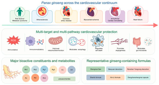
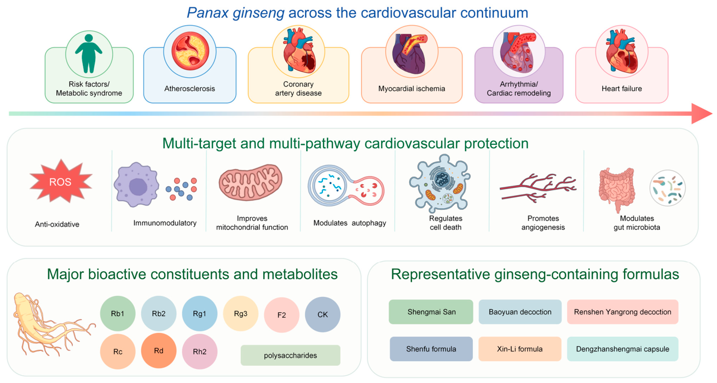
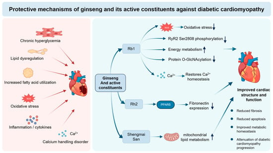
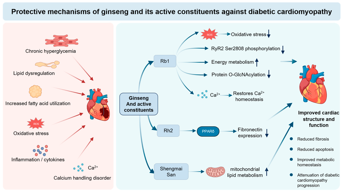
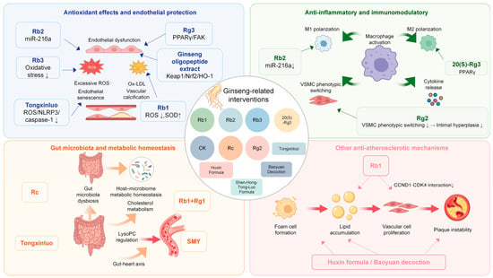
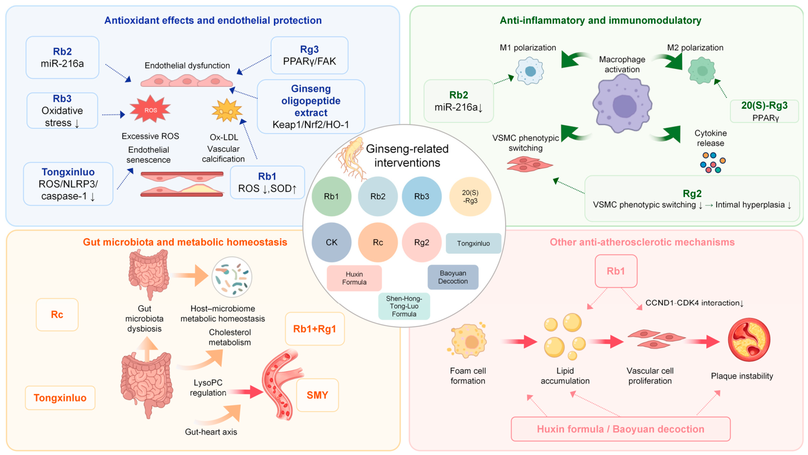
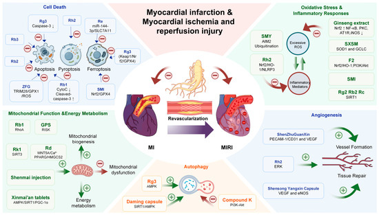
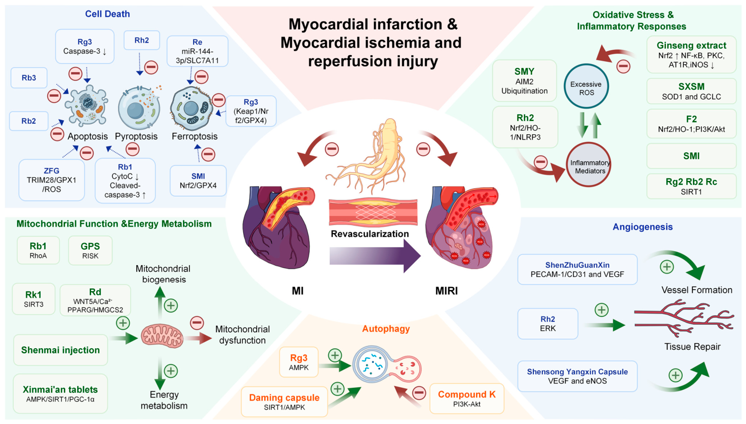

Disclaimer/Publisher’s Note: The statements, opinions and data contained in all publications are solely those of the individual author(s) and contributor(s) and not of MDPI and/or the editor(s). MDPI and/or the editor(s) disclaim responsibility for any injury to people or property resulting from any ideas, methods, instructions or products referred to in the content. |
© 2026 by the authors. Licensee MDPI, Basel, Switzerland. This article is an open access article distributed under the terms and conditions of the Creative Commons Attribution (CC BY) license.