


Abstract
Myxobacteria are prolific producers of structurally diverse secondary metabolites and represent a valuable source of anticancer lead compounds. In this study, quinaldic acid (QA) was putatively annotated from a myxobacterial metabolite extract by LC–MS/MS spectral matching and evaluated for its antitumor activity against non-small cell lung cancer (NSCLC) cells. QA inhibited the viability of four tumor cell lines in a concentration-dependent manner, with A549 lung adenocarcinoma cells displaying the highest sensitivity, with an IC50 of 1.07 mM after 24 h of treatment. QA also significantly suppressed clonogenic growth in A549 cells. Network pharmacology analysis suggested that QA may regulate NSCLC-related targets and pathways involved in cell-cycle control, apoptosis, p53, JAK–STAT, and NF-κB signaling. Experimental validation showed that QA induced S-phase accumulation in A549 cells, increasing the S-phase population by 14.70 percentage points at 4 mM. This effect was accompanied by increased cyclin A1 expression and decreased cyclin B1 and CDK1 expression. QA also promoted apoptosis in a concentration-dependent manner, increasing the apoptotic population by 40.40 percentage points at 2 mM. Apoptosis was associated with elevated BAX and caspase-3 expression and reduced BCL-2 expression. These findings suggest that QA suppresses A549 cell growth by inducing S-Phase accumulation and apoptosis, supporting its potential as a myxobacteria-derived anticancer scaffold.
1. Introduction
Lung cancer remains one of the leading causes of cancer-related morbidity and mortality worldwide [1]. Non-small cell lung cancer (NSCLC) accounts for approximately 85% of all lung cancer cases [2,3,4]. As of 2022, approximately 2,480,675 new lung cancer cases were reported worldwide, with a crude mortality rate of 23.1 per 100,000 population. Despite advances in chemotherapy, targeted therapy, and immunotherapy, drug resistance, systemic toxicity, tumor heterogeneity, and disease recurrence continue to limit long-term clinical benefit. Therefore, identifying bioactive small molecules with distinct mechanisms of action remains an important strategy for anticancer lead discovery.
Myxobacteria are Gram-negative bacteria that are widely distributed in soil ecosystems. They typically possess large genomes (9–16 Mb) and numerous biosynthetic gene clusters for secondary metabolites, and genome size is closely associated with secondary metabolic capacity [5,6,7]. Owing to the substantial structural diversity and novel chemical scaffolds of their metabolites, myxobacteria have emerged as important sources of bioactive natural products and rank second only to actinomycetes among microbial producers of drug-like molecules [7,8,9]. To date, a variety of compounds have been isolated from myxobacteria, including polyketides, nonribosomal peptides, polyketide–nonribosomal peptide hybrids, alkaloids, terpenoids, and phenylpropanoids. These compounds exhibit broad biological activities, including antibacterial, antifungal, antitumor, antiviral, immunosuppressive, and antioxidant activities [10,11]. Their mechanisms of action include disruption of the cytoskeleton, modulation of protein phosphorylation, inhibition of the respiratory chain, alteration of membrane permeability, and metabolic reprogramming [12,13].
Myxobacterial natural products have demonstrated considerable potential for antitumor drug development. Epothilones are among the most representative examples. By stabilizing microtubules and inhibiting mitosis in tumor cells, epothilones exert anticancer effects. Their semisynthetic derivative, ixabepilone, has been approved for the treatment of advanced or metastatic breast cancer and represents a notable example of the successful clinical translation of a myxobacterial natural product. In addition to epothilones, numerous myxobacterial metabolites remain under preclinical investigation and exhibit distinct antitumor mechanisms. For example, tubulysins inhibit microtubule polymerization, display potent cytotoxicity, and have been developed as payloads for antibody–drug conjugates [14,15]. Disorazoles also target the microtubule system and exert marked inhibitory effects against multiple tumor cell types [16]. Compounds such as rhizopodin and chivosazole disrupt the actin cytoskeleton, thereby affecting tumor cell morphology and proliferation [17,18]. Collectively, these studies indicate that myxobacteria produce structurally novel antitumor compounds that target key cellular processes, including microtubule dynamics and actin cytoskeletal organization. Accordingly, myxobacteria represent a promising source of novel anticancer lead compounds and may offer opportunities to address drug resistance.
In this study, quinaldic acid (QA) was tentatively identified in an antitumor-active fraction of a myxobacterial metabolite extract. QA contains a quinoline scaffold that is present in numerous molecules with antitumor activity and is regarded as a privileged structure in medicinal chemistry. This structural feature suggests that QA may possess biological activity and be amenable to structural optimization. Previous studies have suggested that QA may inhibit the proliferation and migration of glioblastoma cells, renal cancer cells, and LS180 colon cancer cells through modulation of tumor suppressor proteins, including p53 [19,20,21,22,23]. However, evidence regarding the anticancer activity of QA remains limited. Its broader antitumor activity, tumor-cell selectivity, direct intracellular targets, and key signaling pathways remain unclear.
In addition, QA has been proposed to be a derivative of kynurenic acid (KYNA) [24]. KYNA is a tryptophan metabolite in the tryptophan–kynurenine pathway (KP) and has demonstrated antiproliferative activity against cancer cells in vitro. QA may therefore be associated with tryptophan metabolism; however, its in vivo origin and biological functions remain to be elucidated. Although upstream KP metabolites have received extensive attention, the roles of QA in tumor development, tumor progression, and the tumor microenvironment remain unclear. Therefore, a systematic evaluation of the anticancer activity of QA and its potential mechanisms is warranted.
In this study, we systematically evaluated the cytotoxic effects of QA against multiple tumor cell lines and further investigated the potential molecular mechanisms underlying its inhibitory effects in A549 NSCLC cells. Our findings expand the repertoire of myxobacteria-derived antitumor metabolites and provide an experimental basis for the further development of QA as a candidate lead compound for NSCLC through structural optimization and preclinical evaluation.
2. Materials and Methods
2.1. Reagents and Chemicals
QA (purity ≥ 99%; CAS No. 93-10-7; catalog no. 258165) was purchased from J&K Scientific Ltd. (Beijing, China). QA was dissolved in sterile water to prepare a 40 mM stock solution and freshly diluted with complete culture medium to the required working concentrations before each experiment. Vehicle-control wells received the same volume of sterile water.
The Cell Counting Kit-8 (CCK-8; catalog no. C0037) and Annexin V-fluorescein isothiocyanate/propidium iodide (Annexin V-FITC/PI) Apoptosis Detection Kit (catalog no. C1062M) were purchased from Beyotime Biotechnology (Shanghai, China). XAD-16 macroporous adsorption resin was obtained as a commercially pretreated product (catalog no. S51657; Yuanye Bio-Technology Co., Ltd., Shanghai, China). All other reagents were of analytical grade or suitable for cell culture. Commercial Ham’s F-12K medium supplemented with GlutaPLUS was purchased from Servicebio (Wuhan, China; catalog no. G4560-500ML). Rabbit polyclonal primary antibodies against BAX (catalog no. GB11690; 1:1000), BCL-2 (catalog no. GB11223; 1:1000), total caspase-3 (catalog no. GB11767C; 1:1000), cyclin A1 (catalog no. GB11547; 1:1000), cyclin B1 (catalog no. GB11224; 1:1000), cyclin-dependent kinase 1 (CDK1; catalog no. GB11225; 1:1000), and β-actin (catalog no. GB11011; 1:5000) were purchased from Servicebio Technology Co., Ltd. (Wuhan, China). The horseradish peroxidase (HRP)-conjugated goat anti-rabbit immunoglobulin G (IgG; H + L) secondary antibody (catalog no. GB23303; 1:5000) was obtained from the same supplier.
The human lung adenocarcinoma cell line A549, human hepatocellular carcinoma cell line SK-HEP-1, human breast cancer cell line MCF-7, human glioblastoma cell line U87MG, and human embryonic kidney cell line HEK293T were kindly provided by Professor Fang Wan from the Molecular Pharmacology Laboratory, Inner Mongolia Agricultural University. All cell cultures tested negative for Mycoplasma by PCR within 3 months of the reported experiments.
A549 cells were maintained in Ham’s F-12K medium supplemented with 10% fetal bovine serum (FBS) and 1% penicillin–streptomycin. U87MG and SK-HEP-1 cells were maintained in minimum essential medium (MEM) supplemented with 10% FBS and 1% penicillin–streptomycin. MCF-7 and HEK293T cells were maintained in Dulbecco’s modified Eagle’s medium (DMEM) supplemented with 10% FBS and 1% penicillin–streptomycin. All cells were cultured at 37 °C in a humidified atmosphere containing 5% CO2.
2.2. Culture and Identification of Myxobacterial Strain E51
Myxobacterial strain E51 was previously isolated and preserved in our laboratory. The strain stored at −80 °C was inoculated onto VY/2 solid medium for recovery and used in subsequent experiments after satisfactory growth was observed. Genomic DNA was extracted using an alkaline lysis method, and the 16S rRNA gene was amplified using the universal primers 27F, 5′-AGAGTTTGATCCTGGCTCAG-3′, and 1495R, 5′-CTACGGCTACCTTGTTACGA-3′. PCR amplification was performed in a 30 µL reaction mixture containing 15.0 µL of Premix Taq DNA polymerase mix, 0.3 µL of 27F primer, 0.3 µL of 1495R primer, 3.0 µL of genomic DNA, and 11.4 µL of ddH2O. The PCR amplification program was as follows: initial denaturation at 94 °C for 3 min; 30 cycles of denaturation at 94 °C for 30 s, annealing at 55 °C for 45 s, and extension at 72 °C for 90 s; followed by a final extension at 72 °C for 10 min.
The PCR products were examined by agarose gel electrophoresis and then subjected to bidirectional sequencing by Sangon Biotech Co., Ltd. The obtained sequences were assembled and corrected, followed by homology analysis using NCBI BLAST 2.17.0 (https://blast.ncbi.nlm.nih.gov/Blast.cgi; accessed on 13 October 2023). The sequence was deposited in the GenBank database under accession number OR668733.1. Closely related sequences of reference strains with high homology were selected for multiple sequence alignment using MEGA 7.0, and a phylogenetic tree was constructed using the neighbor-joining NJ method. The robustness of the tree topology was evaluated by bootstrap analysis with 1000 replicates. The strain was comprehensively identified in combination with its morphological characteristics.
2.3. Preparation of Secondary Metabolites from Strain E51 and Evaluation of Their Antitumor Activity
An activated seed culture of strain E51 was inoculated at 2% (v/v) into a 10 L fermenter containing 5 L of VY/2 liquid medium. The VY/2 medium was composed of yeast, 5 g/L; CaCl2·2H2O, 1 g/L; and MgSO4·7H2O, 0.5 mg/L, with the pH adjusted to 7.2. At the start of fermentation, commercially pretreated XAD-16 macroporous adsorption resin was added at 2% (w/v) for in situ adsorption. Fermentation was carried out at 28 °C with continuous aeration for 7 days.
At the end of fermentation, the fermentation broth and resin were discharged together, and the resin was collected by filtration through a sieve. The resin was thoroughly washed with distilled water and air-dried at room temperature. The dried resin was extracted three times with methanol (800 mL per extraction). The combined extracts were concentrated under reduced pressure at a temperature not exceeding 40 °C to yield a crude extract of secondary metabolites from strain E51.
The inhibitory effects of the crude extract of secondary metabolites from strain E51 on human glioblastoma U87MG cells and human embryonic kidney HEK293T cells were evaluated using the Cell Counting Kit-8 (CCK-8) assay. U87MG and HEK293T cells in the logarithmic growth phase were seeded into 96-well plates. After cell attachment, the cells were treated with the E51 crude extract at concentrations ranging 1000 µg/mL for 24 h. Subsequently, CCK-8 reagent was added, and the cells were incubated according to the manufacturer’s instructions. Absorbance was then measured, and cell proliferation inhibition was calculated as follows:
Note: OD control represents the optical density of untreated cells from the same cell line, OD treatment represents the optical density of cells treated with the E51 crude extract, and OD blank represents the optical density of cell-free wells containing culture medium and CCK-8 reagent.
Where OD control, OD treatment, and OD blank represent the optical density values of the control, treatment, and blank wells, respectively. The calculated inhibition rates were used to assess the cytotoxic or antiproliferative effects of the E51 crude extract on the two cell lines. HEK293T cells were included as a non-tumor-derived comparison cell line, while the inhibition rate for each cell line was calculated relative to its own untreated control. For morphological assessment, representative images were acquired using a 10× objective.
2.4. Silica Gel Column Chromatographic Fractionation of Antitumor-Active Components from E51 Metabolites and Assessment of Antitumor Activity
The crude extract was dissolved in a small volume of methanol and thoroughly mixed with silica gel for column chromatography (200–300 mesh) at an approximate sample-to-silica ratio of 1:50 (w/w). The mixture was evaporated to dryness under reduced pressure to prepare a dry-loaded sample. The sample was fractionated by silica gel column chromatography (column dimensions: 5 cm inner diameter × 15 cm length; silica gel, 130 g). Elution was performed with gradients of petroleum ether–ethyl acetate (PE–EtOAc; v/v, 100:0 to 0:100) and dichloromethane–methanol (DCM–MeOH; v/v, 100:0 to 0:100).
Fractions were analyzed by thin-layer chromatography (TLC) using silica gel GF254 plates (Qingdao Marine Chemical Factory, Qingdao, China). The developing solvent was DCM–MeOH (15:1, v/v), and the plates were visualized under ultraviolet light at 254 nm. Fractions with similar TLC profiles were combined.
The activities of nine silica gel column chromatography fractions from strain E51 were evaluated in human embryonic kidney cells (HEK293T), human lung adenocarcinoma cells (A549), human breast cancer cells (MCF-7), human glioblastoma cells (U87MG), and human hepatocellular carcinoma cells (SK-HEP-1). U87MG, A549, SK-HEP-1, MCF-7, and HEK293T cells were seeded separately in 96-well tissue culture plates at 5 × 10³ cells per well and incubated at 37 °C in 5% CO2 for 24 h to allow cell attachment. Cells were then exposed to crude fractions at concentrations of 1.5625, 3.125, 6.25, 12.5, 25, 50, 100, or 200 μg/mL for 24 h at 37 °C. Cell viability was assessed using CCK-8, and absorbance was measured at 450 nm using a spectrophotometer.
2.5. Mass Spectrometric Identification of Fractions with High Antitumor Activity
Silica gel column chromatography fractions with high antitumor activity were subjected to LC-MS analysis. Fraction 5 was dissolved in LC-MS-grade methanol to a concentration of 1 mg/mL, filtered through a 0.22 µm membrane filter, and 5 µL of the filtrate was injected into the LC-MS system. Chromatographic separation was performed using a Hypersil GOLD C18 column (100 mm × 2.1 mm i.d., 1.9 µm) maintained at 40 °C. The mobile phases consisted of 0.1% formic acid in water (solvent A) and methanol (solvent B) (Table 1), delivered at a flow rate of 0.2 mL/min.
Table 1.
Gradient elution conditions for chromatography.
Mass spectrometric analysis was performed using a Q Exactive HF-X high-resolution mass spectrometry system (Thermo Fisher Scientific, Waltham, MA, USA). The scan range was set to m/z 100–1500. Electrospray ionization source parameters were as follows: spray voltage, 3.5 kV; sheath gas flow rate, 35 arbitrary units; auxiliary gas flow rate, 10 L/min; capillary temperature, 320 °C; S-lens radio frequency level, 60; and auxiliary gas heater temperature, 350 °C. Data were acquired separately in positive- and negative-ion modes. Data-dependent acquisition was used for tandem mass spectrometry analysis.
Raw data were processed using Compound Discoverer version 3.3. The workflow included peak extraction, retention-time correction, and peak-area quantification. Metabolites were identified by matching accurate molecular masses, with a mass error < 5 ppm, and MS/MS fragmentation information against the mzCloud and other relevant databases.
2.6. Determination of the IC50 of QA
A549 cells were seeded in 96-well plates at 5 × 10³ cells per well and incubated overnight for 24 h at 37 °C in a humidified atmosphere containing 5% CO2 to allow cell attachment. Cells were subsequently exposed to QA at concentrations ranging from 0.5 to 8 mM for 24 h under the same culture conditions. The half-maximal inhibitory concentration (IC) was determined from percentage cell-viability data by nonlinear regression using GraphPad Prism version 9.5.1 (2023).
2.7. Network Pharmacology-Based Prediction of the Mechanisms Underlying QA Activity Against NSCLC
2.7.1. Network Pharmacology Databases and Software
The following databases and software were used for network pharmacology analysis: PubChem database (https://pubchem.ncbi.nlm.nih.gov/), SwissTargetPrediction database (http://www.swisstargetprediction.ch/), SuperPred database (https://bio.tools/superpred), ChEMBL database (https://www.ebi.ac.uk/chembl/), GeneCards database (http://www.genecards.org/), Online Mendelian Inheritance in Man (OMIM) database (https://www.omim.org/), Venny 2.1.0 (https://bioinfogp.cnb.csic.es/tools/venny/index.html), Metascape database (https://metascape.org/), All databases and web tools were accessed on 24 March 2025. Cytoscape versions 3.7.0 and 3.9.1 were used for network analysis and visualization.
2.7.2. Identification and Screening of Potential QA Targets
Potential active targets of QA were screened using the PharmMapper and SwissTargetPrediction databases. The UniProt database was used to standardize target names and convert them into official gene symbols, thereby obtaining a standardized gene-naming system for subsequent analysis.
2.7.3. Identification of NSCLC-Related Targets
Using “lung cancer” as the search term, lung cancer-related disease targets were retrieved from the GeneCards database. Targets with a GeneCards Relevance Score ≥ 20 were selected as core regulatory targets associated with lung cancer. The target gene names were standardized using the UniProt database.
2.7.4. Identification of Overlapping Targets and Network Construction
The QA-related target genes and lung cancer-related therapeutic target genes were imported into the Bioinformatics online platform. Intersection analysis was performed using a Venn diagram tool to obtain the shared gene set between the compound and disease targets, which was defined as the potential therapeutic target set of QA against lung cancer.
2.7.5. Protein–Protein Interaction Network Construction and Hub-Target Screening
The intersecting genes were imported into the STRING 12.0 database to construct a protein–protein interaction (PPI) network. The species was set to “Homo sapiens”, the confidence threshold was set to a combined score > 0.7, and all other parameters were retained at their default settings. After excluding non-core associated genes, a core-gene PPI network was generated, and the network data were exported in TSV format. Network topological analysis was performed using the CytoNCA plugin in Cytoscape 3.7.1. The PPI network map was generated by ranking nodes in descending order according to degree values.
2.7.6. Gene Ontology Functional and Kyoto Encyclopedia of Genes and Genomes Pathway Enrichment Analyses of Core Targets
Core targets were uploaded to the Metascape database for Gene Ontology (GO) enrichment analysis, including biological process (BP), cellular component (CC), and molecular function (MF) categories, as well as Kyoto Encyclopedia of Genes and Genomes (KEGG) pathway enrichment analysis. The species was set to “H. sapiens”, and p < 0.05 was considered statistically significant. The top 10 GO terms ranked by p value were selected, and the top 20 KEGG signaling pathways significantly associated with lung cancer were screened. GO three-in-one bar charts and KEGG enrichment bubble plots were visualized using the Bioinformatics online platform.
2.7.7. Construction of the Multidimensional Interaction Network
The data of the active compound, core targets, and key pathways were integrated, and Cytoscape 3.7.1 was used to construct a “compound–target–pathway–disease” network model for the anti-lung cancer effect of QA. The interaction strength among network elements was evaluated based on network topological parameters.
2.8. Wound-Healing Assay
A549 cells were seeded into 6-well plates at a density of 5 × 10⁵ cells/well. After the cells reached approximately 100% confluence, a linear wound was created in each well using a sterile 10 µL pipette tip. The detached cells were removed by gentle washing, and the remaining cells were treated with QA at final concentrations of 0, 0.5, 1, 2, and 4 mM. Images of three fixed fields per well were acquired using an optical microscope equipped with a 4× objective at 0, 24, and 48 h after treatment. Changes in wound width were quantified using ImageJ software (version 1.53t, National Institutes of Health, USA), and wound closure was calculated as a percentage of the initial wound width at 0 h. Data are presented as the mean ± standard deviation.
2.9. Colony Formation Assay
To assess the long-term proliferative survival of individual A549 cells after drug treatment, cells were seeded in 6-well plates at 500 cells per well in 2 mL of complete medium containing 10% FBS and cultured at 37 °C in a humidified incubator containing 5% CO2. After cells had attached for 12–24 h, they were treated with 4 mM QA dissolved in DMSO, with a final DMSO concentration of ≤0.1%, or with an equal volume of vehicle control (0.1% DMSO) for 24 h. The drug-containing medium was removed, and cells were gently washed once with phosphate-buffered saline (PBS). Fresh complete medium containing 10% FBS was then added, and cells were cultured for an additional 14 days. The culture medium was replaced every 2–3 days.
At the end of the culture period, cells were washed twice with PBS, fixed with absolute methanol for 15 min, and stained with 0.1% crystal violet (w/v in 20% methanol) at room temperature for 20 min. Excess stain was gently removed under running water, and the plates were air-dried overnight at room temperature. Colonies containing more than 50 cells were counted under an inverted microscope. Each condition was tested in triplicate wells in three independent experiments. Data are expressed as the mean ± standard deviation (SD).
2.10. Cell Cycle Analysis
A549 cells were seeded in T25 cell culture flasks at a density of 3 × 10⁵ cells per flask and allowed to attach overnight. The cells were then treated with QA at final concentrations of 0, 0.5, 1, 2, and 4 mM for 24 h. Following treatment, the cells, including both adherent and floating cells, were collected by trypsinization and centrifugation. The cell pellets were washed twice with ice-cold phosphate-buffered saline (PBS).
The cells were fixed by slowly adding pre-chilled 70% ethanol while gently vortexing and were incubated overnight at 4 °C. The fixed cells were then centrifuged to remove the ethanol and washed twice with PBS. Subsequently, the cells were resuspended in a staining solution containing RNase A at a final concentration of 100 µg/mL and propidium iodide (PI) at a final concentration of 50 µg/mL. The cells were incubated at 37 °C for 30 min in the dark.
Cellular DNA content and cell-cycle distribution were analyzed using a CytoFLEX S flow cytometer (Beckman Coulter, Indianapolis, IN, USA). At least 10,000 single-cell events were acquired for each sample.
2.11. Apoptosis Analysis
Apoptosis was assessed and quantified by flow cytometry using Annexin V–fluorescein isothiocyanate (FITC) and propidium iodide (PI) dual staining. A549 cells were seeded in T25 cell culture flasks at a density of 3 × 10⁵ cells per flask and allowed to adhere overnight. Cells were subsequently treated with QA at final concentrations of 0, 0.5, 1, 2, and 4 mM for 24 h.
Following treatment, nonadherent cells in the culture supernatant were collected and combined with adherent cells harvested by trypsinization to avoid excluding detached apoptotic cells. Cells were washed twice with ice-cold phosphate-buffered saline (PBS), centrifuged, and resuspended in 1× binding buffer. The cell density was adjusted to approximately 1 × 10⁶ cells/mL, and 100 µL of the cell suspension was transferred to a flow cytometry tube. Each sample was stained with 1 µL of Annexin V–FITC and 1.5 µL of PI for 30 min in the dark. After incubation, 400 µL of 1× binding buffer was added to each sample to obtain a final staining volume of 500 µL. Flow cytometric analysis was completed within 1 h after staining.
Annexin V–FITC and PI fluorescence signals were acquired using a CytoFLEX S flow cytometer (Beckman Coulter, USA). Spectral overlap between FITC and PI was compensated using single-stained controls. Quadrant gates were set using unstained cells, Annexin V–FITC single-stained controls, and PI single-stained controls. Cell debris and doublets were excluded before analysis. Viable cells were defined as Annexin V−/PI−, early apoptotic cells as Annexin V+/PI−, and late apoptotic and/or secondary necrotic cells as Annexin V+/PI+. At least 10,000 single-cell events were acquired for each sample.
2.12. Western Blotting
A549 cells were seeded in 10 cm culture dishes at a density of 1 × 10⁶ cells per dish, allowed to adhere overnight, and then treated with QA at final concentrations of 0, 0.5, 1, 2, and 4 mM for 24 h. Following treatment, the culture medium was removed, and the cells were washed twice with ice-cold phosphate-buffered saline (PBS). Cells were lysed in pre-chilled radioimmunoprecipitation assay (RIPA) buffer supplemented with protease and phosphatase inhibitors. After lysis on ice for 30 min, the lysates were centrifuged at 12,000× g for 10 min at 4 °C, and the supernatants were collected for total protein analysis.
Protein concentrations were determined using a bicinchoninic acid (BCA) assay. Equal amounts of total protein (30 µg per lane) were separated by sodium dodecyl sulfate-polyacrylamide gel electrophoresis (SDS-PAGE) and transferred onto polyvinylidene difluoride (PVDF) membranes. Owing to differences in molecular weight among the target proteins, individual target proteins were analyzed on separate, independently prepared membranes using the same batch of protein samples and the same loading scheme. The membranes were blocked with 5% nonfat milk in Tris-buffered saline containing Tween 20 (TBST) for 1–2 h at room temperature and incubated overnight at 4 °C with the corresponding primary antibodies. After washing with TBST, the membranes were incubated with horseradish peroxidase (HRP)-conjugated secondary antibodies for 1–1.5 h at room temperature.
Protein bands were visualized by enhanced chemiluminescence (ECL) using a G:BOX Chemi XT4 imaging system and quantified by densitometric analysis using ImageJ. Relative protein expression was calculated by normalizing the intensity of each target protein to that of the corresponding β-actin loading control, followed by normalization to the untreated control (0 concentration), which was assigned a value of 1.0. All experiments were performed independently at least three times.
2.13. In Silico Prediction of QA Drug-likeness
The drug-development potential of QA was predicted using the ProTox and SwissTargetPrediction databases.
2.14. Statistical Analysis
All experiments were performed in triplicate, and mean values were used for analysis. Data were analyzed using statistical software. Differences among groups were compared using one-way analysis of variance. A value of p < 0.05 was considered statistically significant.
3. Results
3.1. Myxobacterial Strain E51 Produces Metabolites with Cytotoxic Activity Against Cancer Cells
A myxobacterial strain, designated E51, was previously isolated from a soil sample. During the late growth phase, strain E51 formed orange to orange-red, spherical or hemispherical fruiting bodies. Its 16S rRNA gene sequence showed 100% similarity to that of Myxococcus fulvus. In the phylogenetic tree, strain E51 clustered with the type strain of M. fulvus in a distinct clade. Based on its morphological characteristics and phylogenetic analysis, strain E51 was identified as Myxococcus fulvus (Figure 1C).
Figure 1.
Identification of strain E51 and inhibitory activity of its metabolite crude extract. (A) Colony morphology; (B) morphology of the fruiting bodies of strain E51; (C) phylogenetic tree constructed based on 16S rRNA gene sequences; and (D) inhibitory activity of the crude extract from strain E51.
Strain E51 was fermented, and a crude metabolite extract was prepared. Its inhibitory effects on human glioblastoma U87MG cells and human embryonic kidney HEK293T cells were measured to evaluate its antitumor activity and cytotoxicity. The crude extract inhibited U87MG and HEK293T cells by 80.90% and 30.41%, respectively (Figure 1D). These findings indicate that the crude extract exerted substantial inhibitory activity against U87MG cells while showing lower inhibitory activity against HEK293T cells, suggesting a degree of selectivity toward tumor cells.
3.2. Silica Gel Chromatographic Fractionation Identifies Strain E51 Metabolite Fractions with Cytotoxic Activity Against Cancer Cells
The crude extract of metabolites from strain E51 was preliminarily fractionated by silica gel column chromatography, yielding nine fractions. Among these, three fractions had relatively limited effects on the viability of the normal cell line HEK293T, with inhibition rates below 20% (Figure 2). In contrast, these fractions exhibited inhibition rates exceeding 80% in certain tumor cell lines.
Figure 2.
Antitumor activity of silica gel column chromatography fractions of metabolites from strain E51. Statistical analysis of inhibition rates in different cell lines treated with different fractions. In the inhibition-rate plots, panels are arranged from left to right and from top to bottom as follows: Fraction 2, Fraction 5, and Fraction 9. For each fraction, the cell lines are HEK293T, A549, U87MG, SK-HEP-1, and MCF-7 under the indicated treatment conditions. Data are presented as mean ± standard deviation (SD); n = 3.
Specifically, Fraction 2 showed pronounced inhibitory activity against U87MG and A549 cells, with maximum inhibition rates of 86.40% and 82.91%, respectively. Fraction 5 exhibited the strongest inhibitory effect on SK-HEP-1 cells, with a maximum inhibition rate of 85.08%. This fraction also inhibited U87MG and MCF-7 cells, with maximum inhibition rates of 63.24% and 52.79%, respectively. Fraction 9 showed the strongest inhibitory effect on MCF-7 cells, with a maximum inhibition rate of 94.66%. This fraction also inhibited A549 and SK-HEP-1 cells, with maximum inhibition rates of 49.17% and 36.58%, respectively.
After treatment with the active fractions, cancer cells displayed apparent features of cell death and morphological alterations. Compared with the control group, treated cells were markedly reduced in number, showed decreased adherence, became smaller and more rounded, and exhibited weakened intercellular contacts. In addition, the cells showed indistinct borders, irregular morphology, increased cytoplasmic granularity, and greater accumulation of cellular debris. These changes were consistent with morphological features of cell injury (Figure 3).
Figure 3.
Morphological changes in different cell lines following treatment with different fractions. The images on the left show microscopic observations of each cell line under representative treatment conditions, illustrating cell density, adherence status, and morphological changes.
3.3. QA Is Identified in Silica Gel Chromatographic Fraction 5 from Strain E51
Fraction 5, obtained by silica gel column chromatography of metabolites from strain E51, exhibited significant inhibitory activity against U87MG, SK-HEP-1, and MCF-7 cells, suggesting broad-spectrum antitumor potential. To characterize its putative bioactive constituents, the chemical composition of fraction 5 was analyzed by untargeted liquid chromatography–tandem mass spectrometry (LC–MS/MS).
Based on relative MS signal intensity, relative peak-area ranking, and concordant “Full match” results in the mzCloud, mzVault, and MassList databases, 30 metabolites were detected in fraction 5 (Table 2). Among these substances, QA was detected as a deprotonated molecular ion, [M−H]−, with a mass error of +0.03 ppm. Its major fragment ions at m/z 128.04976, 129.05318, and 144.04471 were highly consistent with the mzCloud reference spectrum, supporting the reliable identification of QA (Figure 4). QA is a quinoline-2-carboxylic acid derivative with a characteristic quinoline heterocyclic scaffold. Previous studies have shown that QA inhibits the growth of colon cancer cells and may be associated with regulation of the p53 pathway [20]. However, its anticancer activity and underlying molecular mechanisms in other tumor types remain insufficiently characterized. Considering its relative abundance in fraction 5, reliable mass spectrometric identification, reported antitumor activity, and potential relevance to metabolic regulation, QA was selected as the priority candidate compound for further investigation of its anticancer activity and mechanism of action.
Table 2.
Annotated metabolites detected by LC–MS in negative-ion (ESI−) and positive-ion (ESI+) modes.
Figure 4.
Mirror MS/MS spectral comparison between QA and the mzCloud reference spectrum. The upper (forward) spectrum represents the reference spectrum, whereas the lower (reverse) spectrum represents the sample spectrum. Green annotations indicate matched fragment-ion peaks; gray annotations indicate unmatched or low-contribution ion peaks; and red annotations indicate selected peaks, apex positions, matching anchors, or related features. The m/z values labeled in the figure are experimentally measured peak values from the representative MS/MS spectrum and may differ slightly from the software-annotated values reported in the table.
3.4. QA Suppresses Cancer Cell Viability
The inhibitory activity of QA against multiple cancer cell lines was evaluated using the Cell Counting Kit-8 (CCK-8) assay. QA exhibited marked dose-dependent inhibitory effects on tumor cells. The greatest inhibition of all four tumor cell lines was observed at 4 mM, with inhibition rates ranging from 27.64% to 91.72% (Figure 5). Among the cell lines tested, A549 cells were the most sensitive to QA, exhibiting an inhibition rate of 91.72%. At the same concentration, QA inhibited human embryonic kidney 293T (HEK293T) cells, the sole normal-cell control in this study, by only 14.42%. These findings suggest that QA preferentially inhibits tumor cells relative to HEK293T cells under the experimental conditions.
Figure 5.
Inhibitory rates of QA in HEK293T cells and four cancer cell lines. (A−E) Inhibitory rates of quinaldic acid in HEK293T, A549, MCF-7, SK-HEP-1, and U87MG cells, respectively. (F) Determination of the IC 50 of quinaldic acid in A549 cells.
Morphological observations showed that QA induced prominent vacuolization and cellular shrinkage in A549 cells, whereas no obvious morphological changes were observed in HEK293T cells (Figure 6). Nonlinear regression analysis (R2 > 0.95) yielded a half-maximal inhibitory concentration (IC 50) of 1.07 mM for QA in A549 cells (Figure 5).
Figure 6.
Morphological changes in HEK293T and A549 cells following treatment with quinaldic acid (QA). (A) Panels (a–g) show A549 cells treated with quinaldic acid at 0, 0.5, 1, 2, 3, 4, and 5 mM, respectively. (B) Panels (a–g) show HEK293T cells treated with quinaldic acid at 0, 0.5, 1, 2, 3, 4, and 5 mM, respectively.
3.5. Network Pharmacology Predicts the Core Targets and Signaling Pathways of QA in NSCLC
In this study, the potential targets of QA and disease-associated targets of non-small cell lung cancer (NSCLC) were predicted using the SwissTargetPrediction, SuperPred, and ChEMBL databases. A total of 189 putative QA targets and 1746 NSCLC-associated targets were identified. Intersection analysis yielded 47 shared targets (Figure 7A–C). Topological analysis of the protein–protein interaction (PPI) network identified nine core targets: prostaglandin-endoperoxide synthase 2 (PTGS2), matrix metallopeptidase 9 (MMP9), nuclear factor kappa B subunit 1 (NFKB1), matrix metallopeptidase 2 (MMP2), poly(ADP-ribose) polymerase 1 (PARP1), signal transducer and activator of transcription 1 (STAT1), myeloid cell leukemia sequence 1 (MCL1), protein tyrosine phosphatase non-receptor type 11 (PTPN11), and thymidylate synthase (TYMS) (Figure 7D).
Figure 7.
Network pharmacology and Gene Ontology (GO)/Kyoto Encyclopedia of Genes and Genomes (KEGG) enrichment analyses of QA targets. (A) Venn diagram of drug and disease targets. (B) Gene network of shared drug–disease targets. (C) QA–target–NSCLC network. (D) Protein–protein interaction network of shared targets. (E) GO enrichment analysis of QA targets. (F) KEGG pathway enrichment analysis of QA targets.
Gene Ontology (GO) and Kyoto Encyclopedia of Genes and Genomes (KEGG) enrichment analyses of the shared targets were subsequently performed using Metascape to characterize their potential biological functions. GO enrichment analysis indicated that the putative effects of QA were primarily associated with oxidative stress, inflammatory responses, apoptosis, and metabolic regulation. KEGG pathway analysis identified 13 significantly enriched pathways, predominantly including the p53, Janus kinase–signal transducer and activator of transcription (JAK–STAT), and nuclear factor kappa B (NF-κB) signaling pathways, as well as microRNAs in cancer. Notably, enrichment of the p53 signaling pathway and DNA adduct-related pathways suggests that QA may induce A549 cell death by compromising DNA integrity or activating cell cycle checkpoints.
3.6. QA Does Not Significantly Affect Wound Closure but Reduces Colony Formation in A549 Cells
The effects of QA on the migration and proliferative capacity of A549 cells were evaluated using wound-healing and clonogenic assays. QA treatment did not significantly alter the migration rate or wound-healing rate of A549 cells (Figure 8A,B), indicating that QA did not inhibit cell migration under the tested conditions. In contrast, QA significantly suppressed the colony-forming capacity of A549 cells (Figure 8C,D). Compared with the control group, no apparent colony formation was observed in the 4 mM QA-treated group, and the number of colonies was significantly reduced (p < 0.0001), indicating that QA at this concentration markedly impaired the colony-forming capacity of non-small cell lung cancer (NSCLC) cells. In addition, QA-treated cells exhibited reduced cell size, suggesting marked alterations in cellular morphology.
Figure 8.
Effects of QA on wound closure and colony formation in A549 cells. (A) Representative images from the wound-healing assay. (B) Quantitative analysis of wound-healing rates. (C) Representative images from the colony-formation assay. (D) Quantitative analysis of colony numbers. ****: p < 0.0001.
Collectively, these findings indicate that QA did not significantly alter the wound-closure capacity of A549 cells but significantly inhibited their colony-forming capacity. This result suggests that QA may impair the long-term proliferative potential of A549 cells and may have the potential to suppress malignant phenotypes associated with clonogenic proliferation in NSCLC cells.
3.7. QA Induces Cell-Cycle Arrest in A549 Cells
To investigate the effects of QA on cell cycle progression in A549 cells, cell cycle analysis was performed. As shown in Figure 9, after 24 h of treatment, QA significantly inhibited cell cycle progression compared with the control group. The proportion of cells in the S phase increased significantly, whereas the proportion of cells in the G2/M phase decreased significantly. These changes became more pronounced with increasing QA concentration. At 1 mM, the proportion of S-phase cells increased by 10.20 percentage points, whereas the proportion of G2/M-phase cells decreased by 0.53 percentage points. At 4 mM, the proportion of S-phase cells increased by 14.70 percentage points, whereas the proportion of G2/M-phase cells decreased significantly by 7.72 percentage points. These results indicate that QA disrupts cell cycle progression in tumor cells and may induce S-Phase accumulation.
Figure 9.
Effects of QA on cell cycle progression in A549 cells. A549 cells were treated with the indicated concentrations of QA for 24 h. (A) Flow cytometric analysis of cell cycle distribution. (B) Quantification of the proportions of cells in different cell cycle phases. (C) Western blot analysis of cell cycle-related proteins. Western blot images are provided in Supplementary Figure S2. (D–F) Quantification of the relative expression levels of proteins assessed by Western blotting.
To further support this finding, the expression of cell cycle phase-specific proteins was examined by Western blotting. As shown in Figure 9C, QA treatment significantly increased the expression of cyclin A1, an S-phase-associated protein, while significantly decreasing the expression of cyclin B1, a G2-phase-associated protein, and cyclin-dependent kinase 1 (CDK1), an M-phase-associated protein. These findings were consistent with the cell cycle analysis and further suggest that QA may induce S-Phase accumulation in A549 cells. The original, uncropped Western blot images are provided in the Supplementary Materials (Figure S1).
3.8. QA Induces Apoptosis in A549 Cells
The effects of QA on apoptosis in A549 cells were evaluated using annexin V/propidium iodide (PI) dual staining. As shown in Figure 10, QA induced apoptosis in a significant dose-dependent manner over the concentration range of 0.5–2 mM. At 2 mM, QA increased the apoptotic rate by 40.4 percentage points relative to that of the control group. Morphological observations (Figure 10A) showed that QA-treated cells exhibited characteristic apoptotic features, including cytoplasmic condensation, cell shrinkage and rounding, and vacuolization. To further investigate the associated molecular events, the expression of apoptosis-related proteins was assessed. Western blot analysis (Figure 10C) showed that QA treatment significantly increased the expression of the proapoptotic proteins BAX and caspase-3 while decreasing the expression of the antiapoptotic protein BCL-2. These findings suggest that QA-induced apoptosis in A549 cells may be associated with the regulation of BAX, BCL-2, and caspase-3. The original, uncropped Western blot images are provided in the Supplementary Materials (Figure S2).
Figure 10.
Effects of QA on apoptosis in A549 cells. A549 cells were treated with the indicated concentrations of QA for 24 h. (A) Flow cytometric analysis of apoptosis. (B) Quantification of the proportions of cells at different stages of apoptosis. (C) Western blot analysis of apoptosis-related proteins.Original. Western blot images are provided in Supplementary Figure S1. (D–F) Quantification of the relative expression levels of proteins assessed by Western blotting.
3.9. In Silico Analysis Predicts the Drug-likeness and Toxicity Profiles of QA
In this study, the drug-likeness and safety profiles of quinaldic acid (QA) were evaluated using the ProTox-II platform through multidimensional in silico simulations. As shown in Figure 11, the predicted oral median lethal dose (LD 50) of QA was 1550 mg/kg. QA was classified as a Globally Harmonized System (GHS) Category 4 compound, indicating relatively low acute toxicity and a potentially broad therapeutic window. Physicochemical property analysis indicated that QA showed favorable compliance with Lipinski’s rule of five, with a low molecular weight (173.17 g/mol), moderate lipophilicity (logP = 1.93), and an optimized topological polar surface area (TPSA = 50.19 Å2). In population-distribution analyses, these properties were notably more favorable than the database average, suggesting favorable potential for bioavailability and tissue penetration.
Figure 11.
Toxicity assessment of QA. (A) Chemical structure, predicted LD 50, toxicity class, prediction confidence, and physicochemical properties of QA, together with comparisons of molecular-weight and dose distributions against compounds in the database. (B) Predicted potential toxicity targets, with colors indicating the probability of target binding. The results suggested that estrogen receptor 2 (ESR2) and prostaglandin G/H synthase 1 (PGH1) may exhibit potential interactions with QA. Pharmacophore-fit scores and ligand similarities for candidate toxicity targets are also shown. The assessment was performed using the ProTox-II server.
In the multidimensional toxicity profile, QA was predicted to be inactive with respect to carcinogenicity, mutagenicity, and immunotoxicity and showed no predicted binding tendency toward 87.5% of common toxicity targets. Although the model indicated potential hepatotoxicity and weak predicted binding to AOFA/PGH1, the very low pharmacophore-fit score and 0% similarity to known toxic ligands further supported its favorable predicted chemical safety profile. Overall, these in silico findings provide a preliminary characterization of the predicted drug-likeness and toxicity profiles of QA and warrant further experimental evaluation. (https://tox-new.charite.de/protox_II/; accessed on 4 February 2026).
QA demonstrated favorable drug-likeness, satisfying Lipinski, Ghose, and Veber criteria, as well as favorable predicted solubility and gastrointestinal absorption. It also exhibited a high bioavailability score (0.85), supporting its further investigation as a drug candidate (http://www.swissadme.ch/; accessed on 14 August 2025; Figure 12).
Figure 12.
Drug-likeness assessment of quinaldic acid.
4. Discussion
Myxobacteria are a valuable microbial resource for natural-product discovery, and their secondary metabolites exhibit substantial diversity in chemical structure, bioactivity, and mechanisms of action [6,10,25]. Among these bacteria, the genus Myxococcus is one of the most extensively studied, and its strains produce structurally diverse bioactive compounds targeting distinct biological processes. Representative examples include myxalamids, which interfere with energy metabolism-related processes and exhibit antibacterial activity [26]; myxothiazol, a thiazole-containing compound that inhibits mitochondrial respiratory complex III [27]; and myxovirescins, macrolide antibiotics that inhibit type II signal peptidase [28,29]. Collectively, these findings indicate that Myxococcus produces structurally diverse natural products that target distinct biological processes, including energy metabolism, respiratory-chain function, and key enzymatic activities. These features suggest that this genus has considerable potential as a source of antitumor and other bioactive lead compounds [6,30]. However, the chemical diversity of Myxococcus metabolites remains incompletely characterized, and the antitumor mechanisms of many Myxococcus-derived compounds have not been fully elucidated.
To our knowledge, this is the first report of a compound putatively annotated as QA in myxobacterial metabolites based on mass spectrometric analysis. This observation suggests that myxobacteria may possess the metabolic capacity to produce, transform, or accumulate QA-related compounds. However, evidence for the broad distribution of QA among plants, animals, and microorganisms remains limited. In plants, direct reports are scarce and primarily involve QA detected at increased relative abundance in Carex breviculmis under drought stress, suggesting that QA may occur only in specific plant species under particular stress conditions [31,32]. In animals, QA has been detected in urine, saliva, plasma, and gut-associated samples from humans and other mammals [33,34,35]. However, these observations do not establish QA as a widely synthesized and accumulated endogenous metabolite in animals. In microorganisms, QA biosynthesis has mainly been reported in limited Streptomyces species that produce thiopeptide natural products containing a QA structural moiety, such as thiostrepton from Streptomyces laurentii and Streptomyces azureus, and siomycin-class antibiotics from Streptomyces sioyaensis [36,37,38]. Therefore, QA is more appropriately considered a structural building block of specialized metabolites in a limited number of microorganisms than a widely distributed free metabolite. The detection of QA in myxobacteria in the present study expands the known microbial sources of QA and provides additional evidence of the chemical diversity of myxobacterial metabolites. Although information on the distribution of QA in microorganisms remains limited, its microbial transformation and utilization have been relatively well characterized. Several bacteria have been shown to metabolize QA through specific oxidoreductases and degradation pathways [39,40].
Building on these observations, the present study showed that QA significantly inhibited the growth of A549 cells, a human non-small cell lung cancer (NSCLC) cell line. Network pharmacology analysis provided preliminary insight into the potential mechanisms underlying QA activity. Network pharmacology analysis suggested TYMS, MCL1, NFKB1, PTGS2, and MMP9 as potential QA-associated targets. Specifically, TYMS is involved in thymidylate synthesis and is closely associated with DNA replication; MCL1 is an important anti-apoptotic protein, while NFKB1 is associated with cell survival, inflammatory responses, and anti-apoptotic signaling; PTGS2 and MMP9 are more closely involved in the inflammatory microenvironment, tumor invasion, and metastasis [41,42,43,44]. These network pharmacology results suggest that QA may exert antitumor effects through modulation of networks involved in DNA replication, cell-cycle progression, and apoptosis rather than through a single molecular target.
In the present study, QA-induced accumulation of A549 cells in S phase was accompanied by altered expression of cyclin A1, cyclin B1, and CDK1. Previous studies have reported aberrant cyclin A1 expression in several cancers and its potential involvement in cell-cycle regulation [45] whereas cyclin B1 and CDK1 form a key regulatory complex involved in mitotic entry [46]. S-phase accumulation has also been associated with impaired DNA replication, replication-fork stalling, disrupted nucleotide synthesis, and activation of the DNA damage response [47,48]. For example, thymidylate synthase (TYMS) is a key enzyme in thymidylate synthesis, and its inhibition can disrupt DNA replication and result in S-phase arrest [49]. In addition, activation of ATM/ATR–Chk1/Chk2 signaling can suppress CDK activity and delay cell-cycle progression [50,51]. Network pharmacology analysis identified TYMS as a potential QA-associated target, providing a possible hypothesis for the observed S-phase accumulation. However, this computational prediction does not establish a functional association between TYMS and the altered cell-cycle distribution, and there is currently no evidence that QA directly interacts with or inhibits TYMS. This hypothesis therefore requires validation through binding and enzyme-activity assays.
In addition, QA treatment significantly increased the proportion of apoptotic A549 cells and was accompanied by increased BAX expression, decreased BCL-2 expression, an elevated BAX/BCL-2 ratio, and a concentration-dependent increase in total caspase-3 protein expression. As a pro-apoptotic BCL-2 family member, BAX promotes mitochondrial outer-membrane permeabilization and cytochrome c release, whereas BCL-2 maintains mitochondrial membrane integrity and suppresses mitochondria-associated apoptotic signaling [52,53,54,55,56,57]. Accordingly, an increased BAX/BCL-2 ratio is commonly considered an indicator of a shift toward a pro-apoptotic state. However, because cleaved caspase-3, mitochondrial membrane potential, reactive oxygen species, and cytochrome c release were not assessed, the involvement of a mitochondria-dependent caspase cascade cannot be confirmed and requires further investigation. These findings are broadly consistent with previous studies of quinoline compounds. Some quinolines and their derivatives have been reported to induce tumor-cell apoptosis by regulating BCL-2 family proteins, disrupting mitochondrial function, or activating caspase pathways [58,59,60,61]. Previous studies of QA have focused primarily on colon cancer cell models. QA has been reported to inhibit HT-29 cell proliferation and affect signaling pathways involving extracellular signal-regulated kinase 1/2 (ERK1/2), p38, cAMP response element-binding protein (CREB), and protein kinase B (Akt) [19]. Another study suggested that QA may contribute to growth inhibition in LS180 cells by regulating p53 expression and its phosphorylation status [20]. Compared with these previous studies focused on colon cancer models, the present study provides the first evidence that QA induces apoptosis in NSCLC cells, thereby broadening the known antitumor spectrum of QA and suggesting that its mechanism of action may be context-dependent across different tumor types.
Network pharmacology analysis also predicted MCL1 and NFKB1 as potential QA-associated targets. MCL1 is an important anti-apoptotic protein that suppresses BAX/BAK activation and promotes tumor-cell survival [62,63,64,65]. NFKB1 commonly promotes cell survival by regulating the transcription of multiple anti-apoptotic genes [66,67]. Although MCL1 and NFKB1 expression or activation were not directly assessed in the present study, the marked changes in the BAX/BCL-2 axis suggest that QA affects apoptosis-regulatory networks. Thus, at a functional level, the experimental findings are consistent with the network pharmacology predictions related to apoptosis and cell-survival regulation. However, whether QA directly inhibits MCL1 or regulates downstream anti-apoptotic transcriptional programs through NF-κB signaling remains to be determined. In addition, although PTGS2 and MMP9 were identified as potential QA-associated targets, their regulatory effects, as well as those of other targets associated with inflammation, invasion, and metastasis, were not experimentally evaluated and require further investigation.
Following QA treatment, the proportions of A549 cells in S phase and undergoing apoptosis both increased significantly; however, neither phenotype encompassed the entire cell population. This observation suggests that A549 cells may exhibit heterogeneous responses to QA treatment. Further studies using single-cell analyses and functional assays are required to define the cellular heterogeneity and potential adaptive responses to QA treatment.
Based on the cell cycle and apoptosis data, we propose that QA treatment induces S-phase accumulation accompanied by increased apoptosis in A549 cells (Figure 13). In silico network pharmacology prediction suggests that this effect may involve pathways related to DNA replication, cell cycle regulation and apoptosis. However, markers of replication stress, DNA damage and checkpoint response were not examined in the present study, and time-course experiments were not performed. Therefore, the causal relationship and temporal sequence between cell cycle redistribution and apoptosis remain unclear. The model illustrated in Figure 11 serves only as a working hypothesis. Further validation of this hypothesis will require target verification, mechanistic intervention and in vivo studies.
Figure 13.
Proposed molecular mechanism of QA-induced S-phase accumulation and apoptosis in A549 cells. Proposed working model of QA action in A549 cells. QA treatment was associated with S-phase accumulation, altered expression of Cyclin A1, Cyclin B1, and CDK1, increased apoptosis, and changes in BAX, BCL-2, and total Caspase-3 expression. Solid arrows indicate experimentally observed changes; dashed arrows indicate predicted mechanisms requiring further validation. Mitochondrial apoptotic signaling is hypothetical.
In the present study, QA induced S-phase accumulation and significantly increased the proportion of apoptotic A549 cells, suggesting that its effects are not limited to nonspecific cytotoxicity but may involve defined interference with tumor-cell proliferation and survival. Unlike conventional antitumor agents, such as paclitaxel and vinca alkaloids, which primarily disrupt microtubule dynamics and induce G2/M-phase arrest [68,69,70], QA may act through DNA replication-related processes or S-phase checkpoint regulation rather than through the microtubule system. Furthermore, compared with other myxobacteria-derived antitumor molecules, the S-phase accumulation and apoptosis-inducing effects observed for QA differ from the mechanisms of epothilones, which mainly induce G2/M-phase arrest [71]; nannocystin A, which primarily inhibits translation elongation [11]; and argyrin A, which predominantly affects proteasome function [72]. These differences highlight the mechanistic heterogeneity of myxobacteria-derived natural products with antitumor activity. As a relatively small quinoline-2-carboxylic acid, QA may exhibit a mode of action characterized by interference with DNA replication or cell-cycle checkpoint regulation and induction of apoptosis, providing a rationale for further mechanistic investigation and exploration of QA as a potential anticancer scaffold.
In summary, the present study showed that QA exerted inhibitory effects on A549 cells, a human non-small cell lung cancer (NSCLC) cell line. QA treatment reduced A549 cell viability, increased the proportion of cells in S phase, altered the expression of cyclin A1, cyclin B1, and CDK1, and increased apoptosis, accompanied by changes in BAX and BCL-2 expression. Network pharmacology analysis suggested TYMS, MCL1, and NFKB1 as potential QA-associated targets. However, the direct molecular targets of QA, the involvement of mitochondria-associated apoptotic processes, and several predicted signaling pathways require further validation. Overall, QA warrants further investigation in NSCLC models. However, because the present study assessed QA only at millimolar concentrations, broad cellular stress and nonspecific cytotoxicity cannot be excluded. The cancer-cell selectivity and concentration-dependent mechanisms of QA remain to be determined. Further studies are also required to evaluate its stability, pharmacokinetic properties, and in vivo relevance.
5. Conclusions
Quinaldic acid (QA), putatively identified in a metabolite fraction of Myxococcus fulvus E51 by LC–MS/MS library matching, demonstrated potent inhibitory effects against various tumor cell lines, with the most pronounced effect observed in A549 cells. QA inhibits A549 cell proliferation by inducing S-phase accumulation and apoptosis, while modulating the expression of crucial cell cycle and apoptosis-associated proteins including Cyclin A1, Cyclin B1, CDK1, BAX, BCL-2, and caspase-3. Through network pharmacological analysis, key candidate targets such as TYMS, MCL1, NFKB1, PTGS2, and MMP9 were identified, linking QA’s mechanism of action to cancer-related signaling pathways. These findings underscore QA’s potential as a naturally occurring anti-tumor agent and its promising prospects for further exploration in lung cancer research.
Supplementary Materials
The following supporting information can be downloaded at: https://www.mdpi.com/article/10.3390/biom16111551/s1, Figure S1: The original WB images of Figure 9C; Figure S2: The original WB images of Figure 10C.
Author Contributions
S.L.: Investigation, Methodology, Formal analysis, Data curation, Validation, Visualization, Writing—original draft, Writing—review & editing. L.W.: Data curation, Validation, Visualization. L.B.: Data curation, Writing—review & editing. H.H.: Data curation, Validation, Visualization. Y.Z.: Data curation, Validation, Visualization. Y.Q.: Data curation, Validation, Visualization. L.Z.: Data curation, Validation, Visualization. M.M.: Data curation, Validation, Visualization. H.L.: Conceptualization, Funding acquisition, Project administration, Supervision, Writing—review & editing. All authors have read and agreed to the published version of the manuscript.
Funding
This study was supported by the Natural Science Foundation of Inner Mongolia Autonomous Region (Grant No. 2019MS03066), the National Natural Science Foundation of China (Grant No. 32260696), and the Basic Scientific Research Business Expenses for Universities Directly under the Inner Mongolia Autonomous Region (Project Nos. BR251306 and BR261025), and the Innovation and Entrepreneurship Training Program for College Students of Inner Mongolia Agricultural University (Project No. 202510129017).
Institutional Review Board Statement
Not applicable.
Informed Consent Statement
Not applicable.
Data Availability Statement
The original contributions presented in this study are included in the article/supplementary material. Further inquiries can be directed to the corresponding author.
Acknowledgments
The authors thank Wan Fang of Inner Mongolia Agricultural University, Hohhot, China, for kindly providing the A549, U87MG, MCF-7, SK-HEP-1, and HEK293T cell lines.
Conflicts of Interest
The authors declare that they have no known competing financial interests or personal relationships that could have appeared to influence the work reported in this paper.
References
- International Agency for Research on Cancer. Global Cancer Observatory: Cancer Today. In Global Cancer Observatory; International Agency for Research on Cancer: Lyon, France, 2024. [Google Scholar]
- Bray, F.; Laversanne, M.; Sung, H.; Ferlay, J.; Siegel, R.L.; Soerjomataram, I.; Jemal, A. Global cancer statistics 2022: GLOBOCAN estimates of incidence and mortality worldwide for 36 cancers in 185 countries. CA A Cancer J. Clin. 2024, 74, 229–263. [Google Scholar] [CrossRef] [Scilit]
- Thai, A.A.; Solomon, B.J.; Sequist, L.V.; Gainor, J.F.; Heist, R.S. Lung cancer. Lancet 2021, 398, 535–554. [Google Scholar]
- Ettinger, D.S.; Wood, D.E.; Aisner, D.L.; Akerley, W.; Bauman, J.R.; Bharat, A.; Bruno, D.S.; Chang, J.Y.; Chirieac, L.R.; D’Amico, T.A.; et al. Non-Small Cell Lung Cancer, Version 3.2022, NCCN Clinical Practice Guidelines in Oncology. J. Natl. Compr. Cancer Netw. 2022, 20, 497–530. [Google Scholar] [CrossRef] [Scilit]
- Delgado-Baquerizo, M.; Oliverio, A.M.; Brewer, T.E.; Benavent-González, A.; Eldridge, D.J.; Bardgett, R.D.; Maestre, F.T.; Singh, B.K.; Fierer, N. A Global Atlas of the Dominant Bacteria Found in Soil. Science 2018, 359, 320–325. [Google Scholar] [CrossRef] [Scilit]
- Weissman, K.J.; Müller, R. Myxobacterial secondary metabolites: Bioactivities and modes-of-action. Nat. Prod. Rep. 2010, 27, 1276–1295. [Google Scholar] [CrossRef] [Scilit]
- Wang, C.Y.; Hu, J.Q.; Wang, D.G.; Li, Y.-Z.; Wu, C. Recent advances in discovery and biosynthesis of natural products from myxobacteria: An overview from 2017 to 2023. Nat. Prod. Rep. 2024, 41, 905–934. [Google Scholar] [CrossRef] [Scilit]
- Salimimarand, M.; Rizzacasa, M.A. Synthesis of isochromanone containing natural products from myxobacteria. Org. Biomol. Chem. 2023, 21, 1341–1355. [Google Scholar] [CrossRef] [Scilit]
- Bhat, M.A.; Mishra, A.K.; Bhat, M.A.; Banday, M.I.; Bashir, O.; Rather, I.A.; Rahman, S.; Shah, A.A.; Jan, A.T. Myxobacteria as a source of new bioactive compounds: A perspective study. Pharmaceutics 2021, 13, 1265. [Google Scholar] [CrossRef] [Scilit]
- Herrmann, J.; Fayad, A.A.; Müller, R. Natural products from myxobacteria: Novel metabolites and bioactivities. Nat. Prod. Rep. 2017, 34, 135–160. [Google Scholar] [CrossRef] [Scilit]
- Krastel, P.; Roggo, S.; Schirle, M.; Ross, N.T.; Perruccio, F.; Aspesi, P.; Aust, T.; Buntin, K.; Estoppey, D.; Liechty, B.; et al. Nannocystin A: An Elongation Factor 1 Inhibitor from Myxobacteria with Differential Anti-Cancer Properties. Angew. Chem. Int. Ed. 2015, 54, 10149–10154. [Google Scholar] [CrossRef] [Scilit]
- Jansen, R.; HōFle, G.; Reichenbach, H. The corallopyronins, new inhibitors of bacterial RNA synthesis from myxobacteria. J. Antibiot. 1985, 38, 145–152. [Google Scholar] [CrossRef] [Scilit]
- Liu, X.-L.; Li, Y.-Z. Myxobacterial Secondary Metabolites and Their Potential Applications in Agriculture. Res. Microbiol. 2007, 9, 44–51. [Google Scholar]
- Sasse, F.; Sieinmetz, H.; Heil, J.; Höfle, G.; Reichenbach, H. Tubulysins, New Cytostatic Peptides from Myxobacteria Acting on Microtubuli. Production, Isolation, Physico-chemical and Biological Properties. J. Antibiot. 2000, 53, 879–885. [Google Scholar] [CrossRef] [Scilit]
- Khalil, M.W.; Sasse, F.; Lünsdorf, H.; Elnakady, Y.A.; Reichenbach, H. Mechanism of Action of Tubulysin, an Antimitotic Peptide from Myxobacteria. ChemBioChem 2006, 7, 678–683. [Google Scholar] [CrossRef] [Scilit]
- Elnakady, Y.A.; Sasse, F.; Lünsdorf, H.; Reichenbach, H. Disorazol A1, a highly effective antimitotic agent acting on tubulin polymerization and inducing apoptosis in mammalian cells. Biochem. Pharmacol. 2004, 67, 927–935. [Google Scholar] [CrossRef] [Scilit]
- Diestel, R.; Irschik, H.; Jansen, R.; Khalil, M.W.; Reichenbach, H.; Sasse, F. Chivosazoles A and F, Cytostatic Macrolides from Myxobacteria, Interfere with Actin. ChemBioChem 2009, 10, 2900–2903. [Google Scholar] [CrossRef] [Scilit]
- Hagelueken, G.; Albrecht, S.; Steinmetz, H.; Jansen, R.; Heinz, D.; Kalesse, M.; Schubert, W.-D. The Absolute Configuration of Rhizopodin and Its Inhibition of Actin Polymerization by Dimerization†. Angew. Chem. Int. Ed. 2009, 48, 595–598. [Google Scholar] [CrossRef] [Scilit]
- Langner, E.; Walczak, K.; Jeleniewicz, W.; Turski, W.A.; Rajtar, G. Quinaldic acid inhibits proliferation of colon cancer HT-29 cells in vitro: Effects on signaling pathways. Eur. J. Pharmacol. 2015, 757, 21–27. [Google Scholar] [CrossRef] [Scilit]
- Langner, E.; Jeleniewicz, W.; Turski, W.A.; Plech, T. Quinaldic acid induces changes in the expression of p53 tumor suppressor both on protein and gene level in colon cancer LS180 cells. Pharmacol. Rep. 2019, 71, 189–193. [Google Scholar] [CrossRef] [Scilit]
- Walczak, K.; Zurawska, M.; Kiś, J.; Starownik, R.; Zgrajka, W.; Bar, K.; Turski, W.A.; Rzeski, W. Kynurenic acid in human renal cell carcinoma: Its antiproliferative and antimigrative action on Caki-2 cells in human renal cell carcinoma: Its antiproliferative and antimigrative action on Caki-2 cells. Amino Acids 2012, 43, 1663–1670. [Google Scholar] [CrossRef] [Scilit]
- Röhrig, U.F.; Zoete, V.; Michielin, O. Inhibitors of the kynurenine pathway. Top. Med. Chem. 2018, 28, 371–397. [Google Scholar] [CrossRef] [Scilit]
- Walczak, K.; Deneka-Hannemann, S.; Jarosz, B.; Zgrajka, W.; Stoma, F.; Trojanowski, T.; Turski, W.A.; Rzeski, W. Kynurenic acid inhibits proliferation and migration of human glioblastoma T98G cells. Pharmacol. Rep. 2014, 66, 130–136. [Google Scholar] [CrossRef] [Scilit]
- Brown, R.R.; Price, J.M. Quantitative studies on metabolites of tryptophan in the urine of the dog, cat, rat, and man. J. Biol. Chem. 1956, 219, 985–997. [Google Scholar] [CrossRef] [Scilit]
- Sihag, S.; Sinha, S.; Kaur, R. Anticancer Compounds from Myxobacteria: Current Scenario and Future Perspectives. Anti-Cancer Agents Med. Chem. 2026, 26, 731–755. [Google Scholar] [CrossRef] [Scilit]
- Gerth, K.; Jansen, R.; Reifenstahl, G.; Höfle, G.; Irschik, H.; Kunze, B.; Reichenbach, H.; Thierbach, G. The myxalamids, new antibiotics from Myxococcus xanthus (Myxobacterales) I. production, physico-chemical and biological properties, and mechanism of action. J. Antibiot. 1983, 36, 1150–1156. [Google Scholar] [CrossRef] [Scilit]
- Gerth, K.; Irschik, H.; Reichenbach, H.; Trowitzsch, W. Myxothiazol, an antibiotic from Myxococcus fulvus. (myxobacterales) I. cultivation, isolation, physico-chemical and biological properties. J. Antibiot. 1980, 33, 1474–1479. [Google Scholar] [CrossRef] [Scilit]
- Gerth, K.; Irschik, H.; Reichenbach, H.; Trowitzsch, W. The myxovirescins, a family of antibiotics from Myxococcus virescens (Myxobacterales). J. Antibiot. 1982, 35, 1454–1459. [Google Scholar] [CrossRef] [Scilit]
- Xiao, Y.; Gerth, K.; Müller, R.; Wall, D. Myxobacterium-Produced Antibiotic TA (Myxovirescin) Inhibits Type II Signal Peptidase. Antimicrob. Agents Chemother. 2012, 56, 2014–2021. [Google Scholar] [CrossRef] [Scilit]
- Wenzel, S.C.; Müller, R. Myxobacteria—‘microbial factories’ for the production of bioactive secondary metabolites. Mol. Biosyst. 2009, 5, 567. [Google Scholar] [CrossRef] [Scilit]
- Mi, Z.; Ma, Y.; Liu, P.; Zhang, H.; Zhang, L.; Jia, W.; Zhu, X.; Wang, Y.; Zhang, C.; Du, L.; et al. Combining Metabolic Analysis with Biological Endpoints Provides a View Into the Drought Resistance Mechanism of Carex breviculmis. Front. Plant Sci. 2022, 13, 945441. [Google Scholar] [CrossRef] [Scilit]
- Liu, H.; Sultan, M.A.R.F.; Liu, X.L.; Zhang, J.; Yu, F.; Zhao, H.X. Physiological and Comparative Proteomic Analysis Reveals Different Drought Responses in Roots and Leaves of Drought-Tolerant Wild Wheat (Triticum boeoticum). PLoS ONE 2015, 10, e0121852. [Google Scholar] [CrossRef] [Scilit]
- Chang, H.; Wang, X.; Zeng, H.; Zhai, Y.; Huang, N.; Wang, C.; Han, Z. Comparison of ruminal microbiota, metabolomics, and milk performance between Montbéliarde × Holstein and Holstein cattle. Front. Vet. Sci. 2023, 10, 1178093. [Google Scholar] [CrossRef] [Scilit]
- Marín-García, P.J.; Piles, M.; Sánchez, J.P.; Pascual, M.; Llobat, L.; Pascual, J.J.; Hedemann, M.S. Untargeted urine metabolomics suggests that ascorbic acid may serve as a promising biomarker for reduced feed intake in rabbits. Sci. Rep. 2024, 14, 29180. [Google Scholar] [CrossRef] [Scilit]
- Turunen, S.; Puurunen, J.; Auriola, S.; Kullaa, A.M.; Kärkkäinen, O.; Lohi, H.; Hanhineva, K. Metabolome of canine and human saliva: A non-targeted metabolomics study. Metabolomics 2020, 16, 90. [Google Scholar] [CrossRef] [Scilit]
- Liao, R.; Duan, L.; Lei, C.; Pan, H.; Ding, Y.; Zhang, Q.; Chen, D.; Shen, B.; Yu, Y.; Liu, W. Thiopeptide Biosynthesis Featuring Ribosomally Synthesized Precursor Peptides and Conserved Posttranslational Modifications. Chem. Biol. 2009, 16, 141–147. [Google Scholar] [CrossRef] [Scilit]
- Kelly, W.L.; Pan, L.; Li, C. Thiostrepton Biosynthesis: Prototype for a New Family of Bacteriocins. J. Am. Chem. Soc. 2009, 131, 4327–4334. [Google Scholar] [CrossRef] [Scilit]
- Zhou, P.; O’Hagan, D.; Mocek, U.; Zeng, Z.; Yuen, L.D.; Frenzel, T.; Unkefer, C.J.; Beale, J.M.; Floss, H.G. Biosynthesis of the antibiotic thiostrepton. Methylation of tryptophan in the formation of the quinaldic acid moiety by transfer of the methionine methyl group with net retention of configuration. J. Am. Chem. Soc. 1989, 111, 7274–7276. [Google Scholar] [CrossRef] [Scilit]
- Sauter, M.; Tshisuaka, B.; Fetzner, S.; Lingens, F. Microbial Metabolism of Quinoline and Related Compounds. XX. Quinaldic Acid 4-Oxidoreductase from Pseudomonas sp. AK-2 Compared to other Procaryotic Molybdenum-Containing Hydroxylases. Biol. Chem. 1993, 374, 1037–1046. [Google Scholar] [CrossRef] [Scilit]
- Dembek, G.; Lingens, F. Isolation and characterization of a meta-cleavage product in the degradation of quinaldic acid by Azotobacter sp. FEMS Microbiol. Lett. 1988, 56, 261–264. [Google Scholar] [CrossRef] [Scilit]
- Egeblad, M.; Werb, Z. New functions for the matrix metalloproteinases in cancer progression. Nat. Rev. Cancer 2002, 2, 161–174. [Google Scholar] [CrossRef] [Scilit]
- Gomes, R.N.; Felipe Da Costa, S.; Colquhoun, A. Eicosanoids and cancer. Clinics 2018, 73, e530s. [Google Scholar] [CrossRef] [Scilit]
- Youle, R.J.; Strasser, A. The BCL-2 protein family: Opposing activities that mediate cell death. Nat. Rev. Mol. Cell Biol. 2008, 9, 47–59. [Google Scholar] [CrossRef] [Scilit]
- Malumbres, M.; Barbacid, M. Cell cycle, CDKs and cancer: A changing paradigm. Nat. Rev. Cancer 2009, 9, 153–166. [Google Scholar] [CrossRef] [Scilit]
- Joshi, A.R.; Jobanputra, V.; Lele, K.M.; Wolgemuth, D.J. Distinct Properties of Cyclin-Dependent Kinase Complexes Containing Cyclin A1 and Cyclin A2. Biochem. Biophys. Res. Commun. 2009, 378, 595–599. [Google Scholar] [CrossRef] [Scilit]
- Czabotar, P.E.; Lessene, G.; Strasser, A.; Adams, J.M. Control of apoptosis by the BCL-2 protein family: Implications for physiology and therapy. Nat. Rev. Mol. Cell Biol. 2014, 15, 49–63. [Google Scholar] [CrossRef] [Scilit]
- Lecona, E.; Fernandez-Capetillo, O. Targeting ATR in cancer. Nat. Rev. Cancer 2018, 18, 586–595. [Google Scholar] [CrossRef] [Scilit]
- Stark, G.R.; Taylor, W.R. Analyzing the G2/M Checkpoint. Methods Mol. Biol. 2004, 280, 51–82. [Google Scholar] [CrossRef] [Scilit]
- Holub, E.; Szajkó, M.B.; Felföldi, A.; Vértessy, B.G.; Békési, A. Thymidylate synthase inhibitory drugs induce p53-dependent pathways differently. PLoS ONE 2026, 21, e0332491. [Google Scholar] [CrossRef] [Scilit]
- Wang, Q.; Bode, A.M.; Zhang, T. Targeting CDK1 in cancer: Mechanisms and implications. npj Precis. Oncol. 2023, 7, 58. [Google Scholar] [CrossRef] [Scilit]
- Grallert, A.; Chan, K.Y.; Alonso-Nuñez, M.L.; Madrid, M.; Biswas, A.; Alvarez-Tabarés, I.; Connolly, Y.; Tanaka, K.; Robertson, A.; Ortiz, J.M.; et al. Removal of Centrosomal PP1 by NIMA Kinase Unlocks the MPF Feedback Loop to Promote Mitotic Commitment in S.pombe. Curr. Biol. 2013, 23, 213–222. [Google Scholar] [CrossRef] [Scilit]
- Banfalvi, G. Methods to detect apoptotic cell death. Apoptosis 2017, 22, 306–323. [Google Scholar] [CrossRef] [Scilit]
- Kroemer, G.; Pouyssegur, J. Tumor Cell Metabolism: Cancer’s Achilles’ Heel. Cancer Cell 2008, 13, 472–482. [Google Scholar] [CrossRef] [Scilit]
- Choi, Y.; Kim, S.A.; Jung, H.; Kim, E.; Kim, Y.K.; Kim, S.; Kim, J.; Lee, Y.; Jo, M.K.; Woo, J.; et al. Novel insights into paclitaxel’s role on tumor-associated macrophages in enhancing PD-1 blockade in breast cancer treatment. J. Immunother. Cancer 2024, 12, e008864. [Google Scholar] [CrossRef] [Scilit]
- Wang, F.F.; Shi, C.; Yang, Y.; Fang, Y.; Sheng, L.; Li, N. Medicinal mushroom Phellinus igniarius induced cell apoptosis in gastric cancer SGC-7901 through a mitochondria-dependent pathway. Biomed. Pharmacother. 2018, 102, 18–25. [Google Scholar] [CrossRef] [Scilit]
- Singh, R.; Letai, A.; Sarosiek, K. Regulation of apoptosis in health and disease: The balancing act of BCL-2 family proteins. Nat. Rev. Mol. Cell Biol. 2019, 20, 175–193. [Google Scholar] [CrossRef] [Scilit]
- Pfeffer, C.; Singh, A. Apoptosis: A Target for Anticancer Therapy. Int. J. Mol. Sci. 2018, 19, 448. [Google Scholar] [CrossRef] [Scilit]
- Fouda, A.M.; Youssef, A.M.S.; Afifi, T.H.; Mora, A.; El-Agrody, A.M. Cell cycle arrest and induction of apoptosis of newly synthesized pyranoquinoline derivatives under microwave irradiation. Med. Chem. Res. 2019, 28, 668–680. [Google Scholar] [CrossRef] [Scilit]
- Beker, H.K.; Yıldırım, I. Anticancer Activity–Structure Relationship of Quinolinone-Core Compounds: An Overall Review. Pharm. Chem. J. 2023, 56, 1333–1343. [Google Scholar] [CrossRef] [Scilit]
- Pradhan, V.; Salahuddin; Kumar, R.; Mazumder, A.; Abdullah, M.M.; Yar, M.S.; Ahsan, M.J.; Ullah, Z. Molecular target interactions of quinoline derivatives as anticancer agents: A review. Chem. Biol. Drug Des. 2023, 101, 977–997. [Google Scholar] [CrossRef] [Scilit]
- Achour, I.; Addoum, B.; Zennouhi, W.; Khallouki, F.; El Mzibri, M.; Tagajdid, M.R.; Chaoui, I.; Attaleb, M.; Elmakssoudi, A.; Benbacer, L. From quinoline to cancer therapy: A systematic overview of its biological mechanisms and therapeutic roles. Arab. J. Chem. 2026, 19, 6152025. [Google Scholar] [CrossRef] [Scilit]
- Wyżewski, Z.; Stępkowska, J.; Kobylińska, A.M.; Mielcarska, A.; Mielcarska, M.B. Mcl-1 Protein and Viral Infections: A Narrative Review. Int. J. Mol. Sci. 2024, 25, 1138. [Google Scholar] [CrossRef] [Scilit]
- Thomas, L.W.; Lam, C.; Edwards, S.W. Mcl-1; the molecular regulation of protein function. FEBS Lett. 2010, 584, 2981–2989. [Google Scholar] [CrossRef] [Scilit]
- Zhang, L.P.; Wei, Y.M.; Luo, M.J.; Ren, S.-Y.; Zhan, X.-W.; Wang, C.; Li, Z.-F.; Zhu, R.-M.; Yan, S.; Cheng, Y.; et al. Both direct and indirect suppression of MCL1 synergizes with BCLXL inhibition in preclinical models of gastric cancer. Cell Death Dis. 2025, 16, 170. [Google Scholar] [CrossRef] [Scilit]
- Iksen; Witayateeraporn, W.; Hardianti, B.; Pongrakhananon, V. Comprehensive review of Bcl-2 family proteins in cancer apoptosis: Therapeutic strategies and promising updates of natural bioactive compounds and small molecules. Phyther. Res. 2024, 38, 2249–2275. [Google Scholar] [CrossRef] [Scilit]
- Khan, A.; Zhang, Y.; Ma, N.; Shi, J.; Hou, Y. NF-κB role on tumor proliferation, migration, invasion and immune escape. Cancer Gene Ther. 2024, 31, 1599–1610. [Google Scholar] [CrossRef] [Scilit]
- Lukas, K.; Nguyen, J.; Necas, C.; Dave, K.; Venketaraman, V. Targeting the NF-κB Pathway in Cancer: Mechanisms, Resistance, and Therapeutic Potential Across Tumor Types. Pharmaceuticals 2025, 18, 1764. [Google Scholar] [CrossRef] [Scilit]
- Wu, K.; Jian, S.; Han, Z.; Ding, C.; Li, Y.; Wen, Y.; Nie, Y.; Zhu, J.; Li, T.; Zhang, P. Disintegrin Accutin inhibits A549 cell migration though suppression of EMT and FAK/AKT signaling pathway. Int. J. Biol. Macromol. 2024, 275, 133593. [Google Scholar] [CrossRef] [Scilit]
- Hsu, F.F.; Lin, T.Y.; Chen, J.Y.; Shieh, S.-Y. P53-Mediated transactivation of LIMK2b links actin dynamics to cell cycle checkpoint control. Oncogene 2010, 29, 2864–2876. [Google Scholar] [CrossRef] [Scilit]
- Wang, B. Analyzing Cell Cycle Checkpoints in Response to Ionizing Radiation in Mammalian Cells. Methods Mol. Biol. 2014, 1170, 313–320. [Google Scholar] [CrossRef] [Scilit]
- Wang, X.; Gigant, B.; Zheng, X.; Chen, Q. Microtubule-targeting agents for cancer treatment: Seven binding sites and three strategies. MedComm Oncol. 2023, 2, e46. [Google Scholar] [CrossRef] [Scilit]
- Nickeleit, I.; Zender, S.; Sasse, F.; Geffers, R.; Brandes, G.; Sörensen, I.; Steinmetz, H.; Kubicka, S.; Carlomagno, T.; Menche, D.; et al. Argyrin A Reveals a Critical Role for the Tumor Suppressor Protein p27kip1 in Mediating Antitumor Activities in Response to Proteasome Inhibition. Cancer Cell 2008, 14, 23–35. [Google Scholar] [CrossRef] [Scilit]
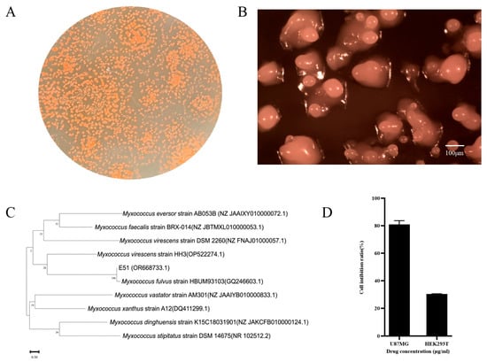
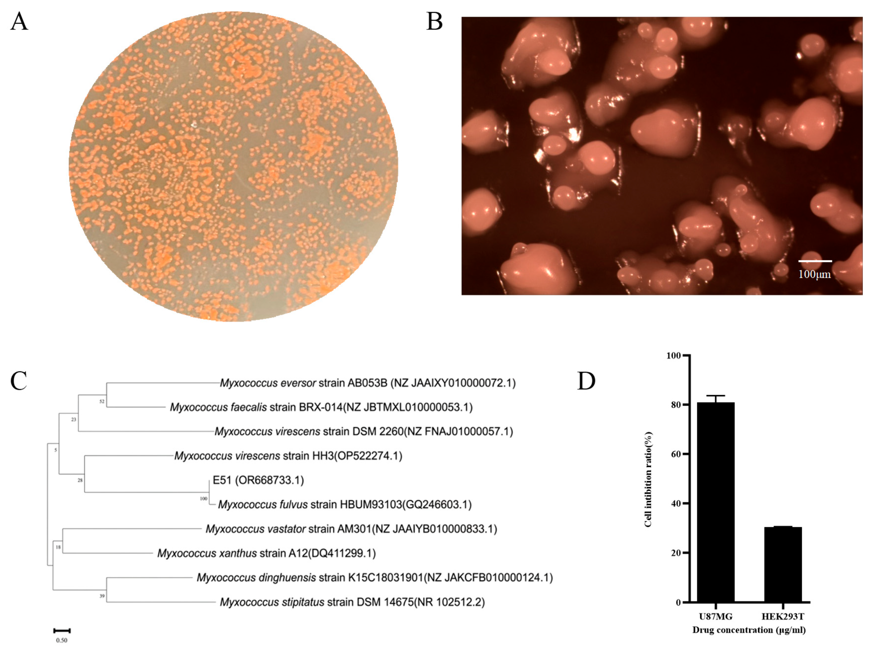
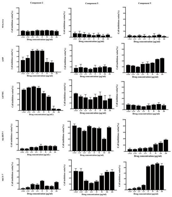
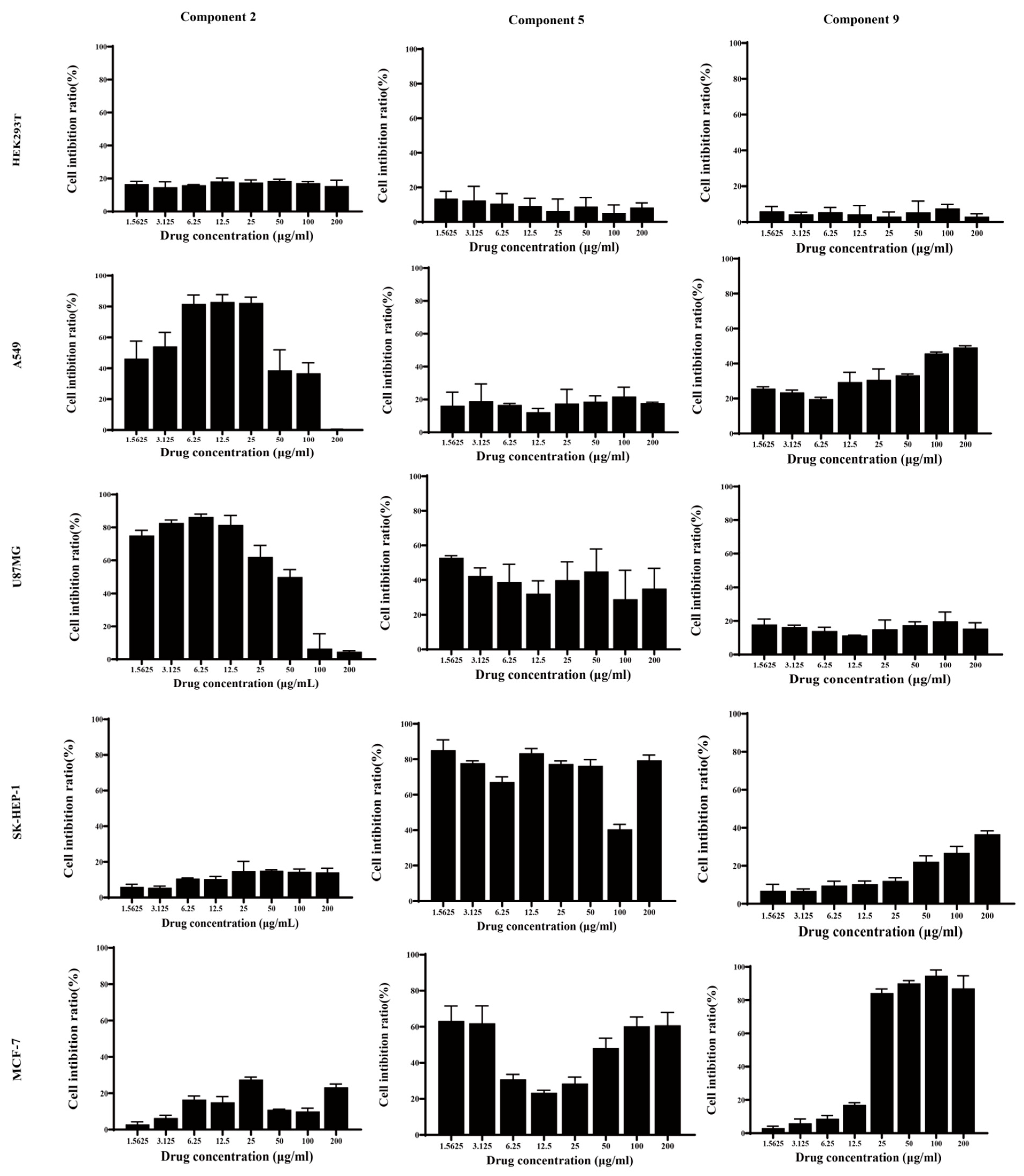
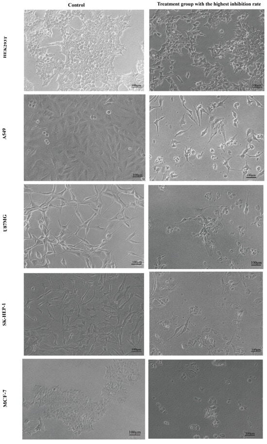
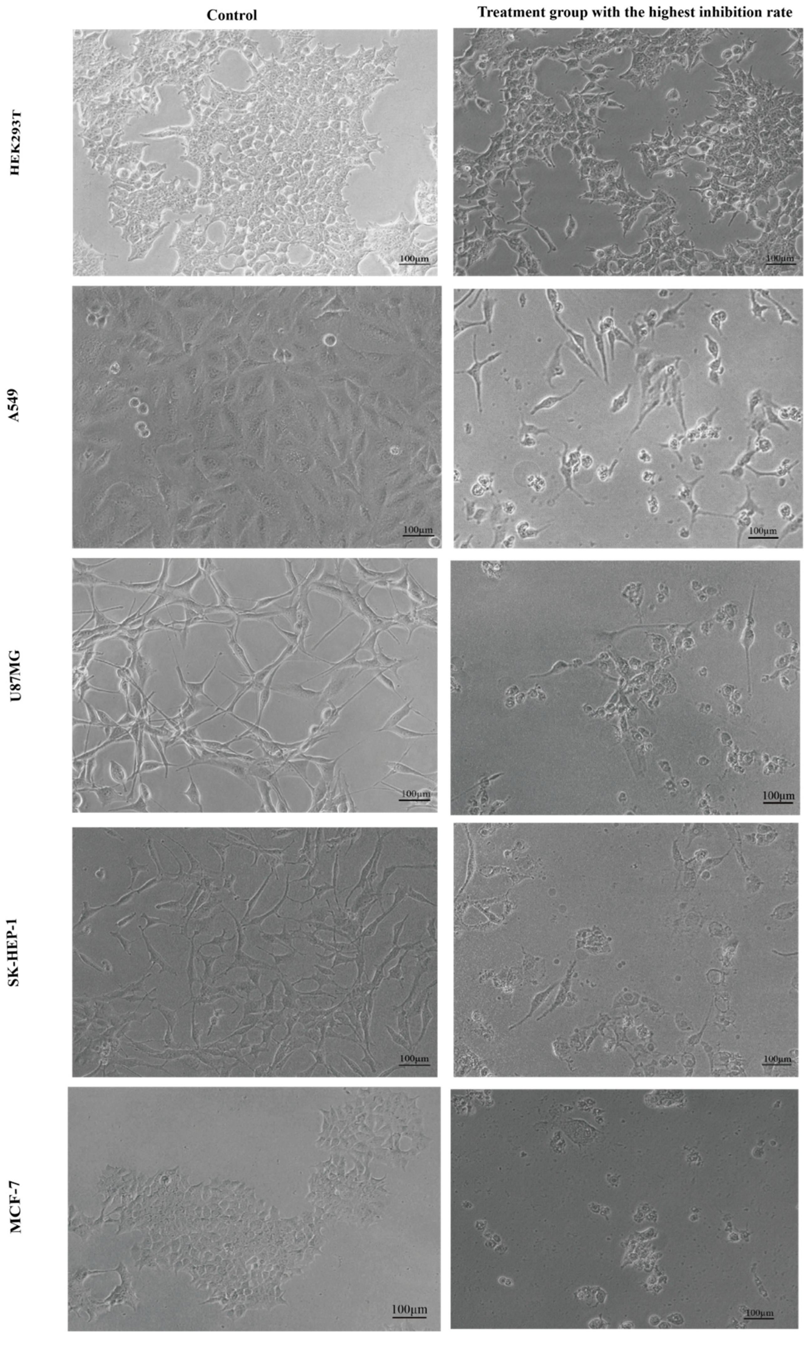
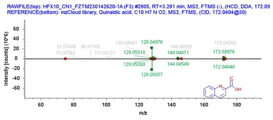
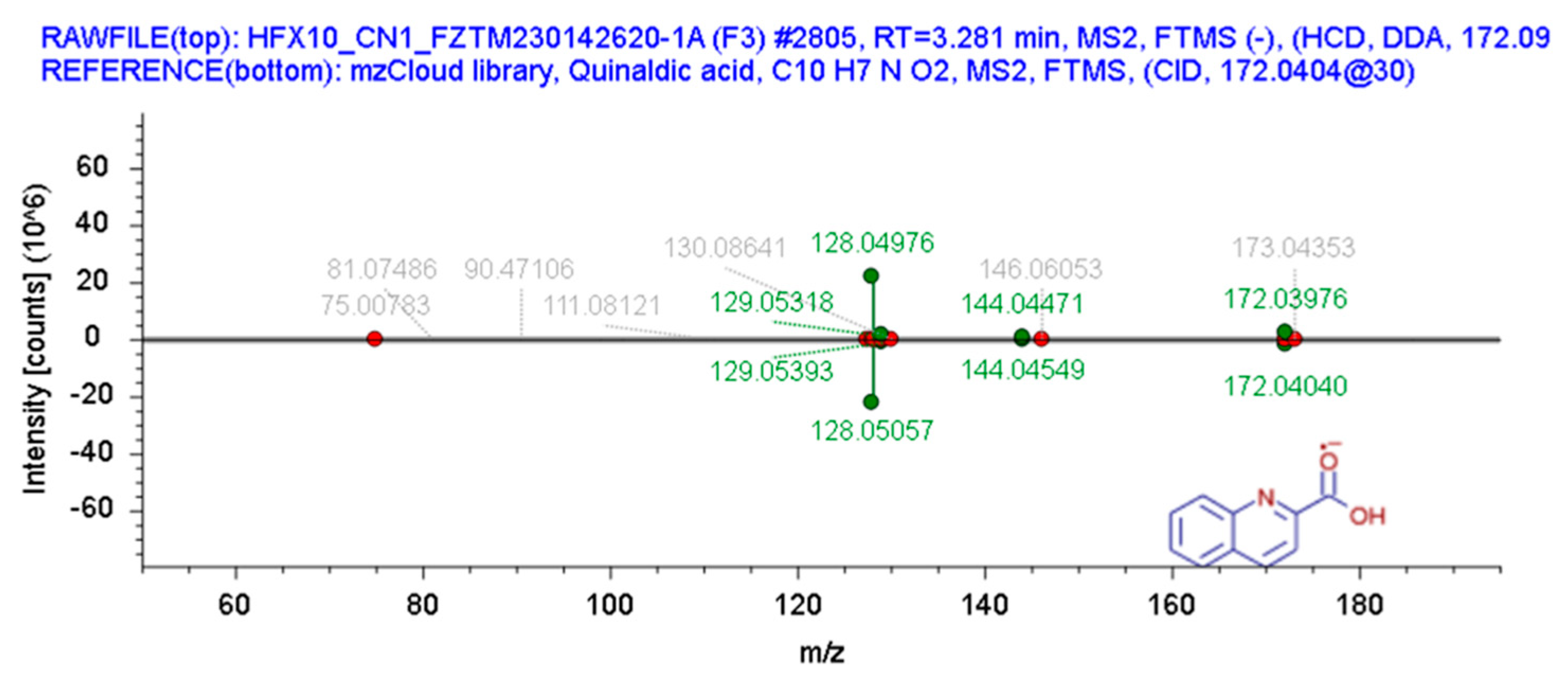
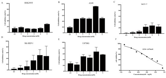
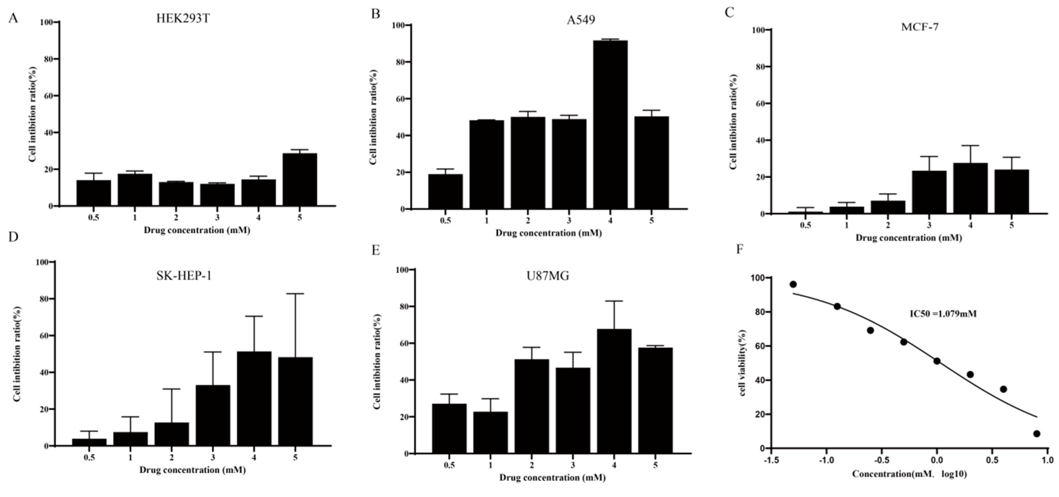
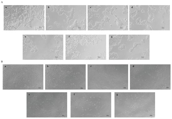
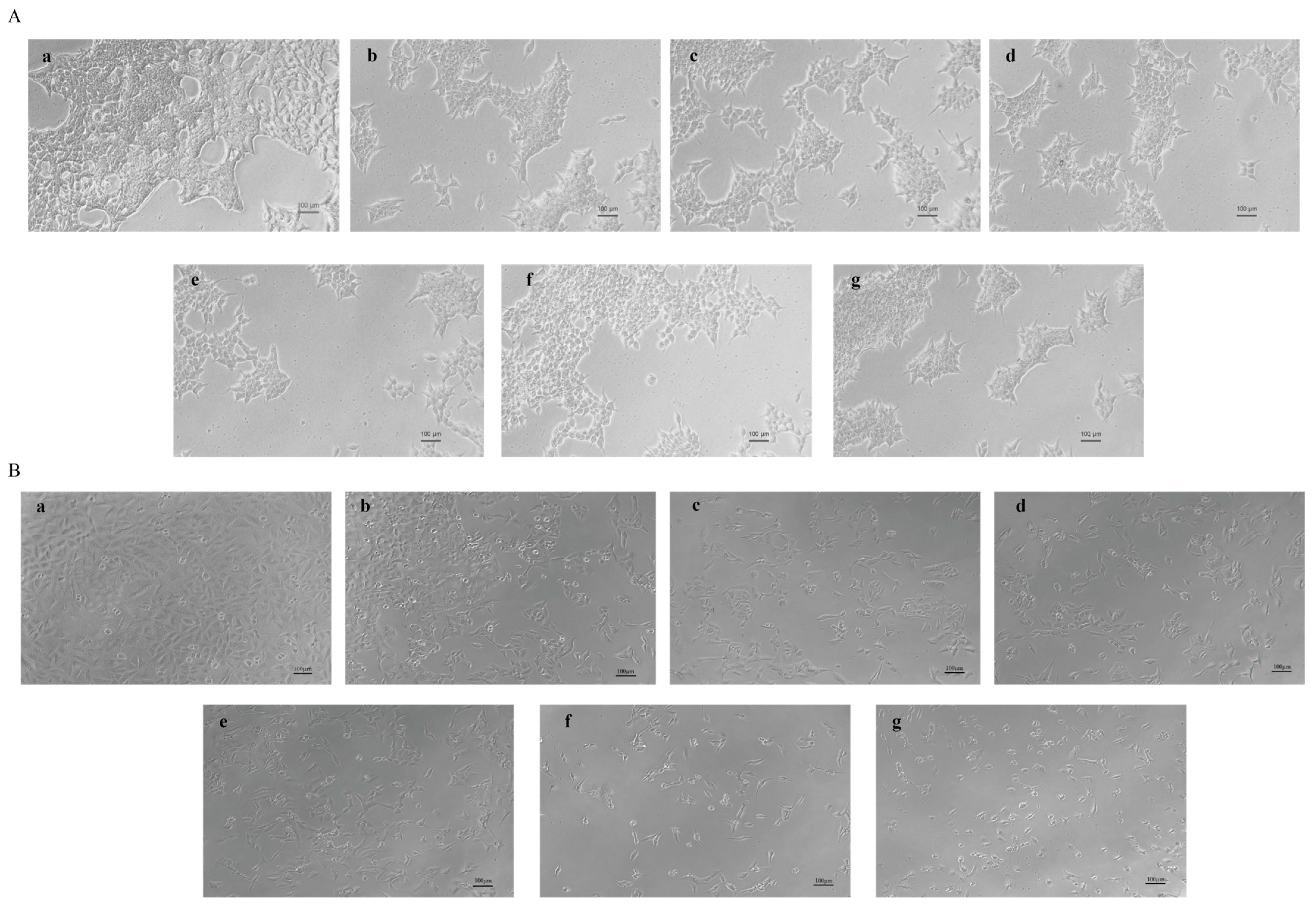
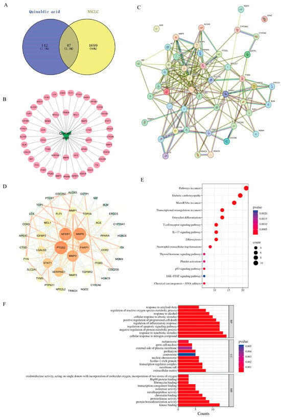
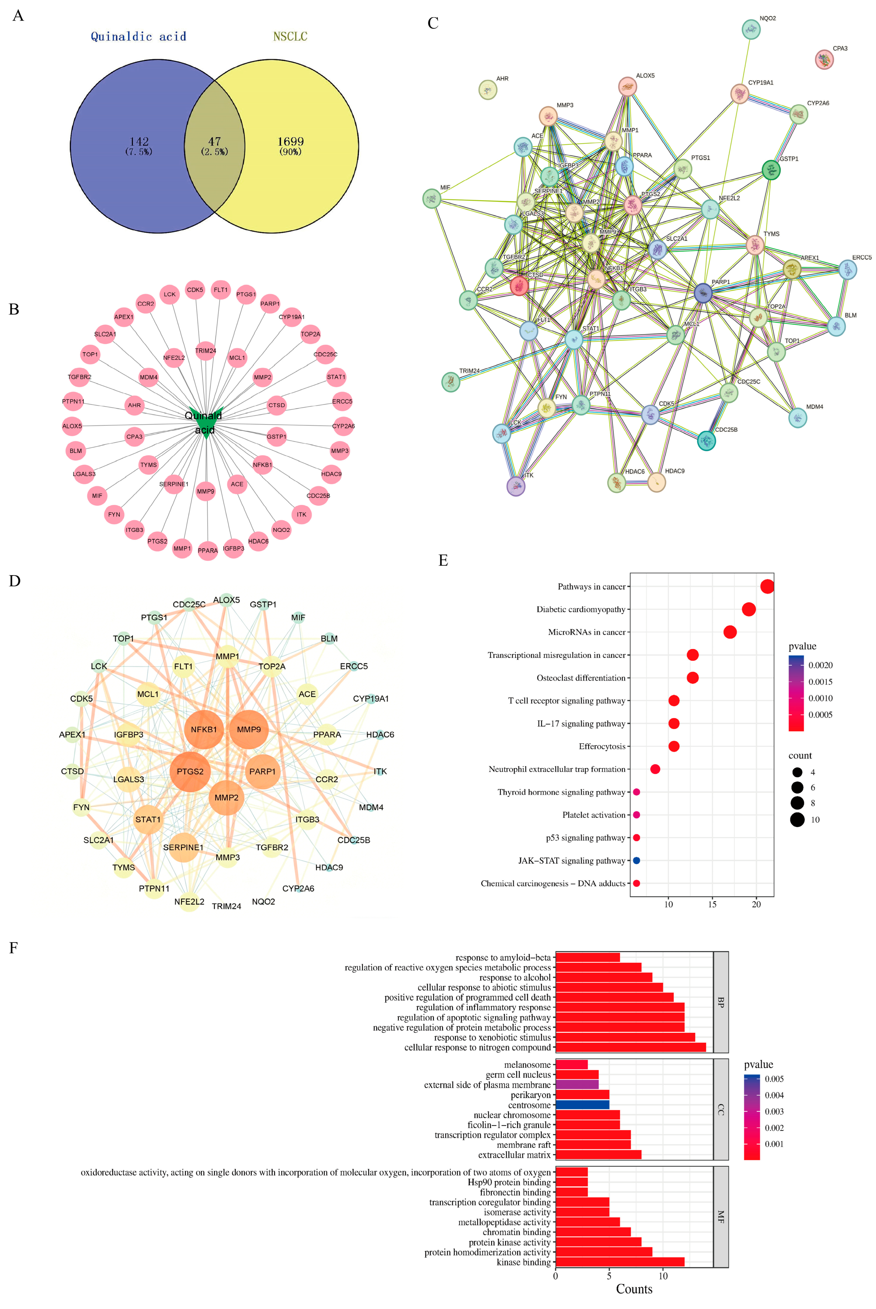
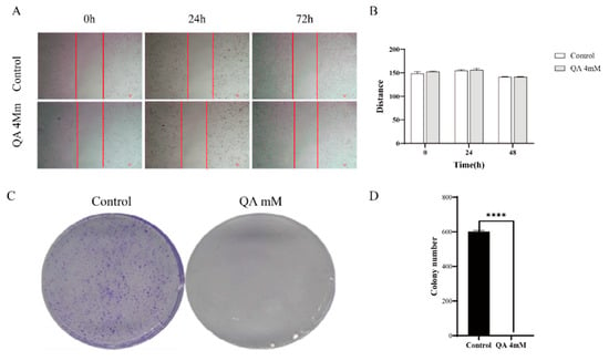
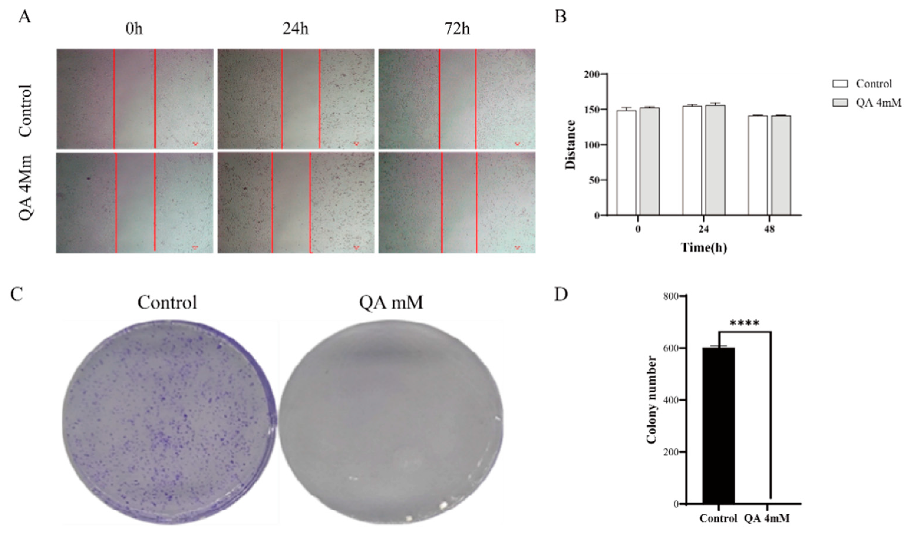
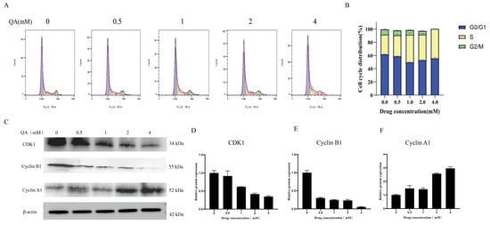
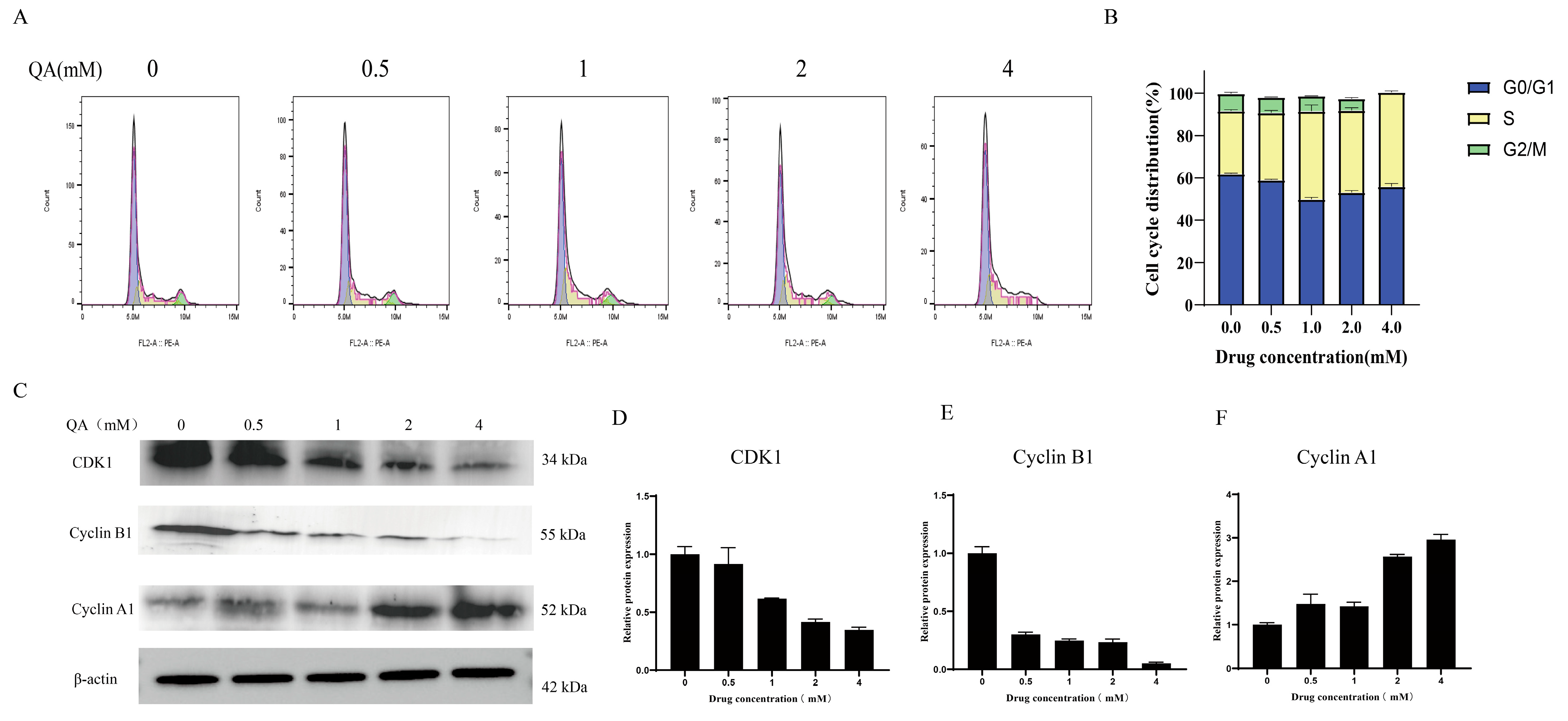
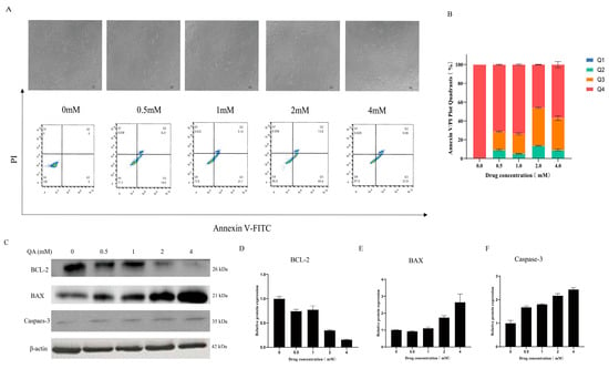
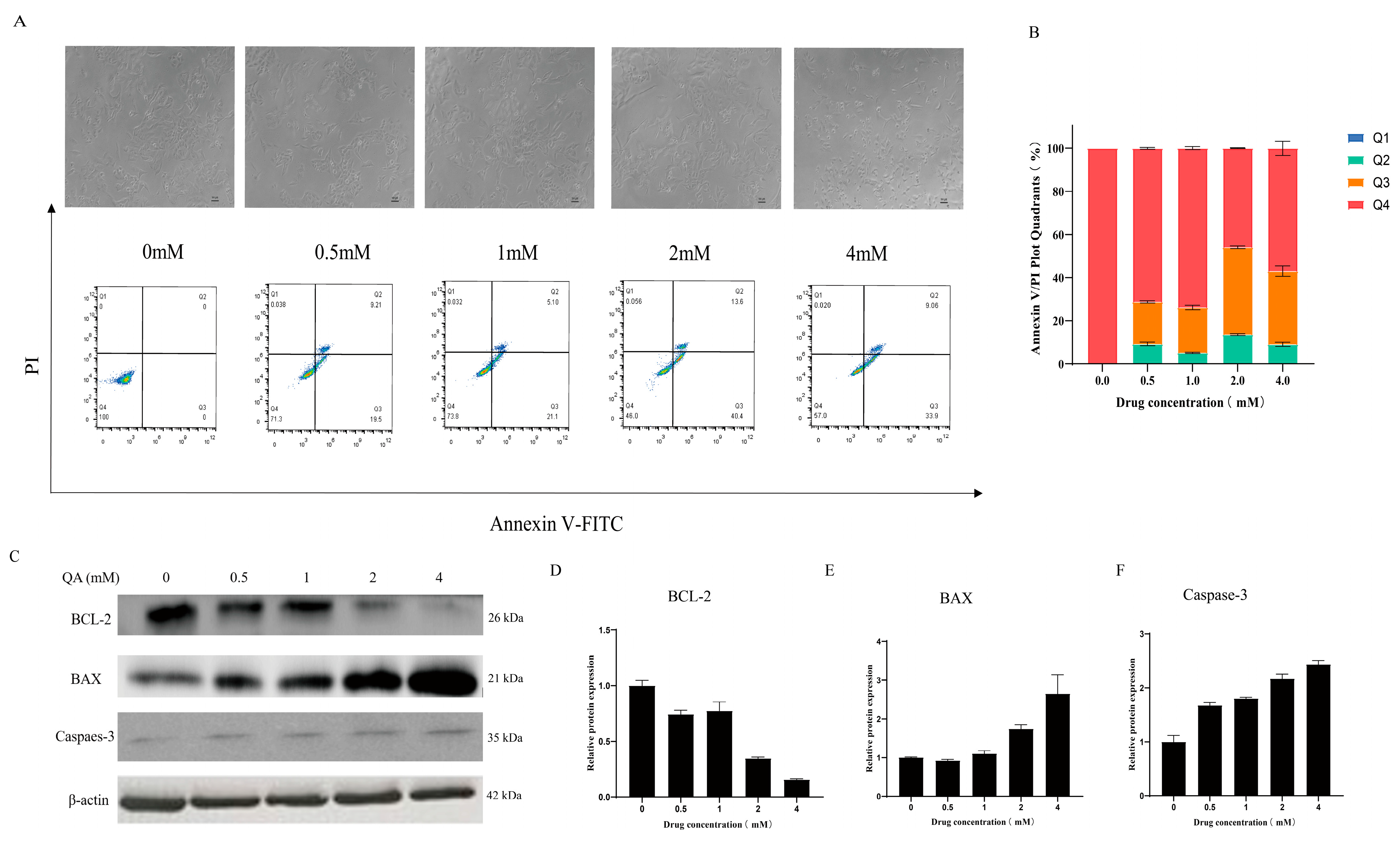
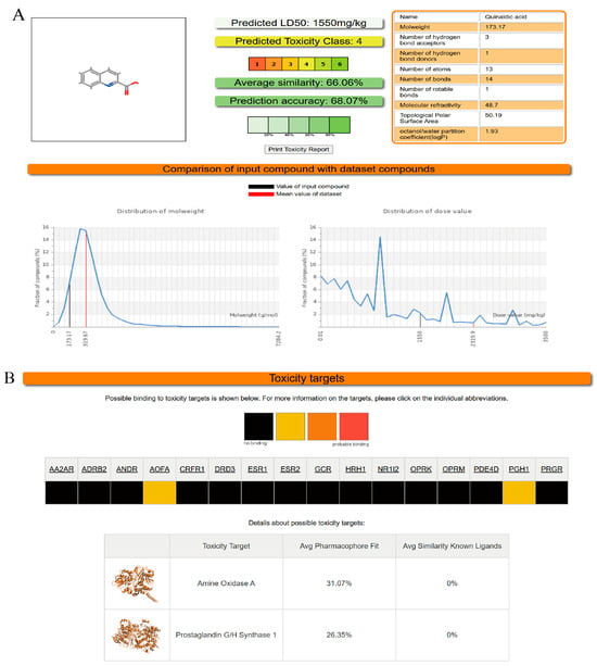
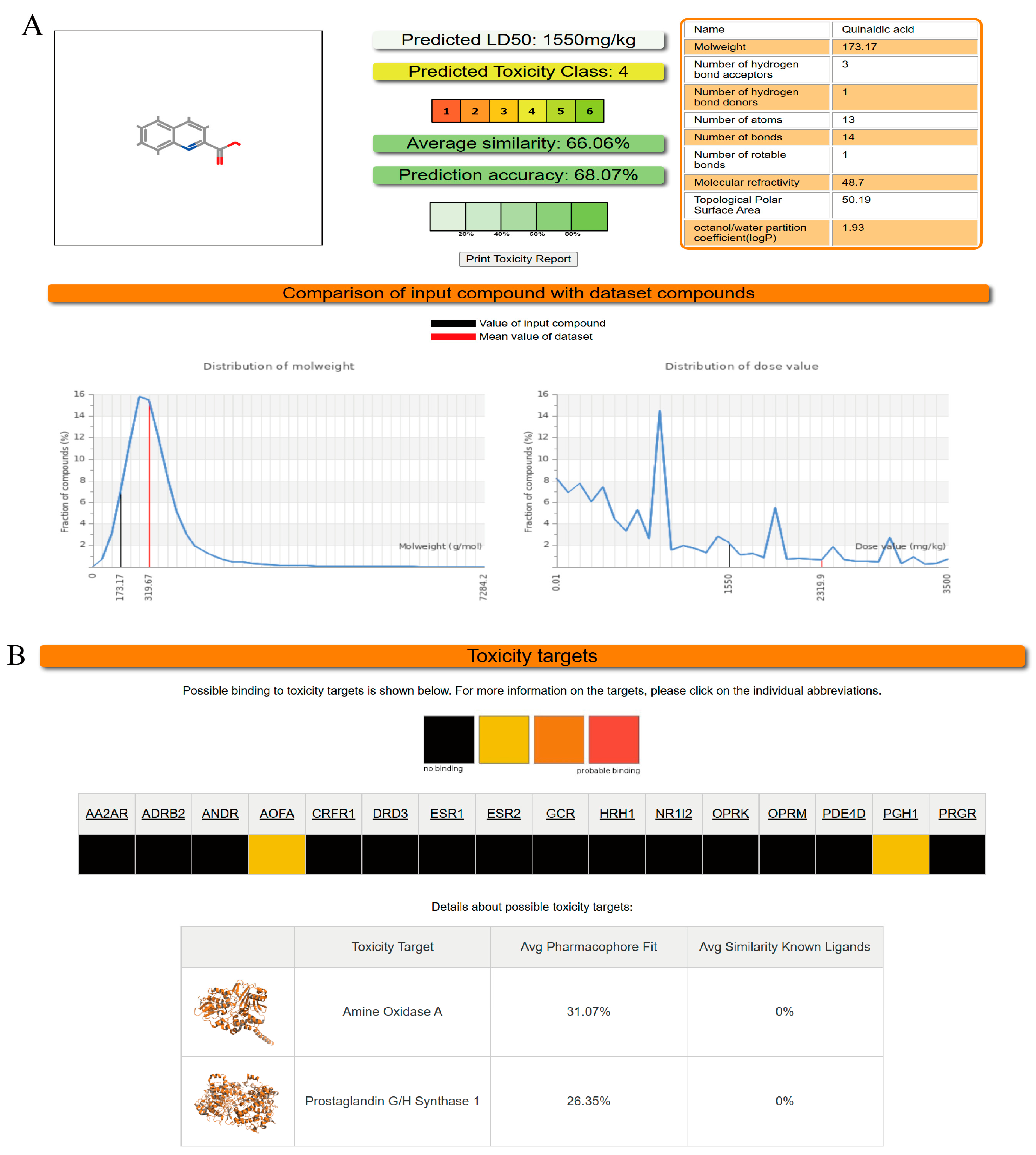
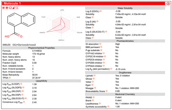
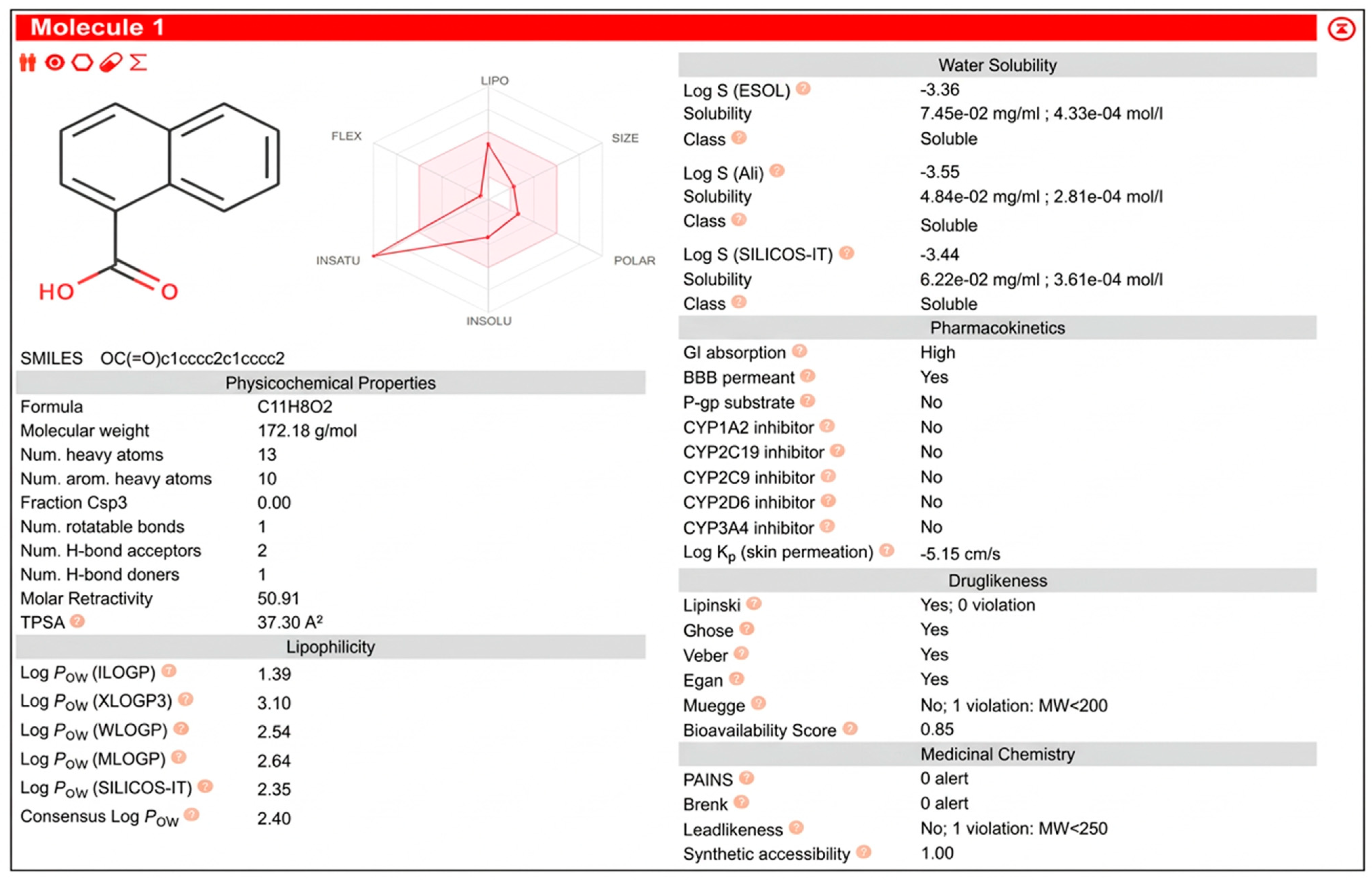
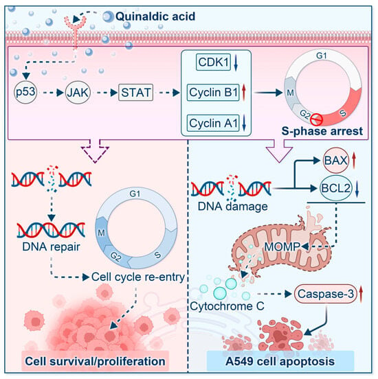
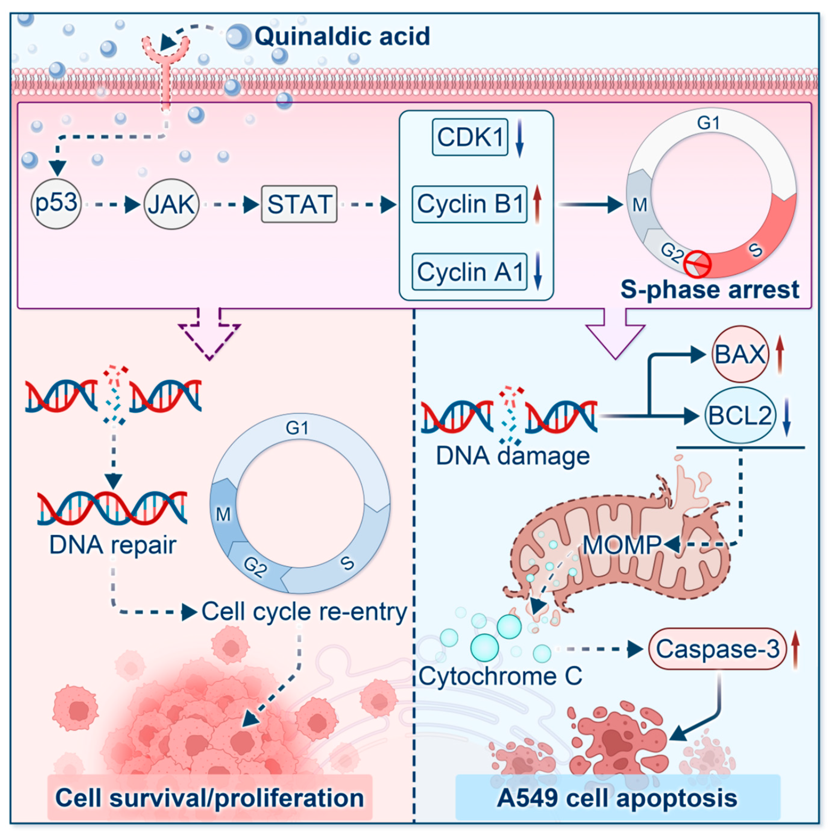

Disclaimer/Publisher’s Note: The statements, opinions and data contained in all publications are solely those of the individual author(s) and contributor(s) and not of MDPI and/or the editor(s). MDPI and/or the editor(s) disclaim responsibility for any injury to people or property resulting from any ideas, methods, instructions or products referred to in the content. |
© 2026 by the authors. Licensee MDPI, Basel, Switzerland. This article is an open access article distributed under the terms and conditions of the Creative Commons Attribution (CC BY) license.