


Abstract
Congenital heart disease (CHD) is the most common anatomical malformation occurring in live-born infants and an increasing cause of morbidity and mortality all over the world. Population-based observations have described associations between maternal cardiometabolic disorders and the risk of CHD in offspring. The present article is a narrative review. The aim of this study was to review the epidemiological evidence and clinical observations relating maternal obesity and diabetes mellitus to the risk of CHD in offspring, with particular attention paid to first trimester disturbances of fetal cardiac development and the influence of genetic, epigenetic and environmental factors. Studies have shown that maternal diabetes is a risk factor associated with nearly all subtypes of CHDs in offspring, while obesity and overweight are associated with increased risk for complex defects and outflow tract obstruction and decreased risk for ventricular septal defects. Diabetes and obesity share several phenotypes, which could be transmissible from mother to fetus via the placenta. This means that an increase in maternal glucose could be responsible for the prevalence of CHD in newborns of obese women. On the other hand, maternal diabetes may induce epigenetic modifications in the developing fetus. DNA methylation changes can impact gene expression patterns relevant to heart development. The abovementioned studies are heterogenous, express different opinions and are often difficult to compare. Therefore, the results from these meta-analyses must be interpreted with caution. Optimal diabetes control is responsible for the prevention of oxidative stress in diabetic pregnancies, and a deeper understanding of maternal risk factors holds the potential to improve both prenatal detection of CHDs by identifying at-risk pregnancies and primary prevention of diseases by improving preconception management.
1. Introduction
Congenital heart disease (CHD) arises from anomalies in the heart and large blood vessel formation in the course of embryonic development. The prevalence of CHD affects ≈1% of live births and accounts for 30% of fetal deaths [1,2]. The spectrum of CHDs varies. They can be diagnosed as an isolated finding or as part of a collection of findings. The etiology of CHDs is complex and multifactorial, with 80% of cases attributed to the interplay between genetic and environmental factors [3,4,5]. Maternal environmental risk factors, such as gestational diabetes mellitus, obesity, maternal age, drug use, air pollution, smoking, alcohol consumption, febrile illnesses during pregnancy, viral infections, pregnancy through artificial reproductive technologies, and socioeconomic factors, have been linked to CHD development in offspring [3,4,6,7,8,9].
Among genetic factors, single-gene disorders, chromosomal anomalies, and double-gene disorders can be distinguished [5,10,11]. The key genes involved in transcriptional control, signaling and morphogenesis have been identified thanks to advances in understanding of the molecular mechanisms of heart development. Some of these genes are involved in the development of CHDs. The most common genes or genetic loci associated with CHD anomalies include NKX2-5, GATA4, TBX5, SRD5A2, MTHFR, MTRR and MTR, CFTR, and 1p22 and 20q12 anomalies [3,5,10,11,12]. Most genetic CHDs exhibit an autosomal dominant inheritance pattern, but several investigations have led to the identification of non-Mendelian factors that contribute to CHD [3,5,10,11]. In addition to genetics, epigenetics plays a crucial role in heart development, with mounting evidence suggesting its dysregulation in CHD pathogenesis [12,13,14]. Both high and low miRNA expression levels have been shown to cause CHDs [15,16,17,18].
Among environmental contributors, metabolic diseases such as obesity, maternal pregestational diabetes mellitus (PGDM) and gestational diabetes (GDM) are highly prevalent and well-established risk factors for CHDs in offspring worldwide, especially in industrialized countries [4,19,20,21,22,23,24,25]. Although maternal factors are recognized risk factors for CHDs, the underlying molecular mechanisms still remain unclear [19,26,27]. The link between obesity and diabetes is thought to be mediated by chronic inflammation, oxidative stress, and dysregulated angiogenic signaling, which may contribute to endothelial dysfunction and abnormal placental development, which in turn is implicated to the pathogenesis of CHDs [28,29].
Several meta-analyses conducted by different research groups have consistently identified that the increased risk associated with maternal obesity includes a wide range of different heart defects, including transposition of the great arteries (TGA); tetralogy of Fallot (ToF); atrial, ventricular and atrioventricular septal defects (ASDs, VSDs, and AVSDs); aortic arch defects; persistent ductus arteriosus (PDA); left ventricular outflow tract obstruction defects (LVOTOs) and right ventricular outflow tract defects (RVOTOs); and univentricular heart (UVH) [21,30,31,32]. Although some studies have reported no increased risk for conotruncal defects (CTDs), other studies reported risk elevations for TGA, as well as defects of the great vessels and truncus arteriosus [33,34,35,36].
Also, meta-analyses carried out by several groups have identified the increased likelihood of specific CHD phenotypes upon exposure to maternal hyperglycemia. Maternal pregestational diabetes mellitus (PGDM) was associated with a higher risk of TGA, persistent truncus arteriosus (PTA), heterotaxy, VSDs and ASDs, ToF, double-outlet right ventricle (DORV) and hypoplastic left heart syndrome (HLHS) in offspring [23,37,38,39,40].
Maternal gestational diabetes mellitus (GDM) has been associated with higher risks of ASDs, VSDs, and ToF [41]. The study by Chen et al. [42] suggested that maternal diabetes mellitus (matDM) was significantly associated with most phenotypes of CHDs, particularly double outlet of the right ventricle, atrioventricular septal defect and truncus arteriosus. Furthermore, this study suggests a significantly higher risk of CHDs among mothers with PGDM than in those with GDM [42].
Understanding the molecular mechanisms of induction of CHDs by a number of environmental and genetic factors and identifying the stages of heart formation that are most vulnerable to these factors will aid the design of epidemiological studies to assess the extent of these risks in human CHDs. A prerequisite for prevention of CHDs is a better understanding of potential modifiable risk factors. Therefore, primary prevention of overweight and obesity in women of reproductive age and careful treatment of PGDM may hold the opportunity to reduce the burden of CHDs.
2. Methodology
The present article is a narrative review. In this review, we studied the factors associated with maternal obesity and GDM. We also discussed the potential underlying mechanisms of the observed associations and direct complications in the fetus/offspring in the form of CHDs. This work is a compendium of knowledge derived from clinical and genetic studies in humans and animal models. It is based on the latest publications and available data on the occurrence of CHDs in the offspring of mothers with diabetes and obesity.
A comprehensive literature search in this narrative review was conducted using the following inclusion criteria: (a) publications in English describing the impact of obesity and the impact of diabetes in mothers on the development of CHDs in their offspring; (b) observational studies involving women and experimental studies involving animals; (c) laboratory tests with assessment of obesity—body mass index (BMI)—and assessment of diabetes—fasting blood glucose, oral glucose tolerance test (OGTT), and glycated hemoglobin A (HbA1c); (d) patients with confirmed diagnosis of CHDs; (e) clinical studies. All possible articles, whether original studies or reviews, i.e., cohort studies, retrospective studies, prospective studies and meta-analyses, were considered for this review. The review also provides an overview of the current understanding of the influence of genetic, epigenetic and environmental factors on the cardiac development and the development of CHDs in the offspring of obese and diabetic mothers. The final search included the most valuable and highest-rated peer-reviewed articles published in the last two decades until the end of 2025, all of which are available through the Pub-Med/Medline/EMBASE/Cochrane Library database. The databases were searched using the following keywords: “metabolic syndrome”, “maternal obesity”, “maternal diabetes”, “cardiac development”, “congenital heart defects”, “genetic factors”, “epigenetic factors”, and “environmental factors”. Additional records were identified using cross-references. A manual search was also conducted combining “AND” or “OR” operators. All identified problems were described in the order of occurrence of CHDs in the offspring of diabetic and obese mothers on different continents of the world and the key mechanisms influencing the development of CHDs in the offspring of diabetic and obese mothers were described.
The following exclusion criteria were applied: (a) unpublished articles or conference proceedings; (b) editorials, opinions, case reports, and letters to the editor; (c) studies in which the patient’s diagnosis is uncertain; (d) abstracts.
This review has been conceptualized to address the need for an up-to-date combined review of the mechanisms of CHD development in the fetuses/offspring of mothers with diabetes and obesity aiming to include all the new data in a simple and comprehensive manner.
3. Metabolic Syndrome (MetS)—Definition and Diagnostic Criteria
In 1988, Reaven described metabolic syndrome (MetS), initially known as syndrome X, for the first time [43,44]. The most common definition of MetS includes a co-occurrence of abdominal obesity and at least two cardiometabolic risk factors such as hypertension, insulin resistance (IR), and dyslipidemia, as well as a low concentration of high-density cholesterol (HDL) (Table 1 and Table 2) [45,46,47,48].
Table 1.
The criteria applicable for the diagnosis of metabolic syndrome (MetS) in women according to the National Cholesterol Education Program—Adult Treatment Panel III. MetS is diagnosed when 3 of the following 5 criteria are met.
Table 2.
Criteria for MetS definition.
A controversy regarding the definition of MetS and its usefulness has arisen recently. Different clinical criteria for the definition of MetS have been adopted by international organizations such as the International Diabetes Federation (IDF) and the World Health Organization (WHO).
Table 2 summarizes three of the most commonly used definitions of metabolic syndrome [49].
Their criteria are similar in many aspects, but they also reveal fundamental differences in their positioning of the predominant causes of the syndrome. All groups agree on the core components of metabolic syndrome: obesity, insulin resistance, dyslipidemia, and hypertension. These conditions are interrelated and share the underlying mediators, mechanisms and signaling pathways.
However, the pathophysiology of metabolic syndrome is not fully understood. Environmental and genetic factors are considered to play an important role in the development of MetS. The genes involved in the development of MetS include genes influencing insulin sensitivity (encoding PPAR-γ and CAPN10), lipid metabolism genes (encoding CDS36 and 11-β-HSD), genes regulating free fatty acid metabolism (encoding adiponectin and β-adrenergic receptor), monogenic obesity genes (leptin genes) and genes associated with inflammatory conditions (TNF-α and CRP genes) [50,51]. Patients with MetS have increased levels of inflammatory markers, including C-reactive protein, TNF-α, interleukin 6 (IL-6), and fibrinogen. In addition to inflammation, oxidative stress is also attributed to play some role in the genesis of MetS [48]. During the reproductive period, the first manifestation of metabolic syndrome in women may be the onset of gestational diabetes or preeclampsia [49,52].
MetS is an increasingly common disorder affecting thousands of people worldwide, especially in industrialized countries. Its average prevalence reaches 30% in adults [53] and between 6 and 39% in children/teenagers [54]. Approximately 24% of adults in the USA, 12–37% of the Asian population and 12–26% of the European population suffer from this disease [55]. With the improvement in living standards and an increasing number of women of advanced maternal age, there is an increase in the prevalence of chronic metabolic diseases, such as obesity and diabetes, among pregnant women [4,56,57]. MetS was assessed in pregnant women (5628) in a multicenter study from Australia, New Zealand, Ireland and the United Kingdom and was reported to occur with an incidence of 12.3% at 15 weeks’ gestation [58]. An association between increased risk of CHDs and maternal metabolic disorders such as obesity [30,31], diabetes [23,38,59], hypertension [39,60], and preeclampsia (PE) [61,62] was reported by large cohort studies from Scandinavia and North America. All the aforementioned metabolic disorders can be associated with hyperglycemia and underlying insulin resistance, and thereby with MetS [46,63].
MetS increases the risk of cardiovascular complications occurrence 2-fold and even up to 5-fold in the case of type 2 diabetes mellitus (T2DM) [64]. It is possible that combinations of several maternal metabolic disorders in the same pregnancy are likely to be associated with higher risk of CHDs in offspring as compared to women with a single disorder [21]. Women with MetS are at increased risk of complications during pregnancy. The most common include miscarriage, gestational diabetes, hypertension, and thromboembolic events.
4. Cardiac Development
Prior to delineation of the complex mechanisms underlying the etiology of congenital heart defects (CHDs) associated with metabolic syndrome diseases, it is essentially important to understand the normal heart development and the cellular and molecular pathways that regulate its development [65,66,67]. Their disruption in each step of organogenesis by genetic, epigenetic and environmental factors acting on the mother may lead to the occurrence of CHDs in the offspring [3,4,8,9]. A possible impact of maternal metabolic status, including diabetes and obesity, on both the morphological and functional development of the offspring heart has been demonstrated by several large-scale cohort studies [68,69].
Cardiac development is a highly complex process involving the interplay of several cell lineages and cellular processes, all tightly controlled by their corresponding genetic programs [65].
The heart is one of the earliest organs to develop in the course of embryonic development [67]. During cardiogenesis, differentiating precursor cell clusters interact to generate specialized heart cells with a well-defined three-dimensional architecture in the restricted regions. The process starts at approximately 15–19 days of human pregnancy from the specification of cardiac progenitor cells [9,70,71]. First, mesodermal cells constitute a heart-forming region known as the cardiac crescent, which contains myocardial precursor cells. The precursor cells are classified as either first heart field (FHF) or second heart field (SHF) cells. The FHF cells ultimately become the heart tube, which then forms the definitive left ventricle and atrioventricular canal. Growth of the early heart tube occurs by the progressive addition of SHF progenitor cells to the cardiac poles. The SHF gives rise to ventricular septal, right ventricular and outflow tract (OFT) myocardium at the arterial pole, and atrial, including atrial septal, myocardium, at the venous pole. SHF deployment creates the template for subsequent cardiac septation and has been implicated in cardiac looping and in orchestrating outflow tract development with neural crest cells [66,72].
As SHF progenitors incorporate, the heart tube elongates, buckles and loops to the right from around 20 to 25 days of human development to ensure the proper cardiac segment alignment template, in preparation for chamber septation [72]. An additional source of the formation of cardiac structures is the “heart pool” of neural crest cells, which are not of mesodermal origin but are derived from the neuroectoderm. The cardiac neural crest cells, which migrate towards the primary heart tube, are involved in the formation of the arteries of the pharyngeal arches, the outflow tract of the right ventricle, the cardiac ganglia, and, together with the pre-epicardial organ, the conduction system of the heart [9,72].
Atrial and ventricular septation begins at 31–35 days of human pregnancy. At that stage, differentiation of the ventriculoarterial and atrioventricular valve systems, including the tendinous cords and papillary muscles, occurs. Concurrently, the conduction system and coronary vessels are developed [73].
During days 37–44, the development of the heart tube is completed and the basic structure of the four heart chambers is formed (Figure 1) [8,74,75,76,77]. We described the development of the heart in detail in Part I of the review by Zubrzycki et al. [9].
Figure 1.
Scheme of the main stages of human cardiac development. Schematic representation of the formation of a heart loop: (A) cardiac crescent configuration; (B) the cardiac crescent is transformed into a transiently linear heart tube; (C) rightward looping; (D) the mature four-chamber heart is formed. Abbreviations: FHF: first heart field; SHF: second heart field; RV: right ventricle; LV: left ventricle; RA: right atrium; LA: left atrium; Ap: arterial pole; Vp: venous pole; PA: pulmonary artery; Ao: aorta. This figure has been taken from the article of Ibrahim et al. [8] distributed under the terms and conditions of the Creative Commons Attribution License (CC BY-NC-ND/4.0).
Heart development relies on crucial spatiotemporal interactions between distinct multipotent cardiac progenitors during early embryogenesis. Precise regulation of gene expression is crucial for correct cardiogenesis and cardiac differentiation. A highly coordinated signaling network of NODAL, NOTCH, BMP, WNT, HIF1α, TGFβ and FGF induces expression of a core group of cardiac transcription factors, including NKX2–5, GATA4/5/6, HAND1/2, MEF2, TBX1/5/20, and ISL1, that function in a mutually reinforcing cascade to drive lineage restriction and differentiation of progenitor cell populations (i.e., FHF and SHF) to chamber-specific cardiac cell types [77,78,79]. Any dysregulation of these pathways during embryonic development may disrupt the spatiotemporal regulation of complex three-dimensional heart structures, leading to CHDs in the fetus. The severity of the CHD phenotype depends on the timing and type of dysregulation.
5. Classification of Congenital Heart Defects (CHDs)
Congenital heart defects (CHDs) are anatomical abnormalities caused by disruption of normal development of the heart and/or large blood vessels during embryonic development [80]. CHDs are the most prevalent type of birth defect worldwide and are the leading cause of death for the fetus and in the first year of life [81,82,83]. Although advances in clinical management have improved the survival of children with CHDs, adult survivors commonly experience cardiac and non-cardiac comorbidities, which affect their quality of life and prognosis [84,85,86].
CHDs account for 0.9–1% of all live births and 10% of stillbirths [2,81,82,87,88,89,90,91]. The prevalence of CHDs is increasing worldwide, most probably with the rising incidence of metabolic disorders, including diabetes and obesity [56,81,88].
There are more than 20 specific types of CHDs, which can be subdivided into several categories depending on their spatiotemporal origin during embryogenesis [8,92]. The most common CHD types are summarized in Figure 2.
Figure 2.
Schematic representation of the normal four-chambered heart and the structural abnormalities of the most common forms of congenital heart defects (CHDs). Abbreviations: TGA: Transposition of the Great Arteries; DORV: Double-Outlet Right Ventricle; PTA: Persistent Truncus Arteriosus; TOF: Tetralogy of Fallot; VSD: Ventricular Septal Defect; ASD: Atrial Septal Defect; HLHS: Hypoplastic Left Heart Syndrome; BAV: Bicuspid Aortic Valve; CoA: Coarctation of the Aorta; RA: Right Atrium; RV: Right Ventricle; LA: Left Atrium; LV: Left Ventricle; PA: Pulmonary Artery; Ao: Aorta. Black arrows indicate the direction of blood flow. This figure has been taken from the article of Ibrahim et al. [8] distributed under the terms and conditions of the Creative Commons Attribution License (CC BY-NC-ND 4.0).
Firstly, there are conotruncal defects resulting from an abnormal formation of the outflow tract (OFT) of the heart. They include (1) transposition of the great arteries (TGA) [93], in which the pulmonary trunk and the aorta are inverted and connected to the left and right ventricle, respectively; (2) double-outlet right ventricle (DORV) [94], involving the two major arteries, the pulmonary artery and the aorta, connected to the right ventricle with ventricular septal defects (VSDs), i.e., when the ventricular septum fails to form correctly, commonly observed with DORV; (3) persistent truncus arteriosus (PTA), also referred to as common arterial trunk (CAT) [95], when the aorta and the pulmonary artery fail to separate into two distinct structures, so the common arterial trunk aligns over a large VSD; (4) tetralogy of Fallot (ToF) [95] is the combination of several malformations: an overriding aorta that induces VSDs, pulmonary artery stenosis, and right ventricle hypertrophy. An extreme form of ToF is pulmonary atresia with VSDs.
Secondly, there are septation defects that are found to be either isolated or associated with other forms of CHDs. They include (1) the aforementioned VSDs [95]; (2) atrial septal defects (ASDs) [96], involving incorrect formation of the septum between the two atria; and (3) atrioventricular septal defects (AVSDs) [97], in which the four chambers of the heart communicate due to failure of the formation of the atrioventricular septum.
Thirdly, there are left-side obstructive lesions, occurring due to abnormal formation of the aorta. These lesions include (1) hypoplastic left heart syndrome (HLHS) [98], where the underdeveloped left ventricle has no outlet; (2) bicuspid aortic valve (BAV) [99], the most common type of CHD, where the aortic valve has two leaflets instead of three; (3) coarctation of the aorta (CoA), a localized narrowing of the aortic lumen leading to hypertension in the upper limbs; hypertrophy of the left ventricle, if severe; and poor vascularization of the abdominal organs and lower limbs [100].
These defects arise as a result of disorders in the processes of migration, differentiation or proliferation of progenitor cells in the first weeks of fetal life. Progenitor cells are involved in this process through SHF-related disorders involving the abnormal addition of progenitor cells from SHF to the main heart tube. Abnormal migration, or premature differentiation of these cells prevents the proper alignment of the aorta and pulmonary artery in relation to the ventricles. Disorders in this process lead to arterial conotruncal defects, the right ventricle and the septa of the heart. SHF is closely associated with nerve crest cells that divide the common trunk into two separate vessels. Nerve crest cells must migrate to the heart to form septa and large vessels. Disorders of this migration result in structural defects. Septal defects (VSDs and ASDs) are caused by impaired distribution of cells from both cardiac fields (FHF and SHF) and mesenchyme. Most defects concern the membranous part of the septum, formed by the combination of tissues derived from the endocardial cushions and the conotruncal septum. Left heart defects (HLHS) are primarily associated with defects in the FHF, which is the primary scaffold for the left ventricle. Hypoplasia results from an insufficient number of cardiomyocytes formed from FHF progenitors or their premature death in fetal life [9].
The pathomechanisms underlying the development of CHD have been heavily investigated using model systems like mice, zebrafish, and human pluripotent stem cells (hPSCs). These models have revealed that CHD is a complex, often multifactorial condition driven by genetic, epigenetic, and environmental factors that disrupt the key stages of cardiac morphogenesis.
6. Prevalence of Congenital Heart Defects (CHDs) Worldwide
The prevalence of CHDs is increasing worldwide, most probably with the rising incidence of metabolic disorders, including diabetes and obesity, the older age of women giving birth, and greater availability of diagnostic methods [56]. It has been suggested that the incidence of CHDs in newborns changes over time and varies according to maternal race, country of residence and continent [101]. Over time, the reported total CHD birth prevalence increased substantially, from <1 per 1000 live births in 1930 to 9 per 1000 live births in recent years, which is probably related to under-reporting in the past in countries with a low socioeconomic status [2,102]. Significant geographical differences were found across the continents. The highest total CHD birth prevalence was reported in Asia (especially in Iran and India) (9.3 per 1000 live births) and the lowest in Africa (1.9 per 1000 live births) [103]. The reported total CHD birth prevalence in Asia was significantly higher than in Europe, North America, South America, Oceania and Africa. The second highest total CHD birth prevalence (8.2 per 1000 live births) was reported in Europe. It was significantly higher than in North America, South America, Oceania and Africa.
On average, the total CHD birth prevalence in China from 1980 to 2019 was 2.502 (95% CI: 2.397, 2.607) per 1000 births. It increased significantly over time, from 0.201 per 1000 births (95% CI: 0.004, 0.398) in 1980–1984 to 4.905 per 1000 births (95% CI: 4.288, 5.521) in 2015–2019 [104]. The total CHD birth prevalence has been on the rise, except for a slight decline during the period 1990–1994, with a slow rise from 1980 to 2004 and a substantial rise from 2005 to 2019 [104].
Among total CHDs, the prevalence of most common subtypes varies, but VSDs, ASDs and PDA are consistently among the most frequent ones.
The birth prevalence (per 1000 live births) of the eight most common CHD subtypes reported worldwide was as follows: VSDs (2.62); ASDs (1.64); PDA (0.87); pulmonary stenosis (PS) (0.50); ToF (0.34); CoA (0.34); TGA (0.31); and aortic valve stenosis (AS) (0.22) (Figure 3).
Figure 3.
Birth prevalence of CHD subtypes over time. Time course of birth prevalence of the 8 most common CHD subtypes from 1945 to 2010. Abbreviations: AS: aortic stenosis; ASD: atrial septal defect; CoA: aortic coarctation; PDA: patent ductus arteriosus; PS: pulmonary stenosis; TGA: transposition of the great arteries; ToF: tetralogy of Fallot; VSD: ventricular septal defect. This figure has been taken from the article of Linde et al. [2] distributed under the terms and conditions of the Creative Commons Attribution License (CC BY-NC-ND 4.0).
Significant geographic differences were detected in the reported birth prevalence of the eight most common CHD subtypes (Figure 4).
Figure 4.
Birth prevalence of the 8 most common CHD subtypes in newborns on each continent. Distribution of subtypes among total CHDs is shown as percentages above bars. * Reported PS and ToF birth prevalence in Asia was significantly higher than in Europe (p < 0.001) and North America (p < 0.001). † Reported CoA birth prevalence in Asia was significantly lower than in Europe (p < 0.001). ‡ Reported TGA and AS birth prevalence in Asia was significantly lower than in Europe (p < 0.001), North America (p < 0.001) and Oceania (p < 0.001). § No data on ToF or AS birth prevalence in Africa were available. Abbreviations as in Figure 3. This figure has been taken from the article of Linde et al. [2] distributed under the terms and conditions of the Creative Commons Attribution License (CC BY-NC-ND 4.0).
It is interesting that the birth prevalence of pulmonary outflow tract obstructions (PS and ToF) is relatively high while the birth prevalence of left ventricular outflow tract obstructions (CoA and AS) is low in Asia. The above findings confirm the results of Jacobs et al. [105], who found that White children seem to have more left ventricular obstructive anomalies (LVOTOs), whereas Chinese children have more right ventricular outflow tract anomalies (RVOTOs). Furthermore, a lower birth prevalence of TGA in Asia compared with Europe, North America, South America, and Oceania (p < 0.001) has been reported. A possible explanation might be found in genetic origin.
In most of the studies from India, VSDs have been found to be the most common anomaly, followed by PDA. The other common anomalies are ASDs, PS and CoA. A community-based study by Bhat et al. [106] in Uttarkhand reported VSDs (30.4%) as the most common congenital heart disease followed by ASDs (17.63%), PDA (9.62%), and PS (6.41%). The burden of CHD in India is likely to be the largest among all nations in the world simply because of the fact that there are more children born in India than anywhere else [107]. In most low- and middle-income nations, including India, only a small fraction of children with heart disease can expect to receive comprehensive care in today’s times. The majority of children with CHDs in this region escape detection due to the lack of awareness, poor socioeconomic status, and poor availability of echocardiography.
7. Etiological Factors Contributing to CHD
The etiology of CHD is complex, involving both hereditary and non-hereditary risk factors [6,83]. Disturbances of any stage of organogenesis may lead to the occurrence of CHDs that could be initiated by various genetic, epigenetic, or environmental factors [3,4,8,9,108].
During the past several decades, a consensus has emerged that genetic factors (e.g., chromosomal abnormalities, smaller copy number variants, and point mutations) and maternal exposure to environmental factors (air pollution and toxic chemicals), parental smoking, maternal history (infectious diseases during pregnancy and pregestational and gestational diabetes mellitus), maternal obesity, maternal drug intake, and pregnancy through artificial reproductive technologies, as well as socioeconomic factors, are related to the occurrence of CHDs in the fetus [3,4,7].
The breakdown of human CHD etiologies is presented in Figure 5. Approximately 45% of CHD cases can be attributed to a known etiologic factor, where ~35% are due to the presence of pathogenic genetic loci and ~10% to exposure to modifiable environmental contributors [3].
Figure 5.
Pie chart representing the percentage of known and unknown etiologies in human CHD cases. From outside in, it shows the proportion of known vs. unknown etiologies, the proportion of genetic vs. environmental etiologies, and the proportion of known genetic etiologies and copy number variation (CNV). All percentages are approximations. This figure has been taken from the article of Choudhury et al. [3] distributed under the terms and conditions of the Creative Commons Attribution License (CC BY-NC-ND 4.0).
7.1. Environmental Factors Contributing to the Occurrence of CHDs
Environmental factors contributing to the occurrence of CHDs are extremely heterogeneous and can be broadly classified into extrinsic and intrinsic factors.
7.1.1. Extrinsic Factors
Extrinsic factors are generally considered highly modifiable, and there has been a consensus to identify and prevent such exposures in order to decrease the associated incidence of CHDs. Extrinsic environmental factors include prenatal maternal exposure to heavy metals (lead, cadmium, mercury, and arsenic), organic solvents (cleaning fluids), various therapeutic drugs (thalidomide, anticonvulsants, and antidepressants), nutritional deficiencies (folic acid/vitamin B12, B3, D, and iron), and retinol/vitamin A, as well as alcohol consumption and marijuana use [4,6,7]. Parental exposure to toxicants (such as paternal smoking) has been demonstrated to increase the risk of having children with CHDs [109].
A conscious lifestyle can modify the risk of exposure to these environmental factors and reduce the incidence of associated CHDs. The emerging body of recent empirical studies suggests that air pollution is an important extrinsic contributor to the development of CHDs, especially when this exposure occurs during the months of preconception and continues to around eight weeks’ gestation [110,111,112,113,114]. There are significant associations between increased risk of specific CHD subtypes and exposure to fine particulate matter with a diameter ≤ 5 μm and ≤10 μm, NO2, CO, O3, SO2, or polycyclic aromatic hydrocarbons [111,112,113,115,116]. High versus low CO and SO2 exposure was associated with an increased risk of ToF, whereas particulate matter and ozone (O3) increased the risk of ASDs and categorical NO2 exposure was associated with an increased risk of CoA [112,115]. Exposure to air pollutants may alter epigenetic modifications such as DNA methylation, which, in turn, may affect inflammation, leading to increased oxidative stress and mitochondrial dysfunction, and result in cardiac malformations [117,118]. It has been reported that gestational exposure to particulate matter ≤ 5 μm may increase the risk of mutations in the transcription factors GATA4 and NKX2.5, which are involved in fetal cardiac development and may cause CHDs in offspring [119,120,121,122]. Recent investigations by Yan et al. [123] have summarized five primary mechanisms through which fine particulate matter (PM2.5) affects adverse birth outcomes. These are: transcriptional and translational regulation, oxidative stress (OS) and inflammatory responses, and epigenetic regulation.
Other major pollutants in modern times are micro- and nano-plastics. A recent study using chick embryos showed that nano-plastics are capable of dysregulating neural crest cell migration, resulting in congenital cardiac and craniofacial anomalies [124].
7.1.2. Intrinsic Factors
Intrinsic factors are much more difficult to control because they relate to internal physiologic conditions during embryogenesis, including maternal age, diseases and infections. These are generally regarded as less modifiable than the external environment and represent the complex interplay of biological processes that occur between the maternal intrauterine environment and the fetus in the course of fetal development and may alter the intrauterine environment of the mother [4].
Moreover, recent studies suggest that abnormal function and inflammation of the placenta is sufficient to lead to CHD, and there exists a placenta–heart axis that must be maintained for normal cardiac development [28,29]. In fact, impaired maternal metabolism via maternal microbiome profiling has been linked with several subtypes of CHDs indicative of an unfavorable maternal environment during cardiogenesis [125,126,127].
Due to the fact that increasing numbers of women in developed countries are delaying childbearing to an older age, maternal age has increased in the last decades, consequently causing a higher birth prevalence of CHDs [9,128,129]. Studies have shown that advanced maternal age (>35 years) is associated with an increased prevalence of several CHD phenotypes: laterality defects (aPR = 2.06; CI: 1.22–3.48), all conotruncal defects (aPR = 1.30; CI: 1.03–1.65), and specifically D-TGA (aPR = 1.65; CI: 1.10–2.48), CoA (aPR = 1.54; CI: 1.10–2.16), VSDs (aPR = 1.20; CI: 1.06–1.36), and ASDs (aPR = 1.36; CI: 1.05–1.77). The significant adjusted incidence rates ranged from 1.20 to 2.06 [130]. Similarly, Reefhuis and Honein [131] observed that advanced maternal age (35–40 years) was associated with all heart defects (OR = 1.12, 95% CI: 1.03–1.22).
Among environmental contributors, maternal PGDM is a highly prevalent and well-established risk factor for CHD, increasing 3- to 5-fold the risk of having an infant with CHD [4,22,23,24]. Meta-analyses carried out by several groups have identified the increased likelihood of specific CHD phenotypes resulting from exposure to maternal PGDM, including TGA, PTA, heterotaxy, VSDs and ASDs [37,38]. The incidence rates of CHD from pregestational type 1 and type 2 DM are similar, but type 1 has the highest risk for conotruncal defects (CTDs) and AVSDs, while type 2 is more associated with heterotaxy and LVOTO malformations [39]. However, the association between gestational diabetes mellitus (GDM) and CHD in offspring was inconsistent in the conducted meta-analyses. An analysis according to types of CHD showed that maternal GDM was associated with higher risks of ASDs, VSDs, and ToF [41]. However, inconsistent results were obtained in the studies evaluating the association between GDM and different CHD phenotypes in offspring. Chen et al. [42] suggested that the risk of CHD in newborns of mothers with PGDM was significantly higher than in newborns of mothers with GDM.
Similarly, maternal obesity, particularly obesity which comes along with other health complications, such as T2DM, has also been independently linked to CHDs [19,20]. The increased risk associated with maternal obesity includes a wide range of different heart defects, including TGA, ToF, septal defects, aortic arch defects, PDA, LVOTOs, RVOTOs and UVH [21,30,31].
Other maternal pathologies include infections such as rubella and cytomegalovirus, as well as phenylketonuria [4,6]. Recently, an increased incidence of CHDs was reported in cases of maternal COVID-19 infections [132].
The use of assisted reproductive techniques (ARTs) has also been proposed as one of the potential contributory causes of the development of CHDs in newborns, although the risk varies with the assisted reproductive technique applied [133,134,135]. The authors speculate that this may not only be due to the reproductive technology but also due to the underlying reason for the infertility of the couple being strongly related to the environmental conditions. This indicates the need for further research to validate the actual evidence and state the real risk of CHDs following ART pregnancies.
Table 3 presents selected common environmental (teratogenic) factors and the associated CHDs occurring in the fetus.
Table 3.
Common environmental exposures/teratogens and associated CHDs.
Although maternal factors are recognized risk factors for CHDs, the underlying molecular mechanisms remain unclear [19,26,27]. The list of environmental factors that can potentially cause CHD is nonexhaustive. The rapid transformation of society and its subsequent influence on our environment and lifestyle will continually give rise to other unknown CHD risk factors.
7.2. Familial Risk of Inheriting Congenital Heart Defects
The familial risk of CHDs in children is a serious concern for families when either parent has a CHD or when a sibling is born with a CHD. The presence of CHDs in parents significantly elevates the risk for their offspring, with a baby’s risk being increased 3-fold if either parent has been diagnosed with a CHD. Research has demonstrated that if a mother has a CHD, the risk of her child being born with a CHD ranges from 2.5% to 18%, with an average risk of 6.7% [136]. In contrast, if a father has a CHD, the risk for the child is lower, ranging from 1.5% to 3% [136]. Anomalies with the highest recurrence risk include heterotaxy, RVOTOs and LVOTOs [137].
Studies have shown that if one child has had a CHD, the chance of another child being born with the condition ranges from 1.5% to 5%, depending on the type of CHD in the first child [136]. This risk increases further if two or more children have CHDs, with an estimated recurrence rate of 5% to 10% for subsequent births [136].
A large cohort study reported a 60% greater risk of CHD among monochorionic and diamniotic twins [138]. Monochorionicity resulted in a 9-fold increased incidence of CHD. Abnormal placentation and, consequently, an abnormal blood flow distribution among the twins can result in twin–twin transfusion syndrome. A 13-fold increase in the risk of CHD is associated with this syndrome alone, but specific heart defects vary between twins [138]. In donor twins, there is an increased incidence of valvular stenosis and hypoplastic ventricles associated with reduced hemodynamic flow. Among recipient twins, the opposite effect is observed, where the prevalence of obstructive lesions and valvular regurgitation is increased [139]. Since both individuals are assumed to be genetically identical, CHD malformations are likely to result directly from epigenetic modifications, environmental factors, or abnormal placentation.
Some heart defects, such as those with autosomal-dominant inheritance, can arouse particular concern, as a parent with the defect has a 50% chance of passing the defect to each child, regardless of whether it is a male or a female [140]. The above means that there is also a 50% chance that the offspring will not be affected by the defect [140]. Notably, autosomal-dominant inheritance affects males and females equally, making this pattern of inheritance particularly relevant for families with CHDs.
The elevated recurrence rate of CHD in affected families indicates the need to identify its underlying genetic factors. Besides chromosomal abnormalities and copy number variation, several genetic variants linked to CHD were historically believed to follow a monogenic disease inheritance pattern. However, recent technological advances have allowed the identification of novel CHD variants and non-Mendelian genetic mechanisms underlying CHD.
7.3. Genetic Causes of Congenital Heart Defects (CHDs)
Genetic causes are considered to be the main factor in the pathogenesis of CHDs, but the genetic penetrance of variants to the phenotype and clinical course vary significantly [12]. Specific genetic causes can be detected in an estimated ~40% of CHD cases (Figure 6). These include chromosomal anomalies or aneuploidies accounting for ~13% of CHD cases (range: 9% to 18%) and copy number variants (CNVs) estimated at 10–15% (range: 3% to 25%) in syndromic CHD and 3% to 10% in non-syndromic CHD, as well as single-gene disorders (12%) (Figure 6) [10,11,141,142].
Figure 6.
Specific genetic causes of congenital heart disease. Chromosomal abnormalities, copy number variations and single-gene variants are associated with ~40% of congenital heart disease cases, but the genetic causes of the majority (60%) of congenital heart disease cases remain unknown. All percentages are approximates based on recent publications. NR, not reported. This figure has been taken from the article of Yasuhara and Garg [10] distributed under the terms and conditions of the Creative Commons Attribution License (CC BY-NC-ND/4.0).
The genetic basis of CHD can be divided into syndromic CHD and non-syndromic CHD, where congenital abnormalities are isolated to the heart. In non-syndromic and isolated (without a familial history or a Mendelian inheritance) forms of CHDs, a multifactorial pathogenesis with interplay between inherited and non-inherited causes is recognized. Numerous genes have been discovered to be implicated in the pathogenesis of syndromic CHD. However, identification of the genetic contributors of non-syndromic CHD is more challenging because of genetic heterogeneity, incomplete segregation, and potentially oligogenic or polygenic origins [10,11]. A single candidate gene or genetic variant can produce a spectrum of heart malformations. Moreover, it may even occur in phenotypically normal humans [5].
7.3.1. Genetic Abnormalities Associated with Syndromic CHD
Chromosomal abnormalities and copy number variation (CNV) have been strongly associated with syndromic CHD, due to disrupted expression and dosage of crucial developmental genes located within the affected loci [5].
Chromosomal mutations include either anomalies involving the loss or gain of complete chromosomes (aneuploidy), or structural abnormalities in subchromosomal regions (e.g., CNVs). Chromosomal aneuploidies include trisomies (13, 18 and 21) and monosomies such as Turner syndrome. Chromosomal abnormalities are a significant risk factor for CHDs, particularly in individuals with Down syndrome. The extra 21st chromosome present in individuals with Down syndrome is thought to contribute to the development of a diverse range of heart conditions observed in children with this condition [10,143]. Chromosomal aneuploidies and larger CNVs can be identified using karyotyping and chromosomal microarray.
CNVs are large deletions or duplications of DNA segments, and pathogenic CNVs are associated with syndromic CHDs. These include 22q11.2 deletion syndrome (DiGeorge syndrome), 1p36 deletion syndrome, 7q11.23 deletion (Williams–Beuren syndrome), terminal deletions of 11q (Jacobsen syndrome), 1q21.1 deletion/duplication, and 8p23.1 deletion syndrome, which can be detected by fluorescent in situ hybridization and/or chromosomal microarray (CMA) [10].
Single-gene defects play a significant role in the development of CHDs, accounting for approximately 12% of cases [10]. These defects often follow a Mendelian inheritance pattern, most commonly exhibiting an autosomal-dominant trait [144], which implies that only one copy of the mutated gene is necessary to manifest the condition. There are several examples of Mendelian syndromes associated with CHDs, including Alagille syndrome, Holt–Oram syndrome, Costello syndrome, Kabuki syndrome, Char syndrome and Noonan syndrome [10,144]. Table 4 shows the most common syndromic conditions associated with CHDs.
Table 4.
Common syndromes associated with congenital heart defects (CHDs).
Recent advances in cytogenetics and molecular diagnostic techniques have significantly contributed to the identification of new genes and chromosomal regions involved in both syndromic and non-syndromic CHD. The knowledge of them enables a more defined explanation of the underlying pathogenetic mechanisms of CHD [3,10].
7.3.2. Genetic Abnormalities Associated with Non-Syndromic CHD
In recent years, remarkable advances in next-generation sequencing (NGS) technologies have allowed for the identification of novel genetic etiologies for CHD and better understanding of the complex genetic architecture of non-syndromic CHD. Pathogenic variants that result in non-syndromic CHD can be broadly divided into 1) cardiac transcription factors, governing cardiac development (NKX2.5, GATA4, and members of the T-box family [TBX1 and TBX5]; (2) cardiac structural proteins (MYH6, ACTC1, and ELN); and (3) cell signal factors (neurogenic locus notch homolog protein 1 and vascular endothelial growth factor) [5,12,83,145,146,147,148].
Although mutations in the coding sequence or gene regulatory sequences affecting their expression can result in cardiac malformations, the precise genotype/phenotype relationship is often challenging to establish [149,150]. Genetic variants are implicated as the primary drivers of pathogenesis, but genetic penetrance of variants to phenotype and clinical course varies significantly. Table 5 shows selected monogenic causes of non-syndromic CHDs.
Table 5.
Genes associated with non-syndromic congenital heart defects (selected).
Notably, even though more than 400 genes associated with CHDs have been identified, only about one-third of cases have a simple genetic cause, highlighting the substantial contribution of multifactorial inheritance to the disease [3,4,5]. Additionally, mutations in specific genes such as the Notch receptor (NOTH) family, the NKX2 homeobox (NKX2.5) family, the GATA-binding protein 4 (GATA4) family and the T-box transcription factor (TBX) family have been identified as contributing factors to non-syndromic CHDs [9,144,145,151,152].
NKX2.5 is the master regulator of cardiac development. Mutations in NKX2.5 are often associated in humans with ASDs and conduction abnormalities. NKX2.5 gene mutations have also been reported in VSDs, ToF, aortic stenosis, and HLHS, demonstrating that NKX2.5 has multiple roles during heart development [10,12,153].
GATA4 is a potent activator of many cardiac genes, including the genes encoding natriuretic peptides (NPPA and NPPB), cardiac myosin heavy chains (MYH6 and MYH7), and troponin isoforms (TNNI3 and TNNC1), as well as the cardiac muscarinic m2 acetylcholine receptor (CHRM2). GATA4 is a mutual cofactor of NKX2.5 [12,154]. Over 100 GATA4 mutations linked to various cardiac defects and types of CHD have been identified to date. GATA4 is necessary for cardiac septation in a subset of cardiac progenitor cells called the “second heart field” [155]. Variants of GATA4 have been associated with ASDs, VSDs, TOF, PDA and pulmonary stenosis (PS) [10,12,156,157].
In human hearts, TBX5 is expressed in the epicardium, myocardium, and endocardium of both embryonic and adult hearts [158,159]. The T-box family of transcription factors plays an important role in patterning the embryonic germ layers. It functions as a transcriptional activator, interacting with various cofactors, including GATA4, NKX2.5, MEF2C and SALL4, to regulate genes essential for cardiac morphogenesis and development of the conduction system [160]. Variants in TBX1 in humans are commonly associated with abnormalities in pharyngeal arch patterning and ventricular septation [10,12].
Despite tremendous advances in genetic sequencing technologies, the identification of pathogenic variants in patients with non-syndromic CHD has been challenging, even in familial cases of CHD. Direct analyses of human CHD tissues, comprehensive assessments of non-coding sequences and oligogenic variants, and examinations of the effects of environmental exposures may be useful in diagnostics, classification, and clinical care of patients with CHD, but also for better understanding of developmental and genomic biology.
7.4. Epigenetic Modifications in CHDs
Epigenetic modification is an extragenomic mechanism that does not affect a DNA sequence but is capable of regulating gene expression by influencing transcription or inhibiting translation [13,78,161]. There is growing evidence demonstrating the association of epigenetics with the development of polygenic diseases, including CHDs. To understand the mechanisms whereby gene–environment interactions lead to complex CHDs, epigenetic mechanisms such as DNA methylation, histone modifications, chromatin remodeling and non-coding RNAs must be considered [12,13,162].
Methylation of DNA is the best-characterized epigenetic mechanism. Tightly controlled DNA methylation is essential in early fetal development and in the regulation of genomic programing. DNA methylation patterns are tissue-specific and vary depending on cell type. [162,163]. In 2011, Chowdhury et al. [164] provided the first evidence of association between maternal gene-specific DNA methylation and CHD through a case–control study of genome-wide maternal DNA methylation. Other authors reported methylation levels of APOA5 and PCSK9 to be elevated in neonates with aortic valve stenosis (AS), potentially indicating risk factors for adult CHD [165,166]. Patients with ToF and congenital VSDs demonstrated hypermethylation in the promoter region of cytochrome C oxidative synthase protein (SCO2) [167]. Maternal hypermethylation of LINE-1 DNA has also been found to be related to a heightened risk of non-syndromic coronary heart disease. The MTHFR C677T mutation has been demonstrated to be associated with up to 50% of certain CHDs [168]. Among methylated genes, a strong association between prenatal placental DNA methylation and congenital heart disease (CHD) has been revealed by Gene Ontology (GO) analysis, with certain genes such as TLL1, CRABP1, FDFT1 and PCK2 located within the differentially methylated regions associated with clinical phenotypes [167].
Histone modifications are crucial for regulating gene expression by altering chromatin structure, which affects the accessibility of DNA for transcription. In patients with CHD, a marked excess of de novo variants affecting histone-modifying genes, leading to altered methylation, particularly of genes heavily expressed in the heart, has been demonstrated [166,169]. Significant de novo mutations in genes responsible for the writing, erasing, and reading of H3K4 methylation during H3K4 methylation or H2BK120 ubiquitination have been identified. Decreased acetylation of H3K4, H3K9, and H3K27 after the downregulation of histone acetyltransferase has been linked to CHD through decreased GATA4 expression [170]. A lack of histone deacetylase 2 has been shown to cause severe cardiac developmental defects and myocyte hyperproliferation, possibly through hyperacetylation of GATA4 [171]. These findings suggest a potential pathogenic role of abnormal histone methylation in coronary heart disease [169].
Defects in DNA remodeling complexes have also been associated with CHD [172,173,174]. Chromatin remodeling refers to the process of repositioning, ejecting, and restructuring nucleosomes, thus regulating the accessibility of DNA sequences to the transcription machinery [175]. Several studies in mice have shown that a deficiency in Brg1, which encodes an ATPase subunit of a DNA remodeling complex essential for cardiac development, causes congenital heart defects [172,176]. A recent case–control study found significantly lower levels of BRG1 expression in the myocardium of patients with CHDs compared with controls [173]. Interestingly, GATA4 expression was directly correlated with BRG1 expression levels in the myocardium of patients with CHDs, suggesting that the pathogenic effects of BRG1 deficiency could be due to its impact on GATA4 expression. Mutations in another chromatin remodeler, CHD7, have also been associated with CHD, specifically AVSDs and CTDs [177].
Non-coding RNAs (ncRNAs), including microRNAs (miRNAs) and long ncRNAs (lncRNAs), are other epigenetic regulators crucial for normal cardiac development. miRNAs inhibit translation by binding to target mRNAs, whereas lncRNAs can directly engage with chromatin remodeling complexes to regulate transcription [178,179,180,181].
Several studies have identified irregular miRNA expression in ToF patients, with the implication of miR-421 in SOX4 regulation [182]. The overexpression of miR-424 was associated with increased proliferation and decreased expression of HAS2 and NF1 in right ventricular cardiomyocytes from ToF patients [17,18,183].
Both high and low miRNA expression levels have been demonstrated to cause CHDs [15,16,17,18]. For example, excess miR-1 miRNA abundantly expressed in the heart suppressed ventricular cardiomyocyte proliferation, while targeted miR-1-2 deletion caused VSDs [15,184,185]. The above results can be attributed to altered expression levels of miRNA targets, such as the transcription factor Hand2 and the histone-modifying protein HDAC4 [15,184,185]. The repression of histone deacetylase4 (HDAC4) enhances the activity of the myocyte enhancer factor 2A (MEF2) transcription factor, which, in turn, facilitates the expression of miR-1 [184,185].
The downregulation of miR-206 and miR-240 and upregulation of miR-424/424 and miR-421 have also been associated with ToF by causing changes in the expression of the target genes involved in intercellular communication (GJA1), cardiac septation (NF1 and HAS2), and the development of the cardiac outflow tract (JARDI2) [16,17,18,186]. The most common miRNAs associated with CHDs are presented in Table 6.
Table 6.
Congenital heart defects (CHDs) and associated miRNAs.
During the early stages of cardiac development, lncRNAs regulate the differentiation of pluripotent stem cells and cardiac precursors into mature cardiac cells [188]. Then, lncRNAs oversee cellular senescence and other pathways that are pivotal in cardiac pathology [189]. Hundreds of lncRNAs have been reported to date to be associated with cardiac development and diseases [178,179,180,181]. The low endogenous expression level of these lncRNAs and the complexity of identifying their binding partners makes the elucidation of their functional roles a significant challenge [190].
Recent advances have focused on utilizing epigenetic biomarkers for the diagnosis of CHD. As potential diagnostic indicators, aberrant DNA methylation patterns and circulating miRNAs are being investigated [191]. In ToF cases, a genome-wide methylation assay identified 25 genes with high predictive accuracy for ToF [192], while in ventricular septal defect cases it revealed 80 CpG sites in 80 genes highly accurate in predicting VSDs [193]. Circulating miRNAs, such as hsa-let-7a and hsa-let-7b, show a diagnostic value for atrial septal defects [191]. Researchers have found a panel of four miRNAs (miR-19b, miR-22, miR-29c, and miR-375), which were remarkably upregulated in pregnant women who had fetuses with CHDs at 18 to 22 weeks of gestation. Moreover, the combination of the four miRNAs exhibited high efficiency for the early diagnosis of fetal CHD [194,195].
Beyond enhancing our understanding of disease processes, epigenetics may play a pivotal role in advancing innovative treatments and diagnostic approaches.
8. Genomic Techniques
In CHD, clinically overt cases with a high index of suspicion for single-gene defects are increasingly subject to conventional genetic diagnosis using such techniques as karyotyping, which facilitates the visualization and analysis of chromosomal composition at the macroscopic level to elucidate chromosomal aberrations, including aneuploidies and fluorescence in situ hybridization (FISH), enabling the visualization and subsequent detection of the presence and location of specific DNA sequences on chromosomes and in cell nuclei associated with CHD.
Chromosomal microarray analysis (CMA) is a high-resolution genome-wide cytogenetic technique used for the detection of submicroscopic chromosomal imbalances, including copy number variants (CNVs), across the entire genome. Clinical genetic testing in infants with CHDs using karyotyping, FISH, and CMA has an overall diagnostic yield of 15–25%, with a higher likelihood of finding a genetic diagnosis in patients with dysmorphic facial features and extracardiac anomalies [196,197].
Genomic technology has transformed our understanding of CHDs and associated gene–environment interaction. Techniques such as genome-wide association studies (GWASs), single-nucleotide variations (SNVs), and copy number variants (CNVs) have been instrumental in identifying genetic risk factors for CHDs [198,199,200]. The state-of-the-art techniques, such as next-generation sequencing (NGS), single-cell RNA sequencing (scRNA-seq), microRNA sequencing (miRNA-seq), human-induced pluripotent stem cells (hiPSCs), cytogenetics, and epigenetics, have facilitated the discovery of the role of individual cells during cardiac development and provided novel insights into the genetic architecture of CHDs [11,12,201].
Advancements will improve whole-genome sequencing/whole-exome sequencing (WGS/WES), revealing de novo variants (DNVs) and supporting data processing, leading to the development of increasingly accurate prognostic prediction tools.
Next-generation sequencing (NGS) technologies are unravelling the role of oligogenic inheritance, epigenetic modification, genetic mosaicism, and non-coding variants in controlling the expression of candidate CHD-associated genes [11,77,148]. Owing to these molecular diagnostic techniques, new genes and chromosomal regions involved in syndromic and non-syndromic CHD have been identified. Advances in sequencing technologies and functional genomic models of CHD will allow for the integration of genome editing, cardiac bioengineering and cardiac organoid models. The development of these technologies will enable the elaboration of new regenerative and preventive therapeutic approaches to treat the core disease mechanisms in CHD patients in the future. However, the prediction of clinical risk based on these factors remains challenging because the underlying cause of more than half of CHD cases remains unknown [10]. The indications for genetic testing and associated CHD conditions are summarized in Table 7.
Table 7.
Genetic testing indications.
Examples of metabolic disorders are diabetes, obesity, dyslipidemia, metabolic syndrome, nonalcoholic fatty liver disease, and insulin resistance.
9. Diabetes Mellitus (DM)
9.1. Definition and Classification of Diabetes
Diabetes mellitus (DM) is a metabolic disorder characterized by hyperglycemia resulting from an insulin secretion defect, insulin action disorder, or both [202,203,204].
DM can be classified into the following general categories: (1) Type 1 diabetes mellitus (T1DM) (insulin-dependent) is associated with autoimmunity against pancreatic beta cells, i.e., the destruction of beta cells caused by the expression of autoantibodies against insulin (IAAs), antibodies against insular cells (ICAs), antibodies associated with insulinoma protein-2 antibodies (IA-2As), glutamic acid decarboxylase antibodies (GADAs) and zinc transporter antibodies 8 (ZnT8As); insufficient insulin secretion; and hyperglycemia [205,206]. T1DM is especially prevalent among young people and accounts for approximately 5% of all types of diabetes. (2) Type 2 diabetes mellitus (T2DM) (insulin-independent) is characterized by insulin resistance, with the body being unable to use secreted insulin such that symptoms of hyperglycemia occur. T2DM is more common in older and overweight people and affects most diabetic individuals worldwide (90–95%) [207]. It is also noteworthy that in recent years, in younger people (<40 years of age), a two- or three-fold increase in the incidence of T2DM has been noted [208]. (3) Gestational diabetes mellitus (GDM) is defined as a glucose metabolism disorder of both insulin resistance and β-cell dysfunction, with the onset or first recognition occurring during pregnancy.
Reduced insulin-stimulated glucose uptake further contributes to hyperglycemia, overburdening the β-cells, which have to produce additional insulin in response [209,210]. According to the American Diabetes Association (ADA) “Standards of Medical Care in Diabetes” of 2022, GDM is defined as diabetes diagnosed in the second trimester (13–27 gestational weeks) or the third trimester (≥28 gestational weeks) that was not clearly overt diabetes prior to gestation [211]. More and more women are at risk of developing GDM due to the obesity epidemic and late motherhood. It occurs transiently in the 3–9% of pregnant women affected and usually disappears completely after delivery [212]. (4) Secondary types of diabetes, which include several specific causes, such as impaired beta cell function due to monogenic defects, pancreatic diseases and drug/chemical-induced endocrinopathies, appear in 2–5% of individuals [211,213,214,215].
The term matDM encompasses the three clinical types of diabetes, T1DM, T2DM and GDM, encountered during pregnancy [216].
For women with pre-existing diabetes (both type 1 and type 2) who are planning a pregnancy, the classification developed by Priscilla White [217] is currently used. This classification takes into account the age of onset, the duration of diabetes, the presence of any vascular complications, and the need for insulin treatment. Based on this, the patient is assigned to one of nine categories. The classification of gestational diabetes proposed by White is presented in Table 8.
Table 8.
Classification of pregnant diabetic patients according to White.
The aforementioned classes, except for A, require insulin therapy to obtain normoglycemia. This classification system is widely used to assess the risk to the mother and fetus based on the maternal risk factors. The White classification allows assessment of the course of pregnancy both in women who are just planning to have children and in those who are already expecting a child.
Hyperglycemia first diagnosed during pregnancy should be diagnosed and classified according criteria for diagnosis of gestational diabetes presented in Table 9. The GDM diagnostic criteria, according to the recommendations of the WHO and the Polish Diabetes Association (PDA—in accordance with PDA guidelines 2024/2025), are unified and based on performing an oral glucose load test (OGTT) between 24 and 28 weeks of gestation (or earlier if there is a risk).
Table 9.
Criteria for diagnosis of gestational diabetes mellitus (GDM) and diabetes in pregnancy—based on Polish Diabetes Association: Clinical recommendations for management in people with diabetes 2025. Position of the Polish Diabetes Association [218].
9.2. The Incidence of Diabetes (DM)
The incidence of DM has increased dramatically in recent decades, making the condition a significant public health concern. Current estimates suggest that the global prevalence of diabetes has reached 6.1% [219]. It is estimated that 537 million people worldwide (aged between 20 and 79), or approximately 1 out of 10 adults, have diabetes, and it is predicted that this will rise to 643 million by 2030 and 783 million by 2045, making it one of the fastest growing diseases worldwide [204,220]. Accordingly, 129.4 million women of childbearing age (20–49 years) are affected by DM, and 20.9 million live births worldwide are at risk of maternal hyperglycemia [24].
The global incidence of symptomatic T1DM is markedly varied. It was estimated that in 2025 there were 9.5 million people living with T1DM globally (compared to 8.4 million in 2021, a 13% increase), with 1.0 million of those aged 0–14 and 0.8 million aged 15–19 years. In lower-income countries, the prevalence has increased by 20% from 1.8 million in 2021 to 2.1 million in 2025. Incident cases in 2025 are estimated at 513,000 (164,000 aged 0–14 and 58,000 aged 15–19 years), with the incidence having increased by 2.4% in the last year. The projected T1DM population for 2040 is estimated to be 14.7 million [221].
The most common type of diabetes mellitus is T2DM. In 2017, approximately 6.28% of the world’s population was affected by T2DM, and around 1 million deaths yearly can be attributed to T2DM alone [222].
On a global scale, in 2021, an estimated 21.1 million or approximately 16.7% of births were adversely affected by diabetes in pregnancy. Of these, 80.3% were classified as GDM; 10.6% as diabetes detected before pregnancy, including type 1 or type 2 diabetes mellitus; and the remaining 9.1% were cases of diabetes first detected during pregnancy (including type 1 and type 2 diabetes) [223,224].
Gestational diabetes mellitus (GDM) is diagnosed in 5.8–12.9% of pregnant women. Pregestational diabetes mellitus (PGDM) occurs in 0.4–1.1% of pregnant women [202,203,204]. The prevalence of GDM varies substantially between populations, with a range of 1.7–11.6% (Figure 7). The prevalence seems to vary considerably within Europe, with a trend towards lower prevalence of GDM in Northern or Atlantic seaboard parts of Europe, where the estimates are mostly lower than 4%, whereas in the South or Mediterranean seaboard region, estimates higher than 6% predominate (Figure 7).
Figure 7.
Reported prevalence of gestational diabetes in Europe. This figure has been taken from the article of Buckley et al. [225] distributed under the terms and conditions of the Creative Commons Attribution License (CC BY-NC-ND/4.0).
The reported incidence of GDM varies widely by world region, race/ethnicity and the socioeconomic status of patients.
In Europe, it occurs in 3–5.4% of pregnant women [226]. GDM in Europe has been found to be more common among Asian women than among European women [227]. In Poland, the incidence of GDM is 6.2%, while for PGDM it is 1% [228].
In the United States (US), diabetes affects more than 25 million Americans, or over 8% of the current population, and this figure is predicted to reach 10.8% in 2050 [229,230]. Even within countries, the incidence of GDM varies. In the US, Native American, Asian, Hispanic, and African-American women are at higher risk for GDM than non-Hispanic Caucasian women [231,232,233,234,235,236,237,238,239]. In Australia, the GDM prevalence was found to be higher in women whose country of birth was China or India than in women born in Europe or Northern Africa [240]. It was also higher in Aboriginal women than in non-Aboriginal women [202,241]. The frequency of GDM worldwide is highest in Southeast Asia (26.6%). The proportion of pregnancies complicated by GDM in Asian countries has been reported to be lower than the proportion observed in Asian women living in other continents [242]. GDM is least common in the Chinese population of Taiwan and southern India. In India, GDM has been found to be more common in women living in urban areas than in women living in rural areas [243]. The prevalence of diabetes mellitus among women aged 20–39 years is low in Japan; the prevalence of strongly suspected diabetes was 1.9% according to the National Health and Nutrition Survey Japan in 2019 [244,245]. The estimated prevalence of GDM in Japanese pregnant women based on the criteria issued by the Japan Diabetes Society was 2.4–6.6% [246].
Øyen et al. [23] describe the results of a large national cohort study in Denmark that examined trends in the prevalence of CHD cases attributable to PGDM among 2,025,727 births from 1998 to 2011, with a low prevalence of exposure to maternal PGDM (0.36%) in comparison with that reported in North America (>1%). The prevalence of births with PGDM in this cohort did demonstrate an increase over time (from 0.23% in 1978–1986 to 0.42% in 1994–2011), and, in comparison with the overall prevalence of CHD among births to mothers without pregestational diabetes (8/1000), the overall prevalence of CHD among births to mothers with PGDM was four times higher, which is consistent with previous observations. A finding of this study that causes concern was that the high proportion of CHDs attributable to PGDM among births to mothers with PGDM in 1978 to 1986 (79%) showed only a 6% decline by the end of the study period from 1997 to 2011 (to 74%). The authors were able to corroborate the strong associations between maternal PGDM and CHDs reported in previous case–control studies, that the association of PGDM with CHDs is evident for specific CHD phenotypes and groupings of CHD phenotypes, that this association is stronger for certain CHD phenotype groupings (e.g., heterotaxia and conotruncal defects) than for others, and that diabetes mellitus is also associated with noncardiac defects.
There is also some evidence that GDM prevalence varies by season, with more diagnoses of GDM in summer than winter [247]. The discrepancies in the data may be caused primarily by different diagnostic methods for diagnosing carbohydrate metabolism disorders, different diagnostic criteria, as well as cultural and ethnic differences in the studied populations.
9.3. The Role of Glucose Metabolism in a Normal Pregnancy
In the course of pregnancy, the mother’s body undergoes a series of physiological changes in order to support the demands of the growing fetus. A growing fetus requires constant access to nutrients. During early embryogenesis, glucose is the main source of energy [248,249]. In the late-embryonic, fetal and neonatal stages, there is a switch in the energy substrate of cardiomyocytes to fatty acid metabolism as the main source of cellular energy (ATP) [250,251]. This metabolic shift is due to adverse maternal environments and changes in the expression of genes encoding metabolic enzymes and transporters, and it plays a key role in fetal heart maturation [252,253].
Several glucose transporter proteins are responsible for the facilitated diffusion of glucose from the maternal to the fetal circulation via the placental barrier. Glucose transporter type 1 (GLUT1), insulin-independent, ensures a constant supply of glucose to cells, irrespective of insulin levels. It is encoded by the Solute Carrier Family 1 Member 2 (Slc1a2) gene and is considered the major transporter isoform in early embryogenesis [254,255,256]. Throughout development, GLUT1 is gradually downregulated with concomitant upregulation of glucose transporter type 4 (GLUT4), an insulin-dependent glucose transporter which translocates from intracellular compartments upon insulin binding to the insulin receptor in muscle, which allows for glucose uptake. GLUT 4 becomes predominant by the end of gestation [257,258]. Over the course of gestation, insulin sensitivity shifts depending on the requirements of pregnancy. With gestational age, insulin resistance (IR) increases. As a result of this process, a growing fetus has constant access to energy [259]. This forces the flow of energetic substrates through the placenta [255]. The level of glucose in the blood of the fetus is adequate to the level of glucose in the mother, but slightly lower. Despite increased gluconeogenesis, glycemic values are lower in healthy pregnant women. This is due to an increase in insulin secretion and increased glucose consumption by fetal tissues [256]. The common denominator of diabetes is chronic hyperglycemia resulting from an insufficient amount of insulin. In the case of the IR-related substrate, hyperglycemia is a consequence of its relative deficiency. During pregnancy, physiological insulin resistance increases. This is related to ensuring greater availability of glucose for the fetus. This is due to the increased caloric needs of the pregnant woman; higher body weight; and the activity of placental hormones such as human placental lactogen, leptin, adiponectin, cortisol, progesterone, estrogens, placental growth hormone and prolactin [260]. The metabolic status of a pregnant woman leads to an increased amount of cholesterol, triglycerides and ketones, and these, passing easily through the placenta, stimulate the growth of the fetus [256,261]. In addition, during pregnancy, insulin degradation by the liver is reduced. To compensate for this insulin resistance, the pancreatic cells increase insulin secretion, thereby preventing the blood glucose concentration from exceeding normal values [260]. In the case of their failure, the regulation of carbohydrate metabolism is disturbed. As far as β cell dysfunction is concerned, it has been observed that it is reduced by 30–70% in GDM, which indicates that β cells are unable to compensate for the increase in insulin resistance, leading to the development of GDM. The mechanisms underlying β cell dysfunction are not fully understood, but they probably partially overlap with those described in T2DM diabetes [262,263,264].
These data suggest that high glucose suppresses cardiac maturation, providing a possible mechanistic basis for congenital heart defects in diabetic pregnancy.
9.4. Risk Factors for Diabetes Development
The risk factors for T2DM include complex combinations of genetic, metabolic and environmental factors that interact with one another and comprise both non-modifiable (ethnicity and family history/genetic predispositions) and modifiable risk factors (obesity, low physical activity, and an unhealthy diet high in sugars and fats, as well as smoking and excessive alcohol consumption). Risk factors for developing GDM include overweight and obesity; excessive gestational weight gain; a Westernized diet; ethnicity; genetic polymorphisms; advanced maternal age; intrauterine environment (low or high birthweight); family and personal history of GDM; and other diseases of insulin resistance, such as polycystic ovarian syndrome. These conditions affect cell function, resulting in a complex network of pathological changes that influence one another and lead to the perpetuation of insulin dysfunction [265]. GDM increases the risk of a number of short-term and long-term health consequences for both the mother and the fetus [256].
Recently, it has been suggested that maternal exposure to fine particulate matter (PM2.5) during the first trimester of pregnancy contributes to the development of GDM and is a risk factor for T2DM because it is associated with impaired glucose metabolism [266,267]. After being inhaled into the lungs, PM2.5 can enter the fetus through the air–blood barrier and placental barrier, generating large amounts of harmful products such as pro-inflammatory cytokines and reactive oxygen species, thereby causing cardiac developmental toxicity to the fetus. The underlying mechanisms involved include: interference with genes related to cardiac development, dysfunction of genes associated with heart function, OS, inflammation, mitochondrial impairment, epigenetic modification, endoplasmic reticulum stress, autophagy, apoptosis, Aryl hydrogen receptor signaling, Wnt signaling, DNA damage and disorders of Ca2+ homeostasis. It has been suggested that gestational exposure to PM2.5 increases the incidence of congenital diseases in offspring, including CHD. In addition, animal model studies have revealed that gestational exposure to PM2.5 can disrupt normal heart development in offspring, although the potential molecular mechanisms have yet to be fully elucidated [121].
Recent advances in microbiome research have provided new insights into the complex role of the gut microbiome in metabolic regulation [268,269,270,271,272]. Numerous studies have shown that the composition and functions of the gut microbiota may be important factors influencing the risk of development of diabetes. They indicate that changes in dysbiosis can promote IR and T2DM [265,273]. A high-fat diet can induce up to threefold production of lipopolysaccharides (from Gram-negative bacteria) in mouse models, thus contributing to low-grade inflammation and insulin resistance [274,275]. In addition, intestinal dysbiosis can reduce short-chain fatty acid synthesis that promotes gut barrier integrity, pancreatic β cell proliferation and insulin biosynthesis [276,277]. Dysbiosis can also compromise the production of other metabolites, such as branched amino acids and trimethylamine, disrupting glucose homeostasis and triggering T2DM development [278,279]. As a result, the gut microbiome is increasingly considered a potential target for new therapeutic strategies in the prevention and management of diabetes [269,280].
9.5. Mechanisms Influencing the Risk of CHD in the Offspring of Diabetic Mothers
The mechanism by which maternal diabetes increases CHD risk is far from clear and still a matter of debate [79]. This is because it is a complex metabolic disease. In addition to hyperglycemia, patients may also present with hyperlipidemia [281], protein misfolding and glycation [282,283], as well as impaired glucose tolerance and impaired insulin sensitivity [283]. Abnormal early pregnancy maternal lipid profiles have been associated with increased risk for CHD in offspring. Despite this assumption, there is no consensus on how hyperglycemia causes CHD. Extensive studies in a variety of animal models have led to many divergent hypotheses, including hypoxia and/or increased oxidative stress [284], activation of the polyol or hexosamine pathways [285,286], increased apoptosis [287], and endoplasmic reticulum stress [288,289]. In experimental studies using mice, oxidative stress and/or inflammation appeared to increase vascular insulin resistance (IR) and/or impair hepatic glucose metabolism [290,291].
In GDM, low-grade inflammation arises and leads to the sequential activation of a variety of inflammatory mechanisms in maternal and gestational tissues [292,293]. Key maternal immunological mediators such as neutrophiles, monocytes/macrophages, natural killer (NK) cells and T cells must be balanced by the maternal immune system to avoid adverse pathology and/or complications of pregnancy [292]. The placenta is known to play an important role in shielding the fetus from the maternal immune system [294,295]. The accumulation of pro-inflammatory molecules in the placenta may be a cause of disruption of trophoblast physiology and a lower fetal-to-placenta weight ratio, an abnormal placenta villi structure and placenta vascularization, decreased apoptosis, increased autophagy and oxidative stress, as well as mitochondrial damage [292,296,297]. In an experiment using a human trophoblast cell line from the first trimester, hyperglycemia triggered trophoblast secretion of inflammatory cytokines, suggesting that excess glucose leads to trophoblast dysfunction and inhibits adequate placentation development [298]. The placenta requires an initial inflammatory response for tissue remodeling and the support of angiogenesis. Inflammation further worsens pancreatic β cell function and increases IR in peripheral tissues such as the muscles, adipose tissue and placenta [293,299,300]. The influence of circulating and tissue-infiltrating (i.e., visceral adipose tissue and placental tissue) mediators on immune system cell populations in normal pregnancies and those complicated by GDM is presented in Figure 8.
Figure 8.
An overview of immune cell phenotypes in maternal circulation, adipose tissue and placental tissue in healthy uncomplicated pregnancy compared to pregnancy complicated by GDM. ↑, increased; ↓, decreased; GDM: gestational diabetes mellitus; NK: natural killer; Th2: T-helper 2 cell; Th1: T-helper 1 cell; Th17: T-helper 17 cell; Treg: regulatory T cell; dNK: decidual NK cell. This figure has been taken from the article of McElwain et al. [292] distributed under the terms and conditions of the Creative Commons Attribution License (CC BY-NC-ND/4.0/).
However, there is still uncertainty concerning specific immune cell populations in GDM pathology, such as how circulating levels of NKT cells and dendritic cells are influenced, the extent of macrophage infiltration and polarization in placental tissue, and the identity and role of additional immune populations in mediating adipose tissue inflammation and metabolic dysfunction [292].
Abnormal levels of various inflammatory mediators such as tumor necrosis factor-α (TNF-α), transforming growth factor-β (TGF-β), interleukins (ILs), and adhesion molecules cause an intracellular inflammatory response and block insulin signal transmission [301,302]. In high-glucose environments, levels of IL-6 and IL-1β are significantly increased, which is closely related to IR [303,304]. IL-1β not only inhibits insulin signal transduction in macrophages, causing abnormal insulin secretion [304], but also causes proinflammatory cells to migrate to islets and damage them [305]. Epidemiological studies have also demonstrated that a systemic inflammatory marker, C-reactive protein, is associated with an increase in glucose intolerance and IR [306,307].
In addition to inflammatory mediators, cell signaling pathways are involved in GDM [308,309,310,311,312]. A high maternal blood glucose level acts as a major teratogenic agent by altering many normal signaling pathways involved in fetal development and organogenesis [313,314]. Several studies have shown that activation of phosphatidylinositol 3-kinase/protein kinase B (PI3K/AKT) and MAPK signaling pathways could promote the occurrence of hyperglycemia and IR [308,309]. The C-Jun N-terminal kinase (JNK) [315], JAK/STAT [310] and nuclear factor kappa B cells (NF-κB) pathways [312] are closely related to IR. The activity of JNK and insulin receptor substrate 1 (IRS-1) serine phosphorylation inhibits the occurrence of IR [316,317]. It has also been found that JAK/STAT could affect the secretion of proinflammatory cytokines, such as tumor necrosis factor alpha (TNF-α) and IL-6, thus mediating IR [303,310]. Also, the IKK/NF-κB signal pathway was involved in IR in DM [316,318] and the Toll-like receptor (TLR) pathway [314].
This intriguing notion that pro-inflammatory mediators, metabolic and transcriptional mediated pathways, are decisively involved in provoking the pathogenesis of IR has also been supported by many clinical observations where IR has been strongly correlated with systemic and/or local low-grade chronic inflammation. Nevertheless, the etiology of GDM and the activity of particular pro-inflammatory molecular pathways are still under debate.
9.6. Genetic Basis of Diabetes
A major goal in human genetics is to use natural variation to understand the consequences of altering each protein-coding gene in the genome. Twin and family studies provide evidence of genetic factors contributing to T1DM and T2DM risk and estimates of the familial aggregation of the disease based on risk in the relatives of an affected individual.
Twin studies have shown that concordance for T1DM and T2DM is greater for monozygotic twins, who share 100% of their genes. When a member of the pair has type 1 and type 2 diabetes (proband twin), the risk to the other twin is approximately 50%, suggesting that both genetic and nongenetic factors contribute to the risk [319,320,321]. Dizygotic twins and non-twin siblings share only 50% of their genes, and, accordingly, their concordance rate for T1DM and T2DM is lower [319,321,322,323]. It has been reported that individuals with T2DM-affected siblings are at a two- to threefold increased risk of developing T2DM compared with the general population [324]. Having one parent with diabetes increases the risk of T2DM by 30–40%, and having both parents with diabetes increases the risk by 70% [325].
Furthermore, there are specific forms of monogenic diabetes that suggest a genetic etiology. The genes known to cause monogenic diabetes include GCK, HNF1A, HNF4A, and PDX1 [326,327]. Genetic factors may relate to the occurrence of mutations in genes (maturity onset diabetes of the young, MODY), as well as anti-insulin or anti-islet antibodies (latent autoimmune diabetes in adults, LADAs) [328]. Disorders in the functioning of insulin receptors are also causes of diabetes. A correlation between polymorphism in melatonin receptor genes (melatonin receptor 1B, MTNR1B), transcription factor 7-like 2 (TCF7L2) and tumor necrosis factor alpha (TNF-α) has been demonstrated [329,330]. Hayes and colleagues found an association between glucokinase regulator (GCKR), glucose-6-phosphatase catalytic subunit 2 (G6PC2), proprotein convertase subtilisin/kexin type 1 (PCSK1), protein phosphatase 1 regulatory subunit 3B (PPP1R3B) and MTNR1B and fasting glucose levels [331].
Genes and their variants within the human major histocompatibility complex (MHC) and the human leukocyte antigen (HLA) loci, including class I (HLA-A, -B, and -C) and class II (HLA-DR, -DQ, and -DP), account for ~50% of the genetic risk of T1DM. In addition to the MHC region, T1DM risk loci were initially identified through candidate gene and linkage studies, including variants in or near the INS genes (encoding insulin), PTPN22 (protein-altering structure), and CTLA4 and IL2RA (affecting T cell activation and differentiation) [332,333].
T2DM, as a polygenic condition, is well appreciated to be genetically determined by thousands of variants. Hundreds of independent SNPs have been associated with T2DM and glycemic traits using genome-wide association studies (GWASs), and their numbers continue to increase. Table 10 presents the 75 independent genetic loci associated with T2DM.
Table 10.
Common genetic variant association loci for T2DM.
The major critics of genetic research on T2DM highlight the fact that the common variants with a relatively low effect size (an odds ratio between 1.10 and 1.40) explain only 10–15% of the T2DM heritability [335]. In addition, most of the variants are located in intergenic or intronic regions, where it is difficult to explain their functional consequences.
9.7. Epigenetics of Diabetes in Humans
In addition to genetic factors, epigenetic changes in the genome have been implicated in diabetes. Epigenetics is one of the mechanisms linking environmental factors to altered genes. Epigenetic modifications such as increased DNA methylation, histone modifications and disturbed microRNA (miRNA) expression are associated with hyperglycemia and the development of diseases, including CHDs in humans [336]. Furthermore, since the epigenetic patterns are cell-specific, it is essential to study tissues of importance for a certain disease. The development of T2DM requires disturbances of multiple biological mechanisms in different organs, including the pancreas, liver, skeletal muscle and adipose tissue [337]. Figure 9 illustrates tissues and genes with alterations in DNA methylation observed in subjects with T2DM compared with non-diabetic controls.
Figure 9.
Epigenetics of type 2 diabetes in humans. Type 2 diabetes (T2DM) is associated with differential DNA methylation in human tissues. This figure has been taken from the article of Ling and Rönn [338] distributed under the terms and conditions of the Creative Commons Attribution License (CC BY-NC-ND/4.0/).
Some of the genes shown in Figure 9 also show differential gene expression and have been shown to functionally affect diabetes-related phenotypes such as insulin secretion. DNA methylation of the genes ABCG1, FAM123C, FHL2, KLF14, PHOSPHO1, and ZNF518B indicated as being related to blood has been associated with future risk of T2DM in prospective cohorts [338].
DNA methylation of candidate genes for T2D, such as INS (encoding insulin), PDX1, PPARGC1A (encoding PGC1, which regulates insulin expression), and GLP1R (encoding the GLP-1 receptor), in human pancreatic islets from donors with T2D and non-diabetic controls was studied. Recent epigenome-wide association studies (EWASs) identified several DNA methylation markers associated with T2DM, fasting glucose and HbA1c levels [339]. Pancreatic islets from T2DM donors were found to have increased DNA methylation and decreased expression of the key genes associated with impaired insulin secretion. High glucose and glycated hemoglobin (HbA1c) levels increased DNA methylation of these genes [340,341,342,343,344].
On the other hand, Dayeh et al. [345] found CDKN1A, PDE7B and SEPT9 genes with reduced DNA methylation and increased gene expression in pancreatic islets from donors with T2DM [345]. To mimic the situation of T2DM, these three genes were overexpressed in clonal β cells, which resulted in decreased glucose-stimulated insulin secretion.
In pancreatic islets, Dayeh et al. [345] also found differential DNA methylation of CpG sites annotated to several candidate genes for T2DM and obesity, as identified by genome-wide association studies (GWASs), such as ADCY5, FTO, HHEX, IRS1, KCNQ1, PPARG, and TCF7L2 [345].
DNA methylation was also analyzed in human adipose tissue, liver, and skeletal muscle from subjects with T2DM and non-diabetic controls [346,347,348,349,350]. These studies identified numerous CpG sites with altered DNA methylation in target tissues from patients with T2DM, supporting the role of epigenetics in the pathogenesis of diabetes.
In addition to DNA methylation, studies have profiled changes in histone modification in T1DM. For example, researchers have measured levels of histone modifications, such as dimethylation and acetylation of H3 lysine 9 (H3K9me2 and H3K9ac), in lymphocytes and monocytes and found altered levels in T1DM compared to controls, including those measured in T1DM risk loci, e.g., CTLA4 and class II MHC [351,352]. Changes in histone modification levels, as well as histone acetyltransferase localization, have been observed in immune cells in disease-relevant environmental conditions, such as hyperglycemia [353,354]. Hyperglycemia alters epigenetic landscapes by increasing DNA methylation, suppressing histone deacetylation and perturbing microRNA (miRNA) expression.
There are many miRNAs that may have an impact on DM. They can inhibit insulin signaling, inhibit glucose uptake, promote insulin signaling, and reduce insulin secretion [265,355]. Deregulation of miRNA expression can directly impair β-cell function, leading to the development of T2DM [356]. More than 2600 miRNAs have been described within the human genome, and multiple miRNAs, including miR-200, miR-7, miR-184, miR-212/miR132 and miR-130a/b/miR-152, have been shown to be involved in the pathogenesis of T2DM [357].
Animal studies further support the hypothesis that epigenetic modifications in pancreatic islets may lead to altered gene expression, impaired insulin secretion and subsequently diabetes [358,359]. Studies using rodent models of maternal diabetes and single-cell transcriptomic and epigenetic profiling have identified several embryonic cardiac developmental processes and cardiac cell lineages that are impaired under hyperglycemic conditions [360,361].
Genetics and epigenetics improve the ability to predict T1DM and T2DM in individuals at risk, shedding light on the heterogeneity of the disease, and may provide evidence of underlying pathophysiological mechanisms that could be targeted for prevention and treatment.
10. Fetal Congenital Heart Defects as a Complication of Maternal Diabetes
Numerous epidemiological studies have demonstrated that matDM is a significant risk factor for structural birth defects in offspring, the most common anomalies being CHDs and neural tube defects [8,19,79]. However, the associations between maternal PGDM and GDM, as well as the risk of specific types of CHDs, remain under debate [362]. The offspring of mothers with preexisting diabetes (types 1 and 2) have an approximately 3-fold increased risk of any type of CHD, and GDM carries an approximately 1.5-fold increased CHD risk [38].
Clinical and epidemiological studies have established a strong link between maternal PGDM and an increased risk of neonatal CHDs and other adverse pregnancy outcomes [25]. “Diabetic embryopathies” affecting the cardiovascular and nervous system can be observed in the offspring of women with pregestational T1DM [363,364]. PGDM causes structural and morphological abnormalities in the developing embryonic heart, whereas GDM induces functional disorders in the fetal heart and long-term metabolic risks for both the woman and her offspring [365]. Nevertheless, the presence of PGDM carries a greater risk of complications in the newborn. This may be due to the presence of vascular changes in type 1 and 2 diabetes and its longer duration than that of GDM. However, the prevalence of offspring CHDs correlates with increased or poorly controlled maternal blood glucose levels, measured by glycated hemoglobin (HbA1c) [366]. In the case of PGDM (type 1 or 2), proper glycemic control and (HbA1c < 6.5%) in the pregestational period and in the first trimester of pregnancy significantly reduces the incidence of complications in the newborn [367,368].
A variety of cardiac malformations have been reported in the offspring of women with PGDM. Several meta-analyses conducted by different research groups have consistently identified specific CHD phenotypes occurring with an increased prevalence upon exposure to maternal hyperglycemia. These phenotypes include TGA, heterotaxia, VSDs, and ASDs [37,38]. While T1DM and T2DM have similar incidences of CHDs, T1DM is associated with a higher risk of conotruncal defects and AVSDs, whereas T2DM is more commonly associated with heterotaxia and left ventricular OFT obstructive abnormalities [39].
Population-based studies on the offspring of PGDM mothers demonstrated CHDs originating from an altered differentiation of the anterior SHF progenitor cells (PTA, TOF, DORV, HLHS, and VSDs) to be more frequent than other CHD types [23]. The above suggests that progenitors of the anterior SHF are more sensitive to maternal hyperglycemia than those of the posterior SHF [40,369]. Nevertheless, ASD cases in diabetic pregnancies have also been reported [23]. Although less prevalent, these ASDs suggest that maternal hyperglycemia impacts the patterning of the posterior SHF [37,40].
However, inconsistent results were retrieved from studies evaluating the association between gestational diabetes mellitus (GDM) and CHDs in offspring. Subsequent analysis according to the types of CHD showed that maternal GDM was associated with higher risks of ASDs, VSDs, and ToF [41]. To examine the independent associations between maternal exposure to diabetes during the periconceptional period and the occurrence of CHDs in offspring, odds ratios (ORs) and 95% confidence intervals (CIs) were calculated using a multivariate logistic regression model.
Chen et al. [42] conducted a systematic review and meta-analysis to assess the risk of CHDs and the specific phenotypes associated with matDM, including PGDM and GDM. Their study suggests that the risk of CHDs is significantly higher among mothers with PGDM than those with GDM. Additionally, this study suggested that matDM is significantly associated with most phenotypes of CHDs; of these, double outlet of the right ventricle (OR = 10.89; 95% CI: 8.77–13.53), atrioventricular septal defects (OR = 5.74; 95% CI: 3.20–10.27) and truncus arteriosus (OR = 5.06; 95% CI: 2.65–9.65) were identified as the first three of the most common phenotypes of CHDs associated with matDM.
In a large prospective cohort study conducted between January 2011 and March 2014 in Japan, it was observed that matDM, including both PGDM and GDM, was associated with an increased risk of CHD in offspring: multivariable OR (95% CI) = 1.81 (1.40–2.33) for matDM, 2.39 (1.05–5.42) for PGDM and 1.77 (1.36–2.30) for GDM. A higher risk of offspring CHD was observed in pre-pregnancy BMI ≥ 25.0 kg/m2 (OR = 2.55, 95% CI: 1.74–3.75) than in pre-pregnancy BMI < 25.0 kg/m2 (OR = 1.49, 95% CI: 1.05–2.10, p for interaction = 0.04) [245].
A large population-based study using health care records in China reported the association between matDM and the risk of offspring CHD (OR = 1.80, 95% CI: 1.31–2.46) [370].
Another large population-based study of 48,249 patients with CHD that used data from the Texas Birth Defects Registry and state-wide vital records for deliveries demonstrated an association with offspring CHD, even after adjustment for race/ethnicity (34.0% White, 10.8% Black, 52.3% Hispanic, and 2.9% others), for maternal diabetes (PR = 1.93, 95% CI: 1.84–2.03), pre-pregnancy diabetes (PR = 3.24, 95% CI: 2.86–3.67), and GDM (PR = 1.49, 95% CI: 1.39–1.60) [38].
A Canadian study conducted on a population-based sample of nearly 2.3 million infants born in Canada between 2002 and 2010 also investigated the link between type 1 and type 2 matDM and specific CHD subtypes. The researchers found type 2 matDM to be associated with the highest risk of heterotaxy and left ventricular outflow tract obstructive malformations, while type 1 matDM carried the highest risk for conotruncal malformations and atrioventricular septal defects [39]. It is noteworthy that type 1 and type 2 matDM raised, although to lower levels, the risk of other types of CHD examined (right ventricular outflow tract obstructive malformations and atrial and ventricular septal defects).
Among births in Norway in 1994–2009, in women with PGDM (adjusted RR = 2.23, 95% CI: 1.39–3.59) or GDM (adjusted RR = 2.73, 95% CI: 1.53–4.85), giving birth to a large-for-gestational-age neonate was associated with a 2- to 3-fold increased risk of cardiac defects in offspring compared to neonates with a normal birth weight [59]. Other studies have reported a similar association between matDM, including PGDM and GDM, and the risk of CHD in offspring [23,59,371].
The above results emphasize the fact that, today, PGDM is an important modifiable risk factor for CHDs, and for other birth defects as well, in many populations around the world experiencing an increasing prevalence of pregnancies complicated by PGDM.
In a systematic review and meta-analysis of population-based studies of over 80 million participants, Tie-Ning Zhang et al. investigated the associations between maternal PGDM and GDM and CHDs [362]. The authors suggested a statistically significant increase in the risk of CHDs in offspring of women with PGDM (RR = 3.46, 95% CI: 2.77 to 4.32, I2 = 98.2%, p < 0.001). Similarly, maternal type 1 and type 2 diabetes were associated with increased risk of CHDs in offspring (type 1: RR = 3.75, 95% CI: 1.86 to 7.57, I2 = 99.1%, p < 0.001; type 2: RR = 3.15, 95% CI: 1.72 to 5.78, I2 = 93.6%, p < 0.001). Maternal PGDM was associated with an increased risk of all specific types of CHDs available for examination in the present study. The RRs of specific types of CHDs ranged from 2.23 (for hypoplastic left heart; 95% CI: 1.07 to 4.64, I2 = 64.0%, p = 0.040) to 12.16 (for truncus arteriosus; 95% CI: 7.52 to 19.68, I2 = 0%, p = 0.866). However, maternal GDM is associated with CHDs (RR = 1.50, 95% CI: 1.38 to 1.64, I2 = 81.2%, p < 0.001). With respect to specific types of CHDs, the authors found that the offspring of women with GDM were at an increased risk of heterotaxia (RR = 5.70, 95% CI: 1.09 to 29.92, I2 = 85.7%, p = 0.008), ToF (RR = 1.41, 95% CI: 1.20 to 1.66, I2 = 0%, p = 0.600), LVOTOs (RR = 1.67, 95% CI: 1.15 to 2.41, I2 = 50.0%, p = 0.112), CoA (RR = 1.50, 95% CI: 1.23 to 1.83, I2 = 35.4%, p = 0.213), RVOTOs (RR = 1.25, 95% CI: 1.03 to 1.53, I2 = 0%, p = 0.739), VSDs (RR = 1.31, 95% CI: 1.24 to 1.38, I2 = 0%, p = 0.960), and ASDs (RR = 1.45, 95% CI: 1.40 to 1.50, I2 = 0%, p = 0.426).
Table 11 presents an overview of the association between the 18 analyzed subtypes of congenital heart defects in offspring and maternal PGDM and GDM. The RRs of overall CHDs in the offspring of women with PGDM were higher than those in the offspring of women with GDM. This means that the risk of CHDs in offspring was higher in women with PGDM than in those with GDM [362]. In earlier studies, a similar result was observed with respect to the risk of occurrence of most CHD phenotypes in the offspring of women with PGDM [38,42] and of women with GDM [372,373,374,375].
Table 11.
Pooled RR and 95% confidence intervals for associations between PGDM and GDM and any type of CHD.
Other population-based studies have consistently indicated a 3–5-fold higher prevalence of CHD among offspring when exposed to pre-existing matDM [23,38,376,377].
11. Obesity
11.1. Definition and Classification of Obesity
Obesity is a multifaceted disease widely regarded currently as the main risk condition for developing metabolic syndrome. The basis of this disorder is an imbalance between energy consumption and energy intake [378]. According to the WHO (World Health Organization) definition, obesity is the abnormal or excessive accumulation of body fat, leading to a deterioration in the patient’s health. Precise definitions of obesity are under debate. Obesity affects the patient’s quality and length of life. Obesity in pregnancy has an adverse effect on both fetal and neonatal outcomes, including increased risks of major congenital malformations [379]. The development of obesity is influenced by many factors, genetic, environmental, socioeconomic, psychological and hormonal, the coexistence of which can intensify the disease process. Excessive energy supply with food and insufficient energy expenditure are the main factors promoting obesity [378,380].
Many authors distinguish between primary and secondary obesity.
Primary obesity (simple, alimentary) is in 90% of cases associated with a disorder in the supply and expenditure of energy consumed by the body. In the case of the population of developmental age, a sustained positive energy balance increases the number and volume of adipocytes (hypertrophy and hyperplasia of fat cells), which predisposes to obesity in later years [378].
Secondary (symptomatic) obesity occurs in approximately 10% of patients. The secondary occurrence of excess body weight is caused by hormonal disorders (hypothyroidism, Cushing’s disease and syndrome, and growth hormone deficiencies) and genetic conditions (Down, Turner, Prader–Wille, Bardet–Biedl, Carpenter and Alstrom syndromes). Also, the use of glucocorticoids, antidepressants and antiepileptic drugs may affect the development of this type of obesity [381,382].
11.2. Classification of Obesity Based on Body Mass Index (BMI) According to WHO
According to the World Health Organization (WHO), BMI is most often used to assess the amount of adipose tissue in the body. Maternal BMI is categorized as underweight (BMI < 18.5 kg/m2); normal weight (BMI 18.5 to <25 kg/m2); overweight (BMI 25 to <30 kg/m2); and class I (BMI 30 to <35 kg/m2), class II (BMI 35 to <40 kg/m2), and class III (BMI ≥ 40 kg/m2) obesity [383,384] (Table 12).
Table 12.
WHO obesity classification based on BMI (own modification).
11.3. Epidemiology of Obesity
Obesity is classed as a global epidemic by the World Health Organization (WHO) [383]. Although in the past it was observed only in high-income countries, the rate of obesity is now increasing in prevalence in many middle- and low-income countries. The dynamics of obesity growth in Europe are greater for men than for women (3.09% per year vs. 1.92% per year). With the growth rate remaining at the estimated level, in 2030 there will probably be more obese men (36.6%) than women (32.0%) in Europe [385].
In Europe, the proportion of obese subjects has increased over the past four decades four times among men and twice among women, and the number of diagnosed obesity cases ranges from 19% (Denmark) to 31% (Malta) [386]. The lowest rates of obesity were found among men in Portugal and Slovenia and among women in Denmark, and the highest rates of obesity among both men and women were found in Malta.
The lowest percentages of overweight people (34–46%) live in Romania, France, Russia and Ukraine, and the lowest percentages of overweight women (33–39%) live in Estonia, France, Italy and Denmark [387,388]. In 2030, the percentage of obese men is likely to exceed 40% in six countries: Bulgaria, Ireland, Romania, the United Kingdom, Malta and Hungary (up to 43.6%); and the percentage of obese women is likely to exceed 42.0% in two countries: the United Kingdom and Ireland [386].
Among the analyzed non-European countries, the highest numbers of obese men in 2016 were recorded in the USA, Kuwait and Canada, and the highest numbers of obese women were recorded in Kuwait, Saudi Arabia and the United Arab Emirates [386]. The percentage of obese people, especially among women, was much higher than the average in the European Union. The lowest number of obese people was recorded in the male population in Uganda, Bangladesh, Nepal and India (less than 3%) and in the female population (less than 5%) in Japan, Bangladesh, Nepal and India. Globally, a significantly smaller percentage of people with excess body weight is observed in Southeast Asia—India, Thailand, Vietnam, Indonesia, Korea, and China—although the obesity rate is also increasing in that region [389]. In North Africa, a 3-fold increase in overweight and obesity has been shown in 20 years, which is associated with lower-education societies [388,390]. An alarming increase in the percentage of people with a BMI > 25 kg/m2 is observed in developed countries. It is predicted that, in about 15 years, in the USA approximately 79% of people will have a BMI above 25 kg/m2, including about 50% with a BMI above 30 kg/m2 [391].
The increase in overweight and obesity in women occurs mainly at reproductive age, which is alarming because excessive pregestational weight is associated with the development of maternal pregnancy-related complications and has significant consequences on the short- and long-term health of offspring [392,393].
The prevalence of obesity varies by demographic factors, such as age, race and origin, educational attainment, and socioeconomic status [57,394].
11.4. Prevalence of Obesity Among Women of Reproductive Age
Obesity among women of reproductive age is increasing worldwide and is now a common health problem in pregnancy [56,57,395,396]. Figure 10 presents data on the prevalence of excess body weight among women of reproductive age from the member countries of the Organization for Economic Cooperation and Development only.
Figure 10.
Prevalence of excessive weight in women. Prevalence data are shown only for member countries of the Organization for Economic Cooperation and Development. Women aged 18 to 44 were included in most countries. This figure has been taken from the article of Valencia-Ortega et al. [397] distributed under the terms and conditions of the Creative Commons Attribution License (CC BY-NC-ND/4.0).
There is evidence that overweight and obesity are prevalent among women of reproductive age, accounting for 40% to 60% in developed countries and 30% to 40% in developing countries. In European countries, the prevalence of obesity before pregnancy ranges from 7.8% to 25.6% [398,399]. In Sweden, from 1992 to 2014, the prevalence of early pregnancy obesity (BMI ≥ 30) increased from 6.0% to 12.9%, and the prevalence of obesity class III (BMI ≥ 40 kg/m2) has more than tripled [400].
In Australia, the prevalence of obesity (BMI 30+) increased from 7.1% in 1980 to 18.4% in 2000, and by 2000 22.6% of 25- to 34-year-old Australian women were overweight (BMI ≥ 20 to <30) and 12.4% were obese [401,402]. In the United States, the prevalence of early pregnancy obesity was nearly 3 in 10 women, with only 45% of mothers having a normal weight when becoming pregnant [403,404], while nearly 4% were underweight [405]. According to estimates, the overall prevalence of obesity in women aged between 20 and 39 years in the United States increased from 15% in 1976–1980 to 34% in 2008 [406,407,408].
He et al. [409] presented national estimates of the prevalence of overweight or obesity and underweight among reproductive-age women based on a national mega-survey conducted between 2010 and 2014 in rural China. They found that among reproductive-age women (20–49 years old), the prevalence of overweight or obesity was 16.5% (95% CI: 16.4%, 16.6%) and that of underweight was 7.8% (95% confidence interval [CI]: 7.7%, 7.9%) according to the WHO criteria. According to Chinese criteria, the overweight and obesity prevalence reached 24.8% (95% CI: 24.7%, 24.9%) for these women [409]. Moghimi-Dehkordi et al. reported that in Iran the overall prevalence of overweight and obesity was 34.1% and 15.4% and that their prevalence in the general Iranian population is moderately high [410].
Globally, it has been reported that the number of women aged 18 years and older with a BMI ≥ 35 doubled from approximately 50 million to 100 million between 2000 and 2010 [411]. It has been projected that by 2025 more than 21% of women in the world will be severely obese [412]. As suggested by linear time trend forecasts, 51% of the population will be obese by 2030. The model estimates a much lower obesity prevalence of 42% and severe obesity prevalence of 11% [413].
Figure 11 summarizes studies reporting the prevalence of pre-pregnancy overweight and obesity among pregnant women from different countries [414]. This increasing trend is occurring in many countries Figure 12.
Figure 11.
Prevalence (%) of overweight and obesity in pregnant women. IOM, Institute of Medicine; WHO, World Health Organization. This figure has been taken from the article of Guelinckx et al. [414] distributed under the terms and conditions of the Creative Commons Attribution License (CC BY-NC-ND/4.0).
Figure 12.
Obesity in pregnant women. The estimated distribution of obese pregnant women—the global perspective (184 countries, the time-related trend from 2005 to 2014). This figure has been taken from the article of Chen et al. [57] distributed under the terms and conditions of the Creative Commons Attribution License (CC BY-NC-ND/4.0).
11.5. Pathogenesis of Obesity and Factors Influencing Its Development
The pathophysiology of obesity is not well understood. The development of obesity is influenced by many factors from the prenatal period to adulthood. These include environmental, hormonal, psychological, genetic and epigenetic factors, as well as changes in the gut microbiome [415,416,417,418]. All these factors, in turn, create a network of relationships which are the basis for the concept of the multifactorial pathogenesis of obesity and its comorbidities.
Among the hormonal factors, insulin, which activates lipogenesis, inhibits lipolysis and is involved in the differentiation of fat cells, playing a key role in the development of obesity [419]. Cortisol has an effect on fat accumulation and mobilization and inhibits the antilipolytic effect of insulin in adipocytes, especially in visceral adipose tissue. An important weight regulator is growth hormone. Its deficiency leads to an increase in the amount of subcutaneous fat, which in adults is deposited mainly in the trunk area. Untreated hypothyroidism can also be a cause of obesity, as low levels of T3 and T4 result in a slowdown in metabolism and the accumulation of fat and water. Gonadal steroids cause a change in the ratio of fat to lean body mass in girls and boys. During menopause, a sharp drop in estrogen levels causes an increase in visceral fat mass and an increase in insulin resistance. Progesterone helps to maintain normal estrogen levels and stabilizes blood glucose levels, while women with low progesterone levels may have fluctuations in insulin blood levels and problems with maintaining a healthy weight. Testosterone reduces fat mass in men and increases muscle mass during puberty; however, with age, low levels cause the accumulation of visceral fat. Excess androgens also play an important role in the development of visceral obesity [420]. The hyperandrogenic state is also associated with metabolic disorders in the course of polycystic ovary syndrome, contributing to the occurrence of complications ranging from obesity to insulin resistance [421].
Weight gain can also develop with long-term exposure to chronic stress, which is characterized by increased activity of the hypothalamic–pituitary–adrenal and sympathetic system axes [420]. Leptin affects receptors in the hypothalamus, inducing a feeling of satiety and giving a signal to stop eating. Leptin signal disorders due to too low levels or lack of sensitivity of the receptors to its action cause a lack of satiety and excessive food intake. A long-term increase in leptin levels in the blood can lead to leptin resistance, a condition in which leptin receptors stop recognizing it. Ghrelin, also referred to as the “hunger hormone”, together with leptin, is responsible for the balance between hunger and satiety [422]. During the fasting period, the endocrine cells of the stomach secrete ghrelin, which, like leptin, goes to the hypothalamus and connects to ghrelin receptors. Then the neuropeptide Y is activated, which stimulates the appetite and causes a feeling of hunger. After a meal, ghrelin levels drop and a feeling of satiety appears. Ghrelin negatively correlates with the concentrations of glucose and insulin in the blood. The most common hormonal syndromes associated with obesity include hypothyroidism, Cushing’s disease and syndrome, and polycystic ovary syndrome (PCOS) in women, as well as T2DM.
Deficiencies in most vitamins, especially fat-soluble vitamins, folic acid, vitamin B12 and vitamin C, have been found in obese subjects [423]. Vitamin D plays an intermediate but important role in carbohydrate and lipid metabolism [424,425]. A correlation between vitamin D receptor expression and obesity has been demonstrated [426], as well as the effect of vitamin D3 on the development of overweight and obesity [427]. Lipophilic vitamin D3 is stored in adipose tissue, among other areas, which reduces its concentration in the blood and its bioavailability [428]. The serum levels of 25(OH)D in obese individuals were shown to account for 71% of the values in non-obese subjects (p = 0.001) [429]. Too low levels of vitamin D3 in the blood coexisted with impaired insulin function and carbohydrate metabolism disorders. In a group of school-age children, it was observed that reduced blood levels of vitamin D3 were associated with an increase in BMI and the development of obesity (especially visceral obesity) and, regardless of the above correlation, with an increased risk of T2DM [430]. Studies have shown that vitamin A may also influence the development of obesity and obesity-related diseases, including insulin resistance, T2DM, and fatty liver and steatohepatitis, as well as cardiovascular diseases [431,432]. This vitamin is involved in lipid metabolism and acts on the storage and catabolism of fatty acids and adipocytes.
Some medications often contribute to weight gain, mainly through appetite stimulation. These include glucocorticoids, excessive insulin doses, sulfonylurea derivatives, progesterone, estrogens, cyproheptadine, some antiepileptic drugs (e.g., valproic acid), antidepressants (e.g., amitriptyline and mianserin) and neuroleptics (e.g., phenothiazine derivatives and olanzapine) [433]. Beta-blockers may also promote weight gain, possibly by inhibiting thermogenesis [434].
Based on the latest research, it has also been hypothesized that highly processed foods may induce addictive behaviors [435]. In a group of 625 people, a higher score on the Yale Food Addiction Scale (Yale Food Addiction Scale, YFAS) was associated with impulsive and compulsive food consumption, higher BMI, neuroticism, and stress [436]. Neuroanatomical correlations between obesity and symptoms of food addiction have also been demonstrated. Increased BMI values were accompanied by lower volumes of some areas of the cerebral cortex, mainly in the frontal lobes [436,437,438]. It has been found that structural differences in the frontal areas of the brain, genetically determined or induced by factors related to obesity, can exacerbate abnormal eating behaviors and lead to weight gain or decreased dietary success [439]. Women with overweight or obesity and food dependence according to the YFAS, compared to women who were only overweight or obese, showed different responses to food signals (highly and minimally processed) in the upper right frontal gyrus on fMRI [440]. In a similar group of obese people, impaired control of binge eating inhibition was found [441], and in cases of morbid obesity, lower binding of the dopamine D2/D3 receptor in the striatum was demonstrated [442]. An important positive correlation between symptoms of food addiction and higher activation of the basolateral part of the amygdala, which is associated with susceptibility to weight gain and promotion of appetite behavior, has been identified [440].
A growing number of studies support the concept of bidirectional signaling within the brain–gut–microbiome axis in the pathophysiology of obesity, mediated by metabolic, endocrine, neural and immune system mechanisms. An imbalance in the gut microbiota has been indicated as a potential factor leading to obesity [416,443,444,445]. The gut microbiota affects host metabolism and obesity through several pathways involving gut barrier integrity, production of metabolites affecting satiety and insulin resistance, and epigenetic factors, as well as metabolism of bile acids and subsequent changes in metabolic signaling [443,445]. Gut microbiota management has become a new method of treatment of obesity.
Psychological factors include disorders of the self-regulation mechanism, difficulties in coping with stress, and eating disorders. Psychological factors can lead to compulsive eating and excessive calorie consumption. Eating disorders such as bulimia and anorexia can contribute to obesity.
Another important reason for the development of obesity is limited physical activity, a sedentary lifestyle, passive rest, a reduction in time spent on and a reduction in physical exertion during various household chores, reduced work-related energy expenditure, and the absence or limitation of recreational physical activity.
11.6. Genetic Basis of the Development of Obesity
The genetics of obesity is a complex issue, as metabolism, appetite regulation, and fat accumulation can be affected by many different genes. Currently, it is estimated that a genetic basis is important in approximately 25–40% of cases of overweight [446]. It has been observed that obesity is twice as common in identical twins as in non-twin siblings, and the risk of its occurrence increases 4–5 times and 13 times when one or both parents are found to be overweight [447]. The inheritance of the vast majority of cases of obesity is very complex and polygenic in nature [448]. Single-gene obesity is rarely observed (about 200 such cases have been described, e.g., mutations in the leptin gene, leptin receptor, and melanocortin receptor 4), as is obesity that is part of a genetic syndrome (e.g., Prader–Willi syndrome or Bardet–Biedel syndrome) [382,449].
The recent advent of sophisticated genetic and genomic methods has led to the identification of numerous genes associated with obesity and diabetes mellitus. Mutations associated with overweight may occur in genes regulating appetite and food intake, metabolism or adipocyte maturation. One of the most commonly studied genes associated with obesity is the fat mass and obesity-associated (FTO) gene. The FTO gene is located on chromosome 16, and its various variants can affect appetite regulation, overall metabolism, and how fat is stored [450]. In particular, two studies have demonstrated that the variants rs9939609 and rs9930506 in the first intron of the FTO gene are significantly associated with BMI, thus suggesting that the presence of these variants may increase the risk of obesity in both adults and children [451,452].
In genome-wide association studies (GWASs), more than thirty sites strongly associated with obesity diagnosed by BMI have been identified [453,454]. These loci of susceptibility to obesity are found in the GNPDA2, SH2B1, TMEM18, MTCH2, CDKAL1, FAIM2, and MC4R genes, among others. There are also other genes that affect body weight, e.g., melanocortin 4 receptor (MC4R). The obesity risk alleles in MC4R have been shown to be associated with increased calorie intake [455,456] and the SH2B1 alleles with increased fat intake [457]. Kang et al. [458] found that loci near TMEM18 (rs6548238), CDKAL1 (rs7754840), and FAIM2 (rs7138803) may be associated with obesity-related indicators, and loci near TMEM18 (rs6548238) and FAIM2 (rs7138803) may increase susceptibility to the coexistence of obesity and T2DM. Recent GWASs include the identification of novel loci associated with obesity (SEMA-4D, PRKCA, and WARS2) [459]. The polymorphism of the Pro12Ala isoform PPARγ2 (peroxisome proliferator activated receptor, PPAR) has been shown to be manifested in increased BMI, hip circumference, lipid and carbohydrate metabolism disorders, leptin and adiponectin concentrations, and higher blood pressure values [456].
Since there are several publications reporting the critical pathogenic role of pro-inflammatory cytokines in obesity, further genetic association studies have focused on the impact of variants in genes encoding for these molecules in obesity [460]. Increased production and secretion of a wide range of inflammatory molecules, including tumor necrosis factor-alpha (TNF-a) and interleukin-6 (IL-6), has been reported in white adipose tissue in obesity [461]. TNF-a was found to be involved in lipid metabolism leading to hypertriglyceridemia as a result of decreased lipoprotein lipase activity and increased hepatic de novo synthesis of fatty acids, while IL-6 was found to be mainly associated with liver and adipose tissue inflammation [462]. The rs1800629 variant in the TNF-a gene, associated with increased expression of the cytokine in adipose tissue, has been reported to occur more frequently in obese than in lean subjects [463,464].
A study investigating genetic influences on extreme obesity indicated several loci that are linked to either the circadian rhythm of food consumption or hypothalamic signaling related to food intake [465,466]. Growing evidence shows that genetic variation (typically SNPs) can also affect epigenetic profiles independently of, or in combination with, environmental factors. Over the next decade, we can anticipate huge advances in capitalization on the biological promise of human genetics for metabolic diseases, delivering causal pathways and mechanisms for these diseases. These advances will provide a platform for improvements in obesity and diabetes diagnosis, prevention and management.
11.7. The Influence of Epigenetics on the Development of Obesity
Epigenetic changes in the human genome are defined as heritable regulatory mechanisms of gene expression overlaying the information enclosed in the DNA sequence. Obesity and related phenotypes induce epigenetic dysregulation, seen as increased variability in DNA methylation. DNA methylation, one of the most frequent and well-characterized epigenetic modifications, reflects at the molecular level a wide range of environmental exposures and genetic influences [467]. By stabilizing chromatin structure and altering gene expression, DNA methylation may influence an individual’s susceptibility to obesity and to the development of adiposity-related diseases such as diabetes, dyslipidemia, cardiovascular disease and CHDs [468]. It has been reported that DNA sequence variants or mutations affecting enzymes responsible for modifying or sensing epigenetic marks have been identified in patients with CHDs [469]. Furthermore, since the epigenetic patterns are cell-specific, it is essential to study tissues of importance for a certain disease, e.g., blood, adipose tissue and skeletal muscle stem cells for obesity. Increased methylation with increased BMI was confirmed in blood with altered expression of the ABCG1, CD38, CPT1A, HIF3A, PHGDH, SOCS3, and SREBF1 genes [470,471,472,473,474,475]. An interaction between stress and DNA methylation of one CpG site in SOCS3 in whole blood and its influence on obesity was also identified (Figure 13) [471,475].
Figure 13.
Epigenetics of obesity. Obesity is associated with differential DNA methylation and increased epigenetic variability. GBP, gastric bypass. This figure has been taken from the article of Ling and Rönn [338] distributed under the terms and conditions of the Creative Commons Attribution License (CC BY-NC-ND/4.0/).
Several studies have also investigated the association between DNA methylation and obesity-related traits and confirmed the altered gene expression of HIF3A, FTO, IRS1, and KCNQ1 in subcutaneous adipose tissue [474,476] and FTO, DNMT3B, HDAC4, KCNQ1, MC4R, PDE7B, and SLC9A3 in adipose tissue after gastric bypass associated with weight loss [477,478], as well as IL18 and MECP2 in skeletal muscle stem cells (Figure 13) [479].
Figure 13 illustrates tissues and genes associated with observed alterations in DNA methylation due to obesity and related phenotypes (BMI and waist circumference), some of which are also associated with gene expression.
It has been suggested that perturbation of hypoxia-inducible transcription factor pathways could also have an important role in the response to increased weight in humans [474,480,481]. Increased BMI in adults of European origin has been shown to be associated with increased methylation at the HIF3A locus in blood cells and in adipose tissue. The DNA methylation data from human adipose tissue supports a role for epigenetics in the pathogenesis of the disease. Importantly, the DNA methylation sites associated with obesity predicted future risk of T2DM, which is a major clinical condition associated with obesity [481].
A high-fat maternal diet, especially in conjunction with maternal diabetes, has been demonstrated to induce lipotoxic effects in the developing offspring’s heart [482]. Together, these studies support an impact of BMI on epigenetic variation in candidate genes for both obesity and T2DM, which seems to affect gene expression and metabolism [338,468].
In addition to DNA methylation, studies have profiled changes in histone modification in obesity. Blin et al. found by measuring histone marks and global DNA methylation levels that maternal exposure to a high-fat diet induced long-term derepressive chromatin marks in the offspring’s heart. There is evidence suggesting that maternal high-fat exposure upregulates cardiac developing genes, such as isl lim homeobox 1 (ISL1) and six homeobox 1 (SIX1), by decreasing di- and trimethylated histone H3 and ubiquitinated histone H2A levels [483]. As demonstrated by chromatin immunoprecipitation sequencing studies, the offspring of obese mothers show a differential peak distribution on gene promoters related to acetylation of lysine 9 and 14 and trimethylation of lysine 4 and 27 in histone H3. Many of these genes are associated with metabolic processes and cardiac disease susceptibility [484].
Non-coding RNAs, especially microRNAs (miRNAs), are another class of epigenetic markers that have been linked to obesity and risk for other metabolic diseases [485]. These miRNAs can affect mRNA stability and degradation by binding anywhere along the length of the mRNA transcript [486,487]. Like genetic variants, several loci that correspond to miRNAs have been identified. Kunej et al. [488] identified 1736 genomic loci associated with obesity, of which 221 correspond to miRNAs. Several studies have also found these miRNAs to be correlated with diet and lifestyle [489,490], the key ones being the miR-17/20/93, 21-590-5p, 200 b/c, 221/222, let-7/miR-98, and miR-203 families of miRNAs. In a study conducted in children, a sex-specific association with obesity was found for the following miRNAs: 26 b-3p, hsa-576-5p, hsa-31-5p, hsa-10b-5p, and hsa-31-5p [489]. The understanding of the human epigenome and its interactions with the environment, particularly diet, may provide greater insight into the pathogenesis of obesity and help develop personalized nutritional interventions.
Epigenome-wide association studies (EWASs) using tissues would provide a comprehensive insight into the etiology of the disease; however, access to such samples is not possible on a large scale. That is why most EWASs have been conducted using whole blood [491]. Although more evidence is needed, literature reports strongly suggest that the analysis of the epigenetic architecture at the interface between gene expression and the epigenetic environment could be relevant for a better understanding of obesity and its associated comorbidities.
Understanding the role of diet-induced early epigenetic cues in the pathogenesis of disease is essential to halting its progression before the onset of symptoms and could be a key to reducing the global burden of metabolic diseases.
11.8. Mechanisms Influencing the Occurrence of CHDs in the Offspring of Obese Mothers
Maternal pre-pregnancy obesity is known to be associated with increased risk for gestational diabetes mellitus, and it is likely that some of the effect in obese individuals may be mediated by glycemic dysregulation [492]. In addition to glycemic dysregulation, a wide range of metabolic and endocrine abnormalities are present in obese subjects. Obesity is associated with hyperinsulinemia and insulin resistance [493,494], dyslipidemia [493,495], and oxidative stress [495,496,497,498]. Obesity is associated closely with a chronic low-grade inflammatory condition, which can give rise to the generation of reactive oxygen species, ultimately exerting detrimental effects on cells and tissues. Exposure to oxidative stress during fetal development can interfere with normal heart development and lead to CHD. The imbalance between reactive oxygen species (ROS) production and antioxidant defense mechanisms can lead to significant cellular and molecular alterations, contributing to the formation of structural anomalies. One of the primary mechanisms by which oxidative stress contributes to CHDs is through direct damage to the cellular components. Elevated ROS levels can induce lipid peroxidation, leading to membrane damage and altered fluidity, which compromises cell integrity and function. Additionally, ROS can modify proteins through oxidation, impairing their function and disrupting signaling pathways essential for cardiac development. Oxidative modification of transcription factors can alter gene expression, resulting in impaired cellular differentiation and growth. Oxidative stress can affect the key signaling pathways involved in cardiac morphogenesis. The development of the heart is regulated by various signaling cascades, including the Wnt/β-catenin pathway, transforming growth factor-beta (TGF-β) signaling, and the fibroblast growth factor (FGF) signaling pathway. Oxidative stress can interfere with these pathways, leading to abnormal cell proliferation, apoptosis, and improper cardiac tissue remodeling. Disruption of these signaling mechanisms during critical developmental windows can result in structural heart defects, highlighting the importance of maintaining redox balance for proper cardiac formation [496,497].
There is an increasing recognition that structural abnormalities and functional changes in the placenta can have deleterious effects on the fetal heart and development which can lead to CHD. The fetal heart and the placenta are directly linked because they develop concurrently with shared regulatory and signaling pathways. The mechanisms underlying this placental–fetal axis of interaction potentially include genetic factors, oxidative stress, chronic hypoxia, and/or angiogenic imbalance and nutrient transfer from the mother. Chronic hypoxia alters blood flow and affects the production of growth factors, leading to abnormal development of cardiomyocytes and, consequently, structural defects. Hemodynamic changes (an increase in cardiac afterload) can lead to abnormal development of the heart chambers and, in extreme cases, their underdevelopment. Disorders in signaling pathways (e.g., abnormal expression of the HOXA13 genes) impair heart growth and vascular development. Defects of early placentation, such as abnormal placental implantation and incomplete remodeling of the spiral arteries, result in reduced uterine–placental flow, which induces chronic stress for the developing heart. Research also suggests that in some cases heart defects and placental pathologies result from the same genetic defects (placental–heart axis), which makes both structures susceptible to damage at the same time [498].
Maternal obesity can directly influence the functionality of the placenta, which has been identified as a contributing factor to changes in the development of fetal organs [499]. Obese women exhibit inefficient placental blood flow and compromised delivery of oxygen and nutritional factors that are important for fetal cardiac development [500,501]. Placental villous tissues from overweight and obese women were found to have a 6- and 14-fold increase in reactive oxygen species production, respectively [502]. Additionally, during pregnancy, women with obesity frequently exhibit decreased levels of circulating adiponectin [503], which has been linked to the development of placental insulin resistance [504] and increased placental nutrient transfer [505]. This is particularly significant because cardiac development occurs primarily during the first trimester, a period in which the fetus is unable to regulate glucose.
Epidemiological studies demonstrate that maternal obesity and maternal diabetes are often grouped together and considered on the same spectrum because they are associated with similar mechanisms through which they influence the development of a range of complex CHDs in offspring [30,31,56]. However, the precise mechanism by which maternal obesity and maternal diabetes influence crucial stages of fetal cardiac development remains largely unexplored and is hypothesized to be multifactorial [79]. The significant phenotypic overlap between diabetes mellitus, obesity, and cardiometabolic risk is complex; it has not been established yet which of these factors is responsible for risk to the fetus when present in the mother during early pregnancy. Figure 14 presents experimental models suggesting a variety of potential mechanisms by which maternal metabolic factors may disturb development of the heart, which occurs early in pregnancy during the first trimester.
Figure 14.
Potential mechanisms for transmission of maternal metabolic risk for congenital heart disease (CHD) in the fetus. Illustration of potential mechanisms of transmission of maternal factors during pregnancy influencing risk for CHD in offspring. Maternal diabetes mellitus and obesity share a variety of intermediate phenotypes (bidirectional gray arrow), which could be transmissible from mother to fetus in the blood across the placenta (red arrow) or transmitted genetically at the time of conception by pleiotropic variants, conferring risk for both metabolic phenotypes and CHD (green arrow). Specific differences in placental function related to maternal obesity may also contribute to risk (purple arrow). This figure has been taken from the article of Helle and Priest [56] distributed under the terms of the Creative Commons Attribution License (CC BY-NC-ND 4.0).
Experimental models suggest multiple potential mechanisms by which maternal metabolic factors may disrupt cardiac development during the first trimester of pregnancy [9].
11.9. Congenital Defects of the Fetal Heart as a Complication of Maternal Obesity
Numerous epidemiological studies have demonstrated that maternal obesity during pre-pregnancy and pregnancy is associated with increased risks of severe fetal complications, heart defects (conotruncal heart defects) and malformations (neural tube defects, orofacial clefts, limb reduction defects, and urinary tract defects) [19,31,32,506,507,508]. The severity of CHDs varies but is often accompanied by increased mortality and long-term morbidity risks [214].
The increased risk associated with maternal obesity includes a wide range of different heart defects, including transposition of the great arteries, tetralogy of Fallot, septal defects, aortic arch defects, persistent ductus arteriosus, conotruncal defects, left ventricular outflow tract obstruction defects, right ventricular outflow tract defects and univentricular heart (UVH) [21,30,31].
Among numerous reports on associations of CHD phenotypes in infants born to obese mothers, the results of cohort studies and meta-analyses based on large populations of infants born in Nordic countries (Sweden, Denmark, Finland, Iceland and Norway) and non-Nordic countries [19,31,509,510] are particularly interesting. The authors calculated adjusted prevalence rate ratios (PRRs) with 95% confidence intervals (CIs) using a multivariate logistic regression model for each group of congenital heart defects in newborns in relation to the severity of maternal overweight and obesity.
In the largest nationwide Swedish cohort study to date, including more than 2 million infants born between 1992 and 2012 to severely obese mothers without pregestational diabetes, the authors used the European Surveillance of Congenital Heart Malformations to classify CHDs and examined multiple CHD phenotypes [31]. This study showed that the PRR of aortic arch defects and TGA were increased in the neonates born to these mothers. In aortic arch defects, the adjusted PRR was 30% in offspring born to mothers with class I obesity, and it almost doubled in newborns to mothers with class III obesity. In the case of TGA, although this study showed an association with the more severe obesity category (class II obesity), it did not show evidence of a dose–effect relationship. In the case of ASDs and PDA, PRR prevalence in newborns also increased with increasing maternal BMI. For ASDs, the adjusted PRRs were 1.08 (95% CI: 1.02 to 1.14) and 1.65 (95% CI: 1.34 to 2.03) in the offspring of overweight mothers and the offspring of mothers with class III obesity, respectively. For PDA in term infants, the corresponding PRRs were 1.16 (95% CI: 1.06 to 1.27) and 2.32 (95% CI: 1.73 to 3.12), respectively. It was also observed that in the case of tetralogy of Fallot, atrioventricular septal defects, ventricular septal defects, aortic valve defects, right ventricular defects or univentricular heart, there was no clear correlation with maternal BMI. The PRRs of mitral–tricuspid valve and pulmonary valve defects increased with the severity of obesity but were not increased in the adjusted models. However, in the offspring of mothers with class I obesity, the risk of pulmonary valve defects was increased (Figure 15) [31].
Figure 15.
Maternal obesity and the risk of specific heart defects. This figure has been taken from the article of Persson et al. [31] distributed under the terms and conditions of the Creative Commons Attribution License (CC BY-NC-ND/4.0).
The patterns of dose–effect relationships for maternal obesity severity and prevalence of ASDs and aortic arch defects found in the study by Persson et al. [31] are generally consistent with those observed in previous studies. For TGA, the study by Persson et al. [31] found associations that were in contrast to the results of the meta-analysis of maternal obesity and TGA by Cai et al. [511], who found no evidence of an association between TGA and any of the categories of maternal obesity, the sample size being larger than that reported in the study by Persson et al. [31]. Also, in the case of VSDs, for which no evidence of association with any obesity category was shown, the results are inconsistent with previous reports. Therefore, more studies are needed to explain these new observations. Although some studies reported no increased risk for CTDs, other studies reported risk elevations for TGA and defects of the great vessels and truncus arteriosus [33,34,35]. Waller et al. [33] described an elevated OR for TGA and great arteries defects, but not for the defects of septal closure. Queisser-Luft et al. [34], in their study, reported elevated ORs among mothers with a BMI > 30 for some heart anomalies, including truncus arteriosus and TGA.
In another study on a Swedish population-based cohort conducted from 2001 to 2014, it was observed that risks of congenital heart defects also progressively increased with maternal BMI (women with both pregestational diabetes and gestational diabetes were excluded) [31,56]. In this study, the adjusted risk ratios for CHDs by maternal BMI were 1.05 (95% CI: 1.01–1.08) for mothers who were overweight, 1.15 (1.09–1.20) for mothers with class I obesity, 1.26 (1.16–1.37) for mothers with class II obesity, and 1.44 (1.27–1.63) for mothers with class III obesity [31]. This is consistent with findings from a meta-analysis which also reported dose–response associations between the severity of maternal overweight and obesity and the overall risk of congenital heart defects, as well as risks of a few specific heart defects [511,512]. However, a Mendelian randomization study did not demonstrate a causal association between maternal BMI and the incidence of CHDs in the offspring [36].
Among all children born between 2006 and 2016 in Finland, maternal overweight has been found to be associated with increased odds of LVOTOs (OR = 1.28 [95% CI: 1.10–1.49]) compared with normal maternal BMI. Maternal obesity was associated with increased odds of complex defects (OR = 2.70 [95% CI: 1.14–6.43]), LVOTOs (OR = 1.24 [95% CI: 1.03–1.50]), and RVOTOs (OR = 1.31 [95% CI: 1.09–1.58]) in comparison with normal maternal BMI. Finally, maternal overweight was associated with lower odds of VSDs in offspring (OR = 0.92 [95% CI: 0.86–0.98]), and maternal underweight was associated with increased odds of pulmonary venous anomalies (OR = 6.75 [95% CI: 2.43–18.77]) compared with normal maternal BMI [19].
In a nationwide study conducted in Denmark between 2008 and 2018, 5442 out of the 547,105 pregnancies included in the cohort had CHDs (1.0%). The risk of CHDs became gradually higher with higher maternal BMI: for BMI 25–29.9 kg/m2, the adjusted odds ratio (aOR) was 1.17 (95% CI: 1.10–1.26); for BMI 30–34.9 kg/m2, the aOR was 1.21 (95% CI: 1.09–1.33); for BMI 35–39.9 kg/m2, the aOR was 1.29 (95% CI: 1.11–1.50); and for BMI ≥ 40 kg/m2, the aOR was 1.85 (95% CI: 1.54–2.21). The same pattern was observed in the severe CHD subgroup.
The proportions of CHD cases with one of the five specific CHD (UVH, TGA, AVSD, CoA and ToF) diagnoses was as follows: UVH: 4.2%; TGA: 2.8%; AVSDs: 4.2%; CoA: 4.1%; and ToF: 2.1%. No significant associations with maternal BMI were seen for UVH, TGA, CoA or ToF. However, maternal BMI ≥ 30 kg/m2 was positively correlated with a risk of AVSDs in offspring (OR = 1.67, 95% CI: 1.13–2.44) [510].
The authors also demonstrated a significant association between interpregnancy maternal weight gain ≥2 BMI units and higher risk of fetal CHDs in the second pregnancy (BMI 2 to <4 kg/m2: aOR = 1.29, 95% CI: 1.09–1.53; BMI ≥ 4 kg/m2: aOR = 1.36, 95% CI: 1.08–1.68) [510].
In a large US cohort study, obese and overweight women were more likely than normal-weight women to deliver an infant with any CHD, and obese women (BMI ≥ 30 kg/m2) had higher odds of having an infant with CTDs (OR = 1.34, 95% CI: 1.04–1.72), ASD (OR = 1.22, 95% CI: 1.04–1.43), or VSD (OR = 1.38, 95% CI: 1.06–1.79) [30].
Another population-based study conducted in New York State between 1993 and 2003 showed that maternal obesity increased the risk of ASDs, hypoplastic left heart syndrome, AS, PS and ToF. In all the women with obesity, there was a higher risk of having children with a CHD than in normal-weight women (OR = 1.15; 95% CI: 1.07, 1.23; p, 0.0001). On the other hand, in overweight women, no increased congenital defect risk to their newborns was observed [513]. Also, according to the findings of a recent meta-analysis, overweight women, as a group, were not at a significantly increased risk of any CHD or individual defects [507].
There are limited Australian data on the risk of birth defects associated with maternal pre-pregnancy obesity. A small case–control study conducted between September 1997 and March 2000 in western Australia provides evidence of an increased risk of congenital CTDs in newborns associated with maternal pre-pregnancy obesity [402]. Another Australian study we found on obesity and birth defects was that of Callaway et al. from Queensland, and they observed an overall increase in birth defects of 58%, but individual birth defects were not investigated [514]. These findings are supported by similar, statistically significant and more precise results from other overseas authors.
An observational study in China involving 1206 fetuses with congenital heart defects and 1112 fetuses without defects was conducted. The research showed an increase in congenital heart defects in underweight mothers compared to mothers with overweight or obesity. The lack of correlation between maternal obesity and congenital heart defects was attributed to the difference in the prevalence of obesity between the West and the East. A small number of overweight or obese mothers (BMI ≥ 24.0 kg/m2) took part in the study [515]. This explains why the result was inconclusive in comparison with the results from other studies conducted in Europe or America. Ghaderian et al. [516], examining a population of Iranian women in 2011–2012, also suggested that there may be no relation between maternal BMI and having a child with a congenital heart defect and that the most frequent congenital heart defects were VSDs (39%), PDA (11%), AVSDs (10%), PS (9.1%), and ASDs (8.5%) [516]. The results of recent meta-analyses by other authors suggest a moderate association between maternal obesity (BMI ≥ 30 kg/m2) and CHDs in offspring, with an odds ratio (OR) of 1.2 (95% CI: 1.1–1.2) [512] or 1.3 (95% CI: 1.2–1.4) [517]. A recent systematic review on the topic demonstrated great heterogeneity among the studies concerning the design, exposure definition, outcome definition, and choice of covariates, and only the populations of Northern European or Chinese descent were examined to a reasonable extent [21,510].
Liu et al. [91] conducted a large meta-analysis that included 6 cohort studies and 13 case–control studies conducted in North America, Europe, Oceania and Asia. Of the included studies, 9 reported that maternal obesity significantly increased the risk of CHDs in infants and 10 reported that there was no significant association between increased maternal BMI and increased CHD risk in the offspring. Subgroup analysis by study design showed that the significant association between maternal overweight and increased risk of infants with CHDs existed only in case–control studies, while the significant association between maternal obesity and increased risk of infants with CHDs existed in both cohort studies and case–control studies. A dose–response meta-analysis showed that each 5 kg/m2 increase in maternal BMI is accompanied by a 7% increment in CHD risk in infants. This meta-analysis discovered a risk increase of 8% for infants with CHDs in the maternal overweight group and an 23% risk increase in the maternal obesity group compared with the mothers with normal weight [91]. These findings are similar to those of a meta-analysis by Cai et al., who examined the association between maternal BMI and CHDs in offspring and reported a similar summary for overweight and obese individuals [511].
Salmeri et al. [32], in a meta-analysis conducted up to April 2023 that included 31 studies with 4,861,693 patients and 86,136 cases of CHDs, demonstrated the association of obesity with a 1.5-fold increase in the risk of severe CHD (pooled OR = 1.48 (95% CI: 1.03–2.13). Severe obesity was associated with an even higher risk, with 1.8-times higher odds compared with the reference group for specific subtypes of CHDs. All increased BMI categories were associated with a significantly higher risk of CTDs, and, in particular, the severe obesity category was associated with an elevated risk of both TGA (pooled OR = 1.39 (95% CI: 1.10–1.74)) and, to an even greater extent, ToF (pooled OR = 1.72 (95% CI: 1.38–2.16).
Increased BMI categories were also associated with a significantly higher risk of LVOTOs. In particular, all of these categories were associated with an elevated risk of HLHS. None of the BMI categories was associated with an increased risk of AS. All categories of increased BMI were associated with a significantly higher risk of RVOTOs, with the pooled OR escalating progressively with the increase in BMI. It is noteworthy that the risk of PS increased for all BMI categories, again with a progressive effect.
Compared with the reference category, women with moderate and severe obesity were at a significantly higher overall risk of septal defects (SDs). Specifically, the risk of VSDs and AVSDs increased only in the offspring of women with severe obesity, whereas the risk of ASDs was elevated in all categories of increased BMI [32].
The above studies express different opinions and are often difficult to compare. Therefore, the results from these meta-analyses must be interpreted with caution. The conflicting results of these studies suggest that the effect of maternal obesity on the offspring’s heart remains uncertain.
Table 13 presents an overview of associations between the 13 analyzed subtypes of congenital heart defects in offspring in relation to maternal exposure to metabolic disorders (obesity, diabetes, hypertension, preeclampsia, dyslipidemia, or MetS) [21]. The authors demonstrated that some disorders increased the risk of CHDs marginally, whereas pregestational diabetes and early-onset preeclampsia were strongly associated with CHDs, without consistent differences between CHD subtypes. The results are presented for maternal obesity (BMI ≥ 30 kg/m2).
Table 13.
Overview of association between congenital heart defect (CHD) subtypes and maternal exposure to metabolic syndrome (MetS) diseases.
Since prenatal medical intervention for the fetus is limited, and structural anomalies cannot be reversed during fetal development, primary prevention of congenital anomalies based on preconception care is critical at the population level. Identification of risk factors for congenital anomalies is fundamental for such primary prevention. Additional robust evidence and analytical methods are necessary to control for potential confounding factors.
12. Limitations
It is worth noting that the clinical studies conducted to date are beset by a number of characteristics that make them difficult to compare, such as small group sizes, heterogeneity of patient samples and insufficient sample sizes, which may cause errors and potential systematic bias, thus hindering comparative analysis of patients and control groups; also, different observation periods, study methods and rates of CHDs, as well as varying numbers of repeated CHDs, may also contribute to discrepancies in the final conclusions. Furthermore, definitions of maternal obesity vary widely, from pre-pregnancy BMI to weight gain during pregnancy, as is the case with the assessment of diabetes—fasting blood glucose, oral glucose tolerance test (OGTT), and glycated hemoglobin (HbA1c)—which gives rise to inconsistencies across the literature. In addition, many researchers fail to consider the variability in activity which can be influenced by cultural behaviors, air pollution, smoking exposure, alcohol consumption, medication use during pregnancy, and socioeconomic status in patients.
Furthermore, study populations may consist of heterogeneous ethnic groups with different cultural behaviors, diets and lifestyles. Additionally, many studies are based on heterogeneous clinical data, which also prevents in-depth comparative analysis or precise stratification and sensitivity analyses. Studies may be conducted in different laboratories at different times, using different research methods, which also limits the generalizability of the final results. Many mechanistic insights are drawn from animal studies, which may not fully capture the complexity of human physiology and maternal–fetal interactions. Additionally, most studies are short- to medium-term, with limited longitudinal follow-up into adolescence or adulthood, restricting the ability to fully capture intergenerational effects. Therefore, conclusions should be interpreted with caution. Importantly, clinical studies investigating the co-occurrence of CHDs in the offspring of mothers with diabetes and obesity are primarily cohort studies, which are burdened with a low level of evidence. Therefore, a systematic review based on randomized controlled trials is necessary to confirm the association between the coexistence of obesity and diabetes, their interplay and the development of CHDs.
13. Summary
This narrative review summarizes the current state of knowledge on the development of CHDs in patients with mothers affected by obesity or diabetes. The association between maternal diabetes or obesity and the development of CHD in offspring remains complex and multifaceted. The work analyses the mechanisms and cell signaling pathways through which maternal diabetes or obesity increases the risk of CHDs.
Maternal obesity and diabetes (pre-pregnancy and gestational diabetes) are important environmental factors that, through intrauterine metabolic disorders, affect the key molecular pathways of fetal heart development, significantly increasing the risk of CHDs. High levels of glucose (hyperglycemia), free fatty acids and inflammation in the mother’s body lead to increased oxidative stress, overproduction of ROS and DNA damage in the developing fetal cardiomyocytes, which disrupts the following signaling pathways: the TGF-\( \beta\) (Transforming Growth Factor-beta) pathway, the activation of which leads to increased cardiac fibrosis and cardiomyocyte hypertrophy, which disrupts the proper formation of the septa and valves; the Wnt/(beta)-ketamine pathway, the disruption of which in maternal diabetes can lead to abnormal Wnt signaling, which results in defects in the septum—ventricular septal defects (VSDs) and conotruncal defects (CTDs); the Hif1α pathway, disorders in which in maternal diabetes contribute to fetal CHDs; the insulin pathway (IGF/PI3K/AKT), where inappropriate regulation of the insulin pathway in response to fetal hyperinsulinemia (resulting from maternal diabetes) leads to growth disorders and functional immaturity of the heart cells; Notch signaling, which plays a role in the formation of coronary vessels; the renin–angiotensin–aldosterone system (RAAS), which is activated by obesity, resulting in the remodeling of the heart; Rho GTPase and actin cytoskeleton signaling, disturbances in which affect cell migration and cardiac development, leading to septal defects; inflammatory pathways (VCAM-1, E-selectin, and NOX), which are responsible for the creation of a pro-inflammatory environment (induced by obesity and diabetes) that activates adhesion molecules and increases oxidative stress, which damages heart structures.
Additionally, the development of the fetal heart in diabetes and obesity may be negatively affected by: (1) oxidative stress and apoptosis: excess glucose and lipids generate reactive oxygen species (ROS), which cause DNA damage and accelerated, abnormal cell death (apoptosis) during the critical period of organogenesis (the first trimester); (2) epigenetic disorders: intrauterine conditions (obesity/diabetes) alter the expression of genes important for heart development through DNA methylation and histone modifications, which permanently programs cardiac functions (fetal programming); (3) extracellular matrix remodeling (ECM) disorders: changes in the activity of matrix metalloproteinases (MMPs) disrupt the formation of the heart structure, leading to defects in valves and septa; 4) energy metabolism disorders: fetal cardiomyocytes exposed to diabetes show impaired glucose utilization, resulting in their immaturity and poorer ability to contract.
Studies conducted in Europe and America have demonstrated that maternal diabetes is a risk factor associated with nearly all subtypes of CHD in offspring, while obesity and overweight are associated with an increased risk for complex defects and outflow tract obstruction and a decreased risk for ventricular septal defects. The risk of CHDs in the fetus increased gradually with increasing maternal BMI.
In contrast, observational studies conducted in China showed an increase in CHDs in underweight mothers compared to mothers with overweight or obesity. The lack of correlation between maternal obesity and congenital heart defects was attributed to the difference in the prevalence of obesity between the West and the East. A small number of mothers with overweight or obesity participated in that research. This explains why the results were inconsistent with the results from other studies that were conducted in Europe or America. Subgroup analysis by study design showed that the significant association between maternal overweight and increased risk of infants with CHDs existed only in case–control studies, while the significant association between maternal obesity and increased risk of infants with CHDs existed in both cohort studies and case–control studies. Dose–response studies demonstrated that each 5 kg/m2 increase in maternal BMI is accompanied by a 7% increment in the risk of infants with CHDs. The risk of CHDs in offspring was demonstrated to be higher in women with PGDM than in those with GDM in the conducted studies. However, the prevalence of offspring CHDs correlates with increased or poorly controlled maternal blood glucose levels, measured by glycated hemoglobin (HbA1c).
Many inconsistent results of observational studies, systemic reviews and meta-analyses were retrieved from research work evaluating the association between gestational diabetes mellitus and obesity and CHDs in offspring. These studies also demonstrated great heterogeneity in their design, exposure definitions, outcome definitions, and choice of covariates. Taking into account that these conditions often occur in parallel, better understanding of the contribution of each factor to offspring risk for CHD in general and for specific CHD subgroups may not only be helpful in prevention, but may also provide cues to direct future research in unraveling the underlying molecular-level mechanisms through the use of modern genomic methods.
14. Conclusions
Six key conclusions emerge from this narrative review:
- Maternal obesity is an independent risk factor for overall congenital heart defects in offspring.
- PGDM partially mediates the association between maternal obesity and CHDs in offspring.
- Disturbed metabolic status of the mother during pregnancy complicated by obesity and diabetes results in an increased incidence of complex forms of CHD in offspring.
- The risk of CHDs is significantly higher among mothers with PGDM than in those with GDM.
- Screening for PGDM and the adjustment of glucose levels play an important role in reducing the risk of fetal heart defects in obese women.
- The mechanisms underlying the associations between maternal obesity and the risk of offspring CHDs need to be further investigated to provide individualized treatment plans for high-risk-populations.
Author Contributions
Conceptualization, M.Z. (Marek Zubrzycki) and M.Z. (Maria Zubrzycka); literature search and article selection, M.Z. (Marek Zubrzycki); writing—original draft preparation, M.Z. (Marek Zubrzycki) and M.Z. (Maria Zubrzycka); final revision and editing, M.Z. (Marek Zubrzycki), M.K., J.F.G., A.C.-J., L.P., T.H., I.B., A.Z. and M.Z. (Maria Zubrzycka); funding acquisition, M.Z. (Maria Zubrzycka). All authors have read and agreed to the published version of the manuscript.
Funding
This study was supported by grant no. 503/1-079-01/503-11-001 from the Medical University of Lodz.
Institutional Review Board Statement
Not applicable.
Informed Consent Statement
Not applicable.
Data Availability Statement
No new data were created or analyzed in this study. Data sharing is not applicable to this article.
Conflicts of Interest
The authors declare that they have no conflicts of interest.
Abbreviations
| 95% CI | 95% confidence interval |
| aOR | Adjusted odds ratio |
| ARTs | Assisted reproductive techniques |
| AS | Aortic valve stenosis |
| ASD | Atrial septal defect |
| AVSD | Atrioventricular septal defect |
| BMI | Body mass index |
| CHD | Congenital heart disease / defect |
| CI | Confidence interval |
| CoA | Aortic coarctation |
| CTD | Conotruncal defect |
| CVD | Cardiovascular disease |
| DCM | Diabetic cardiomyopathy |
| DM | Diabetes mellitus |
| DNV | De novo variant |
| DORV | Double-outlet right ventricle |
| EUROCAT | European Surveillance of Congenital Anomalies |
| EWAS | Epigenome-wide association study |
| FHF | First heart field |
| GDM | Gestational diabetes mellitus |
| GH | Growth hormone |
| GLUT-1 | Glucose transporter-1 |
| GLUT-4 | Glucose transporter-4 |
| GnRH | Gonadotrophin releasing hormone |
| GWAS | Genome-wide association study |
| HbA1C | Glycosylated hemoglobin A |
| HDL | High-density lipoprotein |
| HTX | Heterotaxy |
| HLHS | Hypoplastic left heart syndrome |
| ICD | International Classification of Diseases |
| IGF-1 | Insulin-like growth factor 1 |
| IR | Insulin resistance |
| LADA | Latent autoimmune diabetes in adults |
| LDL | Low-density lipoprotein |
| LVOTO | Left ventricular outflow tract obstruction |
| matDM | Maternal diabetes mellitus |
| MODY | Maturity onset diabetes of the young |
| MR | Mendelian randomization |
| MetS | Metabolic syndrome |
| ncRNA | Non-coding RNA |
| NGS | Next-generation sequencing |
| NF-κB | Nuclear factor kappa B cell |
| OFT | Cardiac outflow tract |
| OGTT | Oral glucose tolerance test |
| OR | Odds ratio |
| OS | Oxidative stress |
| PA | Pulmonary artery |
| PDA | Patent ductus arteriosus |
| PE | Preeclampsia |
| PGDM | Pregestational diabetes mellitus |
| PM2.5 | Fine particulate matter |
| PRR | Prevalence rate ratio |
| PS | Pulmonary valve stenosis |
| PTA | Persistent truncus arteriosus |
| RNA | Ribonucleic acid |
| ROS | Reactive oxygen species |
| RVOTO | Right ventricular outflow tract obstruction |
| SD | Septal defect |
| SHF | Second heart field |
| T1DM | Type 1 diabetes mellitus |
| T2DM | Type 2 diabetes mellitus |
| TA | Truncus arteriosus |
| TGA | Transposition of the great arteries |
| TNF-α | Tumor necrosis factor alpha |
| ToF | Tetralogy of Fallot |
| UVH | Univentricular heart |
| VSD | Ventricular septal defect |
| WHO | World Health Organization |
References
- Hoffman, J.I.E.; Kaplan, S.; Liberthson, R.R. Prevalence of congenital heart disease. Am. Heart J. 2004, 147, 425–439. [Google Scholar] [CrossRef] [Scilit]
- van der Linde, D.; Konings, E.E.M.; Slager, M.A.; Witsenburg, M.; Helbing, W.A.; Takkenberg, J.J.M.; Roos-Hesselink, J.W. Birth prevalence of congenital heart disease worldwide: A systematic review and meta-analysis. J. Am. Coll. Cardiol. 2011, 58, 2241–2247. [Google Scholar] [CrossRef] [Scilit] [PubMed]
- Choudhury, T.Z.; Gilbert, B.L.; Garg, V. Genetic and Environmental Contributors To Congenital Heart Disease. Curr. Treat. Options Cardiovasc. Med. 2025, 27, 36. [Google Scholar] [CrossRef] [Scilit]
- Kalisch-Smith, J.I.; Ved, N.; Sparrow, D.B. Environmental Risk Factors for Congenital Heart Disease. Cold Spring Harb. Perspect. Biol. 2020, 12, 037234. [Google Scholar] [CrossRef] [Scilit]
- Zhang, X.; Qi, M.; Fu, Q. Molecular genetics of congenital heart disease. Sci. China Life Sci. 2025, 68, 2225–2242. [Google Scholar] [CrossRef] [Scilit] [PubMed]
- Jenkins, K.J.; Correa, A.; Feinstein, J.A.; Botto, L.; Britt, A.E.; Daniels, S.R.; Elixson, M.; Warnes, C.A.; Webb, C.L.; American Heart Association Council on Cardiovascular Disease in the Young. Noninherited risk factors and congenital cardiovascular defects: Current knowledge: A scientific statement from the American Heart Association Council on Cardiovascular Disease in the Young: Endorsed by the American Academy of Pediatrics. Circulation 2007, 115, 2995–3014. [Google Scholar] [CrossRef] [Scilit] [PubMed]
- Wu, L.; Li, N.; Liu, Y. Association Between Maternal Factors and Risk of Congenital Heart Disease in Offspring: A Systematic Review and Meta-Analysis. Matern. Child Health J. 2022, 27, 29–48. [Google Scholar] [CrossRef] [Scilit]
- Ibrahim, S.; Gaborit, B.; Lenoir, M.; Collod-Beroud, G.; Stefanovic, S. Maternal Pre-Existing Diabetes: A Non-Inherited Risk Factor for Congenital Cardiopathies. Int. J. Mol. Sci. 2023, 24, 16258. [Google Scholar] [CrossRef] [Scilit]
- Zubrzycki, M.; Schramm, R.; Costard-Jackle, A.; Grohmann, J.; Gummert, J.F.; Zubrzycka, M. Cardiac Development and Factors Influencing the Development of Congenital Heart Defects (CHDs): Part I. Int. J. Mol. Sci. 2024, 25, 7117. [Google Scholar] [CrossRef] [Scilit]
- Yasuhara, J.; Garg, V. Genetics of congenital heart disease: A narrative review of recent advances and clinical implications. Transl. Pediatr. 2021, 10, 2366–2386. [Google Scholar] [CrossRef] [Scilit]
- Rachamadugua, S.I.; Millerc, K.A.; Leed, I.H.; Zoub, Y.S. Genetic detection of congenital heart disease. Gynecol. Obstet. Clin. Med. 2022, 2, 109–123. [Google Scholar] [CrossRef] [Scilit]
- Chhatwal, K.; Smith, J.J.; Bola, H.; Zahid, A.; Venkatakrishnan, A.; Brand, T. Uncovering the Genetic Basis of Congenital Heart Disease: Recent Advancements and Implications for Clinical Management. CJC Pediatr. Congenit. Heart Dis. 2023, 2, 464–480. [Google Scholar] [CrossRef] [Scilit] [PubMed]
- Bird, A. Perceptions of epigenetics. Nature 2007, 447, 396–398. [Google Scholar] [CrossRef] [Scilit]
- Wang, G.; Shen, W.B.; Chen, A.W.; Reece, A.E.; Yang, P. Diabetes and Early Development: Epigenetics, Biological Stress, and Aging. Am. J. Perinatol. 2025, 42, 977–987. [Google Scholar] [CrossRef] [Scilit]
- Zhao, Y.; Ransom, J.F.; Li, A.; Vedantham, V.; von Drehle, M.; Muth, A.N.; Tsuchihashi, T.; McManus, M.T.; Schwartz, R.J.; Srivastava, D. Dysregulation of cardiogenesis, cardiac conduction, and cell cycle in mice lacking miRNA-1-2. Cell 2007, 129, 303–317. [Google Scholar] [CrossRef] [Scilit] [PubMed]
- Wu, Y.; Ma, X.J.; Wang, H.J.; Li, W.C.; Chen, L.; Ma, D.; Huang, G.Y. Expression of Cx43-related microRNAs in patients with tetralogy of Fallot. World J. Pediatr. 2014, 10, 138–144. [Google Scholar] [CrossRef] [Scilit]
- Zhang, J.; Chang, J.J.; Xu, F.; Ma, X.J.; Wu, Y.; Li, W.C.; Wang, H.J.; Huang, G.Y.; Ma, D. MicroRNA deregulation in right ventricular outflow tract myocardium in nonsyndromic tetralogy of fallot. Can. J. Cardiol. 2013, 29, 1695–1703. [Google Scholar] [CrossRef] [Scilit]
- Bittel, D.C.; Kibiryeva, N.; Marshall, J.A.; O’Brien, J.E. MicroRNA-421 dysregulation is associated with tetralogy of Fallot. Cells 2014, 3, 713–723. [Google Scholar] [CrossRef] [Scilit]
- Turunen, R.; Pulakka, A.; Metsälä, J.; Vahlberg, T.; Ojala, T.; Gissler, M.; Kajantie, E.; Helle, E. Maternal diabetes and overweight and congenital heart defects in offspring. JAMA Netw. Open 2024, 7, 2350579. [Google Scholar] [CrossRef] [Scilit]
- McMullan, A.; Zwierzynski, J.B.; Jain, N.; Haneline, L.S.; Shou, W.; Kua, K.L.; Hota, S.K.; Durbin, M.D. Role of Maternal Obesity in Offspring Cardiovascular Development and Congenital Heart Defects. J. Am. Heart Assoc. 2025, 14, 039684. [Google Scholar] [CrossRef] [Scilit]
- Hedermann, G.; Hedley, P.L.; Thagaard, I.N.; Krebs, L.; Ekelund, C.K.; Sørensen, T.I.A.; Christiansen, M. Maternal obesity and metabolic disorders associate with congenital heart defects in the offspring: A systematic review. PLoS ONE 2021, 16, 0252343. [Google Scholar] [CrossRef] [Scilit] [PubMed]
- Helle, E.I.; Biegley, P.; Knowles, J.W.; Leader, J.B.; Pendergrass, S.; Yang, W.; Raven, G.H.; Shaw, G.M.; Ritchie, M.; Priest, J.R. First trimester plasma glucose values in women without diabetes are associated with risk for congenital heart disease in offspring. J. Pediatr. 2018, 195, 275–278. [Google Scholar] [CrossRef] [Scilit]
- Øyen, N.; Diaz, L.J.; Leirgul, E.; Boyd, H.A.; Priest, J.; Mathiesen, E.R.; Quertermous, T.; Wohlfahrt, J.; Melbye, M. Prepregnancy Diabetes and Offspring Risk of Congenital Heart Disease: A Nationwide Cohort Study. Circulation 2016, 133, 2243–2253. [Google Scholar] [CrossRef] [Scilit]
- Ogurtsova, K.; da Rocha Fernandes, J.D.; Huang, Y.; Linnenkamp, U.; Guariguata, L.; Cho, N.H.; Cavan, D.; Shaw, J.E.; Makaroff, L.E. IDF Diabetes Atlas: Global Estimates for the Prevalence of Diabetes for 2015 and 2040. Diabetes Res. Clin. Pract. 2017, 128, 40–50. [Google Scholar] [CrossRef] [Scilit]
- Correa, A.; Gilboa, S.M.; Besser, L.M.; Botto, L.D.; Moore, C.A.; Hobbs, C.A.; Cleves, M.A.; Riehle-Colarusso, T.J.; Waller, D.K.; Reece, E.A. Diabetes mellitus and birth defects. Am. J. Obstet. Gynecol. 2008, 199, 1237–1239. [Google Scholar] [CrossRef] [Scilit] [PubMed]
- Robinson, G.I.; Ye, F.; Lu, X.; Laviolette, S.R.; Feng, Q. Maternal delta-9- tetrahydrocannabinol exposure induces abnormalities of the developing heart in mice. Cannabis Cannabinoid Res. 2024, 9, 121–133. [Google Scholar] [CrossRef] [Scilit]
- Suarez, E.A.; Bateman, B.T.; Straub, L.; Hernández-Díaz, S.; Jones, H.E.; Gray, K.J.; Connery, H.S.; Davis, J.M.; Lester, B.; Terplan, M.; et al. First trimester use of buprenorphine or methadone and the risk of congenital malformations. JAMA Intern. Med. 2024, 184, 242–251. [Google Scholar] [CrossRef] [Scilit]
- Ward, E.J.; Bert, S.; Fanti, S.; Malone, K.M.; Maughan, R.T.; Gkantsinikoudi, C.; Prin, F.; Volpato, L.K.; Piovezan, A.P.; Graham, G.J.; et al. Placental inflammation leads to abnormal embryonic heart development. Circulation 2023, 147, 956–972. [Google Scholar] [CrossRef] [Scilit] [PubMed]
- Cromb, D.; Slator, P.J.; Hall, M.; Price, A.; Alexander, D.C.; Counsell, S.J.; Hutter, J. Advanced magnetic resonance imaging detects altered placental development in pregnancies affected by congenital heart disease. Sci. Rep. 2024, 14, 12357. [Google Scholar] [CrossRef] [Scilit]
- Brite, J.; Laughon, S.K.; Troendle, J.; Mills, J. Maternal overweight and obesity and risk of congenital heart defects in offspring. Int. J. Obes. 2014, 38, 878–882. [Google Scholar] [CrossRef] [Scilit] [PubMed]
- Persson, M.; Razaz, N.; Edstedt Bonamy, A.K.; Villamor, E.; Cnattingius, S.J. Maternal overweight and obesity and risk of congenital heart defects. Am. Coll. Cardiol. 2019, 73, 44–53. [Google Scholar] [CrossRef] [Scilit] [PubMed]
- Salmeri, N.; Seidenari, A.; Cavoretto, P.I.; Papale, M.; Candiani, M.; Farina, A. Maternal prepregnancy weight as an independent risk factor for congenital heart defect: Systematic review and meta-analysis stratified by subtype and severity of defect. Ultrasound Obstet. Gynecol. 2024, 64, 294–307. [Google Scholar] [CrossRef] [Scilit]
- Waller, D.K.; Mills, J.L.; Simpson, J.L.; Cunningham, G.C.; Conley, M.R.; Lassman, M.R.; Rhoads, G.G. Are obese women at higher risk for producing malformed offspring? Am. J. Obstet. Gynecol. 1994, 170, 541–548. [Google Scholar] [CrossRef] [Scilit]
- Queisser-Luft, A.; Kieninger-Baum, D.; Menger, H.; Stolz, G.; Schlaefer, K.; Merz, E. Does maternal obesity increase the risk of fetal abnormalities? Analysis of 20,248 newborn infants of the Mainz Birth Register for detecting congenital abnormalities. Ultraschall Med. 1998, 19, 40–44. [Google Scholar] [CrossRef] [Scilit]
- Shaw, G.M.; Todoroff, K.; Schaffer, D.M.; Selvin, S. Maternal height and prepregnancy body mass index as risk factors for selected congenital anomalies. Paediatr. Perinat. Epidemiol. 2000, 14, 234–239. [Google Scholar] [CrossRef] [Scilit]
- Taylor, K.; Wootton, R.E.; Yang, Q.; Oddie, S.; Wright, J.; Yang, T.C.; Magnus, M.; Andreassen, O.A.; Borges, M.C.; Caputo, M.; et al. The effect of maternal BMI, smoking and alcohol on congenital heart diseases: A Mendelian randomisation study. BMC Med. 2023, 21, 35. [Google Scholar] [CrossRef] [Scilit]
- Lisowski, L.A.; Verheijen, P.M.; Copel, J.A.; Kleinman, C.S.; Wassink, S.; Visser, G.H.A.; Meijboom, E.J. Congenital Heart Disease in Pregnancies Complicated by Maternal Diabetes Mellitus. An International Clinical Collaboration, Literature Review, and Meta-Analysis. Herz 2010, 35, 19–26. [Google Scholar] [CrossRef] [Scilit] [PubMed]
- Hoang, T.T.; Marengo, L.K.; Mitchell, L.E.; Canfield, M.A.; Agopian, A.J. Original findings and updated metaanalysis for the association between maternal diabetes and risk for congenital heart disease phenotypes. Am. J. Epidemiol. 2017, 186, 118–128. [Google Scholar] [CrossRef] [Scilit]
- Liu, S.; Joseph, K.S.; Lisonkova, S.; Rouleau, J.; Van den Hof, M.; Sauve, R.; Kramer, M.S.; Canadian Perinatal Surveillance System (Public Health Agency of Canada). Association between Maternal Chronic Conditions and Congenital Heart Defects: A Population-Based Cohort Study. Circulation 2013, 128, 583–589. [Google Scholar] [CrossRef] [Scilit] [PubMed]
- Stefanovic, S.; Laforest, B.; Desvignes, J.-P.; Lescroart, F.; Argiro, L.; Maurel-Zaffran, C.; Salgado, D.; Plaindoux, E.; De Bono, C.; Pazur, K.; et al. Hox-Dependent Coordination of Mouse Cardiac Progenitor Cell Patterning and Differentiation. eLife 2020, 9, 55124. [Google Scholar] [CrossRef] [Scilit] [PubMed]
- Liu, Y.; Yue, L.; Chang, L. Maternal Gestational Diabetes Mellitus and Congenital Heart Disease in Offspring: A Meta-Analysis. Horm. Metab. Res. 2024, 56, 574–584. [Google Scholar] [CrossRef] [Scilit]
- Chen, L.; Yang, T.; Chen, L.; Wang, L.; Wang, T.; Zhao, L.; Ye, Z.; Zhang, S.; Luo, L.; Zheng, Z.; et al. Risk of congenital heart defects in offspring exposed to maternal diabetes mellitus: An updated systematic review and meta-analysis. Arch. Gynecol. Obstet. 2019, 300, 1491–1506. [Google Scholar] [CrossRef] [Scilit] [PubMed]
- Reaven, G.M. Role of insulin resistance in human disease. Diabetes 1988, 37, 1595–1607. [Google Scholar] [CrossRef] [Scilit]
- Sarafidis, P.A.; Nilsson, P.M. The metabolic syndrome: A glance at its history. J. Hypertens. 2006, 24, 621–626. [Google Scholar] [CrossRef] [Scilit] [PubMed]
- Szmuilowicz, E.D.; Josefson, J.L.; Metzger, B.E. Gestational Diabetes Mellitus. Endocrinol. Metab. Clin. N. Am. 2019, 48, 479–493. [Google Scholar] [CrossRef] [Scilit]
- Cornier, M.A.; Dabelea, D.; Hernandez, T.L.; Lindstrom, R.C.; Steig, A.J.; Stob, N.R.; Van Pelt, R.E.; Wang, H.; Eckel, R.H. The metabolic syndrome. Endocr. Rev. 2008, 29, 777–822. [Google Scholar] [CrossRef] [Scilit]
- O’Neill, S.; O’Driscoll, L. Metabolic syndrome: A closer look at the growing epidemic and its associated pathologies. Obes. Rev. 2015, 16, 1–12. [Google Scholar] [CrossRef] [Scilit]
- Martemucci, G.; Fracchiolla, G.; Muraglia, M.; Tardugno, R.; Dibenedetto, R.S.; D’Alessandro, A.G. Metabolic Syndrome: A Narrative Review from the Oxidative Stress to the Management of Related Diseases. Antioxidants 2023, 12, 2091. [Google Scholar] [CrossRef] [Scilit]
- Bartáková, V.; Chalásová, K.; Pácal, L.; Tápalová, V.; Máchal, J.; Jank, P.; Kaňková, K. Metabolic Syndrome Prevalence in Women with Gestational Diabetes Mellitus in the Second Trimester of Gravidity. J. Clin. Med. 2024, 13, 1260. [Google Scholar] [CrossRef] [Scilit] [PubMed]
- Song, Q.; Wang, S.S.; Zafari, A.M. Genetics of the metabolic syndrome. Hosp. Physician 2006, 42, 51–61. [Google Scholar]
- Sookoian, S.; Pirola, C.J. Genetics of the cardiometabolic syndrome: New insights and therapeutic implications. Ther. Adv. Cardiovasc. Dis. 2007, 1, 37–47. [Google Scholar] [CrossRef] [Scilit]
- Opichka, M.A.; Rappelt, M.W.; Gutterman, D.D.; Grobe, J.L.; McIntosh, J.J. Vascular Dysfunction in Preeclampsia. Cells 2021, 10, 3055. [Google Scholar] [CrossRef] [Scilit]
- Engin, A. The definition and prevalence of obesity and metabolic syndrome. Adv. Exp. Med. Biol. 2017, 960, 1–17. [Google Scholar] [CrossRef] [Scilit] [PubMed]
- Weihe, P.; Weihrauch-Blüher, S. Metabolic syndrome in children and adolescents: Diagnostic criteria, therapeutic options and perspectives. Curr. Obes. Rep. 2019, 8, 472–479. [Google Scholar] [CrossRef] [Scilit]
- Ranasinghe, P.; Mathangasinghe, Y.; Jayawardena, R.; Hills, A.P.; Misra, A. Prevalence and trends of metabolic syndrome among adults in the asia-pacific region: A systematic review. BMC Public Health 2017, 17, 101. [Google Scholar] [CrossRef] [Scilit] [PubMed]
- Helle, E.; Priest, J.R. Maternal Obesity and Diabetes Mellitus as Risk Factors for Congenital Heart Disease in the Offspring. J. Am. Heart Assoc. 2020, 9, 011541. [Google Scholar] [CrossRef] [Scilit]
- Chen, C.; Xu, X.; Yan, Y. Estimated global overweight and obesity burden in pregnant women based on panel data model. PLoS ONE 2018, 13, e0202183. [Google Scholar] [CrossRef] [Scilit]
- Grieger, J.A.; Bianco-Miotto, T.; Grzeskowiak, L.E.; Leemaqz, S.Y.; Poston, L.; McCowan, L.M.; Kenny, L.C.; Myers, J.E.; Walker, J.J.; Dekker, G.A.; et al. Metabolic syndrome in pregnancy and risk for adverse pregnancy outcomes: A prospective cohort of nulliparous women. PLoS Med. 2018, 15, 1002710. [Google Scholar] [CrossRef] [Scilit] [PubMed]
- Leirgul, E.; Brodwall, K.; Greve, G.; Vollset, S.E.; Holmstrøm, H.; Tell, G.S.; Øyen, N. Maternal Diabetes, Birth Weight, and Neonatal Risk of Congenital Heart Defects in Norway, 1994–2009. Obstet. Gynecol. 2016, 128, 1116–1125. [Google Scholar] [CrossRef] [Scilit]
- Fisher, S.C.; Van Zutphen, A.R.; Werler, M.M.; Lin, A.E.; Romitti, P.A.; Druschel, C.M.; Browne, M.L. National Birth Defects Prevention. Maternal Antihypertensive Medication Use and Congenital Heart Defects: Updated Results From the National Birth Defects Prevention Study. Hypertension 2017, 69, 798–805. [Google Scholar] [CrossRef] [Scilit]
- Boyd, H.A.; Basit, S.; Behrens, I.; Leirgul, E.; Bundgaard, H.; Wohlfahrt, J.; Melbye, M.; Øyen, N. Association Between Fetal Congenital Heart Defects and Maternal Risk of Hypertensive Disorders of Pregnancy in the Same Pregnancy and Across Pregnancies. Circulation 2017, 136, 39–48. [Google Scholar] [CrossRef] [Scilit] [PubMed]
- Auger, N.; Fraser, W.D.; Healy-Profito´s, J.; Arbour, L. Association Between Preeclampsia and Congenital Heart Defects. JAMA 2015, 314, 1588–1598. [Google Scholar] [CrossRef] [Scilit]
- Jeyabalan, A.; Hubel, C.A.; Roberts, J.M. Metabolic Syndrome and Preeclampsia. In Chesley’s Hypertensive Disorders in Pregnancy, 4th ed; Taylor, R.N., Roberts, J.M., Cunningham, F.G., Lindheimer, M.D., Eds.; Academic Press: Cambridge, MA, USA, 2015; pp. 133–160. [Google Scholar]
- Samson, S.L.; Garber, A.J. Metabolic syndrome. Endocrinol. Metab. Clin. N. Am. 2014, 43, 1–23. [Google Scholar] [CrossRef] [Scilit]
- Li, Y.; Du, J.; Deng, S.; Liu, B.; Jing, X.; Yan, Y.; Liu, Y.; Wang, J.; Zhou, X.; She, Q. The molecular mechanisms of cardiac development and related diseases. Signal Transduct. Target. Ther. 2024, 9, 368. [Google Scholar] [CrossRef] [Scilit]
- Kelly, R.G.; Buckingham, M.E.; Moorman, A.F. Heart fields and cardiac morphogenesis. Cold Spring Harb. Perspect. Med. 2014, 4, 015750. [Google Scholar] [CrossRef] [Scilit]
- Christoffels, V.; Jensen, B. Cardiac Morphogenesis: Specification of the Four-Chambered Heart. Cold Spring Harb. Perspect. Biol. 2020, 12, 037143. [Google Scholar] [CrossRef] [Scilit]
- DeLaughter, D.M.; Bick, A.G.; Wakimoto, H.; McKean, D.; Gorham, J.M.; Kathiriya, I.S.; Hinson, J.T.; Homsy, J.; Gray, J.; Pu, W.; et al. Single-Cell Resolution of Temporal Gene Expression during Heart Development. Dev. Cell 2016, 39, 480–490. [Google Scholar] [CrossRef] [Scilit]
- Knutson, A.K.; Williams, A.L.; Boisvert, W.A.; Shohet, R.V. HIF in the heart: Development, metabolism, ischemia, and atherosclerosis. J. Clin. Investig. 2021, 131, 137557. [Google Scholar] [CrossRef] [Scilit] [PubMed]
- Sylva, M.; van den Hoff, M.J.B.; Moorman, A.F.M. Development of the human heart. Am. J. Med. Genet. A 2014, 164A, 1347–1371. [Google Scholar] [CrossRef] [Scilit]
- Rossi, G.; Broguiere, N.; Miyamoto, M.; Boni, A.; Guiet, R.; Girgin, M.; Kelly, R.G.; Kwon, C.; Lutolf, M.P. Capturing Cardiogenesis in Gastruloids. Cell Stem Cell 2021, 28, 230–240. [Google Scholar] [CrossRef] [Scilit] [PubMed]
- Guijarro, C.; Kelly, R.G. On the involvement of the second heart field in congenital heart defects. Comptes Rendus Biol. 2024, 347, 9–18. [Google Scholar] [CrossRef] [Scilit] [PubMed]
- Villavicencio-Guzmán, L.; Sánchez-Gómez, C.; Jaime-Cruz, R.; Ramírez-Fuentes, T.C.; Patino-Morales, C.C.; Salazar-García, M. Human Heart Morphogenesis: A New Vision Based on In Vivo Labeling and Cell Tracking. Life 2023, 13, 165. [Google Scholar] [CrossRef] [Scilit]
- Houyel, L.; Meilhac, S.M. Heart Development and Congenital Structural Heart Defects. Annu. Rev. Genom. Hum. Genet. 2021, 22, 257–284. [Google Scholar] [CrossRef] [Scilit]
- Sizarov, A.; Ya, J.; de Boer, B.A.; Lamers, W.H.; Christoffels, V.M.; Moorman, A.F. Formation of the building plan of the human heart: Morphogenesis, growth, and differentiation. Circulation 2011, 123, 1125–1135. [Google Scholar] [CrossRef] [Scilit] [PubMed]
- Gittenberger-de Groot, A.C.; Bartelings, M.M.; Poelmann, R.E.; Haak, M.C.; Jongbloed, M.R. Embryology of the heart and its impact on understanding fetal and neonatal heart disease. Semin. Fetal Neonatal Med. 2013, 18, 237–244. [Google Scholar] [CrossRef] [Scilit]
- Choudhury, T.Z.; Garg, V. Molecular Genetic Mechanisms of Congenital Heart Disease. Curr. Opin. Genet. Dev. 2022, 75, 101949. [Google Scholar] [CrossRef] [Scilit]
- Jarrell, D.K.; Lennon, M.L.; Jacot, J.G. Epigenetics and Mechanobiology in Heart Development and Congenital Heart Disease. Diseases 2019, 7, 52. [Google Scholar] [CrossRef] [Scilit]
- Basu, M.; Garg, V. Maternal hyperglycemia and fetal cardiac development: Clinical impact and underlying mechanisms. Birth Defects Res. 2018, 110, 1504–1516. [Google Scholar] [CrossRef] [Scilit]
- Sun, R.; Liu, M.; Lu, L.; Zheng, Y.; Zhang, P. Congenital heart disease: Causes, diagnosis, symptoms, and treatments. Cell Biochem. Biophys. 2015, 72, 857–860. [Google Scholar] [CrossRef] [Scilit] [PubMed]
- Zegkos, T.; Ntiloudi, D.; Giannakoulas, G. Parental alcohol exposure and congenital heart diseases in offspring: A causal link with controversial evidence. Eur. J. Prev. Cardiol. 2020, 27, 407–409. [Google Scholar] [CrossRef] [Scilit]
- Chen, Z.Y.; Li, S.; Guo, L.H.; Peng, X.; Liu, Y. Prenatal alcohol exposure induced congenital heart diseases: From bench to bedside. Birth Defects Res. 2021, 113, 521–534. [Google Scholar] [CrossRef] [Scilit]
- Pierpont, M.E.; Brueckner, M.; Chung, W.K.; Garg, V.; Lacro, R.V.; McGuire, A.L.; Mital, S.; Priest, J.R.; Pu, W.T.; Roberts, A.; et al. American Academy of Pediatrics, American Heart Association Council on Cardiovascular Disease in the Young; Council on Cardiovascular and Stroke Nursing; and Council on Genomic and Precision Medicine. Genetic basis for congenital heart disease: Revisited: A scientific statement from the American Heart Association. Circulation 2018, 138, 653–711. [Google Scholar] [CrossRef] [Scilit]
- McCracken, C.; Spector, L.G.; Menk, J.S.; Knight, J.H.; Vinocur, J.M.; Thomas, A.S.; Oster, M.E.; St Louis, J.D.; Moller, J.H.; Kochilas, L. Mortality Following Pediatric Congenital Heart Surgery: An Analysis of the Causes of Death Derived From the National Death Index. J. Am. Heart Assoc. 2018, 7, 010624. [Google Scholar] [CrossRef] [Scilit] [PubMed]
- van der Bom, T.; Zomer, A.; Zwinderman, A.H.; Meijboom, F.J.; Bouma, B.J.; Mulder, B.J. The changing epidemiology of congenital heart disease. Nat. Rev. Cardiol. 2011, 8, 50–60. [Google Scholar] [CrossRef] [Scilit] [PubMed]
- Zhang, Y.; Wang, J.; Zhao, J.; Huang, G.; Liu, K.; Pan, W.; Sun, L.; Li, J.; Xu, W.; He, C.; et al. Current status and challenges in prenatal and neonatal screening, diagnosis, and management of congenital heart disease in China. Lancet Child Adolesc. Health 2023, 7, 479–489. [Google Scholar] [CrossRef] [Scilit] [PubMed]
- Jorgensen, M.; McPherson, E.; Zaleski, C.; Shivaram, P.; Cold, C. Stillbirth: The heart of the matter. Am. J. Med. Genet. A 2014, 164A, 691–699. [Google Scholar] [CrossRef] [Scilit]
- Wu, X.X.; Ge, R.X.; Huang, L.; Tian, F.Y.; Chen, Y.X.; Wu, L.L.; Niu, J.M. Pregestational diabetes mediates the association between maternal obesity and the risk of congenital heart defects. J. Diabetes Investig. 2022, 13, 367–374. [Google Scholar] [CrossRef] [Scilit]
- Chen, Z.Y.; Mao, S.F.; Guo, L.H.; Qin, J.; Yang, L.X.; Liu, Y. Effect of maternal pregestational diabetes mellitus on congenital heart diseases. World J. Pediatr. 2023, 19, 303–314. [Google Scholar] [CrossRef] [Scilit]
- Boyd, R.; McMullen, H.; Beqaj, H.; Kalfa, D. Environmental exposures and congenital heart disease. Pediatrics 2022, 149, 2021052151. [Google Scholar] [CrossRef] [Scilit] [PubMed]
- Liu, Y.; Chen, S.; Zühlke, L.; Black, G.C.; Choy, M.; Li, N.; Keavney, B.D. Global birth prevalence of congenital heart defects 1970–2017: Updated systematic review and meta-analysis of 260 studies. Int. J. Epidemiol. 2019, 48, 455–463. [Google Scholar] [CrossRef] [Scilit]
- Bruneau, B.G. The developmental genetics of congenital heart disease. Nature 2008, 451, 943–948. [Google Scholar] [CrossRef] [Scilit] [PubMed]
- Martins, P.; Castela, E. Transposition of the Great Arteries. Orphanet J. Rare Dis. 2008, 3, 27. [Google Scholar] [CrossRef] [Scilit] [PubMed]
- Obler, D.; Juraszek, A.L.; Smoot, L.B.; Natowicz, M.R. Double Outlet Right Ventricle: Aetiologies and Associations. J. Med. Genet. 2008, 45, 481–497. [Google Scholar] [CrossRef] [Scilit]
- Bartelings, M.M.; Gittenberger-de Groot, A.C. Morphogenetic Considerations on Congenital Malformations of the Outflow Tract. Part 1: Common Arterial Trunk and Tetralogy of Fallot. Int. J. Cardiol. 1991, 32, 213–230. [Google Scholar] [CrossRef] [Scilit]
- Geva, T.; Martins, J.D.; Wald, R.M. Atrial Septal Defects. Lancet 2014, 383, 1921–1932. [Google Scholar] [CrossRef] [Scilit]
- Calkoen, E.E.; Hazekamp, M.G.; Blom, N.A.; Elders, B.B.L.J.; Gittenberger-de Groot, A.C.; Haak, M.C.; Bartelings, M.M.; Roest, A.A.W.; Jongbloed, M.R.M. Atrioventricular Septal Defect: From Embryonic Development to Long-Term Follow-Up. Int. J. Cardiol. 2016, 202, 784–795. [Google Scholar] [CrossRef] [Scilit]
- Barron, D.J.; Kilby, M.D.; Davies, B.; Wright, J.G.; Jones, T.J.; Brawn, W.J. Hypoplastic Left Heart Syndrome. Lancet 2009, 374, 551–564. [Google Scholar] [CrossRef] [Scilit]
- Siu, S.C.; Silversides, C.K. Bicuspid Aortic Valve Disease. J. Am. Coll. Cardiol. 2010, 55, 2789–2800. [Google Scholar] [CrossRef] [Scilit]
- Franklin, R.C.G.; Béland, M.J.; Colan, S.D.; Walters, H.L.; Aiello, V.D.; Anderson, R.H.; Bailliard, F.; Boris, J.R.; Cohen, M.S.; Gaynor, J.W.; et al. Nomenclature for Congenital and Paediatric Cardiac Disease: The International Paediatric and Congenital Cardiac Code (IPCCC) and the Eleventh Iteration of the International Classification of Diseases (ICD-11). Cardiol. Young 2017, 27, 1872–1938. [Google Scholar] [CrossRef] [Scilit] [PubMed]
- Hall, K.C.; Robinson, J.C.; Karimi, M.; Sung, J.H. Effect of Maternal Race, Residential Rurality, and Social Vulnerability on Critical Congenital Heart Defect Risk. Pediatr. Cardiol. 2025, 46, 604–609. [Google Scholar] [CrossRef] [Scilit]
- Hoffman, J.I. Incidence of congenital heart disease: I. Postnatal incidence. Pediatr. Cardiol. 1995, 16, 103–113. [Google Scholar] [CrossRef] [Scilit]
- Abqari, S.; Gupta, A.; Shahab, T.; Rabbani, M.U.; Ali, S.M.; Firdaus, U. Profile and risk factors for congenital heart defects: A study in a tertiary care hospital. Ann. Pediatr. Cardiol. 2016, 9, 216–221. [Google Scholar] [CrossRef] [Scilit]
- Zhao, L.; Chen, L.; Yang, T.; Wang, T.; Zhang, S.; Chen, L.; Ye, Z.; Luo, L.; Qin, J. Birth prevalence of congenital heart disease in China, 1980–2019: A systematic review and meta-analysis of 617 studies. Eur. J. Epidemiol. 2020, 35, 631–642. [Google Scholar] [CrossRef] [Scilit]
- Jacobs, E.G.; Leung, M.P.; Karlberg, J. Distribution of symptomatic congenital heart disease in Hong Kong. Pediatr. Cardiol. 2000, 21, 148–157. [Google Scholar] [CrossRef] [Scilit]
- Bhat, N.K.; Dhar, M.; Kumar, R.; Patel, A.; Rawat, A.; Kalra, B.P. Prevalence and pattern of congenital heart disease in Uttakhand, India. Indian J. Pediatr. 2013, 80, 281–285. [Google Scholar] [CrossRef] [Scilit] [PubMed]
- Kumar, R.K. Universal heart coverage for children with heart disease in India. Ann. Pediatr. Cardiol. 2015, 8, 177–183. [Google Scholar] [CrossRef] [Scilit] [PubMed]
- Li, M.; Li, J.; Wei, C.; Lu, Q.; Tang, X.; Erickson, S.W.; MacLeod, S.L.; Hobbs, C.A. A three-way interaction among maternal and fetal variants contributing to congenital heart defects. Ann. Hum. Genet. 2016, 80, 20–31. [Google Scholar] [CrossRef] [Scilit]
- Vecoli, C.; Pulignani, S.; Foffa, I.; Andreasi, M.G. Congenital heart disease: The crossroads of genetics, epigenetics and environment. Curr. Genom. 2014, 15, 390–399. [Google Scholar] [CrossRef] [Scilit] [PubMed]
- Padula, A.M.; Tager, I.B.; Carmichael, S.L.; Hammond, S.K.; Yang, W.; Lurmann, F.; Shaw, G.M. Ambient air pollution and traffic exposures and congenital heart defects in the San Joaquin Valley of California. Pediatr. Perinat. Epidemiol. 2013, 27, 329–339. [Google Scholar] [CrossRef] [Scilit]
- Jin, L.; Qiu, J.; Zhang, Y.; Qiu, W.; He, X.; Wang, Y.; Sun, Q.; Li, M.; Zhao, N.; Cui, H.; et al. Ambient air pollution and congenital heart defects in Lanzhou, China. Environ. Res. Lett. 2015, 10, 074005. [Google Scholar] [CrossRef] [Scilit]
- Hu, C.Y.; Huang, K.; Fang, Y.; Yang, X.J.; Ding, K.; Jiang, W.; Hua, X.G.; Huang, D.Y.; Jiang, Z.X.; Zhang, X.J. Maternal air pollution exposure and congenital heart defects in offspring: A systematic review and meta-analysis. Chemosphere 2020, 253, 126668. [Google Scholar] [CrossRef] [Scilit]
- Yuan, X.; Liang, F.; Zhu, J.; Huang, K.; Dai, L.; Li, X.; Wang, Y.; Li, Q.; Lu, X.; Huang, J.; et al. Maternal exposure to PM2.5 and the risk of congenital heart defects in 1.4 million births: A nationwide surveillance-based study. Circulation 2023, 147, 565–574. [Google Scholar] [CrossRef] [Scilit]
- Zhang, D.; Zhang, Y.; Feng, H.; Nie, L.; Yang, Y.; Wang, X.; Tan, Y. Maternal exposure to ambient air pollution and risk of congenital heart defects in offspring: A population-based cohort study in Wuhan, China. Int. J. Hyg. Environ. Health 2025, 269, 114627. [Google Scholar] [CrossRef] [Scilit] [PubMed]
- Vrijheid, M.; Martinez, D.; Manzanares, S.; Dadvand, P.; Schembari, A.; Rankin, J.; Nieuwenhuijsen, M. Ambient air pollution and risk of congenital anomalies: A systematic review and meta-analysis. Environ. Health Perspect. 2011, 119, 598–606. [Google Scholar] [CrossRef] [Scilit] [PubMed]
- Zhang, Q.; Sun, S.; Sui, X.; Ding, L.; Yang, M.; Li, C.; Zhang, C.; Zhang, X.; Hao, J.; Xu, Y.; et al. Associations between weekly air pollution exposure and congenital heart disease. Sci. Total Environ. 2021, 757, 143821. [Google Scholar] [CrossRef] [Scilit]
- Ren, F.; Ji, C.; Huang, Y.; Aniagu, S.; Jiang, Y.; Chen, T. AHR-mediated ROS production contributes to the cardiac developmental toxicity of PM2.5 in zebrafish embryos. Sci. Total Environ. 2020, 719, 135097. [Google Scholar] [CrossRef] [Scilit] [PubMed]
- Chen, J.; Zhang, M.; Zou, H.; Aniagu, S.; Jiang, Y.; Chena, T. PM2.5 induces mitochondrial dysfunction via AHR-mediated cyp1a1 overexpression during zebrafish heart development. Toxicology 2023, 487, 153466. [Google Scholar] [CrossRef] [Scilit]
- Watt, A.J.; Battle, M.A.; Li, J.; Duncan, S.A. GATA4 is essential for formation of the proepicardium and regulates cardiogenesis. Proc. Natl. Acad. Sci. USA 2004, 101, 12573–12578. [Google Scholar] [CrossRef] [Scilit]
- Jiang, S.Y.; Xu, M.; Zhang, Y.Y. Role of GATA-4 in cardiac development and remodeling. Sheng Li Ke Xue Jin Zhan 2008, 39, 302–306. [Google Scholar]
- Meng, X.; Du, W.; Sun, Z. Fine particulate matter-induced cardiac developmental toxicity. Exp. Ther. Med. 2024, 29, 6. [Google Scholar] [CrossRef] [Scilit]
- Li, D.; Xu, W.; Qiu, Y.; Pan, F.; Lou, H.; Li, J.; Jin, Y.; Wu, T.; Pan, L.; An, J.; et al. Maternal air pollution exposure and neonatal congenital heart disease: A multi-city cross-sectional study in eastern China. Int. J. Hyg. Environ. Health 2022, 240, 113898. [Google Scholar] [CrossRef] [Scilit]
- Yan, R.; Ma, D.; Liu, Y.; Wang, R.; Fan, L.; Yan, Q.; Chen, C.; Wang, W.; Ren, Z.; Ku, T.; et al. Developmental toxicity of fine particulate matter: Multifaceted exploration from epidemiological and laboratory perspectives. Toxics 2024, 12, 274. [Google Scholar] [CrossRef] [Scilit]
- Wang, M.; Rücklin, M.; Poelmann, R.E.; de Mooij, C.L.; Fokkema, M.; Lamers, G.E.M.; de Bakker, M.A.G.; Chin, E.; Bakos, L.J.; Federica Marone, F.; et al. Nanoplastics causes extensive congenital malformations during embryonic development by passively targeting neural crest cells. Environ. Int. 2023, 173, 107865. [Google Scholar] [CrossRef] [Scilit]
- Bahado-Singh, R.; Ashrafi, N.; Ibrahim, A.; Aydas, B.; Yilmaz, A.; Friedman, P.; Graham, S.F.; Turkoglu, O. Precision fetal cardiology detects cyanotic congenital heart disease using maternal saliva metabolome and artificial intelligence. Sci. Rep. 2025, 15, 2060. [Google Scholar] [CrossRef] [Scilit]
- Hsu, P.C.; Maity, S.; Patel, J.; Lupo, P.J.; Nembhard, W.N. Metabolomics signatures and subsequent maternal health among mothers with a congenital heart defect-affected pregnancy. Metabolites 2022, 12, 100. [Google Scholar] [CrossRef] [Scilit]
- Wang, T.; Chen, L.; Huang, P.; Yang, T.; Zhang, S.; Zhao, L.; Chen, L.; Ye, Z.; Luo, L.; Qin, J. Association of maternal gut microbiota and plasma metabolism with congenital heart disease in offspring: A multi-omic analysis. Sci. Rep. 2021, 11, 5339. [Google Scholar] [CrossRef] [Scilit] [PubMed]
- Meredith, R.; Taylor, A.I.; Ansi, F.M. High risk of Down’s syndrome at advanced maternal age. Lancet 1978, 1, 564–565. [Google Scholar] [CrossRef] [Scilit] [PubMed]
- Baird, P.A.; Sadovnick, A.D.; Yee, I.M. Maternal age and birth defects: A population study. Lancet 1991, 337, 527–530. [Google Scholar] [CrossRef] [Scilit]
- Miller, A.; Riehle-Colarusso, T.; Siffel, C.; Frias, J.L.; Correa, A. Maternal age and prevalence of isolated congenital heart defects in an urban area of the United States. Am. J. Med. Genet. A 2011, 155A, 2137–2145. [Google Scholar] [CrossRef] [Scilit]
- Reefhuis, J.; Honein, M.A. Maternal age and non-chromosomal birth defects, Atlanta–1968–2000: Teenager or thirty-something, who is at risk? Birth Defects Res. Part A Clin. Mol. Teratol. 2004, 70, 572–579. [Google Scholar] [CrossRef] [Scilit] [PubMed]
- Ren, H.; Zhang, X.; Zhang, S.; Pan, J.; Wang, W. The association of increased incidence of congenital heart disease in newborns with maternal COVID-19 infection during pregnancy. Sci. Rep. 2024, 14, 24866. [Google Scholar] [CrossRef] [Scilit] [PubMed]
- Tararbit, K.; Houyel, L.; Bonnet, D.; De Vigan, C.; Lelong, N.; Goffinet, F.; Khoshnood, B. Risk of congenital heart defects associated with assisted reproductive technologies: A population-based evaluation. Eur. Heart J. 2011, 32, 500–508. [Google Scholar] [CrossRef] [Scilit]
- Gullo, G.; Scaglione, M.; Laganà, A.S.; Perino, A.; Andrisani, A.; Chiantera, V.; Cucinella, G.; Gitas, G.; Barra, F.; Riemma, G. Assisted Reproductive Techniques and Risk of Congenital Heart Diseases in Children: A Systematic Review and Meta-analysis. Reprod. Sci. 2023, 30, 2896–2906. [Google Scholar] [CrossRef] [Scilit]
- Sargisian, N.; Petzold, M.; Furenäs, E.; Gissler, M.; Spangmose, A.L.; Malchau Lauesgaard, S.; Opdahl, S.; Pinborg, A.; Henningsen, A.A.; Westvik-Johari, K.; et al. Congenital heart defects in children born after assisted reproductive technology: A CoNARTaS study. Eur. Heart J. 2024, 45, 4840–4858. [Google Scholar] [CrossRef] [Scilit]
- Cincinnati Children’s. Congenital Heart Disease, Contributing Factors. Available online: https://www.cincinnatichildrens.org/health/c/factors-chd (accessed on 24 February 2025).
- Loffredo, C.A.; Chokkalingam, A.; Sill, A.M.; Boughman, J.A.; Clark, E.B.; Scheel, J.; Brenner, J.I. Prevalence of congenital cardiovascular malformations among relatives of infants with hypoplastic left heart, coarctation of the aorta, and d-transposition of the great arteries. Am. J. Med. Genet. 2004, 124A, 225–230. [Google Scholar] [CrossRef] [Scilit]
- Bahtiyar, M.O.; Dulay, A.T.; Weeks, B.P.; Friedman, A.H.; Copel, J.A. Prevalence of congenital heart defects in monochorionic/diamniotic twin gestations: A systematic literature review. J. Ultrasound Med. 2007, 26, 1491–1498. [Google Scholar] [CrossRef] [Scilit]
- AlRais, F.; Feldstein, V.A.; Srivastava, D.; Gosnell, K.; Moon-Grady, A.J. Monochorionic twins discordant for congenital heart disease: A referral center’s experience and possible pathophysiologic mechanisms. Prenat. Diagn. 2011, 31, 978–984. [Google Scholar] [CrossRef] [Scilit]
- Øyen, N.; Poulsen, G.; Wohlfahrt, J.; Boyd, H.A.; Jensen, P.K.A.; Melbye, M. Recurrence of discordant congenital heart defects in families. Circ. Cardiovasc. Genet. 2010, 3, 122–128. [Google Scholar] [CrossRef] [Scilit] [PubMed]
- Cowan, J.R.; Ware, S.M. Genetics and genetic testing in congenital heart disease. Clin. Perinatol. 2015, 42, 373–393. [Google Scholar] [CrossRef] [Scilit]
- Muntean, I.; Togănel, R.; Benedek, T. Genetics of Congenital Heart Disease: Past and Present. Biochem. Genet. 2017, 55, 105–123. [Google Scholar] [CrossRef] [Scilit] [PubMed]
- Nappi, F. In-Depth Genomic Analysis: The New Challenge in Congenital Heart Disease. Int. J. Mol. Sci. 2024, 25, 1734. [Google Scholar] [CrossRef] [Scilit]
- Blue, G.M.; Kirk, E.P.; Sholler, G.F.; Harvey, R.P.; Winlaw, D.S. Congenital heart disease: Current knowledge about causes and inheritance. Med. J. Aust. 2012, 197, 155–159. [Google Scholar] [CrossRef] [Scilit]
- Fahed, A.C.; Gelb, B.D.; Seidman, J.G.; Seidman, C.E. Genetics of congenital heart disease: The glass half empty. Circ. Res. 2013, 112, 707–720. [Google Scholar] [CrossRef] [Scilit]
- Zaidi, S.; Brueckner, M. Genetics and Genomics of Congenital Heart Disease. Circ. Res. 2017, 120, 923–940. [Google Scholar] [CrossRef] [Scilit]
- Jin, S.C.; Homsy, J.; Zaidi, S.; Lu, Q.; Morton, S.; DePalma, S.R.; Zeng, X.; Qi, H.; Chang, W.; Sierant, M.C.; et al. Contribution of rare inherited and de novo variants in 2,871 congenital heart disease probands. Nat. Genet. 2017, 49, 1593–1601. [Google Scholar] [CrossRef] [Scilit]
- Nawaz, K.; Alifah, N.; Hussain, T.; Hameed, H.; Ali, H.; Hamayun, S.; Mir, A.; Wahab, A.; Naeem, M.; Zakria, M.; et al. From genes to therapy: A comprehensive exploration of congenital heart disease through the lens of genetics and emerging technologies. Curr. Probl. Cardiol. 2024, 49, 102726. [Google Scholar] [CrossRef] [Scilit]
- Olson, E.N. Gene regulatory networks in the evolution and development of the heart. Science 2006, 313, 1922–1927. [Google Scholar] [CrossRef] [Scilit]
- Bruneau, B.G. Transcriptional regulation of vertebrate cardiac morphogenesis. Circ. Res. 2002, 90, 509–519. [Google Scholar] [CrossRef] [Scilit]
- Granados-Riveron, J.T.; Pope, M.; Bu’Lock, F.A.; Thornborough, C.; Eason, J.; Setchfield, K.; Ketley, A.; Kirk, E.P.; Fatkin, D.; Feneley, M.P.; et al. Combined Mutation Screening of NKX2-5, GATA4, and TBX5 in Congenital Heart Disease: Multiple Heterozygosity and Novel Mutations. Congenit. Heart Dis. 2012, 7, 151–159. [Google Scholar] [CrossRef] [Scilit]
- Bruneau, B.G. Signaling and Transcriptional Networks in Heart Development and Regeneration. Cold Spring Harb. Perspect. Biol. 2013, 5, 008292. [Google Scholar] [CrossRef] [Scilit]
- Kolomenski, J.E.; Delea, M.; Simonetti, L.; Fabbro, M.C.; Espeche, L.D.; Taboas, M.; Nadra, A.D.; Bruque, C.D.; Dain, L. An update on genetic variants of the NKX2-5. Hum. Mutat. 2020, 41, 1187–1208. [Google Scholar] [CrossRef] [Scilit]
- Kinnunen, S.; Välimäki, M.; Tölli, M.; Wohlfahrt, G.; Darwich, R.; Komati, H.; Nemer, M.; Ruskoaho, H. Nuclear Receptor-Like Structure and Interaction of Congenital Heart Disease-Associated Factors GATA4 and NKX2-5. PLoS ONE 2015, 10, 0144145. [Google Scholar] [CrossRef] [Scilit]
- Zhou, L.; Liu, J.; Xiang, M.; Olson, P.; Guzzetta, A.; Zhang, K.; Moskowitz, I.P.; Xie, L. Gata4 potentiates second heart field proliferation and Hedgehog signaling for cardiac septation. Proc. Natl. Acad. Sci. USA 2017, 114, 1422–1431. [Google Scholar] [CrossRef] [Scilit]
- Dixit, R.; Narasimhan, C.; Balekundri, V.I.; Agrawal, D.; Kumar, A.; Mohapatra, B. Functionally significant, novel GATA4 variants are frequently associated with Tetralogy of Fallot. Hum. Mutat. 2018, 39, 1957–1972. [Google Scholar] [CrossRef] [Scilit] [PubMed]
- Whitcomb, J.; Gharibeh, L.; Nemer, M. From embryogenesis to adulthood: Critical role for GATA factors in heart development and function. IUBMB Life 2019, 72, 53–67. [Google Scholar] [CrossRef] [Scilit] [PubMed]
- Hatcher, C.J.; Goldstein, M.M.; Mah, C.S.; Delia, C.S.; Basson, C.T. Identification and localization of TBX5 transcription factor during human cardiac morphogenesis. Dev. Dyn. 2000, 219, 90–95. [Google Scholar] [CrossRef] [Scilit]
- Steimle, J.D.; Moskowitz, I.P. TBX5: A key regulator of heart development. Curr. Top. Dev. Biol. 2017, 122, 195–221. [Google Scholar] [CrossRef] [Scilit] [PubMed]
- Greulich, F.; Rudat, C.; Kispert, A. Mechanisms of T-box gene function in the developing heart. Cardiovasc. Res. 2011, 91, 212–222. [Google Scholar] [CrossRef] [Scilit]
- Feinberg, A.P. The Key Role of Epigenetics in Human Disease Prevention and Mitigation. N. Engl. J. Med. 2018, 378, 1323–1334. [Google Scholar] [CrossRef] [Scilit]
- Wang, X.; Teng, X.; Luo, C.; Kong, L. Mechanisms and Advances of Epigenetic Regulation in Cardiovascular Disease. Front. Biosci. 2024, 29, 205. [Google Scholar] [CrossRef] [Scilit]
- Wang, G.; Wang, B.; Yang, P. Epigenetics in Congenital Heart Disease. J. Am. Heart Assoc. 2022, 11, 025163. [Google Scholar] [CrossRef] [Scilit]
- Chowdhury, S.; Erickson, S.W.; MacLeod, S.L.; Cleves, M.A.; Hu, P.; Karim, M.A.; Hobbs, C.A. Maternal genome-wide DNA methylation patterns and congenital heart defects. PLoS ONE 2011, 6, 16506. [Google Scholar] [CrossRef] [Scilit] [PubMed]
- Radhakrishna, U.; Albayrak, S.; Alpay-Savasan, Z.; Zeb, A.; Turkoglu, O.; Sobolewski, P.; Bahado-Singh, R.O. Genome-Wide DNA Methylation Analysis and Epigenetic Variations Associated with Congenital Aortic Valve Stenosis (AVS). PLoS ONE 2016, 11, 0154010. [Google Scholar] [CrossRef] [Scilit]
- Chen, S.; Jin, Q.; Hou, S.; Li, M.; Zhang, Y.; Guan, L.; Pan, W.; Ge, J.; Zhou, D. Identification of recurrent variants implicated in disease in bicuspid aortic valve patients through whole-exome sequencing. Hum. Genom. 2022, 16, 36. [Google Scholar] [CrossRef] [Scilit] [PubMed]
- Grunert, M.; Dorn, C.; Cui, H.; Dunkel, I.; Schulz, K.; Schoenhals, S.; Sun, W.; Berger, F.; Chen, W.; Sperling, S.R. Comparative DNA methylation and gene expression analysis identifies novel genes for structural congenital heart diseases. Cardiovasc. Res. 2016, 112, 464–477. [Google Scholar] [CrossRef] [Scilit]
- Ghaffar, A.A.A.A.; Hamza, M.T.; Ahmad, A.M. Factor V Leiden (FVL), Prothrombin G20210A and MTHFR C677T Mutations among Patients of Budd-Chiari Syndrome. QJM Int. J. Med. 2023, 116, hcad069-179. [Google Scholar] [CrossRef] [Scilit]
- Zaidi, S.; Choi, M.; Wakimoto, H.; Ma, L.; Jiang, J.; Overton, J.D.; Romano-Adesman, A.; Bjornson, R.D.; Breitbart, R.E.; Brown, K.K.; et al. De novo mutations in histone-modifying genes in congenital heart disease. Nature 2013, 498, 220–223. [Google Scholar] [CrossRef] [Scilit]
- Zhou, W.; Jiang, D.; Tian, J.; Liu, L.; Lu, T.; Huang, X.; Sun, H. Acetylation of H3K4, H3K9, and H3K27 mediated by p300 regulates the expression of GATA4 in cardiocytes. Genes Dis. 2018, 6, 318–325. [Google Scholar] [CrossRef] [Scilit]
- Trivedi, C.M.; Zhu, W.; Wang, Q.; Jia, C.; Kee, H.J.; Li, L.; Hannenhalli, S.; Epstein, J.A. Hopx and Hdac2 interact to modulate Gata4 acetylation and embryonic cardiac myocyte proliferation. Dev. Cell 2010, 19, 450–459. [Google Scholar] [CrossRef] [Scilit]
- Takeuchi, J.K.; Lou, X.; Alexander, J.M.; Sugizaki, H.; Delgado-Olguín, P.; Holloway, A.K.; Mori, A.D.; Wylie, J.N.; Munson, C.; Zhu, Y.; et al. Chromatin remodelling complex dosage modulates transcription factor function in heart development. Nat. Commun. 2011, 2, 187. [Google Scholar] [CrossRef] [Scilit] [PubMed]
- Qian, Y.; Xiao, D.; Guo, X.; Chen, H.; Hao, L.; Ma, X.; Huang, G.; Ma, D.; Wang, H. Hypomethylation and decreased expression of BRG1 in the myocardium of patients with congenital heart disease. Birth Defects Res. 2017, 109, 1183–1195. [Google Scholar] [CrossRef] [Scilit]
- Meisner, J.K.; Martin, D.M. Congenital heart defects in CHARGE: The molecular role of CHD7 and effects on cardiac phenotype and clinical outcomes. Am. J. Med. Genet. C Semin. Med. Genet. 2020, 184, 81–89. [Google Scholar] [CrossRef] [Scilit]
- Ramazi, S.; Allahverdi, A.; Zahiri, J. Evaluation of post-translational modifications in histone proteins: A review on histone modification defects in developmental and neurological disorders. J. Biosci. 2020, 45, 135. [Google Scholar] [CrossRef] [Scilit]
- Stankunas, K.; Hang, C.T.; Tsun, Z.Y.; Chen, H.; Lee, N.V.; Wu, J.I.; Shang, C.; Bayle, J.H.; Shou, W.; Iruela-Arispe, M.L.; et al. Endocardial Brg1 represses ADAMTS1 to maintain the microenvironment for myocardial morphogenesis. Dev. Cell 2008, 14, 298–311. [Google Scholar] [CrossRef] [Scilit] [PubMed]
- Corsten-Janssen, N.; Kerstjens-Frederikse, W.S.; du Marchie Sarvaas, G.J.; Baardman, M.E.; Bakker, M.K.; Bergman, J.E.H.; Hove, H.D.; Heimdal, K.R.; Rustad, C.F.; Hennekam, R.C.M.; et al. The cardiac phenotype in patients with a CHD7 mutation. Circ. Cardiovasc. Genet. 2013, 6, 248–254. [Google Scholar] [CrossRef] [Scilit]
- Han, X.; Zhang, J.; Liu, Y.; Fan, X.; Ai, S.; Luo, Y.; Li, X.; Jin, H.; Luo, S.; Zheng, H.; et al. The lncRNA Hand2os1/Uph locus orchestrates heart development through regulation of precise expression of Hand2. Development 2019, 146, 176198. [Google Scholar] [CrossRef] [Scilit] [PubMed]
- Taliani, V.; Buonaiuto, G.; Desideri, F.; Setti, A.; Santini, T.; Galfrè, S.; Schirone, L.; Mariani, D.; Frati, G.; Valenti, V.; et al. The long noncoding RNA Charme supervises cardiomyocyte maturation by controlling cell differentiation programs in the developing heart. eLife 2023, 12, 81360. [Google Scholar] [CrossRef] [Scilit]
- Yang, H.; Xuan, L.; Wang, S.; Luo, H.; Duan, X.; Guo, J.; Cui, S.; Xin, J.; Hao, J.; Li, X.; et al. LncRNA CCRR maintains Ca2+ homeostasis against myocardial infarction through the FTO-SERCA2a pathway. Sci. China Life Sci. 2024, 67, 1601–1619. [Google Scholar] [CrossRef] [Scilit]
- Yuan, S.; Zhang, X.; Zhan, J.; Xie, R.; Fan, J.; Dai, B.; Zhao, Y.; Yin, Z.; Liu, Q.; Wang, D.W.; et al. Fibroblast-localized lncRNA CFIRL promotes cardiac fibrosis and dysfunction in dilated cardiomyopathy. Sci. China Life Sci. 2024, 67, 1155–1169. [Google Scholar] [CrossRef] [Scilit] [PubMed]
- Li, J.; Su, T.; Zou, C.; Luo, W.; Shi, G.; Chen, L.; Fang, C.; Li, C. Long Noncoding RNA H19 Regulates Porcine Satellite Cell Differentiation Through miR-140-5p/SOX4 and DBN1. Front. Cell Dev. Biol. 2020, 8, 518724. [Google Scholar] [CrossRef] [Scilit]
- Huesca-Gomez, C.; Gonzalez-Moyotl, N.; Romero-Maldonado, S.; Samano, R.; Gamboa, R. Role of circulating microRNAs in tetralogy of Fallot. Adv. Interv. Cardiol. 2025, 21, 247–254. [Google Scholar] [CrossRef] [Scilit]
- Zhao, Y.; Samal, E.; Srivastava, D. Serum response factor regulates a muscle-specific microRNA that targets Hand2 during cardiogenesis. Nature 2005, 436, 214–220. [Google Scholar] [CrossRef] [Scilit]
- Chen, J.F.; Mandel, E.M.; Thomson, J.M.; Wu, Q.; Callis, T.E.; Hammond, S.M.; Conlon, F.L.; Wang, D.Z. The role of microRNA-1 and microRNA-133 in skeletal muscle proliferation and differentiation. Nat. Genet. 2006, 38, 228–233. [Google Scholar] [CrossRef] [Scilit]
- Barth, J.L.; Clark, C.D.; Fresco, V.M.; Knoll, E.P.; Lee, B.; Argraves, W.S.; Lee, K.H. Jarid2 is among a set of genes differentially regulated by Nkx2.5 during outflow tract morphogenesis. Dev. Dyn. 2010, 239, 2024–2033. [Google Scholar] [CrossRef] [Scilit]
- Rodríguez-Pérez, J.M.; Ortega-Zhindón, D.B.; Lara-Ortiz, J.S.; Villamil-Castañeda, C.; Borgonio-Cuadra, V.M.; Cervantes-Salazar, J.L.; Calderón-Colmenero, J.; Escalante-Ruiz, Z.N.; Retama-Méndez, E.; Hernández-García, Y.C.; et al. Congenital Heart Diseases: Recent Insights into Epigenetic Mechanisms. Cells 2025, 14, 820. [Google Scholar] [CrossRef] [Scilit]
- Ramos, T.A.R.; Urquiza-Zurich, S.; Kim, S.Y.; Gillette, T.G.; Hill, J.A.; Lavandero, S.; do Rêgo, T.G.; Maracaja-Coutinho, V. Single-cell transcriptional landscape of long non-coding RNAs orchestrating mouse heart development. Cell Death Dis. 2023, 14, 841. [Google Scholar] [CrossRef] [Scilit] [PubMed]
- Hobuß, L.; Bär, C.; Thum, T. Long non-coding RNAs: At the heart of cardiac dysfunction? Front. Physiol. 2019, 10, 30. [Google Scholar] [CrossRef] [Scilit] [PubMed]
- Turton, N.; Swan, R.; Mahenthiralingam, T.; Pitts, D.; Dykes, I.M. The functions of long non-coding RNA during embryonic cardiovascular development and its potential for diagnosis and treatment of congenital heart disease. J. Cardiovasc. Dev. Dis. 2019, 6, 21. [Google Scholar] [CrossRef] [Scilit] [PubMed]
- Song, Y.; Higgins, H.; Guo, J.; Harrison, K.; Schultz, E.N.; Hales, B.J.; Moses, E.K.; Goldblatt, J.; Pachter, N.; Zhang, G. Clinical significance of circulating microRNAs as markers in detecting and predicting congenital heart defects in children. J. Transl. Med. 2018, 16, 42. [Google Scholar] [CrossRef] [Scilit]
- Radhakrishna, U.; Vishweswaraiah, S.; Veerappa, A.M.; Zafra, R.; Albayrak, S.; Sitharam, P.H.; Saiyed, N.M.; Mishra, N.K.; Guda, C.; Bahado-Singh, R. Newborn blood DNA epigenetic variations and signaling pathway genes associated with Tetralogy of Fallot (TOF). PLoS ONE 2018, 13, 0203893. [Google Scholar] [CrossRef] [Scilit]
- Radhakrishna, U.; Albayrak, S.; Zafra, R.; Baraa, A.; Vishweswaraiah, S.; Veerappa, A.M.; Mahishi, D.; Saiyed, N.; Mishra, N.K.; Guda, C.; et al. Placental epigenetics for evaluation of fetal congenital heart defects: Ventricular Septal Defect (VSD). PLoS ONE 2019, 14, 0200229. [Google Scholar] [CrossRef] [Scilit]
- Zhu, S.; Cao, L.I.; Zhu, J.; Kong, L.; Jin, J.; Qian, L.; Zhu, C.; Hu, X.; Li, M.; Guo, X.; et al. Identification of maternal serum microRNAs as novel non-invasive biomarkers for prenatal detection of fetal congenital heart defects. Clin. Chim. Acta 2013, 424, 66–72. [Google Scholar] [CrossRef] [Scilit]
- Sun, Y.; Wang, Q.; Zhang, Y.; Geng, M.; Wei, Y.; Liu, Y.; Liu, S.; Peterson, R.B.; Yue, J.; Huang, K.; et al. Multigenerational maternal obesity increases the incidence of HCC in offspring via miR-27a-3p. J. Hepatol. 2020, 73, 603–615. [Google Scholar] [CrossRef] [Scilit]
- Ahrens-Nicklas, R.C.; Khan, S.; Garbarini, J.; Woyciechowski, S.; D’Alessandro, L.; Zackai, E.H.; Deardorff, M.A.; Goldmuntz, E. Utility of genetic evaluation in infants with congenital heart defects admitted to the cardiac intensive care unit. Am. J. Med. Genet. Part A 2016, 170, 3090–3097. [Google Scholar] [CrossRef] [Scilit] [PubMed]
- Nees, S.N.; Chung, W.K. Genetic Basis of Human Congenital Heart Disease. Cold Spring Harb. Perspect. Biol. 2020, 12, 036749. [Google Scholar] [CrossRef] [Scilit]
- Ehrlich, L.; Prakash, S.K. Copy-number variation in congenital heart disease. Curr. Opin. Genet. Dev. 2022, 77, 101986. [Google Scholar] [CrossRef] [Scilit]
- Liu, W.; Cheng, L.; Chen, K.; Chen, K.; Wu, J.; Peng, R.; Tang, Y.L.; Chen, J.; Yang, Y.; Li, P.; et al. Identification of Novel Single-Nucleotide Variants With Potential of Mediating Malfunction of MicroRNA in Congenital Heart Disease. Front. Cardiovasc. Med. 2021, 8, 739598. [Google Scholar] [CrossRef] [Scilit]
- Lahm, H.; Jia, M.; Dreßen, M.; Wirth, F.; Puluca, N.; Gilsbach, R.; Keavney, B.D.; Cleuziou, J.; Beck, N.; Bondareva, O.; et al. Congenital heart disease risk loci identified by genome-wide association study in European patients. J. Clin. Investig. 2021, 131, 141837. [Google Scholar] [CrossRef] [Scilit] [PubMed]
- de Soysa, T.Y.; Ranade, S.S.; Okawa, S.; Ravichandran, S.; Huang, Y.; Salunga, H.T.; Schricker, A.; Del Sol, A.; Gifford, C.A.; Srivastava, D. Single-cell analysis of cardiogenesis reveals basis for organ-level developmental defects. Nature 2019, 572, 120–124. [Google Scholar] [CrossRef] [Scilit]
- Zhu, Y.; Zhang, C. Prevalence of Gestational Diabetes and Risk of Progression to Type 2 Diabetes: A Global Perspective. Curr. Diabetes Rep. 2016, 16, 7. [Google Scholar] [CrossRef] [Scilit]
- Peng, T.Y.; Ehrlich, S.F.; Crites, Y.; Kitzmiller, J.L.; Kuzniewicz, M.W.; Hedderson, M.M.; Ferrara, A. Trends and racial and ethnic disparities in the prevalence of pregestational type 1 and type 2 diabetes in Northern California: 1996–2014. Am. J. Obstet. Gynecol. 2017, 216, 177.e1–177.e8. [Google Scholar] [CrossRef] [Scilit]
- International Diabetes Federation. 2021. Available online: https://diabetesatlas.org/data-by-location/global/ (accessed on 10 December 2025).
- Katsarou, A.; Gudbjörnsdottir, S.; Rawshani, A.; Dabelea, D.; Bonifacio, E.; Anderson, B.J.; Jacobsen, L.M.; Schatz, D.A.; Lernmark, Å. Type 1 diabetes mellitus. Nat. Rev. Dis. Primers 2017, 3, 17016. [Google Scholar] [CrossRef] [Scilit]
- DiMeglio, L.A.; Evans-Molina, C.; Oram, R.A. Type 1 diabetes. Lancet 2018, 391, 2449–2462. [Google Scholar] [CrossRef] [Scilit]
- Ahmad, E.; Lim, S.; Lamptey, R.; Webb, D.R.; Davies, M.J. Type 2 diabetes. Lancet 2022, 400, 1803–1820. [Google Scholar] [CrossRef] [Scilit]
- Strati, M.; Moustaki, M.; Psaltopoulou, T.; Vryonidou, A.; Paschou, S.A. Early onset type 2 diabetes mellitus: An update. Endocrine 2024, 85, 965–978. [Google Scholar] [CrossRef] [Scilit]
- Zhang, X.; Gregg, E.W.; Williamson, D.F.; Barker, L.E.; Thomas, W.; Bullard, K.M.; Imperatore, G.; Williams, D.E.; Albright, A.L. A1C level and future risk of diabetes: A systematic review. Diabetes Care 2010, 33, 1665–1673. [Google Scholar] [CrossRef] [Scilit]
- Seino, Y.; Nanjo, K.; Tajima, N.; Kadowaki, T.; Kashiwagi, A.; Araki, E.; Ito, C.; Inagaki, N.; Iwamoto, Y.; Kasuga, M.; et al. Report of the committee on the classification and diagnostic criteria of diabetes mellitus. J. Diabetes Investig. 2010, 1, 212–228. [Google Scholar] [CrossRef] [Scilit]
- American Diabetes Association Professional Practice Committee Classification and Diagnosis of Diabetes: Standards of Medical Care in Diabetes-2022. Diabetes Care 2021, 45, 17–38.
- Saravanan, P.; Diabetes in PregnancyWorking Group; Maternal Medicine Clinical Study Group; Royal College of Obstetricians and Gynaecologists, UK. Gestational diabetes: Opportunities for improving maternal and child health. Lancet Diabetes Endocrinol. 2020, 8, 793–800. [Google Scholar] [CrossRef] [Scilit]
- ElSayed, N.A.; Aleppo, G.; Aroda, V.R.; Bannuru, R.R.; Brown, F.M.; Bruemmer, D.; Collins, B.S.; Hilliard, M.E.; Isaacs, D.; Johnson, E.L.; et al. Classification and Diagnosis of Diabetes: Standards of Care in Diabetes-2023. Diabetes Care 2023, 46, 19–40. [Google Scholar] [CrossRef] [Scilit]
- American Diabetes Association. Diagnosis and classification of diabetes mellitus. Diabetes Care 2013, 36, 67–74. [Google Scholar] [CrossRef] [Scilit]
- Centers for Disease Control and Prevention. National Diabetes Statistics Report: Estimates of Diabetes and Its Burden in the United States; U.S. Department of Health and Human Services, Centers for Disease Control and Prevention: Atlanta, GA, USA, 2014.
- Eletri, L.; Mitanchez, D. How Do the Different Types of Maternal Diabetes during Pregnancy Influence Offspring Outcomes? Nutrients 2022, 14, 3870. [Google Scholar] [CrossRef] [Scilit]
- White, P. Classification of obstetric diabetes. Am. J. Obstet. Gynecol. 1978, 130, 228–230. [Google Scholar] [CrossRef] [Scilit]
- Araszkiewicz, A.; Borys, S.; Broncel, M.; Budzyński, A.; Cyganek, K.; Cyranka, K.; Czupryniak, L.; Dąbrowski, M.; Dzida, G.; Dziedzic, T.; et al. Standards of Care in Diabetes. The position of Diabetes Poland—2025. Curr. Top. Diabetes 2025, 5, 1–158. [Google Scholar] [CrossRef] [Scilit]
- GBD 2021 Diabetes Collaborators. Global, regional, and national burden of diabetes from 1990 to 2021, with projections of prevalence to 2050: A systematic analysis for the Global Burden of Disease Study 2021. Lancet 2023, 402, 203–234. [Google Scholar] [CrossRef] [Scilit]
- Saeedi, P.; Petersohn, I.; Salpea, P.; Malanda, B.; Karuranga, S.; Unwin, N.; Colagiuri, S.; Guariguata, L.; Motala, A.A.; Ogurtsova, K.; et al. Global and regional diabetes prevalence estimates for 2019 and projections for 2030 and 2045: Results from the International Diabetes Federation Diabetes Atlas, 9th edition. Diabetes Res. Clin. Pract. 2019, 157, 107843. [Google Scholar] [CrossRef] [Scilit]
- Ogle, G.D.; Wang, F.; Haynes, A.; Gregory, G.A.; King, T.W.; Deng, K.; Dabelea, D.; James, S.; Jenkins, A.J.; Li, X.; et al. Global type 1 diabetes prevalence, incidence, and mortality estimates 2025: Results from the International diabetes Federation Atlas, 11th Edition, and the T1D Index Version 3.0. Diabetes Res. Clin. Pract. 2025, 225, 112277. [Google Scholar] [CrossRef] [Scilit]
- Khan, M.A.B.; Hashim, M.J.; King, J.K.; Govender, R.D.; Mustafa, H.; Kaabi, J.A. Epidemiology of Type 2 Diabetes—Global Burden of Disease and Forecasted Trends. J. Epidemiol. Glob. Health 2020, 10, 107–111. [Google Scholar] [CrossRef] [Scilit]
- Magliano, D.J.; Chen, L.; Islam, R.M.; Carstensen, B.; Gregg, E.W.; Pavkov, M.E.; Andes, L.J.; Balicer, R.; Baviera, M.; Boersma-van Dam, E.; et al. Trends in the incidence of diagnosed diabetes: A multicountry analysis of aggregate data from 22 million diagnoses in high-income and middle-income settings. Lancet Diabetes Endocrinol. 2021, 9, 203–211. [Google Scholar] [CrossRef] [Scilit]
- Magliano, D.J.; Boyko, E.J. IDF Diabetes Atlas, 10th ed.; International Diabetes Federation: Brussels, Belgium, 2021. [Google Scholar]
- Buckley, B.S.; Harreiter, J.; Damm, P.; Corcoy, R.; Chico, A.; Simmons, D.; Vellinga, A.; Dunne, F.; DALI Core Investigator Group. Gestational diabetes mellitus in Europe: Prevalence, current screening practice and barriers to screening. Diabet. Med. 2012, 29, 844–854. [Google Scholar] [CrossRef] [Scilit]
- Dornhorst, A.; Paterson, C.M.; Nicholls, J.S.; Wadsworth, J.; Chiu, D.C.; Elkeles, R.S.; Johnston, D.G.; Beard, R.W. High prevalence of gestational diabetes in women from ethnic minority groups. Diabet. Med. 1992, 9, 820–825. [Google Scholar] [CrossRef] [Scilit] [PubMed]
- Eades, C.E.; Cameron, D.M.; Evans, J.M.M. Prevalence of gestational diabetes mellitus in Europe: A meta-analysis. Diabetes Res. Clin. Pract. 2017, 129, 173–181. [Google Scholar] [CrossRef] [Scilit]
- Wojtyla, C.; Stanirowski, P.; Gutaj, P.; Ciebiera, M.; Wojtyla, A. Perinatal outcomes in a population of diabetic and obese pregnant women-the results of the Polish National Survey. Int. J. Environ. Res. Public Health 2021, 18, 560. [Google Scholar] [CrossRef] [Scilit]
- Cowie, C.C.; Rust, K.F.; Byrd-Holt, D.D.; Gregg, E.W.; Ford, E.S.; Geiss, L.S.; Bainbridge, K.E.; Fradkin, J.E. Prevalence of diabetes and high risk for diabetes using A1C criteria in the U.S. population in 1988–2006. Diabetes Care 2010, 33, 562–568. [Google Scholar] [CrossRef] [Scilit]
- Boyle, J.P.; Thompson, T.J.; Gregg, E.W.; Barker, L.E.; Williamson, D.F. Projection of the year 2050 burden of diabetes in the US adult population: Dynamic modeling of incidence, mortality, and prediabetes prevalence. Popul. Health Metr. 2010, 8, 29. [Google Scholar] [CrossRef] [Scilit] [PubMed]
- Doery, J.C.; Edis, K.; Healy, D.; Bishop, S.; Tippett, C. Very high prevalence of gestational diabetes in Vietnamese and Cambodian women (Letter). Med. J. Aust. 1989, 151, 111. [Google Scholar] [CrossRef] [Scilit]
- Green, J.R.; Pawson, I.G.; Schumacher, L.B.; Perry, J.; Kretchmer, N. Glucose tolerance in pregnancy: Ethnic variation and influence of body habitus. Am. J. Obstet. Gynecol. 1990, 163, 86–92. [Google Scholar] [CrossRef] [Scilit]
- James-Todd, T.; Teresa Janevic, T.; Brown, F.M.; Savitz, D.A. Race/ethnicity, educational attainment, and pregnancy complications in New York City women with pre-existing diabetes. Paediatr. Perinat. Epidemiol. 2014, 28, 157–165. [Google Scholar] [CrossRef] [Scilit]
- Dooley, S.L.; Metzger, B.E.; Cho, N.H. Gestational diabetes mellitus: Influence of race on disease prevalence and perinatal outcome in a U.S. population. Diabetes 1991, 40, 25–29. [Google Scholar] [CrossRef] [Scilit]
- Ferrara, A. Increasing Prevalence of Gestational Diabetes Mellitus: A public health perspective. Diabetes Care 2007, 30, 141–146. [Google Scholar] [CrossRef] [Scilit] [PubMed]
- Dabelea, D.; Snell-Bergeon, J.K.; Hartsfield, C.L.; Bischoff, K.J.; Hamman, R.F.; McDuffie, R.S. Increasing prevalence of gestational diabetes mellitus (GDM) over time and by birth cohort: Kaiser Permanente of Colorado GDM Screening Program. Diabetes Care 2005, 28, 579–584. [Google Scholar] [CrossRef] [Scilit]
- Thorpe, L.E.; Berger, D.; Ellis, J.A.; Bettegowda, V.R.; Brown, G.; Matte, T.; Bassett, M.; Frieden, T.R. Trends and racial/ethnic disparities in gestational diabetes among pregnant women in New York City, 1990–2001. Am. J. Public Health 2005, 95, 1536–1539. [Google Scholar] [CrossRef] [Scilit] [PubMed]
- Yuen, L.; Wong, V.W. Gestational diabetes mellitus: Challenges for different ethnic groups. World J. Diabetes 2015, 6, 1024–1032. [Google Scholar] [CrossRef] [Scilit]
- Britton, L.E.; Hussey, J.M.; Crandell, J.L.; Berry, D.C.; Brooks, J.L.; Bryant, A.G. Racial/Ethnic Disparities in Diabetes Diagnosis and Glycemic Control Among Women of Reproductive Age. J. Womens Health 2018, 27, 1271–1277. [Google Scholar] [CrossRef] [Scilit] [PubMed]
- Beischer, N.A.; Oats, J.N.; Henry, O.A.; Sheedy, M.T.; Walstab, J.E. Incidence and severity of gestational diabetes mellitus according to country of birth in women living in Australia. Diabetes 1991, 40, 35–38. [Google Scholar] [CrossRef] [Scilit]
- Ishak, M.; Petocz, P. Gestational diabetes among Aboriginal Australians: Prevalence, time trend, and comparisons with non-Aboriginal Australians. Ethn. Dis. 2003, 13, 55–60. [Google Scholar] [PubMed]
- Yang, X.; Hsu-Hage, B.; Zhang, H.; Yu, L.; Dong, L.; Li, J.; Shao, P.; Zhang, C. Gestational diabetes mellitus in women of single gravidity in Tianjin City, China. Diabetes Care 2002, 25, 847–851. [Google Scholar] [CrossRef] [Scilit]
- Zargar, A.H.; Sheikh, M.I.; Bashir, M.I.; Masoodi, S.R.; Laway, B.A.; Wani, A.I.; Bhat, M.H.; Dar, F.A. Prevalence of gestational diabetes mellitus in Kashmiri women from the Indian subcontinent. Diabetes Res. Clin. Pract. 2004, 66, 139–145. [Google Scholar] [CrossRef] [Scilit]
- Nishi, N.; Takimoto, H. Current Findings from the Japan National Health and Nutrition Survey. Nutrients 2023, 15, 2213. [Google Scholar] [CrossRef] [Scilit]
- Nagasawa, M.; Ikehara, S.; Aochi, Y.; Tanigawa, K.; Kitamura, T.; Sobue, T.; Iso, H.; the Japan Environment and Children’s Study (JECS) Group. Maternal diabetes and risk of offspring congenital heart diseases: The Japan Environment and Children’s Study. Environ. Health Prev. Med. 2024, 29, 23. [Google Scholar] [CrossRef] [Scilit]
- Morikawa, M.; Yamada, T.; Yamada, T.; Akaishi, R.; Nishida, R.; Cho, K.; Minakami, H. Change in the number of patients after the adoption of IADPSG criteria for hyperglycemia during pregnancy in Japanese women. Diabetes Res. Clin. Pract. 2010, 90, 339–342. [Google Scholar] [CrossRef] [Scilit]
- Moses, R.G.; Wong, V.C.K.; Lambert, K.; Morris, G.J.; Gil, F.S. Seasonal Changes in the Prevalence of Gestational Diabetes Mellitus. Diabetes Care 2016, 39, 1218–1221. [Google Scholar] [CrossRef] [Scilit] [PubMed]
- Lager, S.; Powell, T.L. Regulation of Nutrient Transport across the Placenta. J. Pregnancy 2012, 2012, 179827. [Google Scholar] [CrossRef] [Scilit] [PubMed]
- Kolwicz, S.C., Jr.; Purohit, S.; Tian, R. Cardiac metabolism and its interactions with contraction, growth, and survival of cardiomyocytes. Circ. Res. 2013, 113, 603–616. [Google Scholar] [CrossRef] [Scilit]
- Cox, S.J.; Gunberg, D.L. Metabolite Utilization by Isolated Embryonic Rat Hearts in Vitro. J. Embryol. Exp. Morphol. 1972, 28, 235–245. [Google Scholar] [CrossRef] [Scilit]
- Gibb, A.A.; Hill, B.G. Metabolic Coordination of Physiological and Pathological Cardiac Remodeling. Circ. Res. 2018, 123, 107–128. [Google Scholar] [CrossRef] [Scilit]
- Nakano, H.; Minami, I.; Braas, D.; Pappoe, H.; Wu, X.; Sagadevan, A.; Vergnes, L.; Fu, K.; Morselli, M.; Dunham, C.; et al. Glucose Inhibits Cardiac Muscle Maturation through Nucleotide Biosynthesis. eLife 2017, 6, 29330. [Google Scholar] [CrossRef] [Scilit]
- Maréchal, L.; Sicotte, B.; Caron, V.; Brochu, M.; Tremblay, A. Fetal Cardiac Lipid Sensing Triggers an Early and Sex-related Metabolic Energy Switch in Intrauterine Growth Restriction. J. Clin. Endocrinol. Metab. 2021, 106, 3295–3311. [Google Scholar] [CrossRef] [Scilit]
- Jansson, T.; Wennergren, M.; Illsley, N.P. Glucose Transporter Protein Expression in Human Placenta throughout Gestation and in Intrauterine Growth Retardation. J. Clin. Endocrinol. Metab. 1993, 77, 1554–1562. [Google Scholar] [CrossRef] [Scilit]
- Di Cianni, G.; Miccoli, R.; Volpe, L.; Lencioni, C.; Del Prato, S. Intermediate metabolism in normal pregnancy and in gestational diabetes. Diabetes Metab. Res. Rev. 2003, 19, 259–270. [Google Scholar] [CrossRef] [Scilit] [PubMed]
- Plows, J.F.; Stanley, J.L.; Baker, P.N.; Reynolds, C.M.; Vickers, M.H. The Pathophysiology of Gestational Diabetes Mellitus. Int. J. Mol. Sci. 2018, 19, 3342. [Google Scholar] [CrossRef] [Scilit]
- Abel, E.D. Glucose Transport in the Heart. Front. Biosci. 2004, 9, 201–215. [Google Scholar] [CrossRef] [Scilit]
- Shao, D.; Tian, R. Glucose Transporters in Cardiac Metabolism and Hypertrophy. In Comprehensive Physiology; Terjung, R., Ed.; John Wiley and Sons: Hoboken, NJ, USA, 2015; pp. 331–351. [Google Scholar]
- Czech, M.P. Insulin action and resistance in obesity and type 2 diabetes. Nat. Med. 2017, 23, 804–814. [Google Scholar] [CrossRef] [Scilit]
- Catalano, P.M.; Tyzbir, E.D.; Roman, N.M.; Amini, S.B.; Sims, E.A. Longitudinal changes in insulin release and insulin resistance in nonobese pregnant women. Am. J. Obstet. Gynecol. 1991, 165, 1667–1672. [Google Scholar] [CrossRef] [Scilit]
- Herrera, E. Metabolic adaptations in pregnancy and their implications for the availability of substrates to the fetus. Eur. J. Clin. Nutr. 2000, 54, 47–51. [Google Scholar] [CrossRef] [Scilit] [PubMed]
- Baeyens, L.; Hindi, S.; Sorenson, R.L.; German, M.S. β-Cell adaptation in pregnancy. Diabetes Obes. Metab. 2016, 18, 63–70. [Google Scholar] [CrossRef] [Scilit]
- Butler, A.E.; Cao-Minh, L.; Galasso, R.; Rizza, R.A.; Corradin, A.; Cobelli, C.; Butler, P.C. Adaptive changes in pancreatic beta cell fractional area and beta cell turnover in human pregnancy. Diabetologia 2010, 53, 2167–2176. [Google Scholar] [CrossRef] [Scilit] [PubMed]
- Buchanan, T.A.; Xiang, A.H. Gestational diabetes mellitus. J. Clin. Investig. 2005, 115, 485–491. [Google Scholar] [CrossRef] [Scilit] [PubMed]
- Galicia-Garcia, U.; Benito-Vicente, A.; Jebari, S.; Larrea-Sebal, A.; Siddiqi, H.; Uribe, K.B.; Ostolaza, H.; Martín, C. Pathophysiology of Type 2 Diabetes Mellitus. Int. J. Mol. Sci. 2020, 21, 6275. [Google Scholar] [CrossRef] [Scilit]
- Yang, B.Y.; Fan, S.; Thiering, E.; Seissler, J.; Nowak, D.; Dong, G.H.; Heinrich, J. Ambient air pollution and diabetes: A systematic review and meta-analysis. Environ. Res. 2020, 180, 108817. [Google Scholar] [CrossRef] [Scilit]
- Michikawa, T.; Morokuma, S.; Yamazaki, S.; Yoshino, A.; Sugata, S.; Takami, A.; Nakahara, K.; Saito, S.; Hoshi, J.; Kato, K.; et al. Maternal Exposure to Fine Particulate Matter and Its Chemical Components Increasing the Occurrence of Gestational Diabetes Mellitus in Pregnant Japanese Women. JMA J. 2022, 5, 480–490. [Google Scholar] [CrossRef] [Scilit] [PubMed]
- Wu, J.; Wang, K.; Wang, X.; Pang, Y.; Jiang, C. The role of the gut microbiome and its metabolites in metabolic diseases. Protein Cell 2021, 12, 360–373. [Google Scholar] [CrossRef] [Scilit]
- Wu, Z.; Gong, C.; Wang, B. The relationship between dietary index for gut microbiota and diabetes. Sci. Rep. 2025, 15, 6234. [Google Scholar] [CrossRef] [Scilit]
- Lê, S.; Cecchin-Albertoni, C.; Thomas, C.; Kemoun, P.; Minty, M.; Blasco-Baque, V. The role of dysbiotic oral microbiota in cardiometabolic diseases: A narrative review. Diagnostics 2023, 13, 3184. [Google Scholar] [CrossRef] [Scilit] [PubMed]
- Mostafavi Abdolmaleky, H.; Zhou, J.R. Gut microbiota dysbiosis, oxidative stress, inflammation, and epigenetic alterations in metabolic diseases. Antioxidants 2024, 13, 985. [Google Scholar] [CrossRef] [Scilit] [PubMed]
- Mostafavi, S.; Nohesara, S.; Pirani, A.; Mostafavi Abdolmaleky, H.M.; Thiagalingam, S. Oral-Gut Microbiota Crosstalk and Epigenetic Targets in Metabolic and Neuropsychiatric Diseases. Nutrients 2025, 17, 3367. [Google Scholar] [CrossRef] [Scilit]
- Sircana, A.; Framarin, L.; Leone, N.; Berrutti, M.; Castellino, F.; Parente, R.; De Michieli, F.; Paschetta, E.; Musso, G. Altered Gut Microbiota in Type 2 Diabetes: Just a Coincidence? Curr. Diabetes Rep. 2018, 18, 98. [Google Scholar] [CrossRef] [Scilit]
- Li, X.; Watanabe, K.; Kimura, I. Gut Microbiota Dysbiosis Drives and Implies Novel Therapeutic Strategies for Diabetes Mellitus and Related Metabolic Diseases. Front. Immunol. 2017, 8, 1882. [Google Scholar] [CrossRef] [Scilit]
- Cani, P.D.; Amar, J.; Iglesias, M.A.; Poggi, M.; Knauf, C.; Bastelica, D.; Neyrinck, A.M.; Fava, F.; Tuohy, K.M.; Chabo, C.; et al. Metabolic endotoxemia initiates obesity and insulin resistance. Diabetes 2007, 56, 1761–1772. [Google Scholar] [CrossRef] [Scilit]
- Tan, J.; McKenzie, C.; Potamitis, M.; Thorburn, A.N.; Mackay, C.R.; Macia, L. The role of short-chain fatty acids in health and disease. Adv. Immunol. 2014, 121, 91–119. [Google Scholar] [CrossRef] [Scilit]
- Tang, C.; Ahmed, K.; Gille, A.; Lu, S.; Gröne, H.J.; Tunaru, S.; Offermanns, S. Loss of FFA2 and FFA3 increases insulin secretion and improves glucose tolerance in type 2 diabetes. Nat. Med. 2015, 21, 173–177. [Google Scholar] [CrossRef] [Scilit]
- Neis, E.P.; Dejong, C.H.; Rensen, S.S. The role of microbial amino acid metabolism in host metabolism. Nutrients 2015, 7, 2930–2946. [Google Scholar] [CrossRef] [Scilit]
- Shan, Z.; Sun, T.; Huang, H.; Chen, S.; Chen, L.; Luo, C.; Yang, W.; Yang, X.; Yao, P.; Cheng, J.; et al. Association between microbiota-dependent metabolite trimethylamine-N-oxide and type 2 diabetes. Am. J. Clin. Nutr. 2017, 106, 888–894. [Google Scholar] [CrossRef] [Scilit]
- Sharma, S.; Tripathi, P. Gut microbiome and type 2 diabetes: Where we are and where to go? J. Nutr. Biochem. 2019, 63, 101–108. [Google Scholar] [CrossRef] [Scilit]
- Abbate, S.L.; Brunzell, J.D. Pathophysiology of Hyperlipidemia in Diabetes Mellitus. J. Cardiovasc. Pharmacol. 1990, 16, S1–S7. [Google Scholar] [CrossRef]
- Scheuner, D.; Kaufman, R.J. The unfolded protein response: A pathway that links insulin demand with β-cell failure and diabetes. Endocr. Rev. 2008, 29, 317–333. [Google Scholar] [CrossRef] [Scilit]
- Singh, V.P.; Bali, A.; Singh, N.; Jaggi, A.S. Advanced glycation end products and diabetic complications. Korean J. Physiol. Pharmacol. 2014, 18, 1–14. [Google Scholar] [CrossRef] [Scilit]
- Li, R.; Chase, M.; Jung, S.K.; Smith, P.J.; Loeken, M.R. Hypoxic stress in diabetic pregnancycontributesto impaired embryo gene expression and defective development by inducing oxidative stress. Am. J. Physiol. Endocrinol. Metab. 2005, 289, 591–599. [Google Scholar] [CrossRef] [Scilit]
- Sussman, I.; Matschinsky, F.M. Diabetes affects sorbitol and myo-inositol levels of neuroectodermal tissue during embryogenesis in rat. Diabetes 1988, 37, 974–981. [Google Scholar] [CrossRef] [Scilit]
- Horal, M.; Zhang, Z.; Stanton, R.; Virkamäki, A.; Loeken, M.R. Activation of the hexosamine pathway causes oxidative stress and abnormal embryo gene expression: Involvement in diabetic teratogenesis. Birth Defects Res. A Clin. Mol. Teratol. 2004, 70, 519–527. [Google Scholar] [CrossRef] [Scilit]
- Gäreskog, M.; Cederberg, J.; Eriksson, U.J.; Wentzel, P. Maternal diabetes in vivo and high glucose concentration in vitro increases apoptosis in rat embryos. Reprod. Toxicol. 2007, 23, 63–74. [Google Scholar] [CrossRef] [Scilit]
- Wang, F.; Reece, E.A.; Yang, P. Superoxide dismutase 1 overexpression in mice abolishes maternal diabetes-induced endoplasmic reticulum stress in diabetic embryopathy. Am. J. Obstet. Gynecol. 2013, 209, 34.e1–34.e7. [Google Scholar] [CrossRef] [Scilit]
- Wang, F.; Reece, E.A.; Yang, P. Oxidative stress is responsible for maternal diabetes-impaired transforming growth factor beta signaling in the developing mouse heart. Am. J. Obstet. Gynecol. 2015, 212, 650.e1–650.e11. [Google Scholar] [CrossRef] [Scilit]
- Haberzettl, P.; O’Toole, T.E.; Bhatnagar, A.; Conklin, D.J. Exposure to fine particulate air pollution causes vascular insulin resistance by inducing pulmonary oxidative stress. Environ. Health Perspect. 2016, 124, 1830–1839. [Google Scholar] [CrossRef] [Scilit]
- Zheng, Z.; Xu, X.; Zhang, X.; Wang, A.; Zhang, C.; Hüttemann, M.; Grossman, L.I.; Chen, L.C.; Rajagopalan, S.; Sun, Q.; et al. Exposure to ambient particulate matter induces a NASH-like phenotype and impairs hepatic glucose metabolism in an animal model. J. Hepatol. 2013, 58, 148–154. [Google Scholar] [CrossRef] [Scilit]
- McElwain, C.J.; McCarthy, F.P.; McCarthy, C.M. Gestational Diabetes Mellitus and Maternal Immune Dysregulation: WhatWeKnow So Far. Int. J. Mol. Sci. 2021, 22, 4261. [Google Scholar] [CrossRef] [Scilit]
- Rehman, K.; Akash, M.S. Mechanisms of inflammatory responses and development of insulin resistance: How are they interlinked? J. Biomed. Sci. 2016, 23, 87. [Google Scholar] [CrossRef] [Scilit]
- Evain-Brion, D.; Malassine, A. Human placenta as an endocrine organ. Growth Horm. IGF Res. 2003, 13, 34–37. [Google Scholar] [CrossRef] [Scilit]
- Gude, N.M.; Roberts, C.T.; Kalionis, B.; King, R.G. Growth and function of the normal human placenta. Thromb. Res. 2004, 114, 397–407. [Google Scholar] [CrossRef] [Scilit]
- Edu, A.; Teodorescu, C.; Dobjanschi, C.G.; Socol, Z.Z.; Teodorescu, V.; Matei, A.; Albu, D.F.; Radulian, G. Placenta changes in pregnancy with gestational diabetes. Rom. J. Morphol. Embryol. 2016, 57, 507–512. [Google Scholar]
- Correa-Silva, S.; Alencar, A.P.; Moreli, J.B.; Borbely, A.U.; Lima, L.d.S.; Scavone, C.; Damasceno, D.C.; Rudge, M.V.C.; Bevilacqua, E.; Calderon, I.M.P. Hyperglycemia induces inflammatory mediators in the human chorionic villous. Cytokine 2018, 111, 41–48. [Google Scholar] [CrossRef] [Scilit]
- Han, C.S.; Herrin, M.A.; Pitruzzello, M.C.; Mulla, M.J.; Werner, E.F.; Pettker, C.M.; Flannery, C.A.; Abrahams, V.M. Glucose and metformin modulate human first trimester trophoblast function: A model and potential therapy for diabetes-associated uteroplacental insufficiency. Am. J. Reprod. Immunol. 2015, 73, 362–371. [Google Scholar] [CrossRef] [Scilit]
- Taricco, E.; Radaelli, T.; Nobile de Santis, M.S.; Cetin, I. Foetal and placental weights in relation to maternal characteristics in gestational diabetes. Placenta 2003, 24, 343–347. [Google Scholar] [CrossRef] [Scilit]
- Arcot, A.; Walker, R.E.; Gallagher, K.; Goldstein, J.A.; Gernand, A.D. Gestational diabetes mellitus and vascular malperfusion lesions in the placenta: A systematic review and meta-analysis. Int. J. Gynecol. Obstet. 2025, 170, 1071–1083. [Google Scholar] [CrossRef] [Scilit]
- Mou, X.; Zhou, D.Y.; Zhou, D.Y.; Ma, J.-R.; Liu, Y.-H.; Chen, H.-P.; Hu, Y.-B.; Shou, C.-M.; Chen, J.-W.; Liu, W.-H.; et al. Serum TGF-β1 as a biomarker for type 2 diabetic nephropathy: A meta-analysis of randomized controlled trials. PLoS ONE 2016, 11, 0149513. [Google Scholar] [CrossRef] [Scilit]
- Zhao, Y.; Deng, W.; Wang, Z.; Wang, Y.; Zheng, H.; Zhou, K.; Xu, Q.; Bai, L.; Liu, H.; Ren, Z.; et al. Genetics of congenital heart disease. Clin. Chim. Acta 2024, 552, 117683. [Google Scholar] [CrossRef] [Scilit]
- Bako, H.Y.; Ibrahim, M.A.; Isah, M.S.; Ibrahim, S. Inhibition of JAK-STAT and NF-κB signalling systems could be a novel therapeutic target against insulin resistance and type 2 diabetes. Life Sci. 2019, 239, 117045. [Google Scholar] [CrossRef] [Scilit] [PubMed]
- Grant, R.W.; Dixit, V.D. Mechanisms of disease: Inflammasome activation and the development of type 2 diabetes. Front. Immunol. 2013, 4, 50. [Google Scholar] [CrossRef] [Scilit]
- Youm, Y.H.; Adijiang, A.; Vandanmagsar, B.; Burk, D.; Ravussin, A.; Dixit, V.D. Elimination of the NLRP3-ASC inflammasome protects against chronic obesity-induced pancreatic damage. Endocrinology 2011, 152, 4039–4045. [Google Scholar] [CrossRef] [Scilit] [PubMed]
- Lowe, L.P.; Metzger, B.E.; Lowe, W.L., Jr.; Dyer, A.R.; McDade, T.E.; McIntyre, H.D.; HAPO Study Cooperative Research Group. Inflammatory mediators and glucose in pregnancy: Results from a subset of the Hyperglycemia and Adverse Pregnancy Outcome (HAPO) Study. J. Clin. Endocrinol. Metab. 2010, 95, 5427–5434. [Google Scholar] [CrossRef] [Scilit] [PubMed]
- Herder, C.; Faerch, K.; Carstensen-Kirberg, M.; Lowe, G.D.; Haapakoski, R.; Witte, D.R.; Brunner, E.J.; Roden, M.; Tabak, A.G.; Kivimäki, M.; et al. Biomarkers of subclinical inflammation and increases in glycaemia, insulin resistance and beta-cell function in non-diabetic individuals: The Whitehall II study. Eur. J. Endocrinol. 2016, 175, 367–377. [Google Scholar] [CrossRef] [Scilit]
- Wang, C.; Chen, Z.; Li, S.; Zhang, Y.; Jia, S.; Li, J.; Chi, Y.; Miao, Y.; Guan, Y.; Yang, J. Hepatic overexpression of ATP synthase β subunit activates PI3K/Akt pathway to ameliorate hyperglycemia of diabetic mice. Diabetes 2014, 63, 947–959. [Google Scholar] [CrossRef] [Scilit]
- Zhao, Y.; Tang, Z.; Zhu, X.; Wang, X.; Wang, C.; Zhang, W.; Xia, N.; Wang, S.; Huang, J.; Cui, S. TAB3 involves in hepatic insulin resistance through activation of MAPK pathway. Gen. Comp. Endocrinol. 2015, 224, 228–234. [Google Scholar] [CrossRef] [Scilit] [PubMed]
- Heo, Y.J.; Choi, S.E.; Jeon, J.Y.; Han, S.J.; Kim, D.J.; Kang, Y.; Lee, K.W.; Kim, H.J. Visfatin induces inflammation and insulin resistance via the NF-κB and STAT3 signaling pathways in hepatocytes. J. Diabetes Res. 2019, 2019, 4021623. [Google Scholar] [CrossRef] [Scilit]
- Moshapa, F.T.; Riches-Suman, K.; Palmer, T.M. Therapeutic targeting of the proinflammatory IL-6-JAK/STAT signalling pathways responsible for vascular restenosis in type 2 diabetes mellitus. Cardiol. Res. Pract. 2019, 2019, 9846312. [Google Scholar] [CrossRef] [Scilit]
- Li, A.; Lin, C.; Xie, F.; Jin, M.; Lin, F. Berberine ameliorates insulin resistance by inhibiting IKK/NF-κB, JNK, and IRS-1/AKT signaling pathway in liver of gestational diabetes mellitus rats. Metab. Syndr. Relat. Disord. 2022, 20, 480–488. [Google Scholar] [CrossRef] [Scilit]
- Nguyen-Ngo, C.; Jayabalan, N.; Salomon, C.; Lappas, M. Molecular pathways disrupted by gestational diabetes mellitus. J. Mol. Endocrinol. 2019, 63, 51–72. [Google Scholar] [CrossRef] [Scilit]
- Zgutka, K.; Tkacz, M.; Tomasiak, P.; Piotrowska, K.; Ustianowski, P.; Pawlik, A.; Tarnowski, M. Gestational Diabetes Mellitus-Induced Inflammation in the Placenta via IL-1β and Toll-like Receptor Pathways. Int. J. Mol. Sci. 2024, 25, 11409. [Google Scholar] [CrossRef] [Scilit]
- Sun, B.; Zhou, J.; Gao, Y.; He, F.; Xu, H.; Chen, X.; Zhang, W.; Chen, L. Fas-associated factor 1 promotes hepatic insulin resistance via JNK signaling pathway. Oxidative Med. Cell. Longev. 2021, 2021, 3756925. [Google Scholar] [CrossRef] [Scilit] [PubMed]
- Li, P.; Oh, D.Y.; Bandyopadhyay, G.; Lagakos, W.; Talukdar, S.; Osborn, O.; Johnson, A.; Chung, H.; Maris, M.; Ofrecio, J.M.; et al. LTB4 promotes insulin resistance in obese mice by acting on macrophages, hepatocytes and myocytes. Nat. Med. 2015, 21, 239–247. [Google Scholar] [CrossRef] [Scilit] [PubMed]
- Woo, J.R.; Bae, S.H.; Wales, T.E. The serine phosphorylations in the IRS-1 PIR domain abrogate IRS-1 and IR interaction. Proc. Natl. Acad. Sci. USA 2024, 121, 2401716121. [Google Scholar] [CrossRef] [Scilit] [PubMed]
- Luo, C.; Yang, H.; Tang, C.; Yao, G.; Kong, L.; He, H.; Zhua, Y. Kaempferol alleviates insulin resistance via hepatic IKK/NF-κB signal in type 2 diabetic rats. Int. Immunopharmacol. 2015, 28, 744–750. [Google Scholar] [CrossRef] [Scilit]
- Barnett, A.H.; Eff, C.; Leslie, R.D.; Pyke, D.A. Diabetes in identical twins. A study of 200 pairs. Diabetologia 1981, 20, 87–93. [Google Scholar] [CrossRef] [Scilit]
- Poulsen, P.; Kyvik, K.O.; Vaag, A.; Beck-Nielsen, H. Heritability of type II (non-insulin-dependent) diabetes mellitus and abnormal glucose tolerance—A population-based twin study. Diabetologia 1999, 42, 139–145. [Google Scholar] [CrossRef] [Scilit]
- Kaprio, J.; Tuomilehto, J.; Koskenvuo, M.; Romanov, K.; Reunanen, A.; Eriksson, J.; Stengård, J.; Kesäniemi, A. Concordance for type 1 (insulin-dependent) and type 2 (non-insulin-dependent) diabetes mellitus in a population-based cohort of twins in Finland. Diabetologia 1992, 35, 1060–1067. [Google Scholar] [CrossRef] [Scilit] [PubMed]
- Hyttinen, V.; Kaprio, J.; Kinnunen, L.; Koskenvuo, M.; Tuomilehto, J. Genetic liability of type 1 diabetes and the onset age among 22,650 young Finnish twin pairs: A nationwide follow-up study. Diabetes 2003, 52, 1052–1055. [Google Scholar] [CrossRef] [Scilit] [PubMed]
- Triolo, T.M.; Fouts, A.; Pyle, L.; Yu, L.; Gottlieb, P.A.; Steck, A.K.; Type 1 Diabetes Trial Net Study Group. Identical and nonidentical twins: Risk and factors involved in development of islet autoimmunity and type 1 diabetes. Diabetes Care 2019, 42, 192–199. [Google Scholar] [CrossRef] [Scilit]
- Hemminki, K.; Li, X.; Sundquist, K.; Sundquist, J. Familial risks for type 2 diabetes in Sweden. Diabetes Care 2010, 33, 293–297. [Google Scholar] [CrossRef] [Scilit]
- Meigs, J.B.; Cupples, L.A.; Wilson, P.W. Parental transmission of type 2 diabetes: The Framingham Offspring Study. Diabetes 2000, 49, 2201–2207. [Google Scholar] [CrossRef] [Scilit]
- Deaton, A.M.; Parker, M.M.; Ward, L.D.; Flynn-Carroll, A.O.; BonDurant, L.; Hinkle, G.; Akbari, P.; Lotta, L.A.; Regeneron Genetics Center, DiscovEHR Collaboration; Baras, A.; et al. Gene-level analysis of rare variants in 379,066 whole exome sequences identifies an association of GIGYF1 loss of function with type 2 diabetes. Sci. Rep. 2021, 11, 21565. [Google Scholar] [CrossRef] [Scilit]
- Nag, A.; Dhindsa, R.S.; Mitchell, J.; Vasavda, C.; Harper, A.R.; Vitsios, D.; Ahnmark, A.; Bilican, B.; Madeyski-Bengtson, K.; Zorrouki, B.; et al. Human genetics uncovers MAP3K15 as an obesity-independent therapeutic target for diabetes. Sci. Adv. 2022, 8, 5430. [Google Scholar] [CrossRef] [Scilit] [PubMed]
- Tosur, M.; Philipson, L.H. Precision diabetes: Lessons learned from maturity-onset diabetes of the young (MODY). J. Diabetes Investig. 2022, 13, 1465–1471. [Google Scholar] [CrossRef] [Scilit] [PubMed]
- Wu, Y.; Reece, E.A.; Zhong, J.; Dong, D.; Shen, W.B.; Harman, C.R.; Yang, P. Type 2 diabetes mellitus induces congenital heart defects in murine embryos by increasing oxidative stress, endoplasmic reticulum stress, and apoptosis. Am. J. Obstet. Gynecol. 2016, 215, 366.e1–366.e10. [Google Scholar] [CrossRef] [Scilit]
- Gaulton, K.J.; Ferreira, T.; Lee, Y.; Raimondo, A.; Mägi, R.; Reschen, M.E.; Mahajan, A.; Locke, A.; Rayner, N.W.; Robertson, N.; et al. Genetic fine mapping and genomic annotation defines causal mechanisms at type 2 diabetes susceptibility loci. Nat. Genet. 2015, 47, 1415–1425. [Google Scholar] [CrossRef] [Scilit]
- Hayes, M.G.; Urbanek, M.; Hivert, M.F.; Armstrong, L.L.; Morrison, J.; Guo, C.; Lowe, L.P.; Scheftner, D.A.; Pluzhnikov, A.; Levine, D.M.; et al. Identification of HKDC1 and BACE2 as Genes Influencing Glycemic Traits During Pregnancy Through Genome-Wide Association Studies. Diabetes 2013, 62, 3282–3291. [Google Scholar] [CrossRef] [Scilit] [PubMed]
- Bjørnvold, M.; Undlien, D.E.; Joner, G.; Dahl-Jørgensen, K.; Njølstad, P.R.; Akselsen, H.E.; Gervin, K.; Rønningen, K.S.; Stene, L.C. Joint effects of HLA, INS, PTPN22 and CTLA4 genes on the risk of type 1 diabetes. Diabetologia 2008, 51, 589–596. [Google Scholar] [CrossRef] [Scilit]
- Steck, A.K.; Rewers, M.J. Genetics of Type 1 Diabetes. Clin. Chem. 2011, 57, 176–185. [Google Scholar] [CrossRef] [Scilit]
- Kwak, S.H.; Park, K.S. Recent progress in genetic and epigenetic research on type 2 diabetes. Exp. Mol. Med. 2016, 48, 220. [Google Scholar] [CrossRef] [Scilit] [PubMed]
- McCarthy, M.I. Genomics, type 2 diabetes, and obesity. N. Engl. J. Med. 2010, 363, 2339–2350. [Google Scholar] [CrossRef] [Scilit]
- Ling, C.; Groop, L. Epigenetics: A molecular link between environmental factors and type 2 diabetes. Diabetes 2009, 58, 2718–2725. [Google Scholar] [CrossRef] [Scilit]
- Kahn, S.E.; Cooper, M.E.; Del Prato, S. Pathophysiology and treatment of type 2 diabetes: Perspectives on the past, present, and future. Lancet 2014, 383, 1068–1083. [Google Scholar] [CrossRef] [Scilit]
- Ling, C.; Rönn, T. Epigenetics in Human Obesity and Type 2 Diabetes. Cell Metab. 2019, 29, 1028–1044. [Google Scholar] [CrossRef] [Scilit] [PubMed]
- Walaszczyk, E.; Luijten, M.; Spijkerman, A.M.W.; Bonder, M.J.; Lutgers, H.L.; Snieder, H.; Wolffenbuttel, B.H.R.; van Vliet-Ostaptchouk, J.V. DNA methylation markers associated with type 2 diabetes, fasting glucose and HbA1c levels: A systematic review and replication in a case-control sample of the Lifelines study. Diabetologia 2018, 61, 354–368. [Google Scholar] [CrossRef] [Scilit] [PubMed]
- Hall, E.; Dayeh, T.; Kirkpatrick, C.L.; Wollheim, C.B.; Dekker Nitert, M.; Ling, C. DNA methylation of the glucagon-like peptide 1 receptor (GLP1R) in human pancreatic islets. BMC Med. Genet. 2013, 14, 76. [Google Scholar] [CrossRef] [Scilit]
- Hall, E.; Dekker-Nitert, M.; Volkov, P.; Malmgren, S.; Mulder, H.; Bacos, K.; Ling, C. The effects of high glucose exposure on global gene expression and DNA methylation in human pancreatic islets. Mol. Cell. Endocrinol. 2018, 472, 57–67. [Google Scholar] [CrossRef] [Scilit]
- Ling, C.; Del Guerra, S.; Lupi, R.; Rönn, T.; Granhall, C.; Luthman, H.; Masiello, P.; Marchetti, P.; Groop, L.; Del Prato, S. Epigenetic regulation of PPARGC1A in human type 2 diabetic islets and effect on insulin secretion. Diabetologia 2008, 51, 615–622. [Google Scholar] [CrossRef] [Scilit]
- Yang, B.T.; Dayeh, T.A.; Kirkpatrick, C.L.; Taneera, J.; Kumar, R.; Groop, L.; Wollheim, C.B.; Nitert, M.D.; Ling, C. Insulin promoter DNA methylation correlates negatively with insulin gene expression and positively with HbA(1c) levels in human pancreatic islets. Diabetologia 2011, 54, 360–367. [Google Scholar] [CrossRef] [Scilit]
- Yang, B.T.; Dayeh, T.A.; Volkov, P.A.; Kirkpatrick, C.L.; Malmgren, S.; Jing, X.; Renströ, M.E.; Wollheim, C.B.; Nitert, M.D.; Ling, C. Increased DNA methylation and decreased expression of PDX-1 in pancreatic islets from patients with type 2 diabetes. Mol. Endocrinol. 2012, 26, 1203–1212. [Google Scholar] [CrossRef] [Scilit]
- Dayeh, T.; Volkov, P.; Salö, S.; Hall, E.; Nilsson, E.; Olsson, A.H.; Kirkpatrick, C.L.; Wollheim, C.B.; Eliasson, L.; Rönn, T.; et al. Genome-wide DNA methylation analysis of human pancreatic islets from type 2 diabetic and non-diabetic donors identifies candidate genes that influence insulin secretion. PLoS Genet. 2014, 10, 1004160. [Google Scholar] [CrossRef] [Scilit]
- Kirchner, H.; Sinha, I.; Gao, H.; Ruby, M.A.; Schönke, M.; Lindvall, J.M.; Barrès, R.; Krook, A.; Näslund, E.; Dahlman-Wright, K.; et al. Altered DNA methylation of glycolytic and lipogenic genes in liver from obese and type 2 diabetic patients. Mol. Metab. 2016, 5, 171–183. [Google Scholar] [CrossRef] [Scilit] [PubMed]
- Nilsson, E.; Jansson, P.A.; Perfilyev, A.; Volkov, P.; Pedersen, M.; Svensson, M.K.; Poulsen, P.; Ribel-Madsen, R.; Pedersen, N.L.; Almgren, P.; et al. Altered DNA methylation and differential expression of genes influencing metabolism and inflammation in adipose tissue from subjects with type 2 diabetes. Diabetes 2014, 63, 2962–2976. [Google Scholar] [CrossRef] [Scilit]
- Nilsson, E.; Matte, A.; Perfilyev, A.; de Mello, V.D.; Käkelä, P.; Pihlajamäki, J.; Ling, C. Epigenetic alterations in human liver from subjects with type 2 diabetes in parallel with reduced folate levels. J. Clin. Endocrinol. Metab. 2015, 100, 1491–1501. [Google Scholar] [CrossRef] [Scilit]
- Ribel-Madsen, R.; Fraga, M.F.; Jacobsen, S.; Bork-Jensen, J.; Lara, E.; Calvanese, V.; Fernandez, A.F.; Friedrichsen, M.; Vind, B.F.; Højlund, K.; et al. Genome-wide analysis of DNA methylation differences in muscle and fat from monozygotic twins discordant for type 2 diabetes. PLoS ONE 2012, 7, 51302. [Google Scholar] [CrossRef] [Scilit]
- Nitert, M.D.; Dayeh, T.; Volkov, P.; Elgzyri, T.; Hall, E.; Nilsson, E.; Yang, B.T.; Lang, S.; Parikh, H.; Wessman, Y.; et al. Impact of an exercise intervention on DNA methylation in skeletal muscle from first-degree relatives of patients with type 2 diabetes. Diabetes 2012, 61, 3322–3332. [Google Scholar] [CrossRef] [Scilit]
- Miao, F.; Smith, D.D.; Zhang, L.; Min, A.; Feng, W.; Natarajan, R. Lymphocytes from patients with type 1 diabetes display a distinct profile of chromatin histone H3 lysine 9 dimethylation: An epigenetic study in diabetes. Diabetes 2008, 57, 3189–3198. [Google Scholar] [CrossRef] [Scilit]
- Miao, F.; Chen, Z.; Zhang, L.; Liu, Z.; Wu, X.; Yuan, Y.C.; Natarajan, R. Profiles of epigenetic histone post-translational modifications at type 1 diabetes susceptible genes. J. Biol. Chem. 2012, 287, 16335–16345. [Google Scholar] [CrossRef] [Scilit]
- Miao, F.; Gonzalo, I.G.; Lanting, L.; Natarajan, R. In vivo chromatin remodeling events leading to inflammatory gene transcription under diabetic conditions. J. Biol. Chem. 2004, 279, 18091–18097. [Google Scholar] [CrossRef] [Scilit]
- Miao, F.; Wu, X.; Zhang, L.; Yuan, Y.C.; Riggs, A.D.; Natarajan, R. Genome-wide analysis of histone lysine methylation variations caused by diabetic conditions in human monocytes. J. Biol. Chem. 2007, 282, 13854–13863. [Google Scholar] [CrossRef] [Scilit]
- LaPierre, M.P.; Stoffel, M. MicroRNAs as stress regulators in pancreatic beta cells and diabetes. Mol. Metab. 2017, 6, 1010–1023. [Google Scholar] [CrossRef] [Scilit]
- Esguerra, J.L.S.; Nagao, M.; Ofori, J.K.; Wendt, A.; Eliasson, L. MicroRNAs in islet hormone secretion. Diabetes Obes. Metab. 2018, 20, 11–19. [Google Scholar] [CrossRef] [Scilit]
- Ofori, J.K.; Salunkhe, V.A.; Bagge, A.; Vishnu, N.; Nagao, M.; Mulder, H.; Wollheim, C.B.; Eliasson, L.; Esguerra, J.L. Elevated miR-130a/miR130b/miR-152 expression reduces intracellular ATP levels in the pancreatic beta cell. Sci. Rep. 2017, 7, 44986. [Google Scholar] [CrossRef] [Scilit]
- Park, J.H.; Stoffers, D.A.; Nicholls, R.D.; Simmons, R.A. Development of type 2 diabetes following intrauterine growth retardation in rats is associated with progressive epigenetic silencing of Pdx1. J. Clin. Investig. 2008, 118, 2316–2324. [Google Scholar] [CrossRef] [Scilit]
- Sandovici, I.S.; Smith, N.H.; Dekker-Nitert, M.; Ackers-Johnson, M.; Jones, R.H.; O’Neill, L.P.; Marquez, V.E.; Cairns, W.J.; Tadayyon, M.; Ling, C.; et al. Dynamic epigenetic regulation by early-diet and aging of the type 2 diabetes susceptibility gene Hnf4a in pancreatic islets. Proc. Natl. Acad. Sci. USA 2011, 108, 5449–5454. [Google Scholar] [CrossRef] [Scilit] [PubMed]
- Choudhury, T.Z.; Majumdar, U.; Basu, M.; Garg, V. Impact of maternal hyperglycemia on cardiac development: Insights from animal models. Genesis 2021, 59, 23449. [Google Scholar] [CrossRef] [Scilit]
- Manivannan, S.; Mansfield, C.; Zhang, X.; Kodigepalli, K.M.; Majumdar, U.; Garg, V. Single-cell transcriptomic profiling unveils dysregulation of cardiac progenitor cells and cardiomyocytes in a mouse model of maternal hyperglycemia. Commun. Biol. 2022, 5, 820. [Google Scholar] [CrossRef] [Scilit] [PubMed]
- Zhang, T.N.; Huang, X.M.; Zhao, X.Y.; Wang, W.; Wen, R.; Gao, S.Y. Risks of specific congenital anomalies in offspring of women with diabetes: A systematic review and meta-analysis of population-based studies including over 80 million births. PLoS Med. 2022, 19, 1003900. [Google Scholar] [CrossRef] [Scilit]
- Zhao, Z.; Reece, E.A. New Concepts in Diabetic Embryopathy. Clin. Lab. Med. 2013, 33, 207–233. [Google Scholar] [CrossRef] [Scilit] [PubMed]
- Boardley, E.; Shanks, A.L.; Thada, P.K.; Khattar, D. Diabetic Embryopathy. In StatPearls; Stat Pearls Publishing: Treasure Island, FL, USA, 2025. [Google Scholar]
- Kc, K.; Shakya, S.; Zhang, H. Gestational Diabetes Mellitus and Macrosomia: A Literature Review. Ann. Nutr. Metab. 2015, 66, 14–20. [Google Scholar] [CrossRef] [Scilit] [PubMed]
- Priest, J.R.; Yang, W.; Reaven, G.; Knowles, J.W.; Shaw, G.M. Maternal Midpregnancy Glucose Levels and Risk of Congenital Heart Disease in Offspring. JAMA Pediatr. 2015, 169, 1112–1116. [Google Scholar] [CrossRef] [Scilit]
- Sibiak, R.; Jankowski, M.; Gutaj, P.; Mozdziak, P.; Kempisty, B.; Wender-Ozegowska, E. Placental Lactogen as a Marker of Maternal Obesity, Diabetes, and Fetal Growth Abnormalities: Current Knowledge and Clinical Perspectives. J. Clin. Med. 2020, 9, 1142. [Google Scholar] [CrossRef] [Scilit]
- Suda-Całus, M.; Dąbrowska, K.; Gulczyńska, E. Infant of a diabetic mother: Clinical presentation, diagnosis and treatment. Pediatr. Endocrinol. Diabetes Metab. 2024, 30, 36–41. [Google Scholar] [CrossRef] [Scilit]
- Stefanovic, S.; Etchevers, H.C.; Zaffran, S. Outflow Tract Formation—Embryonic Origins of Conotruncal Congenital Heart Disease. J. Cardiovasc. Dev. Dis. 2021, 8, 42. [Google Scholar] [CrossRef] [Scilit]
- Liu, X.; Liu, G.; Wang, P.; Huang, Y.; Liu, E.; Li, D.; Ren, S.; Pan, L.; Li, N.; Yang, X.; et al. Prevalence of congenital heart disease and its related risk indicators among 90 796 Chinese infants aged less than 6 months in Tianjin. Int. J. Epidemiol. 2015, 44, 884–893. [Google Scholar] [CrossRef] [Scilit] [PubMed]
- Liu, Z.; Yu, Y.; Li, X.; Wu, A.; Mu, M.; Li, N.; Deng, Y.; Jina, X.; Li, S.; Lin, Y.; et al. Maternal lead exposure and risk of congenital heart defects occurrence in offspring. Reprod. Toxicol. 2015, 51, 1–6. [Google Scholar] [CrossRef] [Scilit]
- Schraw, J.M.; Langlois, P.H.; Lupo, P.J. Comprehensive assessment of the associations between maternal diabetes and structural birth defects in offspring: A phenome-wide association study. Ann. Epidemiol. 2021, 53, 14–20. [Google Scholar] [CrossRef] [Scilit] [PubMed]
- Arendt, L.H.; Pedersen, L.H.; Pedersen, L.; Ovesen, P.G.; Henriksen, T.B.; Lindhard, M.S.; Olsen, J.; Sørensen, H.T.; Ramlau-Hansen, C.H. Glycemic Control in Pregnancies Complicated by Pre-Existing Diabetes Mellitus and Congenital Malformations: A Danish Population-Based Study. Clin. Epidemiol. 2021, 13, 615–626. [Google Scholar] [CrossRef] [Scilit]
- Wu, Y.; Liu, B.; Sun, Y.; Du, Y.; Santillan, M.K.; Santillan, D.A.; Snetselaar, L.G.; Bao, W. association of maternal prepregnancy diabetes and gestational diabetes mellitus with congenital anomalies of the newborn. Diabetes Care 2020, 43, 2983–2990. [Google Scholar] [CrossRef] [Scilit]
- Yang, G.R.; Dye, T.D.; Li, D. Effects of pre-gestational diabetes mellitus and gestational diabetes mellitus on macrosomia and birth defects in upstate New York. Diabetes Res. Clin. Pract. 2019, 155, 107811. [Google Scholar] [CrossRef] [Scilit]
- Eidem, I.; Stene, L.C.; Henriksen, T.; Hanssen, K.F.; Vangen, S.; Vollset, S.E.; Joner, G. Congenital anomalies in newborns of women with type 1 diabetes: Nationwide population-based study in Norway, 1999–2004. Acta Obstet. Gynecol. Scand. 2010, 89, 1403–1411. [Google Scholar] [CrossRef] [Scilit]
- Simeone, R.M.; Tinker, S.C.; Gilboa, S.M.; Agopian, A.J.; Oster, M.E.; Devine, O.J.; Honein, M.A. National Birth Defects Prevention Study. Proportion of selected congenital heart defects attributable to recognized risk factors. Ann. Epidemiol. 2016, 26, 838–845. [Google Scholar] [CrossRef] [Scilit] [PubMed]
- Ahmed, S.K.; Mohammed, R.A. Obesity: Prevalence, causes, consequences, management, preventive strategies and future research directions. Metabol. Open 2025, 27, 100375. [Google Scholar] [CrossRef] [Scilit]
- Abenhaim, H.A.; Kinch, R.A.; Morin, L.; Benjamin, A.; Usher, R. Effect of prepregnancy body mass index categories on obstetrical and neonatal outcomes. Arch. Gynecol. Obstet. 2007, 275, 39–43. [Google Scholar] [CrossRef] [Scilit]
- Rodgers, A.; Sferruzzi-Perri, A.N. Developmental programming of offspring adipose tissue biology and obesity risk. Int. J. Obes. 2021, 45, 1170–1192. [Google Scholar] [CrossRef] [Scilit]
- Babcock-Gilbert, S.; Roth, L.W. Hypothalamic obesity. Minerva Endocrinol. 2015, 40, 61–70. [Google Scholar] [PubMed]
- Horwitz, A.; Birk, R. Adipose Tissue Hyperplasia and Hypertrophy in Common and Syndromic Obesity-The Case of BBS Obesity. Nutrients 2023, 15, 3445. [Google Scholar] [CrossRef] [Scilit]
- World Health Organization (WHO). Obesity and Overweight. 2020. Available online: https://www.who.int/news-room/fact-sheets/detail/obesity-and-overweight (accessed on 4 March 2025).
- World Obesity. Global Obesity Observatory. Available online: https://data.worldobesity.org/ (accessed on 4 March 2025).
- Krzysztoszek, J.; Laudańska-Krzemińska, I.; Bronikowski, M. Assessment of Epidemiological obesity among adults in EU countries. Ann. Agric. Environ. Med. 2019, 26, 341–349. [Google Scholar] [CrossRef] [Scilit] [PubMed]
- Kreling, G.A.D.; de Almeida Neto, N.R.; Dos Santos Neto, P.J. Migrainous infarction: A rare and often overlooked diagnosis. Autops. Case Rep. 2017, 7, 61–68. [Google Scholar] [CrossRef] [Scilit] [PubMed]
- Jakubowska-Pietkiewicz, E.; Prochowska, A.; Fendler, W.; Szadkowska, A. Comparison of body fat measurement methods in children. Pediatr. Endocrinol. Diabetes Metab. 2009, 15, 246–250. [Google Scholar] [PubMed]
- Overweight and Obesity BMI Statistics in EU. 2024. Available online: https://ec.europa.eu/eurostat/statistics-explained/index.php?title=Overweight_and_obesity_-_BMI_statistics (accessed on 4 March 2025).
- An, R.; Guan, C.; Liu, J.; Chen, N.; Clarke, C. Trade openness and the obesity epidemic: A cross-national study of 175 countries during 1975–2016. Ann. Epidemiol. 2019, 37, 31–36. [Google Scholar] [CrossRef] [Scilit]
- Lee, D.C.; Sui, X.; Artero, E.G.; Lee, I.M.; Church, T.S.; McAuley, P.A.; Stanford, F.C.; Kohl, H.W.; Blair, S.N. Long-term effects of changes in cardiorespiratory fitness and body mass index on all-cause and cardiovascular disease mortality in men: The Aerobics Center Longitudinal Study. Circulation 2011, 124, 2483–2490. [Google Scholar] [CrossRef] [Scilit]
- Wang, Z.; Zhou, Y.; Galper, B.; Gao, F.; Yeh, R.W.; Mauri, L. Association of body mass index with mortality and cardiovascular events for patients with coronary artery disease: A systematic review and meta-analysis. Heart 2015, 101, 1631–1638. [Google Scholar] [CrossRef] [Scilit]
- Barker, D.J. The origins of the developmental origins theory. J. Intern. Med. 2007, 261, 412–417. [Google Scholar] [CrossRef] [Scilit] [PubMed]
- Arima, Y.; Fukuoka, H. Developmental origins of health and disease theory in cardiology. J. Cardiol. 2020, 76, 14–17. [Google Scholar] [CrossRef] [Scilit]
- Autret, K.; Bekelman, T.A. Socioeconomic Status and Obesity. J. Endocr. Soc. 2024, 8, 176. [Google Scholar] [CrossRef] [Scilit]
- Ng, M.; Fleming, T.; Robinson, M.; Thomson, B.; Graetz, M.; Margono, K.; Mullany, E.C.; Biryukov, S.; Abbafati, C.; Abera, S.F.; et al. Global, regional, and national prevalence of overweight and obesity in children and adults during 1980–2013: A systematic analysis for the Global Burden of Disease Study. Lancet 2014, 384, 766–781. [Google Scholar] [CrossRef] [Scilit]
- Hruby, A.; Hu, F.B. The Epidemiology of Obesity: A Big Picture. Pharmacoeconomics 2015, 33, 673–689. [Google Scholar] [CrossRef] [Scilit]
- Valencia-Ortega, J.; Solis-Paredes, J.M.; Saucedo, R.; Estrada-Gutierrez, G.; Camacho-Arroyo, I. Excessive Pregestational Weight and Maternal Obstetric Complications: The Role of Adipokines. Int. J. Mol. Sci. 2023, 24, 14678. [Google Scholar] [CrossRef] [Scilit] [PubMed]
- Devlieger, R.; Benhalima, K.; Damm, P.; Van Assche, A.; Mathieu, C.; Mahmood, T.; Dunne, F.; Bogaerts, A. Maternal obesity in Europe: Where do we stand and how to move forward?: A scientific paper commissioned by the European Board and College of Obstetrics and Gynaecology (EBCOG). Eur. J. Obstet. Gynecol. Reprod. Biol. 2016, 201, 203–208. [Google Scholar] [CrossRef] [Scilit]
- Zeitlin, J.; Alexander, S.; Barros, H.; Blondel, B.; Delnord, M.; Durox, M.; Gissler, M.; Hindori-Mohangoo, A.D.; Hocquette, A.; Szamotulska, K.; et al. Perinatal health monitoring through a European lens: Eight lessons from the Euro-Peristat report on 2015 births. BJOG 2019, 126, 1518–1522. [Google Scholar] [CrossRef] [Scilit]
- The National Board of Health and Welfare. Graviditeter, Förlossningar Och Nyfödda Barn. Available online: https://www.socialstyrelsen.se/publikationer/graviditeter-forlossningar-och-nyfodda-barn--medicinska-fodelseregistret-19732014-assisterad-befruktning-19912013--2015-12-27/ (accessed on 9 September 2018).
- Cameron, A.J.; Welborn, T.A.; Zimmet, P.Z.; Dunstan, D.W.; Owen, N.; Salmon, J.; Dalton, M.; Jolley, D.; Shaw, J.E. Overweight and obesity in Australia: The 1999–2000 Australian Diabetes, Obesity and Lifestyle Study (AusDiab). Med. J. Aust. 2003, 178, 427–432. [Google Scholar] [CrossRef] [Scilit] [PubMed]
- Oddy, W.H.; De Klerk, N.H.; Miller, M.; Payne, J.; Bower, C. Association of maternal pre-pregnancy weight with birth defects: Evidence from a case-control study in Western Australia. Aust. N. Z. J. Obstet. Gynaecol. 2009, 49, 11–15. [Google Scholar] [CrossRef] [Scilit]
- Deputy, N.P.; Dub, B.; Sharma, A.J. Prevalence and trends in prepregnancy normal weight—48 states, New York City, and District of Columbia, 2011–2015. MMWR Morb. Mortal. Wkly. Rep. 2018, 66, 1402–1407. [Google Scholar] [CrossRef] [Scilit]
- Driscoll, A.K.; Gregory, E.C.W. Increases in prepregnancy obesity: United States, 2016–2019. NCHS Data Brief 2020, 1–8. [Google Scholar]
- Branum, A.M.; Kirmeyer, S.E.; Gregory, E.C. Prepregnancy body mass index by maternal characteristics and state: Data from the birth certificate, 2014. Natl. Vital Stat. Rep. 2016, 65, 1–11. [Google Scholar]
- Freedman, D.S. Obesity-United States, 1988–2008. MMWR 2011, 60, 73–77. [Google Scholar]
- Mokdad, A.H.; Serdula, M.K.; Dietz, W.H.; Bowman, B.A.; Marks, J.S.; Koplan, J.P. The spread of the obesity epidemic in the United States, 1991–1998. JAMA 1999, 282, 1519–1522. [Google Scholar] [CrossRef] [Scilit] [PubMed]
- Mokdad, A.H.; Bowman, B.A.; Ford, E.S.; Vinicor, F.; Marks, J.S.; Koplan, J.P. The continuing epidemics of obesity and diabetes in the United States. JAMA 2001, 286, 1195–1200. [Google Scholar] [CrossRef] [Scilit] [PubMed]
- He, Y.; Pan, A.; Yang, Y.; Wang, Y.; Xu, J.; Zhang, Y.; Liu, D.; Wang, Q.; Shen, H.; Zhang, Y.; et al. Prevalence of Underweight, Overweight, and Obesity Among Reproductive-Age Women and Adolescent Girls in Rural China. Am. J. Public Health 2016, 106, 2103–2110. [Google Scholar] [CrossRef] [Scilit]
- Moghimi-Dehkordi, B.; Safaee, A.; Vahedi, M.; Pourhoseingholi, A.; Pourhoseingholi, M.; Ashtari, S.; Zali, M. Overweight and obesity and related factors in urban Iranian population aged between 20 to 84 years. Ann. Med. Health Sci. Res. 2013, 3, 171–176. [Google Scholar] [CrossRef] [Scilit] [PubMed]
- NCD Risk Factor Collaboration (NCD-RisC). Trends in adult body-mass index in 200 countries from 1975 to 2014: A pooled analysis of 1698 population-based measurement studies with 19.2 million participants. Lancet 2016, 387, 1377–1396. [Google Scholar] [CrossRef] [Scilit]
- Inbaraj, L.R. Global, regional, and national prevalence of adult overweight and obesity, 1990–2021, with forecasts to 2050: A forecasting study for the Global Burden of Disease Study 2021. Lancet 2025, 405, 813–838. [Google Scholar] [CrossRef] [Scilit]
- Finkelstein, E.A.; Khavjou, O.A.; Thompson, H.; Trogdon, J.G.; Pan, L.; Sherry, B.; Dietz, W. Obesity and severe obesity forecasts through 2030. Am. J. Prev. Med. 2012, 42, 563–570. [Google Scholar] [CrossRef] [Scilit]
- Guelinckx, I.; Devlieger, R.; Beckers, K.; Vansant, G. Maternal obesity: Pregnancy complications, gestational weight gain and nutrition. Obes. Rev. 2008, 9, 140–150. [Google Scholar] [CrossRef] [Scilit]
- Mekkaoui, B.; Auajjar, N.; Lahman, H.; Ammari, L.E.; Rami, A.; Gamih, H.; Aguenaou, H.; Yahyane, A.; Benjeddou, K.; Kari, K.E. Obesity and associated risk factors among women of reproductive age in Morocco. Rocz. Państw. Zakł. Hig. 2025, 76, 177–187. [Google Scholar] [CrossRef] [Scilit]
- Gupta, A.; Osadchiy, V.; Mayer, E.A. Brain–gut–microbiome interactions in obesity and food addiction. Nat. Rev. Gastroenterol. Hepatol. 2020, 17, 655–672. [Google Scholar] [CrossRef] [Scilit]
- John, G.K.; Mullin, G.E. The Gut Microbiome and Obesity. Curr. Oncol. Rep. 2016, 18, 45. [Google Scholar] [CrossRef] [Scilit]
- Meijnikman, A.S.; Gerdes, V.E.; Nieuwdorp, M.; Herrema, H. Evaluating Causality of Gut Microbiota in Obesity and Diabetes in Humans. Endocr. Rev. 2018, 39, 133–153. [Google Scholar] [CrossRef] [Scilit]
- Dehlendorff, C.; Andersen, K.; Olsen, T.S. Body Mass Index and Death by Stroke No Obesity Paradox. JAMA Neurol. 2014, 71, 978–984. [Google Scholar] [CrossRef] [Scilit]
- Pasquali, R.; Oriolo, C. Obesity and Androgens in Women. Front. Horm. Res. 2019, 53, 120–134. [Google Scholar] [CrossRef] [Scilit]
- Sanchez-Garrido, M.; Tena-Sempere, M. Metabolic dysfunction in polycystic ovary syndrome: Pathogenic role of androgen excess and potential therapeutic strategies. Mol. Metab. 2020, 35, 100937. [Google Scholar] [CrossRef] [Scilit]
- Akalu, Y.; Molla, M.D.; Dessie, G.; Ayelign, B. Physiological Effect of Ghrelin on Body Systems. Int. J. Endocrinol. 2020, 25, 1385138. [Google Scholar] [CrossRef] [Scilit]
- Thomas-Valdes, S.; Tostes, M.D.G.V.; Anunciacao, P.C.; da Silva, B.P.; Sant’Ana, H.M.P. Association between vitamin deficiency and metabolic disorders related to obesity. Crit. Rev. Food Sci. Nutr. 2017, 57, 3332–3343. [Google Scholar] [CrossRef] [Scilit]
- Pramono, A.; Jocken, J.; Blaak, E. Vitamin D deficiency in the aetiology of obesityrelated insulin resistance. Diabetes Metab. Res. Rev. 2019, 35, 3146. [Google Scholar] [CrossRef] [Scilit] [PubMed]
- Wimalawansa, S.J. Associations of vitamin D with insulin resistance, obesity, type 2 diabetes, and metabolic syndrome. J. Steroid Biochem. Mol. Biol. 2018, 175, 177–189. [Google Scholar] [CrossRef] [Scilit] [PubMed]
- Yuzbashian, E.; Asghari, G.; Hedayati, M.; Zarkesh, M.; Mirmiran, P.; Khalaj, A. Determinants of vitamin D receptor gene expression in visceral and subcutaneous adipose tissue in non-obese, obese, and morbidly obese subjects. J. Steroid Biochem. Mol. Biol. 2019, 187, 82–87. [Google Scholar] [CrossRef] [Scilit] [PubMed]
- Yu, S.; Feng, Y.; Qu, C.; Huo, W.; Mao, Z.; Wang, C.; Li, W.; Li, X. CYP27B1 as an instrument gene to investigate the causal relationship between vitamin D deficiency and obesity: A family-based study. Eur. J. Clin. Nutr. 2020, 74, 806–810. [Google Scholar] [CrossRef] [Scilit]
- McGill, A.; Stewart, J.; Lithander, F.; Strik, C.; Poppitt, S. Relationships of low serum vitamin D3 with anthropometry and markers of the metabolic syndrome and diabetes in overweight and obesity. Nutr. J. 2008, 7, 4. [Google Scholar] [CrossRef] [Scilit]
- Leung, K.; Fong, W.; Freedman, B.; Bajorek, B.; Lee, V.W.Y. Association between beta-blocker use and obesity in Hong Kong Chinese elders: A post-hoc analysis. Hong Kong Med. J. 2020, 26, 27–34. [Google Scholar] [CrossRef] [Scilit]
- Rodriguez, A.; Gomez-Ambrosi, J.; Catalan, V.; Becerril, S.; Sainz, N.; Gil, M.J.; Silva, C.; Salvador, J.; Barba, J.; Colina, I.; et al. Association of plasma acylated ghrelin with blood pressure and left ventricular mass in patients with metabolic syndrome. J. Hypertens. 2010, 28, 560–567. [Google Scholar] [CrossRef] [Scilit]
- Blum, A.; Simsolo, C.; Sirchan, R. “Obesity paradox” in chronic obstructive pulmonary disease. Isr. Med. Assoc. J. 2011, 13, 672–675. [Google Scholar]
- Mody, N. Alterations in vitamin A/retinoic acid homeostasis in diet-induced obesity and insulin resistance. Proc. Nutr. Soc. 2017, 76, 597–602. [Google Scholar] [CrossRef] [Scilit]
- Ding, C.; Chan, Z.; Magkos, F. Lean, but not healthy: The ‘metabolically obese, normalweight’ phenotype. Curr. Opin. Clin. Nutr. Metab. Care 2016, 19, 408–417. [Google Scholar] [CrossRef] [Scilit] [PubMed]
- Leporq, B.; Lambert, S.A.; Ronot, M.; Vilgrain, V.; Van Beers, B.E. Simultaneous MR quantification of hepatic fat content, fatty acid composition, transverse relaxation time and magnetic susceptibility for the diagnosis of nonalcoholic steatohepatitis. NMR Biomed. 2017, 30, 3766. [Google Scholar] [CrossRef] [Scilit]
- Schienle, A.; Unger, I.; Wabnegger, A. Comparison of women with high vs. low food addiction tendency: A pilot study with voxel-based morphometry. J. Eat. Disord. 2020, 8, 13. [Google Scholar] [CrossRef] [Scilit]
- Bergman, R.N.; Stefanovski, D.; Buchanan, T.A.; Sumner, A.E.; Reynolds, J.C.; Sebring, N.G.; Xiang, A.H.; Watanabe, R.M. A better index of body adiposity. Obesity 2011, 19, 1083–1089. [Google Scholar] [CrossRef] [Scilit]
- Funcke, J.; Scherer, P. Beyond adiponectin and leptin: Adipose tissue-derived mediators of inter-organ communication. J. Lipid. Res. 2019, 60, 1648–1684. [Google Scholar] [CrossRef] [Scilit]
- Liu, W.; Li, N.; Tang, D.; Qin, L.; Zhu, Z. Multimodal Neuroimaging of Obesity: From Structural-Functional Mechanisms to Precision Interventions. Brain Sci. 2025, 15, 446. [Google Scholar] [CrossRef] [Scilit] [PubMed]
- Del Brutto, O.; Mera, R. Atahualpa Project Investigators. Indices of abdominal obesity may be better than the BMI to discriminate Latin American natives/mestizos with a poor cardiovascular status. Diabetes Metab. Syndr. 2014, 8, 115–118. [Google Scholar] [CrossRef] [Scilit]
- Schulte, E.M.; Yokum, S.; Jahn, A.; Gearhardt, A.N. Food cue reactivity in food addiction: A functional magnetic resonance imaging study. Physiol. Behav. 2019, 208, 112574. [Google Scholar] [CrossRef] [Scilit] [PubMed]
- Hao, Y.; Xin, M.; Feng, L.; Wang, X.; Ma, D.; Feng, J. Review Cerebral Ischemic Tolerance and Preconditioning: Methods, Mechanisms, Clinical Applications, and Challenges. Front. Neurol. 2020, 11, 12. [Google Scholar] [CrossRef] [Scilit]
- Wang, G.; Volkow, D.; Logan, J.; Pappas, N.R.; Wong, C.T.; Zhu, W.; Netusil, N.; Fowler, J.S. Brain dopamine and obesity. Lancet 2001, 357, 354–357. [Google Scholar] [CrossRef] [Scilit]
- Lee, C.J.; Sears, C.L.; Maruthur, N. Gut microbiome and its role in obesity and insulin resistance. Ann. N. Y. Acad. Sci. 2020, 1461, 37–52. [Google Scholar] [CrossRef] [Scilit]
- Nardelli, C.; Granata, I.; D’Argenio, V.; Tramontano, S.; Compare, D.; Guarracino, M.; Nardone, G.; Pilone, V.; Sacchetti, L. Characterization of the Duodenal Mucosal Microbiome in Obese Adult Subjects by 16S rRNA Sequencing. Microorganisms 2020, 8, 485. [Google Scholar] [CrossRef] [Scilit]
- Liu, B.N.; Liu, X.T.; Liang, Z.H.; Wang, J.H. Gut microbiota in obesity. World J. Gastroenterol. 2021, 27, 3837–3850. [Google Scholar] [CrossRef] [Scilit] [PubMed]
- Beyer, F.; Masouleh, S.K.; Kratzsch, J.; Schroeter, M.L.; Rohr, S.; Riedel-Heller, S.G.; Villringer, A.; Witte, A.V. A Metabolic Obesity Profile Is Associated With Decreased Gray Matter Volume in Cognitively Healthy Older Adults. Front. Aging Neurosci. 2019, 11, 202. [Google Scholar] [CrossRef] [Scilit] [PubMed]
- Bouchard, C. Genetics of Obesity: What We Have Learned Over Decades of Research. Obesity 2021, 29, 802–820. [Google Scholar] [CrossRef] [Scilit]
- Khera, A.V.; Chaffin, M.; Wade, K.H.; Zahid, S.; Brancale, J.; Xia, R.; Distefano, M.; Senol-Cosar, O.; Haas, M.E.; Bick, A.; et al. Polygenic prediction of weight and obesity trajectories from birth to adulthood. Cell 2019, 177, 587–596–599. [Google Scholar] [CrossRef] [Scilit]
- Kim, C.; Ryu, W.; Kim, B.J.; Lee, S.H. Paradoxical effect of obesity on hemorrhagic transformation after acute ischemic stroke. BMC Neurol. 2013, 13, 123. [Google Scholar] [CrossRef] [Scilit]
- Larder, R.; Cheung, M.K.; Tung, Y.C.; Yeo, G.S.; Coll, A.P. Where to go with FTO? Trends Endocrinol. Metab. 2011, 22, 53–59. [Google Scholar] [CrossRef] [Scilit]
- Frayling, T.M.; Timpson, N.J.; Weedon, M.N.; Zeggini, E.; Freathy, R.M.; Lindgren, C.M.; Perry, J.R.B.; Elliott, K.S.; Lango, H.; Rayner, N.W.; et al. A common variant in the FTO gene is associated with body mass index and predisposes to childhood and adult obesity. Science 2007, 316, 889–894. [Google Scholar] [CrossRef] [Scilit] [PubMed]
- Scuteri, A.; Sanna, S.; Chen, W.M.; Uda, M.; Albai, G.; Strait, J.; Najjar, S.; Nagaraja, R.; Orru, M.; Usala, G.; et al. Genome-wide association scan shows genetic variants in the FTO gene are associated with obesity-related traits. PLoS Genet. 2007, 3, 115. [Google Scholar] [CrossRef] [Scilit] [PubMed]
- Abadi, A.; Alyass, A.; Robiou-du-Pont, S.; Bolker, B.; Singh, P.; Mohan, V.; Diaz, R.; Engert, J.C.; Yusuf, S.; Gerstein, H.C.; et al. Penetrance of Polygenic Obesity Susceptibility Loci across the Body Mass Index Distribution. Am. J. Hum. Genet. 2017, 101, 925–938. [Google Scholar] [CrossRef] [Scilit]
- Rasouli, N.; Kern, F.A. Adipocytokines and the metabolic complications of obesity. J. Clin. Endocrinol. Metab. 2008, 93, 64–73. [Google Scholar] [CrossRef] [Scilit]
- Qi, L.; Kraft, P.; Hunter, D.; Hu, F. The common obesity variant near MC4R gene is associated with higher intakes of total energy and dietary fat, weight change and diabetes risk in women. Hum. Mol. Genet. 2008, 17, 3502–3508. [Google Scholar] [CrossRef] [Scilit]
- Voruganti, V.S. Precision Nutrition: Recent Advances in Obesity. Physiology 2022, 38, 42–50. [Google Scholar] [CrossRef] [Scilit]
- Perez-Diaz-del-Campo, N.; Abete, I.; Cantero, I.; Marin-Alejandre, B.A.; Monreal, J.I.; Elorz, M.; Herrero, J.I.; Benito-Boillos, A.; Riezu-Boj, J.I.; Milagro, F.I.; et al. Association of the SH2B1 rs7359397 Gene Polymorphism with Steatosis Severity in Subjects with Obesity and Non-Alcoholic Fatty Liver Disease. Nutrients 2020, 12, 1260. [Google Scholar] [CrossRef] [Scilit]
- Kang, J.; Guan, R.; Zhao, Y.; Chen, Y. Obesity-related loci in TMEM18, CDKAL1 and FAIM2 are associated with obesity and type 2 diabetes in Chinese Han patients. BMC Med. Genet. 2020, 21, 65. [Google Scholar] [CrossRef] [Scilit]
- Doumatey, A.P.; Ekoru, K.; Adeyemo, A.; Rotimi, C.N. Genetic Basis of Obesity and Type 2 Diabetes in Africans: Impact on Precision Medicine. Curr. Diabetes Rep. 2019, 19, 105. [Google Scholar] [CrossRef] [Scilit]
- Moghbeli, M.; Khedmatgozar, H.; Yadegari, M.; Avan, A.; Ferns, G.A.; Ghayour Mobarhan, M. Cytokines and the immune response in obesity-related disorders. Adv. Clin. Chem. 2021, 101, 135–168. [Google Scholar] [CrossRef] [Scilit] [PubMed]
- Trayhurn, P.; Wood, I.S. Adipokines: Inflammation and the pleiotropic role of white adipose tissue. Br. J. Nutr. 2004, 92, 347–355. [Google Scholar] [CrossRef] [Scilit] [PubMed]
- Panera, N.; Della Corte, C.; Crudele, A.; Stronati, L.; Nobili, V.; Alisi, A. Recent advances in understanding the role of adipocytokines during non-alcoholic fatty liver disease pathogenesis and their link with hepatokines. Expert Rev. Gastroenterol. Hepatol. 2016, 10, 393–403. [Google Scholar] [CrossRef] [Scilit] [PubMed]
- Brand, E.; Schorr, U.; Kunz, I.; Kertmen, E.; Ringel, J.; Distler, A.; Sharma, A.M. Tumor necrosis factor-alpha–308 G/A polymorphism in obese caucasians. Int. J. Obs. Relat. Metab. Disord. 2001, 25, 581–585. [Google Scholar] [CrossRef] [Scilit]
- Popko, K.; Górska, E.; Pyrzak, B.; Telmaszczyk-Emmel, A.; Wisniewska, A.; Majcher, A.; Wasik, M.; Demkow, U. Influence of proinflammatory cytokine gene polymorphism on childhood obesity. Eur. J. Med. Res. 2009, 14, 59–62. [Google Scholar] [CrossRef] [Scilit]
- Mariman, E.C.; Bouwman, F.G.; Aller, E.E.; van Baak, M.A.; Wang, P. Extreme obesity is associated with variation in genes related to the circadian rhythm of food intake and hypothalamic signaling. Physiol. Genom. 2015, 47, 225–231. [Google Scholar] [CrossRef] [Scilit]
- Valeeva, F.V.; Medvedeva, M.S.; Khasanova, K.B.; Valeeva, E.V.; Kiseleva, T.A.; Egorova, E.S.; Pickering, C.; Ahmetov, I.I. Association of gene polymorphisms with body weight changes in prediabetic patients. Mol. Biol. Rep. 2022, 49, 4217–4224. [Google Scholar] [CrossRef] [Scilit] [PubMed]
- Greenberg, M.V.C.; Bourc’his, D. The diverse roles of DNA methylation in mammalian development and disease. Nat. Rev. Mol. Cell Biol. 2019, 20, 590–607. [Google Scholar] [CrossRef] [Scilit]
- Li, Y.; Pollock, C.A.; Saad, S. Aberrant DNA methylation mediates the transgenerational risk of metabolic and chronic disease due to maternal obesity and overnutrition. Genes 2021, 12, 1653. [Google Scholar] [CrossRef] [Scilit]
- Jain, R.; Epstein, J.A. Epigenetics. Adv. Exp. Med. Biol. 2024, 1441, 341–364. [Google Scholar] [CrossRef] [Scilit]
- Xu, X.; Su, S.; Barnes, V.A.; De Miguel, C.; Pollock, J.; Ownby, D.; Shi, H.; Zhu, H.; Snieder, H.; Wang, X. A genome-wide methylation study on obesity: Differential variability and differential methylation. Epigenetics 2013, 8, 522–533. [Google Scholar] [CrossRef] [Scilit]
- Xu, K.; Zhang, X.; Wang, Z.; Hu, Y.; Sinha, R. Epigenome-wide association analysis revealed that SOCS3 methylation influences the effect of cumulative stress on obesity. Biol. Psychol. 2018, 131, 63–71. [Google Scholar] [CrossRef] [Scilit] [PubMed]
- Dick, K.J.; Nelson, C.P.; Tsaprouni, L.; Sandling, J.K.; Aïssi, D.; Wahl, S.; Meduri, E.; Morange, P.E.; Gagnon, F.; Grallert, H.; et al. DNA methylation and body-mass index: A genome-wide analysis. Lancet 2014, 383, 1990–1998. [Google Scholar] [CrossRef] [Scilit]
- Mendelson, M.M.; Marioni, R.E.; Joehanes, R.; Liu, C.; Hedman, A.K.; Aslibekyan, S.; Demerath, E.W.; Guan, W.; Zhi, D.; Yao, C.; et al. Association of body mass index with DNA methylation and gene expression in blood cells and relations to cardiometabolic disease: A Mendelian randomization approach. PLoS Med. 2017, 14, 1002215. [Google Scholar] [CrossRef] [Scilit]
- Demerath, E.W.; Guan, W.; Grove, M.L.; Aslibekyan, S.; Mendelson, M.; Zhou, Y.H.; Hedman, A.K.; Sandling, J.K.; Li, L.A.; Irvin, M.R.; et al. Epigenome-wide association study (EWAS) of BMI, BMI change and waist circumference in African American adults identifies multiple replicated loci. Hum. Mol. Genet. 2015, 24, 4464–4479. [Google Scholar] [CrossRef] [Scilit]
- Wang, X.; Pan, Y.; Zhu, H.; Hao, G.; Huang, Y.; Barnes, V.; Shi, H.; Snieder, H.; Pankow, J.; North, K. An epigenome-wide study of obesity in African American youth and young adults: Novel findings, replication in neutrophils, and relationship with gene expression. Clin. Epigenet. 2018, 10, 3. [Google Scholar] [CrossRef] [Scilit]
- Rönn, T.; Volkov, P.; Gillberg, L.; Kokosar, M.; Perfilyev, A.; Jacobsen, A.L.; Jørgensen, S.W.; Brøns, C.; Jansson, P.A.; Eriksson, K.F.; et al. Impact of age, BMI and HbA1c levels on the genome-wide DNA methylation and mRNA expression patterns in human adipose tissue and identification of epigenetic biomarkers in blood. Hum. Mol. Genet. 2015, 24, 3792–3813. [Google Scholar] [CrossRef] [Scilit]
- Benton, M.C.; Johnstone, A.; Eccles, D.; Harmon, B.; Hayes, M.T.; Lea, R.A.; Griffiths, L.; Hoffman, E.P.; Stubbs, R.S.; Macartney-Coxson, D. An analysis of DNA methylation in human adipose tissue reveals differential modification of obesity genes before and after gastric bypass and weight loss. Genome Biol. 2015, 16, 8. [Google Scholar] [CrossRef] [Scilit]
- Dahlman, I.; Sinha, I.; Gao, H.; Brodin, D.; Thorell, A.; Rydén, M.; Andersson, D.P.; Henriksson, J.; Perfilyev, A.; Ling, C. The fat cell epigenetic signature in post-obese women is characterized by global hypomethylation and differential DNA methylation of adipogenesis genes. Int. J. Obes. 2015, 39, 910–919. [Google Scholar] [CrossRef] [Scilit]
- Davegårdh, C.; Broholm, C.; Perfilyev, A.; Henriksen, T.; García-Calzón, S.; Peijs, L.; Hansen, N.S.; Volkov, P.; Kjøbsted, R.; Wojtaszewski, J.F.P.; et al. Abnormal epigenetic changes during differentiation of human skeletal muscle stem cells from obese subjects. BMC Med. 2017, 15, 39. [Google Scholar] [CrossRef] [Scilit]
- Sayols-Baixeras, S.; Subirana, I.; Fernandez-Sanles, A.; Senti, M.; Lluis-Ganella, C.; Marrugat, J.; Elosua, R. DNA methylation and obesity traits: An epigenome-wide association study. The REGICOR study. Epigenetics 2017, 12, 909–916. [Google Scholar] [CrossRef] [Scilit]
- Wahl, S.; Drong, A.; Lehne, B.; Loh, M.; Scott, W.R.; Kunze, S.; Tsai, P.C.; Ried, J.S.; Zhang, W.; Yang, Y.; et al. Epigenome-wide association study of body mass index, and the adverse outcomes of adiposity. Nature 2017, 541, 81–86. [Google Scholar] [CrossRef] [Scilit]
- Mdaki, K.S.; Larsen, T.D.; Wachal, A.L.; Schimelpfenig, M.D.; Weaver, L.J.; Dooyema, S.D.; Louwagie, E.J.; Baack, M.L. Maternal high-fat diet impairs cardiac function in offspring of diabetic pregnancy through metabolic stress and mitochondrial dysfunction. Am. J. Physiol. Heart Circ. Physiol. 2016, 310, 681–692. [Google Scholar] [CrossRef] [Scilit]
- Blin, G.; Liand, M.; Mauduit, C.; Chehade, H.; Benahmed, M.; Simeoni, U.; Siddeek, B. Maternal Exposure to High-Fat Diet Induces Long-Term Derepressive Chromatin Marks in the Heart. Nutrients 2020, 12, 181. [Google Scholar] [CrossRef] [Scilit]
- Upadhyaya, B.; Larsen, T.; Barwari, S.; Louwagie, E.J.; Baack, M.L.; Dey, M. Prenatal Exposure to a Maternal High-Fat Diet Affects Histone Modification of Cardiometabolic Genes in Newborn Rats. Nutrients 2017, 9, 407. [Google Scholar] [CrossRef] [Scilit]
- O’Brien, J.; Hayder, H.; Zayed, Y.; Peng, C. Overview of microRNA biogenesis, mechanisms of actiones, and circulation. Front. Endocrinol. 2018, 9, 402. [Google Scholar] [CrossRef] [Scilit]
- Landrier, J.F.; Derghal, A.; Mounien, L. MicroRNAs in obesity and related metabolic disorders. Cells 2019, 8, 859. [Google Scholar] [CrossRef] [Scilit]
- Formichi, C.; Nigi, L.; Grieco, G.E.; Maccora, C.; Fignani, D.; Brusco, N.; Licata, G.; Sebastiani, G.; Dotta, F. Non-coding RNAs: Novel players in insulin resistance and related diseases. Int. J. Mol. Sci. 2021, 22, 7716. [Google Scholar] [CrossRef] [Scilit]
- Kunej, T.; Skok, D.J.; Zorc, M.; Ogrinc, A.; Michal, J.J.; Kovac, M.; Jiang, Z. Obesity gene atlas in mammals. J. Genom. 2013, 1, 45–55. [Google Scholar] [CrossRef] [Scilit]
- Lauria, F.; Iacomino, G.; Russo, P.; Venezia, A.; Marena, P.; Ahrens, W.; De Henauw, S.; Eiben, G.; Foraita, R.; Hebestreit, A.; et al. Circulating miRNAs are associated with inflammation biomarkers in children with overweight and obesity: Results of the I. Family Study. Genes 2022, 13, 632. [Google Scholar] [CrossRef] [Scilit] [PubMed]
- Cantaro, G.; Filardi, T.; Sabato, C.; Vacca, A.; Migliaccio, S.; Morano, S.; Ferretti, E. Tissue and circulating microRNAs as biomarkers of response to obesity treatment strategies. J. Endocrinol. Investig. 2021, 44, 1159–1174. [Google Scholar] [CrossRef] [Scilit]
- Houseman, E.A.; Kim, S.; Kelsey, K.T.; Wiencke, J.K. DNA methylation in whole blood: Uses and challenges. Curr. Environ. Health Rep. 2015, 2, 145–154. [Google Scholar] [CrossRef] [Scilit]
- Torloni, M.R.; Betrbn, A.P.; Horta, B.L.; Nakamura, M.U.; Atallah, A.N.; Moron, A.F.; Valente, O. Prepregnancy BMI and the risk of gestational diabetes: A systematic review of the literature with meta-analysis. Obes. Metabol. 2009, 6, 48–49. [Google Scholar] [CrossRef] [Scilit]
- Ramsay, J.E.; Ferrell, W.R.; Crawford, L.; Wallace, A.M.; Greer, I.A.; Sattar, N. Maternal obesity is associated with dysregulation of metabolic, vascular, and inflammatory pathways. J. Clin. Endocrinol. Metab. 2002, 87, 4231–4237. [Google Scholar] [CrossRef] [Scilit]
- Catalano, P.M.; Ehrenberg, H.M. Review article: The short-and long-term implications of maternal obesity on the mother and her offspring. BJOG 2006, 113, 1126–1133. [Google Scholar] [CrossRef] [Scilit]
- Pou, K.M.; Massaro, J.M.; Hoffmann, U.; Vasan, R.S.; Maurovich-Horvat, P.; Larson, M.G.; Keaney, J.F., Jr.; Meigs, J.B.; Lipinska, I.; Kathiresan, S.; et al. Visceral and subcutaneous adipose tissue volumes are cross-sectionally related to markers of inflammation and oxidative stress: The Framingham Heart Study. Circulation 2007, 116, 1234–1241. [Google Scholar] [CrossRef] [Scilit] [PubMed]
- Obeagu, E.I.; Obeagu, G.U. Antioxidants and the Prevention of Congenital Heart Defects. Int. J. Med. Sci. Pharma Res. 2024, 10, 16–21. [Google Scholar] [CrossRef] [Scilit]
- Toboła-Wróbel, K.; Pietryga, M.; Dydowicz, P.; Napierała, M.; Brązert, J.; Florek, E. Association of Oxidative Stress on Pregnancy. Oxidative Med. Cell. Longev. 2020, 15, 6398520. [Google Scholar] [CrossRef] [Scilit] [PubMed]
- Sultana, Z.; Maiti, K.; Aitken, J.; Morris, J.; Dedman, L.; Smith, R. Oxidative stress, placental ageing-related pathologies and adverse pregnancy outcomes. Am. J. Reprod. Immunol. 2017, 77, 12653. [Google Scholar] [CrossRef] [Scilit] [PubMed]
- Fowden, A.L.; Camm, E.J.; Sferruzzi-Perri, A.N. Effects of maternal obesity on placental phenotype. Curr. Vasc. Pharmacol. 2021, 19, 113–131. [Google Scholar] [CrossRef] [Scilit]
- Holland, O.; Dekker Nitert, M.; Gallo, L.A.; Vejzovic, M.; Fisher, J.J.; Perkins, A.V. Review: Placental mitochondrial function and structure in gestational disorders. Placenta 2017, 54, 2–9. [Google Scholar] [CrossRef] [Scilit]
- Bedell, S.; Hutson, J.; de Vrijer, B.; Eastabrook, G. Effects of maternal obesity and gestational diabetes mellitus on the placenta: Current knowledge and targets for therapeutic interventions. Curr. Vasc. Pharmacol. 2021, 19, 176–192. [Google Scholar] [CrossRef] [Scilit]
- Mele, J.; Muralimanoharan, S.; Maloyan, A.; Myatt, L. Impaired mitochondrial function in human placenta with increased maternal adiposity. Am. J. Physiol. Endocrinol. Metab. 2014, 307, 419–425. [Google Scholar] [CrossRef] [Scilit]
- Case, A.P.; Ramadhani, T.A.; Canfield, M.A.; Beverly, L.; Wood, R. Folic acid supplementation among diabetic, overweight, or obese women of childbearing age. J. Obstet. Gynecol. Neonatal. Nurs. 2007, 36, 335–341. [Google Scholar] [CrossRef] [Scilit]
- Jones, H.N.; Jansson, T.; Powell, T.L. Full-length adiponectin attenuates insulin signaling and inhibits insulin-stimulated amino Acid transport in human primary trophoblast cells. Diabetes 2010, 59, 1161–1170. [Google Scholar] [CrossRef] [Scilit]
- Acosta, O.; Ramirez, V.I.; Lager, S.; Gaccioli, F.; Dudley, D.J.; Powell, T.L.; Jansson, T. Increased glucose and placentalGLUT-1 in large infants of obese nondiabetic mothers. Am. J. Obstet. Gynecol. 2015, 212, 227.e1–227.e7. [Google Scholar] [CrossRef] [Scilit]
- Heslehurst, N.; Simpson, H.; Ells, L.J.; Rankin, J.; Wilkinson, J.; Lang, R.; Brown, T.J.; Summerbell, C.D. The impact of maternal BMI status on pregnancy outcomes with immediate short-term obstetric resource implications: A meta-analysis. Obes. Rev. 2008, 9, 635–683. [Google Scholar] [CrossRef] [Scilit]
- Stothard, K.J.; Tennant, P.W.; Bell, R.; Rankin, J. Maternal overweight and obesity and the risk of congenital anomalies: A systematic review and meta-analysis. JAMA 2009, 301, 636–650. [Google Scholar] [CrossRef] [Scilit]
- Ren, Z.; Luo, S.; Cui, J.; Tang, Y.; Huang, H.; Ding, G. Research Progress of Maternal Metabolism on Cardiac Development and Function in Offspring. Nutrients 2023, 15, 3388. [Google Scholar] [CrossRef] [Scilit] [PubMed]
- Persson, M.; Cnattingius, S.; Villamor, E.; Söderling, J.; Pasternak, B.; Stephansson, O.; Neovius, M. Risk of major congenital malformations in relation to maternal overweight and obesity severity: Cohort study of 1.2 million singletons. BMJ 2017, 357, 2563. [Google Scholar] [CrossRef] [Scilit] [PubMed]
- Hedermann, G.; Hedley, P.L.; Gadsbøll, K.; Thagaard, I.N.; Krebs, L.; Hagen, C.M.; Sørensen, T.I.A.; Christiansen, M.; Ekelund, C.K. Maternal obesity, interpregnancy weight changes and congenital heart defects in the offspring: A nationwide cohort study. Int. J. Obes. 2024, 48, 1126–1132. [Google Scholar] [CrossRef] [Scilit] [PubMed]
- Cai, G.J.; Sun, X.X.; Zhang, L.; Hong, Q. Association between maternal body mass index and congenital heart defects in offspring: A systematic review. Am. J. Obstet. Gynecol. 2014, 211, 91–117. [Google Scholar] [CrossRef] [Scilit]
- Zhu, Y.; Chen, Y.; Feng, Y.; Yu, D.; Mo, X. Association between maternal body mass index and congenital heart defects in infants: A meta-analysis. Congenit. Heart Dis. 2018, 13, 271–281. [Google Scholar] [CrossRef] [Scilit] [PubMed]
- Mills, J.L.; Troendle, J.; Conley, M.R.; Carter, T.; Druschel, C.M. Maternal obesity and congenital heart defects: A population-based study. Am. J. Clin. Nutr. 2010, 91, 1543–1549. [Google Scholar] [CrossRef] [Scilit] [PubMed]
- Callaway, L.K.; Prins, J.B.; Chang, A.M.; McIntyre, H.D. The prevalence and impact of overweight and obesity in an Australian obstetric population. Med. J. Aust. 2006, 184, 56–59. [Google Scholar] [CrossRef] [Scilit]
- Yuan, X.; Liu, Z.; Zhu, J.; Yu, P.; Deng, Y.; Chen, X.; Li, N.; Li, S.; Yang, S.; Li, J.; et al. Association between prepregnancy body mass index and risk of congenital heart defects in offspring: An ambispective observational study in China. BMC Pregnancy Childbirth 2020, 20, 444. [Google Scholar] [CrossRef] [Scilit] [PubMed]
- Ghaderian, M.; Emami-Moghadam, A.R.; Khalilian, M.R.; Riahi, K.; Ghaedi, F. Prepregnancy Maternal Weight and Body Mass Index of Children with and without Congenital Heart Disease. Iran. J. Pediatr. 2014, 24, 313–318. [Google Scholar]
- Zheng, Z.; Yang, T.; Chen, L.; Wang, L.; Zhang, S.; Wang, T.; Zhao, L.; Ye, Z.; Chen, L.; Quin, J. Increased maternal Body Mass Index is associated with congenital heart defects: An updated metaanalysis of observational studies. Int. J. Cardiol. 2018, 273, 112–120. [Google Scholar] [CrossRef] [Scilit]
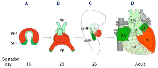
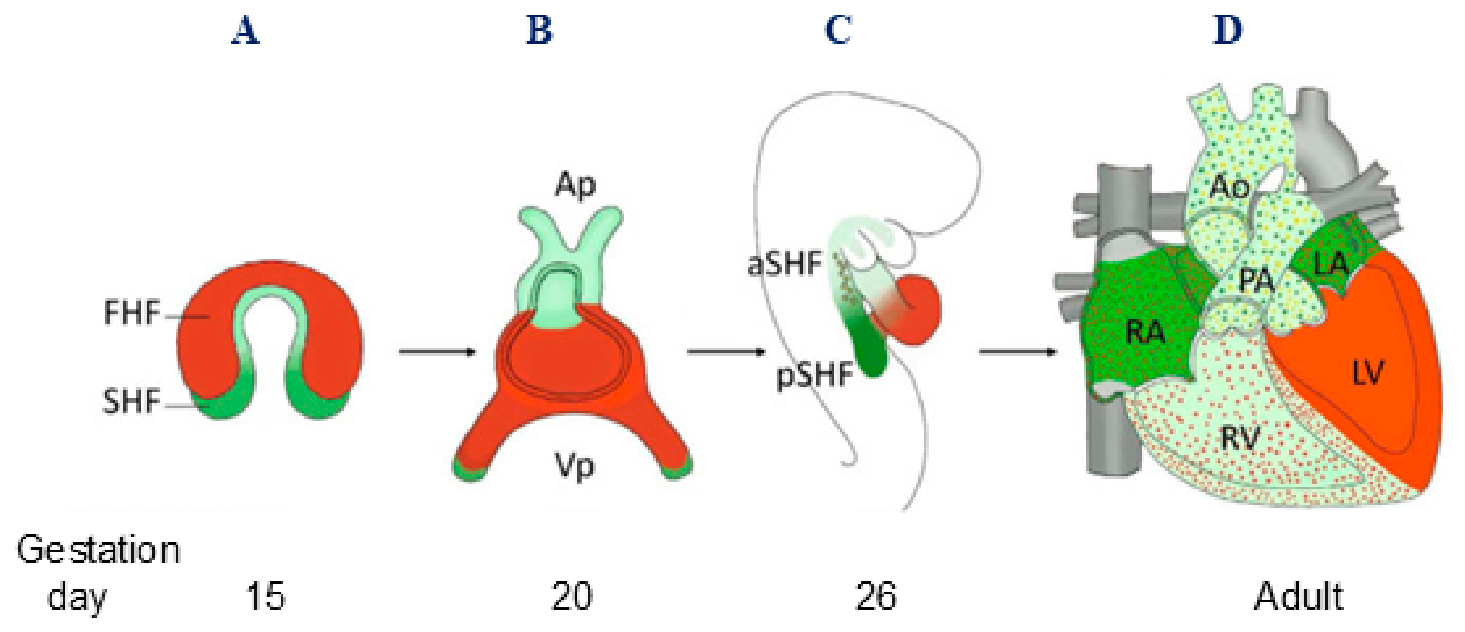
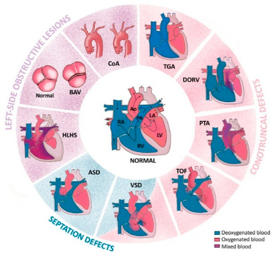
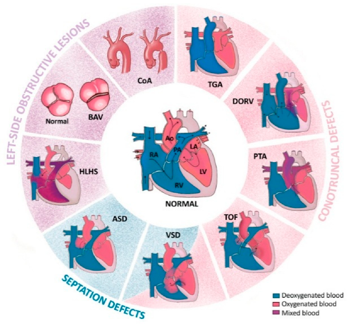
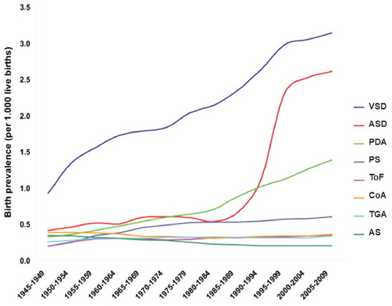
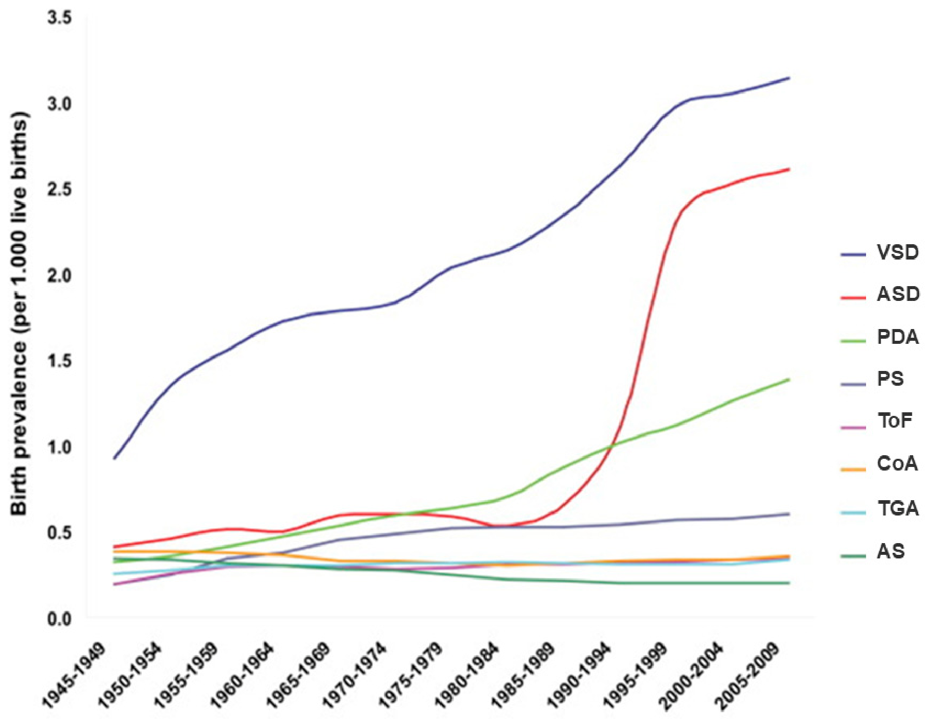
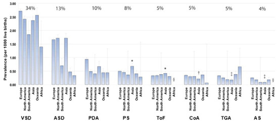
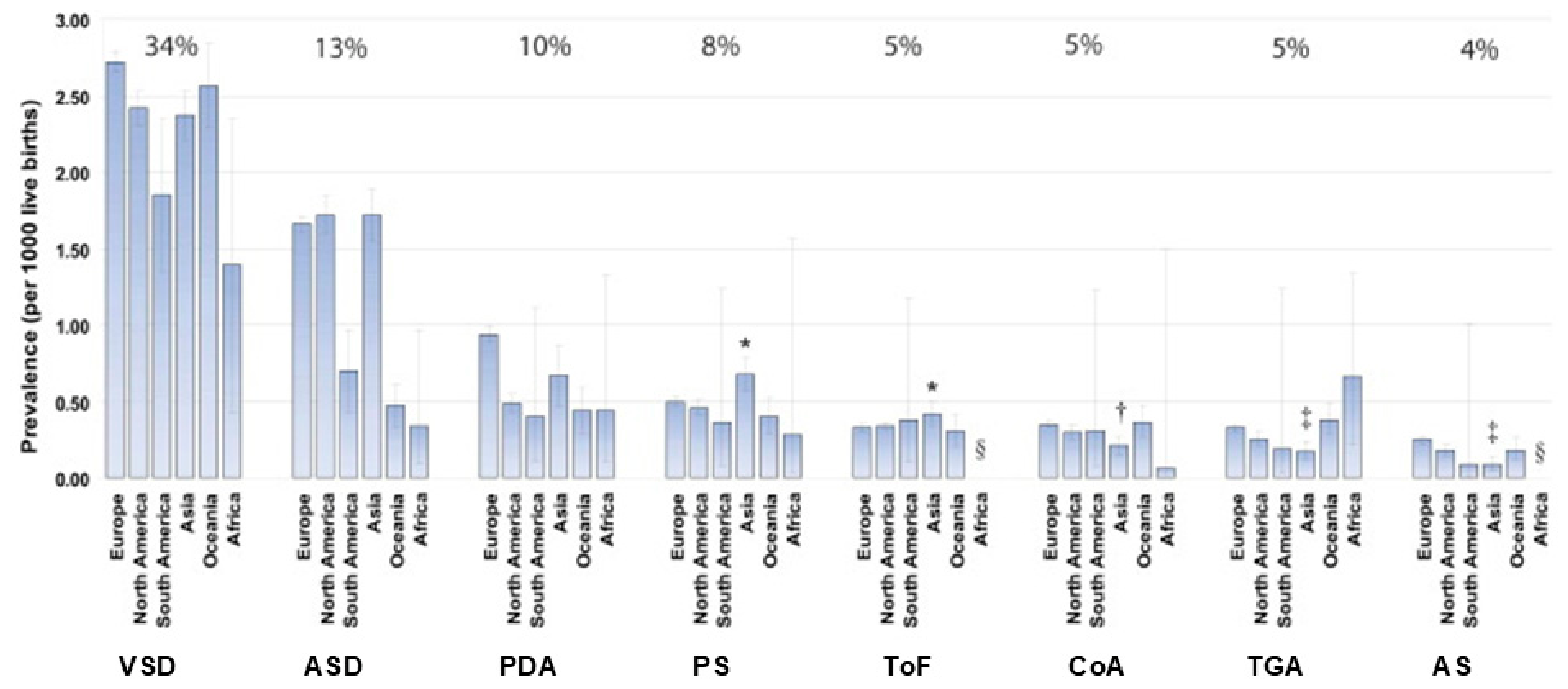
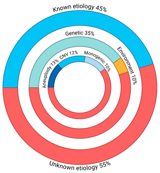
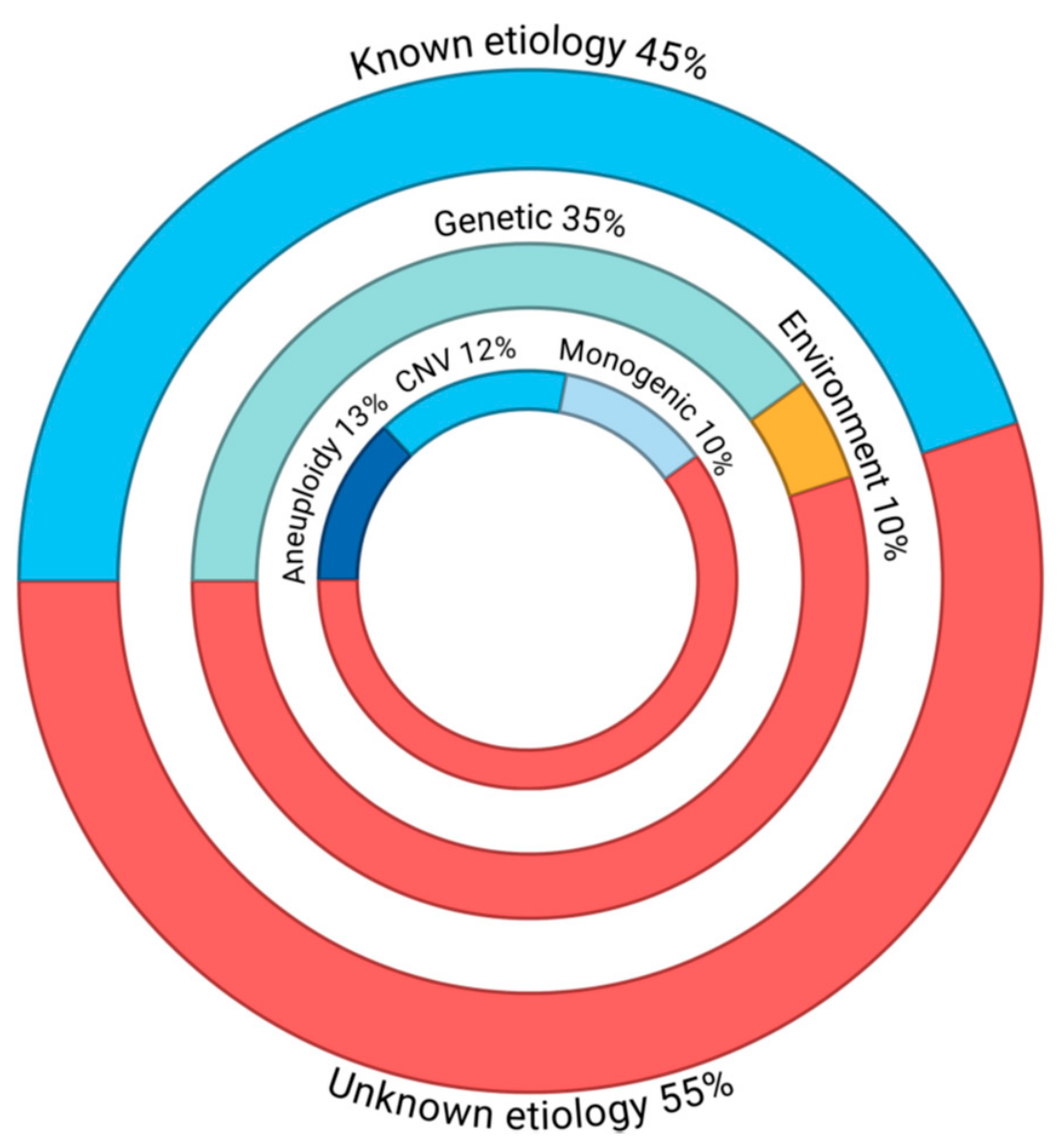
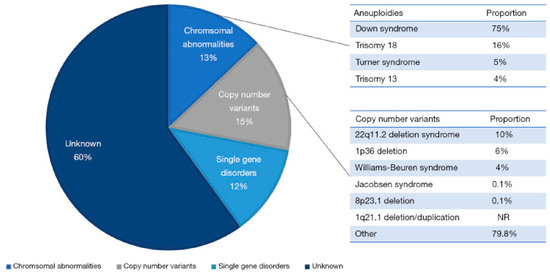
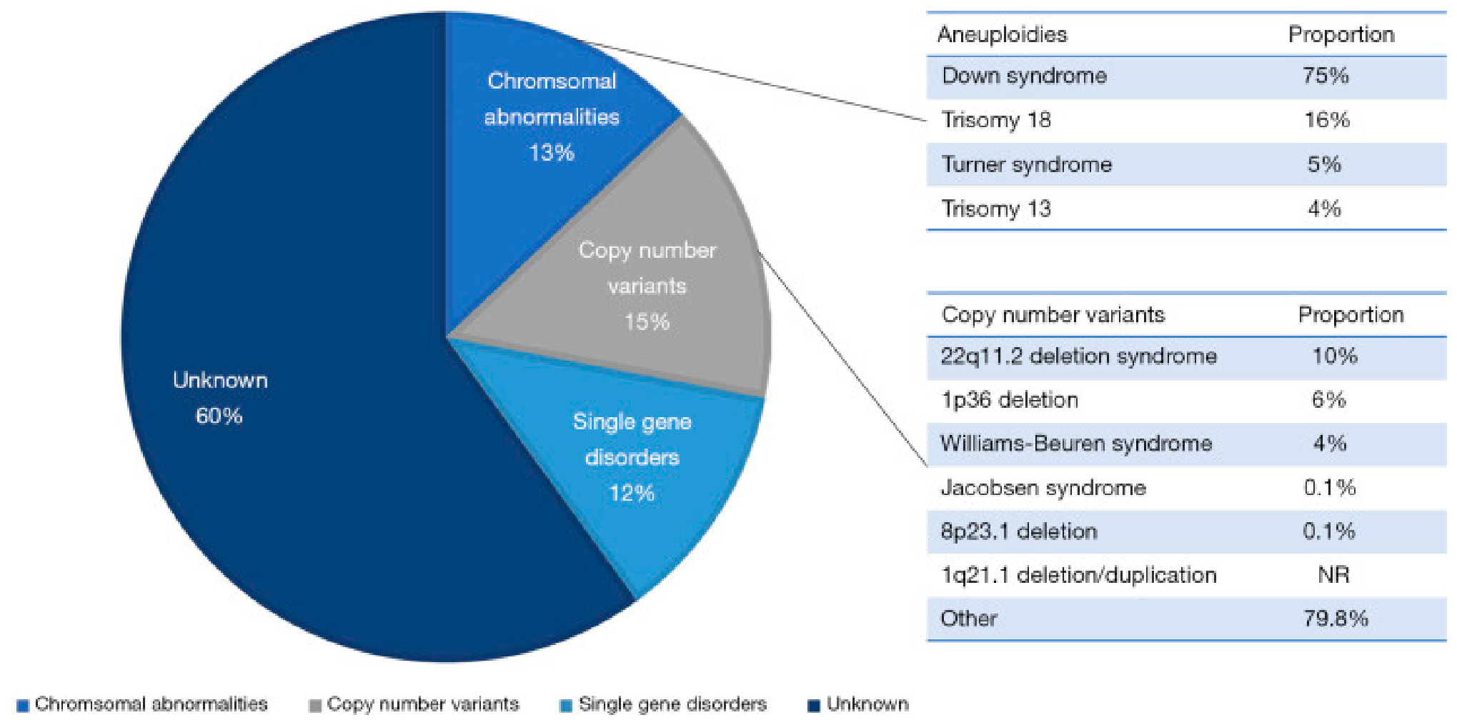
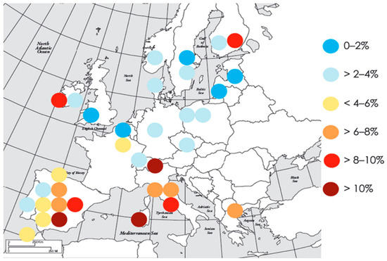
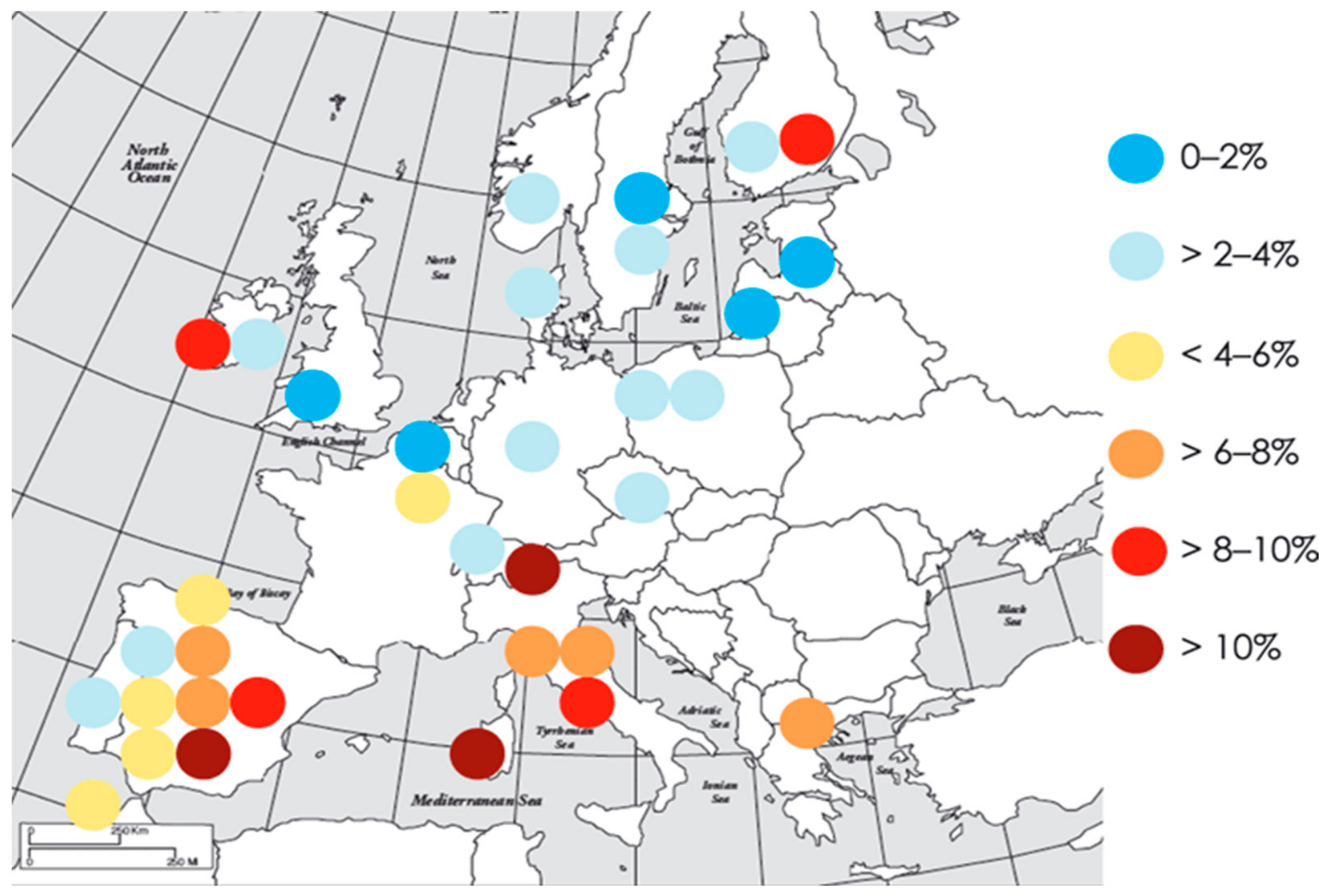
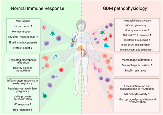
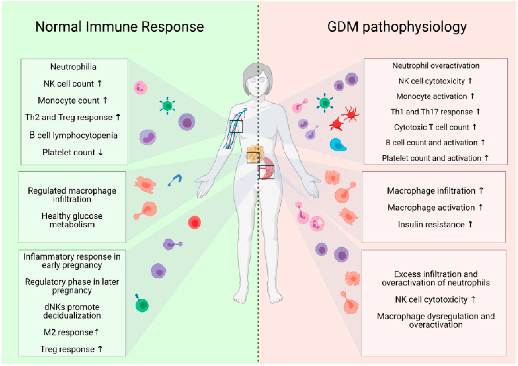
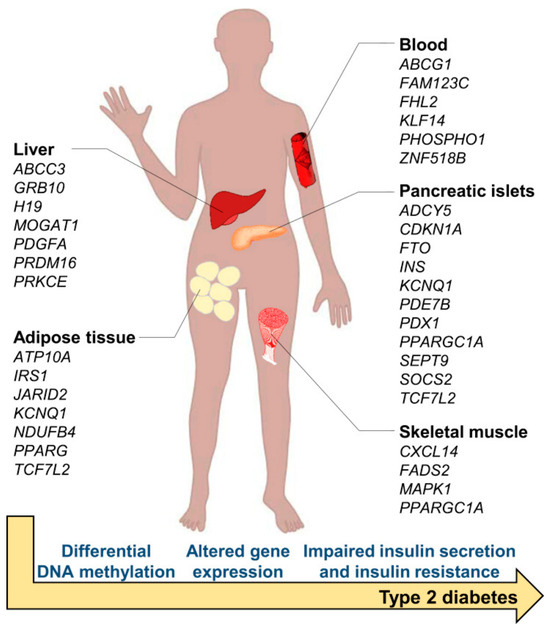
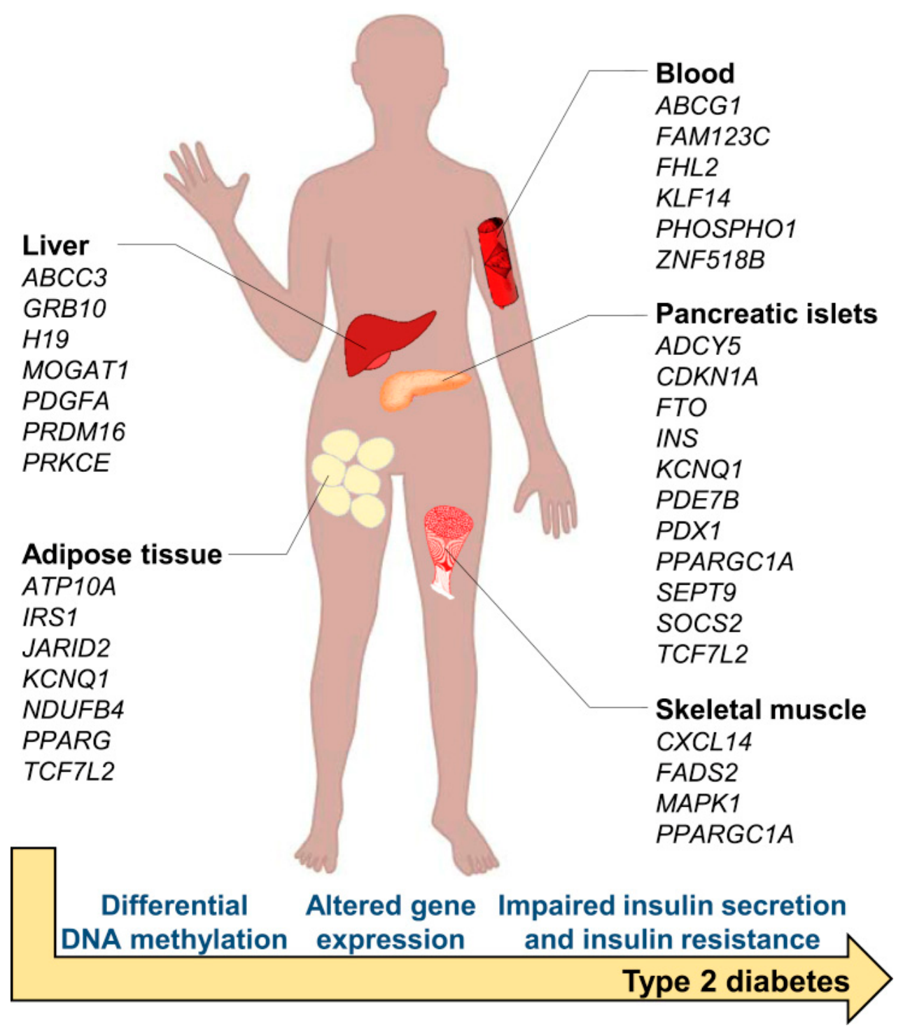
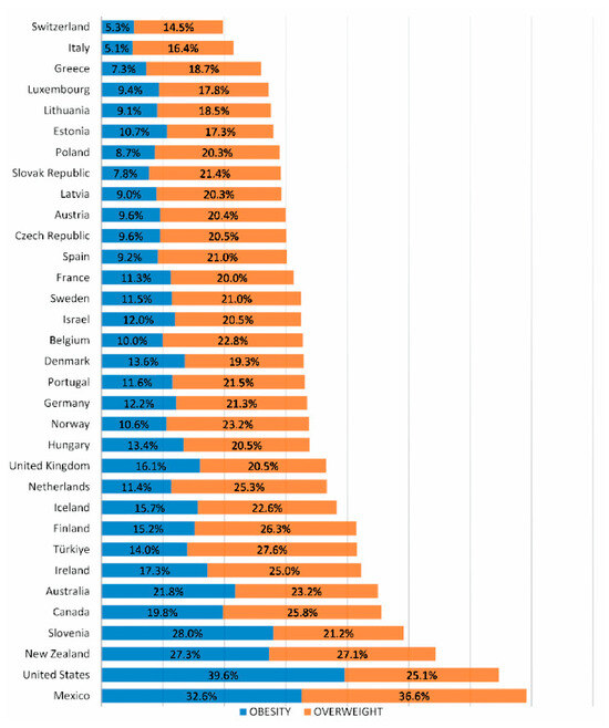
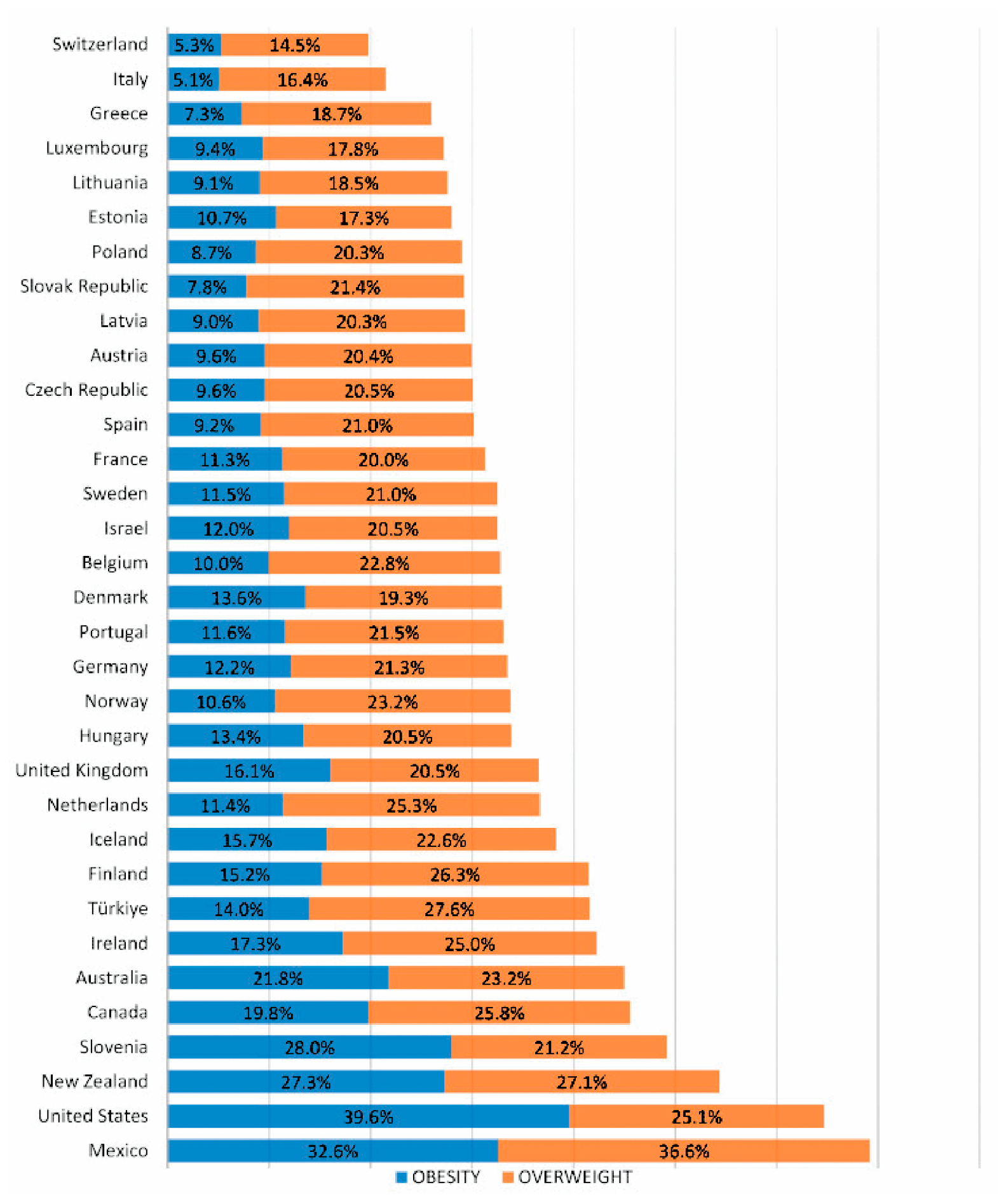
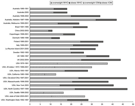
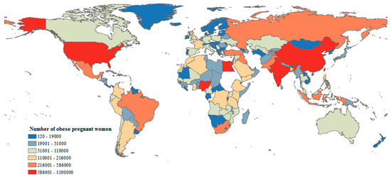
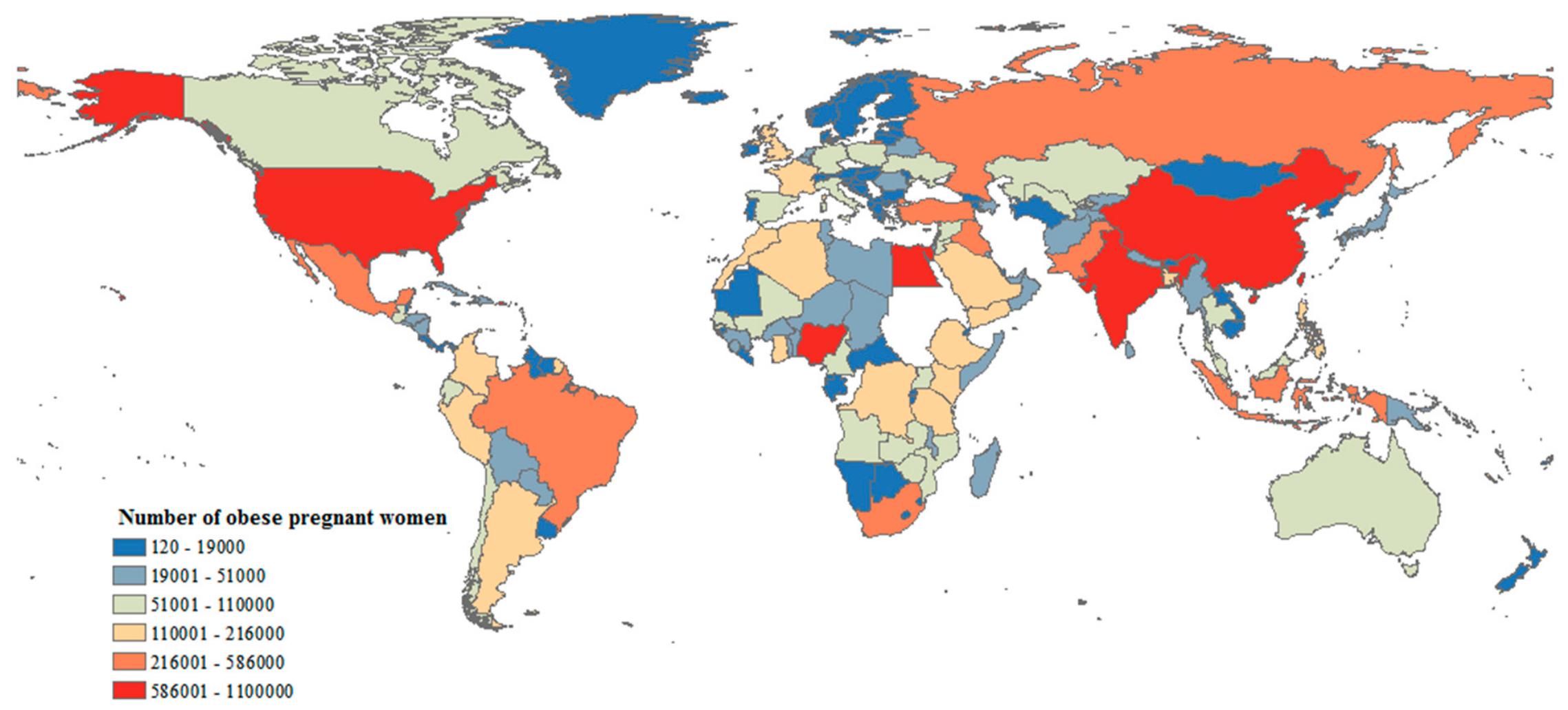
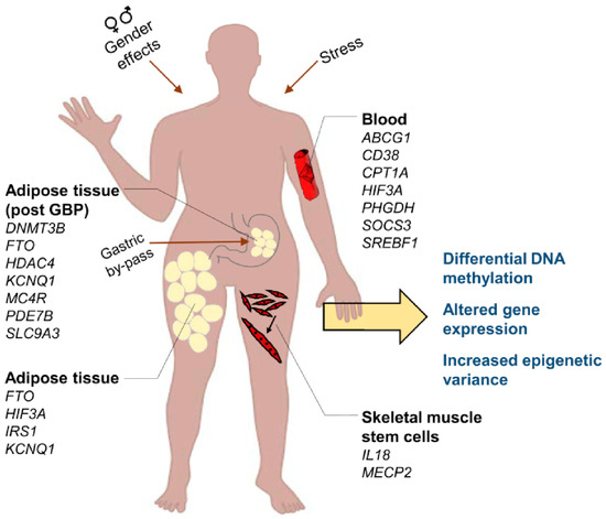
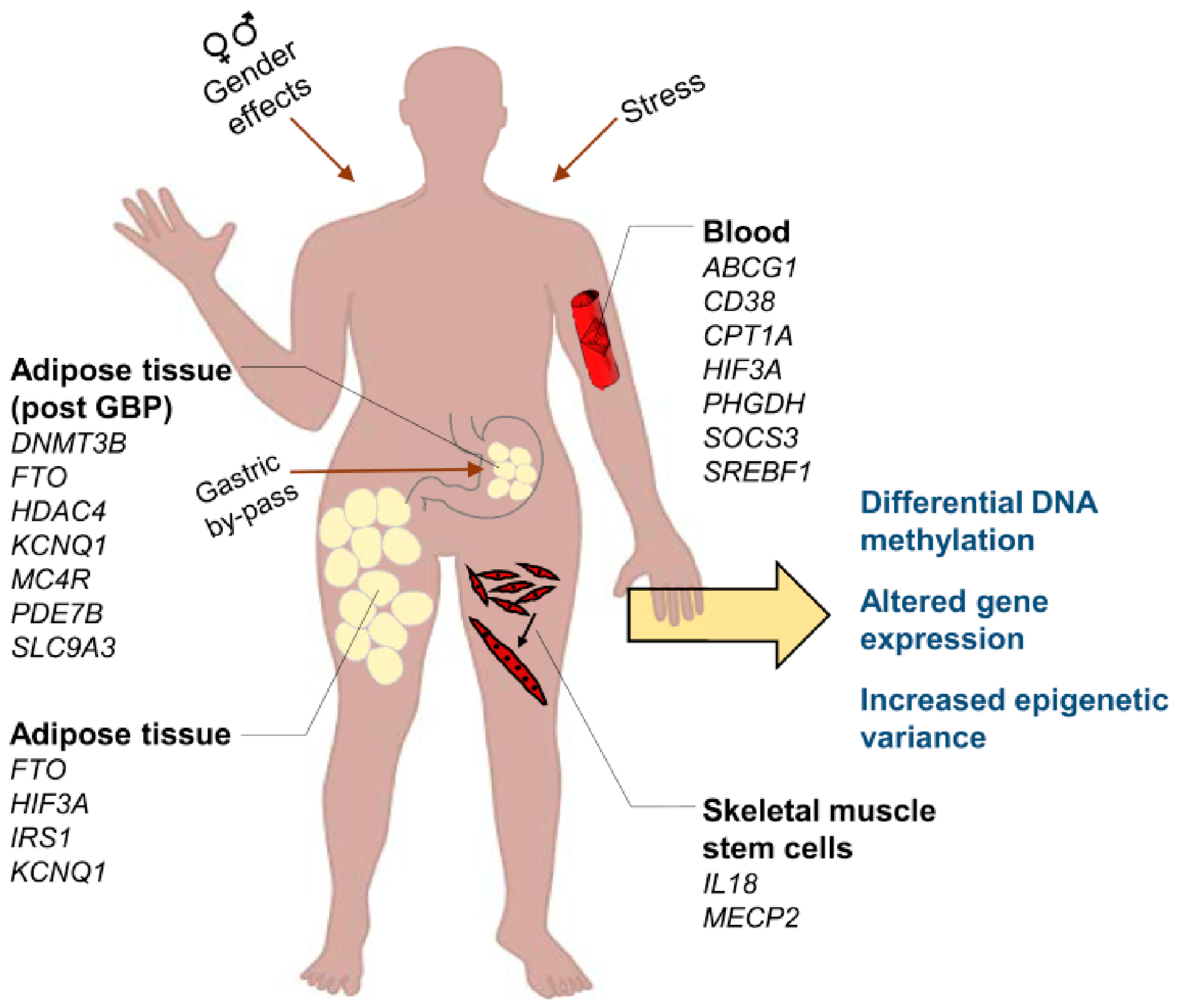
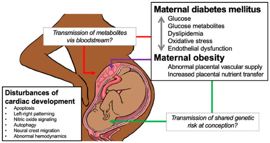
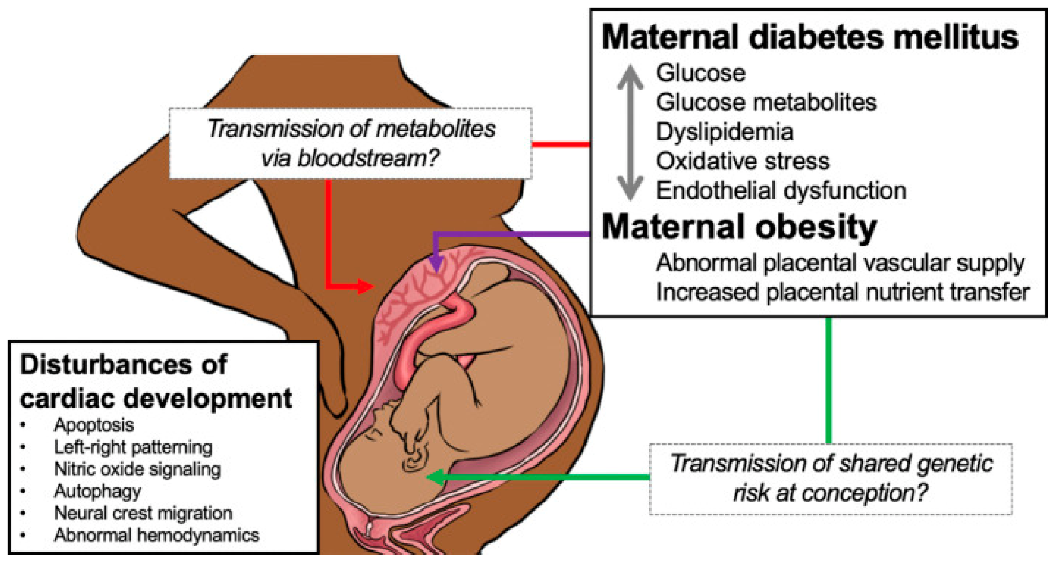
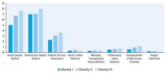
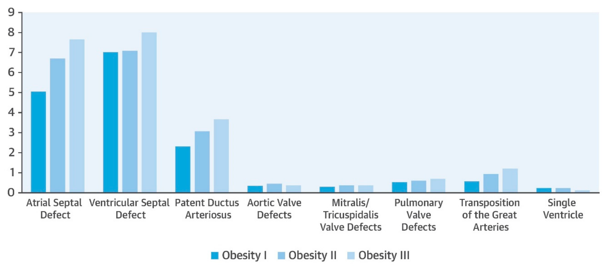

Disclaimer/Publisher’s Note: The statements, opinions and data contained in all publications are solely those of the individual author(s) and contributor(s) and not of MDPI and/or the editor(s). MDPI and/or the editor(s) disclaim responsibility for any injury to people or property resulting from any ideas, methods, instructions or products referred to in the content. |
© 2026 by the authors. Licensee MDPI, Basel, Switzerland. This article is an open access article distributed under the terms and conditions of the Creative Commons Attribution (CC BY) license.