


Abstract
The gut immune microenvironment and the liver engage in intricate information exchange via the gut–liver axis. The disruption of these interactions plays a pivotal role in the formation and exacerbation of pathological damage to the liver. The gut immune microenvironment is not an independent layer of the gut barrier; rather, it permeates and regulates all other barrier functions, serving as the core coordinator. Disruption of the immune microenvironment in the gut–liver axis drives progression across the full disease spectrum—from steatosis to hepatitis, fibrosis, and even liver cancer—through the continuous influx of immune-stimulatory signals that overwhelm the liver’s intrinsic immune regulatory mechanisms. Dysfunction of innate immunity components, amplification of inflammatory factors and key cellular signaling pathways, activation of adaptive immune T cells, and systemic effects mediated by liver-derived inflammatory factors collectively form a disordered immune microenvironment. This damages the intestinal barrier and exacerbates liver disease via the gut–liver axis, leading to further intestinal injury, thus establishing a self-reinforcing vicious cycle. Current therapeutic strategies based on modulating the gut–liver axis microenvironment remain limited, yet studies have demonstrated that suppressing gut immune cells, cytokines, and signaling pathways can help delay liver disease progression. Hopefully, future combined, precise, and cutting-edge gut immunotherapies will provide more effective strategies for liver disease treatment.
1. Introduction
According to the 2010 Global Burden of Disease Project assessment, liver cirrhosis (LC) accounted for 2% of all deaths, as did hepatocellular carcinoma (HCC) and acute hepatitis, indicating that liver disease constitutes a substantial public health burden [1]. Between 2010 and 2019, the burden of liver disease caused by virus-related hepatitis gradually decreased due to the successful implementation of vaccination programs, aflatoxin reduction initiatives, and aggressive antiviral therapy. However, with improved living standards, increased obesity, and rising alcohol consumption, the incidence of liver disease driven by metabolic dysfunction-associated steatotic liver disease (MASLD) and alcoholic liver disease (ALD) has dramatically increased [2]. The spectrum of liver diseases is shifting from a predominance of viral hepatitis to a predominance of metabolic liver diseases. Over the past few decades, the global occurrence rate and prevalence of autoimmune liver diseases (AILDs) have shown an overall increasing trend, which is associated with improvements in clinical diagnostic capabilities and enhanced patient awareness of the disease [3]. Consequently, the “true” burden of AILDs may be severely underestimated. Effectively treating these conditions also represents a crucial component in alleviating the overall burden of liver disease. Current treatment modalities for these diseases primarily entail surgery, antifibrotic drugs, and immunosuppressants. However, their efficacy still faces fundamental challenges in treating the most prevalent metabolic liver diseases, autoimmune liver diseases, and advanced cirrhosis. Furthermore, there are obstacles in translating therapeutic innovations into public health practice [1]. Therefore, a new, promising direction is needed to revitalize the search for better liver disease treatments.
The unique anatomical, physiological, and immunological relationship between the liver and the gut has shifted liver disease treatment strategies from the traditional hepatocentric approach to a holistic view of the gut–liver axis. A substantial body of research demonstrates that dysregulation of the gut–liver axis serves as a central nexus in the pathogenesis and progression of various liver diseases. Consequently, numerous restorative interventions targeting gut barrier function have offered novel therapeutic avenues. For instance, targeting the gut microbiota (through probiotics, prebiotics, dietary interventions, or fecal microbiota transplantation) to restore gut barrier function offers promising new strategies for liver disease treatment [4,5]. The advantage of this approach lies in its intervention targeting the “upstream” origin of the disease, potentially offering broad-spectrum efficacy and lower side effects. However, therapeutic outcomes are subject to significant batch-to-batch variability, and the approach lacks precision, specificity and targeting capability. The intestinal immune system engages in dynamic crosstalk with the liver via the gut–liver axis. Following the disruption of intestinal immune tolerance, erroneous recognition and attack by the intestinal immune system lead to aberrant activation of the immune microenvironment and the formation of a pro-inflammatory microenvironment. This process generates numerous pro-inflammatory factors, which damage multiple intestinal barrier systems by, for example, disrupting epithelial tight junctions, inducing epithelial cell apoptosis, impairing the mucus layer, and altering the gut microbiota. These factors subsequently reach the liver via the bloodstream, impacting the hepatic ecosystem, thereby driving and sustaining liver inflammation, steatosis, and fibrosis, and ultimately affecting immune surveillance against hepatocellular carcinoma [6,7,8]. Immune system dysfunction serves as the critical bridge linking intestinal homeostasis imbalance to hepatic pathological injury. Therefore, targeting the intestinal immune microenvironment (e.g., by restoring intestinal immune tolerance or targeting specific immune cell subsets) offers a more stable and precise therapeutic strategy for treating various liver diseases.
Clearly, intricate information exchange occurs between the gut immune microenvironment and the liver via the gut–liver axis. Dysregulation of this communication plays a pivotal role in the formation and exacerbation of pathological damage to the liver. Therefore, we provide a review of the literature on the information exchange within the immune microenvironment via the gut–liver axis and its potential as a novel therapeutic target in liver diseases, aiming to offer new strategies for early intervention and the delay of liver disease progression.
2. The Role of the Gut Immune Microenvironment in the Pathophysiology of the Gut–Liver Axis
2.1. Anatomical and Physiological Basis of the Gut–Liver Axis
The gut–liver axis is a bidirectional communication system based on unique anatomical connections (the portal vein and biliary tract). Its physiological core lies in the circulation of bile acids, nutrients, and microbial metabolites, which mediate the continuous dialogue between the liver and the gut. It is widely recognized that the gut–liver axis rests on three major anatomical and physiological pillars [4,9]: (1) Portal venous system: Serving as the primary conduit, this system directly transports nutrients, microbial products, and metabolites absorbed from the intestine to the liver. (2) Biliary system: Bile acids, synthesized by the liver and secreted into the intestine via the bile ducts, participate in fat digestion and shape the gut microbiota. The microbiota, in turn, converts primary bile acids into secondary bile acids, some of which are reabsorbed by the liver (enterohepatic circulation). (3) Convergence of systemic circulation and mucosal immune system: The liver receives and processes factors derived from the intestinal immune microenvironment via the lymphatic and systemic circulation.
2.2. The Physiological Role of the Immune Microenvironment in the Multi-Tiered Defense System of the Gut Barrier
The gut barrier is a comprehensive functional unit composed of physical barriers (epithelial cells, tight junctions), chemical barriers (mucus layer, antimicrobial peptides), biological barriers (commensal microbiota), and immune barriers gut-associated lymphoid tissue (GALT), secretory IgA, lamina propria immune cells (e.g., macrophages, dendritic cells, T cells, B cells) [10,11]. The immune system is not an independent layer but rather a core coordinator that permeates and regulates all other barrier functions. The homeostasis of the immune microenvironment is crucial for maintaining overall barrier integrity and distinguishing between “friend” and “foe”. Its dysfunction is the root cause of various intestinal and extraintestinal diseases, such as inflammatory bowel disease and liver disease [10].
For instance, processes such as mucus secretion by goblet cells and antimicrobial peptide production by Paneth cells are strictly regulated through immune signaling, exemplified by cytokines like IL-22 and IL-13 [11]; Furthermore, goblet cells secrete mucus and specific proteins (e.g., RELMB) that actively transport luminal antigens to lamina propria dendritic cells via “antigen delivery” to induce immune tolerance. This process also modulates Th2 and Treg cell responses [12]. This clearly demonstrates that the chemical barrier function is finely regulated by the local immune microenvironment, while also actively shaping the immune response, serving as an indispensable component in achieving immune tolerance. Intestinal epithelial cells are far from being passive physical barriers; they constitute an active interface that directly communicates with immune cells through multiple intricate pathways. The immune microenvironment determines the “on–off” state of the barrier by regulating epithelial cell functions and tight junctions, while epithelial cells shape the nature of immune responses through signaling feedback, forming an inseparable bidirectional regulatory loop. The Gut Vascular Barrier (GVB) consists of specific cells and tight junctions in the intestinal capillary endothelium. Immune cells (e.g., resident intestinal macrophages) are crucial for maintaining tight junctions and barrier function in GVB endothelial cells through the secretion of WNT ligands [9]. This directly demonstrates the active role of the lamina propria immune microenvironment in maintaining anatomical barriers deep within tissues. Similarly, the immune microenvironment plays a crucial role in the resistance against colonization by intestinal pathogens. This protective role is regulated by the gut microbiota. For instance, microbial products such as lipopolysaccharide (LPS) and flagellin stimulate TLR4+ stromal cells and TLR5+CD103+ DCs, thereby promoting the expression of REGIIIγ, which inhibits the colonization of Gram-positive vancomycin-resistant enterococcus (VRE), in epithelial cells. Segmented filamentous bacteria (SFB), which adhere to the intestinal epithelium, enhance IgA production by B cells, SAA-dependent Th17 cell differentiation, the production of pro-inflammatory cytokines, and the generation of antimicrobial peptides in epithelial cells. These processes confer protection against citrobacter rodentium [13]. The microbiota and host immune system co-maintain an anti-inflammatory, tolerant, and defensively robust intestinal environment through reciprocal regulation via microbial metabolites. The breakdown of this homeostasis serves as a precursor event to barrier dysfunction (Figure 1A).
Figure 1.
Gut immune microenvironment in the pathophysiology of the gut–liver axis. (A) Physiological homeostasis: A healthy intestinal barrier (intact epithelium, mucus layer, antimicrobial peptides, and secretory IgA) and immune tolerance in the lamina propria (Tregs, tolerogenic DCs) jointly maintain the stability of the gut–liver axis, preventing microbial translocation. (B) Pathological disruption: Disruption of the intestinal barrier (leaky gut) leads to the translocation of pathogen-associated molecular patterns (PAMPs) and damage-associated molecular patterns (DAMPs). Immune homeostasis in the lamina propria is disrupted, shifting towards a pro-inflammatory environment dominated by Th1/Th17 cells, M1-type macrophages, and pro-inflammatory cytokines (e.g., TNF-α, IL-1β), accompanied by reduced production of secretory IgA.
The intact gut barrier constitutes a multi-tiered fortress system wherein the functions of goblet cells, intestinal epithelial cells, the microbiota, and the GVB are highly dependent on signaling support from specific immune cells in the intestinal lamina propria. This provides the most direct molecular and cytological evidence for “immune microenvironment-mediated regulation of physiological barriers”.
2.3. Manifestations of Immune Microenvironment Disruption in the Gut–Liver Axis
The disruption of the intestinal immune microenvironment is primarily manifested in the following three aspects. First is the imbalanced ratio of pro-inflammatory to anti-inflammatory cytokines. For instance, secondary bile acids produced by gut microbiota metabolism can strongly inhibit the activation of the NLRP3 inflammasome in macrophages by activating the receptor TGR5, thereby reducing the maturation and release of the pro-inflammatory cytokines IL-1β and IL-18 [14]. An obesogenic diet can rapidly inhibit IL-22 production by innate lymphoid cells in the small intestine, leading to the suppression of STAT3 in intestinal epithelial cells (IECs) and the activation of the WNT-β-catenin signaling pathway. This causes the absorptive enterocyte compartment to expand, contributing to metabolic dysfunction-associated steatotic liver disease [15]. Second, immune cell subsets become imbalanced. Studies have shown that, compared with controls, mice with alcoholic hepatitis develop more severe liver inflammation, which is associated with increased intestinal permeability and higher bacterial translocation due to an increase in CD4+T and NKT lymphocytes in the mesenteric lymph nodes [16]. This dysregulation of the immune microenvironment in the intestinal lamina propria, in turn, impairs the barrier function of the intestinal epithelium, subsequently triggering liver inflammation. In liver disease, the number of Tregs in the gut and liver is reduced or their function is impaired, weakening their inhibitory effect on effector T cells (e.g., Th1, Th17). This leads to an imbalance between immunosuppressive and effector subsets, resulting in intestinal barrier damage and liver disease progression [17]. Finally, mucosal immune tolerance breaks down. For example, as mentioned earlier, the GVB serves as the final anatomical defense against bacterial translocation. Its integrity is maintained by the resident CX3CR1+ macrophage subset in the intestinal lamina propria through the secretion of WNT ligands [9]. Secretory IgA (sIgA) is the core executor of mucosal immune tolerance. It coats commensal bacteria through an immune exclusion mechanism, limiting their contact with and translocation across the epithelium without triggering strong inflammation. Alterations in the quality or quantity of sIgA can lead to dysbiotic localization of the microbiota and excessive epithelial contact, thereby breaking tolerance and activating local immune responses [18]. Functional defects in sIgA are an early and significant marker of immune microenvironment disruption in the gut–liver axis, directly contributing to gut microbiota dysbiosis and immune activation. Furthermore, recent studies indicate that in MASLD, viable bacteria can translocate to the mesenteric adipose tissue (MAT) and form local microabscesses, activating immune responses within the MAT. This creates a persistent extraintestinal inflammatory focus, in which inflammatory mediators are continuously released into the portal vein, exacerbating liver inflammation [19]. These ectopic, microbiota-driven immune activation foci become independent sources of inflammation that persistently drive liver injury, representing a novel paradigm of immune dysregulation in the gut–liver axis (Figure 1B).
The disruption of the immune microenvironment within the gut–liver axis is, in essence, a pathological shift in the liver from an immune-tolerant organ to a chronic inflammatory milieu, with the breakdown of intestinal immune homeostasis serving as the central driver of this transition.
3. Dynamic Crosstalk Between Liver and Gut: Immune Dysregulation–Intestinal Leakage–Gut–Liver Axis–Liver Disease Progression
Multiple external triggers disrupt immune tolerance within the gut–liver axis, impairing gut barrier function. This leads to overactivation of the immune system, which persistently activates the liver’s innate immune system via the gut–liver axis, thereby exacerbating the disruption of hepatic immune tolerance and establishing a vicious inflammation–injury cycle. This disruption of the intrahepatic immune microenvironment triggered by gut immune dysregulation constitutes a common core pathway driving the progression of multiple liver diseases, including MASLD, ALD, AILD, LC, and HCC (Figure 2).
Figure 2.
Dynamic mechanisms of gut–liver axis disruption driving chronic liver disease progression: From immune dysregulation to liver cancer.
Intestinal immune dysregulation and barrier disruption drive the progression of chronic liver disease to its end-stage through the gut–liver axis. (A) MASLD: Risk factors such as metabolic abnormalities disrupt intestinal homeostasis, and mild intestinal permeability triggers hepatic steatosis and chronic inflammation. (B) ALD: Persistent alcohol exposure exacerbates intestinal inflammation and barrier loss, driving significant hepatic inflammation. (C) AILD: In genetically susceptible individuals, intestinal permeability may lead to the translocation of self/mimic antigens, breaking immune tolerance and triggering autoimmune attacks against the bile ducts or hepatocytes. (D) LC: Severe gut–liver axis disturbance substantially increases the risk of antigen/bacterial translocation, accompanied by systemic inflammation and complications, forming a vicious cycle. (E) HCC: Against the backdrop of chronic inflammation and regeneration, the accumulation of genetic mutations, dysregulation of immune surveillance, and infiltration of tumor-associated macrophages and immunosuppressive cells collectively promote malignant transformation.
3.1. MASLD
MASLD has become the most prevalent chronic liver disease worldwide, with its disease spectrum progressing from simple hepatic steatosis to metabolic dysfunction-associated steatotic hepatitis (MASH), hepatic fibrosis, cirrhosis, and ultimately hepatocellular carcinoma. The traditional “second hit” theory has evolved into a more complex “multiple parallel hits” model, wherein dysfunction of the gut–liver axis–particularly dysregulation of its immune microenvironment–is recognized as the core driver initiating disease progression and the malignant transformation from benign steatosis to inflammatory MASH and fibrosis. The intensive immune and metabolic crosstalk between the gut and liver occurs via the portal vein, biliary tract, and systemic circulation. Disruption of homeostasis along this axis constitutes the pathophysiological cornerstone of MASLD (Table 1).
Table 1.
Key gut immune regulatory targets and pathways in MASLD.
3.1.1. Innate Immunity
Hepatic macrophages play a pivotal role in the progression of MASLD. Studies have demonstrated that intestinal CX3C chemokine receptor 1-positive (CX3CR1+) macrophages contribute to maintaining intestinal barrier stability and inhibiting gut microbiota translocation. Intestinal CX3CR1 knockout mice lack CX3CR1+ macrophages, leading to impaired function of the multi-layered intestinal barrier and accelerated progression of MASH via the gut–liver axis [29,30]. Furthermore, in patients with MASH, intestinal macrophages exhibit increased release of nitric oxide, which further inhibits the repair of the intestinal epithelial barrier mediated by connexin 43. Concurrently, intestinal macrophages release increased amounts of IL-6, which modulates the expression of the tight junction protein claudin-2 in intestinal epithelial cells, thereby affecting intestinal epithelial barrier function and accelerating the progression of MASLD [31]. These findings indicate that the population of intestinal pro-inflammatory macrophage subsets is increased and capable of secreting pro-inflammatory cytokines, leading to intestinal inflammation and intestinal barrier disruption, with a subsequent influence on the MASLD process via the gut–liver axis.
3.1.2. Key Pathway Amplification
As previously mentioned, activation of the NLRP3 inflammasome pathway in the intestinal immune microenvironment generates large amounts of IL-1β and IL-18, which further damage the intestinal barrier and ultimately exacerbate MASLD [14]. The intestinal immune system recognizes pathogen/microbe-associated molecular patterns (P/MAMPs) via pattern recognition receptors such as TLRs and nucleotide-binding oligomerization domain (NOD)-like receptors (NLRs). Subsequently, the P/MAMPs influx resulting from pathological bacterial translocation (BT) and/or excessive increase in susceptibility induces the release of pro-inflammatory cytokines (e.g., TNF-α, IL-1, IL-6), chemoattractants, and eicosanoids from monocytes. Upon reaching the liver, these mediators drive the generation of a local pro-inflammatory hepatic microenvironment [7]. Furthermore, when the transcription factor basic leucine zipper ATF-like transcription factor 3 (BATF3), which is essential for the development of intestinal DCs in mice, is knocked out, the localization of tight junction proteins in intestinal epithelial cells is altered, intestinal permeability increases, the mice develop metabolic syndrome, and the progression of hepatic steatosis is promoted [32].
3.1.3. Adaptive Immunity Is Involved
Gut-derived antigens may disrupt hepatic immune tolerance, activating CD8+ cytotoxic T cells and CD4+ helper T cells (such as Th1 and Th17), while simultaneously impairing Treg function. This shifts immune attacks toward hepatocytes. In mice fed a high-fat diet (HFD), the percentage ratio of Th1/Th2 cells and Th17/Treg cells was imbalanced in the mesenteric lymph nodes (MLNs), and MLN CD4+ T lymphocytes tended to migrate to the liver, promoting hepatic inflammation [33,34]. In mice with MASH, intestinal IL-17 levels are elevated; intestinal Th17 cells migrate to the liver and secrete IL-17, thereby activating monocytes, bile duct epithelial cells, hepatic KC, and hepatic stellate cells (HSCs). This activation further promotes the secretion of pro-inflammatory cytokines and chemoattractants, exacerbating liver inflammation [35]. Conversely, in HFD-fed mice, donor-specific intestinal Th17 cells, induced in an IL-17-dependent manner, were shown to modulate intestinal epithelial lipid absorption by regulating the expression of the intestinal fatty acid transporter CD36, thereby attenuating the development of metabolic syndrome [36].
3.1.4. Systemic Effects
Inflammatory factors produced by the liver can further damage the intestinal barrier, forming a reverse liver–gut injury pathway. Concurrently, hepatic steatosis itself exacerbates oxidative and endoplasmic reticulum stress, acting synergistically with gut-derived inflammatory signals. Studies have reported an increased number of CD8+ T cells in the jejunum of obese patients. Certain soluble factors secreted by these cells significantly inhibit pSer473-Akt activity in intestinal epithelial cells, thereby impairing insulin sensitivity in small intestinal epithelial cells and affecting small intestinal function [26]. However, several other studies have found that in the duodenum of patients with MASLD and metabolic dysregulation, the expression of CD4+ and CD8+ T lymphocytes and ZO-1 is significantly reduced, leading to impaired intestinal mechanical barrier integrity [27,28]. This further contributes to the selective loss of intrahepatic CD4+ T lymphocytes in humans [37].
In summary, disruption of the gut–liver axis immune microenvironment serves as the driving force for the transition of MASLD from benign hepatic fat accumulation to inflammatory, progressive liver diseases (MASH and fibrosis). This is not merely a failure of a single component, but rather a collapse of the entire system involving gut ecology, physical barrier, immune signaling, metabolic regulation, and intrahepatic cellular network. (Figure 2A).
3.2. ALD
The cause of ALD progression is far from being as simple as the direct toxicity of alcohol to hepatocytes. Previous research has established that persistent disruption of the immune microenvironment in the gut–liver axis serves as the core mechanism driving the malignant transformation of the disease from simple steatosis to hepatitis and fibrosis. Alcohol acts as the initiating factor by first disrupting the gut ecology and immune balance, after which injury signals are amplified and transmitted to the liver through multiple pathways (Table 2).
Table 2.
Key gut immune regulatory targets and pathways in ALD.
Initiation of intestinal barrier disruption. Alcohol directly damages the intestinal epithelium or disrupts tight junctions by perturbing local intestinal immunity (e.g., increasing pro-inflammatory IL-17), leading to the translocation of endotoxins such as LPS. Chronic alcohol consumption suppresses the expression of the muscarinic acetylcholine receptor M4 (mAChR4) in small intestinal goblet cells, impairing intestinal immune surveillance. This results in reduced formation of goblet cell-associated antigen passages (GAPs) and inhibition of the ILC3-IL-22 antimicrobial pathway, thereby compromising antimicrobial immunity and facilitating bacterial translocation to the liver [44]. Studies indicate that chronic alcohol intake activates intestinal monocytes/macrophages, prompting them to release TNF-α. This subsequently activates the expression of tumor necrosis factor α receptor I on intestinal epithelial cells, further promoting the activation of myosin light-chain kinase in these cells. This cascade ultimately leads to the loss of tight junction proteins and increased intestinal permeability [45]. Recent research demonstrates that in ALD, inactivated postbiotics can activate intestinal innate immunity by stimulating the intestinal DCs-ILC3 axis, upregulating IL-22 production. This enhances the intestinal barrier upstream, reduces bacterial translocation, and exerts reparative effects in the liver via the gut–liver axis [46]. Alcohol-associated intestinal dysbiosis, which alters gut-derived immune signals, serves as a significant driver of dysfunction in intrahepatic innate immune cells (e.g., NK cells). This constitutes another pathway for immune crosstalk along the gut–liver axis, influencing hepatic immune surveillance and the inflammatory milieu [47].
Multifaceted signal transmission. Along with viable bacteria and their products (e.g., LPS), damage-signaling vesicles (EVs) released by intestinal epithelial cells and imbalanced microbial metabolites also translocate to the liver, collectively forming a “mixed arsenal” that attacks the liver. Interestingly, alcohol stimulation alters both the quantity and cargo of EVs released by intestinal epithelial cells, with an increase in pro-inflammatory cytokines (e.g., TLR4, TNF, IL-1β) and a decrease in ZO-1, leading to increased intestinal permeability. These vesicles reach the liver via the portal vein, where they can directly impair hepatocyte viability and promote lipid accumulation [48]. Alcohol-induced dysbiosis, characterized by an imbalance in the microbial community, also leads to a reduction in microbial metabolites known as indoles. This reduction contributes to decreased levels of IL-22, a cytokine essential for maintaining intestinal integrity. The decline in IL-22 weakens the intestinal barrier, promotes bacterial translocation to the liver, and advances the progression of ALD [49]. Recent studies have shown that in alcohol-fed mice, the number of intestinal conventional type 1 dendritic cells (cDC1s) is significantly reduced, which exacerbates the downregulation of antimicrobial peptides. Consequently, the intestinal barrier is weakened and becomes more permeable, allowing bacterial toxins such as LPS to enter the bloodstream, thereby increasing inflammation and liver injury [50]. A certain concentration of ethanol within the intestinal lumen can activate intestinal mast cells, leading to the release of substances like histamine, which in turn increases intestinal mucosal permeability. This process also contributes, to some extent, to accelerating the progression of liver disease [51].
Vicious cycle. Ethanol-fed mice and patients with alcohol use disorder exhibit reduced duodenal macrophage numbers, dysregulated cytokine secretion, impaired phagocytic function, and diminished intestinal IgA secretion. Furthermore, even in the pre-cirrhotic stage, patients demonstrate reduced intestinal T cell populations and immune dysfunction, further exacerbating the risk of gut-derived liver inflammation [52].
Alcohol disrupts gut immune surveillance and barrier function, leading to leakage of intestinal vesicles, cytokines, immune cells, bacteria, and their products into the liver. This activates hepatic immune cells, triggering severe inflammation and forming a vicious cycle of “gut immune dysregulation–intestinal leakage–liver injury”—a core mechanism exacerbating alcoholic liver disease (Figure 2B).
3.3. AILD (e.g., Primary Biliary Cholangitis, Autoimmune Hepatitis)
AILD is a group of diseases characterized by the loss of immune tolerance in the liver, the presence of autoantibodies, and chronic inflammation, whose exact etiology has not yet been fully elucidated. Over the past decade, advances in microbiome research have positioned the gut–liver axis theory as a novel perspective for understanding environmental triggers and systemic immune dysregulation in AILD (Table 3). Although primary biliary cholangitis (PBC), autoimmune hepatitis (AIH), and primary sclerosing cholangitis (PSC) have distinct targets, clinical manifestations, and classic pathological mechanisms, they share an increasingly recognized common background: immune microenvironmental dysregulation of the gut–liver axis. This section will elaborate on the central role of immune microenvironmental dysregulation within the gut–liver axis in the pathogenesis and progression of AILD from this shared perspective, focusing on three aspects: erroneous homing, molecular mimicry, and signal amplification.
Table 3.
Key gut immune regulatory targets and pathways in AILD.
3.3.1. Aberrant Homing
The inflamed liver/abnormal bile ducts aberrantly express “postal codes” (e.g., CCL25, MAdCAM-1) that are normally exclusive to the intestine. This misdirects immune cells (CCR9+/α4β7+ T cells, IgA+ plasma cells) originally destined for intestinal duty into the hepatobiliary system, resulting in bystander injury [53,59]. Within the intestinal lymph nodes, these antigens drive the differentiation of naïve T cells into pro-inflammatory Th17 cells. These Th17 cells migrate to the liver, where they secrete cytokines such as IL-17 and IL-22, recruiting neutrophils and promoting hepatocyte damage and portal inflammation [55]. Intestinal Bifidobacterium modulates the IL-33-induced Treg/Th17 imbalance via the TLR2/4 signaling pathway, thereby alleviating autoimmune hepatitis [60].
3.3.2. Molecular Mimicry
Certain components of the gut microbiota (e.g., bacterial enzymes) exhibit high similarity to human autoantigens (e.g., mitochondrial enzymes). Antibodies and T cells produced by the gut immune system to combat these bacteria may mistakenly recognize and attack bile duct cells expressing similar autoantigens, triggering autoimmunity [54]. Intestinal pIgA antibodies form immune complexes that deposit around bile ducts, causing bystander injury to bile ducts through the activation of the complement pathway [53].
3.3.3. Signal Amplifier
Abnormal LSECs act as an accomplice by upregulating molecules such as VAP-1, firmly anchoring recruited gut immune cells in the liver and promoting their infiltration into lesion sites, thereby amplifying the damage [58].
Research over the past decade has firmly established the central role of the gut–liver axis in the pathogenesis of AILD. The gut serves not only as the source of immune disruption but also as the epicenter driving liver-specific autoimmune responses (Figure 2C).
3.4. LC
The onset and progression of LC extend beyond the liver itself and are profoundly influenced by the bidirectional vicious cycle of interaction within the gut–liver axis. From hepatic inflammatory changes to fibrosis and ultimately cirrhosis, the gut immune microenvironment progressively deteriorates, imposing an additional burden on liver function. The first stage involves the breach of immune sentinels, where increased gut permeability allows bacteria and their byproducts, fungi, and other pathogens to penetrate and assault the gut immune system. The second stage manifests as sluggish and dysfunctional innate immune responses. The third stage features the failure of adaptive immunity and the loss of tolerance, characterized by functional exhaustion and increased apoptosis of intestinal T and B lymphocytes. Ultimately, irreversible systemic alterations occur, where persistent translocation of gut-derived pathogens results in recurrent, low-grade bacteremia, triggering systemic inflammation (Table 4).
Table 4.
Key gut immune regulatory targets and pathways in LC.
In acute-on-chronic liver failure (ACLF), the intestinal barrier is severely compromised, leading to the massive and persistent translocation of bacteria and their products (e.g., LPS). This sustained stimulation exhausts the phagocytic and clearance capacity of the hepatic and systemic monocyte–macrophage system and induces the production of high levels of the immunosuppressive factor IL-10 (which may be related to type I interferon signaling) [65]. The direct clinical consequence is that patients face an extremely high risk of bacterial infection, which is a primary factor triggering ACLF. Macrophage-derived inflammasomes triggered by bacterial translocation and tissue damage also contribute to inflammatory liver injury [66]. Activated inflammasomes initiate caspase-1-dependent production of pro-inflammatory cytokines such as IL-1β and IL-18, which subsequently exacerbate hepatic inflammation, fibrosis, and injury in conditions like ALD, MASH, and other forms of liver damage [67]. Studies have shown that in MASH, intestinal B cells can directly migrate to the liver and promote the activation of metabolic CD8+ T cells, driving autoimmune liver injury; they also directly facilitate the development of liver fibrosis via the IgA-Fc receptor signaling pathway [68]. Intestinal immune imbalance leads to decreased expression of tight junction proteins, increased intestinal permeability, and the entry of pro-inflammatory factors and PAMPs into the liver, thereby promoting the activation of hepatic stellate cells.
Gut-derived LPS can stimulate TLR4 on quiescent HSCs and regulate TGF-β in a MyD88–NF-κB-dependent manner to activate these cells, thereby inducing a pro-fibrotic transformation and accelerating liver fibrosis [69]. Neutrophils are recruited to the liver via adhesion molecules ICAM-1 and VCAM-1 expressed on LSECs. In cirrhosis, neutrophil function is also disrupted by the dysfunctional gut–liver axis, leading to inflammation-associated hepatocyte injury and IL-17-dependent HSC activation [70].
In summary, in LC, immune dysregulation of the gut–liver axis forms a self-reinforcing vicious cycle. Breaking this cycle requires a dual strategy that both enhances immune defense and suppresses local inflammation/fibrosis, which represents the primary challenge and direction for current therapeutic development (Figure 2D).
3.5. HCC
As summarized previously, alterations in the gut immune microenvironment constitute one of the critical factors driving the late-stage deterioration and qualitative changes in liver cirrhosis. During the HCC phase, the gut immune microenvironment not only persists in the immunodeficient and dysregulated state characteristic of the cirrhotic stage but is also actively reshaped by the tumor itself and its derived signaling molecules, forming a gut–liver axis immune microenvironment conducive to tumor growth and metastasis (Table 5).
Table 5.
Key gut immune regulatory targets and pathways in HCC.
Intestinal immunosuppression and functional exhaustion. Intestinal immunity exhibits functional exhaustion and suppressive polarization, including reduced secretory IgA, T-cell exhaustion (with high expression of PD-1/TIM-3), expansion of regulatory T cells, and macrophage skewing toward immunosuppressive phenotypes (e.g., M2/TAM) [71,72,73,75]. This leads to the collapse of the mucosal barrier, transforming it from a defensive frontline into an open channel that permits the passage of tumor-promoting signals.
The carcinogenic remodeling of gut microbiota. Characteristic gut dysbiosis (such as expansion of Enterobacteriaceae) continuously delivers substances such as endotoxins, driving chronic intrahepatic inflammation and the activation of pro-carcinogenic pathways, including NF-κB, thereby forming a persistent oncogenic signaling engine [72].
The tumor actively shapes an immunosuppressive environment, characterized by a malignant dialogue within the gut–liver axis. HCC actively influences intestinal immunity and microbiota through secreted factors and metabolic alterations; meanwhile, the dysregulated gut continuously delivers PAMPs/DAMPs and carcinogenic metabolites (e.g., DCA) to the liver via a leaky gut [71]. Substances such as DCA can suppress the function of intrahepatic NK/NKT cells and promote M2-type TAM polarization. This bidirectional “malignant crosstalk” results in massive accumulation of myeloid-derived suppressor cells, M2 macrophages, and regulatory T cells in the liver, forming a potent immunosuppressive network [74].
The above mechanisms do not exist in isolation but constitute a self-sustaining immunosuppressive circuit. The exhausted intestinal gateway opens a channel for oncogenic signals, which drive inflammation and assist the tumor in recruiting immunosuppressive cells; in turn, the tumor further exacerbates intestinal dysfunction. The temporal sequence of “cause and effect” within this circuit, as well as its dynamic changes across different molecular subtypes of HCC or different treatment stages, still lacks evidence from human studies. Moreover, the effects of microbial metabolites are pleiotropic and act in a concentration-dependent manner; their actual effector networks in humans are likely far more complex than currently understood (Figure 2E).
4. Therapeutic Targets for Liver Disease: Strategies Based on Enhancing Gut Immune Barrier Function
Disruption of the gut immune microenvironment constitutes the pivotal initiating event that disrupts gut–liver axis homeostasis. This impairment of gut immune homeostasis serves as the core driver, overwhelming the liver’s intrinsic immune regulatory mechanisms through persistent delivery of immune-stimulatory signals. Consequently, it drives full-spectrum progression from steatosis to hepatitis, fibrosis, and ultimately liver cancer. This intrahepatic immune microenvironment disruption, triggered by gut immune dysregulation, represents the central common pathway underlying disease progression across multiple liver conditions, including MASLD, ALD, AILD, liver cirrhosis, and HCC. Therefore, targeted restoration and improvement of the gut immune barrier to restore gut–liver axis homeostasis has emerged as a novel, highly promising strategy for liver disease treatment (Figure 3).
Figure 3.
Targeting the gut immune barrier to improve the gut–liver axis: Innovative therapeutic strategies for liver diseases. This figure systematically summarizes novel therapeutic strategies for liver diseases aimed at breaking the vicious gut–liver axis cycle by improving gut immune barrier function; such approaches include immunotargeted monotherapy and comprehensive intervention strategies. Immunotargeted monotherapy directly modulates local gut immunity, suppresses excessive intestinal inflammation, and alleviates liver inflammation triggered by gut-derived antigens. The comprehensive intervention strategy enhances efficacy and precision by combining immunotargeted drugs with other methods. These strategies share the common goal of repairing the gut immune barrier, fundamentally reducing the translocation of pathogens and harmful substances to alleviate the liver’s immune burden, break the vicious inflammation–fibrosis cycle, and thereby delay or reverse liver disease progression.
4.1. Preclinical-Stage Strategy
4.1.1. Universal Strategies (Applicable to Multiple Liver Diseases)
Currently, the primary universal strategies include broad-spectrum therapies such as prebiotics, fecal microbiota transplantation (FMT), intestinal cleansing, and nutritional support. These approaches aim to modulate the intestinal immune microenvironment by regulating the balance of the gut microbiota, thereby repairing the intestinal barrier and inhibiting common downstream inflammatory pathways, ultimately treating liver diseases via the gut–liver axis [5,76,77]. While these therapies may offer broad-spectrum efficacy and relatively low side effects, their effectiveness varies significantly between different batches. Moreover, they lack precision, specificity, and targeting capability.
Currently, there are few reports on alleviating liver disease by targeting cells and molecular pathways in the gut immune microenvironment. Blocking core gut–liver axis signaling pathways—including cells such as macrophages and T cells, pathways like LPS-TLR4, and antibodies like pIgA—holds theoretical therapeutic value across all relevant stages of liver disease. We hope that future research will provide new hope for treating liver disease through the modulation of the gut immune microenvironment.
4.1.2. Targeted Strategies for Specific Liver Diseases
As previously mentioned, one of the inflammatory pathways in MASLD is mediated by KCs, which can be activated by TLRs. The gut microbiota can exacerbate host inflammation through various mechanisms. Previous studies have demonstrated that the absence of specific TLRs or the use of innate immune adaptors such as MyD88 exerts a protective effect against HFD-induced obesity [78,79]. Targeting the intestinal immune microenvironment provides a novel perspective for MASLD treatment. Multi-level intervention in the “intestinal barrier-microbiota-immunity” axis makes it possible to disrupt the vicious cycle of MASLD progression.
Recent studies indicate that in ALD, inactivated postbiotics can activate intestinal innate immunity through the Gut DCs-ILC3 axis, upregulate IL-22 production, promote intestinal epithelial regeneration, strengthen the gut barrier at the upstream level, reduce bacterial translocation, and subsequently exert reparative effects on the liver via the gut–liver axis [46]. With deeper understanding of gut immunity mechanisms in ALD, more precisely targeted therapies are expected to emerge in the future, offering new management strategies for this refractory liver disease.
Intestinal inflammation increases the number of gut-homing lymphocytes (expressing α4β7/CCR9), while their ligands (MAdCAM-1/CCL25) are abnormally expressed in the liver, causing these cells to launch a misdirected attack on the hepatobiliary system, particularly in PSC [59]. Consequently, the targeted drug vedolizumab, an anti-α4β7 integrin antibody, may offer a novel therapeutic option for AILD [53,59,80].
Regulatory T cell induction, namely, low-dose IL-2 therapy, can suppress excessive hepatic inflammation and fibrotic responses by enhancing gut immune regulatory function [81]. IL-22 Fc fusion protein and S-adenosylmethionine (SAMe) also enhance gut immune regulatory function: IL-22 promotes epithelial repair, while SAMe provides methyl groups to support hepatic anti-inflammatory and antioxidant functions [15].
Multi-omics analysis of patients with HBV-associated HCC has revealed a strong correlation between specific gut microbiota features and the status of the tumor immune microenvironment (e.g., CD8+ T-cell infiltration) as well as patient prognosis. This provides direct evidence for a precision combination strategy aimed at improving the immune microenvironment by modulating the microbiota, thereby enhancing the efficacy of PD-1 inhibitors [82]. Another study indicated that blocking the CCL2/CCR2 axis can inhibit the recruitment of tumor-promoting M2-type macrophages into liver tumor tissue. Given that intestinal inflammation is a major source of circulating CCL2, therapies targeting the intestinal barrier (e.g., reducing LPS entry into the bloodstream) may indirectly suppress this pathway and exert synergistic anti-tumor effects with CCR2 antagonists [83]. Anti-cancer therapies targeting the gut-liver axis may emerge as a novel strategy to enhance the efficacy of immunotherapies for liver cancer, such as PD-1 inhibitors, when used in combination. By improving intestinal immune and barrier functions, it is possible to remodel the hepatic immune microenvironment, thereby converting “cold” tumors into “hot” tumors.
Gut–liver axis-targeted therapy centered on improving the gut immune barrier represents a profound shift in the liver disease management paradigm–from solely treating the liver to treating both the liver and gut. Universal strategies focus on restoring the barrier’s infrastructure and function, providing a therapeutic starting point applicable to various liver diseases. Conversely, targeted strategies for specific liver diseases reflect a personalized therapeutic approach based on a precision-based understanding of pathological mechanisms.
4.2. Clinical Stage Strategy
Currently, drugs that alleviate liver diseases by modulating the gut–liver axis immune microenvironment, with evidence from human clinical trials, are primarily concentrated in the fields of MASH and AILDs.
4.2.1. Obeticholic Acid: Farnesoid X Receptor Agonist
Intestinal FXR activation exerts potent immunomodulatory effects with anti-inflammatory and anti-fibrotic properties: it inhibits the activation of Kupffer and hepatic stellate cells and reduces the production of pro-inflammatory cytokines. The REGENERATE study (Phase III): After 18 months of treatment, the proportion of patients with MASH and stage 2–3 fibrosis whose liver fibrosis improved by ≥1 stage without the worsening of MASH was significantly higher in the obeticholic acid 25 mg dose group compared to the vehicle group (23.1% vs. 11.9%), making it the first MASH drug to meet the primary endpoint in a Phase III trial [84]. Application in PBC: Obeticholic acid has been approved by the FDA for patients with PBC who inadequately respond to ursodeoxycholic acid, with effects including improvement in cholestasis and alleviation of pruritus [85].
4.2.2. Cenicriviroc: Chemokine Receptor 2/5 Dual Antagonist
Intestinal CCL2/CCL5 are key chemoattractants that recruit inflammatory monocytes and T cells to the liver via receptors CCR2 and CCR5, respectively. Specifically inhibiting the migration and retention of pro-inflammatory, pro-fibrotic monocytes/macrophages to the liver thus ameliorates hepatic inflammation and fibrosis. The CENTAUR study (Phase IIb): After one year of treatment, the proportion of patients with MASH and stage 2–3 fibrosis who exhibited “improvement in fibrosis by ≥1 stage without worsening of MASH” was significantly higher in the CVC group compared to the vehicle group (16% vs. 10%) [86].
4.2.3. Lanifibranor: Peroxisome Proliferator-Activated Receptor α/δ Dual Agonist
PPAR-α regulates fatty acid oxidation, while PPAR-δ regulates fatty acid metabolism and possesses anti-inflammatory effects. By activating both receptors, lanifibranor comprehensively modulates lipid metabolism in the liver and systemically, and directly inhibits hepatic inflammation and fibrosis. The NATIVE study (Phase IIb): After 24 weeks of treatment with lanifibranor (1200 mg/day) in patients with MASH, the proportion of patients who achieved the primary endpoint of “MASH resolution without worsening of fibrosis” was significantly higher compared to the vehicle group (55% vs. 33%) [87]. The drug also showed a positive trend in improving fibrosis.
Targeting the gut–liver axis immune microenvironment has become a prominent avenue in liver disease drug development. The aforementioned clinical-stage drugs not only validate the therapeutic potential of several key targets but also provide valuable human data for understanding gut–liver immune crosstalk in liver diseases, driving the field toward a more precise and effective era of treatment.
5. Conclusions and Discussion
The gut immune microenvironment engages in intricate information exchange with the liver via the gut–liver axis. This dysregulated crosstalk serves as a critical driver in the formation and progression of pathological damage to the liver. The gut immune microenvironment is not an independent layer of the gut barrier; rather, it permeates and regulates all other barrier functions, serving as the core coordinator. The homeostasis of the immune microenvironment serves as a cornerstone for maintaining overall barrier integrity and is the root cause of various intestinal and extraintestinal diseases (such as inflammatory bowel disease and liver disease). Disruption of the immune microenvironment in the gut–liver axis drives progression across the full disease spectrum–from steatosis to hepatitis, fibrosis, and even liver cancer–through the continuous influx of immune-stimulatory signals that overwhelm the liver’s intrinsic immune regulatory mechanisms. The disruption of innate immunity mediated by macrophages, the amplification of inflammatory factors and key cellular signaling pathways, the activation of adaptive immune T cells, and the systemic impact of liver-derived inflammatory factors collectively create a disordered immune microenvironment. This impairs the gut barrier, exacerbates liver disease via the gut–liver axis, and damages the hepatic immune microenvironment. Consequently, the gut suffers further impacts, establishing a vicious cycle. Current therapeutic strategies based on modulating the gut–liver axis microenvironment remain limited, yet studies have demonstrated that suppressing gut immune cells, cytokines, and signaling pathways can help delay liver disease progression.
5.1. Gut Microbiome Multi-Omics
Beyond therapeutic innovation, the gut–liver axis paradigm is also revolutionizing the diagnostic landscape for chronic liver diseases. A significant frontier lies in the application of high-throughput multi-omics technologies, particularly metagenomic and metabolomic profiling of the gut microbiome. Unlike traditional culture-based methods, shotgun metagenomic sequencing allows for the comprehensive characterization of the gut microbial community’s taxonomic composition and functional potential (e.g., genes involved in bile acid metabolism or LPS biosynthesis). Complementarily, metabolomics directly measures the repertoire of microbial-derived metabolites (such as secondary bile acids, short-chain fatty acids, and trimethylamine N-oxide) in the stool or blood, providing a functional readout of host–microbe crosstalk [88,89,90].
Emerging studies have identified distinct microbial signatures and metabolite profiles associated with MASLD severity (e.g., increased Escherichia and decreased Faecalibacterium), progression to MASH, and advanced fibrosis [13,19]. These microbial biomarkers hold promise for developing non-invasive diagnostic tools to stratify disease risk, differentiate steatosis from steatohepatitis, and monitor treatment response. However, translating these findings into clinically validated assays requires the standardization of protocols, validation in large, diverse cohorts, and demonstration of cost-effectiveness. Future integration of microbial multi-omics data with host clinical and genetic variables may pave the way for precision diagnostics, enabling earlier intervention and tailored management strategies based on an individual’s gut ecosystem status.
5.2. From the Preclinical to Clinical Treatment Stage: Opportunities and Challenges of Targeted Therapies for the Intestinal Immune Microenvironment
Although targeting the intestinal immune microenvironment offers a promising new paradigm for the treatment of liver diseases, its translation into clinical success remains a significant challenge. This section aims to critically compare the main strategies, analyze their inherent limitations and the reasons for clinical failures, and explore how these obstacles might be overcome through innovative design.
Direct Immune-Targeting Therapies. These approaches target specific immune cells (e.g., Tregs, Th17), cytokines (e.g., IL-17, IL-23), or checkpoints (e.g., local intestinal PD-1/PD-L1). Their advantages include direct action and high potency. However, they also have several disadvantages: systemic administration may disrupt systemic immune homeostasis; significant inter-patient heterogeneity in immune phenotypes makes single-target strategies difficult to generalize universally; blockade of a single pathway may lead to compensatory activation of others; and the therapeutic time window is narrow—intervention may be ineffective or even harmful if administered too late (e.g., after cirrhosis is established). Current research indicates that anti-IL-17/IL-23 antibodies are effective in IBD, but have largely failed to meet primary endpoints in clinical trials for MASLD [46,78,79].
Microbiota-Based Therapies. These approaches aim to holistically reshape the gut microbiota composition (e.g., FMT, probiotics) or its metabolic output (e.g., postbiotics, prebiotics). Their advantages include addressing the root causes and offering pleiotropic effects, potentially improving barrier function, metabolism, and immunity simultaneously, with a relatively favorable safety profile. Their disadvantages encompass unclear causality between microbial shifts and outcomes; vast inter-individual variability in responses; challenges in quality control due to product complexity; and transient effects. Clinical outcomes for specific probiotic combinations have been inconsistent, and FMT lacks large-scale randomized controlled trials (RCTs) [76,77]. Intestinal Barrier Repair Strategies. These strategies focus on enhancing tight junctions (e.g., via lactobacillus-derived products), promoting mucus production, and providing nutrients for enterocytes (e.g., glutamine) [5,76,77]. Their advantages include the potential to prevent or delay disease onset and a low side-effect profile. Their disadvantages are that merely repairing the barrier may be insufficient during active inflammatory flares; they cannot target specific intestinal segments; and the lack of specific biomarkers makes it difficult to identify patients most likely to benefit from barrier repair therapies.
Given the above discussion, the clinical translation of targeting the gut–liver axis in extraintestinal diseases faces considerable difficulties. The complexity of the gut–liver axis system, the fundamental differences between animal models and humans, the lack of patient biomarkers, and the challenge of achieving high local drug concentrations with sustained action in the intestine while minimizing systemic exposure and associated risks are pressing issues that require resolution.
Future therapies for liver diseases targeting the immunomodulation of the gut–liver axis are primarily focused on the following aspects. First, combination strategies, such as integrating immune-targeting agents with probiotics or FMT, aim to achieve synergistic effects [91,92]. Second, precision approaches involve analyzing the intestinal immune profiles of patients to enable personalized interventions. Third, cutting-edge developments, including engineered probiotics, specific bacteriophages, or nanocarriers, are being explored to achieve more precise regulation of the gut microenvironment and immune signaling [48,93,94,95]. Fourth is precision medicine. As mentioned earlier, in the future, clinical trials must integrate multi-dimensional data such as the metagenome of the intestinal microbiota, the map of immune cells, and metabolomics to discover biomarkers that predict therapeutic efficacy and achieve patient stratification. However, challenges remain: the long-term safety and efficacy of many interventions require validation through large-scale clinical trials, and the optimal timing and combination strategies for different stages of liver disease need to be clarified. Future research should further elucidate the molecular mechanisms underlying the crosstalk between specific intestinal immune cell subsets and the liver. In summary, the notion that safeguarding the intestinal immune microenvironment is integral to preserving liver health is currently guiding—and will continue to drive—innovation in the therapeutics for liver diseases.
Author Contributions
W.-T.W. and X.L. conceived the paper. W.-T.W. wrote the paper and conceived the figures and tables. J.-L.T. produced the figures. S.G., M.-B.W., Y.L. and X.L. edited the paper. All authors have read and agreed to the published version of the manuscript.
Funding
This research was funded by the Gansu Province Clinical Medical Research Center of General Surgery, grant number 20JR10FA661.
Institutional Review Board Statement
Not applicable.
Informed Consent Statement
Not applicable.
Data Availability Statement
The original contributions presented in this study are included in the article. Further inquiries can be directed to the corresponding author.
Acknowledgments
We apologize to colleagues whose important work could not be cited due to space limitations.
Conflicts of Interest
The authors declare no conflicts of interest.
Abbreviations
The following abbreviations are used in this manuscript:
| LC | Liver Cirrhosis |
| HCC | Hepatocellular Carcinoma |
| MASLD | Metabolic Dysfunction-Associated Steatotic Liver Disease |
| ALD | Alcoholic Liver Disease |
| AILD | Autoimmune Liver Disease |
| PBC | Primary Biliary Cholangitis |
| AIH | Autoimmune Hepatitis |
| PSC | Primary Sclerosing Cholangitis |
| GALT | Gut-associated Lymphoid Tissue |
| GVB | Gut Vascular Barrier |
| LPS | Lipopolysaccharide |
| VRE | Vancomycin-Resistant Enterococcus |
| SFB | Segmented Filamentous Bacteria |
| IECs | Intestinal Epithelial Cells |
| AMPs | Antimicrobial Peptides |
| sIgA | secretory Immunoglobulin A |
| Treg | Regulatory T Cell |
| DCs | Dendritic Cells |
| PAMPs | Pathogen-associated Molecular Patterns |
| DAMPs | Damage-associated Molecular Patterns |
| TNF-α | Tumor Necrosis Factor-α |
| MAT | Mesenteric Adipose Tissue |
| IL | Interleukin |
| MDSCs | Myeloid-derived Suppressor Cells |
| MASH | Metabolic Dysfunction-Associated Steatotic Hepatitis |
| CX3CR1 | CX3C chemokine receptor 1 |
| NOD | Nucleotide-binding Oligomerization Domain |
| BT | Bacterial Translocation |
| BATF3 | Basic leucine zipper ATF-like Transcription Factor 3 |
| HFD | High-fat Diet |
| MLNs | Mesenteric Lymph Nodes |
| HSCs | Hepatic Stellate Cells |
| GAPs | Goblet cell associated Antigen Passages |
| EVs | Extracellular Vesicles |
| ACLF | Acute-on-Chronic Liver Failure |
| TAMs | Tumor-associated Macrophages |
| FMT | Fecal Microbiota Transplantation |
| SAMe | S-adenosylmethionine |
| FXR | Farnesoid X Receptor |
| RCTs | Randomized Controlled Trials |
References
- Byass, P. The global burden of liver disease: A challenge for methods and for public health. BMC Med. 2014, 12, 159. [Google Scholar] [CrossRef] [Scilit]
- Huang, D.Q.; Singal, A.G.; Kono, Y.; Tan, D.J.H.; El-Serag, H.B.; Loomba, R. Changing global epidemiology of liver cancer from 2010 to 2019: NASH is the fastest growing cause of liver cancer. Cell Metab. 2022, 34, 969–977.e2. [Google Scholar] [CrossRef] [Scilit]
- Trivedi, P.J.; Hirschfield, G.M. Recent advances in clinical practice: Epidemiology of autoimmune liver diseases. Gut 2021, 70, 1989–2003. [Google Scholar] [CrossRef] [Scilit]
- Tripathi, A.; Debelius, J.; Brenner, D.A.; Karin, M.; Loomba, R.; Schnabl, B.; Knight, R. The gut-liver axis and the intersection with the microbiome. Nat. Rev. Gastroenterol. Hepatol. 2018, 15, 397–411. [Google Scholar] [CrossRef] [Scilit] [PubMed]
- Huang, M.; Ji, Q.; Huang, H.; Wang, X.; Wang, L. Gut microbiota in hepatocellular carcinoma immunotherapy: Immune microenvironment remodeling and gut microbiota modification. Gut Microbes 2025, 17, 2486519. [Google Scholar] [CrossRef] [Scilit]
- Yang, X.; Lu, D.; Zhuo, J.; Lin, Z.; Yang, M.; Xu, X. The Gut-liver Axis in Immune Remodeling: New insight into Liver Diseases. Int. J. Biol. Sci. 2020, 16, 2357–2366. [Google Scholar] [CrossRef] [Scilit] [PubMed]
- Wiest, R.; Albillos, A.; Trauner, M.; Bajaj, J.S.; Jalan, R. Targeting the gut-liver axis in liver disease. J. Hepatol. 2017, 67, 1084–1103. [Google Scholar] [CrossRef] [Scilit]
- Wu, H.J.; Wu, E. The role of gut microbiota in immune homeostasis and autoimmunity. Gut Microbes 2012, 3, 4–14. [Google Scholar] [CrossRef] [Scilit] [PubMed]
- Brescia, P.; Rescigno, M. The gut vascular barrier: A new player in the gut-liver-brain axis. Trends Mol. Med. 2021, 27, 844–855. [Google Scholar] [CrossRef] [Scilit]
- Vancamelbeke, M.; Vermeire, S. The intestinal barrier: A fundamental role in health and disease. Expert Rev. Gastroenterol. Hepatol. 2017, 11, 821–834. [Google Scholar] [CrossRef] [Scilit]
- Peterson, L.W.; Artis, D. Intestinal epithelial cells: Regulators of barrier function and immune homeostasis. Nat. Rev. Immunol. 2014, 14, 141–153. [Google Scholar] [CrossRef] [Scilit]
- Knoop, K.A.; Newberry, R.D. Goblet cells: Multifaceted players in immunity at mucosal surfaces. Mucosal Immunol. 2018, 11, 1551–1557. [Google Scholar] [CrossRef] [Scilit]
- Buffie, C.G.; Pamer, E.G. Microbiota-mediated colonization resistance against intestinal pathogens. Nat. Rev. Immunol. 2013, 13, 790–801. [Google Scholar] [CrossRef] [Scilit] [PubMed]
- Guo, C.; Xie, S.; Chi, Z.; Zhang, J.; Liu, Y.; Zhang, L.; Zheng, M.; Zhang, X.; Xia, D.; Ke, Y.; et al. Bile Acids Control Inflammation and Metabolic Disorder through Inhibition of NLRP3 Inflammasome. Immunity 2016, 45, 802–816. [Google Scholar] [CrossRef] [Scilit]
- Zhang, P.; Liu, J.; Lee, A.; Tsaur, I.; Ohira, M.; Duong, V.; Vo, N.; Watari, K.; Su, H.; Kim, J.Y.; et al. IL-22 resolves MASLD via enterocyte STAT3 restoration of diet-perturbed intestinal homeostasis. Cell Metab. 2024, 36, 2341–2354.e6. [Google Scholar] [CrossRef] [Scilit]
- Llopis, M.; Cassard, A.M.; Wrzosek, L.; Boschat, L.; Bruneau, A.; Ferrere, G.; Puchois, V.; Martin, J.C.; Lepage, P.; Le Roy, T.; et al. Intestinal microbiota contributes to individual susceptibility to alcoholic liver disease. Gut 2016, 65, 830–839. [Google Scholar] [CrossRef] [Scilit] [PubMed]
- Atif, M.; Warner, S.; Oo, Y.H. Linking the gut and liver: Crosstalk between regulatory T cells and mucosa-associated invariant T cells. Hepatol. Int. 2018, 12, 305–314. [Google Scholar] [CrossRef] [Scilit]
- Jahnsen, F.L.; Bækkevold, E.S.; Hov, J.R.; Landsverk, O.J. Do Long-Lived Plasma Cells Maintain a Healthy Microbiota in the Gut? Trends Immunol. 2018, 39, 196–208. [Google Scholar] [CrossRef] [Scilit] [PubMed]
- Ha, C.W.Y.; Martin, A.; Sepich-Poore, G.D.; Shi, B.; Wang, Y.; Gouin, K.; Humphrey, G.; Sanders, K.; Ratnayake, Y.; Chan, K.S.L.; et al. Translocation of Viable Gut Microbiota to Mesenteric Adipose Drives Formation of Creeping Fat in Humans. Cell 2020, 183, 666–683.e17. [Google Scholar] [CrossRef] [Scilit]
- Wang, Y.; Chen, X.; Huws, S.A.; Xu, G.; Li, J.; Ren, J.; Xu, J.; Guan, L.L.; Yao, J.; Wu, S. Ileal microbial microbiome and its secondary bile acids modulate susceptibility to nonalcoholic steatohepatitis in dairy goats. Microbiome 2024, 12, 247. [Google Scholar] [CrossRef] [Scilit]
- Hai, S.; Li, X.; Xie, E.; Wu, W.; Gao, Q.; Yu, B.; Hu, J.; Xu, F.; Zheng, X.; Zhang, B.H.; et al. Intestinal IL-33 promotes microbiota-derived trimethylamine N -oxide synthesis and drives metabolic dysfunction-associated steatotic liver disease progression by exerting dual regulation on HIF-1α. Hepatology 2024, 82, 184–198. [Google Scholar] [CrossRef] [Scilit] [PubMed]
- Dudek, M.; Pfister, D.; Donakonda, S.; Filpe, P.; Schneider, A.; Laschinger, M.; Hartmann, D.; Hüser, N.; Meiser, P.; Bayerl, F.; et al. Auto-aggressive CXCR6+ CD8 T cells cause liver immune pathology in NASH. Nature 2021, 592, 444–449. [Google Scholar] [CrossRef] [Scilit]
- Dang, J.; Cai, T.; Tuo, Y.; Peng, S.; Wang, J.; Gu, A.; Li, J.; Ding, L.; Du, S.; Wang, L. Corn Peptides Alleviate Nonalcoholic Fatty Liver Fibrosis in Mice by Inhibiting NLRP3 Inflammasome Activation and Regulating Gut Microbiota. J. Agric. Food Chem. 2024, 72, 19378–19394. [Google Scholar] [CrossRef] [Scilit] [PubMed]
- Zhou, X.; Zhang, X.; Niu, D.; Zhang, S.; Wang, H.; Zhang, X.; Nan, F.; Jiang, S.; Wang, B. Gut microbiota induces hepatic steatosis by modulating the T cells balance in high fructose diet mice. Sci. Rep. 2023, 13, 6701. [Google Scholar] [CrossRef] [Scilit]
- Zhong, B.; Zhu, Q.; Wei, P.; Wang, X. TMAO regulates OTUB1-mediated SLC7A11 stability to promote NAFLD progression. Biochem. Biophys. Res. Commun. 2025, 778, 152352. [Google Scholar] [CrossRef] [Scilit]
- Monteiro-Sepulveda, M.; Touch, S.; Mendes-Sá, C.; André, S.; Poitou, C.; Allatif, O.; Cotillard, A.; Fohrer-Ting, H.; Hubert, E.L.; Remark, R.; et al. Jejunal T Cell Inflammation in Human Obesity Correlates with Decreased Enterocyte Insulin Signaling. Cell Metab. 2015, 22, 113–124. [Google Scholar] [CrossRef] [Scilit]
- Mohamad Nor, M.H.; Ayob, N.; Mokhtar, N.M.; Raja Ali, R.A.; Tan, G.C.; Wong, Z.; Shafiee, N.H.; Wong, Y.P.; Mustangin, M.; Nawawi, K.N.M. The Effect of Probiotics (MCP(®) BCMC(®) Strains) on Hepatic Steatosis, Small Intestinal Mucosal Immune Function, and Intestinal Barrier in Patients with Non-Alcoholic Fatty Liver Disease. Nutrients 2021, 13, 3192. [Google Scholar] [CrossRef] [Scilit]
- Jiang, W.; Wu, N.; Wang, X.; Chi, Y.; Zhang, Y.; Qiu, X.; Hu, Y.; Li, J.; Liu, Y. Dysbiosis gut microbiota associated with inflammation and impaired mucosal immune function in intestine of humans with non-alcoholic fatty liver disease. Sci. Rep. 2015, 5, 8096. [Google Scholar] [CrossRef] [Scilit]
- Li, J.; Zhou, H.; Fu, X.; Zhang, M.; Sun, F.; Fan, H. Dynamic role of macrophage CX3CR1 expression in inflammatory bowel disease. Immunol. Lett. 2021, 232, 39–44. [Google Scholar] [CrossRef] [Scilit]
- Hundertmark, J.; Krenkel, O.; Tacke, F. Adapted Immune Responses of Myeloid-Derived Cells in Fatty Liver Disease. Front. Immunol. 2018, 9, 2418. [Google Scholar] [CrossRef] [Scilit] [PubMed]
- Wang, L.; Cao, Z.M.; Zhang, L.L.; Li, J.M.; Lv, W.L. The Role of Gut Microbiota in Some Liver Diseases: From an Immunological Perspective. Front. Immunol. 2022, 13, 923599. [Google Scholar] [CrossRef] [Scilit]
- Hamade, H.; Stamps, J.T.; Stamps, D.T.; More, S.K.; Thomas, L.S.; Blackwood, A.Y.; Lahcene, N.L.; Castanon, S.L.; Salumbides, B.C.; Shimodaira, Y.; et al. BATF3 Protects Against Metabolic Syndrome and Maintains Intestinal Epithelial Homeostasis. Front. Immunol. 2022, 13, 841065. [Google Scholar] [CrossRef] [Scilit]
- Su, L.; Wu, Z.; Chi, Y.; Song, Y.; Xu, J.; Tan, J.; Cong, X.; Liu, Y. Mesenteric lymph node CD4+ T lymphocytes migrate to liver and contribute to non-alcoholic fatty liver disease. Cell. Immunol. 2019, 337, 33–41. [Google Scholar] [CrossRef] [Scilit]
- Hu, Y.; Zhang, H.; Li, J.; Cong, X.; Chen, Y.; He, G.; Chi, Y.; Liu, Y. Gut-derived lymphocyte recruitment to liver and induce liver injury in non-alcoholic fatty liver disease mouse model. J. Gastroenterol. Hepatol. 2016, 31, 676–684. [Google Scholar] [CrossRef] [Scilit] [PubMed]
- Nagashimada, M.; Honda, M. Effect of Microbiome on Non-Alcoholic Fatty Liver Disease and the Role of Probiotics, Prebiotics, and Biogenics. Int. J. Mol. Sci. 2021, 22, 8008. [Google Scholar] [CrossRef] [Scilit] [PubMed]
- Kawano, Y.; Edwards, M.; Huang, Y.; Bilate, A.M.; Araujo, L.P.; Tanoue, T.; Atarashi, K.; Ladinsky, M.S.; Reiner, S.L.; Wang, H.H.; et al. Microbiota imbalance induced by dietary sugar disrupts immune-mediated protection from metabolic syndrome. Cell 2022, 185, 3501–3519.e20. [Google Scholar] [CrossRef] [Scilit]
- Ma, C.; Kesarwala, A.H.; Eggert, T.; Medina-Echeverz, J.; Kleiner, D.E.; Jin, P.; Stroncek, D.F.; Terabe, M.; Kapoor, V.; ElGindi, M.; et al. NAFLD causes selective CD4+ T lymphocyte loss and promotes hepatocarcinogenesis. Nature 2016, 531, 253–257. [Google Scholar] [CrossRef] [Scilit]
- Gyongyosi, B.; Cho, Y.; Lowe, P.; Calenda, C.D.; Iracheta-Vellve, A.; Satishchandran, A.; Ambade, A.; Szabo, G. Alcohol-induced IL-17A production in Paneth cells amplifies endoplasmic reticulum stress, apoptosis, and inflammasome-IL-18 activation in the proximal small intestine in mice. Mucosal Immunol. 2019, 12, 930–944. [Google Scholar] [CrossRef] [Scilit]
- Niu, B.; Feng, Y.; Cheng, X.; Xiao, Y.; Zhao, J.; Lu, W.; Tian, F.; Chen, W. The alleviative effects of viable and inactive Lactobacillus paracasei CCFM1120 against alcoholic liver disease via modulation of gut microbiota and the Nrf2/HO-1 and TLR4/MyD88/NF-κB pathways. Food Funct. 2024, 15, 8797–8809. [Google Scholar] [CrossRef] [Scilit]
- Qiu, P.; Mi, A.; Hong, C.; Huang, S.; Ma, Q.; Luo, Q.; Qiu, J.; Jiang, H.; Chen, Y.; Chen, F.; et al. An integrated network pharmacology approach reveals that Ampelopsis grossedentata improves alcoholic liver disease via TLR4/NF-κB/MLKL pathway. Phytomedicine 2024, 132, 155658. [Google Scholar] [CrossRef] [Scilit] [PubMed]
- Ambade, A.; Lowe, P.; Kodys, K.; Catalano, D.; Gyongyosi, B.; Cho, Y.; Iracheta-Vellve, A.; Adejumo, A.; Saha, B.; Calenda, C.; et al. Pharmacological Inhibition of CCR2/5 Signaling Prevents and Reverses Alcohol-Induced Liver Damage, Steatosis, and Inflammation in Mice. Hepatology 2019, 69, 1105–1121. [Google Scholar] [CrossRef] [Scilit] [PubMed]
- Moro-Sibilot, L.; Blanc, P.; Taillardet, M.; Bardel, E.; Couillault, C.; Boschetti, G.; Traverse-Glehen, A.; Defrance, T.; Kaiserlian, D.; Dubois, B. Mouse and Human Liver Contain Immunoglobulin A-Secreting Cells Originating From Peyer’s Patches and Directed Against Intestinal Antigens. Gastroenterology 2016, 151, 311–323. [Google Scholar] [CrossRef] [Scilit]
- Zhu, Y.; Cui, G.; Miyauchi, E.; Nakanishi, Y.; Mukohira, H.; Shimba, A.; Abe, S.; Tani-Ichi, S.; Hara, T.; Nakase, H.; et al. Intestinal epithelial cell-derived IL-15 determines local maintenance and maturation of intra-epithelial lymphocytes in the intestine. Int. Immunol. 2019, 32, 307–319. [Google Scholar] [CrossRef] [Scilit]
- Llorente, C.; Raya Tonetti, F.; Bruellman, R.; Brea, R.; Pell, N.; Hartmann, P.; Maccioni, L.; Han, H.; Cabré, N.; Liu, J.; et al. mAChR4 suppresses liver disease via GAP-induced antimicrobial immunity. Nature 2025, 646, 180–189. [Google Scholar] [CrossRef] [Scilit]
- Jung, J.H.; Kim, S.E.; Suk, K.T.; Kim, D.J. Gut microbiota-modulating agents in alcoholic liver disease: Links between host metabolism and gut microbiota. Front. Med. 2022, 9, 913842. [Google Scholar] [CrossRef] [Scilit] [PubMed]
- Yin, R.; Wang, T.; Sun, J.; Dai, H.; Zhang, Y.; Liu, N.; Liu, H. Postbiotics From Lactobacillus Johnsonii Activates Gut Innate Immunity to Mitigate Alcohol-Associated Liver Disease. Adv. Sci. 2024, 12, e2405781. [Google Scholar] [CrossRef] [Scilit]
- Eom, J.A.; Jeong, J.J.; Han, S.H.; Kwon, G.H.; Lee, K.J.; Gupta, H.; Sharma, S.P.; Won, S.M.; Oh, K.K.; Yoon, S.J.; et al. Gut-microbiota prompt activation of natural killer cell on alcoholic liver disease. Gut Microbes 2023, 15, 2281014. [Google Scholar] [CrossRef] [Scilit]
- Lamas-Paz, A.; Morán, L.; Peng, J.; Salinas, B.; López-Alcántara, N.; Sydor, S.; Vilchez-Vargas, R.; Asensio, I.; Hao, F.; Zheng, K.; et al. Intestinal Epithelial Cell-Derived Extracellular Vesicles Modulate Hepatic Injury via the Gut-Liver Axis During Acute Alcohol Injury. Front. Pharmacol. 2020, 11, 603771. [Google Scholar] [CrossRef] [Scilit]
- Gao, H.; Jiang, Y.; Zeng, G.; Huda, N.; Thoudam, T.; Yang, Z.; Liangpunsakul, S.; Ma, J. Cell-to-cell and organ-to-organ crosstalk in the pathogenesis of alcohol-associated liver disease. eGastroenterology 2024, 2, e100104. [Google Scholar] [CrossRef] [Scilit]
- Hao, L.; Zhong, W.; Woo, J.; Wei, X.; Ma, H.; Dong, H.; Guo, W.; Sun, X.; Yue, R.; Zhao, J.; et al. Conventional type 1 dendritic cells protect against gut barrier disruption via maintaining Akkermansia muciniphila in alcoholic steatohepatitis. Hepatology 2023, 78, 896–910. [Google Scholar] [CrossRef] [Scilit] [PubMed]
- Rocco, A.; Compare, D.; Angrisani, D.; Sanduzzi Zamparelli, M.; Nardone, G. Alcoholic disease: Liver and beyond. World J. Gastroenterol. 2014, 20, 14652–14659. [Google Scholar] [CrossRef] [Scilit]
- Maccioni, L.; Fu, Y.; Horsmans, Y.; Leclercq, I.; Stärkel, P.; Kunos, G.; Gao, B. Alcohol-associated bowel disease: New insights into pathogenesis. eGastroenterology 2023, 1, e100013. [Google Scholar] [CrossRef] [Scilit] [PubMed]
- Shapiro, J.M.; de Zoete, M.R.; Palm, N.W.; Laenen, Y.; Bright, R.; Mallette, M.; Bu, K.; Bielecka, A.A.; Xu, F.; Hurtado-Lorenzo, A.; et al. Immunoglobulin A Targets a Unique Subset of the Microbiota in Inflammatory Bowel Disease. Cell Host Microbe 2021, 29, 83–93.e3. [Google Scholar] [CrossRef] [Scilit] [PubMed]
- Adams, D.H.; Eksteen, B. Aberrant homing of mucosal T cells and extra-intestinal manifestations of inflammatory bowel disease. Nat. Rev. Immunol. 2006, 6, 244–251. [Google Scholar] [CrossRef] [Scilit]
- Ma, L.; Zhang, L.; Song, J.; Dai, J.; Ding, F.; Zhang, Y.; Wu, C.; Chen, J. Fecal Microbiota Transplantation Controls Progression of Experimental Autoimmune Hepatitis in Mice by Modulating the TFR/TFH Immune Imbalance and Intestinal Microbiota Composition. Front. Immunol. 2021, 12, 728723. [Google Scholar] [CrossRef] [Scilit]
- Chen, R.C.; Naiyanetr, P.; Shu, S.A.; Wang, J.; Yang, G.X.; Kenny, T.P.; Guggenheim, K.C.; Butler, J.D.; Bowlus, C.; Tao, M.H. Antimitochondrial antibody heterogeneity and the xenobiotic etiology of primary biliary cirrhosis. Hepatology 2013, 57, 1498–1508. [Google Scholar] [CrossRef] [Scilit]
- Klein, R.; Wiebel, M.; Engelhart, S.; Berg, P.A. Sera from patients with tuberculosis recognize the M2a-epitope (E2-subunit of pyruvate dehydrogenase) specific for primary biliary cirrhosis. Clin. Exp. Immunol. 1993, 92, 308–316. [Google Scholar] [CrossRef] [Scilit]
- Weston, C.J.; Shepherd, E.L.; Claridge, L.C.; Rantakari, P.; Curbishley, S.M.; Tomlinson, J.W.; Hubscher, S.G.; Reynolds, G.M.; Aalto, K.; Anstee, Q.M.; et al. Vascular adhesion protein-1 promotes liver inflammation and drives hepatic fibrosis. J. Clin. Investig. 2014, 125, 501–520. [Google Scholar] [CrossRef] [Scilit]
- Grant, A.J.; Lalor, P.F.; Salmi, M.; Jalkanen, S.; Adams, D.H. Homing of mucosal lymphocytes to the liver in the pathogenesis of hepatic complications of inflammatory bowel disease. Lancet 2002, 359, 150–157. [Google Scholar] [CrossRef] [Scilit]
- Song, J.; Dai, J.; Chen, X.; Ding, F.; Ding, Y.; Ma, L.; Zhang, L. Bifidobacterium mitigates autoimmune hepatitis by regulating IL-33-induced Treg/Th17 imbalance via the TLR2/4 signaling pathway. Histol. Histopathol. 2024, 39, 623–632. [Google Scholar] [PubMed]
- Ferolla, S.M.; Armiliato, G.N.; Couto, C.A.; Ferrari, T.C. The role of intestinal bacteria overgrowth in obesity-related nonalcoholic fatty liver disease. Nutrients 2014, 6, 5583–5599. [Google Scholar] [CrossRef] [Scilit]
- Grant, A.J.; Lalor, P.F.; Hübscher, S.G.; Briskin, M.; Adams, D.H. MAdCAM-1 expressed in chronic inflammatory liver disease supports mucosal lymphocyte adhesion to hepatic endothelium (MAdCAM-1 in chronic inflammatory liver disease). Hepatology 2001, 33, 1065–1072. [Google Scholar] [CrossRef] [Scilit] [PubMed]
- Marentette, J.O.; Wang, M.; Michel, C.R.; Powell, R.; Zhang, X.; Reisdorph, N.; Fritz, K.S.; Ju, C. Multi-omics Analysis of Liver Infiltrating Macrophages Following Ethanol Consumption. Sci. Rep. 2019, 9, 7776. [Google Scholar] [CrossRef] [Scilit] [PubMed]
- Marchianò, S.; Biagioli, M.; Giorgio, C.D.; Massa, C.; Bellini, R.; Bordoni, M.; Urbani, G.; Lachi, G.; Sepe, V.; Morretta, E.; et al. Allo-lithocholic acid, a microbiome derived secondary bile acid, attenuates liver fibrosis. Biochem. Pharmacol. 2025, 236, 116883. [Google Scholar] [CrossRef] [Scilit] [PubMed]
- Krenkel, O.; Tacke, F. Liver macrophages in tissue homeostasis and disease. Nat. Rev. Immunol. 2017, 17, 306–321. [Google Scholar] [CrossRef] [Scilit]
- Mridha, A.R.; Wree, A.; Robertson, A.A.B.; Yeh, M.M.; Johnson, C.D.; Van Rooyen, D.M.; Haczeyni, F.; Teoh, N.C.; Savard, C.; Ioannou, G.N.; et al. NLRP3 inflammasome blockade reduces liver inflammation and fibrosis in experimental NASH in mice. J. Hepatol. 2017, 66, 1037–1046. [Google Scholar] [CrossRef] [Scilit]
- Szabo, G.; Petrasek, J. Inflammasome activation and function in liver disease. Nat. Rev. Gastroenterol. Hepatol. 2015, 12, 387–400. [Google Scholar] [CrossRef] [Scilit]
- Kotsiliti, E.; Leone, V.; Schuehle, S.; Govaere, O.; Li, H.; Wolf, M.J.; Horvatic, H.; Bierwirth, S.; Hundertmark, J.; Inverso, D.; et al. Intestinal B cells license metabolic T-cell activation in NASH microbiota/antigen-independently and contribute to fibrosis by IgA-FcR signalling. J. Hepatol. 2023, 79, 296–313. [Google Scholar] [CrossRef] [Scilit]
- Seki, E.; De Minicis, S.; Osterreicher, C.H.; Kluwe, J.; Osawa, Y.; Brenner, D.A.; Schwabe, R.F. TLR4 enhances TGF-beta signaling and hepatic fibrosis. Nat. Med. 2007, 13, 1324–1332. [Google Scholar] [CrossRef] [Scilit]
- Fabre, T.; Molina, M.F.; Soucy, G.; Goulet, J.P.; Willems, B.; Villeneuve, J.P.; Bilodeau, M.; Shoukry, N.H. Type 3 cytokines IL-17A and IL-22 drive TGF-β-dependent liver fibrosis. Sci. Immunol. 2018, 3, eaar7754. [Google Scholar] [CrossRef] [Scilit]
- Ma, C.; Han, M.; Heinrich, B.; Fu, Q.; Zhang, Q.; Sandhu, M.; Agdashian, D.; Terabe, M.; Berzofsky, J.A.; Fako, V.; et al. Gut microbiome-mediated bile acid metabolism regulates liver cancer via NKT cells. Science 2018, 360, eaan5931. [Google Scholar] [CrossRef] [Scilit]
- Shalapour, S.; Lin, X.J.; Bastian, I.N.; Brain, J.; Burt, A.D.; Aksenov, A.A.; Vrbanac, A.F.; Li, W.; Perkins, A.; Matsutani, T.; et al. Inflammation-induced IgA+ cells dismantle anti-liver cancer immunity. Nature 2017, 551, 340–345. [Google Scholar] [CrossRef] [Scilit] [PubMed]
- Yan, F.; Zhang, Q.; Shi, K.; Zhang, Y.; Zhu, B.; Bi, Y.; Wang, X. Gut microbiota dysbiosis with hepatitis B virus liver disease and association with immune response. Front. Cell. Infect. Microbiol. 2023, 13, 1152987. [Google Scholar] [CrossRef] [Scilit] [PubMed]
- Furusawa, Y.; Obata, Y.; Fukuda, S.; Endo, T.A.; Nakato, G.; Takahashi, D.; Nakanishi, Y.; Uetake, C.; Kato, K.; Kato, T.; et al. Commensal microbe-derived butyrate induces the differentiation of colonic regulatory T cells. Nature 2013, 504, 446–450. [Google Scholar] [CrossRef] [Scilit]
- Wang, W.-T.; Jin, W.-L.; Li, X. Intercellular communication in the tumour microecosystem: Mediators and therapeutic approaches for hepatocellular carcinoma. Biochim. Biophys. Acta Mol. Basis Dis. 2022, 1868, 166528. [Google Scholar] [CrossRef] [Scilit]
- Yao, H.; Ma, S.; Huang, J.; Si, X.; Yang, M.; Song, W.; Lv, G.; Wang, G. Trojan-Horse Strategy Targeting the Gut-Liver Axis Modulates Gut Microbiome and Reshapes Microenvironment for Orthotopic Hepatocellular Carcinoma Therapy. Adv. Sci. 2024, 11, e2310002. [Google Scholar] [CrossRef] [Scilit]
- Chen, P.; Yang, C.; Ren, K.; Xu, M.; Pan, C.; Ye, X.; Li, L. Modulation of gut microbiota by probiotics to improve the efficacy of immunotherapy in hepatocellular carcinoma. Front. Immunol. 2024, 15, 1504948. [Google Scholar] [CrossRef] [Scilit]
- Zhu, L.; Baker, S.S.; Gill, C.; Liu, W.; Alkhouri, R.; Baker, R.D.; Gill, S.R. Characterization of gut microbiomes in nonalcoholic steatohepatitis (NASH) patients: A connection between endogenous alcohol and NASH. Hepatology 2013, 57, 601–609. [Google Scholar] [CrossRef] [Scilit]
- Benedé-Ubieto, R.; Cubero, F.J.; Nevzorova, Y.A. Breaking the barriers: The role of gut homeostasis in Metabolic-Associated Steatotic Liver Disease (MASLD). Gut Microbes 2024, 16, 2331460. [Google Scholar] [CrossRef] [Scilit]
- Lynch, K.D.; Chapman, R.W.; Keshav, S.; Montano-Loza, A.J.; Mason, A.L.; Kremer, A.E.; Vetter, M.; de Krijger, M.; Ponsioen, C.Y.; Trivedi, P.; et al. Effects of Vedolizumab in Patients with Primary Sclerosing Cholangitis and Inflammatory Bowel Diseases. Clin. Gastroenterol. Hepatol. 2020, 18, 179–187.e6. [Google Scholar] [CrossRef] [Scilit] [PubMed]
- Xia, Q.; Lei, Y.; Wang, J.; Wang, Q. Probiotic management and inflammatory factors as a novel treatment in cirrhosis: A systematic review and meta-analysis. Open Life Sci. 2023, 18, 20220741. [Google Scholar] [CrossRef] [Scilit]
- Huang, H.; Ren, Z.; Gao, X.; Hu, X.; Zhou, Y.; Jiang, J.; Lu, H.; Yin, S.; Ji, J.; Zhou, L.; et al. Integrated analysis of microbiome and host transcriptome reveals correlations between gut microbiota and clinical outcomes in HBV-related hepatocellular carcinoma. Genome Med. 2020, 12, 102. [Google Scholar] [CrossRef] [Scilit] [PubMed]
- Li, X.; Yao, W.; Yuan, Y.; Chen, P.; Li, B.; Li, J.; Chu, R.; Song, H.; Xie, D.; Jiang, X.; et al. Targeting of tumour-infiltrating macrophages via CCL2/CCR2 signalling as a therapeutic strategy against hepatocellular carcinoma. Gut 2015, 66, 157–167. [Google Scholar] [CrossRef] [Scilit]
- Younossi, Z.M.; Ratziu, V.; Loomba, R.; Rinella, M.; Anstee, Q.M.; Goodman, Z.; Bedossa, P.; Geier, A.; Beckebaum, S.; Newsome, P.N.; et al. Obeticholic acid for the treatment of non-alcoholic steatohepatitis: Interim analysis from a multicentre, randomised, placebo-controlled phase 3 trial. Lancet 2019, 394, 2184–2196. [Google Scholar] [CrossRef] [Scilit]
- Nevens, F.; Andreone, P.; Mazzella, G.; Strasser, S.I.; Bowlus, C.; Invernizzi, P.; Drenth, J.P.; Pockros, P.J.; Regula, J.; Beuers, U.; et al. A Placebo-Controlled Trial of Obeticholic Acid in Primary Biliary Cholangitis. N. Engl. J. Med. 2016, 375, 631–643. [Google Scholar] [CrossRef] [Scilit] [PubMed]
- Harrison, S.A.; Rinella, M.E.; Abdelmalek, M.F.; Trotter, J.F.; Paredes, A.H.; Arnold, H.L.; Kugelmas, M.; Bashir, M.R.; Jaros, M.J.; Ling, L.; et al. NGM282 for treatment of non-alcoholic steatohepatitis: A multicentre, randomised, double-blind, placebo-controlled, phase 2 trial. Lancet 2018, 391, 1174–1185. [Google Scholar] [CrossRef] [Scilit]
- Francque, S.M.; Bedossa, P.; Ratziu, V.; Anstee, Q.M.; Bugianesi, E.; Sanyal, A.J.; Loomba, R.; Harrison, S.A.; Balabanska, R.; Mateva, L.; et al. A Randomized, Controlled Trial of the Pan-PPAR Agonist Lanifibranor in NASH. N. Engl. J. Med. 2021, 385, 1547–1558. [Google Scholar] [CrossRef] [Scilit]
- Aron-Wisnewsky, J.; Vigliotti, C.; Witjes, J.; Le, P.; Holleboom, A.G.; Verheij, J.; Nieuwdorp, M.; Clément, K. Gut microbiota and human NAFLD: Disentangling microbial signatures from metabolic disorders. Nat. Rev. Gastroenterol. Hepatol. 2020, 17, 279–297. [Google Scholar] [CrossRef] [Scilit]
- Loomba, R.; Seguritan, V.; Li, W.; Long, T.; Klitgord, N.; Bhatt, A.; Dulai, P.S.; Caussy, C.; Bettencourt, R.; Highlander, S.K.; et al. Gut Microbiome-Based Metagenomic Signature for Non-invasive Detection of Advanced Fibrosis in Human Nonalcoholic Fatty Liver Disease. Cell Metab. 2017, 25, 1054–1062.e5. [Google Scholar] [CrossRef] [Scilit]
- Sharpton, S.R.; Schnabl, B.; Knight, R.; Loomba, R. Current Concepts, Opportunities, and Challenges of Gut Microbiome-Based Personalized Medicine in Nonalcoholic Fatty Liver Disease. Cell Metab. 2021, 33, 21–32. [Google Scholar] [CrossRef] [Scilit] [PubMed]
- Bajaj, J.S.; Kassam, Z.; Fagan, A.; Gavis, E.A.; Liu, E.; Cox, I.J.; Kheradman, R.; Heuman, D.; Wang, J.; Gurry, T.; et al. Fecal microbiota transplant from a rational stool donor improves hepatic encephalopathy: A randomized clinical trial. Hepatology 2017, 66, 1727–1738. [Google Scholar] [CrossRef] [Scilit]
- Hassouneh, R.; Bajaj, J.S. Gut Microbiota Modulation and Fecal Transplantation: An Overview on Innovative Strategies for Hepatic Encephalopathy Treatment. J. Clin. Med. 2021, 10, 330. [Google Scholar] [CrossRef] [Scilit] [PubMed]
- Lau, H.C.; Zhang, X.; Yu, J. Gut microbiome in metabolic dysfunction-associated steatotic liver disease and associated hepatocellular carcinoma. Nat. Rev. Gastroenterol. Hepatol. 2025, 22, 619–638. [Google Scholar] [CrossRef] [Scilit] [PubMed]
- Duan, Y.; Llorente, C.; Lang, S.; Brandl, K.; Chu, H.; Jiang, L.; White, R.C.; Clarke, T.H.; Nguyen, K.; Torralba, M.; et al. Bacteriophage targeting of gut bacterium attenuates alcoholic liver disease. Nature 2019, 575, 505–511. [Google Scholar] [CrossRef] [Scilit] [PubMed]
- Li, S.; Li, H.; Xu, X.; Saw, P.E.; Zhang, L. Nanocarrier-mediated antioxidant delivery for liver diseases. Theranostics 2020, 10, 1262–1280. [Google Scholar] [CrossRef] [Scilit]
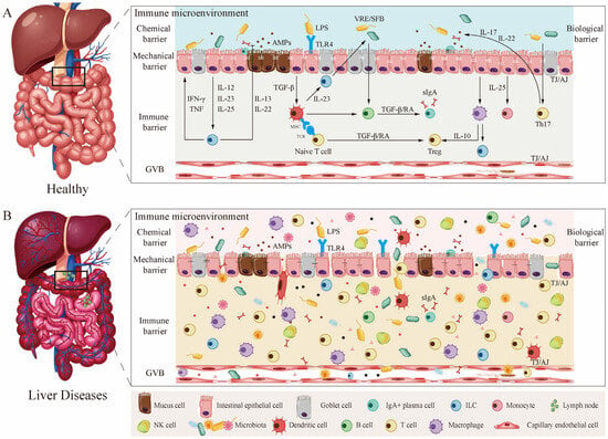
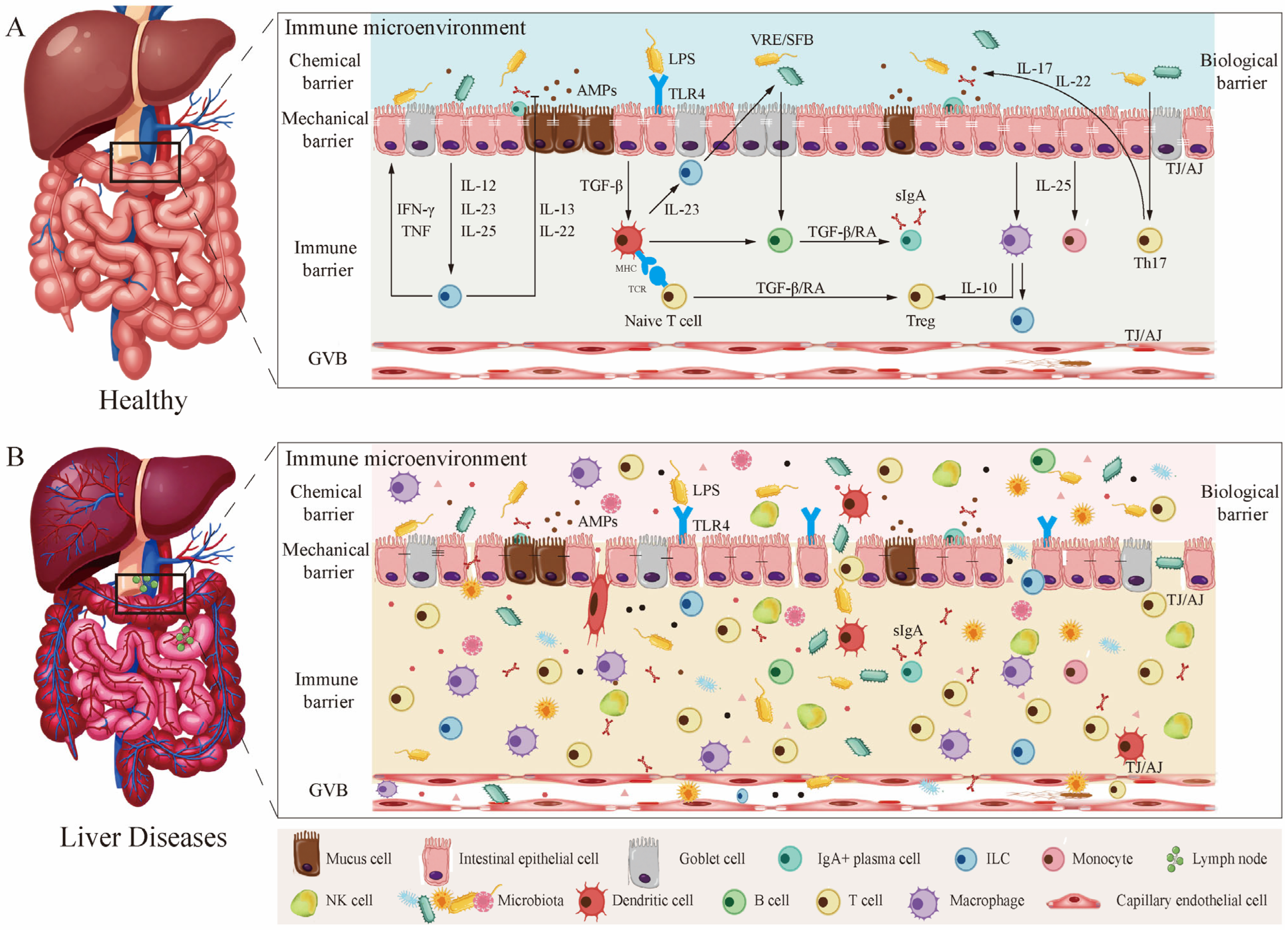
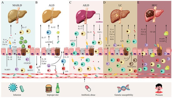
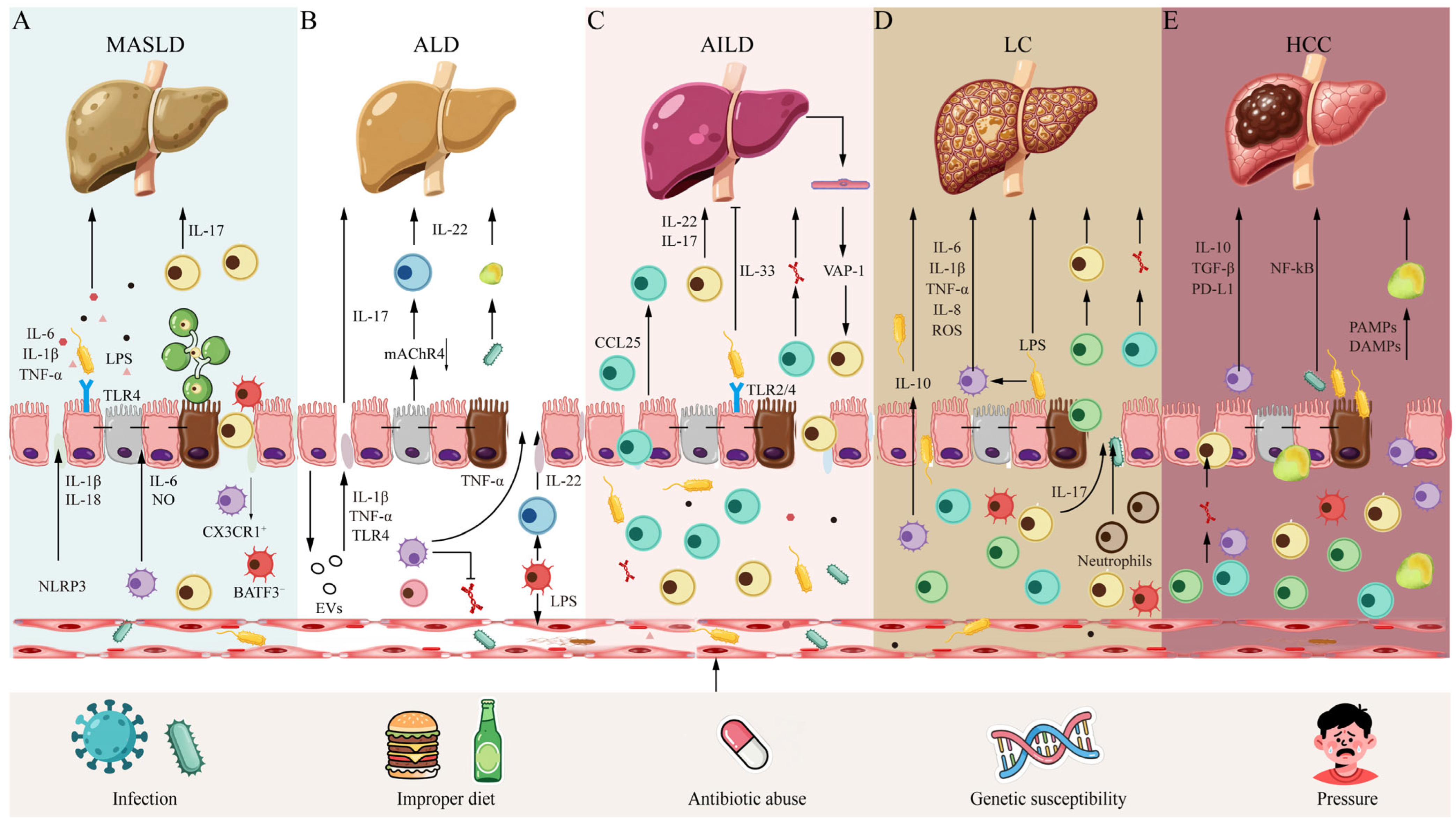
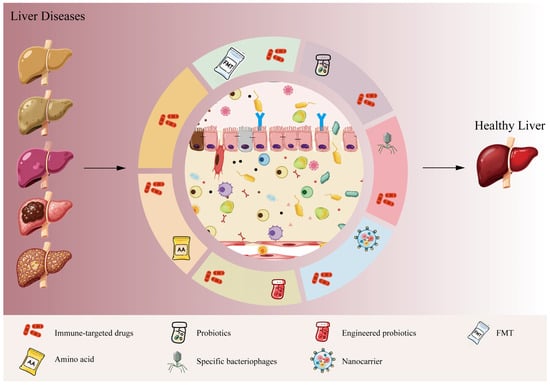
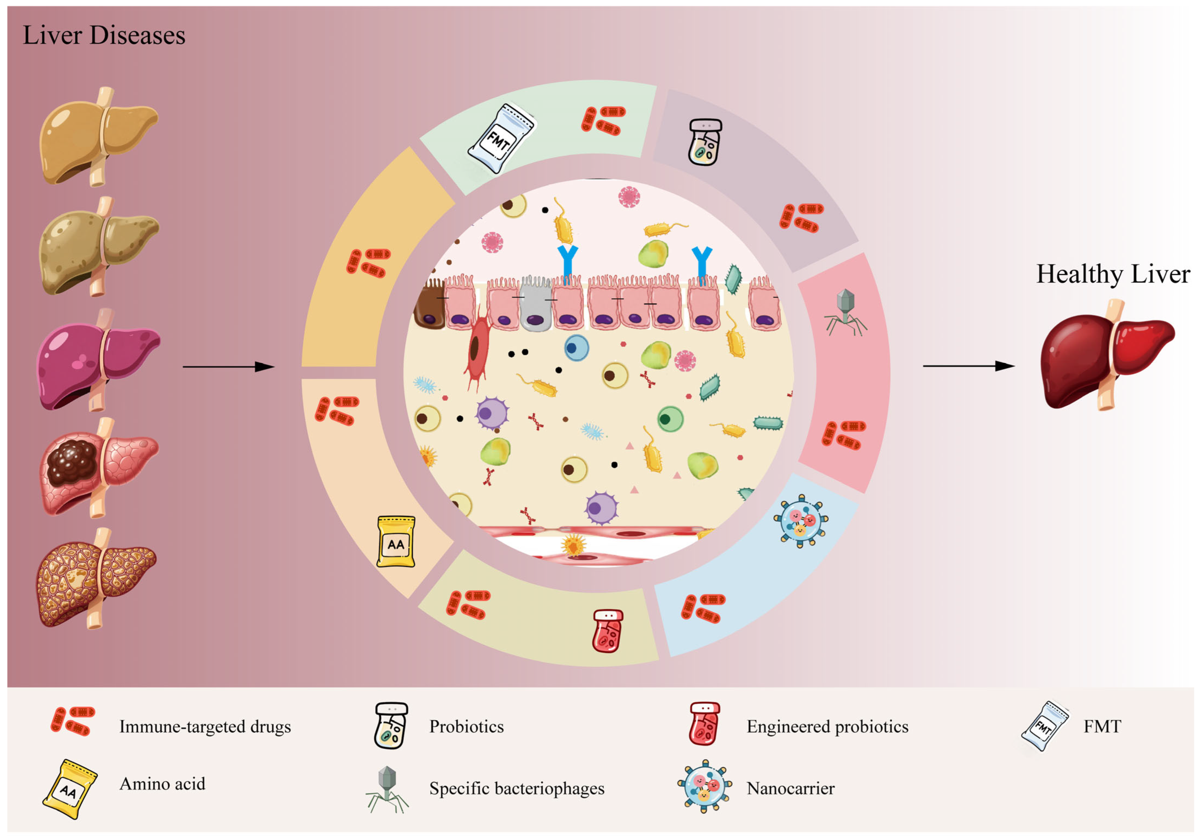

Disclaimer/Publisher’s Note: The statements, opinions and data contained in all publications are solely those of the individual author(s) and contributor(s) and not of MDPI and/or the editor(s). MDPI and/or the editor(s) disclaim responsibility for any injury to people or property resulting from any ideas, methods, instructions or products referred to in the content. |
© 2026 by the authors. Licensee MDPI, Basel, Switzerland. This article is an open access article distributed under the terms and conditions of the Creative Commons Attribution (CC BY) license.