


Abstract
Background/Objectives: Sepsis-associated gastrointestinal dysfunction comprises heterogeneous injury, motility, and feeding phenotypes. This narrative review evaluates their host and microbial metabolic correlates and asks whether metabolic dysfunction-associated steatotic liver disease (MASLD) may modify these relationships. Methods: The literature available through August 2026 was organized into gastrointestinal, systemic metabolic, microbial, and liver-metabolic domains. Human observations, interventions, and experimental mechanisms were evaluated separately. Database query formulations are provided in the manuscript appendices. Results: Small human studies link circulating citrulline and intestinal fatty-acid-binding protein to acute gastrointestinal injury, and fecal short-chain fatty acid profiles to enteral-feeding adequacy. Most lipid, ketone, and amino-acid findings derive from general sepsis cohorts without gastrointestinal endpoints. Steatotic-liver cohorts report heterogeneous systemic outcomes but do not isolate gastrointestinal effects from cardiometabolic confounding. No study identified in the reviewed literature jointly assessed MASLD status, a standardized gastrointestinal endpoint, and host or microbial metabolic profiles in the same participants. Experimental pathways provide biological plausibility, while early microbiome-directed trials do not establish routine treatment utility. Conclusions: The principal contribution is an evidence-gap map and a focused strategy for same-cohort validation. MASLD remains a proposed modifier rather than an established cause of gastrointestinal metabolic dysfunction. Prospective studies should prioritize defined gastrointestinal outcomes, serial measurements, treatment exposures, and added value beyond routine clinical assessment.
1. Introduction
Sepsis is life-threatening organ dysfunction caused by a dysregulated host response to infection [1,2]. Gastrointestinal manifestations include epithelial injury, dysmotility, and impaired enteral feeding. Hypoperfusion, inflammation, medication exposure, nutrition interruption, and altered microbial ecology can contribute, but the same bedside phenotype may arise through different processes. Sepsis-associated gastrointestinal dysfunction (SAGD) is therefore used here as an umbrella description, with acute gastrointestinal injury (AGI), feeding intolerance, and injury biomarkers retained as distinct outcomes.
Metabolic measurements may help characterize this heterogeneity. Small clinical studies associate citrulline with AGI and fecal short-chain fatty acids (SCFAs) with enteral-feeding adequacy [3,4]. Broader sepsis metabolomics identifies amino-acid and lipid abnormalities, but these signals may originate in the liver, kidney, muscle, or immune system rather than the intestine [5,6]. Establishing gastrointestinal relevance requires a measured gastrointestinal endpoint, rather than a pathway’s biological plausibility alone.
MASLD is a common chronic liver-metabolic condition defined by hepatic steatosis with at least one cardiometabolic risk factor [7,8]. Its spectrum includes uncomplicated steatosis, metabolic dysfunction-associated steatohepatitis (MASH), and varying fibrosis stages. Population estimates indicate a substantial burden, making pre-existing liver-metabolic disease relevant to ICU research [9,10]. Altered lipid handling, bile-acid circulation, and inflammatory regulation suggest possible interactions with sepsis. However, systemic associations in liver-disease cohorts do not demonstrate that MASLD modifies intestinal injury or microbial metabolism independently of obesity, diabetes, or fibrosis.
Existing reviews address sepsis dysbiosis, metabolic heterogeneity, or the MASLD gut–liver axis largely as separate subjects [11,12,13,14]. This review connects those literatures by asking which studies actually measured the proposed intersection. No human study identified in the literature reviewed through August 2026 jointly assessed MASLD status, a standardized gastrointestinal or feeding endpoint, and a host or microbial metabolic profile in the same participants. The new insight is this mismatch between mechanistic integration and clinical measurement. We use it to prioritize gastrointestinally anchored biomarkers and a feasible prospective design, retaining MASLD as a hypothesis to test.
2. Scope and Evidence Synthesis
2.1. Narrative Approach and Search Documentation
This is a narrative review, not a systematic review or meta-analysis. The literature base available through August 2026 was organized using PubMed, Web of Science Core Collection, Google Scholar, and reference lists. The available query formulations are reproduced by database in Appendix A, Table A1–Table A3. Four modules address gastrointestinal phenotypes, host metabolism, microbial metabolism, and MASLD or liver-metabolic risk. These tables document the search concepts; they are not a registered protocol or a complete historical search log. No claim of exhaustive coverage or PRISMA-based study identification is made.
2.2. Evidence Selection and Interpretation
English full texts or records with a reliable English translation were eligible. Adult human studies were prioritized when they measured a defined gastrointestinal or feeding phenotype, host or microbial metabolites, or liver-metabolic status in sepsis. Guidelines and focused reviews provide definitions and context. Experimental studies were selected for a measured metabolic or microbial pathway linked to intestinal injury, hepatic injury, or immune function. Combined steatosis–sepsis models, sepsis models without steatosis, and chronic liver-disease models are distinguished. Studies lacking a relevant clinical endpoint or experimentally supported pathway were not used to substantiate gastrointestinal or treatment claims.
Study-level comparisons consider design, population, disease definition, specimen, timing, assay, treatment context, gastrointestinal endpoint, reported estimate, and limitations. Human observational associations, randomized intervention effects, and animal or cellular perturbations remain separate evidence categories. Related reports from the same cohort are not treated as independent replication. Heterogeneous populations and outcomes were not pooled. Concentrations do not establish production or tissue flux, and a biomarker response alone does not establish clinical benefit. These interpretive rules apply throughout rather than being repeated after every pathway.
2.3. Operational Definitions of the Gastrointestinal Phenotype
SAGD denotes gastrointestinal dysfunction occurring during sepsis, rather than a validated diagnostic category. AGI is a graded clinical construct; feeding intolerance is a functional phenotype whose study-specific definition must be stated. Neither is interchangeable with an isolated injury marker, dysbiosis, or cholestasis. Table 1 summarizes these distinctions. In prospective studies, interruptions attributed to vomiting, distension, ileus, or other documented gastrointestinal causes should be separated from procedures, airway care, and planned withholding of nutrition during instability.
Table 1.
Operational definitions and limitations of gastrointestinal phenotypes used in this review.
Table 1 is used as a phenotype key for the subsequent evidence tables. A study reporting cholestasis or an isolated injury marker is therefore not counted as a study of feeding intolerance or AGI unless that endpoint was directly measured.
3. Human Clinical Evidence: Gastrointestinal Phenotypes, Metabolism, and MASLD
3.1. Gastrointestinal Injury, Feeding Tolerance, and Microbial Metabolites
The most direct clinical evidence concerns gastrointestinal phenotypes rather than MASLD. In a prospective cohort of 28 patients with sepsis, 30 with septic shock, and 10 non-infected controls, circulating citrulline was lower in sepsis than in controls and lower in septic shock than in sepsis without shock throughout follow-up. Patients with AGI grade III had lower citrulline than those with AGI grades I–II on days 7 and 10, and a higher AGI score was associated with 28-day mortality [3]. I-FABP was higher in septic shock than in sepsis on days 1 and 3, but the marker differences were not uniform across time. These findings support citrulline and I-FABP as clinically measurable correlates of gastrointestinal injury; they do not make either marker a specific measure of epithelial permeability or enterocyte flux.
Feeding tolerance provides a complementary, functional endpoint. In a 24-patient observational study of non-abdominal infection sepsis, patients were classified by whether they achieved 20 kcal/kg/day by day 3 of enteral nutrition; a total of 14 were classified as tolerant and 10 as intolerant. Fecal microbiota diversity and operational taxonomic units differed on enteral-nutrition day 1, and SCFA concentrations decreased more in the intolerance group by day 3 [4]. The threshold is a nutrition-delivery definition and should not be equated with feeding interruption caused by gastrointestinal symptoms. Nevertheless, the study links a measurable feeding trajectory to microbial and metabolite features and supports prospective studies that record both delivered nutrition and the documented reason for every interruption.
The nutrition exposure itself is a major part of the phenotype. A review of NUTRIREA-2 and NUTRIREA-3 emphasized that bowel ischemia and other gastrointestinal complications were influenced jointly by illness severity, vasopressor dose, and the amount of enteral nutrition delivered during shock [18]. Thus, SCFA or amino-acid measurements obtained during early shock cannot be interpreted without the timing and dose of enteral feeding, hemodynamic support, and the distinction between gastrointestinal intolerance and a planned reduction in nutrition.
3.2. MASLD and Systemic Sepsis Outcomes
Liver-metabolic cohorts provide systemic sepsis evidence, not direct evidence of SAGD. In a United States nationwide analysis of 21,057,911 adult sepsis admissions from 2000–2019, coded NAFLD was associated with higher in-hospital mortality after adjustment (adjusted odds ratio [aOR] 1.19, 95% confidence interval [CI] 1.07–1.32) [19]. Administrative labels do not establish imaging-confirmed steatosis or stage fibrosis. Adjustment for recorded comorbidities cannot exclude residual confounding by cardiometabolic burden, disease recognition, or care patterns.
A prospective cohort of 124 patients with community-acquired sepsis compared participants with and without MASLD. The MASLD group showed higher admission concentrations of IL-17A, IL-23, IL-33, CXCL10, and TGF-beta1 and a different day-5 cytokine trajectory; changes in TGF-beta1 and CCL11 were associated with mortality within the MASLD subgroup [20]. The study is important because it demonstrates dynamic immune divergence, but the sample size and single-cohort design do not define a treatment target.
The SepsisFAT phenotype report is a secondary analysis of a prospective parent cohort, not an independent cohort [21,22]. Among 378 adults, the mixed liver–cardiometabolic phenotype had greater odds of acute kidney injury (AKI; aOR 2.82, 95% CI 1.37–5.90) and continuous renal replacement therapy (CRRT; aOR 3.87, 95% CI 1.50–10.80) than the low-risk phenotype after adjustment for age, sex, and SOFA. These contrasts do not isolate steatosis from accompanying metabolic disease. Another administrative septic-shock study reported an AKI association, but its abstract and tabulated mortality results are internally discordant; its mortality direction is therefore not used to support the framework [23].
3.3. Liver Risk Scores in Acute Sepsis
FIB-4 combines age, aminotransferases, and platelets, all of which can influence acute sepsis prognosis independently of chronic fibrosis. A two-database study included 23,959 patients, comprising 13,983 in MIMIC-IV and 9,976 in eICU [24]. Reported mortality discrimination was AUROC 0.678 (95% CI 0.667–0.689) and 0.713 (95% CI 0.699–0.726), respectively. This is prognostic evidence, not validation of MASLD, fibrosis, or gastrointestinal dysfunction. Pre-illness records or imaging are preferable for chronic phenotyping; acute FIB-4 should remain a covariate.
Long-term susceptibility is a separate question. In a Korean cohort of more than 3.2 million adults, high baseline fatty liver index (FLI) was associated with subsequent sepsis over follow-up of up to 10 years (adjusted hazard ratio [aHR] 1.52, 95% CI 1.49–1.55, versus low FLI) [25]. FLI estimates steatosis probability from metabolic variables; this association neither diagnoses MASLD nor informs acute ICU resuscitation.
3.4. Metabolomics Studies in Sepsis
Human metabolomics studies identify reproducible themes but not a single diagnostic signature. A multicenter study integrating serum metabolomics and proteomics found widespread amino-acid dysregulation and identified pathways related to inflammatory activation, platelet degranulation, arginine biosynthesis, and phenylalanine, tyrosine, and tryptophan metabolism [5]. Other studies using machine learning have reported panels of differential metabolites with promising internal discrimination, but platform, preprocessing, patient selection, and validation practices differ substantially [6,26,27]. For serial data, dataset-stratified, time-aligned comparisons offer a way to handle heterogeneous measurements, as illustrated by secondary LC–MS analysis of exercise recovery outside sepsis [28].
Metabolomic data are therefore most useful when attached to a clinical phenotype. In SAGD, a candidate panel should be evaluated against AGI grade, gastric or post-pyloric feeding tolerance, enteral calorie delivery, stool output, intra-abdominal pressure, liver-metabolic phenotype, SOFA trajectory, and patient-centered outcomes. Repeated sampling is necessary because shock, antibiotics, renal replacement, nutrition, and vasopressors can change metabolites within hours.
3.5. Human Intervention and Cross-Species Metabolic Evidence
In a single-blind synbiotic trial in mechanically ventilated patients with sepsis, 77 patients were randomized, and 72 completed the study, with 35 synbiotic recipients and 37 controls [29]. The formulation contained Bifidobacterium breve, Lactobacillus casei, and galactooligosaccharides. Reported enteritis was 6.3% versus 27.0%, and ventilator-associated pneumonia (VAP) was 14.3% versus 48.6%; bacteremia and mortality did not differ significantly. Fecal organisms and organic acids, particularly acetate, changed alongside these outcomes. The trial did not establish metabolite mediation, a MASLD-specific benefit, or a mortality benefit.
A cross-species study compared stool from 13 participants with sepsis and 13 without sepsis using 16S rRNA sequencing and untargeted metabolomics [30]. Lower fecal L-valine was associated with greater illness severity. Fecal transfer and L-valine supplementation were then studied in mice, where supplementation attenuated intestinal injury. The human component is hypothesis-generating; experimental rescue does not establish that supplementation improves feeding tolerance or AGI in patients.
3.6. Systemic Metabolic Heterogeneity
Sepsis multi-omics identifies lipid, immune, endothelial, and organ-injury patterns that vary over time [31,32,33,34]. Lipidomic studies likewise report phospholipid, sphingolipid, and acylcarnitine disturbances across different sampling and treatment settings [35,36,37,38]. Most lack AGI or feeding endpoints. They explain systemic heterogeneity and nominate assays for testing, but do not establish a gastrointestinal metabolic signature.
Direct intestinal studies are smaller. AGI cohorts using citrulline, I-FABP, or microbial composition and an intra-abdominal infection study linking AGI to cholestasis provide complementary endpoints [3,16,17]. Composition-only studies cannot establish metabolite changes. Nutrition, antibiotics, and organ support must also be considered because they can alter both gastrointestinal outcomes and the measured metabolic pools [39,40].
3.7. Mechanistic Evidence: Combined and Separated Disease Models
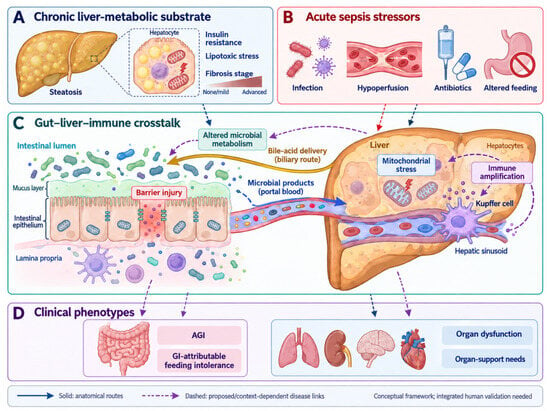
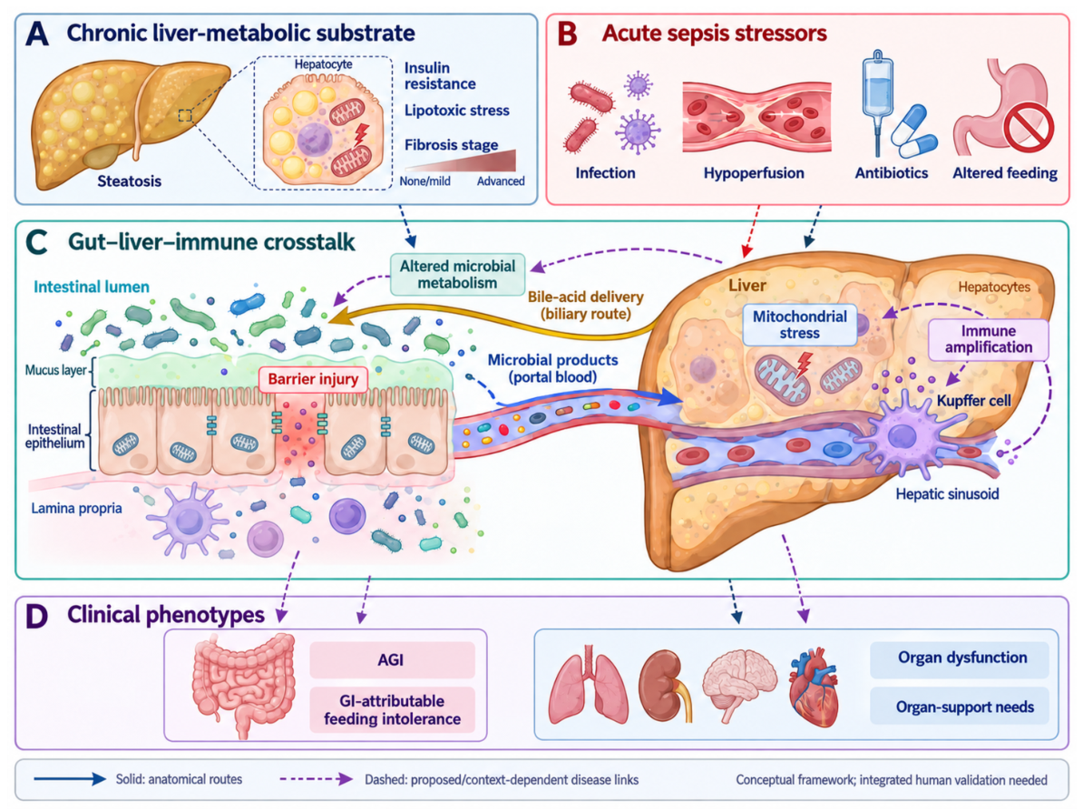
Experimental evidence has distinct scopes. TREM2-related macrophage–hepatocyte coordination was examined in combined steatosis and sepsis models [41]. Fructose-driven lipogenesis and barrier changes in chronic liver-disease models describe the pre-existing substrate without sepsis [42,43]. Microbial hyodeoxycholic acid, granisetron-related signaling, and defined microbial peptides were evaluated in sepsis models without a documented MASLD substrate [44,45,46]. Endotoxin challenge, polymicrobial infection, and pretreatment experiments are not interchangeable with treatment after clinical sepsis onset. Figure 1 is interpreted as a conceptual synthesis of these separate evidence streams.
Figure 1.
Host–microbial metabolic framework for sepsis-associated gastrointestinal dysfunction in MASLD. (A) Chronic liver-metabolic substrate, including steatosis, insulin resistance, lipotoxic stress, and variable fibrosis stage. (B) Acute sepsis stressors, including infection, hypoperfusion, antimicrobial exposure, and altered feeding. (C) Gut–liver–immune crosstalk through barrier injury, microbial products, bile-acid delivery, mitochondrial stress, and immune amplification. (D) Clinical phenotypes that should anchor metabolic measurement. Solid arrows denote anatomical routes; dashed arrows denote proposed or context-dependent disease links. Evidence status for panels (A–D) is conceptual synthesis informed by separate clinical observations and experimental studies; none is original patient evidence or an integrated validation. Abbreviations: AGI, acute gastrointestinal injury; GI, gastrointestinal; MASLD, metabolic dysfunction-associated steatotic liver disease.
3.8. Study-Level Human Evidence at the MASLD–SAGD Intersection
Table 2 reports the principal human studies individually so that population definitions, sampling, gastrointestinal endpoints, and limitations can be compared. The table is intentionally conservative: a study is not treated as direct intersection evidence unless MASLD or liver-metabolic risk, a gastrointestinal phenotype, and a metabolic measurement were assessed in the same participants.
Table 2.
Human evidence grouped by gastrointestinal relevance, followed by liver-metabolic associations. Estimates and timing are reported when directly available; missing estimates are not reconstructed.
No study in Table 2 jointly measures MASLD, a standardized gastrointestinal endpoint, and a host or microbial metabolic profile. The SepsisFAT publications represent related analyses, and FLI susceptibility data address a different clinical timescale from ICU outcomes.
Table 3 integrates the evidence by domain and makes the translational gap explicit. The table separates human endotype and outcome signals from mechanistic gut–liver pathways, then assigns each domain the validation question that must be answered next. Across domains, the recurring requirement is to connect a metabolic readout to a defined gastrointestinal phenotype and a patient-centered outcome rather than to treat pathway enrichment as evidence of clinical utility.
Table 3.
Evidence integration and the specific missing link.
4. How MASLD May Modify the Metabolic Landscape of Sepsis
4.1. MASLD Severity and Cardiometabolic Confounding
MASLD requires hepatic steatosis and at least one cardiometabolic risk factor. Uncomplicated steatosis, MASH, advanced fibrosis, and cirrhosis can differ substantially in hepatic reserve and portal physiology [7,8]. Historical NAFLD cohorts retain their original terminology when alcohol exposure, cardiometabolic criteria, or competing causes are insufficiently documented for reclassification. Administrative diagnoses, FLI, acute FIB-4, and imaging also represent different constructs, as summarized in Appendix A, Table A4.
The human evidence does not isolate a gastrointestinal effect of steatosis from obesity, insulin resistance, diabetes, or treatment differences. Adjustment for selected covariates reduces measured confounding but cannot demonstrate that MASLD independently causes the observed outcomes [19,20,22]. Acute aminotransferase elevations and thrombocytopenia further limit fibrosis-score interpretation during sepsis. Prospective studies should retain pre-illness liver information, describe cardiometabolic factors individually, and examine associations within comparable cardiometabolic and fibrosis-risk strata. Cirrhosis, alcohol-related liver disease, and MetALD require separate characterization. MASLD is therefore a proposed effect modifier, with both disease stage and residual confounding included in the hypothesis.
4.2. Hepatic Lipid Overflow, Insulin Resistance, and Mitochondrial Stress
MASLD is characterized by a mismatch between fatty-acid delivery, de novo lipogenesis, beta-oxidation, triglyceride storage, and export as very-low-density lipoprotein. Inflammation can stimulate hepatic de novo lipogenesis, while dietary fructose can promote a leaky gut, endotoxemia, and fibrogenic signaling in experimental models [42,43]. These chronic dietary and gut–liver relationships are also discussed in a processing-centered MASLD perspective focused on cognitive aging; they provide background rather than sepsis-specific treatment evidence [47].
During sepsis, catecholamines, cytokines, hypoperfusion, and changes in insulin signaling accelerate lipolysis and alter hepatic substrate selection. Mitochondrial electron transport, beta-oxidation, and ketone production may become mismatched with energy demand. Damaged mitochondria can release reactive oxygen species, mitochondrial DNA, phospholipids, and metabolites that act as damage-associated signals. Mitochondrial dysfunction is consequently both a source of energy failure and an amplifier of inflammation [48,49].
The TREM2 study provides a mechanistic example of why pre-existing steatosis may matter. In NAFLD-associated sepsis models, loss of TREM2 in liver macrophages impaired macrophage–hepatocyte metabolic coordination and reduced hepatocyte mitochondrial support; restoring TREM2 improved the experimental sepsis phenotype [41]. The work does not establish a TREM2-directed therapy in humans, but it supports a model in which chronic lipid overload changes the hepatic immune-metabolic response to acute infection.
4.3. Sinusoidal, Cholestatic, and Synthetic-Function Vulnerability
The liver sinusoid is a metabolic and immunological interface rather than a passive vascular bed. In MASLD, sinusoidal endothelial cells undergo capillarization, altered vasoregulation, oxidative stress, and inflammatory activation. These changes can increase intrahepatic resistance and impair exchange between blood, hepatocytes, and Kupffer cells. A recent review positioned liver sinusoidal endothelial cells as a link between MASLD pathogenesis and sepsis susceptibility, while emphasizing that the proposed pathways still require clinical validation [50,51].
Sepsis-associated liver injury can manifest as hypoxic hepatitis, cholestasis, or progressive bile-duct injury. Inflammatory signaling, oxidative stress, cell death, altered bile transport, and gut-derived pathogen-associated molecular patterns interact rather than occurring in isolation [52,53,54]. Bilirubin and aminotransferases remain useful clinical signals, but they capture only part of hepatic function. Albumin, INR, glucose handling, lactate clearance, ammonia, bile-acid composition, and dynamic support requirements may add complementary information while remaining vulnerable to critical-illness confounding.
Figure 1 is a conceptual map. Its anatomical routes should be distinguished from proposed disease interactions; none establishes an integrated human MASLD–SAGD mechanism.
5. Host Metabolism in Sepsis-Associated Gastrointestinal Dysfunction
5.1. Glycolysis, the TCA Cycle, and Redox Capacity
Most evidence in this section comes from general sepsis or experimental systems rather than studies with measured AGI or feeding intolerance. Stress glycolysis, altered pyruvate oxidation, and disrupted cellular redox balance may accompany sepsis. Circulating lactate reflects production, adrenergic stimulation, and clearance; it does not localize intestinal hypoxia. Experimental links between epithelial ATP depletion and barrier dysfunction are biologically relevant, but lactate is a clinical comparator rather than a gastrointestinally specific marker.
TCA intermediates also participate in immune signaling. Their effects depend on cell type, intracellular versus extracellular location, and the phase of inflammation. A compensatory immune-cell response and a high circulating concentration caused by tissue injury or reduced clearance need not indicate the same process. Such compartment dependence is a reason to test pathways against defined outcomes rather than infer intervention direction from a plasma value alone [48,49].
Cellular redox balance involves NAD+/NADH, NADPH, glutathione, thioredoxin, and antioxidant defense against mitochondrial reactive oxygen species. Mitochondrial injury can couple energy disturbance to inflammatory signaling in experimental sepsis [48,49]. Compartment-specific redox interpretation is also emphasized in cardiometabolic gut–mitochondrial research, where the integrated injury pathway remains incompletely validated [55]. A blood redox measurement cannot, by itself, quantify intestinal antioxidant capacity or demonstrate epithelial recovery.
5.2. Lipid Flux, Acylcarnitines, and Ketone Handling
General sepsis lipidomics suggests altered fatty-acid oxidation and membrane remodeling, reflected by acylcarnitines, free fatty acids, and phospholipids [35,37]. These circulating pools combine hepatic, muscular, renal, and immune contributions. A modifying effect of pre-existing steatosis is plausible but has not been established against standardized AGI or feeding endpoints.
Beta-hydroxybutyrate reflects hepatic ketogenesis, peripheral utilization, nutrition, insulin exposure, and acid–base status. Early shock with interrupted feeding differs from later recovery with resumed nutrition. A rising or falling ketone concentration can therefore have different meanings across phases and cannot be assigned a uniformly protective or harmful role. This context dependence also limits translation of metabolite administration in controlled animal models to heterogeneous patients [48,49].
5.3. Amino-Acid and Nitrogen Metabolism
Untargeted metabolomics and proteomics in human sepsis consistently identify disturbances in amino-acid pathways, including phenylalanine, tyrosine, tryptophan, arginine, proline, glycine, serine, and threonine metabolism [5,6,26]. These pathways connect immune-cell activation, nitric-oxide availability, hepatic urea-cycle function, muscle proteolysis, and tissue repair. Citrulline is attractive as a marker of enterocyte mass, but kidney function, nutrition, and systemic inflammation influence its concentration. Glutamine and arginine are similarly affected by organ failure and exogenous supplementation.
Tryptophan is partitioned between host kynurenine production, serotonin synthesis, and microbial indole formation. The kynurenine pathway is induced by inflammatory interferon signaling and may contribute to immune tolerance, whereas microbial indoles can activate aryl hydrocarbon receptor pathways that support epithelial and immune homeostasis [56]. In MASLD, altered bile-acid signaling, intestinal permeability, and diet can change the substrate environment in which these pathways operate. A plasma kynurenine/tryptophan ratio should therefore be interpreted alongside liver function, renal clearance, antibiotic exposure, and stool or dietary data.
6. Microbial Metabolites at the Gut–Liver Interface
6.1. Microbial Composition, Functional Potential, and Activity
Microbial composition, dysbiosis, and microbial metabolic activity describe different observations. Relative abundance from 16S rRNA sequencing characterizes community composition; a taxon can change in relative abundance without a corresponding change in absolute abundance or metabolite production. Predicted functions inferred from 16S data are hypotheses rather than measurements of microbial genes or metabolism [57]. Shotgun metagenomics measures gene content and functional potential, metatranscriptomics measures expressed microbial transcripts, and targeted metabolomics measures the resulting chemical pool, which also reflects host processing and clearance [58]. Matched sampling can test whether an inferred pathway is supported across these levels; agreement alone does not establish metabolic flux.
Human microbiome studies with AGI outcomes therefore support compositional associations when metabolites were not measured [16,17]. A study combining 16S sequencing, metabolomics, and metaproteomics linked microbial features to bilirubin, while fecal transfer to mice provided a separate experimental test [59]. Future studies should align these assays with AGI and feeding outcomes in the same patients, retaining the distinction between human association and experimental perturbation.
6.2. Short-Chain Fatty Acids
Acetate, propionate, and butyrate connect dietary fermentation with epithelial and immune metabolism. Experimental studies support roles for butyrate in colonocyte energy supply and for SCFA signaling in immune regulation [60]. These mechanisms motivate clinical investigation but do not make a stool concentration a measure of epithelial exposure. Stable-isotope work in healthy volunteers demonstrates extensive absorption and metabolism before colonic SCFAs reach the systemic circulation; the quantitative findings should not be transferred directly to patients in shock [61].
Fecal SCFA concentration reflects production, absorption, transit, stool water, nutritional substrate, and sampling conditions. In the small enteral-feeding study, reduced SCFAs accompanied poor nutrition delivery; their temporal association cannot distinguish a contributing deficiency from an effect of illness severity or reduced substrate [4]. The synbiotic trial combined changes in organisms, organic acids, and selected outcomes, without isolating SCFAs as the mediator [29]. Studies should therefore report delivered nutrition, antibiotic exposure, stool consistency and timing, and interpret paired plasma and stool results by compartment. These data do not establish a fecal SCFA threshold for supplementation.
6.3. Bile Acids, FXR/TGR5, and Portal Signaling
The liver synthesizes primary bile acids, and intestinal microbes transform them into secondary bile acids that signal through farnesoid X receptor (FXR), Takeda G-protein-coupled receptor 5 (TGR5), and other pathways. MASLD can change bile-acid synthesis, conjugation, and portal delivery, while sepsis-associated cholestasis and ileal dysfunction can rapidly change the circulating pool. The resulting signals may influence antimicrobial peptide expression, epithelial repair, hepatic inflammation, vascular tone, and glucose and lipid metabolism [13,14].
Bile acids illustrate compartment dependence. Reduced intestinal delivery during cholestasis can coexist with increased circulating concentrations, so plasma accumulation does not imply greater epithelial exposure. Molecular species also differ in receptor activity and toxicity. Experimental protection by a defined microbial derivative does not establish that increasing the total bile-acid pool is beneficial [45]. Targeted plasma and stool species, interpreted with cholestasis and nutrition, are therefore more informative than a single total value [13,14,51].
6.4. Indoles, Microbial Peptides, and Endotoxin
In animal and cellular experiments, microbial indoles and peptides can modify epithelial or innate immune responses. Granisetron-related microbial signaling and an Akkermansia-derived tripeptide protected against selected sepsis outcomes in model systems [44,46]. These are experimental findings without a demonstrated MASLD–SAGD treatment effect in humans.
Metabolic endotoxemia provides a conceptual bridge between MASLD and sepsis. Altered intestinal permeability permits lipopolysaccharide and other microbial products to enter the portal and systemic circulation; chronic exposure can sustain low-grade inflammation, while acute sepsis can greatly amplify the response. Experimental and review evidence links dietary factors, endotoxin handling, and metabolic inflammation, but the contribution of circulating endotoxin varies with assay platform and timing [43,62]. Future studies should pair endotoxin or microbial-product assays with barrier markers, liver-metabolic phenotyping, and organ outcomes.
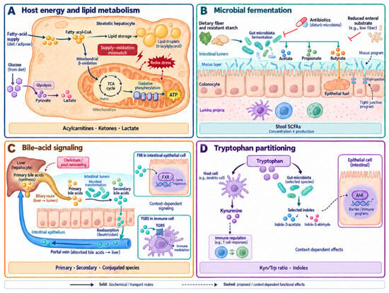
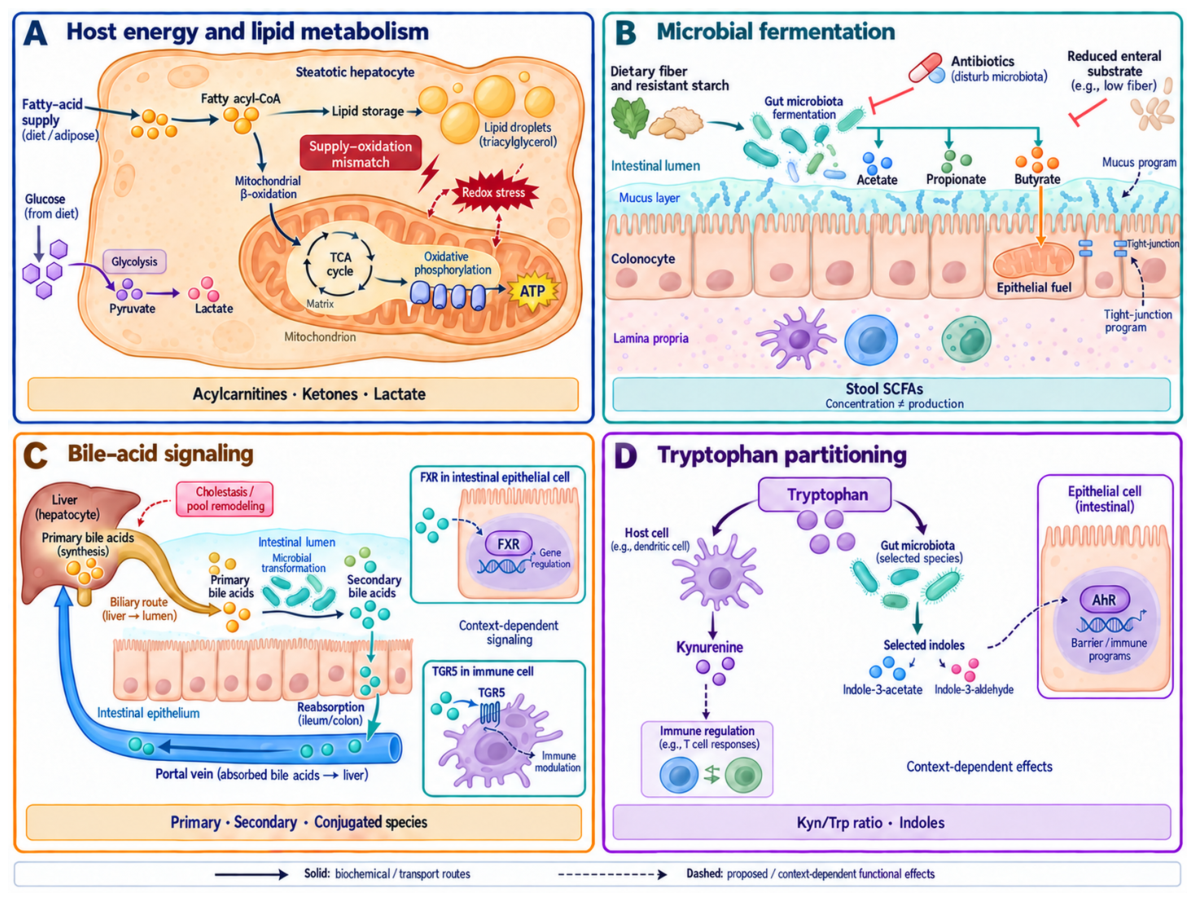
Figure 2 summarizes complementary pathways. The host, fermentation, bile-acid, and tryptophan panels draw on different study populations and experimental systems; they have not been jointly validated in MASLD–SAGD patients.
Figure 2.
Mechanistic metabolic modules at the gut–liver interface in sepsis with MASLD. (A) Host energy and lipid metabolism, including fatty-acid supply, lipid storage, mitochondrial beta-oxidation, glycolysis, lactate, and redox stress. (B) Microbial fermentation of fiber and resistant starch to acetate, propionate, and butyrate, with a context-dependent link to epithelial fuel and barrier programs; stool concentration is not equivalent to production. (C) Bile-acid signaling, distinguishing hepatic synthesis, biliary delivery, microbial transformation, intestinal reabsorption, portal return, and FXR/TGR5-associated signaling. (D) Tryptophan partitioning between host kynurenine metabolism and microbial indole pathways, with proposed AhR-associated epithelial and immune effects. Solid arrows denote biochemical or transport routes; dashed arrows denote proposed or context-dependent functional effects. Evidence status for panels (A–D) is mixed experimental and indirect clinical support, not joint human MASLD–SAGD validation. None is a validated treatment-selection test. Abbreviations: AhR, aryl hydrocarbon receptor; FXR, farnesoid X receptor; MASLD, metabolic dysfunction-associated steatotic liver disease; SCFA, short-chain fatty acid; TGR5, Takeda G-protein-coupled receptor 5.
7. Candidate Biomarkers and Measurement Strategy
Table 4 orders measurements by clinical decision sequence. Routine physiology and gastrointestinal endpoints come first; targeted injury and metabolic assays follow; exploratory microbial measurements form a separate tier. The tier describes research priority and feasibility, not regulatory approval or a validated combined panel.
Table 4.
Measurement priority in clinical decision order. Tiers denote research priority, not validated clinical recommendations.
Tier 1 anchors the phenotype, Tier 2 tests selected host pathways, and Tier 3 explores microbial contributions. The clinical prioritization below distinguishes epithelial injury, microbial metabolism, and systemic disturbance.
7.1. Pre-Analytical Standardization
Comparable measurements require a defined clinical time origin and consistent specimen handling. Nonsepsis chronometabolomic frameworks emphasize clock time and the timing of meals and medications; these complement time since infection or vasopressor initiation, but sparse ICU sampling cannot establish endogenous circadian rhythms [63]. Assay reporting should specify fasting or feeding state, specimen matrix, anticoagulant, processing delay, storage temperature, internal standards, batch correction, missing-value handling, and assay limits of detection. Stool measurements should report water content or a validated normalization strategy. Plasma measurements should report hemolysis, renal replacement, liver support, and medication exposures.
7.2. Clinical Prioritization and Panel Validation
Citrulline and I-FABP have the most direct human AGI relevance among the candidates discussed here, although the supporting cohorts are small and do not establish clinical cutoffs [3]. Citrulline is a metabolite related to enterocyte function; I-FABP is an injury-associated protein measured with a separate assay. Fecal SCFAs provide complementary evidence about microbial fermentation and feeding exposure but require stool collection and targeted analysis [4]. Bile acids, acylcarnitines, ketones, and broader amino-acid panels remain less gastrointestinally specific, while indoles and fecal L-valine are exploratory. This prioritization concerns the present evidence, rather than a recommended bedside panel.
A feasible first comparison would add citrulline and I-FABP to a prespecified clinical model containing SOFA, lactate, routine liver and kidney tests, and nutrition delivery. A stool-metabolite component could then be evaluated in participants with available samples, with missingness reported. Validation should assess calibration, incremental discrimination, and clinical utility in an independent cohort. A combined panel is useful only if it adds information beyond these routine measures; treatment selection additionally requires a biomarker-by-treatment interaction in a trial.
8. Clinical Translation
8.1. Standard Care Remains the Metabolic Context
Source control, appropriate antimicrobials, hemodynamic support, and indicated organ support remain the foundation of care [64]. Metabolic testing should support research phenotyping without delaying treatment. Improving lactate or bilirubin alone does not establish gastrointestinal recovery; cholestatic enzymes or bile-acid abnormalities may persist after bilirubin falls.
Nutrition is a metabolic intervention as well as a calorie prescription. The ESPEN guideline recommends individualized timing and route of nutrition in critical illness, with repeated assessment of tolerance and contraindications [65,66]. Nutrition should be adjusted to illness phase, hemodynamic stability, organ function, and refeeding risk. A MASLD-specific enteral formula has not been validated in sepsis.
8.2. Primary Questions for Prospective Research
An initial cohort should address three questions. First, is documented pre-illness MASLD associated with AGI or GI-attributable feeding intolerance after cardiometabolic factors are considered? Second, do serial citrulline, I-FABP, and fecal SCFA measurements precede or accompany changes in these endpoints? Third, does adding these measurements improve prediction beyond SOFA, lactate, routine liver and kidney tests, and delivered nutrition? These are questions for testing; the proposed framework is not a clinical algorithm.
8.3. A Feasible Prospective Study
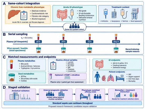
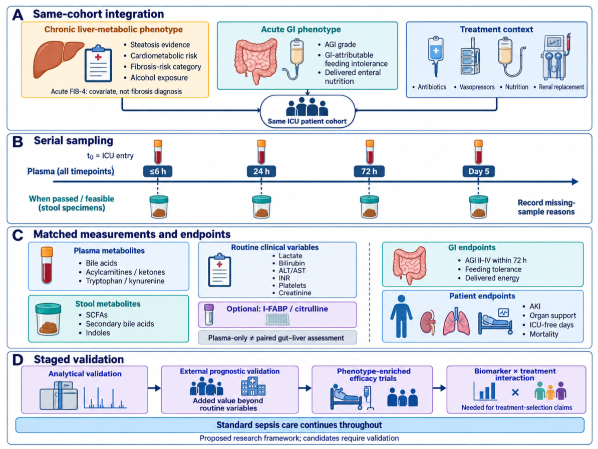
Adults with sepsis or septic shock could be enrolled before or shortly after enteral nutrition begins. Liver classification would use pre-illness records or imaging, cardiometabolic criteria, alcohol exposure, and fibrosis risk; acute FIB-4 would remain a covariate. The proposed primary endpoint is AGI grade II–IV within 72 h, with GI-attributable feeding intolerance and delivered energy as secondary endpoints. Blood could be collected within 6 h of ICU entry, at 24 h, 72 h, and day 5. Routine tests, citrulline, and I-FABP would form the initial panel. Stool SCFAs would be measured when a sample is passed within these windows; bile acids, indoles, and wider omics could be nested exploratory measurements.
Essential metadata include antibiotic timing and spectrum, vasopressors, renal replacement, major metabolic medications, opioids, nutrition prescribed and delivered, stool consistency, and reasons for feeding interruption. Treatment and nutrition exposures should be recorded before each sample. Stool absence should be coded by reason, including ileus, constipation, death, or discharge. Analyses should distinguish the full blood-sampled cohort from the paired-sample subset, assess selection and missing-not-at-random scenarios, and avoid classifying an absent stool sample as a normal result. Sample size should follow the primary endpoint and planned adjustment, with patient-level internal validation and an independent validation cohort before clinical use.
Figure 3 presents the same-cohort integration concept and the distinction between routine variables, optional metabolic measurements, and subsequent validation. Its time points are illustrative research windows rather than mandated clinical sampling.
Figure 3.
Proposed same-cohort integration and staged clinical validation framework. (A) Chronic liver-metabolic phenotype, acute gastrointestinal phenotype, and treatment context converge in one ICU patient cohort. Acute FIB-4 is treated as a covariate rather than a fibrosis diagnosis. (B) Serial plasma sampling at the proposed time points, with stool sampling only when passed or feasible and missing-sample reasons recorded. (C) Matched plasma and stool measurements are separated from routine clinical variables, optional enterocyte-injury assays, GI endpoints, and patient-centered outcomes; plasma-only measurement is not equivalent to a paired gut–liver assessment. (D) Validation progresses from analytical validation to external prognostic validation, phenotype-enriched efficacy trials, and biomarker-by-treatment interaction, while standard sepsis care continues throughout. This is a proposed research framework rather than a clinical decision tree. Abbreviations: AGI, acute gastrointestinal injury; AKI, acute kidney injury; FIB-4, fibrosis-4 index; GI, gastrointestinal; ICU, intensive care unit; I-FABP, intestinal fatty-acid-binding protein; SCFA, short-chain fatty acid.
8.4. Interventions and Safety
Microbiome-directed treatment remains a research strategy for SAGD. The small synbiotic trial reported selected benefits but neither a mortality benefit nor a MASLD-specific effect [29]. In the larger adult PROSPECT trial, Lactobacillus rhamnosus GG did not reduce VAP, and organism-associated adverse events were more frequent with probiotics [67]. Differences in product and population prevent direct equivalence between these trials. The 2026 Surviving Sepsis Campaign suggests against probiotics in adults with sepsis or septic shock, a conditional recommendation based on very low-certainty evidence [64].
Trials in patients with barrier injury or immune dysregulation should specify product quality, contraindications, concomitant antibiotics, adverse-event definitions, and organism-specific bloodstream surveillance. Nutritional substrates, postbiotics, bile-acid signaling agents, or amino-acid supplementation likewise require formulation-specific safety and patient-centered endpoints. Human fecal L-valine associations and mouse supplementation experiments are insufficient to recommend supplementation in patients [30]. Individualized nutrition remains guided by standard critical-care recommendations [65,66].
8.5. Endpoint Hierarchy for Translational Trials
A staged endpoint hierarchy can reduce the risk of declaring success from a biomarker change alone. First, a mechanistic study should demonstrate assay stability, temporal reproducibility, and an association with a prespecified phenotype such as AGI grade, enteral intolerance, cholestasis, or liver-metabolic risk. Second, a prognostic study should quantify incremental discrimination and calibration beyond SOFA, lactate, bilirubin, platelet count, and established nutrition variables. Third, an efficacy trial should assess gastrointestinal or patient-centered benefit and safety, including delivered nutrition and organ-support duration. A treatment-selection claim additionally requires a prespecified biomarker-by-treatment interaction. A metabolite change without improved patient-centered outcomes is a pharmacodynamic observation and does not, by itself, justify clinical targeting.
For MASLD analyses, liver stage and individual cardiometabolic variables should be prespecified rather than inferred from acute biochemical scores. The phenotype and exposure definitions should remain consistent across discovery and validation cohorts.
Table 5 summarizes the distinct steps from measurement to clinical utility. It avoids treating an observational association as a treatment-selection test.
Table 5.
Distinct validation questions for clinical translation.
Progression between these stages requires new evidence. External prognostic validation does not substitute for a trial demonstrating benefit or a biomarker-by-treatment interaction.
9. Limitations and Research Priorities
This narrative synthesis is selective and does not estimate the completeness of the literature. Database query tables improve transparency but do not constitute a systematic search record or formal risk-of-bias assessment. The human gastrointestinal evidence remains small, heterogeneous, and largely non-MASLD-specific. Administrative coding, acute risk scores, mixed liver stages, and cardiometabolic confounding limit interpretation of systemic associations. Related SepsisFAT reports are not independent replications.
The central research priority is to measure chronic liver-metabolic status, a standardized gastrointestinal phenotype, and serial metabolic profiles in the same participants. The focused design in Section 8.3 specifies essential measurements, exposure timing, stool missingness, and validation. Larger exploratory panels should be added only when they answer a defined question; they should not displace reliable endpoint ascertainment or the basic clinical comparator.
10. Conclusions
Human studies associate selected metabolic readouts with AGI or enteral-feeding adequacy, but most sepsis metabolomics findings are systemic rather than gastrointestinally specific. Experimental microbial pathways provide hypotheses, and limited intervention evidence does not establish routine microbiome-directed treatment. MASLD may modify these relationships, yet its independent gastrointestinal contribution remains unproven. The review’s contribution is an explicit evidence-gap map and a practical same-cohort strategy to test that hypothesis, with clinical value assessed beyond routine sepsis, liver, kidney, and nutrition measures.
Author Contributions
Conceptualization, Y.H., X.C., H.C. (Huiling Cai), H.C. (Hairong Cai) and Z.H.; methodology, Y.H., X.C. and P.W.; literature curation, Y.Z., F.H. and D.C.; evidence interpretation, Y.H., X.C., H.C. (Huiling Cai), H.C. (Hairong Cai) and Z.H.; writing—original draft preparation, Y.H. and X.C.; writing—review and editing, P.W., Y.Z., F.H., D.C., H.C. (Huiling Cai), H.C. (Hairong Cai) and Z.H.; visualization, D.C.; supervision, H.C. (Huiling Cai), H.C. (Hairong Cai) and Z.H.; project administration, H.C. (Hairong Cai) and Z.H. All authors have read and agreed to the published version of the manuscript.
Funding
This research received no external funding.
Institutional Review Board Statement
Not applicable.
Informed Consent Statement
Not applicable.
Data Availability Statement
No new participant-level data were generated. Database query formulations are included in Appendix A, Table A1–Table A3, and study-level clinical evidence is summarized in Table 2.
Acknowledgments
The authors acknowledge the investigators and participants in the studies synthesized in this review. During the preparation of this manuscript, the authors used ChatGPT Sol 5.6 (OpenAI) solely for language polishing. The authors reviewed and edited the output and take full responsibility for the content of this publication.
Conflicts of Interest
The authors declare no conflicts of interest.
Appendix A. Database Query Tables and Liver-Metabolic Definitions
Table A1–Table A3 reproduce the query formulations retained with the manuscript materials, grouped by database and evidence module. They document the concepts used for this narrative review through August 2026, rather than a complete historical execution log. The full queries are shown without inferred record counts or a systematic-review flow diagram. Table A4 distinguishes disease definitions from surrogate scores.
Table A1.
PubMed query formulations.
Table A2.
Web of Science Core Collection query formulations.
Table A3.
Google Scholar query formulations.
Table A4.
Liver-metabolic constructs and interpretation, based on the multisociety nomenclature and MASLD guideline [7,8].
References
- Singer, M.; Deutschman, C.S.; Seymour, C.W.; Shankar-Hari, M.; Annane, D.; Bauer, M.; Bellomo, R.; Bernard, G.R.; Chiche, J.D.; Coopersmith, C.M.; et al. The Third International Consensus Definitions for Sepsis and Septic Shock (Sepsis-3). JAMA 2016, 315, 801–810. [Google Scholar] [CrossRef] [Scilit]
- Rudd, K.E.; Johnson, S.C.; Agesa, K.M.; Shackelford, K.A.; Tsoi, D.; Kievlan, D.R.; Colombara, D.V.; Ikuta, K.S.; Kissoon, N.; Finfer, S.; et al. Global, regional, and national sepsis incidence and mortality, 1990–2017: Analysis for the Global Burden of Disease Study. Lancet 2020, 395, 200–211. [Google Scholar] [CrossRef] [Scilit]
- Tyszko, M.; Lemanska-Perek, A.; Smiechowicz, J.; Tomaszewska, P.; Biecek, P.; Gozdzik, W.; Adamik, B. Citrulline, Intestinal Fatty Acid-Binding Protein and the Acute Gastrointestinal Injury Score as Predictors of Gastrointestinal Failure in Patients with Sepsis and Septic Shock. Nutrients 2023, 15, 2100. [Google Scholar] [CrossRef] [Scilit]
- Xu, W.; Zhong, M.; Pan, T.; Qu, H.; Chen, E. Gut Microbiota and Enteral Nutrition Tolerance in Non-Abdominal Infection Septic ICU Patients: An Observational Study. Nutrients 2022, 14, 5342. [Google Scholar] [CrossRef] [Scilit]
- Chen, Q.; Liang, X.; Wu, T.; Jiang, J.; Jiang, Y.; Zhang, S.; Ruan, Y.; Zhang, H.; Zhang, C.; Chen, P.; et al. Integrative analysis of metabolomics and proteomics reveals amino acid metabolism disorder in sepsis. J. Transl. Med. 2022, 20, 123, Correction in J. Transl. Med. 2022, 20, 366. [Google Scholar] [CrossRef] [Scilit]
- She, H.; Du, Y.; Du, Y.; Tan, L.; Yang, S.; Luo, X.; Li, Q.; Xiang, X.; Lu, H.; Hu, Y.; et al. Metabolomics and machine learning approaches for diagnostic and prognostic biomarkers screening in sepsis. BMC Anesthesiol. 2023, 23, 367. [Google Scholar] [CrossRef] [Scilit]
- Rinella, M.E.; Lazarus, J.V.; Ratziu, V.; Francque, S.M.; Sanyal, A.J.; Kanwal, F.; Romero, D.; Abdelmalek, M.F.; Anstee, Q.M.; Arab, J.P.; et al. A multisociety Delphi consensus statement on new fatty liver disease nomenclature. J. Hepatol. 2023, 79, 1542–1556. [Google Scholar] [CrossRef]
- Tacke, F.; Horn, P.; Wai-Sun Wong, V.; Ratziu, V.; Bugianesi, E.; Francque, S.; Zelber-Sagi, S.; Valenti, L.; Roden, M.; Schick, F.; et al. EASL–EASD–EASO Clinical Practice Guidelines on the management of metabolic dysfunction-associated steatotic liver disease (MASLD). J. Hepatol. 2024, 81, 492–542. [Google Scholar] [CrossRef] [Scilit]
- Lee, B.P.; Dodge, J.L.; Terrault, N.A. National prevalence estimates for steatotic liver disease and subclassifications using consensus nomenclature. Hepatology 2024, 79, 666–673. [Google Scholar] [CrossRef] [Scilit]
- Kalligeros, M.; Vassilopoulos, A.; Vassilopoulos, S.; Victor, D.W.; Mylonakis, E.; Noureddin, M. Prevalence of Steatotic Liver Disease (MASLD, MetALD, and ALD) in the United States: NHANES 2017–2020. Clin. Gastroenterol. Hepatol. 2024, 22, 1330–1332.e4. [Google Scholar] [CrossRef] [Scilit]
- Piccioni, A.; Spagnuolo, F.; Candelli, M.; Voza, A.; Covino, M.; Gasbarrini, A.; Franceschi, F. The Gut Microbiome in Sepsis: From Dysbiosis to Personalized Therapy. J. Clin. Med. 2024, 13, 6082. [Google Scholar] [CrossRef] [Scilit]
- Klingensmith, N.J.; Leonard, J.M.; Martinez-Quinones, P.; Chang, C.W.; Nagpal, R.; Efron, P.A.; Ostermann, M.; Cresci, G.A.; Keskey, R.C.; Alverdy, J.C.; et al. The Microbiome in Critical Illness. Crit. Care Med. 2026, 54, 2956–2968. [Google Scholar] [CrossRef] [Scilit]
- Vallianou, N.G.; Kounatidis, D.; Psallida, S.; Vythoulkas-Biotis, N.; Adamou, A.; Zachariadou, T.; Kargioti, S.; Karampela, I.; Dalamaga, M. NAFLD/MASLD and the Gut–Liver Axis: From Pathogenesis to Treatment Options. Metabolites 2024, 14, 366. [Google Scholar] [CrossRef] [Scilit]
- Cui, C.; Gao, S.; Shi, J.; Wang, K. Gut-Liver Axis: The Role of Intestinal Microbiota and Their Metabolites in the Progression of Metabolic Dysfunction-Associated Steatotic Liver Disease. Gut Liver 2025, 19, 479–507. [Google Scholar] [CrossRef] [Scilit]
- Reintam Blaser, A.; Malbrain, M.L.N.G.; Starkopf, J.; Fruhwald, S.; Jakob, S.M.; De Waele, J.; Braun, J.P.; Poeze, M.; Spies, C. Gastrointestinal function in intensive care patients: Terminology, definitions and management. Recommendations of the ESICM Working Group on Abdominal Problems. Intensive Care Med. 2012, 38, 384–394. [Google Scholar] [CrossRef] [Scilit]
- Zhang, B.; Chen, X.; He, C.; Su, T.; Cao, K.; Li, X.; Duan, J.; Chen, M.; Zhu, Z.; Yu, W. Acute gastrointestinal injury and altered gut microbiota are related to sepsis-induced cholestasis in patients with intra-abdominal infection: A retrospective and prospective observational study. Front. Med. 2023, 10, 1144786, Correction in Front. Med. 2023, 10, 1280907. [Google Scholar] [CrossRef] [Scilit]
- Zuo, Z.; Pei, L.; Liu, T.; Liu, X.; Chen, Y.; Hu, Z. Investigation of Gut Microbiota Disorders in Sepsis and Sepsis Complicated with Acute Gastrointestinal Injury Based on 16S rRNA Genes Illumina Sequencing. Infect. Drug Resist. 2023, 16, 7389–7403. [Google Scholar] [CrossRef] [Scilit]
- Patel, J.J.; Lopez-Delgado, J.C.; Stoppe, C.; McClave, S.A. Enteral nutrition in septic shock: A call for a paradigm shift. Curr. Opin. Crit. Care 2024, 30, 165–171. [Google Scholar] [CrossRef] [Scilit]
- Lyu, X.; Liu, B.; Li, Y.; Wang, Y.; Miskovsky, J.; Gaitanis, M.; Promrat, K.; Wu, W.C. Impact of Non-Alcoholic Fatty Liver Disease on Sepsis Inpatient Outcomes: A Nationwide Sample Analysis (2000–2019). J. Clin. Med. 2024, 13, 5737. [Google Scholar] [CrossRef] [Scilit]
- Vrsaljko, N.; Radmanic Matotek, L.; Zidovec-Lepej, S.; Vince, A.; Papic, N. The Impact of Steatotic Liver Disease on Cytokine and Chemokine Kinetics During Sepsis. Int. J. Mol. Sci. 2025, 26, 2226. [Google Scholar] [CrossRef] [Scilit]
- Krznaric, J.; Papic, N.; Vrsaljko, N.; Gjurasin, B.; Kutlesa, M.; Vince, A. Steatotic Liver Disease and Sepsis Outcomes—A Prospective Cohort Study (SepsisFAT). J. Clin. Med. 2024, 13, 798. [Google Scholar] [CrossRef] [Scilit]
- Šamadan Marković, L.; Panić, H.; Krznarić, J.; Gjurašin, B.; Papić, N. Liver–Metabolic Phenotypes and Renal Vulnerability in Community-Acquired Sepsis: Insights from the SepsisFAT Cohort. Metabolites 2026, 16, 468. [Google Scholar] [CrossRef] [Scilit]
- Nguefang Tchoukeu, G.L.; Boateng, S.; Issaka, Y.; Eze, E.; Al-Ajlouni, Y.A.; Ameyaw, P.A.; Landa, E.L.; Verma, A.; Kaur, A.; Mekontso, J.G.K.; et al. Metabolic dysfunction-associated steatotic liver disease increases the risk of acute kidney injury in septic shock: A United States population-based study. Medicine 2026, 105, e48142. [Google Scholar] [CrossRef] [Scilit]
- Kong, X.; Jiang, B.; Xu, C.; Zhou, X.; He, F. Prognostic value of the fibrosis-4 index for predicting in-hospital mortality in sepsis patients: Evidence from MIMIC-IV and eICU databases. Sci. Rep. 2026, 16, 12510. [Google Scholar] [CrossRef] [Scilit]
- Kim, D.; Han, S.H.; Lee, E.H.; Seong, H.; Lee, K.N.; Park, Y.; Han, K.D. Association between a Single-time Measurement of Fatty Liver Index and Occurrence of Sepsis among Individuals without Excessive Alcohol Consumption. Infect. Chemother. 2025, 57, 389–401. [Google Scholar] [CrossRef] [Scilit]
- Li, Y.; Wang, C.; Chen, M. Metabolomics-based study of potential biomarkers of sepsis. Sci. Rep. 2023, 13, 585. [Google Scholar] [CrossRef] [Scilit]
- Pandey, S. Advances in metabolomics in critically ill patients with sepsis and septic shock. Clin. Exp. Emerg. Med. 2025, 12, 4–15. [Google Scholar] [CrossRef] [Scilit]
- Wang, X.; Liu, C.; Chen, Y.; Wang, M.; Zhao, K.; Jiang, W. Metabolomic Signatures of Recovery: A Secondary Analysis of Public Longitudinal LC–MS Datasets Shows Polyphenol-Rich Interventions Attenuate Purine Degradation and Oxidative Stress Following Exhaustive Exercise. Metabolites 2026, 16, 79. [Google Scholar] [CrossRef] [Scilit]
- Shimizu, K.; Yamada, T.; Ogura, H.; Mohri, T.; Kiguchi, T.; Fujimi, S.; Asahara, T.; Yamada, T.; Ojima, M.; Ikeda, M.; et al. Synbiotics modulate gut microbiota and reduce enteritis and ventilator-associated pneumonia in patients with sepsis: A randomized controlled trial. Crit. Care 2018, 22, 239. [Google Scholar] [CrossRef] [Scilit]
- Chen, Y.; Sun, K.; Qi, Y.; Tang, J.; Zhu, H.; Wang, Z. L-valine derived from the gut microbiota protects sepsis-induced intestinal injury and negatively correlates with the severity of sepsis. Front. Immunol. 2024, 15, 1424332. [Google Scholar] [CrossRef] [Scilit]
- Zhang, Z.; Chen, L.; Shen, H.; Wang, J.; Yang, J.; Yang, S.; Zhang, W.; Jiang, X.; Wu, X.; Meng, X.; et al. Deriving consensus sepsis clusters via goal-directed subgroup identification in multi-omics study. Nat. Commun. 2025, 16, 10328. [Google Scholar] [CrossRef] [Scilit]
- Antcliffe, D.B.; Harte, E.; Hussain, H.; Jimenez, B.; Browning, C.; Gordon, A.C. Metabolic septic shock sub-phenotypes, stability over time and association with clinical outcome. Intensive Care Med. 2025, 51, 529–541, Correction in Intensive Care Med. 2026, 52, 1824. [Google Scholar] [CrossRef] [Scilit]
- Augustin, B.; Wu, D.; Black, L.P.; Bertrand, A.; Sulaiman, D.; Hopson, C.; Jacob, V.; Shavit, J.A.; Hofmaenner, D.A.; Labilloy, G.; et al. Multiomic molecular patterns of lipid dysregulation in a subphenotype of sepsis with higher shock incidence and mortality. Crit. Care 2024, 28, 431. [Google Scholar] [CrossRef] [Scilit]
- Alipanah-Lechner, N.; Neyton, L.; Sinha, P.; Leroux, C.; Bardillon, K.; Carrillo, S.A.; Chak, S.; Chao, O.; Hariharan, T.; Hendrickson, C.; et al. Longitudinal multiomic signatures of ARDS and sepsis inflammatory phenotypes identify pathways associated with mortality. J. Clin. Investig. 2026, 136, e196290. [Google Scholar] [CrossRef] [Scilit]
- Salihovic, S.; Eklund, D.; Kruse, R.; Wallgren, U.; Hyotylainen, T.; Sarndahl, E.; Kurland, L. Exploring the circulating metabolome of sepsis: Metabolomic and lipidomic profiles sampled in the ambulance. Metabolomics 2024, 20, 111. [Google Scholar] [CrossRef] [Scilit]
- Sulaiman, D.; Wu, D.; Black, L.P.; Williams, K.J.; Graim, K.; Datta, S.; Reddy, S.T.; Guirgis, F.W. Lipidomic changes in a novel sepsis outcome-based analysis reveals potent proinflammatory and proresolving signaling lipids. Clin. Transl. Sci. 2024, 17, e13745. [Google Scholar] [CrossRef] [Scilit]
- Ahmad, N.S.; Ibrahim, R.; Tan, T.L.; Baharum, S.N.; Sekawi, Z. Clinical lipidomics in bacterial sepsis: A systematic review of serum and plasma evaluations. Metabolomics 2026, 22, 122. [Google Scholar] [CrossRef] [Scilit]
- Pavel, V.; Mester, P.; Horing, M.; Krautbauer, S.; Liebisch, G.; Schmid, S.; Muller, M.; Buechler, C. A Reproducible Ceramide Phenotype of Sepsis Across Aetiologies—A Monocenter Cohort Study. J. Inflamm. Res. 2026, 19, 1–13. [Google Scholar] [CrossRef] [Scilit]
- Bachmann, K.F.; Cotoia, A.; Reintam Blaser, A. Gastrointestinal function and nutritional interventions in septic shock. Curr. Opin. Crit. Care 2025, 31, 599–607. [Google Scholar] [CrossRef] [Scilit]
- Mihajlovic, M.; Rosseel, Z.; De Waele, E.; Vinken, M. Parenteral nutrition-associated liver injury: Clinical relevance and mechanistic insights. Toxicol. Sci. 2024, 199, 1–11. [Google Scholar] [CrossRef] [Scilit]
- Hou, J.; Zhang, J.; Cui, P.; Zhou, Y.; Liu, C.; Wu, X.; Ji, Y.; Wang, S.; Cheng, B.; Ye, H.; et al. TREM2 sustains macrophage-hepatocyte metabolic coordination in nonalcoholic fatty liver disease and sepsis. J. Clin. Investig. 2021, 131, e135197. [Google Scholar] [CrossRef] [Scilit]
- Todoric, J.; Di Caro, G.; Reibe, S.; Henstridge, D.C.; Green, C.R.; Vrbanac, A.; Ceteci, F.; Conche, C.; McNulty, R.; Shalapour, S.; et al. Fructose stimulated de novo lipogenesis is promoted by inflammation. Nat. Metab. 2020, 2, 1034–1045. [Google Scholar] [CrossRef] [Scilit]
- Cho, Y.E.; Kim, D.K.; Seo, W.; Gao, B.; Yoo, S.H.; Song, B.J. Fructose Promotes Leaky Gut, Endotoxemia, and Liver Fibrosis Through Ethanol-Inducible Cytochrome P450-2E1-Mediated Oxidative and Nitrative Stress. Hepatology 2021, 73, 2180–2195. [Google Scholar] [CrossRef] [Scilit]
- Gong, S.; Yan, Z.; Liu, Z.; Niu, M.; Fang, H.; Li, N.; Huang, C.; Li, L.; Chen, G.; Luo, H.; et al. Intestinal Microbiota Mediates the Susceptibility to Polymicrobial Sepsis-Induced Liver Injury by Granisetron Generation in Mice. Hepatology 2019, 69, 1751–1767. [Google Scholar] [CrossRef] [Scilit]
- Li, J.; Chen, Y.; Li, R.; Zhang, X.; Chen, T.; Mei, F.; Liu, R.; Chen, M.; Ge, Y.; Hu, H.; et al. Gut microbial metabolite hyodeoxycholic acid targets the TLR4/MD2 complex to attenuate inflammation and protect against sepsis. Mol. Ther. 2023, 31, 1017–1032. [Google Scholar] [CrossRef] [Scilit]
- Xie, S.; Li, J.; Lyu, F.; Xiong, Q.; Gu, P.; Chen, Y.; Chen, M.; Bao, J.; Zhang, X.; Wei, R.; et al. Novel tripeptide RKH derived from Akkermansia muciniphila protects against lethal sepsis. Gut 2024, 73, 78–91. [Google Scholar] [CrossRef] [Scilit]
- Chen, Y.; Gui, H.; Zhao, T.; Liu, C.; Zhang, Y.; Wang, M.; Yang, R. Ultra-Processed Foods, MASLD, and Cognitive Aging: A Processing-Centered Gut–Liver–Brain Axis Perspective. Nutrients 2026, 18, 2041. [Google Scholar] [CrossRef] [Scilit]
- LeFort, K.R.; Rungratanawanich, W.; Song, B.J. Contributing roles of mitochondrial dysfunction and hepatocyte apoptosis in liver diseases through oxidative stress, post-translational modifications, inflammation, and intestinal barrier dysfunction. Cell. Mol. Life Sci. 2024, 81, 34. [Google Scholar] [CrossRef] [Scilit]
- Lai, B.; Gu, M.; Sun, J.; Zhang, J. Targeting mitochondria: Unveiling novel therapeutic frontiers in sepsis. Int. Immunopharmacol. 2025, 161, 115078. [Google Scholar] [CrossRef] [Scilit]
- Wu, Y.; Le, W.; Zhang, H.; Zhu, Y. Liver sinusoidal endothelial cells: Central mediators of MASLD pathogenesis and sepsis susceptibility. Life Sci. 2025, 379, 123879. [Google Scholar] [CrossRef] [Scilit]
- Baffy, G.; Portincasa, P. Gut Microbiota and Sinusoidal Vasoregulation in MASLD: A Portal Perspective. Metabolites 2024, 14, 324. [Google Scholar] [CrossRef] [Scilit]
- Zhang, X.; Liu, H.; Hashimoto, K.; Yuan, S.; Zhang, J. The gut–liver axis in sepsis: Interaction mechanisms and therapeutic potential. Crit. Care 2022, 26, 213. [Google Scholar] [CrossRef] [Scilit]
- Xu, X.; Yang, T.; An, J.; Li, B.; Dou, Z. Liver injury in sepsis: Manifestations, mechanisms and emerging therapeutic strategies. Front. Immunol. 2025, 16, 1575554. [Google Scholar] [CrossRef] [Scilit]
- Moura, F.A.; Siqueira, A.I.D.A.N. Gut-liver axis in sepsis-associated liver injury: Epidemiology, challenges and clinical practice. World J. Gastroenterol. 2025, 31, 99987. [Google Scholar] [CrossRef] [Scilit]
- Chen, Y.; Zhu, J.; Gui, H.; Liu, M.; Zhang, Y.; Wu, Z.; Liu, C.; Wang, M. Linking Gut Microbiota, Mitochondrial Redox Dysfunction, and Ferroptosis in Cardiometabolic Diseases: A Narrative Review of Mechanistic Evidence and Redox-Targeted Interventions. Antioxidants 2026, 15, 803. [Google Scholar] [CrossRef] [Scilit]
- Su, X.; Gao, Y.; Yang, R. Gut Microbiota-Derived Tryptophan Metabolites Maintain Gut and Systemic Homeostasis. Cells 2022, 11, 2296. [Google Scholar] [CrossRef] [Scilit]
- Douglas, G.M.; Maffei, V.J.; Zaneveld, J.R.; Yurgel, S.N.; Brown, J.R.; Taylor, C.M.; Huttenhower, C.; Langille, M.G.I. PICRUSt2 for prediction of metagenome functions. Nat. Biotechnol. 2020, 38, 685–688. [Google Scholar] [CrossRef] [Scilit]
- Franzosa, E.A.; Morgan, X.C.; Segata, N.; Waldron, L.; Reyes, J.; Earl, A.M.; Giannoukos, G.; Boylan, M.R.; Ciulla, D.; Gevers, D.; et al. Relating the metatranscriptome and metagenome of the human gut. Proc. Natl. Acad. Sci. USA 2014, 111, E2329–E2338. [Google Scholar] [CrossRef] [Scilit]
- Liu, Z.; Li, N.; Fang, H.; Chen, X.; Guo, Y.; Gong, S.; Niu, M.; Zhou, H.; Jiang, Y.; Chang, P.; et al. Enteric dysbiosis is associated with sepsis in patients. FASEB J. 2019, 33, 12299–12310. [Google Scholar] [CrossRef] [Scilit]
- Zhang, L.; Shi, X.; Qiu, H.; Liu, S.; Yang, T.; Li, X.; Liu, X. Protein modification by short-chain fatty acid metabolites in sepsis: A comprehensive review. Front. Immunol. 2023, 14, 1171834. [Google Scholar] [CrossRef] [Scilit]
- Boets, E.; Gomand, S.V.; Deroover, L.; Preston, T.; Vermeulen, K.; De Preter, V.; Hamer, H.M.; Van den Mooter, G.; De Vuyst, L.; Courtin, C.M.; et al. Systemic availability and metabolism of colonic-derived short-chain fatty acids in healthy subjects: A stable isotope study. J. Physiol. 2017, 595, 541–555. [Google Scholar] [CrossRef] [Scilit]
- Mohammad, S.; Thiemermann, C. Role of Metabolic Endotoxemia in Systemic Inflammation and Potential Interventions. Front. Immunol. 2021, 11, 594150. [Google Scholar] [CrossRef] [Scilit]
- Chen, Y.; Gui, H.; Liu, M.; Liu, S.; Liu, C.; Xue, Y. Rhythmic Metabolism in Osteosarcopenia: Emerging Evidence, a Chronometabolomic Framework for the Bone–Muscle Unit, and Research Priorities. Metabolites 2026, 16, 547. [Google Scholar] [CrossRef] [Scilit]
- Prescott, H.C.; Antonelli, M.; Alhazzani, W.; Møller, M.H.; Alshamsi, F.; Azevedo, L.C.P.; Belley-Cote, E.; De Waele, J.; Derde, L.; Dionne, J.C.; et al. Surviving Sepsis Campaign: International Guidelines for Management of Sepsis and Septic Shock 2026. Crit. Care Med. 2026, 54, 725–812. [Google Scholar] [CrossRef] [Scilit]
- Singer, P.; Blaser, A.R.; Berger, M.M.; Alhazzani, W.; Calder, P.C.; Casaer, M.P.; Hiesmayr, M.; Mayer, K.; Montejo, J.C.; Pichard, C.; et al. ESPEN guideline on clinical nutrition in the intensive care unit. Clin. Nutr. 2019, 38, 48–79. [Google Scholar] [CrossRef] [Scilit]
- Singer, P.; Blaser, A.R.; Berger, M.M.; Calder, P.C.; Casaer, M.; Hiesmayr, M.; Mayer, K.; Montejo-Gonzalez, J.C.; Pichard, C.; Preiser, J.C.; et al. ESPEN practical and partially revised guideline: Clinical nutrition in the intensive care unit. Clin. Nutr. 2023, 42, 1671–1689. [Google Scholar] [CrossRef] [Scilit]
- Johnstone, J.; Meade, M.; Lauzier, F.; Marshall, J.; Duan, E.; Dionne, J.; Arabi, Y.M.; Heels-Ansdell, D.; Thabane, L.; Lamarche, D.; et al. Effect of Probiotics on Incident Ventilator-Associated Pneumonia in Critically Ill Patients: A Randomized Clinical Trial. JAMA 2021, 326, 1024–1033. [Google Scholar] [CrossRef] [Scilit]

Disclaimer/Publisher’s Note: The statements, opinions and data contained in all publications are solely those of the individual author(s) and contributor(s) and not of MDPI and/or the editor(s). MDPI and/or the editor(s) disclaim responsibility for any injury to people or property resulting from any ideas, methods, instructions or products referred to in the content. |
© 2026 by the authors. Licensee MDPI, Basel, Switzerland. This article is an open access article distributed under the terms and conditions of the Creative Commons Attribution (CC BY) license.