


Abstract
Bone marrow cell classification and nucleus segmentation in microscopic images are fundamental tasks for computer-aided diagnosis of lymphocytic leukemia. However, bone marrow cells from different subtypes exhibit high morphological similarity, and structural information is often constrained under optical microscopic imaging, posing challenges for stable and effective feature representation. To address this issue, we propose MTLL (Multitask Model on Lymphocytic Leukemia), a novel multitask approach that performs cell classification and nucleus segmentation within a unified network to exploit their complementary information. The model constructs a hybrid backbone for shared feature representation based on a CNN-Transformer architecture, in which Fuse-MBConv modules are tightly integrated with multilayer multi-scale transformers to enable deep fusion of local texture and global semantic information. For the segmentation branch, we design an AM (Atrous Multilayer Perceptron) decoder that combines atrous spatial pyramid pooling with multilayer perceptrons to fuse multi-scale information and accurately delineate nucleus boundaries. The classification branch incorporates prior knowledge of cell nuclei structures to capture subtle variations in cellular morphology and texture, thereby enhancing the model’s ability to distinguish between leukemia subtypes. Experimental results demonstrate that the MTLL model significantly outperforms existing advanced single-task and multi-task models in both lymphocytic leukemia classification and cell nucleus segmentation. These results validate the effectiveness of the multi-task feature-sharing strategy for lymphocytic leukemia diagnosis using bone marrow microscopic images.
1. Introduction
Leukemia is a common hematologic malignancy that can be divided into two main categories: myeloid and lymphoid [1]. Based on the degree of cellular differentiation and the course of the disease, lymphocytic leukemia is further classified into Acute Lymphoblastic Leukemia (ALL) and Chronic Lymphocytic Leukemia (CLL). Cytomorphology is an important method of diagnosing and evaluating hematologic disorders, and imaging bone marrow or peripheral blood smears by light microscopy and identifying various leukocyte types and their numbers or proportions can help to clarify the presence and classification of leukemia [2]. Traditional optical microscopy primarily relies on the visual examination of cell morphology by pathologists under bright-field or phase-contrast microscopes. This process is not only time-consuming but also prone to subjective bias, making it difficult to meet the demands of large-scale, rapid, and accurate diagnosis. In recent years, with the advancement of digital pathology and optical imaging technologies, high-resolution wide-field and confocal optical microscopy has gradually become the mainstream approach for acquiring bone marrow cell images. Based on these optical microscopy images, deep learning-driven automated cell analysis methods have emerged, offering new possibilities for assisting pathological diagnosis. Nevertheless, the high intra-class similarity and low inter-class differences in lymphoblastic leukemia cells in microscopic images of bone marrow make it a challenging task to make the classification and nucleus segmentation of these cells a challenging task.
Cell classification and nucleus segmentation are two central tasks in automated cell analysis methods. ALL cells, CLL cells, and healthy hematopoietic cells in bone marrow micrographic images are represented morphologically with different features. Specifically, ALL cells have reddish nuclei and clearly visible nucleoli; CLL cells have narrow cytoplasm, sparse granules, and regular nuclear contours [3], while healthy cells have normal morphology, and some healthy cells show multinucleated morphology. In morphological examination, the boundary features and shape characteristics of the nucleus are important for both cell classification and segmentation in lymphoblastic leukemia. Therefore, joint training of these two tasks in the same network to facilitate feature sharing between cell classification and nucleus segmentation in lymphocytic leukemia is a promising direction to explore.
In the field of cell classification and segmentation, numerous studies have explored these tasks from different perspectives to provide insights for multi-task joint learning frameworks. Guo et al. [4] proposed a class-balanced classification method for classifying a dataset of bone marrow cells of 15 categories that suffered from the class imbalance problem, and the method achieved a significant performance improvement. In the field of fine-grained leukocyte classification, Qin et al. [5] designed a deep residual neural network leukocyte classifier, which adjusts the topology of the classifier based on prior knowledge of leukocyte classification. Chen et al. [6] proposed a new method called WBC-GLAformer, which integrates a global-local attention encoder to effectively capture both global and local leukocyte features, ultimately enhancing classification accuracy by focusing on discriminative regions. For the extraction and segmentation of cell nuclei, Al-jaboriy et al. [7] achieved cell nuclei segmentation of ALL images by the artificial neural network. Banik et al. [8] developed a two-stage segmentation-classification network, where nuclei are first roughly segmented using color space transformation and k-means clustering, followed by fine leukocyte classification using a lightweight CNN based on the spatial and morphological features of the segmentation results. Although these studies have achieved progress in their respective focuses, they generally treat classification and segmentation as independent processes, failing to fully exploit the complementary information between these two tasks.
In addition, current classification methods based on bone marrow microscopic images are focused on mining discriminative features, ignoring the internal connections in the images. In contrast, multi-task learning jointly trains multiple related tasks to enable shared feature representation. The segmentation task focuses on fine spatial structures, guiding the network to capture detailed cell contours and morphological details, while the classification task emphasizes global semantic consistency, which helps suppress excessive sensitivity of the segmentation process to background noise. This complementary interaction effectively addresses the limitations of single-task approaches. Based on this, we believe that joint learning of cell classification and nucleus segmentation tasks in the same network can not only share the features extracted by the encoder but also facilitate mutual enhancement between the tasks through multi-task constraints, so as to more comprehensively mine the discriminative and morphological features of leukemia cells in bone marrow microscopic images. Therefore, in this paper, we propose a joint training framework based on multi-task learning for cell classification and nucleus segmentation in lymphocytic leukemia. The overall network consists of three parts: a shared feature backbone, a lightweight classification subnetwork, and a segmentation branch equipped with an AM decoder structure. We design the hybrid backbone based on a CNN-Transformer architecture to serve as a shared feature extractor for both cell classification and segmentation tasks. Collaborative constraints are imposed in the shared feature space via the multi-task loss function, encouraging the two branches to leverage complementary information and more effectively capture the texture and morphological characteristics of cells in bone marrow microscopic images.
The high intraclass similarity and low interclass variability of cells in leukocyte images make classification particularly challenging. To address this problem, many approaches have attempted to extract features from fine-grained images using convolutional neural networks or deep residual networks, aiming to achieve automatic leukocyte classification [5,9]. In addition, some methods have been proposed that combine fuzzy logic and attention mechanisms for feature extraction in ALL-specific classification tasks [10]. Many of today’s fine-grained image classification methods focus on mining discriminative features while ignoring connections in images. Unlike existing approaches that extract classification features from a single task, the model proposed in this paper adds a segmentation branch and designs an AM decoder. This decoder enables classification and segmentation tasks to share the same backbone network by connecting feature mappings from a multilayer, multi-scale transformer, thus facilitating finer-grained feature extraction within a multi-task learning framework. To further enhance feature representation in key regions, we introduce the hybrid backbone framework with multi-task feature sharing, in which the Fuse-MBConv block [11] is deeply coupled with the multi-scale transformer [12] to guide the model’s attention toward critical information within the image. We conducted extensive experiments using the LLC-2024 dataset [3] to evaluate the effectiveness of the proposed method. The main contributions of this paper are summarized below:
- We construct a multi-task framework called the MTLL model (Multi-task Model on Lymphocytic Leukemia), which simultaneously supports both lymphocytic leukemia classification and nucleus segmentation. Through joint optimization, the segmentation task learns detailed nucleus structures that serve as structural priors, which enhance the discriminative representations for leukemia classification via the shared backbone.
- The hybrid backbone based on a CNN-Transformer architecture is proposed, in which convolutional operators are deeply integrated with multilayer multi-scale transformer blocks under a unified feature flow. This design establishes a local-to-global information bridging mechanism, enabling adaptive fusion of fine-grained textures and global semantic features.
- In addition, the AM (Atrous-Multilayer Perceptron) decoder is designed to integrate atrous spatial pyramid pooling with multilayer perceptrons for multi-scale contextual fusion and is collaboratively optimized with the hybrid backbone to not only improve nucleus segmentation accuracy but also provide more discriminative shared features for the classification branch.
2. Methodology
2.1. Construction and Processing of the Dataset
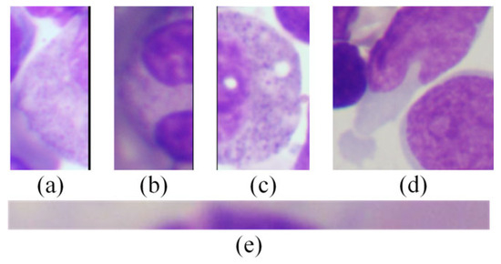
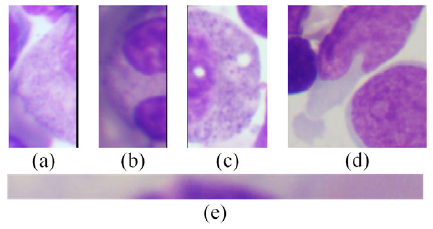
In this study, we selected the LLC-2024 dataset, which consists of bone marrow smear images acquired using an orthogonal optical biological microscope. The dataset encompasses three categories—ALL (Acute Lymphoblastic Leukemia Primary Lymphocytes), CLL (Chronic Lymphocytic Leukemia Mature Monoclonal Lymphocytes), and OTH (Normal bone marrow hematopoietic cells). For the task of nucleus segmentation in bone marrow microscopic images of lymphocytic leukemia, we constructed an efficient and semi-automated annotation process based on the pre-trained SAM_ViT-L (Segment Anything Model, SAM) model [13]. To improve mask quality, the ISAT-SAM (Image Segmentation Annotation Tool) was employed to manually correct and refine the initial masks generated by SAM. The researchers manually calibrated all the masks one by one and performed multiple rounds of fine-tuning on the boundary contours to ensure the segmentation accuracy. This semi-automated process not only drastically reduces the annotation time but also generates accurate masks that lay a solid data foundation for the training of the segmentation branch of the MTLL model. To ensure high-quality supervisory signals for the joint learning framework, a rigorous data screening process was implemented in this study. Specifically, five images (two ALL and three OTH) were identified as ambiguous outliers due to severe nuclear truncation or multiple overlapping cells, resulting in a lack of consensus among annotators regarding the accurate definition of their boundaries (Figure 1). Consequently, these samples were excluded. While maintaining the reliability of the ground truth, this exclusion has a negligible impact on the overall training scale and evaluation results. To facilitate a fair comparison with existing studies, the final dataset follows the original split provided by the dataset, consisting of 10,761 training samples, 3499 validation samples, and 3560 test samples.
Figure 1.
Examples of excluded images. Subfigures (a–e) show five representative examples of excluded images.
All input images were resized to 224 × 224 during the data-loading process before being fed into the network. No additional preprocessing operations were applied, such as image normalization, filtering, sampling frequency adjustment, or sliding-window segmentation, in order to preserve the original morphological characteristics of bone marrow cells. Data augmentation strategies were applied only during the training phase to improve model generalization, while no augmentation was used for validation or testing. Specifically, the following transformations were randomly applied to each training sample:
- Aspect ratio jittering and random scaling (scaling range: 0.5–2; jittering range: ±30%).
- Random horizontal flip with probability 0.5.
- Random panning and filling with a gray border strip.
- Random Gaussian blur, probability 0.25.
- Random rotation ±10°, probability 0.25.
- HSV color gamut jittering (hue ±10%, saturation ±70%, lightness ±30%).
2.2. Overall Structure of the MTLL Model
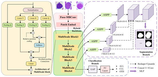
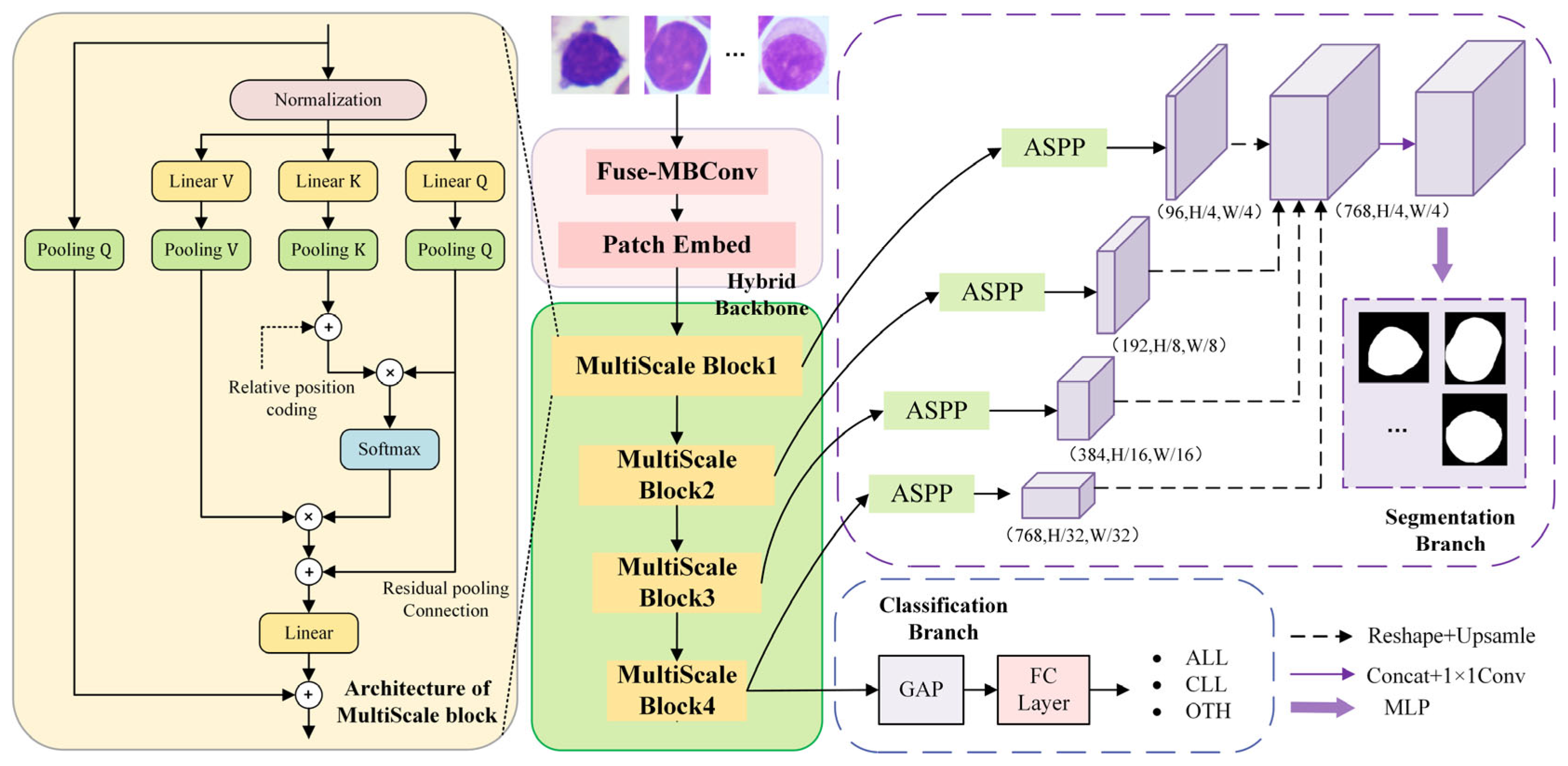
The proposed MTLL model consists of a feature-sharing backbone, a segmentation branch, and a classification branch (Figure 2), which jointly integrates lymphocytic leukemia classification and nucleus segmentation into a unified end-to-end framework. In morphological diagnosis, the shape and structural characteristics of the cell nucleus are important cues for distinguishing different cell types. Therefore, by jointly learning nucleus segmentation and cell classification, the proposed framework enables the structural information captured by the segmentation task to complement the discriminative features required for classification. At its core, the hybrid backbone combines convolutional operators with multilayer transformer blocks. The convolutional layers specialize in extracting fine-grained features such as nuclear edges and textures, while the transformers capture global morphology, spatial distribution, and contextual background information. This local-to-global information bridging mechanism enables adaptive fusion of multi-scale features. The extracted features are then simultaneously fed into two branches. The AM decoder leverages atrous spatial pyramid pooling (ASPP) [14] and channel fusion to progressively reconstruct multi-scale representations for accurate nucleus segmentation. Meanwhile, the classification branch makes full use of the high-level semantic features from the hybrid backbone to produce a three-way prediction (ALL/CLL/OTH) via a fully connected layer. During training, classification and segmentation losses are backpropagated with equal weights, allowing both tasks to jointly constrain and reinforce each other through shared optimization. The detailed designs of each component are described below.
Figure 2.
Architecture of the MTLL model.
2.2.1. Hybrid Backbone for Multi-Task Feature Sharing
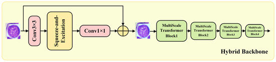
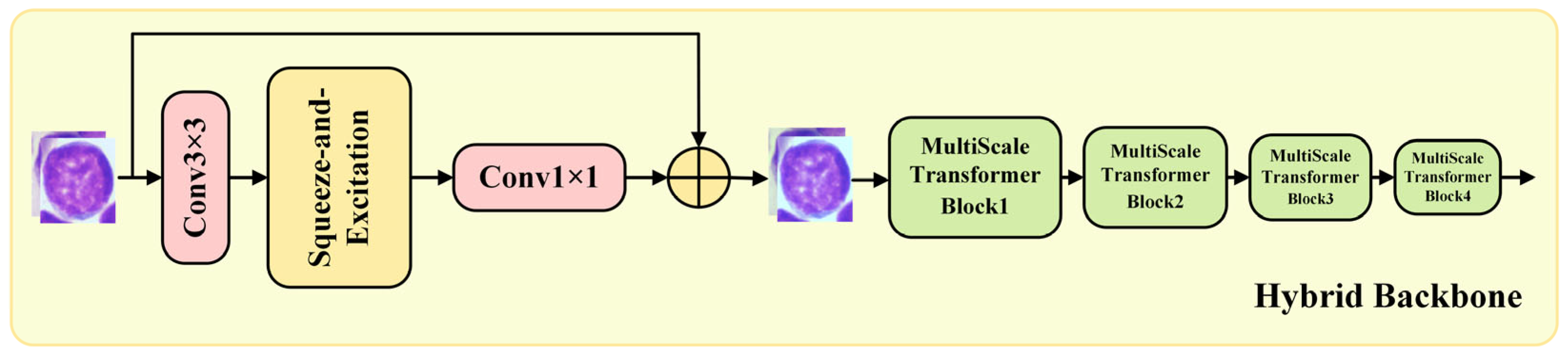
To effectively capture both fine-grained texture details and high-level semantic information in bone marrow microscopic images, we employ a convolution-augmented hybrid backbone that combines convolutional operations with multi-scale Transformer blocks (Figure 3). Specifically, a Fused-MBConv module [12] is introduced before the MViTv2 architecture [11] to capture early-stage local spatial dependencies and provide stronger inductive bias for visual feature extraction. Pure Transformer-based models generally lack the inductive biases of locality and translation invariance, which may limit their ability to model fine-grained morphological structures in images. By incorporating the convolutional stem, the network introduces locality-aware feature modeling at the early stage while preserving the global dependency modeling capability of the Transformer.
Figure 3.
Architecture of the hybrid backbone.
The input images are first passed through a conventional 3 × 3 convolution to capture local spatial information, such as cell edges and textures from the bone marrow images. Then, channel responses are reweighted according to their importance using the Squeeze-and-Excitation attention mechanism [15] to highlight key signals. Subsequently, a 1 × 1 convolution is applied for channel compression and projection, followed by a residual connection to maintain efficient feature flow and gradient propagation. After preliminary feature enhancement and token reconstruction through convolution operators, the model’s sensitivity to local textures and morphological structures is improved, and the resulting feature maps are seamlessly fed into multiple layers of multi-scale transformer blocks. Within each block, cascaded pooling operations progressively reduce spatial resolution to integrate multi-scale information, while decomposable positional distance encoding compensates for the lack of translation invariance. Therefore, for an input image of size H × W, the hybrid backbone produces four multi-scale feature maps with resolutions of .
By combining convolutional inductive biases with the global modeling capability of the Transformer, the proposed hybrid backbone improves the representation of both local morphological patterns and global contextual information. This shared feature representation provides a stable foundation for the downstream tasks of nucleus segmentation and lymphocytic leukemia classification within the multi-task learning framework.
2.2.2. Segmentation Branch Based on AM Decoder
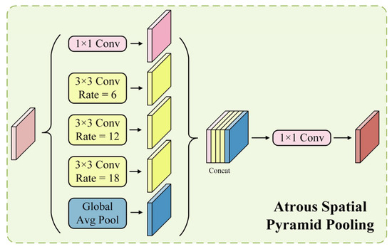
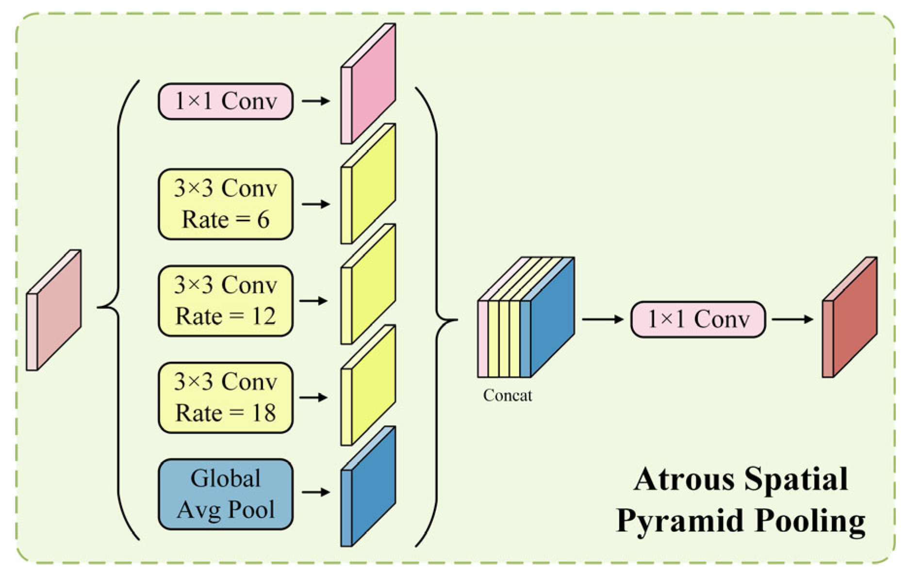
Multi-scale features are sequentially extracted from the four-stage multi-scale transformer blocks of the shared-feature hybrid backbone and fed into the segmentation branch for fine-grained nucleus segmentation. In the shallow stage, the extracted features mainly contain cell texture and edge information. In the middle stage, the features reflect the contour of the cell shape. In the deep stage, richer global semantic information is aggregated, forming a complete and diverse feature representation system. To address the inherent hierarchical characteristics of transformer features, characterized by local attention in shallow layers and global attention in deeper layers, we propose an AM decoder to optimize multi-scale feature fusion. Specifically, in this decoder, the ASPP module (Figure 4) is applied to the features from four different channel resolutions . It expands the receptive field and enhances multi-scale contextual information fusion through dilated convolutions with varying dilation rates. Subsequently, each ASPP output passes through a multilayer perceptron (MLP) layer to unify the channel dimensions. The resulting feature maps are then up-sampled to a size of and concatenated along the channel dimension. Finally, another MLP is applied to the fused features to perform pixel-wise classification. The AM decoder can be formulated as the following equation:
where = 1, 2, 3, 4 denotes the outputs of the four feature map layers, represents the number of input channels, represents the number of output channels, and represents the number of segmentation categories. Here, denotes a linear layer with input dimension and output dimension .
Figure 4.
Architecture of the ASPP module.
From an architectural perspective, the proposed AM decoder differs from the conventional symmetric encoder–decoder design used in U-Net. The latter relies on multiple stages of transposed convolutions and skip connections for spatial reconstruction, which may increase architectural complexity and lead to progressive information loss. Instead, the AM decoder performs parallel alignment and aggregation of multi-level feature maps, shortening the feature propagation path. Compared with the decoder designs used in PSPNet and DeepLab, it is specifically designed for transformer-based features, where lightweight linear projections replace deep convolutional stacks to achieve efficient multi-scale contextual modeling with lower computational cost.
With the AM decoder, the model integrates contextual information at different scales more effectively. It also captures fine boundary details while maintaining global structural consistency in the segmentation of cell nuclei. As a result, the model achieves high segmentation accuracy and strong robustness.
2.2.3. Classification Branch Based on Structural Prior Guidance
The classification branch is designed to exploit the structural priors of cell nuclei reinforced by the AM decoder. After the hybrid backbone performs in-depth refinement of multi-scale features, the high-order semantic feature map aggregates the most informative global semantic cues and discriminative signals. In the segmentation branch, the AM decoder accurately reconstructs cell nucleus boundaries and local contextual details. These structural cues are propagated back to the shared hybrid backbone during joint optimization, enabling the model to encode more discriminative representations for cell morphology. After the hybrid backbone performs multi-scale feature refinement, the high-level semantic feature map aggregates the most informative global contextual information and discriminative signals. To maximize feature utility and minimize redundancy, the classification branch operates directly on the feature stream , which is synergistically optimized through multi-task learning. Consequently, is adopted as the final feature representation for classification. Global average pooling is applied to compress the spatial information in into a semantic vector, which is then passed through a fully connected layer to generate predictions for the three categories: ALL, CLL, and OTH. With minimal computational overhead, the classification branch effectively leverages the deep representations jointly extracted by the hybrid backbone and the AM decoder, achieving efficient and accurate identification of lymphocytic leukemia.
2.3. Multi-Task Loss Function
In this work, the loss function for the segmentation branch consists of two components: Cross-Entropy Loss and Dice Loss. This design aims to balance pixel-level classification accuracy and region-level connectivity. The segmentation loss can be formulated as:
where represents the predicted segmentation result, denotes the corresponding one-hot encoded segmentation mask, and refers to the ground-truth segmentation label with original resolution and integer encoding.
The loss function for the classification branch is defined as the Cross-Entropy Loss:
denotes the predicted labels used for the network, while denotes the true labels used for classification.
Therefore, the total loss of the multi-task model is computed as a weighted sum of the segmentation and classification losses:
Since classification focuses on the overall morphology and staining patterns of cells, while segmentation emphasizes precise boundaries and morphological details, these two tasks have complementary representational requirements. Considering that complex dynamic weighting strategies often introduce additional computational overhead, we adopt a uniform weighting strategy by setting both weights to , so that the classification and segmentation losses are equally weighted during training. This strategy provides a good balance between training efficiency and overall performance. Under this training paradigm, the two tasks mutually regularize the learning process, enabling the hybrid backbone to simultaneously capture global discriminative features and fine-grained cellular structural information, thereby facilitating the synergistic optimization of classification and segmentation.
3. Experimental Results and Analysis
3.1. Model Configuration
All models were implemented using PyTorch 2.2.0 and trained on an NVIDIA GeForce RTX A5000 with 24 GB of graphics memory. The training configuration is summarized in Table 1, and all experiments in this section are conducted under this setting. To accelerate the training process while maintaining stable convergence, Automatic Mixed Precision (AMP) is adopted during training.
Table 1.
Model Configuration.
3.2. Comparison of Classification Networks
To validate the superiority of the proposed MTLL model for classifying lymphoblastic leukemia in bone marrow smears, we compared its classification performance with several mainstream single-task networks under the same experimental settings (Table 2). The experimental results show that the MTLL model outperforms all comparison models in four metrics: Overall Accuracy (OA), Average Precision (AP), Average Recall (AR), and Average F1-score (AF1). Specifically, the MTLL model achieves an average accuracy of 94.27%, exceeding T2T_ViT_14 [16] (89.72%), EfficientNetV2 [11] (91.38%), DeiT3 [17] (91.15%), ConvNeXt [18] (92.22%), EdgeNeXt [19] (92.13%), and TransNeXt [20] (92.44%) by 4.55, 2.89, 3.12, 2.05, 2.14, and 1.83 percentage points, respectively. Moreover, the MTLL model outperforms all single-task classification models in all evaluation metrics, achieving an average precision of 93.90%, an average recall of 91.57%, and an average F1-score of 92.57%. This performance improvement is mainly attributed to the synergy between the multi-task feature-sharing mechanism and the feature fusion module in the MTLL model. Specifically, the AM decoder is introduced in the segmentation branch to effectively capture multi-scale contextual information and enhance fine-grained feature representation. Meanwhile, the hybrid backbone facilitates deep fusion between classification and segmentation features, further improving the discriminative capability of the model. The above design not only ensures the accuracy of cell nucleus segmentation but also significantly improves the classification performance of lymphoid leukemia cell subtypes.
Table 2.
Evaluation of Classification Methods on the LLC-2024 Dataset.
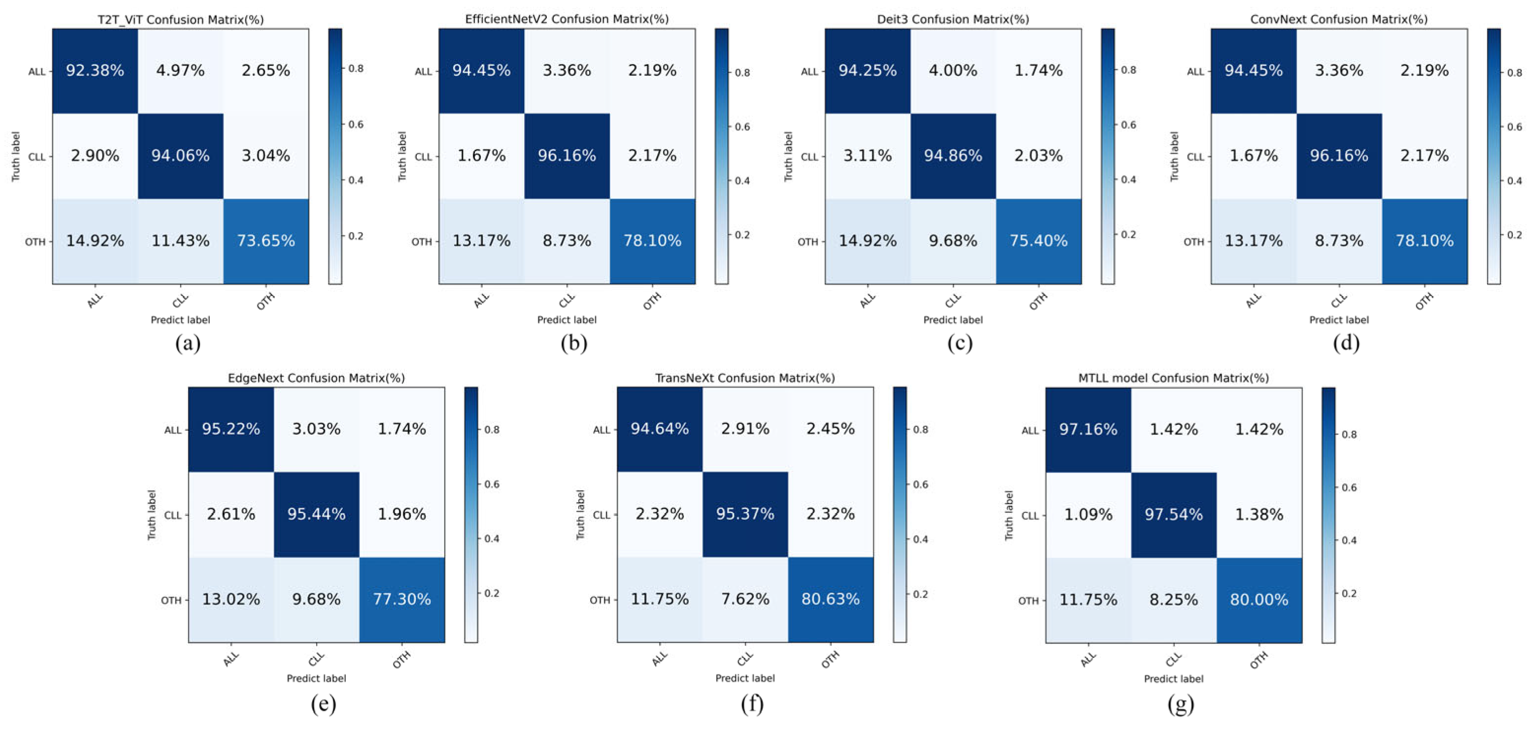
In addition, to systematically evaluate the discriminative ability of the seven models on the ALL, CLL, and OTH classification tasks, the confusion matrices of T2T_ViT (Figure 5a), EfficientNetV2 (Figure 5b), DeiT3 (Figure 5c), ConvNeXt (Figure 5d), EdgeNeXt (Figure 5e), TransNeXt (Figure 5f), and MTLL (Figure 5g) are presented in Figure 5. The classification accuracy is assessed by the proportion of correctly classified samples along the diagonal of each matrix. The MTLL model has the best overall accuracy of 97.16%, 97.54%, and 80.00% for ALL, CLL, and OTH categories, respectively; in contrast, EdgeNeXt, DeiT3, EfficientNetV2, and ConvNeXt have the best diagonal accuracy in ALL, CLL, and OTH categories, respectively. In contrast, EdgeNeXt, DeiT3, EfficientNetV2, and ConvNeXt maintain an accuracy of approximately 94% to 96% for the ALL and CLL categories. However, their performance on the minority class OTH drops significantly, with accuracy ranging from only 74% to 78%, indicating limited classification stability. Taken together, the AM decoder and hybrid backbone deep fusion mechanism employed in the proposed model not only significantly enhance the differentiation between ALL and CLL but also demonstrate stronger robustness in recognizing the minority class OTH. These results further confirm the superior performance of the MTLL model in complex cell classification tasks.
Figure 5.
Comparison of classification confusion matrices for lymphocytic leukemia in bone marrow smears. Sub-figures (a–g) correspond to T2T_ViT, EfficientNetV2, DeiT3, ConvNeXt, EdgeNeXt, TransNeXt, and MTLL, respectively; columns denote predicted classes and rows denote true classes.
3.3. Comparison of Segmentation Networks
In the segmentation performance comparison (Table 3), the MTLL model achieves a Dice coefficient of 98.70% and an IoU of 97.54%, outperforming the ResNet18 [21]-based U-Net [22] (98.54%/97.28%), SegNet [23] (98.41%/97.02%), and Segformer [24] (98.37%/96.99%). In addition, the MTLL model consistently maintains advantages in overall accuracy (OA 98.83%), average precision (AP 98.69%), and average recall (AR 98.83%). In contrast, RefineNet [25] performs relatively worse, with an IoU of 94.44% and a recall of 96.04%. Due to the sufficient sample size of the dataset and the clear boundaries of cell nuclei, classical segmentation networks have already achieved a high level of performance. Consequently, the differences between various methods are typically reflected in only small performance improvements. The AM decoder in the MTLL model integrates multi-scale dilated convolutions with fine-grained mapping. This design not only effectively refines the reconstruction of cell nucleus boundaries but also enhances feature representations. The structured features generated by the segmentation branch can be effectively fed back into the classification branch, further improving the overall discriminative capability of the multi-task framework. These designs not only enable the MTLL model to outperform mainstream methods in segmentation tasks but also provide more discriminative multi-scale features for the identification of lymphocytic leukemia.
Table 3.
Evaluation of Segmentation Methods on the LLC-2024 Dataset.
3.4. Comparison of Multi-Branch Networks
The experimental results show that under the multi-task learning framework, the MTLL model effectively handles both classification and segmentation tasks, achieving state-of-the-art performance in both (Table 4). Specifically, for the classification branch, the MTLL model achieves an OA of 94.27%, an AP of 93.90%, an AR of 91.57%, and an AF1 of 92.57%. These results represent improvements of 3.68, 3.73, 4.08, and 3.96 percentage points over ACSNet [26] (2024), respectively. Compared with MT-Net [27] (2019) and MMRAN [28] (2023), the indicators of OA and AF1 increased by approximately 5.11/8.17 percentage points and 5.75/9.38 percentage points, respectively. For the segmentation branch, the MTLL model outperforms all compared methods, achieving a Dice coefficient of 98.70% and an IoU of 97.54%. In addition, it demonstrates stable improvements in OA (98.83%), AP (98.69%), and AR (98.83%). These results demonstrate that the MTLL model enhances the representation capabilities for both cell nucleus segmentation and lymphocytic leukemia classification under end-to-end joint training, without introducing a significant trade-off between the two tasks. Our method not only ensures superior segmentation accuracy compared to single-task approaches but also effectively injects the fine-grained features generated by the segmentation branch into the classification branch. This design ultimately enables the synergistic optimization of both tasks.
Table 4.
Evaluation of Multi-task Methods on the LLC-2024 Dataset.
3.5. Ablation Experiments
The ablation results in Table 5 further demonstrate the contribution of each key component to the proposed framework under the same training settings. Overall, each module contributes differently to the classification and segmentation tasks. When only the classification branch is used, the model achieves 92.98% OA, 92.51% AP, 90.32% AR, and 91.27% AF1, serving as the baseline for subsequent comparison. When the complete AM decoder is introduced without Fuse-MBConv, the classification performance improves to 94.19% OA, 93.78% AP, 91.51% AR, and 92.49% AF1. Meanwhile, the segmentation branch achieves 98.69% Dice and 97.53% IoU. These results indicate that the segmentation branch and its multi-scale feature aggregation structure can learn useful nucleus structural information, which provides complementary cues for the classification task through shared representations. When Fuse-MBConv is introduced while the ASPP module is removed, the model achieves 93.82% OA, 93.66% AP, 90.54% AR, and 91.80% AF1 for classification, together with 98.70% Dice and 97.53% IoU for segmentation. Compared with the configuration using the complete AM decoder, this result suggests that feature fusion alone, without sufficient multi-scale contextual modeling, provides only limited improvement. This also indicates that the effectiveness of Fuse-MBConv relies on cooperation with richer structural feature representation. When both the complete AM decoder and Fuse-MBConv are jointly adopted, the full MTLL model achieves the best overall performance, reaching 94.27% OA, 93.90% AP, 91.57% AR, and 92.57% AF1 for classification, along with 98.70% Dice and 97.54% IoU for segmentation. These results demonstrate that the AM decoder and Fuse-MBConv play complementary roles in enhancing structural representation and cross-task feature interaction, and their joint use yields the best overall performance.
Table 5.
Ablation study for classification and segmentation.
3.6. Generalization Analysis
In addition, to evaluate the generalization ability of the MTLL model across different tissue types and imaging conditions, we employed the EBHI-Seg dataset [29], which contains six types of colonoscopic biopsy pathology sections along with corresponding annotations, for cross-domain multi-task evaluation (Table 6). The model maintains excellent segmentation accuracy even under varying cell staining intensities and morphological distributions, such as differences in shape, size, and arrangement. In contrast, MMRAN, MT-Net, and ACSNet struggle to simultaneously achieve high performance in both segmentation and classification tasks. In summary, the MTLL model proposed in this paper not only achieves an effective performance balance between classification and segmentation tasks but also demonstrates excellent generalizability and notable advantages in cross-domain medical image analysis.
Table 6.
Evaluation of multi-task methods on the EBHI-Seg dataset.
3.7. Computational Complexity Analysis of Multi-Task Models
To evaluate the practical applicability of the proposed method, we performed a comparative analysis of computational complexity, as summarized in Table 7. The evaluation focuses on the balance between model complexity and inference resources. The results indicate that the proposed MTLL model demonstrates favorable computational efficiency in terms of FLOPs (20.53 G) and Peak Memory (3435.70 MB). These metrics are particularly relevant for ensuring that the model can be effectively deployed for real-time or batch processing in clinical settings. Compared with MT-Net (25.4 G) and ACSNet (20.03 G), MTLL maintains a competitive computational profile, achieving comparable resource consumption despite its more advanced architecture. This efficiency can be largely attributed to the CNN–Transformer hybrid backbone designed for multi-level feature sharing. Although the incorporation of Transformer-based components for long-range dependency modeling increases the total number of parameters, the overall design prevents this from causing a substantial increase in computational cost. By leveraging the local perception of CNNs and the global context of Transformers, MTLL achieves a favorable performance-to-cost ratio. Specifically, MTLL achieves superior performance in both classification and segmentation tasks (Table 4) while maintaining reasonable resource consumption.
Table 7.
The computational complexity of the multi-tasking method.
4. Discussion
One relevant study on the LLC-2024 dataset is the OFAMobileNetV3 method proposed by (2025) [3], which reported 93.35% Precision, 95.22% Recall, and 94.28% F1-score for the ALL category and an overall accuracy (OA) of 92.51% on the same bone marrow smear lymphoid leukemia classification task. The corresponding results are cited from the original paper and summarized in Table 8. Based on the same dataset and evaluation metrics, the proposed MTLL model further improved the ALL category performance, achieving 94.42% Precision, 97.16% Recall, and 95.77% F1-score. The overall accuracy also increased to 94.27%. For the CLL category, the Precision, Recall, and F1-score also improved from 93.32%, 95.15%, and 94.23% to 94.79%, 97.54%, and 96.15%, respectively. Even in the OTH category, where samples are relatively scarce, MTLL increased the Precision from 88.17% to 92.48% and the F1-score from 83.94% to 85.79%, significantly reducing the false positive rate. These performance improvements are mainly attributed to the multi-task learning framework adopted by the MTLL model. This framework enables the classification branch to fully exploit the structured cellular morphological features provided by the segmentation branch, thereby mitigating feature bias and information loss commonly observed in single-task learning. In contrast, OFAMobileNetV3, as a pure classification network, lacks integrated support for cell nucleus segmentation, which limits its discriminative capability when faced with complex cellular morphology. Consequently, the MTLL model proposed in this study significantly outperforms existing methods on the LLC-2024 dataset. These results further demonstrate the effectiveness of multi-task feature sharing and the hybrid backbone-based deep feature fusion strategy in enhancing the classification performance for lymphoblastic leukemia.
Table 8.
Comparison of existing methods for lymphocytic leukemia classification on bone marrow microscopic images.
To further evaluate the statistical stability of the models under different data partitions, an additional 5-fold stratified cross-validation was conducted, in which multiple multi-task models were consistently compared on both classification and segmentation metrics (Table 9 and Table 10). The results show that, although some variation exists across folds, the overall performance trends remain largely consistent with the conclusions drawn from the main experiments based on the official fixed split. Specifically, the proposed MTLL achieved 93.76 ± 0.52% OA, 93.42 ± 0.36% AP, 91.02 ± 0.75% AR, and 92.04 ± 0.60% AF1 for classification, and 98.25 ± 0.36% Dice, 96.85 ± 0.47% IoU, 98.54 ± 0.21% OA, 98.18 ± 0.37% AP, and 98.47 ± 0.30% AR for segmentation, remaining superior overall to the other compared multi-task models. These results indicate that the performance advantage of the proposed method does not rely on a particular data split but can be maintained with good stability and competitiveness across different folds.
Table 9.
Performance comparison of multi-task models on classification under 5-fold stratified cross-validation (mean ± standard deviation).
Table 10.
Performance comparison of multi-task models on segmentation under 5-fold stratified cross-validation (mean ± standard deviation).
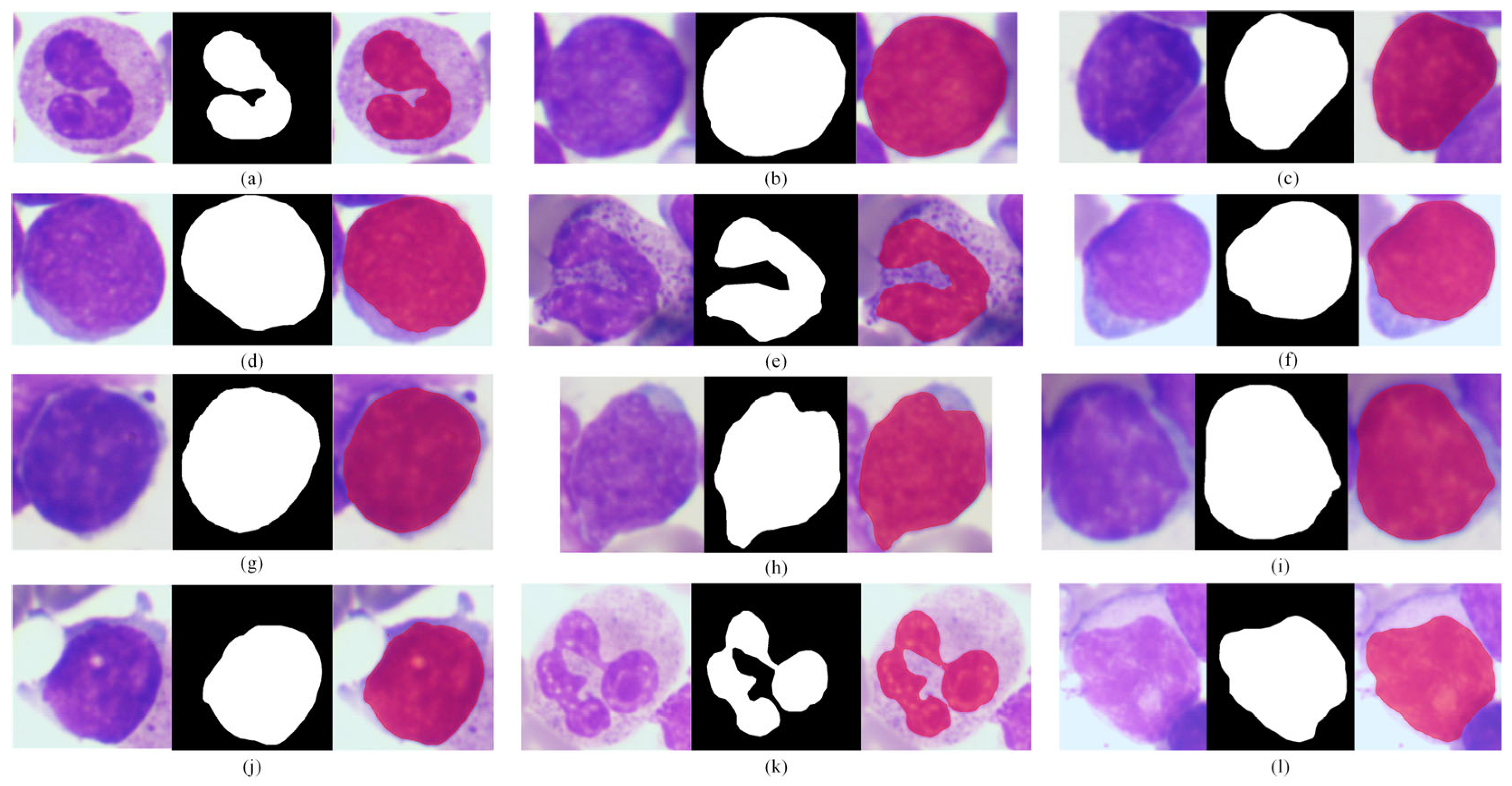
In this study, we randomly selected 12 representative bone marrow microscopic images from the test set and presented the segmentation results generated by the MTLL model (Figure 6). The selected samples cover the following complex morphologies. For instance, in cases where obvious adhesions occur between cell nuclei, the MTLL model can accurately delineate the boundaries of adjacent nuclei, thereby avoiding segmentation errors caused by overlapping morphologies. Similarly, the model effectively suppresses artifacts such as particles or impurities present in the cytoplasm and preserves only the true contours of the nuclear region. For images containing densely packed cells and low-contrast backgrounds, the segmentation masks still maintain clear and continuous edges, effectively accommodating variations in cell size, shape, and staining intensity. In addition, regardless of whether the nuclei exhibit regular round shapes, multinucleated structures, or irregular distorted forms, the MTLL model achieves highly accurate segmentation, with the generated masks visually almost perfectly overlapping with the ground truth contours. These qualitative results demonstrate that the MTLL model exhibits excellent robustness and generalization capability when dealing with variable nuclear morphology, densely distributed cells, and background interference. Moreover, it provides high-quality morphological representations of nuclei, which serve as a reliable foundation for subsequent lymphoblastic leukemia classification.
Figure 6.
MTLL model qualitative results for lymphocytic leukemia nucleus segmentation. Sub-figures (a–l) display 12 typical cases, with each case presenting the raw input (left), annotated ground truth (middle), and predicted segmentation (right).
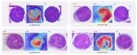
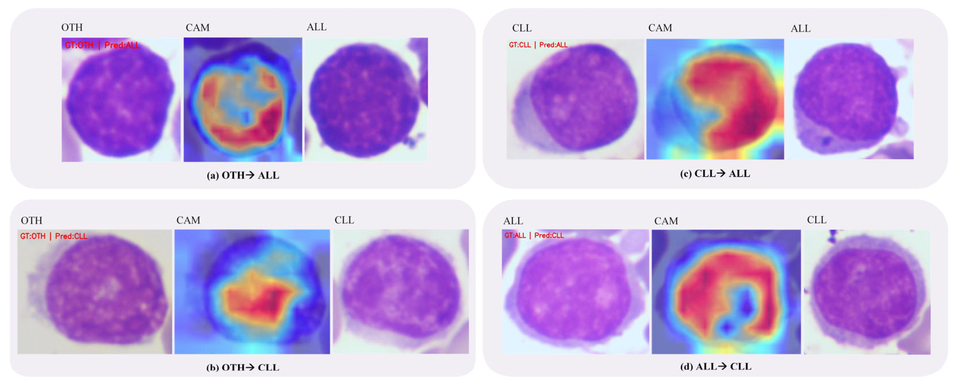
Compared with the ALL and CLL categories, the recognition performance for the OTH category is relatively lower, with a recall rate of approximately 80%. This phenomenon can be attributed to the high heterogeneity of the OTH category, which contains multiple hematopoietic cell types, including granulocytes, erythrocytes, monocytes, mature lymphocytes, and smeared cells. These cells exhibit diverse morphological characteristics and complex structural patterns in bone marrow smear images. In addition, the number of OTH samples is relatively smaller than that of the leukemic categories. To facilitate a more intuitive comparison, each subfigure in Figure 7 contains a misclassified sample from Class A, the corresponding Grad-CAM visualization, and a representative sample from Class B, where Class A → Class B indicates that the sample from Class A is incorrectly predicted as Class B. As illustrated in Figure 7a,b, some OTH cells present nuclear morphology and staining textures that are highly similar to those of ALL or CLL cells, which may lead to misclassification as leukemic categories. Conversely, as shown in Figure 7c,d, certain ALL cells exhibit local chromatin patterns and nuclear structures resembling those of CLL, resulting in incorrect predictions. These observations suggest that the morphological differences among some bone marrow cell types are subtle under microscopic imaging, especially when variations in staining intensity, cell maturation stage, or image acquisition conditions are present. In addition, the Grad-CAM visualizations in Figure 7 provide further insight into the model’s decision-making process. Although some samples are misclassified, the corresponding heatmaps still mainly focus on morphologically relevant cell regions, particularly the nuclear contour, chromatin distribution, and other local structural areas, rather than irrelevant background regions. This indicates that the model has captured meaningful morphological cues for recognition. However, when different cell categories exhibit highly similar microscopic appearances, the attended features may still be insufficiently discriminative, which can eventually lead to incorrect predictions. Therefore, the discrimination between leukemic lymphocytes and other morphologically similar hematopoietic cells remains a challenging task for automated recognition systems.
Figure 7.
Examples of misclassification. Class A → Class B indicates that a sample from Class A is misclassified as Class B.
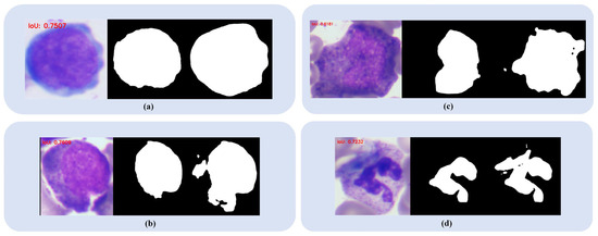
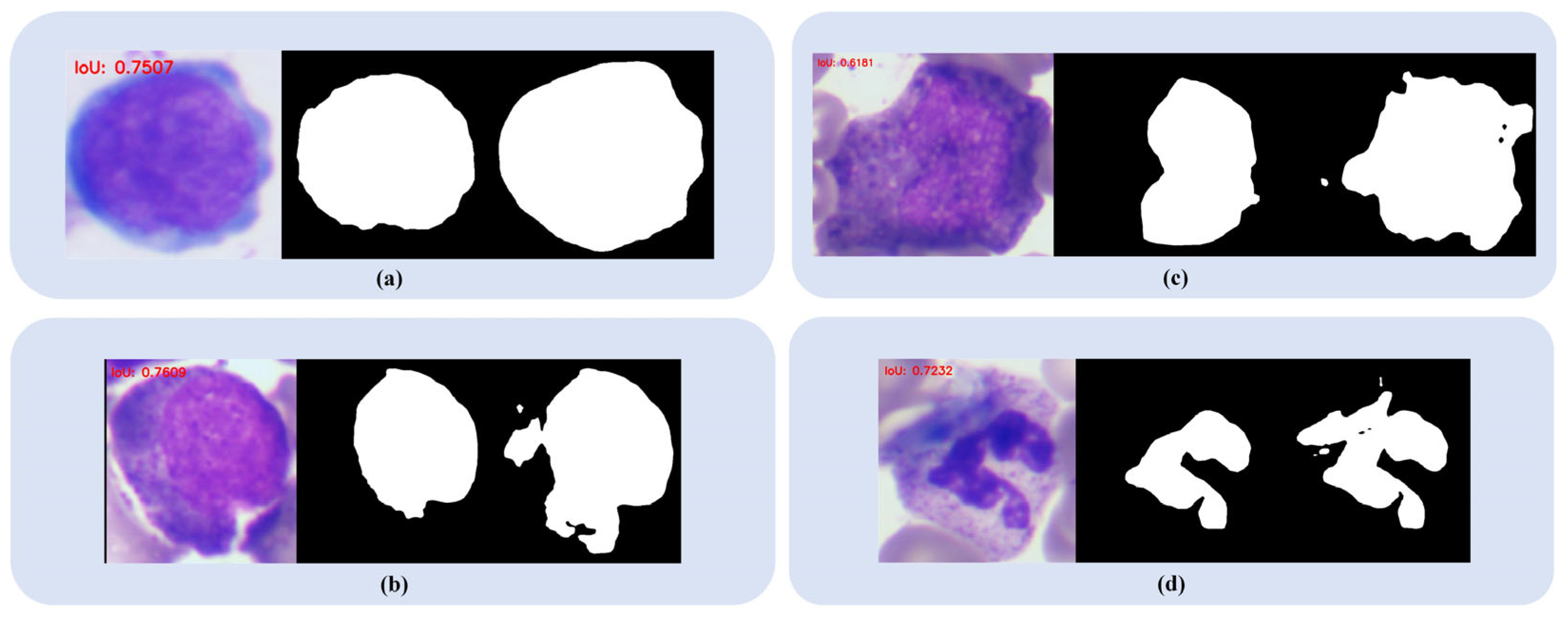
We further analyzed representative cases with suboptimal nucleus segmentation, as shown in Figure 8. These examples correspond to cases with relatively poor segmentation performance in the test set. Such cases typically involve cells with irregular or complex nuclear morphology (Figure 8c,d) or ambiguous boundaries between the nucleus and cytoplasm (Figure 8a,b), both of which pose challenges for accurate segmentation. Further analysis indicates that most suboptimal segmentation cases occur in the OTH category, which includes multiple types of bone marrow cells. Due to the high morphological heterogeneity and structural variability of nuclei in this category, nuclear shapes are more diverse than those observed in leukemic cells, making segmentation more challenging.
Figure 8.
Examples of suboptimal nucleus segmentation. Subfigures (a–d) present four representative cases with relatively unsatisfactory segmentation performance, where each case shows the original image (left), the annotated ground truth (middle), and the predicted result (right).
In future work, incorporating finer-grained structural features and richer contextual information may further improve the discrimination of morphologically similar cell types. Previous studies in medical image analysis have shown that combining multi-task collaborative learning with cross-scale feature interaction can enhance the representation of complex structures and improve both segmentation and recognition performance [30]. At the same time, considering that pixel-level annotation is often labor-intensive, weakly supervised approaches such as scribble supervision have attracted increasing attention. Recent studies have shown that sparse annotations can still provide effective supervision for medical image segmentation, reducing annotation cost while maintaining competitive performance through improved use of contextual and boundary cues [31]. In addition, improving the robustness of the framework under different staining conditions, imaging settings, and cross-dataset scenarios would be important for practical applications. Therefore, integrating weakly supervised strategies together with more robust cross-domain modeling into the current framework may further enhance the practicality, scalability, and generalization ability of the proposed method.
5. Conclusions
In this paper, we propose the MTLL model (multi-task model on lymphocytic leukemia), which integrates lymphocytic leukemia cell classification and nucleus segmentation of bone marrow microscopic images into a unified network and accomplishes both tasks in parallel. On the LLC-2024 dataset containing 17,821 images, the MTLL model achieves an overall accuracy of 94.27% for the classification task and 98.83% for the segmentation task, outperforming advanced single-task and multi-task models. Moreover, cross-domain experiments further validate the robustness and generalization of the model under different imaging conditions and pathology types. These results indicate that the MTLL model, based on a multi-task learning framework and incorporating deep fusion with the hybrid backbone and a multi-scale feature sharing strategy via the AM decoder, effectively improves the accuracy of both lymphoblastic leukemia classification and nucleus segmentation. Furthermore, our method further confirms that introducing the nucleus segmentation task contributes to enhancing the accuracy of cell subtype classification, providing a generalizable technological pathway for end-to-end intelligent diagnosis of complex medical images.
Author Contributions
C.O. participated in the conceptualization and methodology design, wrote the main manuscript text, conducted the investigation and validation, and was responsible for software development, data curation, as well as manuscript review and editing. Z.H. wrote the main manuscript text, contributed to the conceptualization and methodology design, and was responsible for supervision and resource provision. X.W. was in charge of project administration and funding acquisition, supervised the research, participated in the methodology design and validation, developed the related software, and took part in manuscript review and editing. K.C. performed the software development and validation, conducted data curation and visualization, and participated in manuscript review and editing. Y.W. conducted the investigation and validation, and was responsible for data curation and visualization. All authors have read and agreed to the published version of the manuscript.
Funding
Grants were received from the key specialized research and development breakthrough of Henan province (Grant No. 232102211016) and the key scientific research projects of Henan colleges and universities (Grant No. 23A416004).
Institutional Review Board Statement
Not applicable.
Data Availability Statement
The dataset supporting the conclusions of this article, LLC-2024, is available in the Mendeley Data repository, http://data.mendeley.com/datasets/wydknkk5k2 (accessed on 1 December 2025). The dataset supporting the conclusions of this article, EBHI-SEG, is available in the Figshare repository, http://figshare.com/articles/dataset/EBHI-SEG/21540159 (accessed on 1 December 2025).
Conflicts of Interest
The authors declare no conflicts of interest.
Abbreviations
| MTLL model | Multi-task model on Lymphocytic Leukemia |
| AM | Atrous Multilayer Perceptron |
| ALL | Acute Lymphoblastic Leukemia |
| CLL | Chronic Lymphocytic Leukemia |
| OTH | Bone Marrow Hematopoietic Cells from Healthy Subjects |
| SAM | Segment Anything Model |
| ISAT-SAM | Image Segmentation Annotation Tool |
| ASPP | Atrous Spatial Pyramid Pooling |
| MLP | Multilayer Perceptron |
| OA | Overall Accuracy |
| AP | Average Precision |
| AR | Average Recall |
| AF1 | Average F1-score |
References
- Bennett, J.M.; Catovsky, D.; Daniel, M.T.; Flandrin, G.; Galton, D.A.; Gralnick, H.R.; Sultan, C. Proposals for the classification of the acute leukaemias French-American-British (FAB) co-operative group. Br. J. Haematol. 1976, 33, 451–458. [Google Scholar] [CrossRef] [Scilit] [PubMed]
- Arslan, S.; Ozyurek, E.; Gunduz-Demir, C. A color and shape based algorithm for segmentation of white blood cells in peripheral blood and bone marrow images. Cytom. A 2014, 85, 480–490. [Google Scholar] [CrossRef] [Scilit] [PubMed]
- Mei, L.; Lian, C.; Han, S.; Jin, S.; He, J.; Dong, L.; Wang, H.; Shen, H.; Lei, C.; Xiong, B. High-Accuracy and Lightweight Image Classification Network for Optimizing Lymphoblastic Leukemia Diagnosisy. Microsc. Res. Tech. 2025, 88, 489–500. [Google Scholar] [CrossRef] [Scilit] [PubMed]
- Guo, L.; Huang, P.; Huang, D.; Li, Z.; She, C.; Guo, Q.; Zhang, Q.; Li, J.; Ma, Q.; Li, J. A classification method to classify bone marrow cells with class imbalance problem. Biomed. Signal Process. Control 2022, 72, 103296. [Google Scholar] [CrossRef] [Scilit]
- Qin, F.; Gao, N.; Peng, Y.; Wu, Z.; Shen, S.; Grudtsin, A. Fine-grained leukocyte classification with deep residual learning for microscopic images. Comput. Methods Programs Biomed. 2018, 162, 243–252. [Google Scholar] [CrossRef] [Scilit] [PubMed]
- Chen, B.; Qin, F.; Shao, Y.; Cao, J.; Peng, Y.; Ge, R. Fine-grained imbalanced leukocyte classification with global-local attention transformer. J. King Saud Univ. Comput. Inf. Sci. 2023, 35, 101661. [Google Scholar] [CrossRef] [Scilit]
- Al-jaboriy, S.S.; Sjarif, N.N.A.; Chuprat, S.; Abduallah, W.M. Acute lymphoblastic leukemia segmentation using local pixel information. Pattern Recognit. Lett. 2019, 125, 85–90. [Google Scholar] [CrossRef] [Scilit]
- Banik, P.P.; Saha, R.; Kim, K.D. An automatic nucleus segmentation and CNN model based classification method of white blood cell. Expert Syst. Appl. 2020, 149, 113211. [Google Scholar] [CrossRef] [Scilit]
- Sipes, R.; Li, D. Using convolutional neural networks for automated fine grained image classification of acute lymphoblastic leukemia. In Proceedings of the 2018 3rd International Conference on Computational Intelligence and Applications (ICCIA); IEEE: New York, NY, USA, 2018; pp. 157–161. [Google Scholar]
- Zhang, T.; Xue, G. Fuzzy attention-based deep neural networks for acute lymphoblastic leukemia diagnosis. Appl. Soft Comput. 2025, 171, 112810. [Google Scholar] [CrossRef] [Scilit]
- Tan, M.; Le, Q. Efficientnetv2: Smaller models and faster training. In Proceedings of the International Conference on Machine Learning; PMLR: Brookline, MA, USA, 2021; pp. 10096–10106. [Google Scholar]
- Li, Y.; Wu, C.Y.; Fan, H.; Mangalam, K.; Xiong, B.; Malik, J.; Feichtenhofer, C. Mvitv2: Improved multiscale vision transformers for classification and detection. In Proceedings of the IEEE/CVF Conference on Computer Vision and Pattern Recognition; IEEE: New York, NY, USA, 2022; pp. 4804–4814. [Google Scholar]
- Kirillov, A.; Mintun, E.; Ravi, N.; Mao, H.; Rolland, C.; Gustafson, L.; Xiao, T.; Whitehead, S.; Berg, A.C.; Lo, W.Y.; et al. Segment anything. In Proceedings of the IEEE/CVF International Conference on Computer Vision; IEEE: New York, NY, USA, 2023; pp. 4015–4026. [Google Scholar]
- Chen, L.C.; Zhu, Y.; Papandreou, G.; Schroff, F.; Adam, H. Encoder-decoder with atrous separable convolution for semantic image segmentation. In Proceedings of the European Conference on Computer Vision (ECCV); Springer: Berlin/Heidelberg, Germany, 2018; pp. 801–818. [Google Scholar]
- Hu, J.; Shen, L.; Sun, G. Squeeze-and-excitation networks. In Proceedings of the IEEE Conference on Computer Vision and Pattern Recognition; IEEE: New York, NY, USA, 2018; pp. 7132–7141. [Google Scholar]
- Yuan, L.; Chen, Y.; Wang, T.; Yu, W.; Shi, Y.; Jiang, Z.; Tay, F.E.H.; Feng, J.; Yan, S. Tokens-to-token vit: Training vision transformers from scratch on imagenet. In Proceedings of the IEEE/CVF International Conference on Computer Vision; IEEE: New York, NY, USA, 2021; pp. 558–567. [Google Scholar]
- Touvron, H.; Cord, M.; Jégou, H. Deit iii: Revenge of the vit. In Proceedings of the European Conference on Computer Vision; Springer Nature: Cham, Switzerland, 2022; pp. 516–533. [Google Scholar]
- Liu, Z.; Mao, H.; Wu, C.Y.; Feichtenhofer, C.; Darrell, T.; Xie, S. A convnet for the 2020s. In Proceedings of the IEEE/CVF Conference on Computer Vision and Pattern Recognition; IEEE: New York, NY, USA, 2022; pp. 11976–11986. [Google Scholar]
- Maaz, M.; Shaker, A.; Cholakkal, H.; Khan, S.; Zamir, S.W.; Anwer, R.M.; Khan, F.S. Edgenext: Efficiently amalgamated cnn-transformer architecture for mobile vision applications. In Proceedings of the European Conference on Computer Vision; Springer Nature: Cham, Switzerland, 2022; pp. 3–20. [Google Scholar]
- Shi, D. Transnext: Robust foveal visual perception for vision transformers. In Proceedings of the IEEE/CVF Conference on Computer Vision and Pattern Recognition; IEEE: New York, NY, USA, 2024; pp. 17773–17783. [Google Scholar]
- He, K.; Zhang, X.; Ren, S.; Sun, J. Deep residual learning for image recognition. In Proceedings of the IEEE Conference on Computer Vision and Pattern Recognition; IEEE: New York, NY, USA, 2016; pp. 770–778. [Google Scholar]
- Ronneberger, O.; Fischer, P.; Brox, T. U-net: Convolutional networks for biomedical image segmentation. In Proceedings of the Medical Image Computing and Computer-Assisted Intervention—MICCAI 2015: 18th International Conference, Munich, Germany, 5–9 October 2015; Springer International Publishing: Berlin/Heidelberg, Germany, 2015; pp. 234–241. [Google Scholar]
- Badrinarayanan, V.; Kendall, A.; Cipolla, R. Segnet: A deep convolutional encoder-decoder architecture for image segmentation. IEEE Trans. Pattern Anal. Mach. Intell. 2017, 39, 2481–2495. [Google Scholar] [CrossRef] [Scilit] [PubMed]
- Xie, E.; Wang, W.; Yu, Z.; Anandkumar, A.; Alvarez, J.M.; Luo, P. SegFormer: Simple and efficient design for semantic segmentation with transformers. Adv. Neural Inf. Process. Syst. 2021, 34, 12077–12090. [Google Scholar]
- Lin, G.; Milan, A.; Shen, C.; Reid, I. Refinenet: Multi-path refinement networks for high-resolution semantic segmentation. In Proceedings of the IEEE Conference on Computer Vision and Pattern Recognition; IEEE: New York, NY, USA, 2017; pp. 1925–1934. [Google Scholar]
- He, Q.; Yang, Q.; Su, H.; Wang, Y. Multi-task learning for segmentation and classification of breast tumors from ultrasound images. Comput. Biol. Med. 2024, 173, 108319. [Google Scholar] [CrossRef] [Scilit] [PubMed]
- Chen, C.; Bai, W.; Rueckert, D. Multi-task learning for left atrial segmentation on GE-MRI. In Proceedings of the Statistical Atlases and Computational Models of the Heart. Atrial Segmentation and LV Quantification Challenges: 9th International Workshop, STACOM 2018, Held in Conjunction with MICCAI 2018, Granada, Spain, 16 September 2018; Revised Selected Papers 9; Springer International Publishing: Berlin/Heidelberg, Germany, 2018; pp. 292–301. [Google Scholar]
- Li, G.; Hui, X.; Li, W.; Luo, Y. Multitask learning with multiscale residual attention for brain tumor segmentation and classification. Mach. Intell. Res. 2023, 20, 897–908. [Google Scholar] [CrossRef] [Scilit]
- Shi, L.; Li, X.; Hu, W.; Chen, H.; Chen, J.; Fan, Z.; Gao, M.; Jing, Y.; Lu, G.; Ma, D.; et al. EBHI-Seg: A novel enteroscope biopsy histopathological hematoxylin and eosin image dataset for image segmentation tasks. Front. Med. 2023, 10, 1114673. [Google Scholar] [CrossRef] [Scilit]
- Liang, Z.; Wu, J.; Chai, S.; Wang, Y.; Li, C.; Shen, C.; Xin, J. MTA-Net: A Multi-task Assisted Network for Whole-Body Lymphoma Segmentation. In Proceedings of the IFIP International Conference on Artificial Intelligence Applications and Innovations; Springer Nature: Cham, Switzerland, 2024; pp. 174–186. [Google Scholar]
- Hayat, M.; Aramvith, S. Superpixel-Guided Graph-Attention Boundary GAN for Adaptive Feature Refinement in Scribble-Supervised Medical Image Segmentation. IEEE Access 2025, 13, 196654–196668. [Google Scholar] [CrossRef] [Scilit]

Disclaimer/Publisher’s Note: The statements, opinions and data contained in all publications are solely those of the individual author(s) and contributor(s) and not of MDPI and/or the editor(s). MDPI and/or the editor(s) disclaim responsibility for any injury to people or property resulting from any ideas, methods, instructions or products referred to in the content. |
© 2026 by the authors. Licensee MDPI, Basel, Switzerland. This article is an open access article distributed under the terms and conditions of the Creative Commons Attribution (CC BY) license.