


Abstract
Dynamic myocardial CT perfusion imaging enables functional assessment of coronary artery stenosis and myocardial microvascular disease. However, it is susceptible to residual motion artifacts arising from cardiac and respiratory activity. These artifacts introduce temporal misalignments, distorting Time-Enhancement Curves (TECs) and leading to inaccurate myocardial perfusion measurements. Traditional nonrigid registration methods can address such motion but are often computationally expensive and less effective when applied to low-dose images, which are prone to increased noise and structural degradation. In this work, we present a CNN-based motion-correction framework specifically trained for low-dose cardiac CT perfusion imaging. The model leverages spatiotemporal patterns to estimate and correct residual motion between time frames, aligning anatomical structures while preserving dynamic contrast behaviour. Unlike conventional methods, our approach avoids iterative optimization and manually defined similarity metrics, enabling faster, more robust corrections. Quantitative evaluation demonstrates significant improvements in temporal alignment, with reduced Target Registration Error (TRE) and increased correlation between voxel-wise TECs and reference curves. These enhancements enable more accurate myocardial perfusion measurements. Noise from low-dose scans affects registration performance, but this remains an open challenge. This work emphasizes the potential of learning-based methods to perform effective residual motion correction under challenging acquisition conditions, thereby improving the reliability of myocardial perfusion assessment.
1. Introduction
Dynamic myocardial CT perfusion (CTP) imaging has become an essential tool for both qualitative and quantitative assessment of myocardial blood flow (MBF) and perfusion defects, enabling the detection and characterization of hemodynamically significant coronary artery disease [1,2,3]. Unlike static CT acquisitions, myocardial CTP involves acquiring a series of volumetric images after contrast injection, capturing the contrast’s dynamic passage through the myocardium over multiple cardiac phases. This temporal resolution provides valuable diagnostic information but also introduces vulnerability to motion artifacts.
Motion in myocardial CTP results from various physiological and patient-related sources, including cardiac contraction, respiratory motion, and involuntary patient movement [4,5]. While electrocardiographic (ECG) gating and breath-hold techniques can help reduce some of these effects, they cannot fully eliminate frame-to-frame misalignments, especially in patients with irregular heartbeats, difficulty holding their breath, or residual diaphragm movement. Even minor misalignment between sequential frames at the subvoxel level can distort Time Enhancement Curves (TECs), resulting in inaccurate estimates of myocardial blood flow (MBF) and potentially hiding minor perfusion defects in sensitive areas, such as in subendocardial (inner layer of heart muscle) [6].
These challenges are further intensified by the inherently dynamic nature of myocardial CTP, in which rapid changes in myocardial attenuation due to contrast kinetics violate the brightness-constancy assumptions that underpin many traditional image registration algorithms. In addition, the presence of anatomies outside of the cardiac region within the imaging field of view can bias deformation fields if not constrained adequately to the myocardial region of interest [7]. Without robust motion correction, perfusion quantification may suffer from reduced accuracy, increased inter-observer variability, and reduced diagnostic confidence [8,9].
Deformable image registration is the primary motion-correction strategy in dynamic cardiac imaging. Rigid or affine transformations cannot account for the complex, non-linear motion of myocardial tissue, blood flow dynamics, and coronary structures in CT perfusion studies. Unlike global linear transforms, deformable methods locally adapt to physiological motion patterns, including cardiac contraction, perfusion-induced tissue deformation, and respiratory movement. Classical optimization-based methods, including optical flow, B-splines, and diffeomorphic registration, estimate dense voxel-wise deformation fields that spatially align a moving frame to a fixed reference [10]. While these methods have demonstrated success in thoracic CT and cardiac MR perfusion [7,11], their direct application to myocardial CTP is limited by the lack of publicly available cardiac perfusion datasets with ground truth motion fields, intensity variations due to contrast enhancement, and the complex interaction of multiple motion sources [12,13].
The past decade has seen rapid progress in learning-based registration frameworks, particularly Convolutional Neural Networks (CNNs) and, more recently, transformer-based architectures [14]. These models can be trained using supervised, semi-supervised, or unsupervised approaches, leveraging image similarity metrics, deformation smoothness constraints, and adversarial learning to estimate motion fields. Multi-scale and multi-path architectures have been introduced to capture both significant respiratory shifts and small localized myocardial deformations [15,16]. Although such approaches have achieved competitive accuracy in static lung and abdominal imaging, their validation in dynamic myocardial CTP remains limited, with most studies relying on simulated motion or proxy anatomical regions.
Recent modality-specific advances in SPECT (Single-Photon Emission Computed Tomography) and PET (Positron Emission Tomography) myocardial perfusion imaging have demonstrated that effective motion correction can significantly improve diagnostic performance. For example, Zhang et al. [8] developed a deep learning-based framework for cardiac motion correction in SPECT MPI, achieving measurable gains in perfusion defect detection. Similarly, Kuronuma et al. [9] proposed an automated motion-correction approach for PET MPI, yielding MBF estimates with high correlation to manual correction. Although promising, these developments remain modality-specific and do not directly address the unique challenges of myocardial CTP, as in our case.
Given the critical role of accurate motion correction in myocardial CTP, there is a clear need for algorithms/processes designed explicitly for this modality. These methods are robust to dynamic contrast changes, capable of modelling multi-source motion, and computationally efficient for integration into clinical workflows. In this work, we present a motion-correction framework tailored for dynamic myocardial CTP, aiming to quantitatively evaluate its performance and assess its potential to preserve temporal contrast dynamics relevant to downstream perfusion analysis. Our key contributions are: (i) a CTP-specific one-shot CNN-based deformable registration framework extended to 4D for dynamic motion correction, (ii) a unified spatiotemporal Deformation Vector Field (DVF) constructed from slice-wise estimates using cubic B-spline interpolation to ensure smooth continuity across slices and time, and (iii) a perfusion-aware training objective that emphasizes conventional similarity and smoothness losses with a TEC-consistency term to preserve temporal fidelity. Quantitative evaluation of porcine CTP datasets demonstrated consistently reduced TRE and high TEC correlations, yielding an efficient, iteration-free motion-correction pipeline.
2. Related Work
Motion correction by deformable registration is a well-established research area in CT imaging, particularly for anatomical regions such as the lungs, where publicly accessible datasets like DIR-Lab [17,18] have enabled significant algorithmic advances and benchmarking. In contrast, myocardial perfusion imaging, especially dynamic cardiac CT perfusion, lacks annotated reference datasets and standardized benchmarks, which limits the development and validation of deformable registration algorithms tailored to this domain.
Deformable registration methods in the literature are generally divided into optimization-based approaches [10] and learning-based models [12,13,19]. While these techniques effectively model complex cardiac motion across varying image quality levels, classical iterative optimization-based algorithms suffer from high recurring computational costs that discourage clinical adoption, and learning-based approaches struggle with the scarcity of cardiac perfusion datasets containing ground-truth motion fields. Consequently, many recent studies rely on simulated deformations or anatomically related proxy datasets for performance evaluation, limiting real-world applicability.
2.1. Recent Approaches Using Classical Deformable Registration
In the cardiac MR perfusion domain, Benovoy et al. [7] proposed a robust deformable registration framework that leverages proton density cues and the temporal dynamics of optical flow to estimate nonrigid displacement fields. Instead of aligning all frames to a single reference, a strategy prone to compounded errors, they used a frame-to-frame approach, extracting Histogram of Oriented Gradients (HoG) features from consecutive frames to guide optical flow computation. The method improved correlation coefficients and alignment when applied to cropped cardiac regions, though no Target Registration Error (TRE) evaluation was possible due to a lack of landmark annotations.
Huang et al. [20] introduced a reference-free, learning-based similarity metric explicitly designed for motion compensation in cone-beam CT, eliminating the need for ground-truth motion fields or fully aligned reference scans. Their method learns image similarity directly from data, enabling more robust motion estimation in scenarios where conventional intensity-based metrics fail due to artifacts or nonrigid changes. Experimental results demonstrated improved motion-compensated reconstruction quality across multiple datasets, highlighting the value of data-driven similarity measures in challenging CBCT acquisition conditions.
Clinical studies by Mooiweer et al. [4] and Otaki et al. [5] have applied conventional deformable registration tools or manual alignment strategies for myocardial CTP. Although these works highlight the clinical importance of motion correction, they did not introduce new algorithmic approaches.
Hubbard et al. [6] recently introduced a motion-immune (MI) strategy for dynamic myocardial CTP, avoiding the need for registration altogether. In a swine model with controlled stenosis, their MI method achieved myocardial blood flow quantification accuracy () and 93% detection accuracy for flow deficits, outperforming motion susceptible methods (84% accuracy).
2.2. Recent Approaches Using Deep Neural Networks
Lei et al. [21] proposed an unsupervised, multi-scale deep neural network architecture for deformable registration in CT, comprising a GlobalNet for coarse motion estimation and a LocalNet for delicate local refinement. The network was trained using a composite loss function combining image similarity, deformation regularization, and adversarial losses, and evaluated on liver and bowel CT datasets.
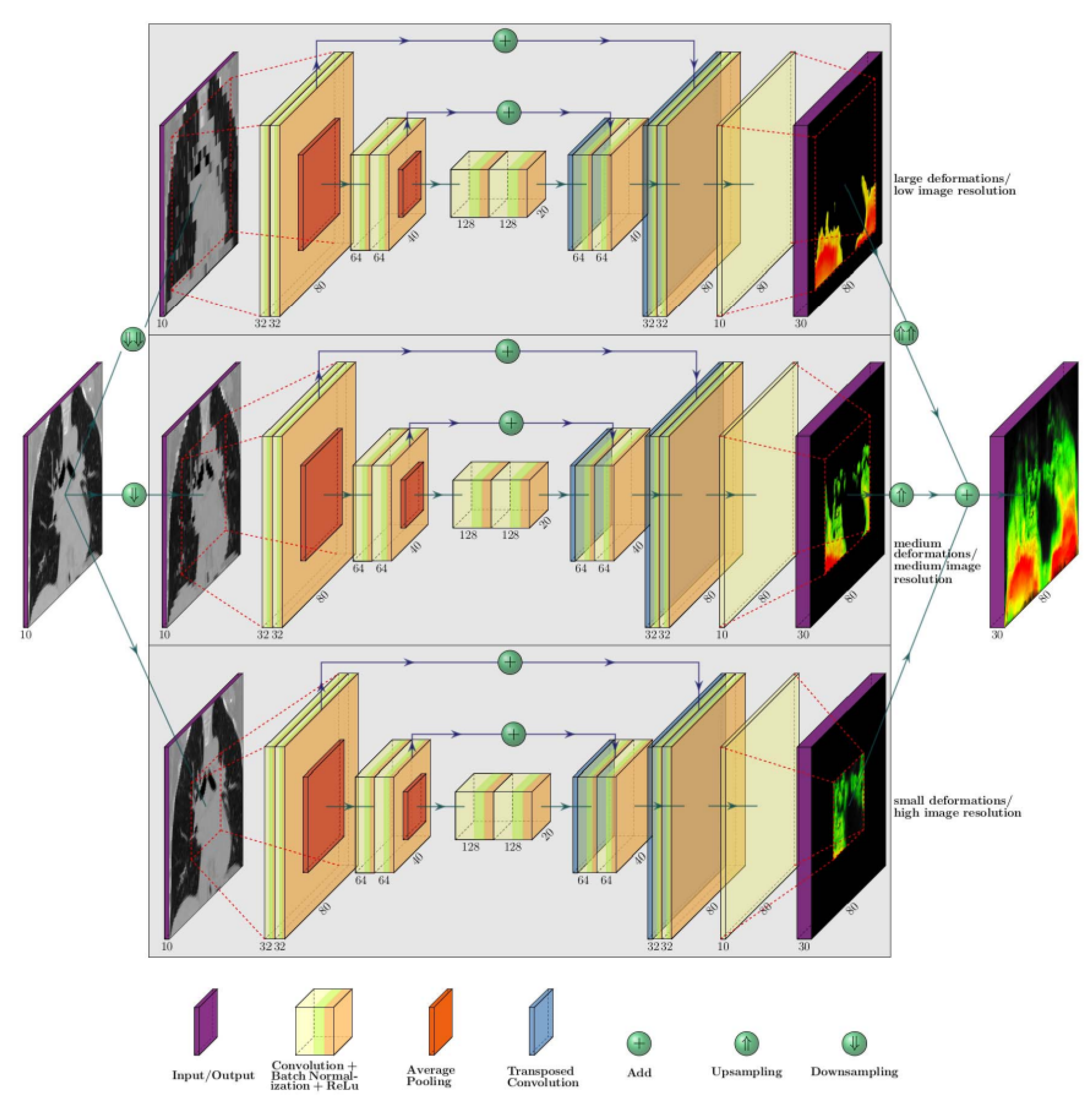
Fechter and Baltas [22] developed a CNN-based framework with three parallel U-Nets, each capturing small, medium, or large-scale deformations. Inputs were resized into multiple resolutions, enabling the network to model broad organ-level motion and fine localized displacements. Evaluations on publicly available POPI and DIR lung datasets, and extensions to cardiac CT, demonstrated improvements in Dice coefficient, Hausdorff Distance, and Average Symmetric Surface Distance.
Han et al. [16] proposed an unsupervised dual-channel deep learning framework for deformable MR–CT image registration, integrating a registration network with a complementary image-synthesis module to improve cross-modality alignment. Their method introduces spatially varying confidence maps to adaptively weight modality contributions, enabling robust registration even in anatomically complex regions of the neurosurgical brain. Quantitative experiments demonstrated improved accuracy compared to existing multi-modal registration baselines, highlighting the effectiveness of combining synthesis and deformation estimation in a unified architecture.
Recent modality-specific developments outside myocardial CTP provide valuable insights. Zhang et al. [8] designed a deep learning network for motion correction in SPECT MPI, improving perfusion defect detection (AUC in reduced-count conditions versus without correction). Kuronuma [9] proposed an automated motion-correction method for dynamic PET-MPI, achieving high agreement with manual correction (r ≥ ) in myocardial blood flow estimates. Urs et al. [23] developed a 3D ResNet-based approach for dynamic PET-MPI motion correction.
2.3. Limitations of Existing Motion-Correction Approaches
Classical methods offer solid algorithmic strategies but often lack quantitative validation in dynamic myocardial CTP. Deep learning approaches have demonstrated strong performance in other modalities and on static datasets, but face challenges generalizing to cardiac perfusion due to the limited availability of annotated datasets. However, no existing framework combines the strengths of deformable registration with perfusion-specific optimization, and it is not validated against standardized metrics such as target registration error (TRE) using reference datasets. Moreover, with few exceptions, existing deformable registration methods are designed for 3D volumetric images rather than 4D perfusion data. Therefore, there remains a need for deformable registration and motion-correction frameworks that are explicitly intended, trained, and validated for dynamic myocardial CTP.
Several recent learning-based approaches have been proposed for motion correction in myocardial CTP imaging, demonstrating improved spatial alignment under cardiac and respiratory motion. However, most of these methods primarily focus on spatial registration accuracy and do not explicitly address preservation of time-enhancement curve (TEC) fidelity, which is critical for perfusion analysis. In addition, many existing approaches rely on iterative optimization strategies or volumetric supervision, which can increase computational complexity and sensitivity to noise in low-dose settings [24]. Therefore, there remains a need for learning-based approaches that explicitly address these limitations.
3. Proposed Method
3.1. One-Shot CNN Registration Framework
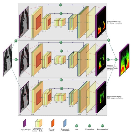
Convolutional Neural Networks (CNNs) have emerged as powerful tools for deformable medical image registration, enabling end-to-end estimation of deformation fields without iterative optimization [22]. Fechter et al. proposed a one-shot multi-scale CNN for 3D registration, consisting of parallel U-Net-like subnetworks that hierarchically capture deformations at different spatial scales. Each subnetwork operates on resampled versions of the fixed and moving volumes, learning motion patterns ranging from coarse displacements to fine anatomical details. Their outputs are fused into a dense displacement field that warps the moving image to align it with the fixed reference. This design achieves efficient one-shot registration while maintaining accuracy across multiple scales. Figure 1 illustrates the architecture.
Figure 1.
CNN-UNet based registration architecture, proposed by Fechter et al. [22].
In this work, we build upon the existing one-shot CNN registration framework proposed by Fechter et al. [22], which employs a CNN-UNet architecture to directly predict dense deformation vector fields between a moving and a fixed image without iterative optimization. The network follows an encoder–decoder design with skip connections and predicts voxel-wise displacement fields at multiple scales. Figure 1 illustrates the CNN-UNet–based baseline registration architecture used in this study.
While initially designed for 3D CT volumes, we extend this framework to a fully 4D spatiotemporal motion-correction method tailored for dynamic myocardial CTP. Unlike iterative optimization-based techniques, our approach performs rapid one-shot deformation estimation and explicitly incorporates temporal consistency, both of which are critical for preserving perfusion dynamics. The core extension lies in (i) combining slice-wise CNN registrations into a unified 4D deformation field and (ii) training with a perfusion-aware loss function so that the single deformation field produced at inference respects time–enhancement curve (TEC) fidelity. Specifically, deformation fields for myocardial CTP are estimated in a slice-wise manner by applying the baseline one-shot CNN to each axial slice across the time axis, and spatial coherence is then introduced by integrating these slice-wise deformation fields across slices using B-spline interpolation. This strategy yields a smooth 4D spatiotemporal deformation field while preserving the efficiency and non-iterative nature of the original 3D framework.
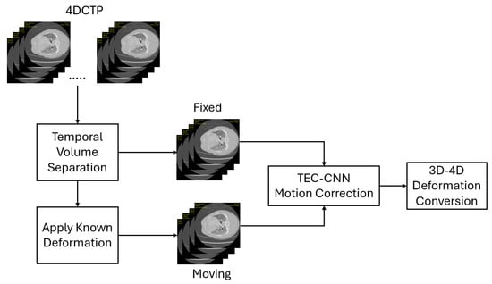
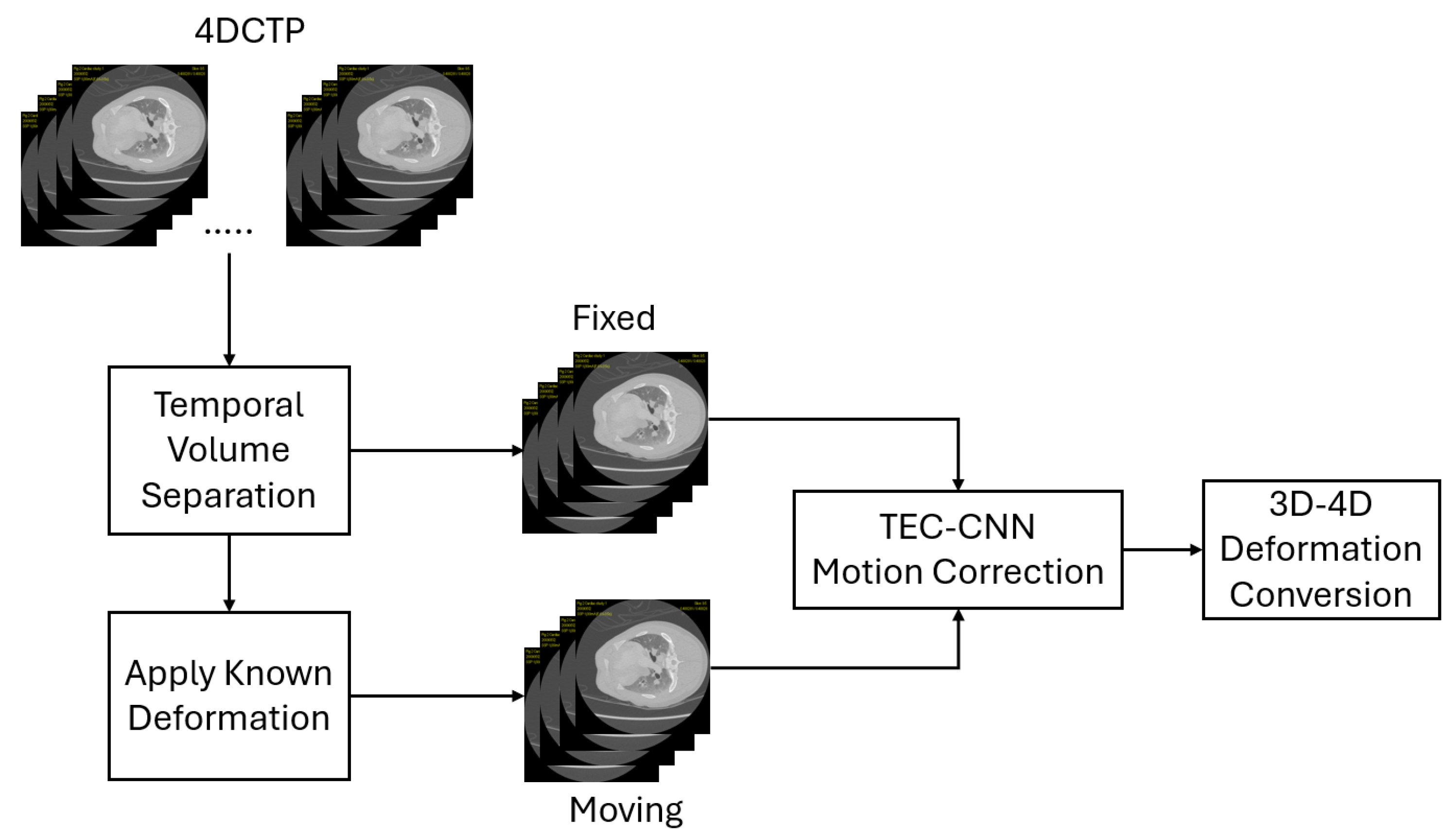
The proposed framework follows a one-shot registration paradigm. Network parameters are learned offline during a separate training phase using supervised optimization. At inference time, motion estimation is performed via a single forward pass through the trained network without any iterative optimization or case-specific parameter tuning. Figure 2 illustrates the proposed extension of the baseline one-shot CNN registration model to dynamic myocardial CTP. The temporal volume is first separated into axial slices along the time axis. Known deformations are then applied to the temporal volume to generate corresponding fixed and moving volumes, as required by the one-shot CNN framework. This process produces a deformation field for each axial slice, which is subsequently interpolated using B-spline interpolation to construct a smooth spatiotemporal deformation vector field. This figure highlights the transition from slice-wise deformation estimation to a unified 4D motion model, demonstrating how deformation fields predicted at individual time frames are interpolated and fused across the temporal dimension. Together, Figure 1 and Figure 2 clarify the progression from the original 3D one-shot registration framework to the proposed 4D motion-correction formulation.
Figure 2.
Proposed 4D motion-correction pipeline (two-stage). The TEC-inspired objective is applied during training, and the model produces the 4D Deformable Vector Field at the end.
3.2. One-Shot 4D Motion-Correction Pipeline
Slice-wise deformation field generation: Each axial slice is treated as a volume of its time series, with the baseline perfusion phase (typically end-diastole) as the fixed frame and subsequent phases as moving frames. The multi-scale CNN is applied to generate deformation fields for each slice. For a CTP dataset with S slices and N time points, this yields S sets of deformation fields (one per slice) encoding motion across time relative to the baseline.
4D field construction: Because slice-wise processing can introduce discontinuities, the estimated deformations are merged into a unified 4D spatiotemporal field using B-spline interpolation of order 3. This enforces continuity across slices and time, yielding physiologically realistic motion trajectories that preserve both anatomical coherence and temporal progression required for perfusion analysis. The slice-wise estimation followed by spatial integration introduces the possibility of error propagation across adjacent slices if localized misregistrations occur. However, the use of B-spline interpolation enforces spatial smoothness across slices, which mitigates the impact of isolated slice-level errors by distributing deformations smoothly in the through-plane direction rather than allowing abrupt discontinuities.
TEC-Influenced Loss Function Design: The original one-shot CNN framework [22] is trained with a composite loss consisting of three terms as shown in Equation (1).
where is an image similarity term (NCC) between the fixed and warped moving volumes, is a spatial smoothness penalty on the gradients of the deformation vector field (DVF), and is a multi-scale consistency constraint ensuring stability across the parallel sub networks. This formulation successfully drives anatomical alignment but does not preserve the fidelity of temporal perfusion signals. To address this, we extend the loss with a fourth component, as shown in the equation.
In Equation (2), is a TEC refinement term, which encourages voxel-wise temporal curves in the warped sequence to remain correlated with those from the fixed reference. This is computed using the correlation coefficient measure between the warped and reference curves for each voxel. No explicit ROI masking or thresholding is applied during training; all voxels contribute equally to the loss formulation. Low similarity is penalized, except in regions of rapid contrast change where variability reflects actual perfusion dynamics. In practice, regions of rapid contrast enhancement are implicitly handled by the correlation-based formulation, which is less sensitive to absolute intensity changes and emphasizes relative temporal trends. This design avoids over-penalizing physiologically valid contrast changes while still discouraging temporally inconsistent deformations.
The weighting coefficients control the trade-off between anatomical alignment, spatial smoothness, multi-scale consistency, and temporal fidelity. While Fechter et al. [22] reported and , we introduce (empirically tuned to be 0.1). The weighting parameters in the loss function were empirically selected during preliminary experiments to balance image similarity, deformation smoothness, and temporal consistency while ensuring stable convergence. Convergence was considered achieved when the absolute difference between the current loss value and the moving average of previous loss values in Equation (2) was less than . Moderate variations around the selected values did not qualitatively change the observed performance trends. A comprehensive sensitivity analysis of these parameters was beyond the scope of this study.
3.3. Data Description
Data Acquisition: Our study utilized porcine cardiac CT perfusion (CTP) datasets acquired at three tube current settings: high-dose (80 mA), mid-dose (40 mA), and low-dose (20 mA). Training, validation, and testing were performed using myocardial CTP datasets acquired at 80 mA and 40 mA dose levels. Data acquired at 20 mA were not included in the present evaluation due to increased noise and degradation, and are explicitly reserved for future investigation. Accordingly, all reported performance results and conclusions are limited to the evaluated dose range. All myocardial CTP scans were acquired using a consistent imaging protocol with a fixed tube voltage of 140 kVp and standardized reconstruction settings. The in-plane voxel spacing was approximately 0.5–0.6 mm, and the slice thickness was 5.00 mm; these values were used consistently for pixel-to-physical unit conversions. Dose level classification is influenced by factors such as exposure duration and tube current. For example, a 20 mA scan with a 0.35 s gantry rotation, 80 kilovolts (kV) and 20 perfusion acquisitions corresponds to a practical exposure of . While 40 mA is typically considered a low dose in humans, the same current may be mid- or high-dose in animal studies. Although this was an animal study, all scans were anonymized before use in training and testing.
Data Splitting: For training, we have 4 myocardial CTP studies with 2240 images each, a total of 8960 images to train the model. For testing, we have 8 myocardial CTP studies available, scanned with different tube current settings ranging from 80 mA for high-dose radiation to 40 mA for low-dose radiation. Table 1 shows the study names and our given aliases to those studies, along with their scan dose.
Table 1.
Summary of Test Data Sets, their Radiation Dose Levels, and Number of Images.
3.4. Training Procedure
To train the motion-correction model, we simulated physiologically plausible motion by applying random nonrigid deformations with a maximum displacement of 3 pixels (approximately 2 mm) to dynamic CTP volumes. The rationale for selecting a maximum displacement of 3 pixels is further detailed in Section 4.1, where this value is related to the physical voxel resolution of the test myocardial CTP images and shown to correspond to realistic residual myocardial motion ranges encountered in practice. Each reference volume was paired with its deformed counterpart, serving as the fixed and moving inputs to the one-shot CNN registration framework. As the name implies, the one-shot model was optimized only on this single pair, without batch training or reliance on large datasets, and directly predicted a deformation field. To capture motion across the entire dynamic sequence, this pairwise registration was repeated for each time frame, yielding slice-specific deformation vector fields.
The one-shot CNN employed a multi-scale architecture that progressively downscaled inputs to capture motion across varying spatial resolutions. Our perfusion-aware loss function (Equation (2)) simultaneously optimized for: (1) anatomical similarity between fixed and warped images, (2) temporal consistency of myocardial Time-Enhancement Curves (TECs), and (3) smoothness of the estimated deformation fields. To ensure spatial and temporal continuity, the slice-specific fields were interpolated using a cubic B-spline scheme, producing a unified 4D spatiotemporal deformation field that preserves both cardiac motion patterns and perfusion kinetics across all phases.
3.5. Challenges in Generalization
Training a deep learning–based motion-correction model for dynamic myocardial CTP presents unique challenges due to interspecific differences in residual motion patterns. While the overall structure and functionality of porcine and human hearts are comparable, their physiological heart rates differ, typically 70 to less than 100 bpm for humans versus 90 to 107 bpm for porcine subjects [25]. This difference alters the temporal characteristics of residual cardiac motion captured in perfusion studies. As a result, a network trained exclusively on porcine datasets may not generalize well to human studies, as it would have learned motion patterns not fully representative of human cardiac dynamics. Therefore, motion correction performance in this setting should be interpreted as a conservative evaluation of the proposed method rather than direct evidence of clinical readiness. In addition, due to increased noise and degradation associated with low-dose myocardial CTP imaging, the present study limits the evaluation of the known-deformation experiments to dose levels down to 40 mA.
4. Results
4.1. Target Registration Error (TRE)
Target Registration Error (TRE) quantifies the error in registration of anatomical landmarks between reference and reconstructed images. It is defined by (3).
where and denote the ground truth and registered landmark positions, respectively, and N is the number of landmarks.
Calculating the Target Registration Error (TRE) is not straightforward. Metrics such as MSE, PSNR, and SSIM are computed per image, independent of the spatial locations of anatomical structures. TRE, on the other hand, requires the identification of specific anatomical landmarks, or virtual fiducial points, followed by the collection of their corresponding spatial coordinates from both the reference and registered image sets. The positional differences between these landmarks are then used to quantify the registration error.
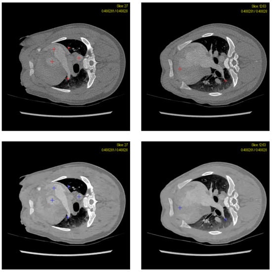
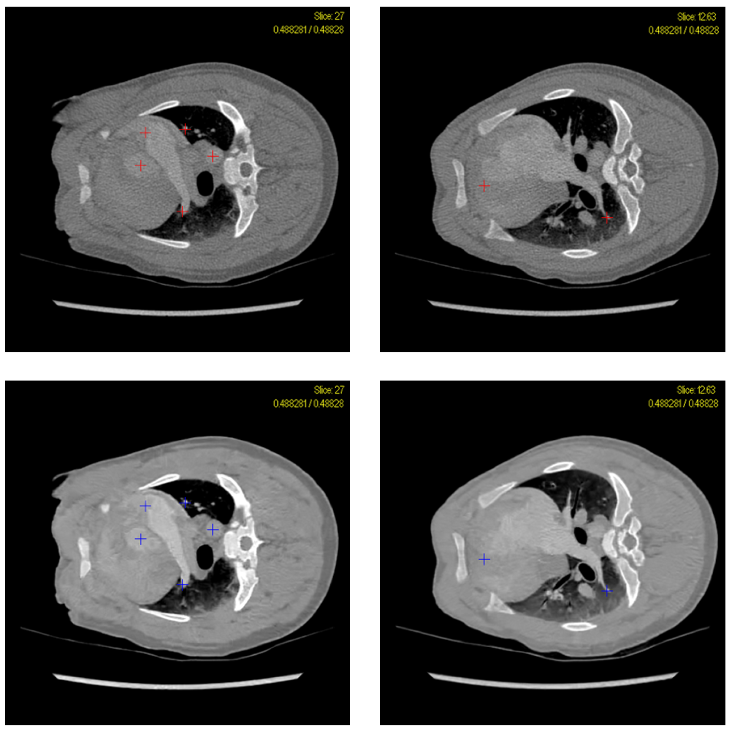
To facilitate this process, eight anatomical landmarks within the myocardium were selected, as listed in Table 2. These anatomical landmarks were used consistently across all datasets and experiments for TRE evaluation. Landmarks were manually annotated by an experienced observer, following consistent anatomical selection criteria, focusing on clearly identifiable, temporally stable structures visible across all frames. The locations of these landmark points were verified by an independent expert (see acknowledgment section). The exact landmark locations were tracked across time for each sequence to ensure consistent TRE computation. Figure 3 shows the visual annotation of these structures using colored markers. The top row displays two representative slices from the reference image stack: one near the top (left) and one from the middle (right), highlighting all the anatomical points listed in Table 2. The bottom row shows the corresponding slices (the top slice on the left and the middle slice on the right) from the registered image stack. For each test case, these landmarks were manually annotated in both the reference and registered images, and their coordinates were subsequently used to compute TRE.
Table 2.
List of Anatomical Locations for TRE Calculation.
Figure 3.
Annotated CT Slice denoting anatomies selected for TRE calculation. The red marks on top row indicate the selected locations on reference images and the blue marks on the bottom row indicate the selected locations on registered images.
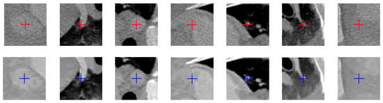
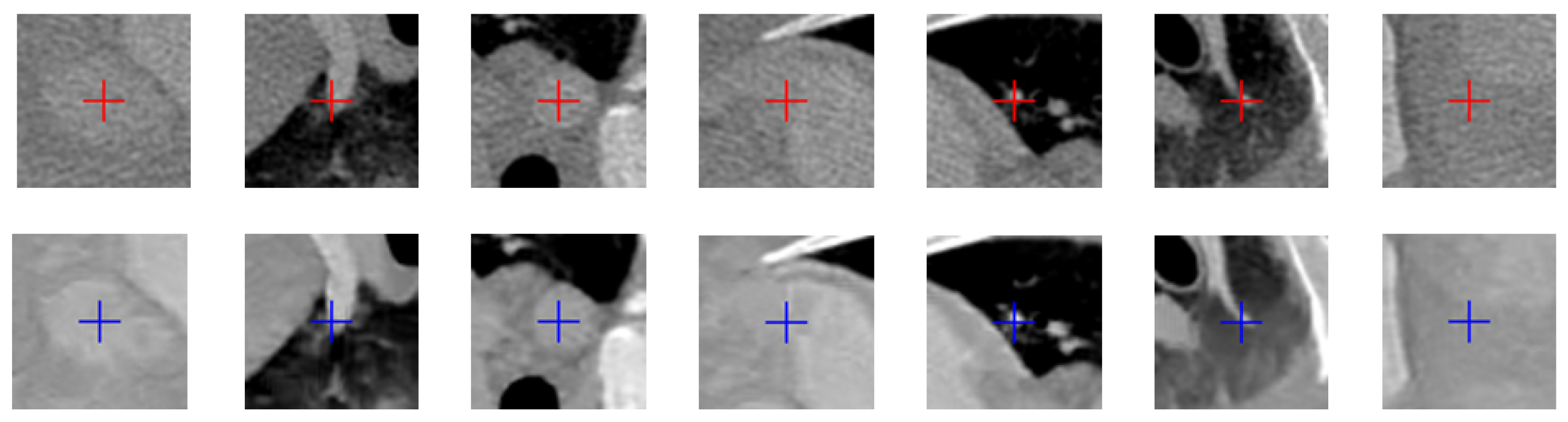
Figure 4 presents a zoomed view of the annotated anatomical points used for TRE calculation. The top row shows the annotated landmarks in the reference image set, while the bottom row displays the corresponding points in the registered image set.
Figure 4.
Zoomed annotated CT Slice denoting anatomies selected for TRE calculation. The red marks on top row indicate the selected locations on reference images and the blue marks on the bottom row indicate the selected locations on registered images.
Table 3 summarizes the Target Registration Error (TRE) values for the synthetic deformation experiments. To simulate nonrigid motion, a known random deformation of 3 pixels was synthetically applied to the image set. This deformation magnitude was selected for two primary reasons. First, our registration model was explicitly trained to accommodate deformations up to 3 pixels, ensuring that the test data remained within the network’s learned deformation regime. Second, considering that the voxel size in standard CT scanners typically ranges from to , and our model was trained using a voxel resolution of ∼0.5–0.6 mm, a 3-pixel displacement effectively corresponds to a ∼1.5–1.8 mm deformation—well within the physical motion range encountered in practical scanning scenarios. The resulting deformed images were subsequently registered back to their original reference using our proposed deformable registration model. A set of 7 soft fiducial points was selected across anatomically relevant regions to evaluate registration accuracy. These points served as ground truth landmarks for calculating the TRE before and after deformation.
Table 3.
Target Registration Error (TRE) for Registered vs. Reference Test Cases.
The TRE values quantify the residual misalignment between corresponding anatomical landmarks in the reference and registered images. In the ideal case of perfect registration, the TRE would be zero, indicating complete correction of the motion artifact. Conversely, a TRE equal to or exceeding the magnitude of the applied deformation would imply registration failure. As shown in Table 3, the TRE in all test cases remained below the applied 3-pixel deformation, demonstrating that the model was able to recover a substantial portion of the synthetic motion. These results highlight the effectiveness of the proposed model in mitigating motion-induced misalignments and producing spatially accurate reconstructions.
4.2. Pearson Correlation Coefficient
Pearson Correlation Coefficient measures similarity in curve shape, independent of scale. It is defined in Equation (4).
where and represent the time-enhancement curves (TECs) of the reference and motion-corrected datasets, respectively, and denote their mean intensities over the N temporal phases, and r quantifies linear correlation with values in . In signal analysis, is typically interpreted as strong similarity, whereas reflects weaker agreement that may indicate underlying discrepancies [26]. Therefore, in myocardial CTP analysis, is expected to indicate preserved temporal patterns, while suggests temporal misalignment requiring correction. We computed Pearson correlation coefficients between the registered and reference Time-Enhancement Curves (TECs) in the descending Aorta across all test cases. As visible from Table 4, the proposed method consistently achieved high temporal fidelity, with correlation coefficients averaging . A single outlier (STC4, ) exhibited reduced performance, likely due to residual respiratory motion artifacts during peak contrast enhancement. During peak enhancement, the contrast-to-noise ratio and the HU gradient between the blood pool and myocardium are at their maximum; consequently, even small residual misalignments can disproportionately affect voxel-wise temporal correlation metrics due to the rapid change in intensity. This case highlights a specific challenging low-performance scenario rather than the typical performance of the proposed method, as demonstrated by consistently higher correlation values observed across the remaining test cases. These observations highlight the sensitivity of temporal metrics to extreme motion conditions and motivate further investigation of motion-robust strategies in future work.
Table 4.
Correlation Coefficient from Registered Test Cases and respective Reference Test Cases for Descending Aorta.
4.3. Time Enhancement Curve Comparison
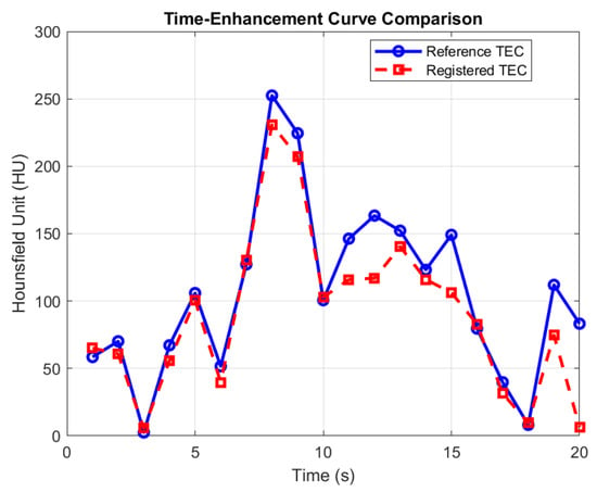
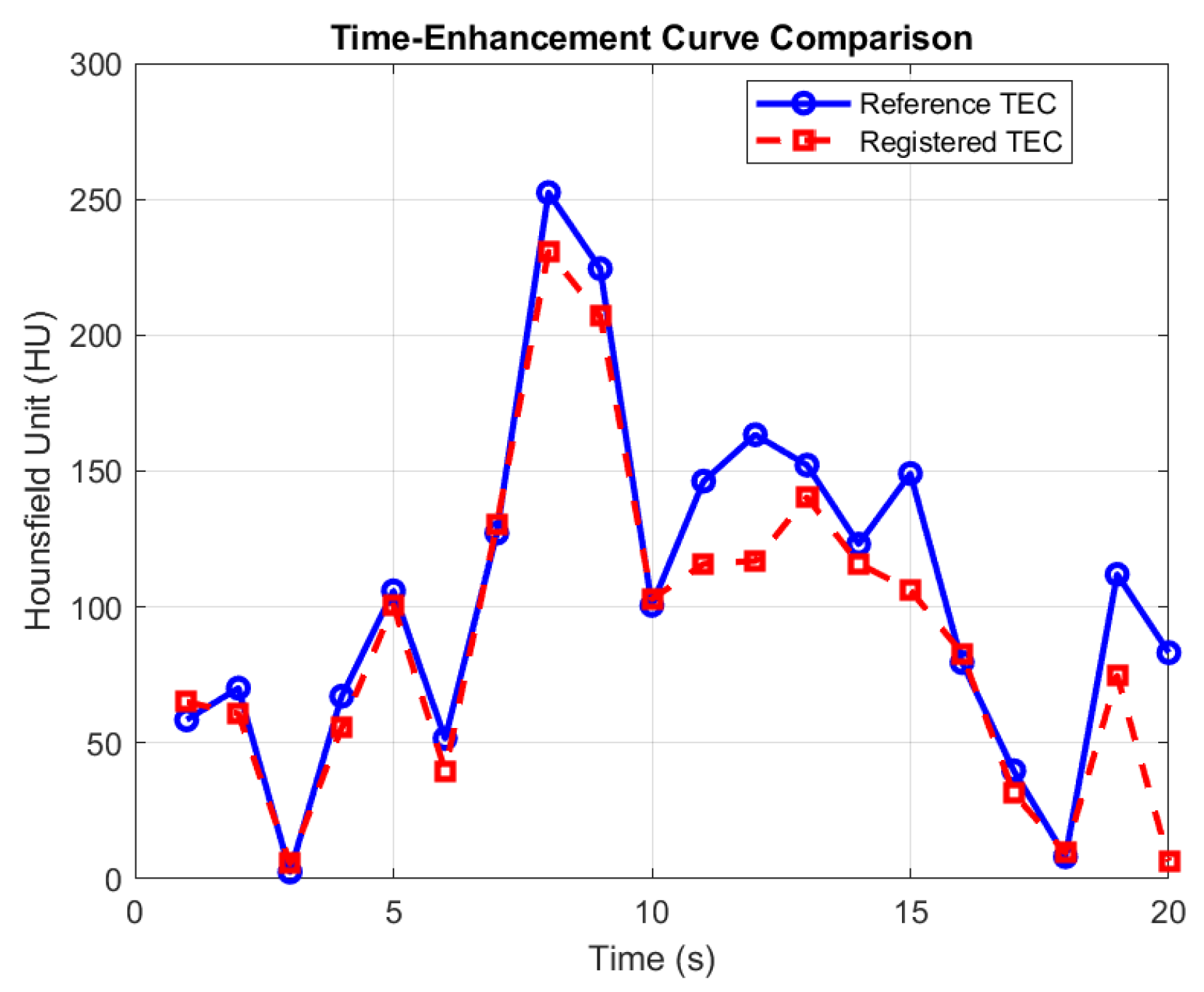
Figure 5 compares the Time-Enhancement Curves (TECs) between the reference and registered images for test case STC5, demonstrating excellent temporal fidelity with a Pearson Correlation Coefficient (PCC) of . The motion-correction framework successfully preserved characteristic wash-in and wash-out patterns relevant to downstream myocardial perfusion analysis. PCC meets diagnostic reliability thresholds in perfusion analysis, and high correlation during rapid contrast changes (e.g., peak enhancement at ) confirms motion artifact resilience. Minor deviations (t = 11–13) may reflect slight misalignment during the late diastolic phase, when cardiac motion becomes less predictable. Clinically, such deviations are usually negligible for MBF quantification.
Figure 5.
Comparison of Time-Enhancement Curves (TECs) between reference (blue solid line) and registered (red dashed line) images for test case STC5.
4.4. Comparative Performance
Although direct comparisons across registration studies are challenging due to substantial differences in anatomy, imaging protocols, and motion characteristics, the TRE values in Table 5 provide valuable insights. Prior works based on lung CT or 4D respiratory datasets, such as Hering et al. [15], Jiang et al. [17], Lei et al. [21], and Fechter & Baltas [22], were evaluated on benchmark datasets (e.g., DIR-Lab, POPI) where image quality is relatively high, and motion is predominantly respiratory. In contrast, our low-dose myocardial CTP data involve dynamic contrast enhancement, increased noise levels, and simultaneous cardiac–respiratory motion, all of which substantially increase registration difficulty.
Table 5.
Representative Target Registration Error (TRE) values reported in recent CT-based deformable image registration studies. These works are not myocardial CTP applications but are included to contextualize the order of magnitude of TRE achieved by the proposed method.
Despite these challenges, the proposed method achieves a mean TRE of 1.08 ± 0.63 mm, which is competitive with the lower end of the range reported by established approaches. Although a direct comparison is infeasible, this result demonstrates that our framework achieves deformation accuracy comparable to that of state-of-the-art CT registration methods, despite being acquired under challenging imaging conditions. Furthermore, while our architecture uses Fechter & Baltas [22] as a baseline, the incorporation of the TEC-guided loss term provides a measurable performance improvement over the original formulation.
Direct numerical comparison with recent deep learning–based myocardial CTP motion-correction methods was not performed, as many existing approaches are evaluated on proprietary datasets, employ different motion models, or report task-specific metrics that are not directly comparable to TRE. Similarly, classical iterative deformable registration methods typically require dataset-specific parameter tuning and iterative optimization, making controlled comparison on identical data challenging.
From a computational perspective, the proposed framework differs fundamentally from classical iterative registration approaches. Motion estimation is performed through a single forward pass of a trained network, without iterative refinement or optimization at inference time. In contrast, classical deformable registration methods rely on repeated optimization steps to converge, with computational complexity that increases with image resolution, deformation complexity, and the number of time frames. The one-shot formulation, therefore, offers a practical efficiency advantage in dynamic myocardial CTP settings, particularly for long temporal sequences, even though explicit runtime measurements are not reported in this study. We emphasize that the efficiency advantage discussed here is based on the non-iterative design of the proposed framework rather than direct empirical runtime comparison.
4.5. Impact of Noise
Although the proposed framework is designed to operate across a wide range of high-to-low dose imaging conditions, performance evaluation in this study was limited to dose levels between 80 mA and 40 mA. Performance beyond this range was not evaluated. The impact of noise on registration accuracy is therefore indirectly reflected in the reported TRE and TEC correlation results across these dose levels. In general, lower signal-to-noise conditions introduce increased uncertainty in local feature correspondence, which can degrade deformation estimation and temporal correlation metrics. Nevertheless, the proposed method maintains consistent performance across the evaluated dose range, indicating robustness to noise-induced degradation. These results suggest that while ultra-low-dose noise remains a challenging factor for motion correction, the proposed framework can preserve structural alignment and temporal consistency under moderate-to-low noise levels.
5. Limitations
Despite the potential of the proposed method, several limitations of the present study should be acknowledged. First, the evaluation in this study focuses on landmark-based TRE and temporal alignment consistency metrics on real myocardial CTP sequences. Segmentation-based anatomical consistency measures, such as ROI overlap, boundary error, or key-structure alignment, were not included. In addition, direct comparison with classical iterative and learning-based registration baselines on identical data was not performed. Such comprehensive benchmarking, including ROI overlap, boundary error analysis, and multi-baseline comparison, represents important future work and would require additional annotations and implementation beyond the scope of the present study.
Second, ablation studies isolating the contribution of the TEC-guided loss term, systematic variation of , and alternative ROI strategies were not performed in this study, since the primary objective was to demonstrate the feasibility and effectiveness of integrating temporal consistency into a one-shot registration framework under controlled conditions. In addition, deformation field sanity metrics, such as Jacobian determinant statistics or folding rates, were not explicitly evaluated, and architectural ablations comparing slice-wise estimation to fused 4D integration or to fully volumetric 3D/4D approaches were beyond the scope of this study.
Third, the TRE evaluation in this study is based on manually annotated landmarks under synthetically applied limited deformations. While this ensures controlled, interpretable benchmarking, it may not accurately reflect real-world motion or physical landmarks. Additionally, formal statistical significance testing and confidence interval estimation were not performed due to the limited number of test cases and the controlled nature of the synthetic deformation experiments.
Fourth, explicit wall-clock runtime and memory usage were not systematically recorded during the original experiments. Direct runtime comparison against conventional iterative deformable registration baselines on identical data was therefore not performed and is left for future work. All experiments were conducted on NVIDIA V100 GPU nodes with 32 GB of VRAM. Because the proposed method performs registration via a single forward pass without iterative refinement, inference-time complexity is fixed and independent of convergence criteria, in contrast to classical iterative deformable registration methods. Future work will include detailed runtime and memory benchmarking under standardized hardware configurations.
Finally, the Pearson correlation analysis in this study was limited to the descending aorta, which provides a stable reference for contrast dynamics but does not fully reflect myocardial perfusion behaviour.
6. Conclusions
This study focused on developing and evaluating a deep learning–based deformable registration framework for motion correction in dynamic myocardial CTP imaging. The method was designed to address residual cardiac and respiratory motion that persists despite ECG gating and breath-hold protocols, thereby improving temporal alignment for accurate myocardial blood flow estimation. A multi-scale CNN architecture, inspired by prior work [22], was employed to capture both large-scale and localized myocardial deformations.
Experiments were conducted using porcine CTP datasets, which share anatomical similarities with human hearts but differ in heart rate and motion patterns, highlighting the importance of domain-specific retraining for clinical translation. The proposed approach was evaluated using anatomical landmarks to compute Target Registration Error (TRE) and the Pearson Correlation Coefficient (PCC), which provide quantitative measures of spatial and temporal alignment in the absence of ground-truth motion fields. In addition, temporal fidelity was assessed using perfusion curve correlation, ensuring that voxel-wise time-intensity dynamics were preserved after registration. We observed that incorporating the TEC correlation term () yielded PCC values consistently above across test cases. This indicates that the TEC-guided constraint successfully maintained temporal coherence, whereas frameworks without such terms often show curve decorrelation () in low-dose data.
Together, these evaluations demonstrated that the proposed method effectively reduced residual motion and improved temporal consistency across frames, thereby enhancing the reliability of myocardial perfusion metrics. Overall, the framework presents a promising step toward robust, efficient, and data-driven motion correction for dynamic myocardial CTP imaging.
7. Future Directions
Future work will focus on retraining the model using human CTP data to capture human-specific motion characteristics better and improve generalization. Cross-species validation using human myocardial CTP data will therefore be an important direction for future work. In addition, extending the validation to include 20 mA dose studies will be investigated in future. Comprehensive ablation and sensitivity analyses remain important future work to further characterize the relative contributions of individual loss components and ROI choices. Additionally, exploring advanced loss functions that incorporate image similarity, temporal smoothness, and biomechanical constraints could further enhance model robustness. Furthermore, future work will extend the evaluation to larger deformation magnitudes and to physical landmark-based evaluation. Also, future work will extend TEC analysis to myocardial regions to more directly evaluate perfusion preservation.
Expanding evaluations to include complete cardiac-cycle acquisitions and diverse patient populations will be essential to assess clinical performance under real-world variability. Moreover, due to the inherently noisy nature of low-dose myocardial CTP data [27], addressing noise-induced degradation remains a significant future challenge for improving motion correction accuracy and overall image quality.
Author Contributions
Conceptualization, M.H., A.S. and M.R.E.-S.; methodology and software/computer program, M.H.; validation, M.R.E.-S., A.S. and M.H.; investigation, M.H.; resources, M.H., M.R.E.-S. and A.S.; data acquisition, A.S.; writing—original draft preparation, M.H.; writing—review and editing, M.R.E.-S. and A.S.; supervision, M.R.E.-S. All authors have read and agreed to the published version of the manuscript.
Funding
This research received no external funding.
Institutional Review Board Statement
Project identification code (Protocol Approval Number): #2009-092; Date of approval: 2009; Name of the ethics committee/institutional review board: The University of Western Ontario, Animal Use Subcommittee/University Council on Animal Care.
Data Availability Statement
The original contributions presented in this study are included in the article. Further inquiries can be directed to the corresponding authors.
Acknowledgments
The authors would like to thank Tamanna Zahan (MBBS, MD -Radiology and Imaging), Department of Radiology and Imaging, Holy Family Red Crescent Medical College & Hospital, Bangladesh, for her valuable contribution as a subject matter expert in the TRE evaluation of the registered CT perfusion images.
Conflicts of Interest
The authors declare no conflicts of interest.
Abbreviations
The following abbreviations are used in this manuscript:
| CTP | Computed Tomography Perfusion |
| DVF | Deformation Vector Field |
| ECG | Electrocardiography/Electrocardiogram |
| HoG | Histogram of Oriented Gradients |
| LDCT | Low-Dose Computed Tomography |
| MBF | Myocardial Blood Flow |
| MI | Motion Immune |
| MPI | Myocardial Perfusion Imaging |
| NCC | Normalized Cross Correlation |
| PET | Positron Emission Tomography |
| SPECT | Single-Photon Emission Computed Tomography |
| TEC | Time Enhancement Curve |
| TRE | Target Registration Error |
References
- Nieman, K.; Balla, S. Dynamic CT myocardial perfusion imaging. J. Cardiovasc. Comput. Tomogr. 2022, 14, 303–306. [Google Scholar] [CrossRef] [Scilit]
- Nous, F.M.A.; van Assen, M.; van Rosendael, A.R.; Alkadhi, H.; Kitagawa, K.; Vliegenthart, R.; Hell, M.M.; Hausleiter, J.; Nguyen, P.K.; Budde, R.P.; et al. Dynamic Myocardial Perfusion CT for the Detection of Hemodynamically Significant Coronary Artery Disease. JACC Cardiovasc. Imaging 2022, 15, 75–87. [Google Scholar] [CrossRef] [Scilit] [PubMed]
- Muscogiuri, G.; Palumbo, P.; Kitagawa, K.; Nakamura, S.; Senatieri, A.; De Cecco, C.N.; Gershon, G.; Chierchia, G.; Usai, J.; Sferratore, D.; et al. State of the Art of CT Myocardial Perfusion. La Radiol. Medica 2025, 130, 438–452. [Google Scholar] [CrossRef] [Scilit] [PubMed]
- Mooiweer, R.; Neji, R.; McElroy, S.; Nazir, M.S.; Razavi, R.; Chiribiri, A.; Roujol, S. A Fast Navigator (fastNAV) for Prospective Respiratory Motion Correction in First-Pass Myocardial Perfusion Imaging. Magn. Reson. Med. 2021, 85, 2661–2671. [Google Scholar] [CrossRef] [Scilit]
- Otaki, Y.; Van Kriekinge, S.D.; Wei, C.C.; Kavanagh, P.; Singh, A.; Parekh, T.; Di Carli, M.; Maddahi, J.; Sitek, A.; Buckley, C.; et al. Improved Myocardial Blood Flow Estimation with Residual Activity Correction and Motion Correction in 18F-Flurpiridaz PET Myocardial Perfusion Imaging. Eur. J. Nucl. Med. Mol. Imaging 2022, 49, 1881–1893. [Google Scholar] [CrossRef] [Scilit]
- Hubbard, L.; Malkasian, S.; Molloi, S. Dynamic CT Myocardial Perfusion without Image Registration. Sci. Rep. 2022, 12, 12608. [Google Scholar] [CrossRef] [Scilit]
- Benovoy, M.; Jacobs, M.; Cheriet, F.; Dahdah, N.; Arai, A.E.; Hsu, L.-Y. Robust Universal Nonrigid Motion Correction Framework for First-Pass Cardiac MR Perfusion Imaging. J. Magn. Reson. Imaging 2017, 46, 1060–1072. [Google Scholar] [CrossRef] [Scilit]
- Zhang, X.; Yang, Y.; Pretorius, P.H.; Slomka, P.J.; King, M.A. Cardiac Motion Correction with a Deep Learning Network for Perfusion Defect Assessment in Single-Photon Emission Computed Tomography Myocardial Perfusion Imaging. JACC Nucl. Cardiol. 2025, 43, 102071. [Google Scholar] [CrossRef] [Scilit]
- Kuronuma, K.; Wei, C.C.; Singh, A.; Lemley, M.; Hayes, S.W.; Otaki, Y.; Hyun, M.C.; Van Kriekinge, S.D.; Kavanagh, P.; Huang, C.; et al. Automated Motion Correction for Myocardial Blood Flow Measurements and Diagnostic Performance of 82Rb PET Myocardial Perfusion Imaging. J. Nucl. Med. 2024, 65, 139–146. [Google Scholar] [CrossRef] [Scilit]
- Nenoff, L.; Amstutz, F.; Murr, M.; Archibald-Heeren, B.; Fusella, M.; Hussein, M.; Lechner, W.; Zhang, Y.; Sharp, G.; Vasquez Osorio, E. Review and Recommendations on Deformable Image Registration Uncertainties for Radiotherapy Applications. Phys. Med. Biol. 2023, 68, 24TR01. [Google Scholar] [CrossRef] [Scilit]
- Chen, J.; Liu, Y.; Wei, S.; Bian, Z.; Subramanian, S.; Carass, A.; Prince, J.L.; Du, Y. A Survey on Deep Learning in Medical Image Registration: New Technologies, Uncertainty, Evaluation Metrics, and Beyond. Med. Image Anal. 2025, 100, 103385. [Google Scholar] [CrossRef] [Scilit]
- Haskins, G.; Kruger, U.; Yan, P. Deep Learning in Medical Image Registration: A Survey. Mach. Vis. Appl. 2020, 31, 8. [Google Scholar] [CrossRef] [Scilit]
- Chen, X.; Diaz-Pinto, A.; Ravikumar, N.; Frangi, A.F. Deep Learning in Medical Image Registration. Prog. Biomed. Eng. 2021, 3, 012003. [Google Scholar] [CrossRef] [Scilit]
- Xu, X.; Wang, P.; Zhao, L.; Quan, G. CT-Value Conservation Based Spatial Transformer Network for Cardiac Motion Correction. X-Ray Spectrom. 2024, 53, 382–391. [Google Scholar] [CrossRef] [Scilit]
- Hering, A.; Häger, S.; Moltz, J.; Lessmann, N.; Heldmann, S.; van Ginneken, B. CNN-Based Lung CT Registration with Multiple Anatomical Constraints. Med. Image Anal. 2021, 72, 102139. [Google Scholar] [CrossRef] [Scilit] [PubMed]
- Han, R.; Jones, C.K.; Lee, J.; Wu, P.; Vagdargi, P.; Uneri, A.; Helm, P.A.; Luciano, M.; Anderson, W.S.; Siewerdsen, J.H. Deformable MR–CT Image Registration Using an Unsupervised, Dual-Channel Network for Neurosurgical Guidance. Med. Image Anal. 2022, 75, 102292. [Google Scholar] [CrossRef] [Scilit] [PubMed]
- Jiang, Z.; Yin, F.-F.; Ge, Y.; Ren, L. A Multi-Scale Framework with Unsupervised Joint Training of Convolutional Neural Networks for Pulmonary Deformable Image Registration. Phys. Med. Biol. 2020, 65, 015011. [Google Scholar] [CrossRef] [Scilit]
- Criscuolo, E.R.; Fu, Y.; Hao, Y.; Zhang, Z.; Yang, D. A Comprehensive Lung CT Landmark Pair Dataset for Evaluating Deformable Image Registration Algorithms. Med. Phys. 2024, 51, 3806–3817. [Google Scholar] [CrossRef] [Scilit]
- Lara-Hernandez, A.; Rienmüller, T.; Juárez, I.; Pérez, M.; Reyna, F.; Baumgartner, D.; Makarenko, V.N.; Bockeria, O.L.; Maksudov, M.; Rienmüller, R.; et al. Deep Learning-Based Image Registration in Dynamic Myocardial Perfusion CT Imaging. IEEE Trans. Med. Imaging 2022, 42, 684–696. [Google Scholar] [CrossRef] [Scilit]
- Huang, H.; Siewerdsen, J.H.; Zbijewski, W.; Weiss, C.R.; Unberath, M.; Ehtiati, T.; Sisniega, A. Reference-Free Learning-Based Similarity Metric for Motion Compensation in Cone-Beam CT. Phys. Med. Biol. 2022, 67, 125020. [Google Scholar] [CrossRef] [Scilit]
- Lei, Y.; Fu, Y.; Wang, T.; Momin, S.; Roper, J.; Patel, P.; Curran, W.J.; Liu, T.; Yang, X. 4D-CT Deformable Image Registration Using Multiscale Unsupervised Deep Learning. Phys. Med. Biol. 2020, 65, 085003. [Google Scholar] [CrossRef] [Scilit]
- Fechter, T.; Baltas, D. One-shot multi-scale CNN for deformable registration in CT. IEEE Trans. Med. Imaging 2020, 39, 2506–2517. [Google Scholar] [CrossRef] [Scilit]
- Urs, M.; Killekar, A.; Builoff, V.; Lemley, M.; Wei, C.C.; Kavanagh, P.; Slomka, P.J. Deep-Learning-Based Automatic Patient Motion Correction for 18F-Flurpiridaz PET-MPI. In Medical Imaging 2025: Image Processing; SPIE: Bellingham, WA, USA, 2025; Volume 13406, pp. 732–738. [Google Scholar] [CrossRef] [Scilit]
- Ketcha, M.D.; De Silva, T.; Han, R.; Uneri, A.; Goerres, J.; Jacobson, M.W.; Vogt, S.; Kleinszig, G.; Siewerdsen, J.H. Effects of Image Quality on the Fundamental Limits of Image Registration Accuracy. IEEE Trans. Med. Imaging 2017, 36, 1997–2009. [Google Scholar] [CrossRef] [Scilit]
- Ou, Q.; Jacobson, Z.; Abouleisa, R.R.; Tang, X.L.; Hindi, S.M.; Kumar, A.; Mohamed, T.M. Physiological Biomimetic Culture System for Pig and Human Heart Slices. Circ. Res. 2019, 125, 628–642. [Google Scholar] [CrossRef] [Scilit]
- Akoglu, H. User’s guide to correlation coefficients. Turk. J. Emerg. Med. 2018, 18, 91–93. [Google Scholar] [CrossRef] [Scilit]
- Hasan, M.; So, A.; El-Sakka, M.R. DnCNN-Based Denoising Model for Low-Dose Myocardial CT Perfusion Imaging. Electronics 2026, 15, 124. [Google Scholar] [CrossRef] [Scilit]

Disclaimer/Publisher’s Note: The statements, opinions and data contained in all publications are solely those of the individual author(s) and contributor(s) and not of MDPI and/or the editor(s). MDPI and/or the editor(s) disclaim responsibility for any injury to people or property resulting from any ideas, methods, instructions or products referred to in the content. |
© 2026 by the authors. Licensee MDPI, Basel, Switzerland. This article is an open access article distributed under the terms and conditions of the Creative Commons Attribution (CC BY) license.