


Abstract
Cerebrovascular diseases, including stroke and intracranial aneurysm, affect millions worldwide and remain a leading cause of mortality and disability. Time-of-Flight Magnetic Resonance Angiography (TOF-MRA) enables non-invasive visualization of intracranial arteries. However, the complex cerebrovascular anatomy, characterized by variable diameters, tortuous trajectories, and intricate branching, renders manual segmentation time-consuming, subjective, and prone to inter-observer variability. While deep learning models achieve strong segmentation performance, existing 3D approaches typically require millions of parameters, limiting deployment in resource-constrained clinical settings. To address this challenge, this paper proposes a Lightweight Intracranial Vascular Segmentation Network (LIVAS-Net), a parameter-efficient 3D encoder–decoder architecture using 3D Ghost convolution modules. It incorporates a novel Vessel Continuity Refinement Branch (VCRB), which aims to correct discontinuities in logit space through per-voxel learnable gating. Two model variants are introduced, LIVAS-Net (129K parameters, 18.3 GFLOPs) and LIVAS-L Net (2.97M parameters, 87.8 GFLOPs), achieving 7.9× and 1.6× fewer FLOPs than the standard 3D U-Net (144.5 GFLOPs), respectively. Evaluation on the multi-center COSTA benchmark shows a DSC of 0.8943 (HD95: 1.97 mm) and 0.9235 (HD95: 0.77 mm) on the ADAM test set, outperforming 3D U-Net (DSC: 0.8762). Cross-center evaluation on three external COSTA datasets yields overall DSCs of 0.7834 and 0.7967 versus 0.6998 for 3D UNet. Further evaluation on the CereVessMRA dataset (N = 271) reveals that LIVAS-Net achieves the highest DSC (0.669), demonstrating promising experimental results warranting future clinical validation in resource-constrained settings.
1. Introduction
Cerebrovascular diseases constitute a significant global health burden [1], accounting for approximately 6.5 million deaths annually and representing the second leading cause of mortality and long-term disability worldwide [2]. Conditions such as ischemic stroke, intracranial aneurysm, and arteriovenous malformation demand accurate delineation of cerebrovascular anatomy for diagnosis and visualization of abnormalities, surgical planning, and monitoring [3]. In this context, image segmentation plays a crucial role in enabling detailed analysis of complex vascular structures [4]. In clinical neuroimaging, Time-of-Flight Magnetic Resonance Angiography (TOF-MRA) is widely used as a non-invasive and contrast-free modality for visualizing intracranial arteries through flow-related enhancement [5]. Consequently, automated segmentation of cerebrovascular structures from TOF-MRA has become an important research problem for quantitative vascular analysis and computer-assisted diagnosis.
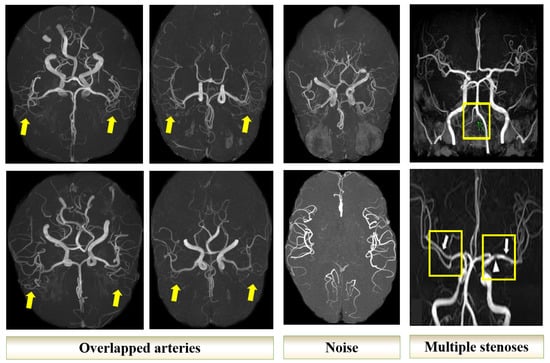
Despite recent advances, accurate cerebrovascular segmentation from TOF-MRA remains challenging. First, the intracranial vascular system consists of thin, elongated, and highly intertwined vessels with severe foreground–background imbalance, where distal branches may span only 1–2 voxels in diameter [6]. Second, vascular abnormalities such as aneurysmal dilatation and focal stenosis may occur at any location within the vascular tree, requiring comprehensive inspection of the entire vasculature, as illustrated in Figure 1 [7]. Third, current clinical workflows typically rely on sequential two-dimensional slice inspection, requiring clinicians to mentally reconstruct complex three-dimensional (3D) vascular anatomy across numerous slices, which is both time-consuming and cognitively demanding. Fourth, subtle morphological changes may extend across multiple slices and remain difficult to detect within individual axial views, increasing the risk of missed diagnoses [8]. Fifth, although Maximum Intensity Projection (MIP) visualization partially alleviates this limitation, overlapping vascular structures and projection artifacts continue to complicate manual interpretation. Furthermore, multi-center TOF-MRA data acquired using different scanners and imaging protocols introduce substantial variability in intensity distributions and spatial resolution, posing additional challenges for robust automated segmentation [9,10].
Figure 1.
Illustration of the clinical visualization challenges in TOF-MRA. Yellow arrows on the highlighted regions that contains overlapping arteries, while yellow boxes indicate the stenotic arteries.
To address these challenges, automated delineation of intracranial arteries through 3D segmentation provides an effective pathway for quantitative vascular analysis that can support the extraction of imaging biomarkers for disease identification. However, deep learning (DL)-based approaches typically rely on heavily parameterized architectures with substantial computational and memory requirements, limiting their deployment in resource-constrained clinical environments and edge computing scenarios.
To address these limitations, this paper proposes the Lightweight Intracranial Vascular Segmentation Network (LIVAS-Net), a parameter-efficient 3D encoder–decoder architecture for cerebrovascular segmentation from TOF-MRA. The proposed framework introduces an anisotropic 3D Ghost convolution design that decomposes feature generation into a pointwise intrinsic convolution followed by a lightweight grouped depthwise operation, substantially reducing computational complexity while preserving in-plane spatial representation. In addition, a Vessel Continuity Refinement Branch (VCRB) is introduced to refine vessel probability maps and improve vascular continuity in predicted segmentations. Alongside the ultra-lightweight LIVAS-Net, a wider variant termed LIVAS-L Net is also developed using standard isotropic Ghost modules to provide a higher-capacity alternative for workstation-based deployment scenarios.
The main contributions of this work are summarized as follows:
- An anisotropic factorization of the 3D Ghost convolution is introduced for parameter-efficient volumetric feature extraction in TOF-MRA segmentation.
- A learnable VCRB is proposed to improve vessel continuity and reduce fragmented vascular predictions.
- Two lightweight DL-based architectures, LIVAS-Net and LIVAS-L Net, are developed to explore parameter-efficient cerebrovascular segmentation under different computational capacity settings.
- Comprehensive experiments are conducted using both whole-volume and patch-based paradigms under internal and cross-dataset validation settings to evaluate segmentation accuracy, parameter efficiency, and generalization capability.
The remainder of this paper is organized as follows. Section 2 highlights related works and research gaps. Section 3 presents the proposed LIVAS-Net architecture and its major components. Section 4 describes the experimental setup and discusses the obtained results. Finally, Section 5 concludes this paper.
2. Related Work
This section reviews existing studies on cerebrovascular segmentation, lightweight medical image segmentation networks, and Ghost convolution-based volumetric architectures, followed by a discussion of the existing research gaps.
Deep learning-based encoder–decoder architectures have become the dominant paradigm for volumetric medical image segmentation. Early approaches such as 3D U-Net [11] and V-Net [12] extended convolutional segmentation frameworks to volumetric data using hierarchical feature extraction and skip connections. Subsequent architectures, including Attention U-Net [13] and UNet++ [14], introduced attention mechanisms and nested skip pathways to improve feature fusion and boundary delineation.
For cerebrovascular analysis, several methods have been specifically designed to address the challenges associated with thin tubular structures and severe foreground–background imbalance. CS2-Net [15] introduced cross-scale and cross-shape attention mechanisms to improve small vessel representation, while ER-Net [16] employed edge-reinforced supervision for boundary refinement. More recently, CESAR [6] combined Swin Transformer encoding with iterative refinement to achieve strong performance on multi-center cerebrovascular datasets.
Despite their segmentation accuracy, most existing cerebrovascular segmentation frameworks rely on heavily parameterized architectures with substantial computational and memory requirements. Such complexity may limit scalability and practical applicability in computationally constrained clinical settings. Representative architectures and their parameter counts are summarized in Table 1.
Table 1.
Representative 3D intracranial vessel segmentation methods and their parameter counts. All methods employ encoder–decoder architectures with millions of parameters.
Beyond architecture-specific improvements, recent studies have increasingly focused on reducing computational complexity and memory consumption in volumetric medical image segmentation. To reduce computational complexity and memory consumption, lightweight convolutional strategies have been increasingly explored in medical image analysis. Techniques such as depthwise separable convolution, grouped convolution, channel shuffling, and feature decomposition have demonstrated improved parameter efficiency while maintaining competitive segmentation performance. Depthwise separable convolutions factorize standard operations into a depthwise spatial filter and a pointwise channel mixer, reducing parameters by approximately one order of magnitude [17]. Group convolutions partition input channels into independent subsets, enabling channel-wise parallelism with reduced cross-channel interaction [18]. Channel shuffle operations have been introduced to compensate for this limited inter-group communication by periodically permuting channel assignments [18].
Ghost convolution [19] reduces parameters by decomposing a standard convolution into a small set of intrinsic feature maps generated by primary convolution, followed by inexpensive linear transformations (cheap operations) to produce additional Ghost feature maps. Originally proposed for efficient 2D image classification, Ghost convolution typically achieves parameter reduction while preserving representational capacity.
Recent studies have extended Ghost-based operations to 3D medical imaging tasks. Ghost-Light-3DNet [20] incorporated 3D Ghost modules with separable convolutions for cardiac segmentation, whereas GSUNet [21] combined Ghost operations with channel shuffling for lightweight brain tumor segmentation. GMetaNet [22] further integrated Ghost-derived spatial pyramid and attention mechanisms within a MetaFormer-based dual-decoder framework. More recently, SwinGhostU-Net [23] combined Ghost-dilated convolutions with transformer blocks, and EDGS-UNet [24] introduced enhanced differential Ghost convolutions for radiotherapy dose estimation.
Although these methods demonstrate the effectiveness of Ghost-based feature generation in reducing model complexity, most existing approaches focus on organ or tumor segmentation tasks involving relatively compact anatomical structures. Their applicability to cerebrovascular segmentation remains insufficiently explored, particularly in the context of preserving vascular continuity and fine distal vessel representation. Furthermore, existing 3D Ghost-based architectures predominantly employ isotropic convolutional kernels and rarely investigate anisotropic factorization strategies tailored to volumetric neurovascular imaging. A comparative summary of existing Ghost convolution-based 3D architectures is presented in Table 2.
Table 2.
Existing works on Ghost convolution-based 3D architectures.
Among the published methods, illustrated in Table 2, GSUNet is the lightest (0.18 M parameters) and thus becomes the most relevant baseline for direct comparison. The remaining architectures either exceed the standard U-Net in parameter count or target fundamentally different tasks, precluding fair benchmarking under identical conditions.
Research Gaps and Motivation
Despite these advances, a critical examination of existing 3D Ghost-based architectures reveals three fundamental limitations that preclude their direct adoption for intracranial vessel segmentation. These limitations can be summarized as follows:
(i) Limited consideration of vascular topology and structural continuity: Existing 3D Ghost-based architectures have primarily been developed for applications such as organ segmentation and radiotherapy dose estimation [20,21,22,23,24], where target structures are relatively compact and volumetrically contiguous. In contrast, intracranial vessels exhibit thin tubular geometries, complex branching patterns, and high susceptibility to discontinuities. Lightweight architectures may further increase the risk of fragmented vessel predictions due to reduced representational capacity. Although prior works improve voxel-wise segmentation accuracy, they generally do not incorporate explicit mechanisms for preserving vascular continuity or topological consistency.
(ii) Isotropic convolution design as an underexplored factorization opportunity: Most existing 3D Ghost variants employ isotropic kernels in both primary and cheap operations, implicitly assuming uniform spatial relevance across dimensions. However, in TOF-MRA, through-plane resolution is often lower than in-plane resolution due to axial acquisition protocols [25]. Under such conditions, replacing the depthwise convolution with an anisotropic formulation reduces per-filter parameters from 27 to 9 while preserving the in-plane spatial modeling capability. Existing approaches such as Ghost-Light-3DNet [20] and GSUNet [21] improve efficiency through separable convolutions and channel operations but retain isotropic Ghost kernel configurations.
(iii) Lack of capacity-aware training strategies in ultra-lightweight regimes: Parameter reduction through Ghost decomposition inherently limits representational capacity, which may hinder the preservation of fine vascular detail across decoder stages. Although deep supervision (DS) improves gradient propagation in high-capacity networks [22,26], its effectiveness in ultra-lightweight architectures remains insufficiently explored. Existing 3D Ghost-based methods either omit DS entirely [20,21,23,24] or apply it in comparatively higher-capacity models [22], leaving its behavior in highly compressed volumetric networks unclear.
To address these limitations, this paper proposes LIVAS-Net, a compact 3D encoder–decoder framework incorporating anisotropic Ghost convolutions and vessel continuity refinement. The proposed architecture achieves a parameter reduction relative to standard 3D U-Net while maintaining competitive segmentation accuracy and vascular continuity. In addition, a wider variant, LIVAS-L Net, is introduced using standard isotropic 3D Ghost modules to provide an alternative trade-off between segmentation performance and computational complexity.
3. Lightweight Intracranial Vascular Segmentation Network Architecture
This section introduces the proposed LIVAS-Net and LIVAS-L Net frameworks, detailing the overall architecture, the 3D Ghost module variants, the VCRB, and the selective DS strategy.
3.1. Architecture Overview
The key distinction between the two introduced models lies in the Ghost module formulation. LIVAS-L Net employs isotropic 3D Ghost modules with kernels in both the primary and cheap operations, yielding 2.97 M parameters. LIVAS-Net replaces these with anisotropic Ghost modules using primary and cheap kernels, reducing the total to 129 K parameters, a compression relative to the baseline 3D U-Net (5.89 M parameters). Both variants integrate VCRB as a post-sigmoid refinement module. LIVAS-Net additionally employs DS at the penultimate decoder stage.
3.2. 3D Ghost Module Formulation
This section presents the formulation of the isotropic and anisotropic 3D Ghost module used in LIVAS-L Net and LIVAS-Net. To facilitate understanding, we first define the necessary preliminaries and notation.
3.2.1. Preliminaries and Notation
Let denote a 3D TOF-MRA volume, from which a set of overlapping sub-volumes , each of size with , , , is extracted using a sliding window strategy. During both training and inference, each sub-volume is treated identically by the network.
Let denote an intermediate feature tensor at an arbitrary layer of the network, where represents the number of input channels. A standard 3D convolution transforms into an output tensor using C learnable kernels of size , resulting in a parameter complexity of .
Ghost convolution [19] reduces this cost by decomposing the mapping into two sequential stages. First, a primary convolution generates a compact set of intrinsic feature maps denoted by
where , r is the Ghost ratio (fixed at ), denotes the primary convolution kernel, is batch normalization, and is the ReLU activation. Second, a computationally inexpensive operation generates additional ghost feature maps by applying depthwise convolution to as
where , is the cheap operation kernel, and denotes depthwise convolution with , such that each intrinsic channel independently produces one Ghost channel through a per-channel spatial filter. The final output is obtained via channel-wise concatenation as
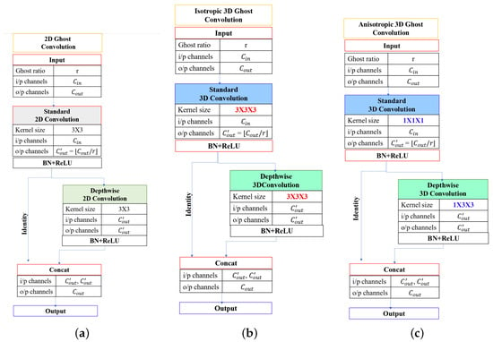
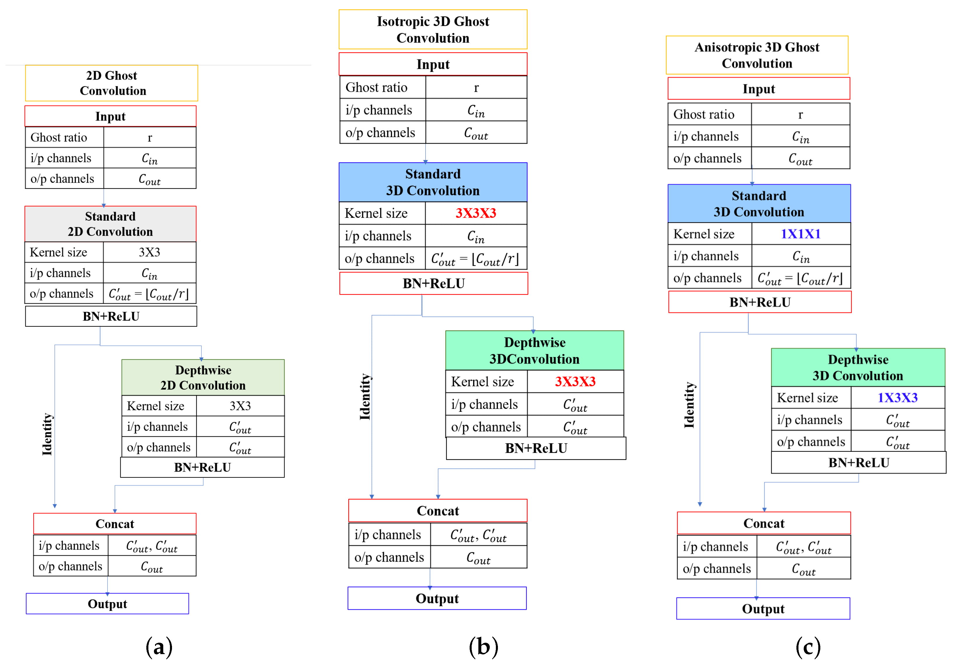
The two Ghost module variants employed in this work are illustrated in Figure 2.
Figure 2.
Comparison of Ghost module variants. (a) Original 2D Ghost module [19]. (b) Isotropic 3D Ghost module (LIVAS-L Net) with kernels. (c) Anisotropic 3D Ghost module (LIVAS-Net) with and kernels.
3.2.2. Description of Isotropic 3D Ghost Module
In LIVAS-L Net, both the primary and cheap operations employ isotropic kernels, directly extending the original 2D GhostNet design [19] to volumetric data. The primary convolution performs full 3D spatial filtering to generate intrinsic features via Equation (1). The cheap operation uses a depthwise kernel with , generating Ghost features via Equation (2). This configuration treats all three spatial dimensions uniformly, providing isotropic spatial modeling. The total parameter count for a single isotropic Ghost module could be calculated as
where . Compared to a standard 3D convolution requiring parameters, the Ghost module achieves an approximate reduction factor of .
3.2.3. Description of Anisotropic 3D Ghost Module
LIVAS-Net introduces an anisotropic Ghost formulation that exploits a key property of TOF-MRA acquisitions: the through-plane (slice) resolution is typically 2–5× coarser than the in-plane resolution. In such anisotropic volumes, spatial filtering along the slice axis at fine scales contributes comparatively less high-frequency spatial detail, as adjacent slices contain substantially less independent spatial detail than adjacent in-plane voxels.
The primary convolution employs a pointwise () kernel , performing channel recombination without spatial filtering. This reduces primary convolution parameters by a factor of approximately 27 relative to the isotropic variant. The cheap operation applies an anisotropic depthwise kernel with , restricting spatial feature generation to the in-plane (axial) dimensions where spatial resolution is highest. The parameter count thus becomes
This design reduces per-module parameters by approximately relative to the isotropic Ghost module for , yielding a total model size of 129K parameters.
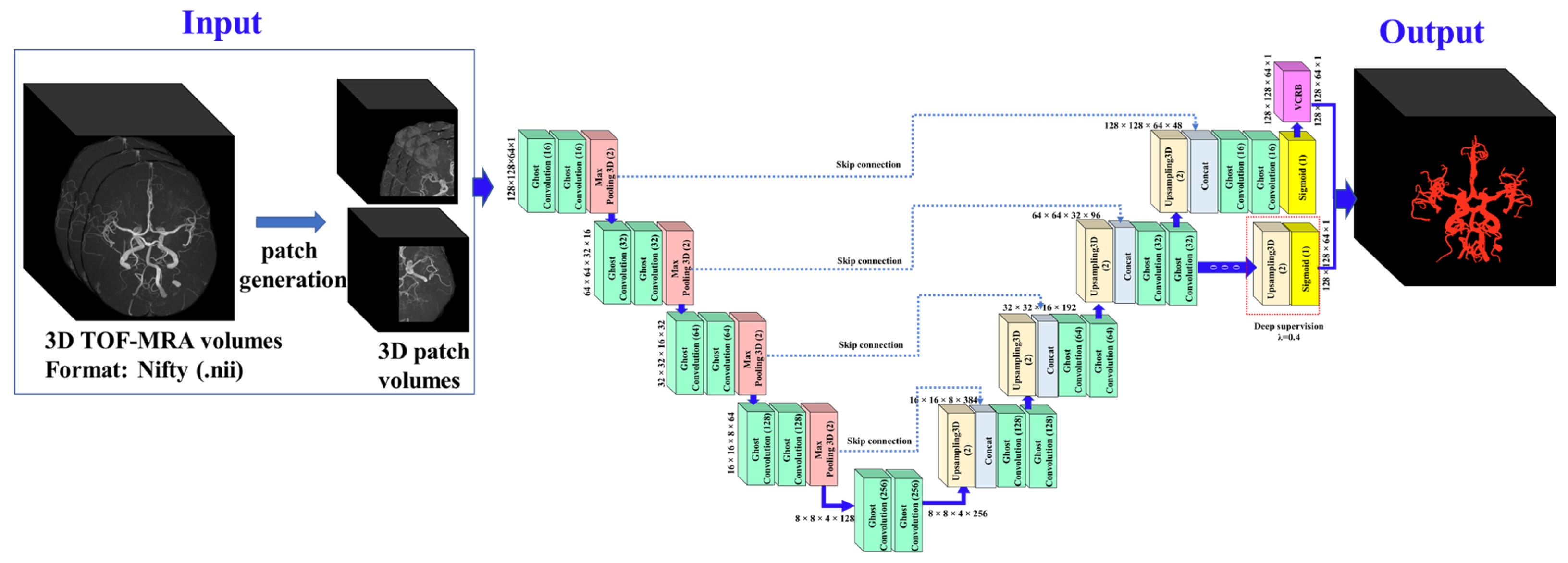
In both variants, each encoder and decoder stage applies two consecutive Ghost modules (denoted Ghost×2 in Figure 3), following the dual-convolution convention of the standard U-Net [27].
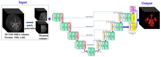
Figure 3.
Overall architecture of the proposed framework. The encoder and decoder each consist of four stages with Ghost×2 blocks. VCRB refines the sigmoid output, and DS is selectively applied at the second decoder level in LIVAS-Net and LIVAS-L Net.
3.3. Vessel Continuity Refinement Branch (VCRB)
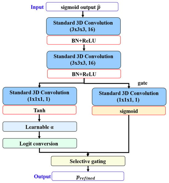
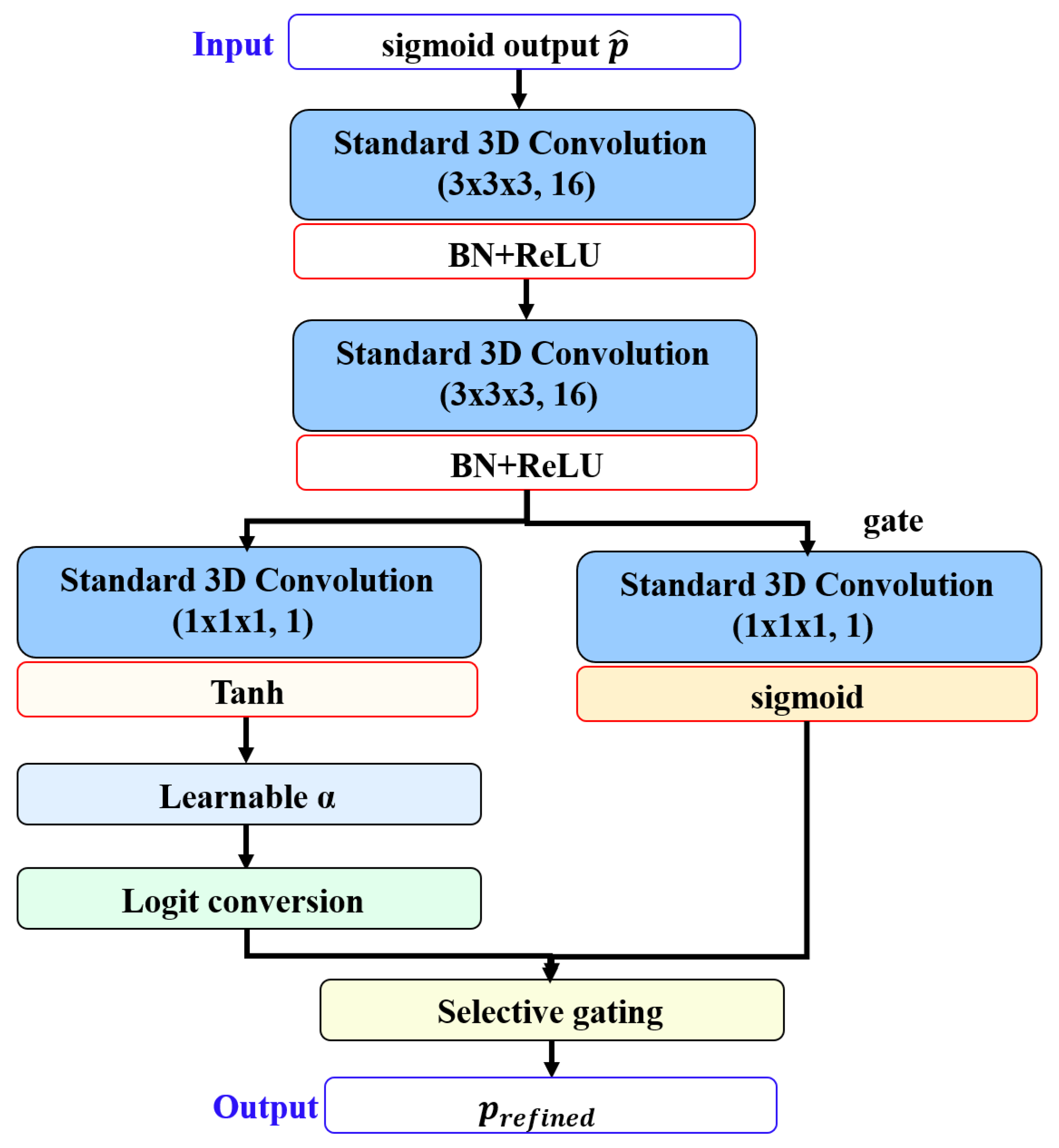
Lightweight Ghost-based decoders, despite their parameter efficiency, exhibit limited reconstruction capacity at decision boundaries where predicted probabilities are close to 0.5. Such uncertain regions frequently correspond to thin vessel segments, bifurcation points, and distal branches where vessel continuity is most fragile. To address this, the VCRB, a lightweight end-to-end trainable module, is proposed to operate in logit space as logit space allows linear additive corrections with greater sensitivity near decision boundaries. Therefore, VCRB aims to refine boundary predictions without disrupting confident vessel or background classifications. The VCRB architecture is illustrated in Figure 4.
Figure 4.
Illustration of the VCRB module.
Let denote the initial sigmoid output at voxel . VCRB first extracts a local context representation through two convolutional layers with BN and ReLU activation as
where is the hidden channel dimension. From this context, a correction signal and a gating signal g are computed as
where is a learnable non-negative scalar (initialized at 0.1) that controls the maximum correction magnitude, and is clipped to for training stability. The correction operates in logit space, where the initial prediction is first converted to logits , then corrected as , and then selectively applied through the learned gate as
The gate g encourages corrections near decision boundaries (where and small logit corrections produce large probability shifts) while remaining near zero in regions of high confidence, thereby preserving already-correct predictions. The entire VCRB introduces negligible computational overhead at inference. Crucially, unlike post hoc refinement strategies such as conditional random fields or morphological operations that require separate inference stages with manually tuned hyperparameters, VCRB is trained jointly with the segmentation network in an end-to-end fashion, requiring no additional inference steps or auxiliary training data.
3.4. Deep Supervision Strategy
In LIVAS-Net, the extreme compression introduced by anisotropic Ghost modules raises a concern: at the coarser decoder levels, vessel structures that occupy only 1–2 voxels in the original resolution become sub-voxel after downsampling, risking complete loss of gradient signal for vessel-relevant features. To mitigate this, LIVAS-Net employs selective DS [26] at the second decoder level (operating at a downsampled resolution).
An auxiliary segmentation head, consisting of a convolution with sigmoid activation, is attached to the second decoder stage output. The auxiliary prediction is upsampled to full resolution and supervised with the same loss as the main output. The total training loss combines the main and auxiliary objectives as follows:
where y denotes the ground-truth segmentation, is the combined binary cross-entropy and Dice loss, and balances the auxiliary contribution. This selective placement at a single intermediate resolution avoids the computational overhead of full multi-scale DS while ensuring that gradient information reaches the encoder through a shorter path, encouraging the network to maintain vessel-discriminative representations at coarser spatial scales.
3.5. Loss Function
The segmentation loss combines binary cross-entropy and Dice loss with equal weighting, which is represented as
This formulation is used for both the main and auxiliary objectives in Equation (9).
4. Experimental Results
This section presents the dataset description, the experimental setup, and a comprehensive evaluation of the proposed methods, including quantitative and qualitative results.
4.1. Dataset Description
The experiments were conducted using the COSTA benchmark [6] and CereVessMRA [28] for cerebrovascular segmentation. The ADAM dataset from COSTA comprises 95 training TOF-MRA volumes and 12 held-out test volumes. From the 95 training cases, 76 volumes were allocated for training and 19 for validation, following an 80/20 split, while the official held-out test set was reserved exclusively for final evaluation. In addition, 10-fold cross-validation was performed on the 95 training volumes using K-Fold with shuffle = True and seed = 42, and all resulting models were further evaluated on the same fixed official test set to assess robustness and generalization. The dataset exhibits heterogeneous voxel spacing, ranging from approximately mm3 to mm3 across volumes.
The final trained models were evaluated on multiple independent test sets to assess both in-domain and cross-domain generalization. Specifically, evaluation was performed on the held-out ADAM test set (12 volumes) and four external test datasets: IXI (34 volumes), LOC H1 (5 volumes), ICBM (10 volumes), and the CereVessMRA dataset (271 volumes). This multi-dataset evaluation enabled a comprehensive assessment of robustness under varying acquisition protocols and population distributions. Detailed acquisition parameters, scanner specifications, and voxel spacing for each center, namely ADAM, IXI, LOC H1, and ICBM, are documented in the original COSTA benchmark [6]. Similarly, scanner-specific information on CereVessMRA is available in the original CereVessMRA benchmark [28].
4.2. Training Configuration
Both LIVAS-Net and LIVAS-L Net were trained using two training paradigms, patch volume and whole volume. In the case of the patch-based paradigm, volumes were processed at native spatial resolution with percentile-based intensity normalization (1st–99th percentile clipping followed by min-max scaling). Patches of size were extracted using a foreground-biased sampling strategy (70% vessel-centered with spatial jittering, 30% random), with six patches per volume. Under the whole-volume paradigm, each volume was resized to voxels (trilinear for images, nearest-neighbor for masks) with min-max normalization. No data augmentation or morphological post-processing was applied under either paradigm. Patch-based inference employed sliding-window aggregation with 25% overlap and Gaussian importance weighting ( patch dimension per axis) to suppress boundary artifacts at patch junctions.
All implementations were carried out using the GPU version of TensorFlow 2.12.0 on the Ubuntu 18.04.3 platform with NVIDIA TITAN RTX 24 GB GPUs and Intel® Xeon® Scalable Processors (Intel Corporation, Santa Clara, CA, USA), 3 UPI up to 10.4 GT/s with 256 GB memory. The training hyperparameters are summarized in Table 3.
Table 3.
Training hyperparameters and configuration details for LIVAS-Net and LIVAS-L Net. All settings are shared unless otherwise noted.
4.3. Quantitative Segmentation Performance on Internal Test Set
The segmentation performance of the proposed architectures, LIVAS Net and LIVAS-L Net, was compared against seven established 3D segmentation architectures and presented in Table 4 and Table 5. The baseline methods include the standard 3D U-Net [11], ResUNet [29], VNet [12], UNet++ [14], attention-augmented variants (SE-UNet [30], CBAM-UNet [31]), and GSUNet [21], a published 3D Ghost-based architecture. All these models were evaluated on the official ADAM test set (N = 12) using six complementary metrics, including the Dice similarity coefficient (DSC), intersection-over-union (IoU), 95th percentile Hausdorff distance (HD95), centerline Dice (clDice), sensitivity (Se), and precision, under identical conditions, including patch size, preprocessing, and hyperparameters.
Table 4.
Quantitative comparison on the ADAM held-out test set (mean) under the patch-based paradigm. ↑ indicates higher values are better, while ↓ indicates lower values are better.
Table 5.
Computational efficiency comparison. Inference measured with patch volume . Vol (s) denotes estimated per-volume inference time based on 60 sliding-window patches per scan.
As evident from Table 4 and Table 5, LIVAS-L Net achieves the highest DSC (0.9235) and clDice (0.9315) among all evaluated models, outperforming the second-best model (UNet++, DSC = 0.9140) by approximately one percentage point while requiring 2.3× fewer parameters (2.97 M vs. 6.87 M) and 5.7× fewer FLOPs (87.8 vs. 503.3 GFLOPs). Its superior clDice score indicates that LIVAS-L Net preserves vessel topology more faithfully than competing architectures, a property attributable to the VCRB operating at full resolution.
On the other hand, LIVAS-Net, ultra-lightweight variant with only 129 K parameters, achieves a DSC of 0.8943, surpassing the standard 3D U-Net (DSC = 0.8762, 5.89 M parameters) with a 45.6× reduction in model size and 7.9× fewer FLOPs (18.3 vs. 144.5 GFLOPs). Notably, LIVAS-Net also outperforms GSUNet, a published 3D Ghost-based architecture (0.18 M parameters), by +4.4 percentage points in DSC while using 30% fewer parameters. GSUNet exhibits high sensitivity (0.945) but substantially lower precision (0.777), indicating systematic over-segmentation that manifests as an HD95 of 7.26 mm compared to LIVAS-Net’s 1.97 mm.
Among the attention-aided baselines, both SE-UNet and CBAM-UNet improve upon the standard 3D U-Net in DSC (0.894 and 0.901, respectively), confirming that channel and spatial attention mechanisms benefit vessel feature discrimination. However, neither surpasses LIVAS-L Net despite requiring approximately twice the parameters. ResUNet achieves strong performance (DSC = 0.912) through residual learning, while VNet’s larger kernel size (5 × 5 × 5) yields competitive results (DSC = 0.904) at the cost of 44 M parameters and 499.6 GFLOPs.
As shown in Table 5, all models exhibit comparable per-patch inference times (107–175 ms), as GPU kernel launch overhead dominates at this patch size. Under sliding-window inference with 25% overlap, each test volume requires approximately 60 patches (varying from 48 to 72 depending on volume dimensions), yielding per-volume inference times of 6–10 seconds across all models. While per-patch inference times remain comparable across architectures due to GPU kernel launch overhead, the substantially lower parameter count and FLOPs of LIVAS-Net (129 K, 18.3 GFLOPs) and LIVAS-L Net (2.97 M, 87.8 GFLOPs) translate to reduced model storage requirements and lower computational cost per forward pass. However, measured inference GPU memory (Table 5) does not reflect a proportional reduction, as activation memory from intermediate feature maps dominates over parameter storage at this patch size. The efficiency advantages of the proposed architectures are therefore most pronounced in terms of parameter count, FLOPs, and model portability rather than inference-time GPU memory.
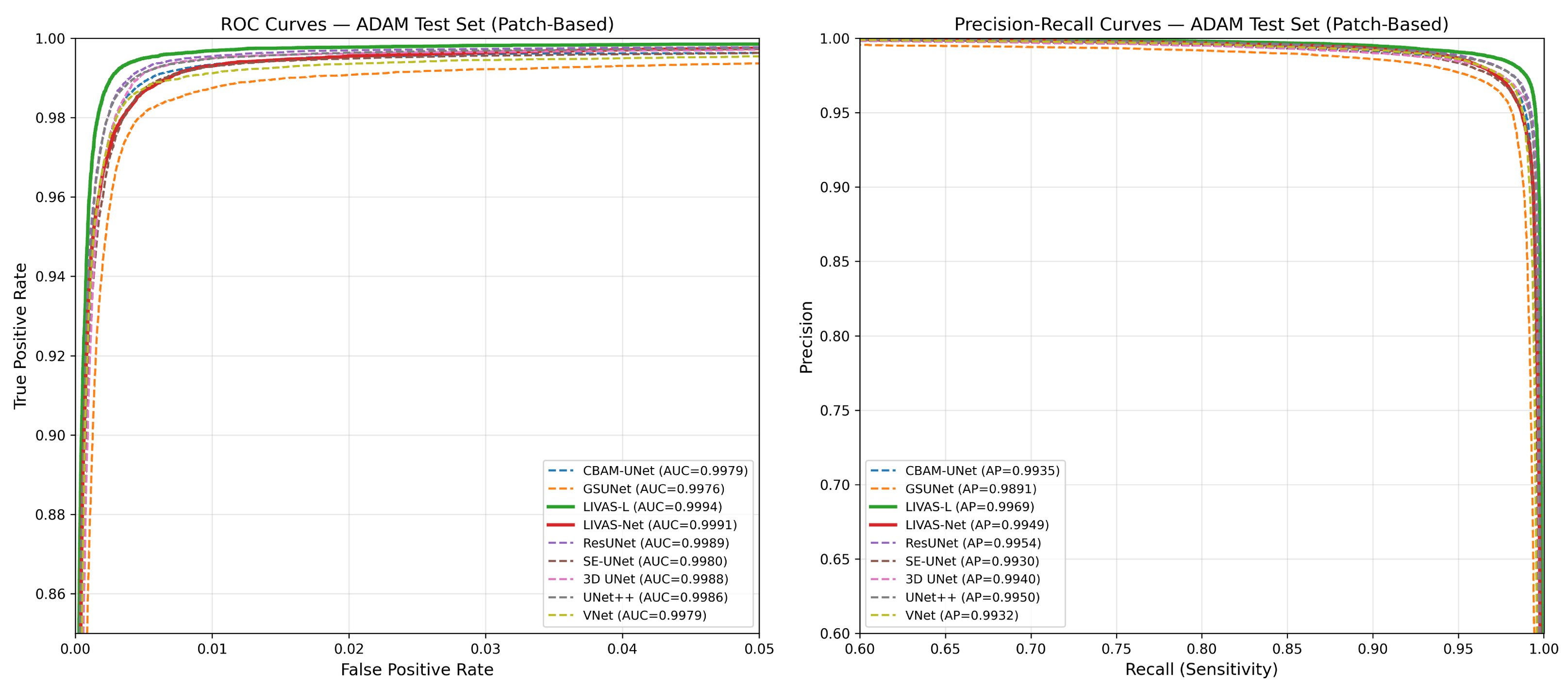
Voxel-wise ROC and precision–recall curves for all evaluated models are shown in Figure 5. All models achieve ROC-AUC scores above 0.997, though this metric is less informative given the severe class imbalance (vessel voxels constitute approximately 0.75% of the volume). Precision–recall curves better differentiate model performance under these conditions. LIVAS-L Net achieves the highest average precision (AP = 0.9970), while GSUNet records the lowest (AP = 0.9908). LIVAS-Net achieves AP = 0.9946, comparable to larger architectures such as ResUNet (0.9961) and UNet++ (0.9956). Differences between models are most apparent in high-recall regions of the PR curve, where LIVAS-L Net maintains higher precision than other methods.
Figure 5.
Voxel-wise ROC and precision–recall curves on the ADAM test set (patch-based).
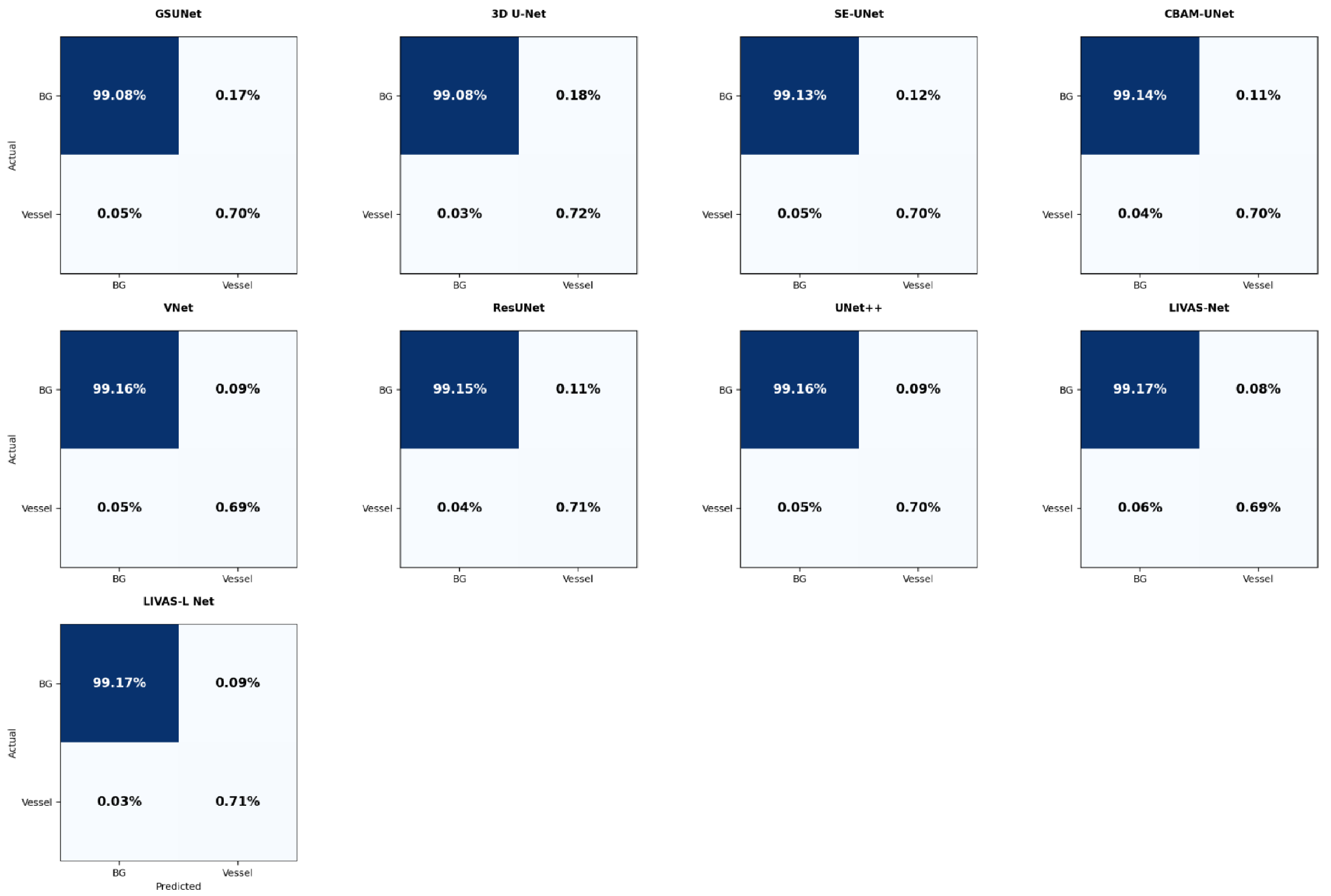
Voxel-level confusion analysis further highlights the effect of class imbalance, with background voxels accounting for over 99% of the volume (Figure 6). LIVAS-Net achieves the lowest FP rate (0.083%), followed by LIVAS-L Net (0.086%) and UNet++ (0.091%), while the standard 3D U-Net and GSUNet exhibit the highest FP rates (0.176% and 0.172%). FN rates remain low across all models (0.03–0.06%), with 3D U-Net achieving the lowest (0.031%), owing to its high sensitivity (0.962), followed by LIVAS-L Net (0.034%). LIVAS-Net shows a slightly higher FN rate (0.063%), reflecting its lower sensitivity under extreme parameter constraints, though this is offset by its superior precision (0.892).
Figure 6.
Normalized voxel-level confusion matrices for all models on the ADAM test set.
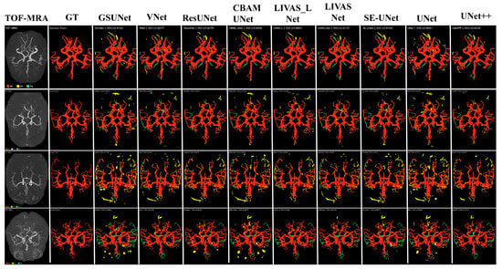
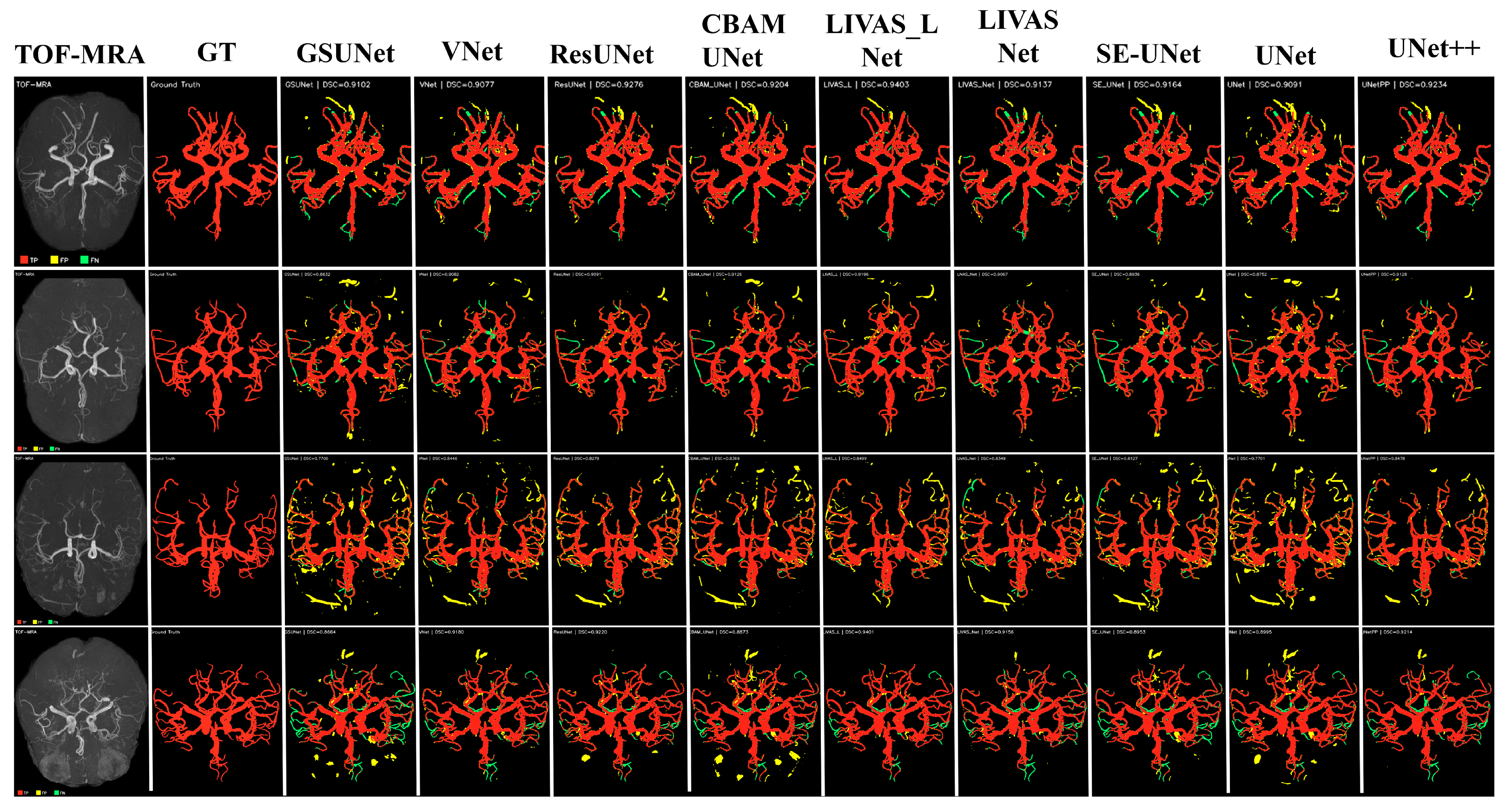
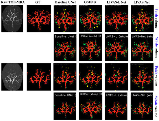
For qualitative comparison of segmentation performance, MIP visualizations are presented for representative 3D TOF-MRA test volumes from the official ADAM dataset of COSTA in Figure 7. To facilitate visual assessment, a color-coding scheme is employed in which true positive (TP), false positive (FP), and false negative (FN) voxels are shown in red, yellow, and green, respectively. This MIP-based visualization strategy is used consistently throughout this paper for qualitative comparison across different models and experimental settings.
Figure 7.
Qualitative segmentation comparison on representative ADAM test volumes using MIP visualization with TP/FP/FN color coding.
For analyses involving training paradigms, cross-center generalization, statistical significance, and ablation studies, we focus on four representative architectures: 3D U-Net, GSUNet, LIVAS-Net, and LIVAS-L Net. These models were chosen to provide comparison across both conventional and lightweight Ghost-based segmentation frameworks under different computational capacities. Unless otherwise stated, subsequent analytical experiments and cross-dataset evaluations are conducted using these four representative models only. All other experiments (Section 4.4.2) report full benchmark comparisons across seven architectures for completeness.
4.4. Ablation and Design Analysis
To systematically evaluate the contribution of architectural design choices, an ablation analysis is conducted across four dimensions: (i) component-wise analysis, (ii) comparative analysis with all seven segmentation models on the whole-slice level, (iii) training paradigm (patch-based vs. whole-volume), and (iv) architectural efficiency and generalization capability.
4.4.1. Component-Wise Analysis
The component-wise ablation study of LIVAS-Net and LIVAS-L Net on the ADAM test set using the patch-volume paradigm is summarized in Table 6 and Table 7, respectively.
Table 6.
Ablation study of LIVAS-Net components on the ADAM test set (patch-based). ✔ = component included; ✗ = excluded. Best per column in bold. ↑ indicates higher values are better, while ↓ indicates lower values are better.
Table 7.
Ablation study of LIVAS-L Net components on the ADAM test set (patch-based). Best per column in bold. ↑ indicates higher values are better, while ↓ indicates lower values are better.
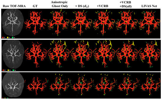
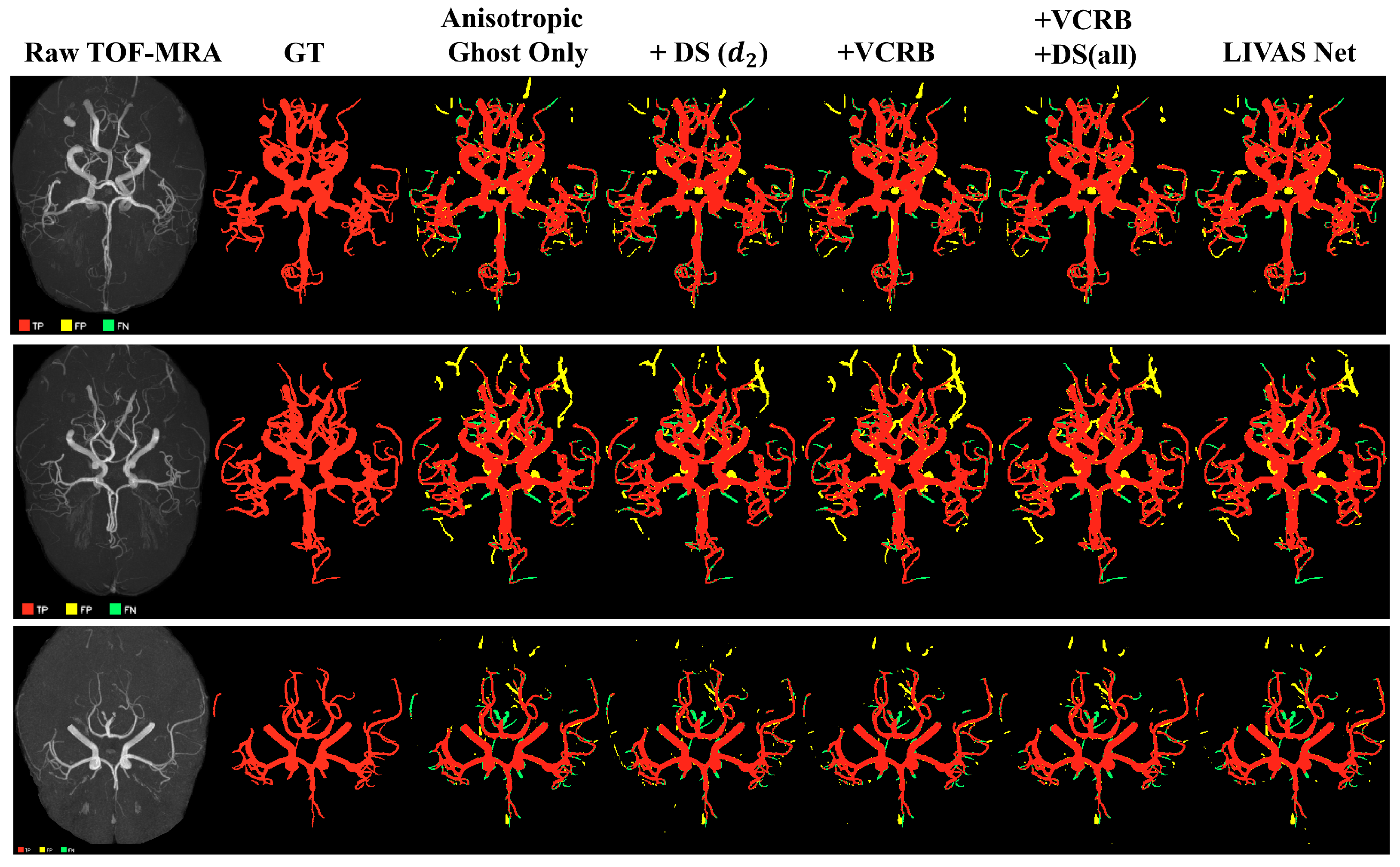
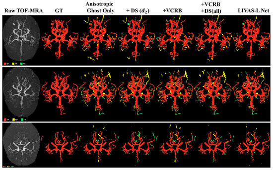
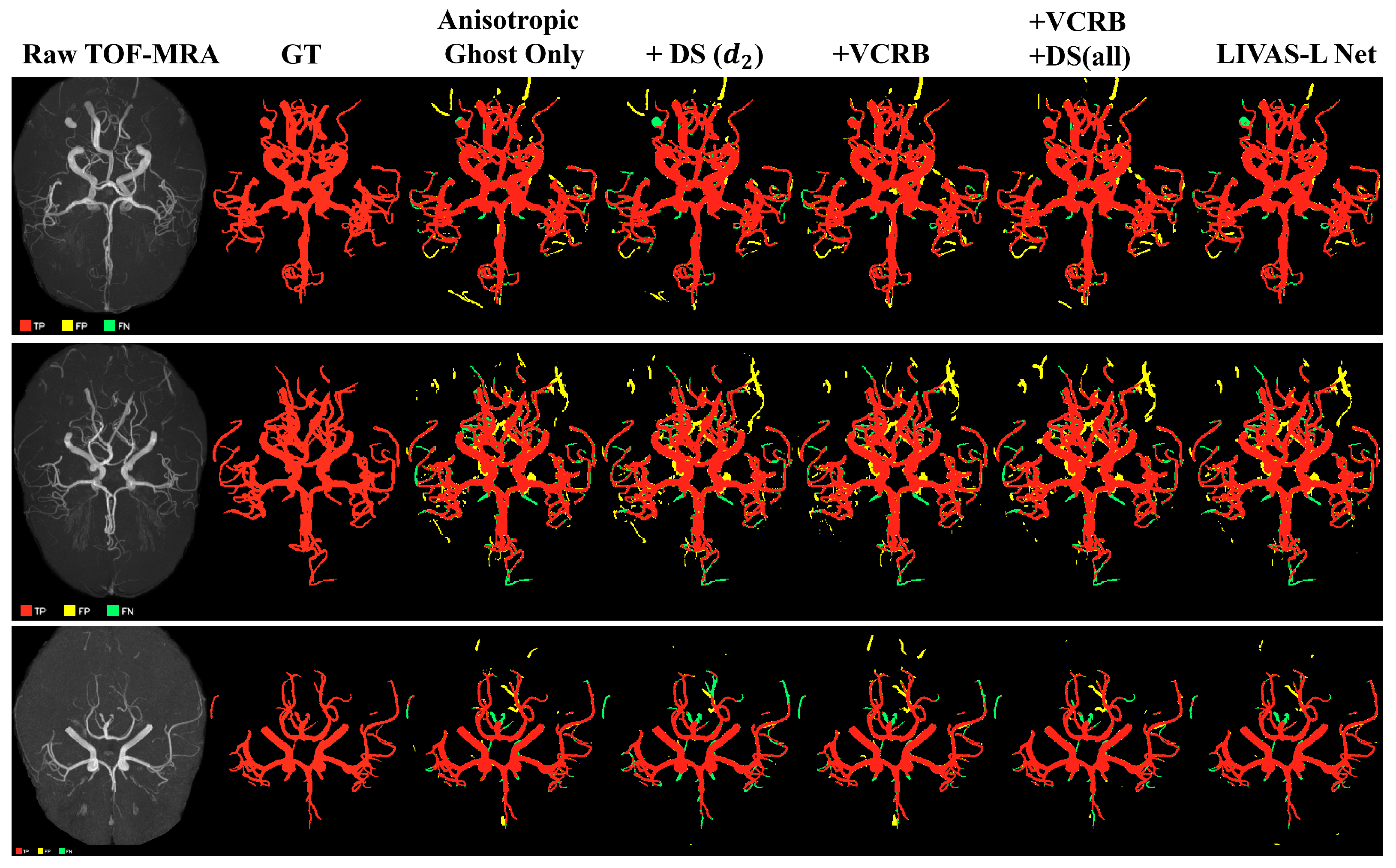
As shown in Table 6, the anisotropic 3D Ghost-only backbone yields a DSC of 0.8860. The inclusion of the VCRB module improves DSC by +0.42% and IoU by +0.69%, indicating that logit-space refinement effectively recovers vessel continuity and suppresses fragmented predictions. Introducing selective DS at decoder stage alone yields a comparable improvement of +0.06% DSC. The combined configuration (LIVAS-Net) achieves the highest DSC (0.8943) and clDice (0.8946), demonstrating that VCRB and selective DS provide additive benefits, with a total improvement of +0.83% over the Ghost-only baseline. Notably, applying DS at all decoder stages does not improve over selective DS at (DSC = 0.8900 vs. 0.8943), validating the design choice to supervise only the second decoder stage where feature resolution balances semantic richness and spatial precision. The quantitative analysis is represented in Figure 8 for LIVAS Net.
Figure 8.
Component-wise ablation study visualization for LIVAS Net.
Similar to LIVAS Net, the isotropic 3D Ghost-only backbone yields a DSC of 0.9081. Adding VCRB alone improves DSC by +0.62% with a substantial HD95 reduction (1.57→0.77 mm), confirming that logit-space refinement benefits both model variants. Adding DS at alone provides a modest +0.28% DSC gain. The combined LIVAS-L Net configuration achieves the highest DSC (0.9235) and clDice (0.9315), representing a +1.54% improvement over the Ghost-only baseline and demonstrating synergistic interaction between VCRB and selective DS. A critical finding is that applying deep supervision at all decoder stages (DSC = 0.8966) substantially degrades performance compared to selective supervision at (DSC = 0.9235, −2.69%). This indicates that auxiliary gradients from coarser decoder stages introduce conflicting supervision signals that destabilize training, whereas restricting supervision to provides optimal regularization without gradient interference. The quantitative analysis is represented in Figure 9 for LIVAS-L Net.
Figure 9.
Component-wise ablation study visualization for LIVAS-L Net.
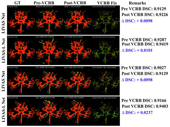
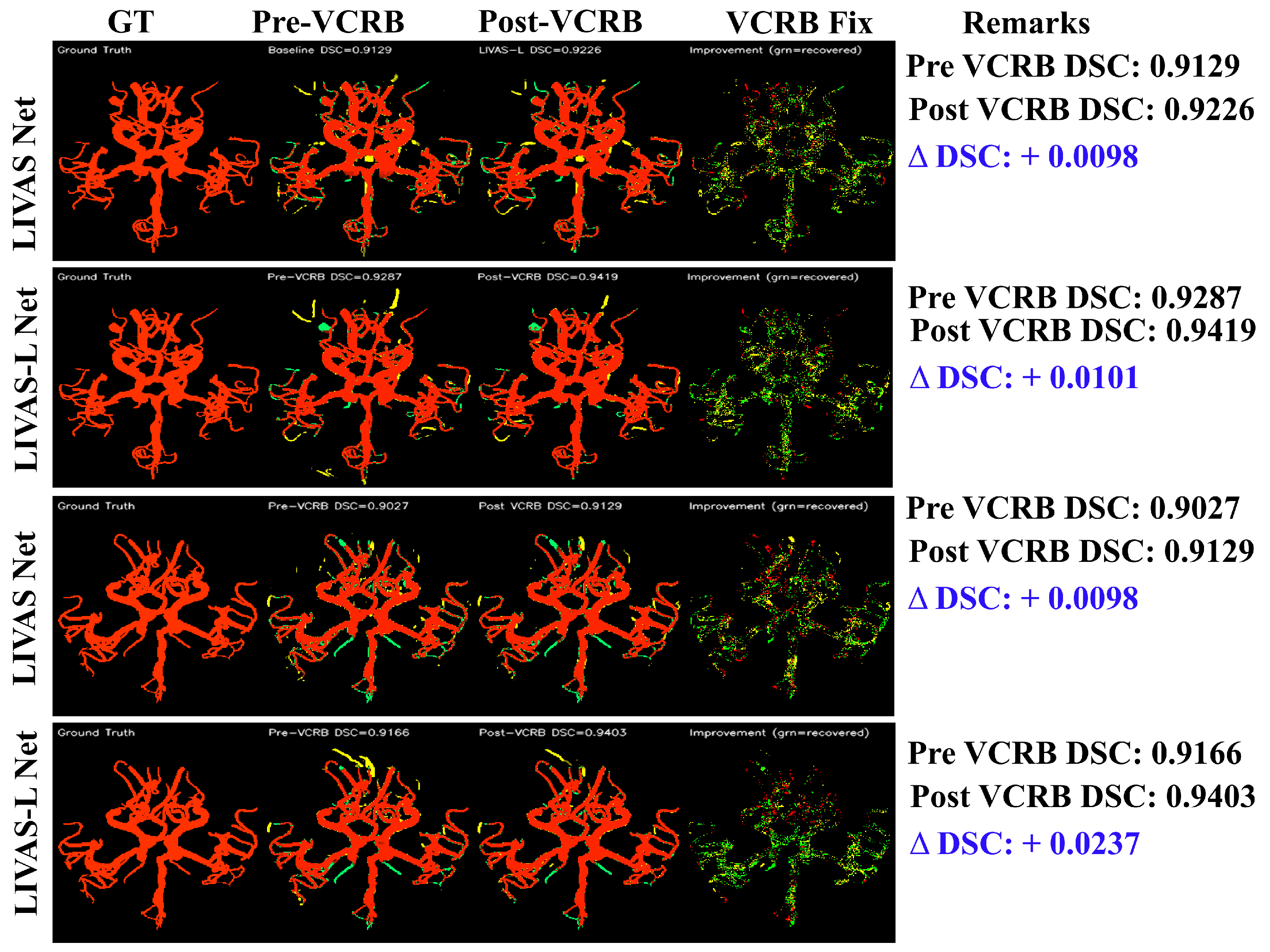
Comparing both LIVAS Net and LIVAS-L Net on the patch-volume paradigm, VCRB contributes more substantially in LIVAS-L Net (+0.62% mean) than in LIVAS-Net (+0.42% mean) (Table 6 and Table 7). Per-case visualization, as seen in Figure 10, confirms this pattern, with LIVAS-L Net showing VCRB-attributable improvements of +1.31% and +2.37% in the two representative cases, compared to +0.98% and +1.01% for LIVAS-Net.
Figure 10.
VCRB correction visualization for LIVAS-Net and LIVAS-L Net under the patch-based paradigm on two representative ADAM test cases. Columns: ground truth, pre-VCRB prediction, post-VCRB prediction, and correction map showing recovered vessels (green) and newly introduced FP (yellow).
4.4.2. Comparative Analysis with Baseline Methods on Whole Volume
To further evaluate robustness under reduced spatial resolution, comparative experiments are additionally conducted using the whole-volume paradigm, where the entire TOF-MRA volume is resized and processed directly. A comprehensive comparison between the proposed LIVAS-Net, LIVAS-L Net, and seven established 3D segmentation architectures is presented in Table 8.
Table 8.
Comparative analysis on the ADAM test set (whole-volume paradigm, 12 test volumes). Best result per metric in bold. ↓ indicates lower is better. ↑ indicates higher values are better, while ↓ indicates lower values are better.
As seen in Table 8, among the baseline methods, the standard 3D U-Net achieves the highest DSC score (0.8652), followed closely by SE-UNet (0.8617) and UNet++ (0.8606). In contrast, V-Net, despite having the largest parameter count (44.04 M), yields the lowest performance (DSC 0.8301), indicating that increased model capacity does not necessarily translate to improved segmentation accuracy under limited data conditions.
The proposed LIVAS-L Net achieves a DSC score of 0.8574, remaining within 0.8 percentage points of the best-performing baseline while using approximately 50% fewer parameters (2.97 M vs. 5.89 M), highlighting the effectiveness of the proposed architectural design. The lightweight variant, LIVAS-Net, further reduces the model size to 0.129 M parameters while maintaining competitive performance (DSC 0.8477), corresponding to 98.0% of the best baseline accuracy with only 2.2% of its parameters. Notably, LIVAS-Net outperforms V-Net despite being over 300× smaller, highlighting the efficiency of the proposed anisotropic design.
Sen analysis reveals that LIVAS-Net (0.8498) achieves a comparable detection capability to 3D U-Net (0.8511), suggesting that the lightweight architecture retains the ability to capture vessel structures despite extreme parameter constraints. Specificity remains consistently high across all models (>0.998), reflecting the inherent class imbalance and indicating that performance differences are primarily driven by vessel detection accuracy rather than background classification.
Although the proposed models do not surpass the standard 3D U-Net under the whole-volume paradigm, their advantages become more evident under the patch-based setting (Table 9), where preservation of native spatial resolution better aligns with the proposed lightweight architectural design. This observation suggests that the parameter-efficiency benefits of Ghost-based architectures are more effectively realized when fine spatial information is preserved, whereas aggressive whole-volume downsampling disproportionately affects lightweight models.
Table 9.
Comparison of whole-volume and patch-based training paradigms on the ADAM test set (12 volumes). Bold indicates the best result per metric. ↑ indicates higher values are better, while ↓ indicates lower values are better.
4.4.3. Impact of Training Paradigm
The influence of the patch-volume and whole-volume paradigms on the ADAM test dataset is summarized in Table 4 and visualized in Figure 11.
Figure 11.
Visualization of whole-volume and patch-volume analysis on ADAM test dataset.
Across all architectures, patch-based training consistently improves DSC, yielding gains of +1.1% (3D U-Net), +4.7% (LIVAS-Net), and +6.6% (LIVAS-L Net). GSUNet is a notable exception: its patch-based DSC (0.8500) is marginally lower than its whole-volume DSC (0.8507), accompanied by a sharp HD95 increase from 1.19 to 7.26 mm. This suggests that GSUNet’s architecture is unable to benefit from native-resolution inputs, with its high sensitivity (0.945) producing scattered false positives at native scale that inflate distance-based metrics.
A similar but less severe HD95 increase is observed for the 3D U-Net (1.13 to 4.02 mm) despite improved DSC, attributable to the over-segmentation of peripheral vessels at native resolution (sensitivity: 0.962). In contrast, both proposed architectures show controlled HD95 behavior under patch-based training, consistent with their improved precision.
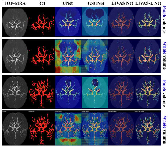
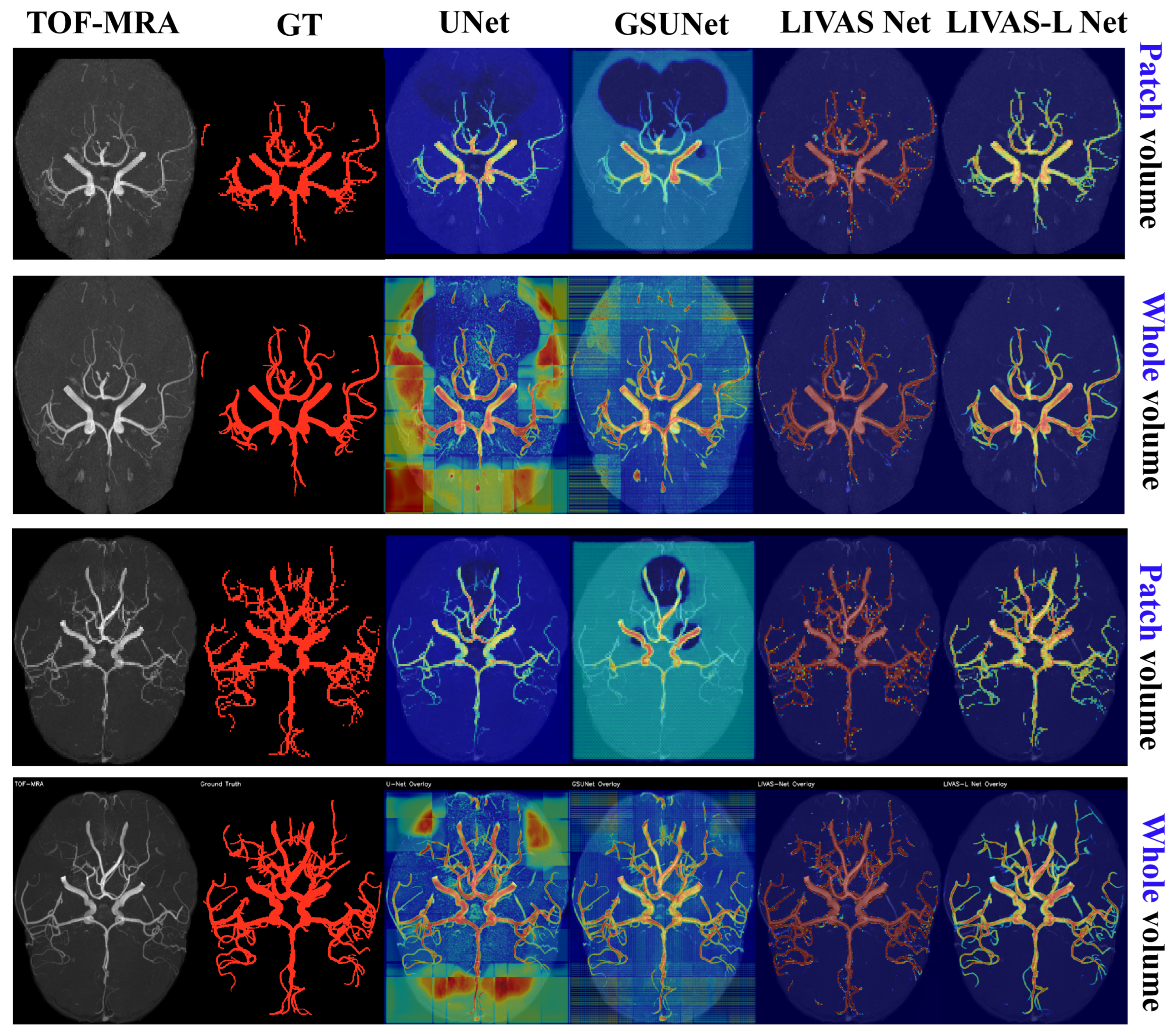
The magnitude of DSC improvement is inversely related to model capacity: LIVAS-L Net exhibits the largest gain (+6.6%), whereas the higher-capacity 3D U-Net shows only +1.1%. This indicates that lightweight architectures are more sensitive to information loss from spatial downsampling, as they lack the representational capacity to compensate for missing fine-scale details. The impact is further reflected in clDice, where LIVAS-L Net improves from 0.8573 (whole) to 0.9315 (patch), demonstrating that native-resolution inputs are essential for preserving distal vessel topology. The corresponding Grad-Cam analysis is displayed in Figure 12.
Figure 12.
Illustration of corresponding Grad-CAM analysis of patch-volume and whole-volume paradigm displayed in Figure 11.
4.4.4. Topological Analysis
As a supportive and exploratory analysis, topology metrics (Table 10) provide additional perspective on structural fragmentation beyond clDice by measuring the number of connected components in the predicted vascular masks. The ground truth contains connected components per volume, whereas GSUNet and 3D U-Net produce over 90 components on average, indicating severe fragmentation caused by scattered FP clusters. LIVAS-Net reduces this fragmentation to components, while LIVAS-L Net further suppresses it to , corresponding to a 2.4× reduction compared to 3D U-Net and a 2.7× reduction compared to GSUNet. Despite differences in fragmentation, component-level recall remains high across all models (≥0.88), indicating that most ground-truth vessel components are still detected with sufficient overlap. However, this metric does not capture over-segmentation effects, which are reflected in the large differences in predicted component counts. The slightly lower recall of LIVAS-L Net (0.885 vs. 0.938 for 3D U-Net) is attributable to its higher precision, where suppression of small spurious components reduces incidental overlaps with minor ground-truth structures rather than true anatomical omissions.
Table 10.
Topology analysis on ADAM test set (mean ± std, N = 12). ↑ indicates higher values are better, while ↓ indicates lower values are better.
These topology metrics in Table 10 are included as complementary observations alongside the primary evaluation metrics. However, they are not sufficient on their own to establish clinically meaningful vessel continuity preservation. More definitive assessment would require additional topology-oriented analyses, such as skeleton precision/recall or branch-level detection evaluation, which were beyond the scope of the current study and remain areas for future investigation.
4.4.5. Cross-Center Dataset Generalization
To evaluate robustness under domain shift, all four representative models, 3D U-Net, GSUNet, LIVAS-Net, and LIVAS-L Net, are evaluated on three external datasets from COSTA that were not used during training (ICBM, IXI, and LocH1) and CereVessMRA. The results are summarized in Table 11.
Table 11.
Cross-dataset generalization (patch-based). Best per column in bold. ↑ indicates higher values are better, while ↓ indicates lower values are better.
Significant performance degradation is observed across all models compared to the in-domain ADAM dataset, reflecting variations in acquisition protocols, scanner characteristics, and population distributions. The baseline U-Net exhibits the most pronounced degradation, with DSC decreasing from 0.8762 (ADAM) to 0.5625 on ICBM and 0.7193 on IXI.
In contrast, the proposed architectures demonstrate comparable robustness under domain shift. LIVAS-L Net achieves consistently higher DSC scores across all external datasets, particularly on ICBM (0.7526 vs. 0.5625), corresponding to a relative improvement of over 33%. Similarly, HD95 is substantially reduced (10.90 mm vs. 27.18 mm), indicating improved boundary localization under challenging conditions.
LIVAS-Net achieves comparable or superior performance on IXI (0.8235 DSC) and LocH1 (0.8318 DSC), despite having two orders of magnitude fewer parameters. This suggests that the lightweight architecture can generalize effectively to datasets with varying spatial resolution and contrast characteristics. To further assess generalization under severe domain shift, all four models were evaluated on the CereVessMRA dataset (N = 271) without retraining. Quantitative results are summarized in Table 12.
Table 12.
Performance on the CereVessMRA dataset (mean ± std, N = 271). ↑ indicates higher values are better, while ↓ indicates lower values are better.
A noticeable performance degradation is observed across all models compared to both in-domain and other external datasets, indicating a substantial domain shift between ADAM and CereVessMRA, likely attributable to differences in acquisition protocols, spatial resolution, and annotation characteristics. Despite this degradation, LIVAS-Net achieves the best DSC and HD95, both outperforming the baseline U-Net. Notably, all models maintain high specificity (>0.999), while reduced sensitivity indicates difficulty in detecting fine vascular structures under altered imaging conditions.
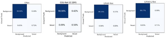
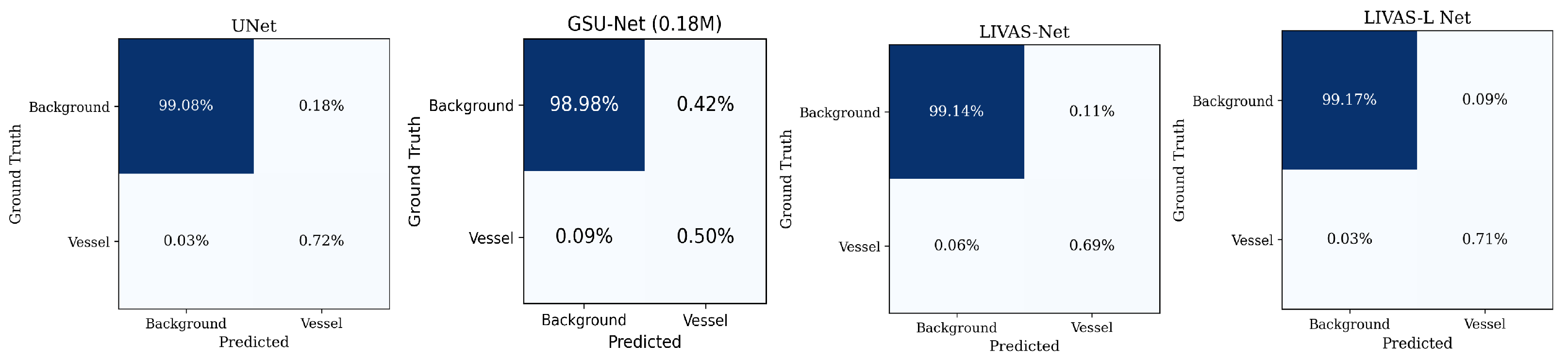
As shown in Figure 13, the confusion matrices reveal that all four models exhibit nearly identical voxel-level classification patterns on CereVessMRA, with the dominant difference occurring in the FP quadrant: LIVAS-L Net achieves the lowest FP rate (0.04%) compared to 0.07% (Baseline U-Net) and 0.06% (LIVAS-Net), consistent with its highest precision (0.8989). The comparable FN rates across models (0.28%) indicate that the sensitivity limitation is driven by the domain shift itself rather than architectural capacity. The reversal in model ranking between COSTA and CereVessMRA is noteworthy, although the current experiments do not provide sufficient evidence to explain the underlying cause. Differences in model capacity may contribute to variations in cross-dataset generalization; however, this remains speculative without additional analyses such as scanner-stratified evaluation, calibration assessment, feature-space visualization, or center-specific error analysis. Therefore, the observed performance differences should be interpreted cautiously, and further investigation is needed to clarify the mechanisms involved.
Figure 13.
Normalized voxel-level confusion matrix for all four models on CereVessMRA dataset.
4.5. Parameter Efficiency Analysis
Parameter efficiency comparisons among all models are reported in Table 13. The compression ratio is computed relative to the 3D U-Net baseline, while DSC/M is used as a normalized indicator of segmentation performance per million parameters. Here, the cross-center DSC indicates the test dataset from IXI, ICBM, and a local hospital from COSTA.
Table 13.
Parameter efficiency comparison. Compression ratio relative to 3D U-Net (5.89 M). DSC/M denotes DSC per million parameters. ↑ indicates higher values are better.
As seen in Table 13, LIVAS-Net achieves the highest parameter efficiency with a DSC/M score of 6.930, substantially outperforming 3D U-Net (0.149) and GSUNet (4.620), indicating that the proposed anisotropic Ghost-based design provides a more favorable trade-off between segmentation accuracy and model complexity. In contrast, GSUNet shows limited efficiency gains despite its high compression ratio, suggesting that parameter reduction alone is insufficient to ensure strong segmentation performance. LIVAS-Net combines high compression () with competitive accuracy, demonstrating the advantage of task-aware architectural design, while LIVAS-L Net achieves the highest absolute DSC (0.924) with moderate compression (), reflecting a balanced trade-off between efficiency and performance across different deployment constraints.
It is important to note that DSC/M is included solely as a descriptive efficiency metric and does not imply a linear relationship between parameter count and segmentation accuracy. Instead, it is most meaningful for relative comparison among models with different computational budgets.
4.6. Statistical Significance Analysis
Pairwise Wilcoxon signed-rank tests were conducted across four models (3D U-Net, GSUNet, LIVAS-Net, LIVAS-L Net) and five metrics, yielding 30 comparisons. The result is reported in Table 14. Bonferroni correction was applied (). Effect sizes are reported as rank-biserial correlation (r), where indicates a large effect [32].
Table 14.
Statistical significance (Wilcoxon signed-rank, Bonferroni-corrected, N = 12). r: Rank-biserial effect size.
After Bonferroni correction, 16 of 30 comparisons remain significant (), with large effect sizes () in all significant cases. Both proposed models significantly outperform 3D U-Net in DSC, HD95, and precision (), while sensitivity differences do not reach significance ( for LIVAS-L), reflecting that performance gains are driven by improved precision rather than detection rate.
Notably, no significant differences are observed between GSUNet and 3D U-Net across any metric, indicating that GSUNet does not improve upon the baseline despite its Ghost-based design. In contrast, LIVAS-Net significantly outperforms 3D U-Net in four of five metrics ( for DSC, HD95, Se, Prec), with clDice approaching but not reaching the corrected threshold (, ), demonstrating that the proposed anisotropic Ghost module combined with VCRB provides meaningful architectural improvements beyond parameter reduction alone. Between the two proposed models, LIVAS-L Net significantly outperforms LIVAS-Net in DSC, clDice, and sensitivity, while precision differences are not significant (), suggesting both models achieve comparable false positive control.
4.7. Cross-Validation Analysis on Proposed Models
To further validate result stability, 10-fold cross-validation is performed on the full ADAM training set (95 volumes), with the results presented in Table 15 under the patch-based paradigm for both models. LIVAS-L Net achieves a fold-level DSC of on validation and on the held-out test set (95% CI: [0.9216, 0.9254]), demonstrating stability with sub-0.4% standard deviation across folds. LIVAS-Net similarly exhibits consistent performance (test DSC , 95% CI: [0.8911, 0.8969]). For both models, held-out test performance exceeds or matches CV validation, confirming that the models generalize effectively and do not overfit to specific training splits.
Table 15.
Ten-fold cross-validation results under the patch-based paradigm. CV Validation: Fold-level mean DSC across 95 ADAM training volumes. Held-out Test: Mean DSC of 10 independently trained models evaluated on the fixed 12-case test set.
4.8. Qualitative Visualization Analysis
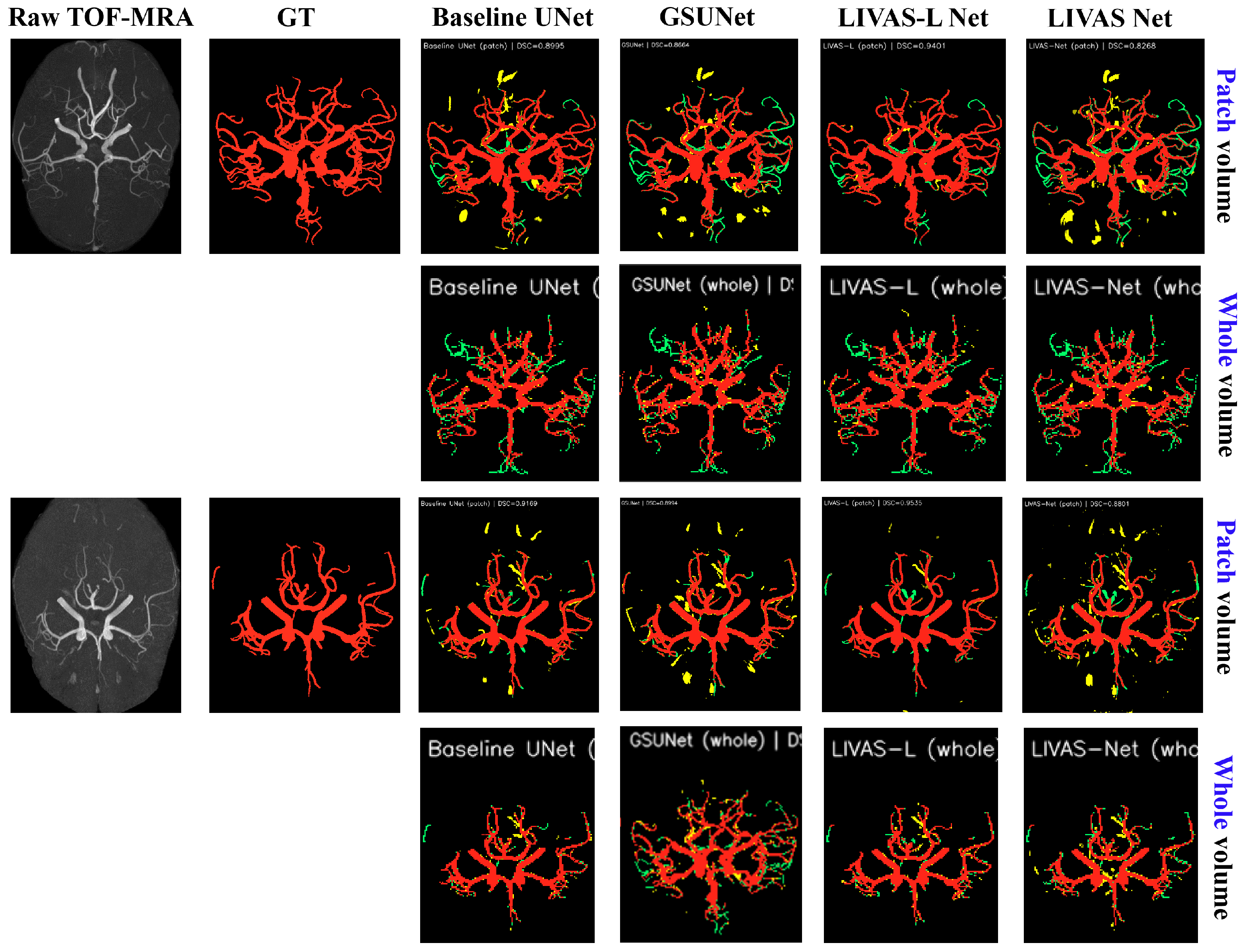
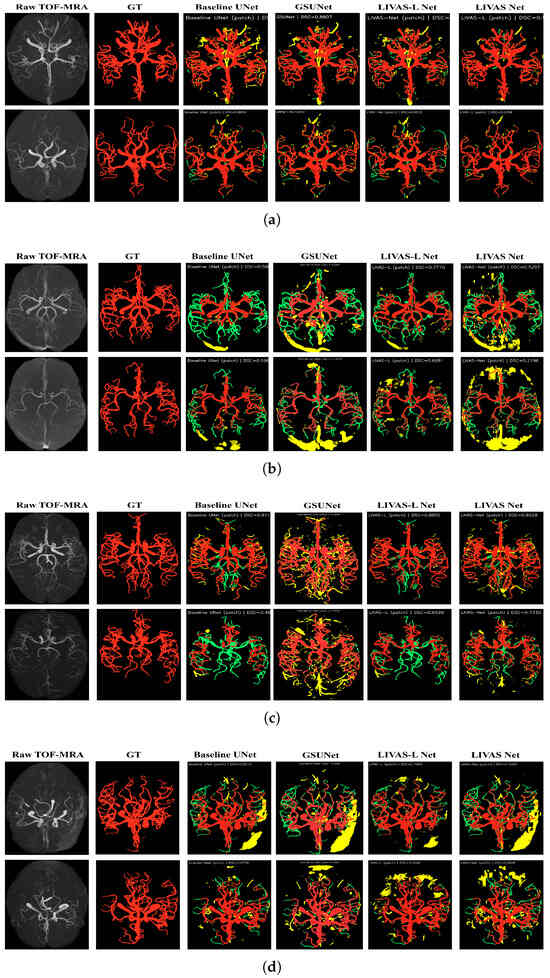
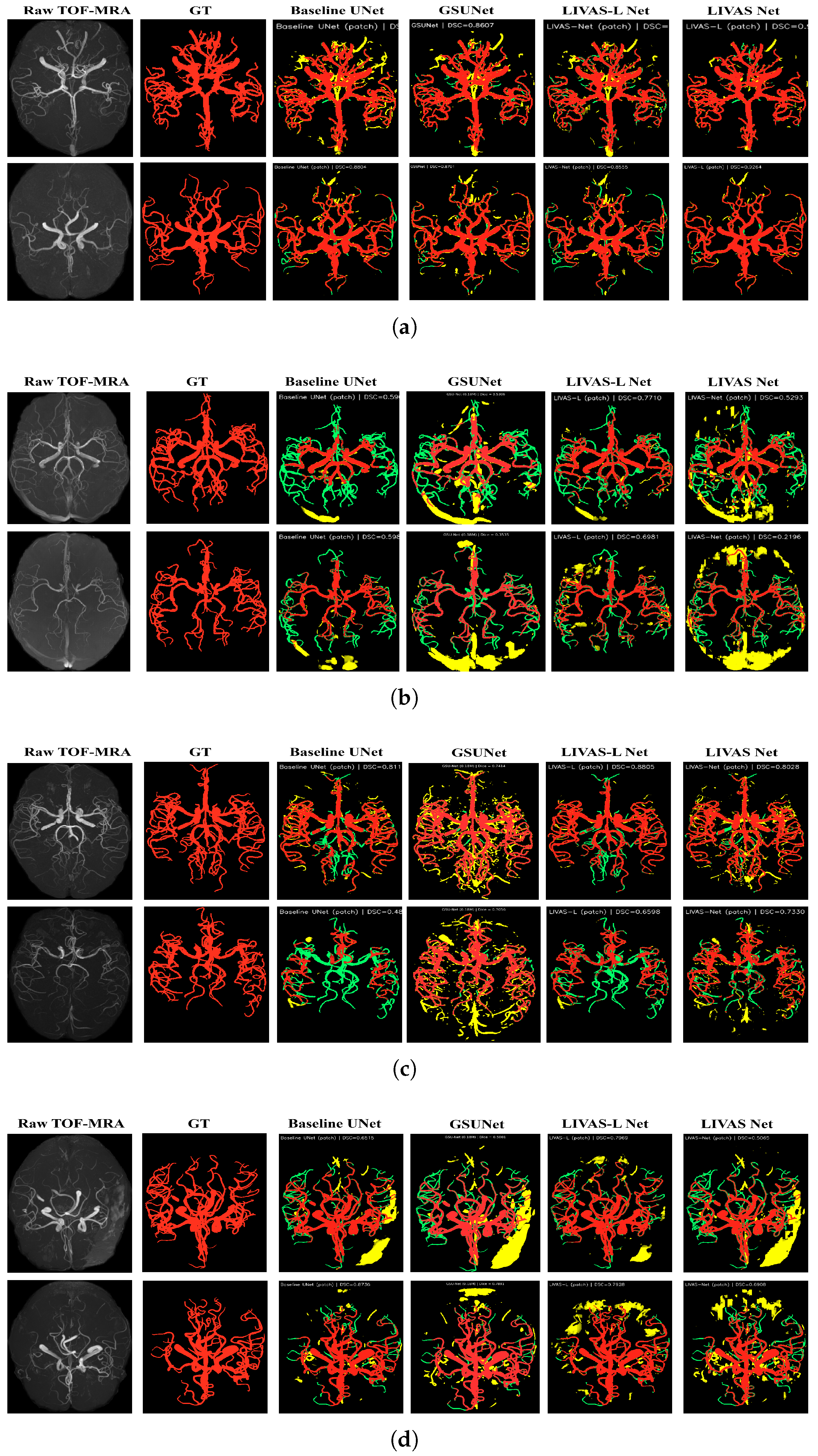
Additional visualization of the 3D TOF-MRA volumes after MIP projection is shown in Figure 14. TP, FP, and FN voxels are color-coded to enable qualitative assessment of segmentation performance.
Figure 14.
Qualitative comparison of segmentation results across multiple datasets using MIP visualization. TP (red), FP (yellow), and FN (green) are overlaid for visual assessment. (a) ADAM test dataset. (b) ICBM test dataset. (c) IXI test dataset. (d) LocH1 test dataset.
The qualitative analysis in Figure 14 supports the quantitative findings presented in the previous sections. The cross-center results reveal a clear generalization hierarchy: LIVAS-L Net generalizes best across all four imaging centers, maintaining the highest average DSC. LIVAS-Net struggles most on out-of-distribution data (ICBM, LocH1), where its ultra-lightweight architecture lacks the capacity to handle domain-shifted intensity distributions, manifesting as extensive FP regions. However, on the larger CereVessMRA external dataset, LIVAS-Net achieves the highest DSC (0.669) among all models. This apparent contradiction can be reconciled by considering the characteristics of the target distributions: on ICBM and LocH1, the domain shift may manifest as a concentrated intensity or resolution discrepancy relative to ADAM, where additional model capacity helps absorb the distribution gap. In contrast, CereVessMRA, comprising 271 subjects, likely encompasses greater acquisition diversity, under which sensitivity to training-set characteristics may become less advantageous. The baseline UNet shows a conservative segmentation pattern, with fewer FPs but more missed vessels (green), reflecting the precision–sensitivity trade-off observed in the quantitative results. The ADAM center results confirm that all models perform best on the in-distribution data, with LIVAS-L Net achieving 0.9235 DSC, validating the architectural design. The performance degradation on external centers (particularly ICBM) highlights the need for domain adaptation strategies, which could be explored in future work.
5. Conclusions
This paper presented LIVAS-Net and LIVAS-L Net, two parameter-efficient 3D encoder–decoder architectures for cerebrovascular segmentation from TOF-MRA. The proposed designs leverage 3D Ghost convolution modules to reduce computational redundancy, a VCRB that refines segmentation discontinuities in logit space, and selective DS to regularize intermediate feature representations. On the ADAM test set, LIVAS-L Net achieved a DSC of 0.9235 and HD95 of 0.77 mm using only 2.97 M parameters, outperforming several established 3D segmentation baselines, including 3D U-Net, ResUNet, SE-UNet, CBAM-UNet, UNet++, and V-Net. LIVAS-Net, with only 129 K parameters (a 45.6× reduction relative to 3D U-Net), achieved a DSC of 0.8943 and HD95 of 1.97 mm, retaining over 98% of the best baseline performance while using only 2.2% of its parameters. These improvements were statistically significant across most metrics (Wilcoxon signed-rank test, ). Cross-center evaluation on three external datasets (ICBM, IXI, LocH1) from COSTA demonstrated improved robustness compared to the baseline 3D U-Net, with LIVAS-L Net and LIVAS-Net achieving overall DSCs of 0.7967 and 0.7834, respectively, compared to 0.6998 for the baseline. On the large-scale CereVessMRA dataset (N = 271), a different ranking was observed: LIVAS-Net achieved the best overall performance (DSC = 0.6686), followed by LIVAS-L Net and the baseline. This suggests that highly compact architectures may generalize more robustly under severe domain shift, although the underlying mechanisms require further investigation. Furthermore, ablation analysis confirmed that patch-based training at native resolution is critical for preserving fine vascular structures, particularly for lightweight architectures, yielding improvements of up to 6.6 percentage points in DSC compared to whole-volume downsampled training.
However, the current study warrants several limitations that should be acknowledged. First, all models were trained exclusively on the ADAM dataset, comprising 76 TOF-MRA volumes from a single imaging center. Although cross-center evaluation demonstrated reasonable generalization, the substantial performance degradation observed on CereVessMRA (DSC dropping from 0.894 to 0.669 for LIVAS-Net) indicates that single-source training remains insufficient for robust multi-site deployment. Future work could incorporate multi-center training data or domain adaptation techniques to reduce this gap. Second, although LIVAS-Net achieves a 45.6× parameter reduction, inference speed was not explicitly benchmarked on edge devices or embedded hardware. The sliding-window patch-based inference strategy, while effective for segmentation accuracy, introduces computational overhead that may limit real-time applicability. Optimizations such as ONNX conversion, quantization, or TensorRT deployment could be explored to bridge this gap. Third, the cross-center performance difference between LIVAS-L Net and LIVAS-Net was hypothesized to relate to model capacity and overfitting to center-specific patterns. However, confirming this interpretation would require scanner-stratified evaluation or feature-space analysis across centers, which remains an avenue for future investigation. Fourth, subjective evaluation by neuroradiologists was not conducted in the current study, as the primary focus was on automated segmentation accuracy rather than clinical workflow integration. Expert clinical assessment could be incorporated in future work when downstream tasks such as aneurysm detection or stenosis grading are developed on top of the proposed segmentation framework.
In summary, LIVAS-Net and LIVAS-L Net demonstrated that accurate cerebrovascular segmentation can be achieved with dramatically reduced model complexity. LIVAS-L Net provides the highest overall segmentation quality, while LIVAS-Net is offered as a promising candidate for resource-constrained settings, pending further clinical validation.
Author Contributions
Conceptualization, M.S., Y.-C.H. and P.K.S.; methodology, M.S.; formal analysis, M.S.; investigation, P.K.S. and Y.-C.H.; writing—original draft preparation, M.S.; writing—review and editing, P.K.S.; visualization, Y.-C.H. and M.S.; supervision, P.K.S.; project administration, P.K.S.; funding acquisition, P.K.S. All authors have read and agreed to the published version of the manuscript.
Funding
This work was supported by the National Science and Technology Council (NSTC), Taiwan, grant number 114-2221-E-182-022-MY3.
Institutional Review Board Statement
Not applicable.
Informed Consent Statement
Not applicable.
Data Availability Statement
Datasets are publicly available: COSTA via https://imed.nimte.ac.cn/costa.html (accessed on 13 April 2026), CereVessMRA via https://www.scidb.cn/en/detail?dataSetId=d3a67e63c0054652b75bfcc3cf15ae3f (accessed on 13 April 2026).
Conflicts of Interest
The authors declare no conflicts of interest.
Abbreviations
The following abbreviations are used in this manuscript:
| 3D U-Net | Three-Dimensional U-Net |
| AP | Average Precision |
| BCE | Binary Cross-Entropy |
| CBAM | Convolutional Block Attention Module |
| clDice | Centerline Dice |
| COSTA | Cerebrovascular Segmentation Toolkit and Analysis |
| CTA | Computed Tomography Angiography |
| DGM | Dense Ghost Module |
| DRG | Dense Residual Ghost |
| DS | Deep Supervision |
| DSA | Digital Subtraction Angiography |
| DSC | Dice Similarity Coefficient |
| EDGConv | Enhanced Differential Ghost Convolution |
| FN | False Negative |
| FP | False Positive |
| GDG | Ghost-Dilated with Global Attention |
| GM | Ghost Module |
| GSA | Ghost Self-Attention |
| GSM | Ghost Shuffle Module |
| GSP | Ghost Spatial Pyramid |
| HD95 | 95th Percentile Hausdorff Distance |
| IoU | Intersection over Union |
| IXI | Information Extraction from Images |
| LIVAS-L Net | Lightweight Intracranial Vascular Segmentation Large Network |
| LIVAS-Net | Lightweight Intracranial Vascular Segmentation Network |
| LR | Learning Rate |
| MIP | Maximum Intensity Projection |
| MRA | Magnetic Resonance Angiography |
| PR | Precision–Recall |
| ResUNet | Residual U-Net |
| ROC | Receiver Operating Characteristic |
| ROC-AUC | Area Under the ROC Curve |
| SE | Squeeze-and-Excitation |
| Sen | Sensitivity |
| TOF-MRA | Time-of-Flight Magnetic Resonance Angiography |
| TP | True Positive |
| TN | True Negative |
| VCRB | Vessel Continuity Refinement Branch |
| V-Net | Volumetric Network |
References
- Mohapatra, S.; Lee, T.H.; Sahoo, P.K.; Wu, C.Y. Localization of early infarction on non-contrast CT images in acute ischemic stroke with deep learning approach. Sci. Rep. 2023, 13, 19442. [Google Scholar] [CrossRef] [Scilit] [PubMed]
- Feigin, V.L.; Abate, M.D.; Abate, Y.H.; Abd ElHafeez, S.; Abd-Allah, F.; Abdelalim, A.; Abdelkader, A.; Abdelmasseh, M.; Abd-Elsalam, S.; Abdi, P.; et al. Global, regional, and national burden of stroke and its risk factors, 1990–2021: A systematic analysis for the Global Burden of Disease Study 2021. Lancet Neurol. 2024, 23, 973–1003. [Google Scholar] [CrossRef] [Scilit] [PubMed]
- Deshmukh, A.S.; Priola, S.M.; Katsanos, A.H.; Scalia, G.; Costa Alves, A.; Srivastava, A.; Hawkes, C. The management of intracranial aneurysms: Current trends and future directions. Neurol. Int. 2024, 16, 74–94. [Google Scholar] [CrossRef] [Scilit] [PubMed]
- Sarkar, M.; Lee, T.H.; Sahoo, P.K. Smart healthcare: Exploring the internet of medical things with ambient intelligence. Electronics 2024, 13, 2309. [Google Scholar] [CrossRef] [Scilit]
- Jeon, Y.H.; Park, C.; Lee, K.H.; Choi, K.S.; Lee, J.Y.; Hwang, I.; Yoo, R.E.; Yun, T.J.; Choi, S.H.; Kim, J.H.; et al. Accelerated intracranial time-of-flight MR angiography with image-based deep learning image enhancement reduces scan times and improves image quality at 3-T and 1.5-T. Neuroradiology 2025, 67, 1203–1213. [Google Scholar] [CrossRef] [Scilit] [PubMed]
- Mou, L.; Lin, J.; Zhao, Y.; Liu, Y.; Ma, S.; Zhang, J.; Lv, W.; Zhou, T.; Liu, J.; Frangi, A.F.; et al. COSTA: A multi-center TOF-MRA dataset and a style self-consistency network for cerebrovascular segmentation. IEEE Trans. Med. Imaging 2024, 43, 4442–4456. [Google Scholar] [CrossRef] [Scilit] [PubMed]
- Gutierrez, J.; Turan, T.N.; Hoh, B.L.; Chimowitz, M.I. Intracranial atherosclerotic stenosis: Risk factors, diagnosis, and treatment. Lancet Neurol. 2022, 21, 355–368. [Google Scholar] [CrossRef] [Scilit] [PubMed]
- Shan, W.; Li, X.; Wang, X.; Li, Q.; Wang, Z. Improving 3D Thin Vessel Segmentation in Brain TOF-MRA via a Dual-space Context-Aware Network. IEEE J. Biomed. Health Inform. 2025, 30, 2354–2367. [Google Scholar] [CrossRef] [Scilit] [PubMed]
- Guan, H.; Liu, M. Domain adaptation for medical image analysis: A survey. IEEE Trans. Biomed. Eng. 2021, 69, 1173–1185. [Google Scholar] [CrossRef] [Scilit] [PubMed]
- Sarkar, M.; Huang, Y.C.; Lee, T.H.; Lee, J.D.; Sahoo, P.K. Analysis of major segmentation models for intracranial artery time-of-flight magnetic resonance angiography images. Mach. Learn. Appl. 2026, 23, 100843. [Google Scholar] [CrossRef] [Scilit]
- Çiçek, Ö.; Abdulkadir, A.; Lienkamp, S.S.; Brox, T.; Ronneberger, O. 3D U-Net: Learning dense volumetric segmentation from sparse annotation. In Proceedings of the International Conference on Medical Image Computing and Computer-Assisted Intervention; Springer: Berlin/Heidelberg, Germany, 2016; pp. 424–432. [Google Scholar]
- Milletari, F.; Navab, N.; Ahmadi, S.A. V-net: Fully convolutional neural networks for volumetric medical image segmentation. In Proceedings of the 2016 Fourth International Conference on 3D Vision (3DV); IEEE: Piscataway, NJ, USA, 2016; pp. 565–571. [Google Scholar]
- Oktay, O.; Schlemper, J.; Folgoc, L.L.; Lee, M.; Heinrich, M.; Misawa, K.; Mori, K.; McDonagh, S.; Hammerla, N.Y.; Kainz, B.; et al. Attention u-net: Learning where to look for the pancreas. arXiv 2018, arXiv:1804.03999. [Google Scholar] [CrossRef] [Scilit]
- Zhou, Z.; Rahman Siddiquee, M.M.; Tajbakhsh, N.; Liang, J. Unet++: A nested u-net architecture for medical image segmentation. In Proceedings of the International Workshop on Deep Learning in Medical Image Analysis; Springer: Berlin/Heidelberg, Germany, 2018; pp. 3–11. [Google Scholar]
- Mou, L.; Zhao, Y.; Fu, H.; Liu, Y.; Cheng, J.; Zheng, Y.; Su, P.; Yang, J.; Chen, L.; Frangi, A.F.; et al. CS2-Net: Deep learning segmentation of curvilinear structures in medical imaging. Med. Image Anal. 2021, 67, 101874. [Google Scholar] [CrossRef] [Scilit] [PubMed]
- Xia, L.; Zhang, H.; Wu, Y.; Song, R.; Ma, Y.; Mou, L.; Liu, J.; Xie, Y.; Ma, M.; Zhao, Y. 3D vessel-like structure segmentation in medical images by an edge-reinforced network. Med. Image Anal. 2022, 82, 102581. [Google Scholar] [CrossRef] [Scilit] [PubMed]
- Chollet, F. Xception: Deep learning with depthwise separable convolutions. In Proceedings of the IEEE Conference on Computer Vision and Pattern Recognition, Honolulu, HI, USA, 21–26 July 2017; pp. 1251–1258. [Google Scholar]
- Zhang, X.; Zhou, X.; Lin, M.; Sun, J. Shufflenet: An extremely efficient convolutional neural network for mobile devices. In Proceedings of the IEEE Conference on Computer Vision and Pattern Recognition, Salt Lake City, UT, USA, 18–23 June 2018; pp. 6848–6856. [Google Scholar]
- Han, K.; Wang, Y.; Tian, Q.; Guo, J.; Xu, C.; Xu, C. Ghostnet: More features from cheap operations. In Proceedings of the IEEE/CVF Conference on Computer Vision and Pattern Recognition, Seattle, WA, USA, 13–19 June 2020; pp. 1580–1589. [Google Scholar]
- Cai, B.; Cheng, E.; Liang, P.; Xiong, C.; Sun, Z.; Zhang, Q.; Song, B. Ghost-light-3dnet: Efficient network for heart segmentation. In Proceedings of the 2021 IEEE 18th International Symposium on Biomedical Imaging (ISBI); IEEE: Piscataway, NJ, USA, 2021; pp. 1704–1708. [Google Scholar]
- Hong, J.; Xie, J.; He, X.; Yang, C. Gsunet: A brain tumor segmentation method based on 3d ghost shuffle u-net. In Proceedings of the International Conference on Multimedia Modeling; Springer: Berlin/Heidelberg, Germany, 2024; pp. 109–120. [Google Scholar]
- Lu, Y.; Chang, Y.; Zheng, Z.; Sun, Y.; Zhao, M.; Yu, B.; Tian, C.; Zhang, Y. GMetaNet: Multi-scale ghost convolutional neural network with auxiliary MetaFormer decoding path for brain tumor segmentation. Biomed. Signal Process. Control 2023, 83, 104694. [Google Scholar] [CrossRef] [Scilit]
- Sima, L.; Chen, J.; Chen, X.; Fang, H.; Song, F.; Zeng, K.; Deng, M. SwinGhostU-Net: A 3D U-shaped network combined with swin transformer and ghost convolution for brain tumor segmentation. Biomed. Signal Process. Control 2026, 113, 109138. [Google Scholar] [CrossRef] [Scilit]
- Wang, X.; Wei, M.; Wang, Y.; Xu, X.G.; Chen, Z. Enhanced difference ghost shuffle U-Net: A hybrid Monte Carlo deep learning denoiser for boron neutron capture therapy dose calculation. Med. Phys. 2025, 52, e70100. [Google Scholar] [CrossRef] [Scilit] [PubMed]
- Haacke, E.M.; Ye, Y. The role of susceptibility weighted imaging in functional MRI. Neuroimage 2012, 62, 923–929. [Google Scholar] [CrossRef] [Scilit] [PubMed]
- Lee, C.Y.; Xie, S.; Gallagher, P.; Zhang, Z.; Tu, Z. Deeply-supervised nets. In Proceedings of the Artificial Intelligence and Statistics; PMLR: Cambridge, MA, USA, 2015; pp. 562–570. [Google Scholar]
- Ronneberger, O.; Fischer, P.; Brox, T. U-net: Convolutional networks for biomedical image segmentation. In Proceedings of the International Conference on Medical Image Computing and Computer-Assisted Intervention; Springer: Berlin/Heidelberg, Germany, 2015; pp. 234–241. [Google Scholar]
- Guo, B.; Chen, Y.; Lin, J.; Huang, B.; Bai, X.; Guo, C.; Gao, B.; Gong, Q.; Bai, X. Self-supervised learning for accurately modelling hierarchical evolutionary patterns of cerebrovasculature. Nat. Commun. 2024, 15, 9235. [Google Scholar] [CrossRef] [Scilit] [PubMed]
- Diakogiannis, F.I.; Waldner, F.; Caccetta, P.; Wu, C. ResUNet-a: A deep learning framework for semantic segmentation of remotely sensed data. ISPRS J. Photogramm. Remote Sens. 2020, 162, 94–114. [Google Scholar] [CrossRef] [Scilit]
- Hu, J.; Shen, L.; Sun, G. Squeeze-and-excitation networks. In Proceedings of the IEEE Conference on Computer Vision and Pattern Recognition, Salt Lake City, UT, USA, 18–23 June 2018; pp. 7132–7141. [Google Scholar]
- Woo, S.; Park, J.; Lee, J.Y.; Kweon, I.S. Cbam: Convolutional block attention module. In Proceedings of the European Conference on Computer Vision (ECCV), Munich, Germany, 8–14 September 2018; pp. 3–19. [Google Scholar]
- Kerby, D.S. The simple difference formula: An approach to teaching nonparametric correlation. Compr. Psychol. 2014, 3, 2165–2228. [Google Scholar] [CrossRef] [Scilit]

Disclaimer/Publisher’s Note: The statements, opinions and data contained in all publications are solely those of the individual author(s) and contributor(s) and not of MDPI and/or the editor(s). MDPI and/or the editor(s) disclaim responsibility for any injury to people or property resulting from any ideas, methods, instructions or products referred to in the content. |
© 2026 by the authors. Licensee MDPI, Basel, Switzerland. This article is an open access article distributed under the terms and conditions of the Creative Commons Attribution (CC BY) license.