


Abstract
Background: Non-invasive energy-based technologies, including high-intensity focused ultrasound (HIFU) and fractional radiofrequency microneedling (RF-MN), are increasingly used for facial rejuvenation due to their ability to induce dermal remodeling and neocollagenesis, although evidence on combined protocols remains limited. Objective: To evaluate the clinical outcomes and safety of a combined treatment protocol using HIFU and RF-MN delivered via the Focus Dual® device for facial skin rejuvenation in patients with moderate-to-severe photo- and chrono-aging. Methods: This observational case series included 37 patients (52–85 years) treated with a combined regimen of HIFU and RF-MN over a 6-month period, consisting of two HIFU sessions and six RF-MN sessions; clinical outcomes were assessed using the Wrinkle Assessment Scale (WAS) across 11 facial anatomical regions and the Global Aesthetic Improvement Scale (GAIS), with evaluations performed on standardized photographs by two blinded independent assessors and safety monitored throughout. Statistical analysis was primarily descriptive and exploratory, including calculation of mean WAS scores, absolute and relative changes, inter-rater reliability assessment, and Pearson’s correlation analysis. Results: The combined treatment was associated with a measurable improvement in wrinkle severity, with median global WAS decreasing from 4.53 at baseline to 3.98 post-treatment, corresponding to a 12.1% overall improvement; the greatest improvements were observed in anatomically lax regions such as the cheeks (25.36%), periorbital lines (25.14%), nasolabial folds (23.92%), neck (23.77%), and marionette lines (21.36%) and corner of mouth lines (20.98%), while dynamic regions such as the glabellar and frontal areas showed a more limited response; a positive correlation between baseline severity and treatment response was observed (r = 0.73), with excellent inter-rater reliability (κ = 0.93–0.95) and no serious adverse events reported. Conclusions: The combined use of HIFU and RF-MN via the Focus Dual® device appears to be a safe and well-tolerated non-invasive approach for facial rejuvenation associated with clinical improvements in wrinkle severity with a region- and severity-dependent response profile; the interaction between deep structural tightening (HIFU) and superficial dermal remodeling (RF-MN) suggests a complementary mechanism of action, although further controlled studies are warranted to confirm these findings and evaluate long-term outcomes.
1. Introduction
The increasing demand for non-invasive esthetic procedures has driven significant advancements in regenerative medicine technologies, particularly for facial and neck rejuvenation [1].
Among these technologies, high-intensity focused ultrasound (HIFU) [2,3,4,5,6,7] and radiofrequency (RF) [8,9,10,11,12,13] have gained considerable attention due to their reported effects on improving skin laxity, wrinkles, and overall skin quality. These methods offer a non-surgical alternative to traditional esthetic procedures, providing patients with effective results while minimizing downtime and potential complications such as scarring, edema, hematoma, pain, swelling, and redness [2,14]. The ability of these treatments to stimulate collagen and elastin production has positioned them as valuable tools in esthetic medicine, particularly for individuals seeking gradual yet long-lasting improvements in skin texture, firmness, and overall appearance [3,4,8,9,11,13].
Facial aging is a multifactorial process influenced by intrinsic and extrinsic factors, including genetic predisposition, lifestyle habits, environmental exposure, and hormonal changes. Over time, the progressive decline in collagen and elastin fibers leads to the loss of skin elasticity, volume depletion, and the formation of wrinkles [15,16].
In response to this challenge, non-invasive technological approaches have emerged as an effective solution to counteract the visible signs of aging without resorting to surgical interventions. Unlike injectables or surgical facelifts, which provide immediate and often dramatic changes, non-invasive treatments work by stimulating the body’s natural regenerative mechanisms. This approach not only enhances esthetic outcomes but also aligns with the growing preference for procedures that support skin health and function over time [17,18].
Ultrasound (US) and RF technologies operate through distinct yet complementary mechanisms. US works by delivering focused US waves at precise depths within the skin, inducing thermal coagulation and triggering neocollagenesis. This process results in tissue tightening and a lifting effect, making it particularly beneficial for improving skin laxity in the face and neck [2,3,4,5,6,7,12]. Conversely, RF generates controlled electromagnetic waves that heat the dermis, stimulating fibroblast activity and promoting collagen remodeling [8,9,10].
Within the evolution of RF-based technologies, radiofrequency microneedling (RF-MN) represents a further refinement of conventional RF systems, enabling more targeted and controlled intradermal energy delivery [19]. By combining mechanical micro-penetration with bipolar RF emission directly within the dermis, RF-MN produces a selective and spatially confined thermal injury, resulting in controlled heating of specific dermal layers while sparing the epidermis [10,19]. This mechanism induces immediate collagen contraction followed by a regenerative cascade characterized by neocollagenesis and neoelastogenesis, leading to progressive dermal tightening and improvement in skin architecture. Owing to its precision and depth-controlled energy delivery, RF-MN has emerged as a particularly effective modality for the treatment of acne scarring and for the management of moderate-to-severe rhytides, including deeply etched and structurally defined wrinkles, where conventional non-fractionated RF systems often demonstrate limited efficacy [10].
The combined use of these two technologies may enhance clinical outcomes, as US can target deeper layers while RF optimizes superficial tissue tightening [20]. The combination of these modalities represents a promising approach in non-invasive esthetic medicine, offering a comprehensive approach to facial rejuvenation [11,13,14].
Despite the increasing popularity of non-invasive esthetic treatments, their clinical efficacy requires rigorous scientific evaluation. Various studies have reported positive outcomes with US and RF treatments [5,7,10,11,12,13,20,21]; however, discrepancies in study methodologies, patient selection criteria, and evaluation techniques necessitate further research to establish standardized protocols. The assessment of treatment efficacy often relies on subjective measures, such as patient satisfaction and physician evaluation, as well as objective tools, including digital imaging, 3D skin analysis, and histological examinations. Standardized evaluation methods are crucial for ensuring reproducibility and reliability in clinical practice, facilitating the optimization of treatment protocols for diverse patient needs.
In addition to efficacy, patient safety remains a fundamental concern in esthetic medicine. While US and RF treatments are generally well-tolerated, they are not entirely devoid of risks [10,21]. Adverse effects, although rare, can include transient erythema, swelling, discomfort, and, in some cases, minor burns or subcutaneous fat atrophy. Understanding the contraindications and potential complications associated with these technologies is essential for optimizing patient outcomes and minimizing risks [5,21]. A thorough pre-treatment assessment, including a comprehensive medical history and skin evaluation, is imperative to identify potential contraindications and tailor treatments to individual patient profiles. Moreover, post-treatment care plays a crucial role in ensuring optimal healing and maximizing therapeutic benefits [5,21].
The aim of this study was to evaluate the clinical outcomes and safety of a combined treatment protocol using HIFU and RF-MN, delivered via the Focus Dual® device, for facial rejuvenation in patients with moderate-to-severe photo- and chrono-aging, and to assess the magnitude, distribution, and severity-dependent pattern of clinical improvement across different facial anatomical regions using standardized photographic analysis and validated wrinkle assessment scales.
2. Materials and Methods
The present case series involved patients presenting with moderate-to-severe photo- and chrono-aging, who were treated using the Focus Dual® device (Med & Tech, Occhiobello, RO, Italy). Patients were treated in a private dental clinic (Societa’ Di Medicina Odontostomatologica S.r.l., Como, Italy) from January to December 2025.
The device utilized (Focus Dual, Med & Tech) holds CE certification for dermatological indications and is routinely employed in clinical settings for skin rejuvenation. The investigation was strictly observational, lacking a control group, randomization, or any experimental interventions, with all procedures adhering to established standard clinical protocols.
Ethical review and approval were not required for this observational case series, as the study involved the retrospective evaluation of clinical outcomes obtained during routine esthetic practice and did not involve an experimental intervention, randomization, or allocation to treatment groups. The HIFU and RF-MN procedures were performed using commercially available, CE-marked medical devices, exclusively according to their intended clinical use and without any modification of the manufacturers’ recommended protocols. No investigational device, experimental procedure, or additional intervention was introduced for the purposes of the study. All patients received comprehensive information regarding the treatment modalities, including their mechanisms of action, procedural details, expected benefits, and contraindications, and provided written informed consent for treatment, protection of privacy, and the use of anonymized clinical data and photographic documentation for scientific purposes. Treatments were performed exclusively by experienced clinicians, and no additional risks beyond those associated with routine clinical practice were introduced. The study was conducted in accordance with applicable national and institutional requirements for non-interventional observational research and adhered to the principles of the Declaration of Helsinki.
2.1. Participant Selection Criteria
Inclusion criteria comprised healthy adult males and females aged 52–85 years with moderate-to-severe facial chronoaging and photoaging, as defined by a Wrinkle Assessment Scale (WAS) score ≥ 3 (moderately deep wrinkles with well-defined edges) in at least two of 11 facial regions and/or Glogau Classification Type II–IV (mild dynamic wrinkles to severe photoaging with gravitational and dynamic changes); participants were required to apply daily sunscreen with SPF 50+ and UVA-PF 5 to sun-exposed areas, with or without atopic diathesis, and to provide written informed consent.
Exclusion criteria encompassed pregnancy or lactation; chronic inflammatory/autoimmune diseases not in remission or remitted <6 months (e.g., psoriasis); multiple chemical sensitivity; addictions (excluding smoking); body dysmorphic disorder; acute/chronic infections (e.g., hepatitis B); recent vaccines (<3 weeks); active/non-induced dermatoses from face mask use; injectable esthetic treatments (e.g., hyaluronic acid, botulinum toxin) <6–8 months prior; implantable devices (e.g., pacemakers), metal implants (e.g., stents), or sensitive organs (e.g., thyroid); skin cancers or endocrine tumors (active or in remission); coagulation disorders/anticoagulant therapy; uncontrolled diabetes; cardiovascular diseases; allergies to treatment gel; damaged skin (e.g., wounds, scars, burns); active skin diseases/inflammation/infections (e.g., herpes, acne, rosacea, dermatitis); facial infections (e.g., conjunctivitis); keloid predisposition; sensitivity to thermal stimulation; ongoing immunosuppressive therapy; facial surgery; or esthetic medicine/ablative/non-ablative laser treatments <12/6 months prior, respectively.
The age range of the study population was not prospectively stratified but reflected the characteristics of patients who voluntarily sought the treatment in the clinical setting and met the predefined inclusion and exclusion criteria. Age was recorded as a baseline patient characteristic, given the potential influence of age-related changes in skin structure and physiology on the response to energy-based treatments.
2.2. Biological Effect, Characteristics and Mechanism of Action of Focus Dual
The Focus Dual is an energy-based esthetic regenerative medical device that delivers controlled thermal, mechanical, or acoustic energy to stimulate skin regeneration and rejuvenation. The Focus Dual comprises two integrated handpieces within a single compact unit: HIFU on one side and fractional RF-MN on the other. This dual configuration synergistically combines fractional RF energy with HIFU’s acoustic thermal action targeting the superficial musculoaponeurotic system (SMAS) and dermal layers, potentially enhancing neocollagenesis, tightening, and rejuvenation across tissue planes. Studies [22] indicate that RF enhances HIFU penetration and efficacy by preconditioning the dermal substrate (improving conductivity and collagen responsiveness), while HIFU amplifies RF-induced remodeling through deeper focal heating, potentially leading to improved outcomes in laxity, wrinkles, and scars versus monotherapy.
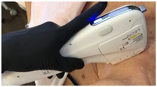
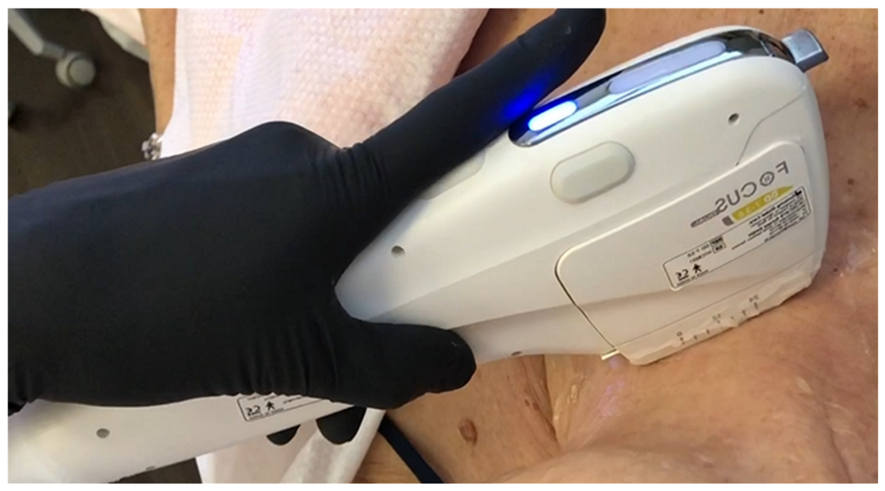
The HIFU handpiece of Focus Dual (Figure 1) employs US waves—mechanical elastic vibrations exceeding 20 kHz—that propagate through elastic media. These waves require a material medium for transmission and exhibit variations in velocity and impedance across different tissues, resulting in phenomena such as reflection, refraction, diffraction, absorption, beam divergence, and deflection. In HIFU, a piezoelectric transducer converts electrical energy into a focused acoustic beam, delivering energy along precise lines of thermocoagulation points (TCPs, <1 mm3) at depths of 1.5–4.5 mm (targeting the papillary/reticular dermis and SMAS) via cartridges with frequencies of 2–9 MHz and energies up to 2.5 J, coupled through gel. Intensity attenuates exponentially through the tissues, inducing focal temperatures >65 °C for 2–3 s, sufficient for coagulative necrosis via protein denaturation, organelle rupture, and enzymatic digestion, while minimizing cavitation (prevalent at lower kHz frequencies). HIFU enables non-invasive facial lifting by targeting multiple depths: 1.5 mm for the superficial dermis (papillary layer, improving texture and fine wrinkles), 3 mm for the deep dermis/reticular layer (filler-like volume restoration), and 4.5 mm for the SMAS (lifting effect on the muscle–aponeurotic system), with an optional depth of 6.5 mm for submental fat. Clinical protocols typically involve three sessions every 3 months (45–60 min each, 350–500 shots per face and 150–200 per neck), using adjustable pitch (0.5–2 mm) to tailor the density of TCPs based on aging severity—denser for advanced laxity and sparser for milder cases. Outcomes include enhanced skin luminosity, nasolabial fold reduction, cheek/jawline contouring, and neocollagenogenesis without anesthesia or downtime, mimicking surgical results while preserving surrounding tissues. Thermal effects dominate via Joule heating, converting mechanical energy into molecular kinetic motion for rapid focal heating (65–70 °C), far exceeding traditional thermoablation needs (43 °C for 60 min) and bypassing perfusion cooling limitations [3]. Non-thermal mechanical effects include stable/inertial cavitation (oscillating gas microbubbles causing membrane shear via microstreaming), radiation forces, and chemical shifts (pH changes, permeabilization), triggering inflammation and tissue remodeling. Post-treatment, coagulative necrosis initiates a regenerative cascade, stimulating fibroblast proliferation for collagen/elastin synthesis, particularly effective on SMAS laxity and dermal flattening in chronoaging. HIFU uniquely reaches SMAS depths inaccessible to RF or superficial devices, offering precision via real-time imaging and cartridges, with minimal cavitation risk at MHz frequencies (vs. kHz for body lipolysis).
Figure 1.
A close-up view of the Focus Dual disposable HIFU handpiece. The handpiece accommodates cartridges with working depths of 1.5 mm, 3.0 mm, or 4.5 mm, depending on the target tissue (superficial dermis, deep dermis, or SMAS) and tailored to the patient’s skin laxity and characteristics.
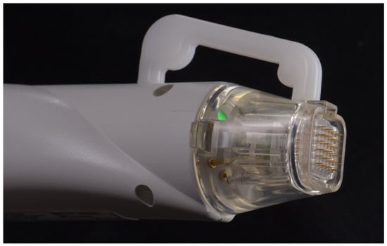
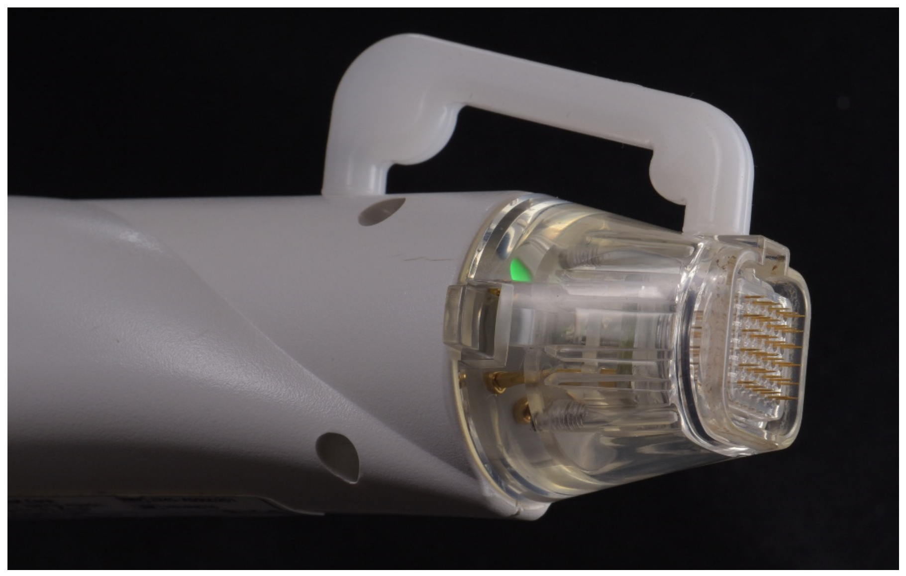
The fractional RF-MN device of Focus Dual incorporates an advanced system combining fractional microneedling with bipolar RF to enhance dermal regeneration through synergistic mechanical and thermal stimulation. In microneedling mode, the system utilizes sterile, gold-plated insulated or non-insulated micro-needles that precisely penetrate the dermis at adjustable depths from 0.5 mm to 3.5 mm, depending on clinical needs (Figure 2). The controlled mechanical insertion of the needles, thanks to the integrated vacuum pump that precisely aspirates the selected skin, triggers a localized inflammatory response that stimulates collagen and elastin production, while bipolar RF energy is simultaneously delivered through the needles, producing targeted volumetric heating of the deep dermis—reaching up to 45–55 °C—thereby inducing controlled collagen denaturation, fibroblast activation, and subsequent tissue remodeling.
Figure 2.
A close-up view of the Focus Dual® disposable RF-MN tip, featuring a high-precision matrix of 36 gold-plated insulated microneedles. The integrated vacuum pump precisely aspirates the skin, allowing controlled and selective needle penetration to specific depths (0.5–3.5 mm with 0.1 mm accuracy), ensuring homogeneous, predictable, and uniform delivery of bipolar RF energy with minimal thermal diffusion and epidermal sparing.
The Focus Dual system supports both insulated and non-insulated needle types, each offering distinct clinical applications. Insulated needles are coated along the shaft, exposing only the tip to deliver RF energy precisely in the deep dermis while preserving the epidermis and upper dermis, thus minimizing the risk of surface damage and post-inflammatory hyperpigmentation; this makes them particularly suitable for medium-to-dark skin types (Fitzpatrick IV–VI), areas with deep dermal laxity, sensitive zones such as the periorbital region and neck, and low-downtime procedures. In contrast, non-insulated needles—also known as full-length needles—deliver RF energy along their entire length, enabling diffuse heating through multiple dermal layers and offering broader stimulation; these are preferred in the treatment of acne scars or post-surgical scars, thick or photo-damaged skin, and in fair skin types (Fitzpatrick I–III), though they carry a higher risk of surface reactions like erythema or superficial irritation. The choice between insulated and non-insulated needles is guided by treatment objectives, anatomical location, skin type, and the desired depth of thermal action. The Focus Dual system allows customizable adjustment of three core parameters: needle penetration depth (0.5–3.5 mm), pulse duration, and RF intensity, which are essential in tailoring treatment to individual skin characteristics and clinical indications. Needle penetration depth defines how deep the micro-needles reach, determining the dermal layer targeted by both mechanical and thermal stimuli; shallow depths (0.5–1.0 mm) are ideal for delicate areas such as periorbital skin, superficial rejuvenation, or early aging signs; intermediate depths (1.0–2.5 mm) are suited to moderate wrinkles, skin laxity, or acne scars; and deeper insertions (2.5–3.5 mm) are preferred for severe atrophic scars, deep wrinkles, and significant laxity in thicker skin areas like the cheeks or jawline. Pulse duration refers to the length of RF energy delivery once the needles are in place; shorter pulses (50–100 ms) generate controlled, focal heating with minimal collateral tissue impact, making them ideal for sensitive skin and lower phototypes, whereas longer pulses (up to 200–300 ms) permit greater thermal diffusion and enhanced collagen remodeling, albeit with an increased risk of erythema and inflammation. RF intensity, indicating the power output during each pulse, may be expressed in energy levels or watts, depending on the interface, and ranges from low (for maintenance or sensitive skin) to moderate or high (to target aging signs, textural irregularities, or scarring); higher intensities, especially when combined with greater needle depth, amplify collagen induction but necessitate close monitoring to prevent over-treatment, particularly in darker phototypes. By fine-tuning these three parameters—depth, duration, and intensity—clinicians can create personalized treatment protocols targeting specific skin structures, optimizing therapeutic outcomes while minimizing adverse effects, and ultimately enhancing both safety and efficacy.
2.3. Protocol
Patients in the treatment group underwent two sessions of HIFU Focus Dual every 3 months and six sessions of RF-MN Focus Dual monthly, for a total of 6 months of treatment. Standardized photographs were acquired at baseline (T0) and 1 month after the last treatment session (T1). The study timepoints were defined as follows:
- T0: baseline photographs before treatment initiation.
- T1: first HIFU and first RF-MN sessions.
- T2 (1 month after T1): second RF-MN session.
- T3 (1 month after T2): third RF-MN session.
- T4 (1 month after T3): second HIFU and fourth RF-MN sessions.
- T5 (1 month after T4): fifth RF-MN session.
- T6 (1 month after T5): sixth RF-MN session.
- T7 (1 month after T6): final control visit with photographs.
All treatments were performed by an experienced operator (O.R.). Clinical evaluations were conducted blindly by two independent evaluators (T.T. and G.P.) using standardized photographic assessments. Baseline facial and neck photographs (T0) were acquired prior to treatment, focusing on the following 11 areas according to the Lemperle Wrinkle Assessment Scale (WAS) [23]: horizontal forehead wrinkles, glabellar frown lines, periorbital wrinkles, preauricular wrinkles, cheek wrinkles, nasolabial folds, upper lip wrinkles, oral commissure wrinkles, marionette lines, labiomental wrinkles and neck lines. Settings for both the HIFU and RF-MN handpieces of the Focus Dual® device were customized for each patient based on wrinkle severity and distribution, as well as skin laxity grade; energy intensity was adjusted according to individual tolerance and clinical response, within predefined parameter ranges according to anatomical region and clinical indication. Prior to each session, the entire face was cleansed using a gentle cleanser. A topical eutectic anesthetic cream (containing 2.5% lidocaine and 2.5% prilocaine) was applied for 60 min and removed before the treatment. In patients with a history of herpes simplex infection, prophylactic antiviral therapy with valacyclovir (500 mg/day) was prescribed for 3 days starting 1 day before the procedure.
Each HIFU session (T1 and T4) was followed by RF-MN treatment. HIFU was applied across the full face prior to RF-MN, which targeted areas with more severe wrinkles and skin laxity. The RF conduction mode featured controlled pulse delivery times and minimal thermal diffusion to reduce the risk of post-inflammatory hyperpigmentation.
After treatment, patients were instructed to apply a non-comedogenic moisturizer several times daily for 3–5 days, avoid sun exposure, and use broad-spectrum sunscreen. They were advised not to use any topical or systemic antibiotics or retinoids throughout the treatment period.
Photographs of each patient were taken using the same camera with fixed camera settings (Reflex Nikon D7200, Nikon Corporation, Tokyo, Japan; ISO 200, f/8, 1/125 s) at baseline (T0) and at the end of the treatment protocol (T7) under consistent lighting and patient positioning with a custom headrest.
2.3.1. HIFU Protocol (Focus Dual System)
HIFU treatments were performed using a focused ultrasound handpiece integrated within the Focus Dual system. A piezoelectric transducer delivered HIFU energy at frequencies ranging from 4 to 7 MHz, generating TCPs at controlled depths of 1.5 mm, 3.0 mm, and 4.5 mm, corresponding respectively to the superficial dermis, deep dermis, and SMAS. An optional 6.0–6.5 mm cartridge was used in selected cases for submental adipose tissue targeting.
Energy delivery was set between 0.8 and 2.4 J per shot, adjusted according to anatomical region and tissue thickness. Treatments were performed using a linear array technique with a spacing (pitch) of 1.0–1.5 mm between lines, allowing controlled thermal overlap and homogeneous energy distribution.
A total of 500–700 shots per full-face/neck session were administered, with approximately 60–80 shots per anatomical region (forehead, periorbital region, midface, lower face, neck and submental area). Each session lasted approximately 45–60 min. Two treatment sessions were performed at 3-month intervals.
Acoustic gel was used as a coupling medium to optimize ultrasound transmission and reduce impedance mismatch. Thermal effects were expected to reach focal temperatures of 65–70 °C for 1 s, inducing controlled coagulative necrosis and subsequent neocollagenesis while preserving surrounding tissues.
2.3.2. Fractional RF Microneedling Protocol (Focus Dual System)
RF-MN treatments were performed using the fractional bipolar RF-MN handpiece of the Focus Dual system. Sterile gold-plated microneedles were used to deliver controlled dermal stimulation. Needle penetration depth was adjusted between 0.5 mm and 3.5 mm according to anatomical region:
- 0.5–1.0 mm for periorbital and superficial dermal rejuvenation;
- 1.0–2.5 mm for moderate rhytides and early laxity;
- 2.5–3.5 mm for deep wrinkles and advanced dermal laxity.
RF energy was delivered in bipolar mode with power settings ranging from 10 to 40 W, depending on skin thickness, phototype, and treatment indication. Pulse duration was set between 50 and 300 ms, allowing modulation of thermal diffusion within the dermis.
Insulated needles were preferentially used in periorbital, neck, and higher-phototype (Fitzpatrick IV–VI) areas to confine thermal energy to the deep dermis and minimize epidermal damage. Each area was treated with 1–2 passes, using a fractional stamping technique combined with controlled vacuum-assisted tissue stabilization to ensure uniform needle insertion. Target dermal temperatures were estimated to be between 45 and 55 °C, sufficient to induce collagen denaturation and fibroblast activation without epidermal ablation.
2.4. Outcomes
The primary outcome of the study was the change in global wrinkle severity between baseline (T0) and post-treatment evaluation (T7), assessed using the mean Wrinkle Assessment Scale (WAS) score across the 11 facial anatomical regions. Secondary outcomes included changes in regional WAS scores; Global Aesthetic Improvement Scale (GAIS) ratings; inter-rater reliability between evaluators; association between baseline wrinkle severity and treatment response; and occurrence of treatment-related adverse events.
2.5. Evaluation Through the WAS and the Global Aesthetic Improvement Scale
Clinical improvement was evaluated using the WAS, which provides a comprehensive evaluation of static wrinkles across 11 facial regions (forehead, glabella, periorbita, preauricular region, cheek, nasolabial region, upper lip, oral commissure, nasolabial folds, labiomental folds and neck). The six ordinal WAS categories were numerically coded from 0 (no wrinkles) to 5 (very deep wrinkles and redundant folds), corresponding to the following grading system: 0 = no wrinkles; 1 = just perceptible wrinkles; 2 = shallow wrinkles; 3 = moderately deep wrinkles; 4 = deep wrinkles with well-defined edges; and 5 = very deep wrinkles and redundant folds.
Two independent esthetic specialists (G.P. and T.T.), blinded to treatment time points, assessed and scored photographs in a randomized, non-chronological order using the WAS.
To objectively assess clinical improvement in skin rejuvenation, the Global Aesthetic Improvement Scale (GAIS) [24] was employed. This validated scale enables standardized evaluation based on photographic comparison. Two esthetic specialists (G.P. and T.T.) independently and blindly reviewed standardized facial photographs taken at baseline and after treatment. Images were presented in a randomized, non-chronological order to minimize potential bias. Each dermatologist assigned a GAIS grade based on the percentage of improvement observed, considering both the number and severity of wrinkles, as well as the overall skin condition (pore size, tone and texture). The GAIS grades were defined as follows:
- Grade 0: worsened;
- Grade 1: 0–25% (minimal improvement);
- Grade 2: 26–50% (moderate improvement);
- Grade 3: 51–75% (marked improvement);
- Grade 4: >75% (near-total clearance).
Adverse effects, including bleeding, erythema, scaling, crusting, dyschromia, and infection, were recorded after each session and again at the final 6-month follow-up.
2.6. Statistical Analysis
Statistical analyses were performed using IBM SPSS Statistics, version 29.0 (IBM Corp., Armonk, NY, USA). Given the observational and descriptive nature of this case series, the statistical analyses were primarily descriptive and exploratory in nature. WAS scores were summarized using median values, and changes in wrinkle severity between baseline (T0) and post-treatment evaluation (T7) were described using the absolute difference (ΔWAS) and relative percentage change for the overall score and for each of the 11 evaluated facial anatomical regions. Median changes in WAS were also calculated descriptively for the predefined baseline severity subgroups. Inter-rater reliability between the two blinded evaluators was assessed using weighted Cohen’s kappa (κ) statistics. GAIS outcomes were summarized using descriptive frequency distributions and percentages. No formal sample size calculation was performed, and no inferential statistical tests were conducted, in accordance with the descriptive nature of the case series and the limited sample size. Age was not included as a covariate in the statistical analysis, and no age-stratified comparisons were performed because of the exploratory nature of the case series, the limited sample size, and the absence of predefined age strata. All analyses were descriptive, and no statistical significance testing or adjustment for multiple comparisons was performed. All analyses were considered exploratory and hypothesis-generating.
3. Results
A total of 37 patients (1 male and 36 females; age range 52–85 years; Fitzpatrick skin types II and III) meeting the inclusion and exclusion criteria were treated with the Focus Dual® (Med & Tech, Italy). Additional anonymized individual patient images and complete clinical documentation are available from the authors upon reasonable request for scientific purposes and are protected under applicable copyright regulations.
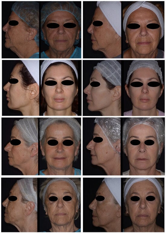
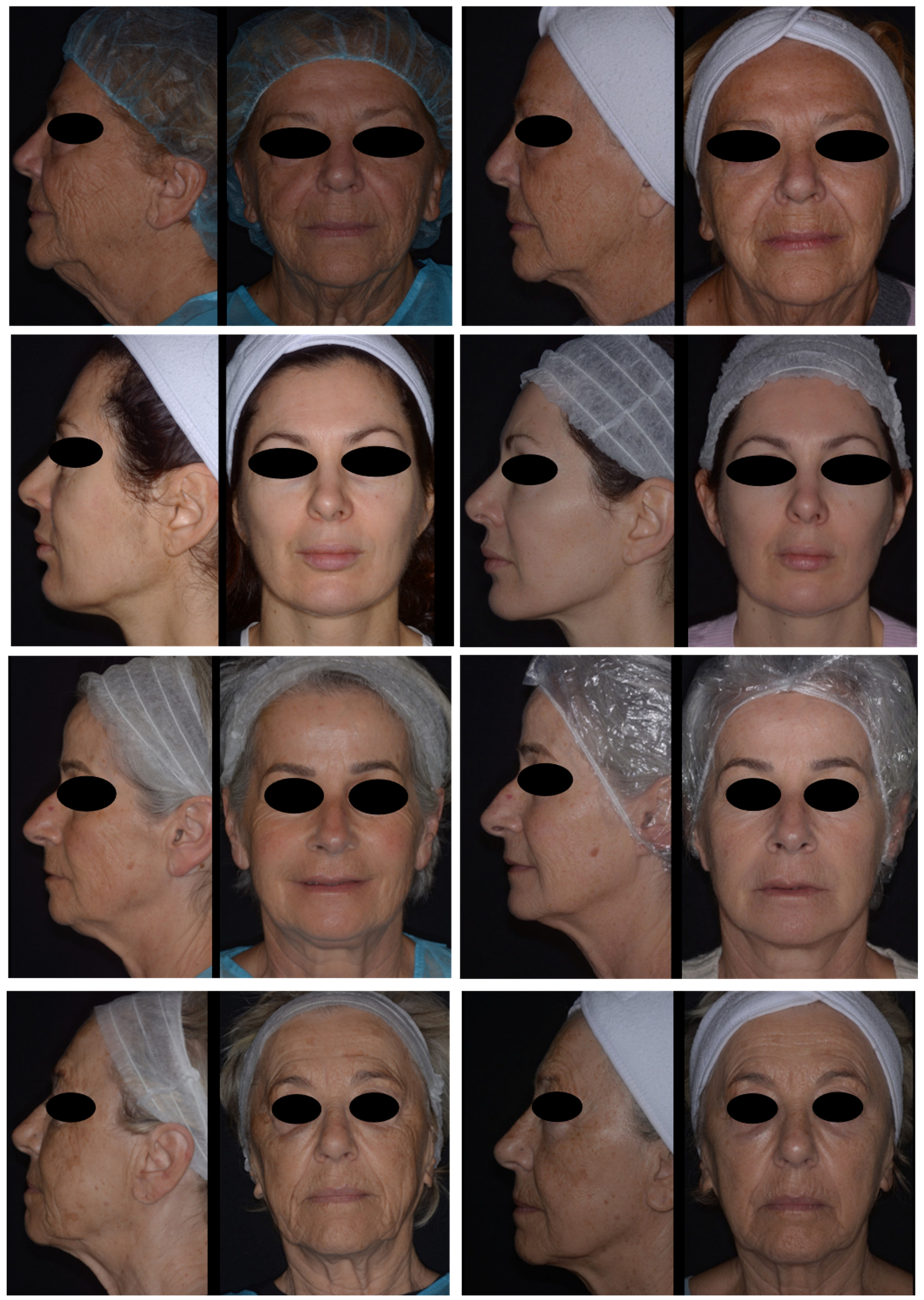
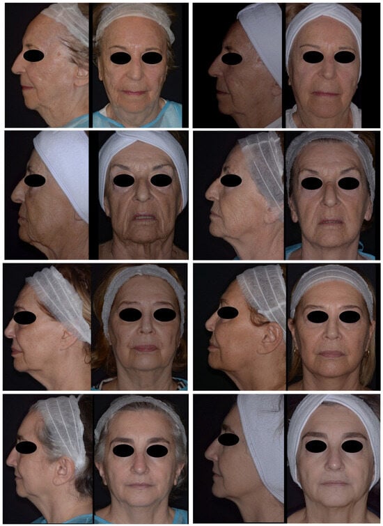
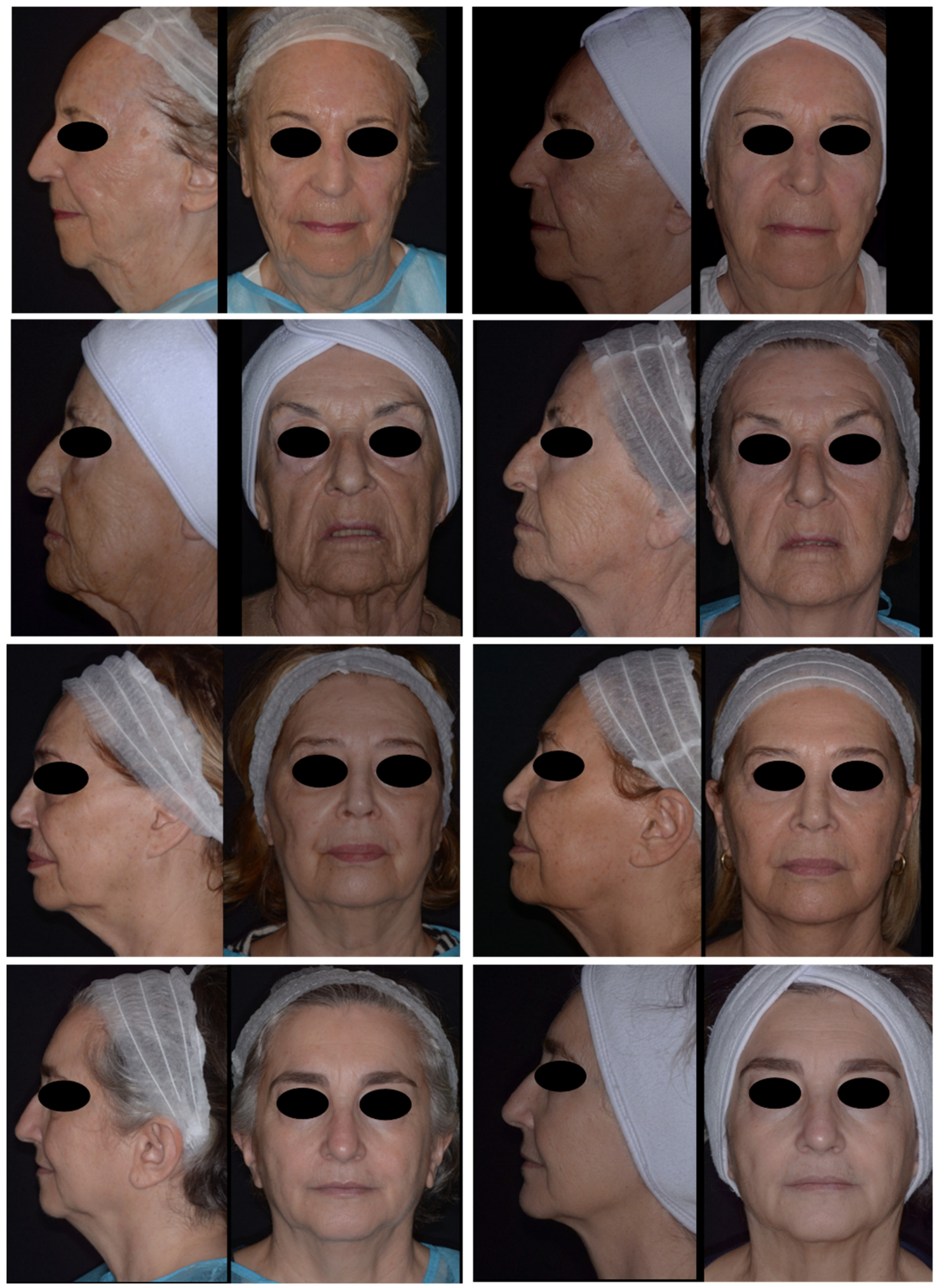
Representative clinical photographs of eight patients were randomly selected from the total study population and are presented in Figure 3 and Figure 4. The corresponding clinical scores for each patient are reported in Table 1, allowing a direct comparison between the photographic outcomes and the quantitative assessment results.
Figure 3.
Representative clinical photographs of Patients 7, 11, 16, and 19. For each patient, frontal and lateral views acquired before treatment (T0) are shown on the left, while the corresponding frontal and lateral views obtained at the end of treatment (T7), according to the study protocol, are shown on the right.
Figure 4.
Representative clinical photographs of Patients 23, 27, 33, and 35. For each patient, frontal and lateral views acquired before treatment (T0) are shown on the left, while the corresponding frontal and lateral views obtained at the end of treatment (T7), according to the study protocol, are shown on the right.
Table 1.
Summary of the scores assigned by the two independent evaluators for Patients 7, 11, 16, 19, 23, 27, 33, and 35. Assessments were performed using the WAS and the GAIS. Individual scores for each patient are reported to illustrate the evaluators’ clinical assessment of treatment outcomes.
The combined application of HIFU and RF-MN resulted in a reduction in wrinkle severity across the full facial assessment. The WAS, calculated as the median of two independent blinded evaluators, decreased from a baseline value of 4.53 to a post-treatment value of 3.98, corresponding to an absolute reduction of 0.55 points and a relative improvement of 12.1% in overall wrinkle severity (Table 2).
Table 2.
Overall WAS scores before and after treatment.
The median baseline and post-treatment WAS scores for each facial region are reported in Table 3. Overall, a reduction in wrinkle severity was observed across all evaluated anatomical areas following the combined HIFU and RF-MN treatment protocol. Based on the mean WAS values, the greatest absolute reductions were observed in the neck folds (mean ΔWAS = 0.97), cheek folds (0.88), marionette lines (0.88), periorbital lines (0.87), and nasolabial folds (0.94). Intermediate reductions were observed in the corner of mouth lines (0.81) and preauricular lines (0.50), whereas smaller changes were detected in the upper lip lines (0.35), horizontal forehead lines (0.35), glabellar lines (0.34), and chin crease (0.21).
Table 3.
Mean and median baseline and post-treatment WAS scores, interquartile ranges (IQRs), absolute changes (ΔWAS), and percentage improvements across the 11 facial regions evaluated after combined HIFU and RF-MN treatment. Greater improvements were observed in anatomical areas predominantly affected by tissue laxity and gravitational aging, including the neck, nasolabial folds, cheeks, marionette lines, and periorbital region, whereas more limited responses were observed in dynamic wrinkle-dominant regions such as the forehead, glabella, and chin crease.
When expressed as percentage improvement based on the mean WAS values, the greatest improvements were observed in the cheek folds (25.36%), periorbital lines (25.14%), marionette lines (21.36%), corner of mouth lines (20.98%), and neck folds (23.77%). Intermediate improvements were observed in the nasolabial folds (23.92%), preauricular lines (17.86%), glabellar lines (12.83%), horizontal forehead lines (12.24%), and upper lip lines (12.11%), whereas the lowest percentage improvement was observed in the chin crease (7.07%).
Stratified analysis according to baseline severity was performed by dividing patients into three groups based on initial WAS scores (moderate, high, and very high severity). The mean WAS improvement within each subgroup was 0.38 points in the moderate-severity group, 0.60 points in the high-severity group, and 0.74 points in the very high-severity group (Table 4).
Table 4.
Treatment response according to baseline wrinkle severity.
Inter-rater reliability analysis showed weighted Cohen’s kappa values of 0.95 at baseline assessment and 0.93 at post-treatment evaluation between the two independent evaluators, both indicating high agreement (Table 5).
Table 5.
Inter-rater reliability: agreement levels were interpreted according to the Landis and Koch classification.
GAIS evaluation showed a consistent positive esthetic response following the combined HIFU and RF-MN treatment protocol. No cases were classified as worsened (Grade 0) by either evaluator. The majority of assessments were categorized as Grade 2 (moderate improvement), accounting for 70.3% and 78.4% of the evaluations performed by GP and TT, respectively. Grade 1 (minimal improvement) represented 27.0% of GP assessments and 21.6% of TT assessments. Only one evaluation (2.7%) was classified as Grade 3 (marked improvement) by GP, whereas no Grade 4 scores were recorded. Overall, GAIS findings indicate that most patients experienced clinically appreciable esthetic improvement, predominantly within the mild-to-moderate improvement range, corroborating the quantitative reduction in wrinkle severity observed through the WAS analysis (Table 6).
Table 6.
Distribution of GAIS scores assigned by the two independent blinded evaluators. n = number of patients assigned to each GAIS grade; % = percentage of patients assigned to each GAIS grade out of the total number of evaluated patients.
Overall, the combined HIFU and RF-MN protocol was well tolerated. Mild and transient adverse effects were observed in a subset of patients, including localized erythema and mild edema immediately after treatment, which typically resolved spontaneously within a few hours to 24–48 h. A small number of patients reported mild procedural discomfort or pain during or shortly after the session, which did not require analgesic treatment and resolved within 1–2 days. Temporary, procedure-related skin changes consistent with microneedling and ultrasound energy delivery were occasionally noted, with no cases of burns, infections, post-inflammatory hyperpigmentation, scarring, or any persistent or clinically relevant adverse events recorded.
4. Discussion
The combined application of HIFU and RF-MN was associated with a measurable improvement in wrinkle severity across the full facial analysis, as assessed using the WAS, a validated semi-quantitative clinician-rated scale evaluating 11 facial anatomical subunits and assigning a severity score ranging from 1 (no wrinkles) to 5 (redundant wrinkles). The WAS, derived from the mean of two independent blinded evaluators, decreased from baseline to after treatment, corresponding to an absolute reduction of 0.55 points and a relative reduction of 12.1%. This change reflects a consistent improvement across the study population, with concordant directional variation observed across most facial subunits. This result is consistent with previous studies indicating clinical improvement with energy-based devices, including HIFU and RF-based technologies, in facial rejuvenation and wrinkle reduction [12,13,20], as well as with evidence supporting their combined application for skin tightening and structural improvement [13,21]. Byun Ji Won et al.’s split-face prospective trial [22] further supports the present findings through objective operator-independent measurements and an instrument-based evaluation framework, using standardized facial imaging systems (digital face scanners with geometric analysis of linear measurements and angular parameters) and skin analysis technologies (US-based skin scanners and probes evaluating elasticity, skin density, and structural properties) that strengthen the objectivity and reliability of the reported outcomes, complementing the clinically relevant but inherently subjective assessments performed by expert operators in the present study. Within this mechanistic framework, the clinical improvement observed after combined HIFU and RF-MN may be related to their complementary depth-dependent effects. High-intensity focused ultrasound primarily targets the deep dermal and SMAS layers, promoting structural tissue contraction and lifting, whereas RF acts within the superficial papillary dermis, enhancing skin quality parameters such as texture, elasticity, and hydration [22]. When delivered through microneedles, however, RF energy can be selectively deposited at greater dermal depths through direct penetration of the microneedles, while minimizing energy delivery to the epidermis and superficial layers. The interaction between these mechanisms may result in complementary tissue remodeling, whereby deep structural tightening is complemented by superficial dermal improvement, contributing to a more uniform enhancement of overall facial appearance [22].
At the regional level, the treatment effect of the combination of HIFU and RF-MN showed a clear anatomical distribution. The greatest improvements were observed in anatomically lax regions such as the cheek folds (25.36%), periorbital lines (25.14%), nasolabial folds (23.92%), neck folds (23.77%), marionette lines (21.36%), and corner of mouth lines (20.98%), whereas more limited responses were observed in dynamic wrinkle-dominant regions such as the chin crease (7.07%), horizontal forehead lines (12.24%), and glabellar lines (12.83%). This regional pattern was also reflected in the median WAS values: the median score decreased by one point in eight of the eleven anatomical regions evaluated, including the glabellar, periorbital, preauricular, cheek, nasolabial, corner of mouth, marionette, and neck regions. In contrast, the median remained unchanged in the horizontal forehead, upper lip, and chin crease regions, despite modest reductions in their corresponding mean WAS values (Table 3). These findings indicate that the observed regional improvements were not solely attributable to changes in mean scores, but were also reflected in a shift in the central distribution of clinician-rated wrinkle severity in most anatomical regions.
These anatomical regions are commonly associated with age-related structural laxity, loss of dermal elasticity, weakening of retaining ligaments, and progressive gravitational descent of soft tissues. The most substantial improvements were consistently observed in the lower and middle facial thirds and in the cervical region, particularly in anatomical areas characterized by structural laxity, gravitational tissue descent, and dermal remodeling. Lower reductions were observed in preauricular lines, upper lip lines, horizontal forehead lines, glabellar lines, and chin crease, which are more influenced by repetitive muscular activity and dynamic facial expression patterns. Conversely, regions such as the glabellar and frontal areas, as well as the chin crease, were associated with more modest improvements. These areas are predominantly influenced by dynamic muscular activity rather than structural laxity, which may be associated with their reduced responsiveness to energy-based tissue tightening procedures.
A positive correlation was observed between baseline wrinkle severity and percentage improvement following treatment (r = 0.73). However, this association should be interpreted with caution, as percentage improvement is mathematically dependent on baseline severity and may therefore introduce a degree of mathematical coupling between the two variables. Accordingly, the observed correlation should not be interpreted as evidence that patients with more severe wrinkles necessarily respond better to treatment. Patients were also grouped into moderate-, high-, and very high-severity categories according to their initial WAS scores. Median absolute WAS reductions were 0.38 in the moderate group, 0.60 in the high-severity group, and 0.74 in the very-high-severity group (Table 4). Although greater absolute reductions were observed in the groups with higher baseline severity, these descriptive differences do not establish a severity-dependent treatment effect and may be influenced by baseline-dependent statistical effects and regression to the mean. Therefore, the present findings should be considered hypothesis-generating rather than confirmatory with regard to the relationship between baseline wrinkle severity and treatment response. Future controlled studies using prespecified statistical models that account for baseline severity are warranted to determine whether baseline wrinkle severity independently influences treatment response. Despite established evidence indicating that energy-based devices such as RF and US are generally more effective in the management of mild to moderate degrees of facial laxity, with limited and often suboptimal outcomes in advanced laxity, for which surgical intervention remains the gold standard [25], the descriptive pattern observed in the present study warrants further investigation. Although the present findings cannot establish greater treatment efficacy in more severe laxity phenotypes, the observed pattern may warrant further investigation into whether combining HIFU with a more selective form of RF delivery, such as RF-MN, could provide complementary effects in patients with more advanced structural laxity.
In line with the findings of the present study, and consistent with the mechanistic framework proposed in the literature, RF-MN may provide enhanced dermal targeting compared with non-microneedling (monopolar or bipolar) RF systems. As highlighted in the comprehensive review by Tan et al. [26], RF-MN delivers thermal energy in a more targeted and intradermal manner, producing controlled micro-injuries within specific dermal layers while minimizing energy dispersion to surrounding tissues. This spatially confined and depth-adjustable delivery is associated with a more efficient induction of neocollagenesis and neoelastogenesis, thereby enhancing dermal tightening and remodeling responses. Such a selective and focused mechanism of action may partly explain the greater improvement observed in regions characterized by gravitational descent and structural laxity in the present study, supporting the concept that microneedle-based RF systems can exert a more relevant biomechanical impact on sagging-related facial compartments than conventional non-fractionated RF modalities. This differential response pattern is of particular relevance for clinical interpretation and is further discussed in relation to treatment selectivity. The observed distribution is consistent with the proposed mechanisms of action of the combined modalities, whereby HIFU induces selective thermal contraction of the SMAS and deep fibrous septa [2,3,4,5,6,7], while RF-MN promotes dermal neocollagenesis and remodeling of elastic fibers [22,26,27,28,29]. HIFU delivers focused acoustic energy to deeper layers, inducing thermal coagulation points within the SMAS and deep fibrous septa, with consequent collagen contraction and tissue repositioning. RF-MN produces controlled fractional thermal injury within the dermis via microneedles, promoting fibroblast activation, neocollagenesis, and remodeling of the extracellular matrix. The combined effect appears to be more relevant in tissue compartments where structural laxity is the dominant aging component, whereas its impact is comparatively reduced in regions primarily driven by hyperdynamic muscular contraction. Inter-rater reliability analysis showed a high level of agreement between the two independent evaluators. Weighted Cohen’s kappa values were 0.95 at baseline and 0.93 after treatment, both corresponding to almost perfect agreement according to standard interpretation criteria. This indicates a high degree of consistency in scoring between observers across both evaluation timepoints. Inter-observer agreement between the two blinded evaluators was excellent, and these results support the robustness and reproducibility of the photographic assessment methodology. The GAIS assessment further supported the clinical findings obtained through the WAS analysis. Most evaluations were classified as moderate improvement (Grade 2), accounting for approximately three-quarters of all assessments, while the remaining cases were predominantly categorized as minimal improvement (Grade 1). No cases of worsening were recorded by either evaluator. These findings suggest that the clinical benefits associated with the combined HIFU and RF-MN protocol were generally perceptible at a global esthetic level and not limited to isolated reductions in wrinkle severity scores. The predominance of Grade 2 responses is consistent with the non-surgical nature of the treatment, which aims to induce progressive tissue remodeling rather than dramatic corrective changes. The limited number of marked improvement ratings may reflect the advanced age and moderate-to-severe baseline photoaging of the study population, in which complete correction of structural aging changes is not generally expected with energy-based therapies alone. Taken together, these findings describe a pattern of anatomical variability in response following combined HIFU and RF-MN treatment, with differences observed across facial regions and baseline severity strata, alongside high inter-observer reliability of outcome assessment. However, the observed differences across severity strata should be interpreted as descriptive and hypothesis-generating rather than as evidence of a severity-dependent treatment effect.
5. Limitations of the Study
This study presents some methodological aspects that should be considered for the correct interpretation of the findings. The case series design, without a control group or randomization, reflects the observational nature of the investigation and the real-world clinical setting in which the treatments were applied.
All procedures were performed by a single experienced clinician. While this approach minimized inter-operator variability and ensured protocol consistency, it may limit the external validity of the results and does not allow assessment of operator-related variability.
The sample size (n = 37) and the single-center setting provide a focused clinical cohort while limiting the extension of the results to broader and more heterogeneous populations.
The study population was predominantly female (36 females and 1 male), reflecting the clinical population voluntarily seeking this type of esthetic treatment. Although male participants were eligible, their limited representation prevents assessment of sex-related differences in treatment response and may limit the generalizability of the findings to males. Future studies with more balanced male and female cohorts are warranted.
The relatively broad age range of the participants (52–85 years) represents another aspect that should be considered when interpreting the findings. Age-related changes in skin thickness, hydration, elasticity, dermal architecture, and other physiological properties may influence the response to energy-based treatments. However, the age distribution was not deliberately imposed by the study design but reflected the population that voluntarily sought treatment and met the eligibility criteria in the clinical setting. Given the limited sample size and observational case series design, no age-stratified or age-adjusted analysis was performed; therefore, age cannot be excluded as a potential confounding factor contributing to inter-individual variability in treatment response. Future studies with larger cohorts should consider predefined age strata or age-adjusted analyses to better characterize the potential influence of age on treatment outcomes.
Given the combined use of HIFU and RF-MN, the specific contribution of each individual modality to the overall clinical outcome cannot be separately quantified within this study design. The results therefore reflect the integrated effect of the combined protocol.
Clinical outcomes were assessed using validated scales (WAS and GAIS) and blinded evaluators based on standardized photographic documentation. Nevertheless, as with all image-based clinical evaluations, the assessment remains dependent on photographic conditions and clinical judgment.
The analysis of the association between baseline wrinkle severity and percentage improvement should also be interpreted cautiously, as percentage improvement is mathematically dependent on baseline severity, potentially introducing mathematical coupling and limiting the interpretation of this association as evidence of a differential treatment response.
The follow-up period was limited to the completion of the treatment protocol and therefore reflects short-term clinical outcomes following the combined intervention.
No histological or advanced imaging assessments (e.g., ultrasound elastography or biopsy) were included; consequently, the mechanistic interpretations are based on the established literature regarding tissue responses to HIFU and RF-based technologies.
Future studies with larger cohorts, controlled designs, and extended follow-up periods may further contribute to characterizing the long-term clinical behavior of combined HIFU and RF-MN protocols in facial rejuvenation.
6. Conclusions
The combined use of HIFU and RF-MN with the Focus Dual® device showed a clinical improvement in facial wrinkle severity in this 37-patient case series. Quantitative assessment using the WAS showed a global reduction in wrinkle scores, with a mean improvement of 12.1% across all evaluated facial regions. Clinical enhancement was further supported by GAIS evaluations, which showed predominantly moderate esthetic enhancement and no cases of worsening, confirming the favorable overall perception of treatment outcomes.
The treatment effect was not uniform across anatomical regions, with greater responses observed in areas characterized by structural laxity and gravitational aging—particularly the neck, nasolabial folds, cheeks, and marionette lines—while more limited changes were detected in regions dominated by dynamic muscular activity, such as the forehead, glabellar area and chin crease. Although a positive association was observed between baseline severity and percentage improvement, this finding should be interpreted cautiously because the improvement measure is mathematically dependent on baseline severity and therefore does not, by itself, demonstrate that more severe wrinkles respond better to treatment.
Overall, the findings suggest the concept of a potential complementary, layer-dependent mechanism of action between HIFU and RF-MN, combining deep structural remodeling with superficial dermal regeneration. Within the limits of an observational design, these results indicate that the combined protocol may represent a useful non-surgical option for facial rejuvenation in appropriately selected patients. Future studies should also incorporate objective instrumental assessments and patient-reported outcomes to provide a more comprehensive evaluation of treatment efficacy and patient-perceived benefit. Further controlled studies with larger cohorts and longer follow-up are required to better define the durability of outcomes and the relative contribution of each modality.
Author Contributions
Conceptualization, O.R., G.P. and T.T.; Methodology, O.R., G.P., T.T. and M.D.F.; Validation, T.T. and M.D.F.; Formal analysis, O.R. and M.D.F.; Investigation, O.R. and M.D.F.; Resources, O.R., G.P. and T.T.; Data curation, O.R., G.P. and M.D.F.; Writing—original draft, O.R.; Supervision, G.P. and T.T.; Project administration, G.P. and T.T.; Funding acquisition M.D.F. All authors have read and agreed to the published version of the manuscript.
Funding
This research received no external funding.
Institutional Review Board Statement
The present case series evaluated the clinical use of HIFU and RF-MN for antiaging treatment in routine practice. The CE-certified device (Focus Dual, Med&Tech) was applied strictly according to its intended clinical purpose. No control group, randomization, or experimental interventions were involved. Ethical review and approval were waived in accordance with national and institutional regulations for non-interventional studies. All procedures were performed by experienced clinicians, and written informed consent for treatment, privacy, and publication of anonymized data and images was obtained from all participants, in compliance with the Declaration of Helsinki.
Informed Consent Statement
Informed consent was obtained from all subjects involved in the study. Each participant was thoroughly informed about the mechanism of action of the device, potential contraindications, expected benefits, and the objectives of the treatment. Written consent regarding privacy and the use of photographic documentation, strictly for scientific purposes, was collected, signed, and archived for every patient. Written informed consent has also been obtained from the patients for the publication of images included in this paper.
Data Availability Statement
The datasets generated and analyzed during the current study are available from the corresponding author upon reasonable request.
Acknowledgments
The authors wish to acknowledge that no additional support, administrative or technical assistance, or donations in kind were received for this study.
Conflicts of Interest
Ornella Rossi, Giovanna Perrotti and Tiziano Testori the authors are affiliated with the Lake Como Institute (Private Practice). The authors declare that no financial or commercial relationships influenced this study. No external funding was received, and all aspects of the study—including its design, data collection, analysis, interpretation, manuscript writing, and the decision to publish—were conducted independently of any commercial entity. All other authors declare that the research was conducted in the absence of any commercial or financial relationships that could be construed as potential conflicts of interest.
References
- Trovato, F.; Ceccarelli, S.; Michelini, S.; Vespasiani, G.; Guida, S.; Galadari, H.I.; Nisticò, S.P.; Colonna, L.; Pellacani, G. Advancements in Regenerative Medicine for Aesthetic Dermatology: A Comprehensive Review and Future Trends. Cosmetics 2024, 11, 49. [Google Scholar] [CrossRef] [Scilit]
- National Council on Radiation Protection and Measurements. Biological Effects of Ultrasound: Mechanisms and Clinical Implications; NCRP Report No. 74; National Council on Radiation Protection and Measurements: Bethesda, MD, USA, 1983. [Google Scholar]
- Miller, M.W.; Miller, D.L.; Brayman, A.A. A review of in vitro bioeffects of inertial ultrasonic cavitation from a mechanistic perspective. Ultrasound Med. Biol. 1996, 22, 1131–1154. [Google Scholar] [CrossRef] [Scilit] [PubMed]
- White, W.M.; Makin, I.R.S.; Barthe, P.G.; Slayton, M.H.; Gliklich, R.E. Selective creation of thermal injury zones in the superficial musculoaponeurotic system using intense focused ultrasound: A new target for noninvasive facial rejuvenation. Arch. Facial Plast. Surg. 2007, 9, 22–29. [Google Scholar] [PubMed]
- Miller, D.; Smith, N.; Bailey, M.; Czarnota, G.; Hynynen, K.; Makin, I.J. Overview of therapeutic ultrasound applications and safety considerations. Ultrasound Med. 2012, 31, 623–634. [Google Scholar] [CrossRef] [Scilit] [PubMed]
- De Lucas, B.; Pérez, L.M.; Bernal, A.; Gálvez, B.G. Ultrasound therapy: Experiences and perspectives for regenerative medicine. Genes 2020, 11, 1086. [Google Scholar] [CrossRef] [Scilit] [PubMed]
- Bader, K.B.; Makin, I.R.S.; Abramowicz, J.S. Ultrasound for aesthetic applications: A review of biophysical mechanisms and safety. J. Ultrasound Med. 2022, 41, 1597–1607. [Google Scholar] [PubMed]
- Ruiz-Esparza, J.; Barba, G.O. The non-ablative radiofrequency for skin tightening. Dermatol. Surg. 2001, 27, 909–911. [Google Scholar]
- Elsaie, M.L. Cutaneous remodeling and photo-rejuvenation using radiofrequency devices. Indian J. Dermatol. 2009, 54, 201–205. [Google Scholar] [CrossRef] [Scilit] [PubMed]
- Austin, G.K.; Struble, S.L.; Quatela, V.C. Evaluating the effectiveness and safety of radiofrequency for face and neck rejuvenation: A systemic review. Lasers Surg. Med. 2022, 54, 27–45. [Google Scholar] [CrossRef] [Scilit] [PubMed]
- Suh, D.H.; Choi, J.H.; Lee, S.J.; Jeong, K.H.; Song, K.Y.; Shin, M.K. Comparative histometric analysis of the effects of high-intensity focused ultrasound and radiofrequency on skin. J. Cosmet. Laser Ther. 2015, 17, 230–236. [Google Scholar] [CrossRef] [Scilit] [PubMed]
- Serdar, Z.A.; Karabay, E.A.; Tatlıparmak, A.; Aksoy, B. Efficacy of high-intensity focused ultrasound in facial and neck rejuvenation. J. Cosmet. Dermatol. 2020, 19, 353–358. [Google Scholar] [CrossRef] [Scilit] [PubMed]
- Mey, S.J.; Khong, L.; Ismail, A.H.; Sujani, S.; Clinic, J.Z.; Devindaran, N.; Medveon, S.K.; Rashid, M.F.A.; Zaman, U.M.S.M. Safety and efficacy of high-intensity focused ultrasound and monopolar radiofrequency combination therapy for skin tightening: A retrospective study in Malaysia. Malays. J. Med. Sci. 2024, 31, 114–123. [Google Scholar] [CrossRef] [Scilit] [PubMed]
- Rossi, O.; Perrotti, G.; Scaini, R.; Del Fabbro, M.; Damiani, G.; Testori, T. How to establish the baseline for non-invasive technological regenerative esthetic medicine in the face and neck region: A literature review. J. Pers. Med. 2025, 15, 283. [Google Scholar] [CrossRef] [Scilit] [PubMed]
- Glogau, R.G. Aesthetic and anatomic analysis of the aging skin. Semin. Cutan. Med. Surg. 1996, 15, 134–138. [Google Scholar] [CrossRef] [Scilit] [PubMed]
- Coleman, S.R.; Grover, R. The anatomy of the aging face: Volume loss and changes in 3-dimensional topography. Aesthet. Surg. J. 2006, 26, S4–S9. [Google Scholar] [CrossRef] [Scilit] [PubMed]
- Goldie, K. The evolving field of regenerative aesthetics. J. Cosmet. Dermatol. 2023, 22, 1–7. [Google Scholar] [CrossRef] [Scilit] [PubMed]
- Zarbafian, M.; Fabi, S.G.; Dayan, S.; Goldie, K. The Emerging Field of Regenerative Aesthetics—Where We Are Now. Dermatol. Surg. 2022, 48, 101–108. [Google Scholar] [CrossRef] [Scilit] [PubMed]
- Weiner, S.F. Radiofrequency microneedling: Overview of technology, advantages, differences in devices, studies, and indications. Facial Plast. Surg. Clin. N. Am. 2019, 27, 291–303. [Google Scholar]
- Zhao, B.; Li, P.; Fu, Y.; Quan, Y.; Wang, H.; Zhao, L.; Pang, X.; Liu, Z.; Tan, L.; Peng, J.; et al. Efficacy and safety of radiofrequency and focused ultrasound in facial rejuvenation: A single-center, single-blind, non-randomized prospective trial. J. Cosmet. Dermatol. 2025, 24, e70407. [Google Scholar] [CrossRef] [Scilit] [PubMed]
- Ayatollahi, A.; Gholami, J.; Saberi, M.; Hosseini, H.; Firooz, A. Systematic review and meta-analysis of safety and efficacy of high-intensity focused ultrasound (HIFU) for face and neck rejuvenation. Lasers Med. Sci. 2020, 35, 1007–1024. [Google Scholar] [CrossRef] [Scilit] [PubMed]
- Byun, J.W.; Kang, Y.R.; Park, S.J.; Hong, W.K. Efficacy of radiofrequency combined with single-dot ultrasound for skin rejuvenation: A non-randomized split-face trial with blinded response evaluation. Skin Res. Technol. 2023, 29, e13452. [Google Scholar] [CrossRef] [Scilit] [PubMed]
- Lemperle, G.; Holmes, R.E.; Cohen, S.R.; Lemperle, S.M. A classification of facial wrinkles. Plast. Reconstr. Surg. 2001, 108, 1735–1745. [Google Scholar] [CrossRef] [Scilit] [PubMed]
- DiBernardo, G.A.; DiBernardo, B.E. Prediction of treatment outcomes for neck rejuvenation utilizing a unique classification system of treatment approach using a 1440-nm side-firing laser. Aesthet. Surg. J. 2018, 38, S43–S51. [Google Scholar] [CrossRef] [Scilit] [PubMed]
- Alexiades-Armenakas, M.; Rosenberg, D.; Renton, B.; Dover, J.; Arndt, K.A. Blinded, randomized, quantitative grading comparison of minimally invasive, fractional radiofrequency and surgical face-lift to treat skin laxity. Arch. Dermatol. 2010, 146, 396–405. [Google Scholar] [CrossRef] [Scilit] [PubMed]
- Tan, M.G.; Jo, C.E.; Chapas, A.; Khetarpal, S.; Dover, J.S. Radiofrequency microneedling: A comprehensive and critical review. Dermatol. Surg. 2021, 47, 755–761. [Google Scholar] [CrossRef] [Scilit] [PubMed]
- Alster, T.S.; Tanzi, E. Improvement of neck and cheek laxity with a nonablative radiofrequency device: A lifting experience. Dermatol. Surg. 2004, 30, 503–507. [Google Scholar] [CrossRef] [Scilit] [PubMed]
- Kumar, N.; Suh, D.H.; Lee, S.J.; Kasif, S.A.; Carruthers, J.D.A. Radiofrequency microneedling for facial rejuvenation: A systematic review. J. Cosmet. Dermatol. 2026, 25, e70845. [Google Scholar] [CrossRef] [Scilit] [PubMed]
- Cook, J.; Waughtel, J.; Lennox, K.P.; Pozner, J.N. Fractional radiofrequency microneedling for skin rejuvenation. Dermatol. Rev. 2020, 1, 16–19. [Google Scholar] [CrossRef] [Scilit]

Disclaimer/Publisher’s Note: The statements, opinions and data contained in all publications are solely those of the individual author(s) and contributor(s) and not of MDPI and/or the editor(s). MDPI and/or the editor(s) disclaim responsibility for any injury to people or property resulting from any ideas, methods, instructions or products referred to in the content. |
© 2026 by the authors. Licensee MDPI, Basel, Switzerland. This article is an open access article distributed under the terms and conditions of the Creative Commons Attribution (CC BY) license.