


Simple Summary
Snake venoms contain many enzymes that alter blood clotting. In this study, a protease was purified from Gloydius ussuriensis venom and examined for its effects on fibrinogen and related coagulation proteins. The enzyme preferentially cleaved the β-chain of fibrinogen but did not convert fibrinogen into a fibrin clot and did not digest preformed fibrin. It remained strongly active after heating and retained approximately 59% of its activity after exposure to 100 °C. The protein was glycosylated, and N-glycosidase F treatment was accompanied by loss of detectable activity toward the preferred chromogenic substrate. These properties distinguish the enzyme from physiological thrombin and support its characterization as a glycosylated, β-fibrinogenolytic serine protease showing marked heat resistance under the tested conditions, together with thrombin-like amidolytic properties. The findings provide a biochemical basis for understanding how snake-venom proteases interact with the coagulation system and provide a foundation for future molecular identification and structure–function studies.
Abstract
Snake-venom serine proteases can alter hemostasis through diverse effects on fibrinogen and coagulation factors. Here, a heat-resistant protease under the tested conditions, designated TLSP, was purified from Gloydius ussuriensis venom by sequential anion-exchange and size-exclusion chromatography and characterized biochemically. TLSP migrated at approximately 35 kDa under reducing conditions and 26 kDa under non-reducing conditions. Periodic acid–Schiff staining and N-glycosidase F treatment indicated N-linked glycosylation. The N-terminal sequence, VVGGDEHNIN, showed high similarity to reported snake-venom thrombin-like serine proteases. TLSP preferentially hydrolyzed Boc-Val-Pro-Arg-pNA, with Km = 0.3 mM and kcat = 8.69 s−1, and its activity was strongly inhibited by aprotinin and moderately reduced by PMSF. TLSP preferentially cleaved the β-chain of human fibrinogen but did not induce fibrin-polymer formation or degrade preformed or factor XIIIa-cross-linked fibrin. The optimum pH was approximately 9.0, and 59% of amidolytic activity remained after heat treatment at 100 °C. N-glycosidase F treatment was accompanied by loss of detectable amidolytic activity. These findings characterize TLSP as a glycosylated β-fibrinogenolytic serine protease showing marked heat resistance under the tested conditions, with thrombin-like substrate preference but without thrombin-like fibrin-clotting activity.
1. Introduction
Snake venoms are complex mixtures of biologically active proteins and peptides that can affect neuromuscular function, tissue integrity, vascular permeability, platelet responses, and the hemostatic system [1,2]. Among the enzymatic components, serine proteases and metalloproteases are particularly important because they can modify multiple steps of blood coagulation and fibrinolysis [3,4,5]. Snake-venom serine proteases include thrombin-like enzymes, kinin-releasing enzymes, factor-activating proteases, and other enzymes that act on coagulation-related substrates [3,6,7].
Snake-venom thrombin-like enzymes (SVTLEs) share functional features with thrombin but differ substantially in substrate recognition and biological outcomes [8]. Many SVTLEs preferentially act on one fibrinogen chain and have therefore been described as α- or β-fibrinogenases, whereas thrombin cleaves fibrinogen in a coordinated manner to generate fibrin monomers that polymerize into a clot [8,9]. Several fibrinogen-active SVTLEs have been characterized across viperid venoms, including halystase and contortrixobin, which preferentially target the fibrinogen β-chain [7,10]. These differences in substrate selectivity are relevant to the effects of venom proteases on coagulation and to their potential use as biochemical tools.
Proteases have previously been described from Gloydius venoms, including thrombin-like serine proteases [11,12]. In the present study, we biochemically characterized a serine protease, designated TLSP, purified from Gloydius ussuriensis venom, historically referred to as Gloydius halys ussuriensis in the original source records. Particular emphasis was placed on its purification, N-terminal sequence, glycosylation, chromogenic-substrate specificity, fibrinogenolytic activity, interactions with coagulation-related proteins, inhibitor sensitivity, and pH and thermal stability. The principal objective was to define the biochemical phenotype of TLSP and to distinguish its thrombin-like amidolytic properties from true thrombin-like fibrin-clotting activity.
2. Materials and Methods
2.1. Materials
Lyophilized crude venom of Gloydius ussuriensis (referred to as Gloydius halys ussuriensis in the original source records) was obtained from Latoxan (Valence, France). HiPrep 16/10 Q FF, Source 15 Q 4.6/100 PE, Superdex 75 10/300 GL, Mono Q HR 5/5, and PD-10 columns were obtained from Amersham Biotech (Uppsala, Sweden). Boc-Val-Pro-Arg-pNA (Boc-VPR-pNA) and Boc-Leu-Gly-Arg-pNA (Boc-LGR-pNA) were obtained from Seikagaku (Tokyo, Japan). S-2222, S-2238, S-2251, S-2288, S-2765, and S-2444 were obtained from Chromogenix (Raleigh, NC, USA). Protease inhibitors and other routine reagents were obtained from Sigma unless otherwise indicated. Human prothrombin was obtained from CalBiochem (San Diego, CA, USA), and human plasminogen and plasmin were obtained from Roche Applied Science (Mannheim, Germany). N-glycosidase F was obtained from New England BioLabs (Ipswich, MA, USA). Human fibrinogen was obtained from Sigma (St. Louis, MO, USA).
2.2. Purification of TLSP
Lyophilized crude venom (103.26 mg) was dissolved in 3 mL of 20 mM Tris-HCl (pH 8.0) and centrifuged at 13,000× g for 1 min. The supernatant was applied to a HiPrep 16/10 Q FF column equilibrated with the same buffer, and bound proteins were eluted with a 0–0.5 M NaCl gradient at 2.5 mL/min. Fractions showing amidolytic activity toward Boc-VPR-pNA were pooled and concentrated using an Amicon YM-10 membrane (Amicon, Beverly, MA, USA). The concentrate was applied to a Source 15 Q 4.6/100 PE column and eluted with a 0–0.25 M NaCl gradient at 0.7 mL/min. Active fractions were then separated on a Superdex 75 10/300 GL column (Cytiva, Uppsala, Sweden) in 20 mM Tris-HCl (pH 8.0) containing 200 mM NaCl at 0.5 mL/min. The active pool was applied to a Mono Q HR 5/5 column (Cytiva, Uppsala, Sweden) and eluted with a 0–0.2 M NaCl gradient at 0.7 mL/min; the major active fraction was subjected to a second Mono Q step. The final active fraction was used as purified TLSP. Protein concentration was determined using Bradford reagent.
2.3. SDS-PAGE and Glycoprotein Analysis
Protein samples were analyzed using 8%, 10%, or 12% SDS-polyacrylamide gels under reducing or non-reducing conditions as appropriate. Protein bands were visualized with Coomassie brilliant blue. Glycoprotein staining was performed with periodic acid–Schiff (PAS) reagent. For deglycosylation, TLSP (2 µg) was treated with N-glycosidase F at 37 °C for 16 h according to the manufacturer’s procedure. Reaction products were analyzed by SDS-PAGE. The completeness of deglycosylation was not independently assessed.
2.4. N-Terminal Amino Acid Sequencing
Purified TLSP was separated by 12% SDS-PAGE and transferred to a PVDF membrane using 10 mM 3-(cyclohexylamino)-1-propanesulfonic acid buffer (pH 11.0) containing 10% methanol. After Coomassie staining and destaining, the target band was excised. N-terminal amino acid sequencing was performed at the Korea Basic Science Institute (Seoul, Republic of Korea).
2.5. Amidolytic Activity and Enzyme Kinetics
Amidolytic activity was measured in a 100 µL reaction containing 1 µg TLSP, 20 mM Tris-HCl (pH 8.0), 0.2 M NaCl, 0.1 mg/mL bovine serum albumin, and 0.4 mM chromogenic substrate. Boc-VPR-pNA and S-2238 were used as thrombin-directed substrates; Boc-LGR-pNA, S-2222, and S-2765 as factor Xa-directed substrates; S-2251 as a chromogenic substrate for plasmin; S-2288 as a t-PA/broad serine-protease substrate; and S-2444 as a urokinase-directed substrate. Reactions were incubated at 37 °C for 20 min, and substrate hydrolysis was monitored at 405 nm. Kinetic measurements were performed in two independent experiments over a Boc-VPR-pNA concentration range of 0–0.6 mM using 0.5 µg TLSP. Km and kcat were estimated by fitting the initial-velocity data to the Michaelis–Menten equation.
2.6. Fibrinogenolytic and Fibrin-Clotting Assays
For the concentration-dependent fibrinogenolysis assay, human fibrinogen (200 µg) was incubated with 1, 3, 5, or 10 µg TLSP in 200 µL of 20 mM Tris-HCl (pH 8.0) at 37 °C for 2 h, followed by SDS-PAGE. For the time-course experiment, TLSP (12.5 µg) was incubated with fibrinogen (250 µg) in 250 µL at 37 °C, and aliquots were collected from 0.5 to 24 h. The concentration-dependent assay used enzyme-to-substrate mass ratios of 1:200 to 1:20, whereas the time-course experiment used a ratio of 1:20. Fibrin-clot formation was assessed by monitoring turbidity at 350 nm after incubation of fibrinogen with TLSP; thrombin served as a positive control.
2.7. Fibrinolytic Activity and Cross-Linked Fibrin
Fibrin polymer was generated by incubating 90 µL of fibrinogen (1 mg/mL) with 10 µL of thrombin (10 U/mL) for 1 h at room temperature. TLSP (5 µg) or plasmin (1 µg) was then added, and changes in turbidity at 350 nm were monitored after 2, 4, and 16 h. Fibrinolysis was also assessed using fibrin plates. To evaluate cross-linked fibrin degradation, fibrin was polymerized in the presence of thrombin, factor XIIIa, and CaCl2 and then incubated with TLSP; products were analyzed by SDS-PAGE.
2.8. Cleavage of Prothrombin and Plasminogen
TLSP (3 µg) was incubated with prothrombin or plasminogen (40 µg) in 80 µL of 20 mM Tris-HCl (pH 8.0) at 37 °C. Aliquots were collected after 4, 8, and 24 h and analyzed by SDS-PAGE. To determine whether TLSP-generated prothrombin fragments possessed thrombin activity, prothrombin was incubated with or without TLSP for 24 h, followed by addition of fibrinogen. Fibrin formation was monitored by turbidity at 350 nm, with thrombin as a positive control.
2.9. Effects of Protease Inhibitors and Divalent Cations
TLSP (1 µg) was pre-incubated for 10 min at 37 °C with EGTA, PMSF, aprotinin, bestatin, 1,10-phenanthroline, EDTA, TPCK, TLCK, or DFP (1 mM each), or with β-mercaptoethanol or DTT (500 mM), before residual activity was measured using Boc-VPR-pNA. In parallel, the effects of Ni2+, Zn2+, Mg2+, Mn2+, Cu2+, and Ca2+ were tested at a final concentration of 1 mM. Activities were expressed relative to an untreated control.
2.10. Effects of pH and Temperature
The pH profile of TLSP was measured at pH 4.0–12.0 using 50 mM sodium acetate (pH 4.0 and 5.0), 50 mM sodium phosphate (pH 6.0 and 7.0), 50 mM Tris-HCl (pH 8.0 and 9.0), 50 mM glycine-NaOH (pH 10.0 and 11.0), and 50 mM sodium carbonate (pH 12.0). For thermal-stability testing, TLSP was pre-incubated from 4 to 100 °C for 30 min in Tris-HCl buffer and then assayed with Boc-VPR-pNA. Preservation of fibrinogenolytic activity after heat treatment was evaluated by pre-incubating TLSP at 37 °C or 100 °C for 30 min and subsequently incubating it with fibrinogen for 1 or 2 h at 37 °C; the corresponding figure legend reports 2 µg TLSP for this experiment.
2.11. Data Presentation
Quantitative assays were generally performed in duplicate unless otherwise specified. Kinetic measurements were performed in two independent experiments (n = 2). Results are presented as mean ± SD where applicable. Electrophoretic images are shown as representative results. No inferential statistical analyses were performed; therefore, the quantitative data are interpreted descriptively.
3. Results
3.1. Purification and Glycosylation of TLSP
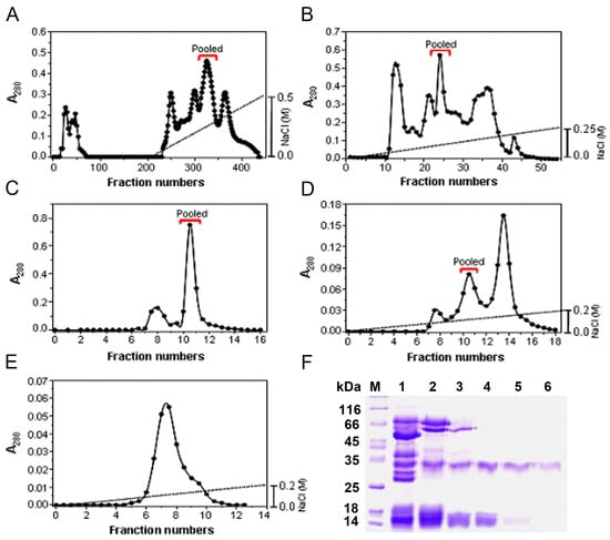
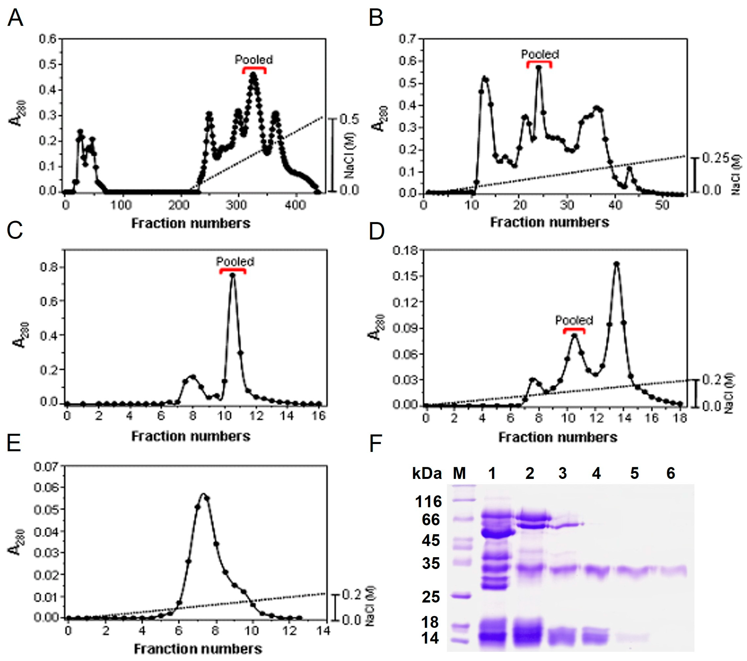
Boc-VPR-pNA-hydrolyzing activity was tracked through five chromatographic steps: HiPrep 16/10 Q FF, Source 15 Q, Superdex 75, and two sequential Mono Q separations (Figure 1A–E). SDS-PAGE showed progressive enrichment of a major protein band during purification (Figure 1F). Starting from 103.26 mg of crude-venom protein and 6195 U of total activity, the final preparation contained 0.14 mg of protein and 140 U of activity, corresponding to a specific activity of 1000 U/mg and a yield of 2.3% (Table 1).
Figure 1.
Purification of TLSP from Gloydius ussuriensis venom. Sequential chromatographic purification using (A) HiPrep 16/10 Q FF, (B) Source 15 Q, (C) Superdex 75, and (D,E) two Mono Q steps. Activity-containing fractions were tracked using Boc-VPR-pNA. (F) SDS-PAGE of proteins obtained during purification. Lane M, molecular-weight markers; lane 1, crude venom; lane 2, HiPrep Q; lane 3, Source Q; lane 4, Superdex 75; lanes 5 and 6, first and second Mono Q fractions.
Table 1.
Purification summary of TLSP.
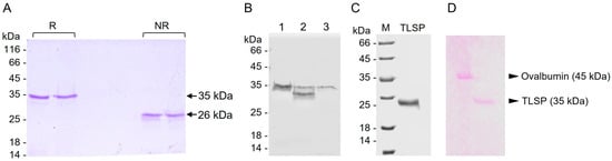
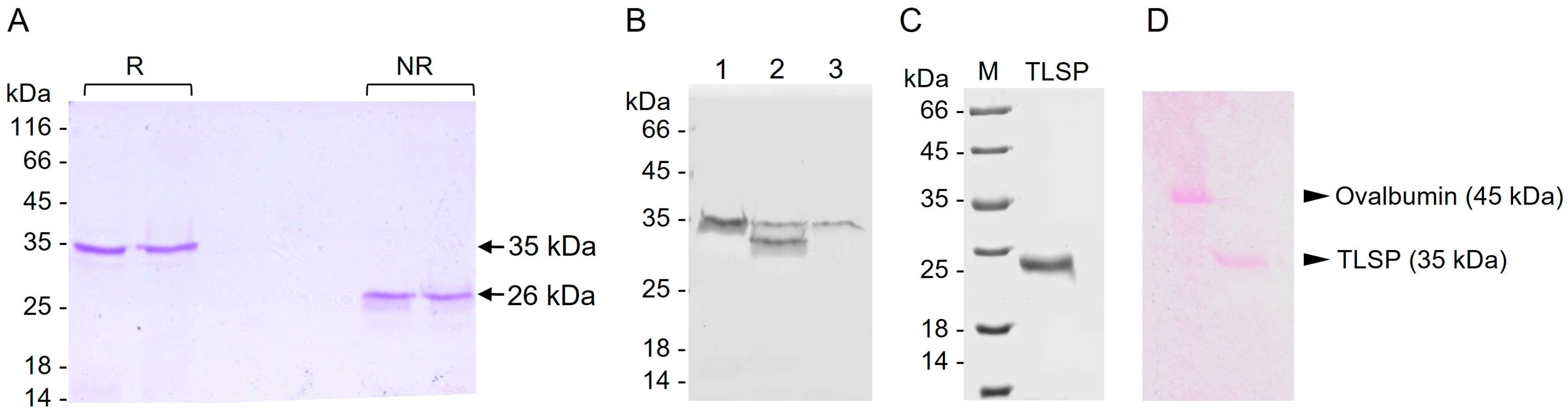
Purified TLSP migrated at approximately 35 kDa under reducing conditions and 26 kDa under non-reducing conditions (Figure 2A). Coomassie brilliant blue staining showed a major purified TLSP band, while PAS staining supported the presence of covalently linked carbohydrate moieties (Figure 2C,D). Treatment with N-glycosidase F reduced the apparent molecular mass of TLSP under reducing conditions from approximately 35 kDa to 33 kDa, corresponding to an apparent shift of approximately 2 kDa (Figure 2B). An N-glycosidase F-only control was included in the electrophoretic analysis. Together, these findings support the presence of N-linked glycosylation in TLSP.
Figure 2.
Electrophoretic and glycoprotein characterization of purified TLSP. (A) SDS-PAGE analysis of purified TLSP under reducing (R) and non-reducing (NR) conditions. The apparent molecular masses of TLSP were approximately 35 kDa and 26 kDa under reducing and non-reducing conditions, respectively. (B) SDS-PAGE analysis of TLSP following treatment with N-glycosidase F. Lane 1, untreated TLSP; lane 2, N-glycosidase F-treated TLSP; lane 3, N-glycosidase F alone. N-glycosidase F treatment resulted in a decrease in the apparent molecular mass of TLSP. (C) Coomassie brilliant blue staining of purified TLSP. Lane M, molecular mass markers; TLSP, purified TLSP. (D) Periodic acid–Schiff (PAS) staining of purified TLSP. Ovalbumin (45 kDa) was used as a glycoprotein-positive control. PAS staining of TLSP supported the presence of covalently linked carbohydrate moieties.
3.2. Kinetic Properties, N-Terminal Sequence, and Chromogenic-Substrate Specificity
Using Boc-VPR-pNA as substrate, TLSP had a Km of 0.3 mM and a kcat of 8.69 s−1, corresponding to a catalytic efficiency (kcat/Km) of 28.8 mM−1 s−1 (Table 2 and Supplementary Figure S1). The N-terminal sequence of TLSP (VVGGDEHNIN) showed 90% identity with VLβF, 80% with calobin, 90% with contortrixobin, and 30% with human α-thrombin (Table 3). These sequence similarities support classification of TLSP within the snake-venom serine-protease family but do not establish its exact molecular identity.
Table 2.
Kinetic parameters of TLSP toward Boc-VPR-pNA.
Table 3.
N-terminal sequence comparison reported for TLSP.
Among the chromogenic substrates tested, Boc-VPR-pNA produced the highest relative activity and was set to 100%. Boc-LGR-pNA and S-2288 also showed substantial hydrolysis, at 85% and 81% relative activity, respectively. Activities toward S-2222, S-2238, S-2251, S-2765, and S-2444 were lower (Table 4). Thus, TLSP displayed a broad amidolytic profile with a preference for Boc-VPR-pNA rather than strict thrombin-like substrate specificity.
Table 4.
Chromogenic-substrate specificity of TLSP.
3.3. TLSP Preferentially Cleaves the Fibrinogen β-Chain but Does Not Form Fibrin Polymer
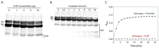
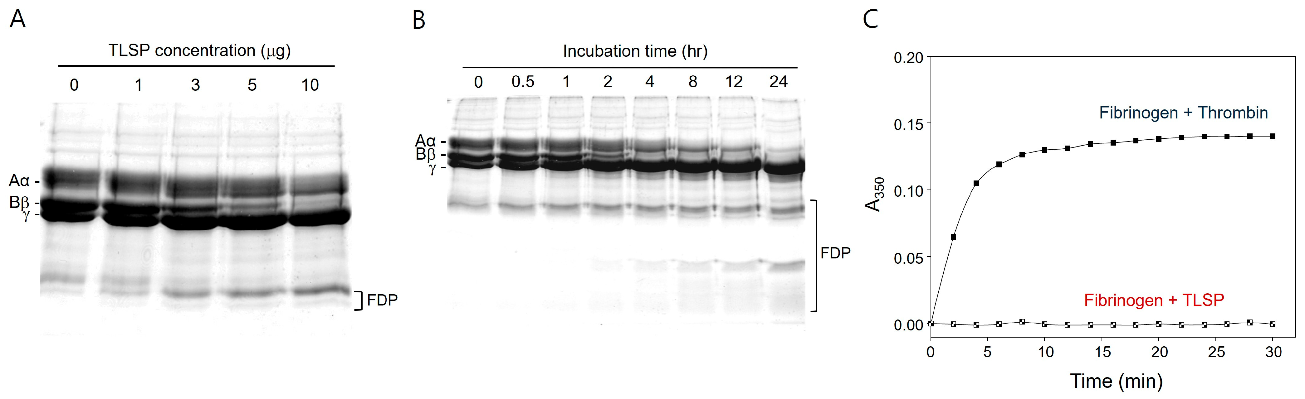
In the concentration-dependent experiment, TLSP cleaved the fibrinogen β-chain strongly within 2 h at 37 °C, whereas the α-chain was less susceptible (Figure 3A). In a separate time-course experiment performed with a different enzyme-to-substrate ratio, complete β-chain degradation was evident by approximately 4 h, while marked α-chain degradation became apparent only after prolonged incubation, approximately 12 h or longer (Figure 3B). The SDS-PAGE data qualitatively showed progressive degradation of the fibrinogen β-chain, followed by α-chain degradation after prolonged incubation. Despite its fibrinogenolytic activity, TLSP did not increase fibrin turbidity, whereas thrombin rapidly induced fibrin-polymer formation (Figure 3C). These observations indicate preferential β-fibrinogenolysis without fibrin-clotting activity.
Figure 3.
Fibrinogenolytic activity of TLSP. (A) Human fibrinogen (200 µg) was incubated with increasing amounts of TLSP (1, 3, 5, or 10 µg) for 2 h at 37 °C and analyzed by SDS-PAGE. (B) Time-dependent degradation of human fibrinogen by TLSP. (C) Fibrin-polymer formation assessed by turbidity at 350 nm. The original protein gel images are provided in Supplementary File S1. TLSP did not induce the turbidity increase observed with thrombin. FDP, fibrinogen degradation products; TLSP, thermostable serine protease.
3.4. Effects on Fibrin, Cross-Linked Fibrin, Plasminogen, and Prothrombin
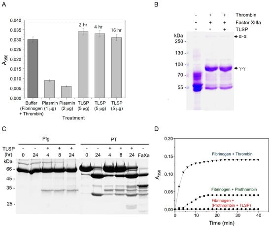
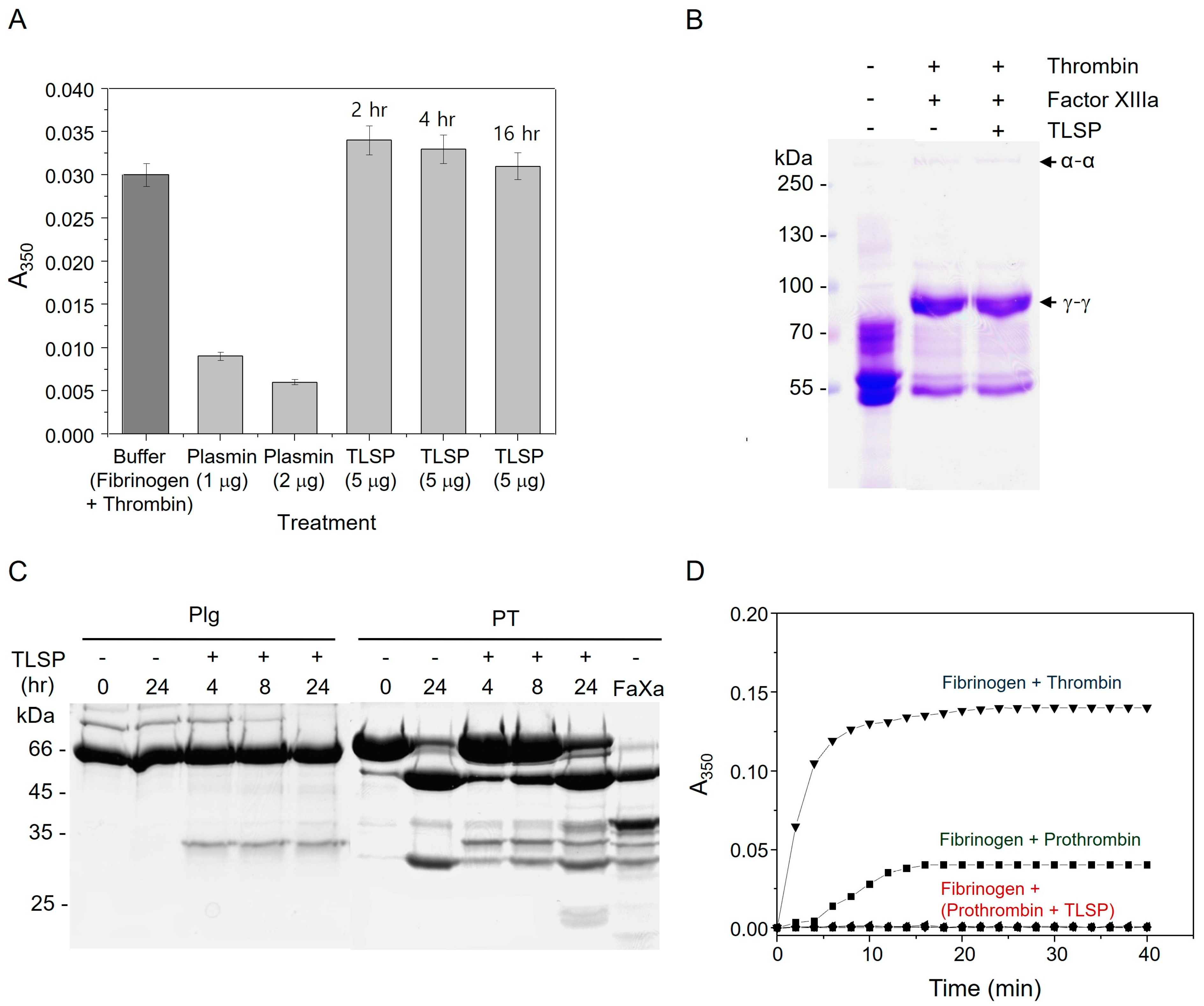
TLSP did not show plasmin-like fibrinolytic activity against preformed fibrin polymer in the turbidity assay, in contrast to plasmin (Figure 4A). It also failed to cleave factor XIIIa-cross-linked fibrin under the conditions tested (Figure 4B). TLSP nevertheless cleaved plasminogen and prothrombin during prolonged incubation. The prothrombin cleavage products did not induce fibrin formation, indicating that TLSP-mediated proteolysis did not generate thrombin activity (Figure 4D). Together, these findings further distinguish the biochemical phenotype of TLSP from both plasmin and physiological thrombin.
Figure 4.
Effects of TLSP on coagulation-related substrates. (A) Fibrinolytic activity measured by changes in fibrin-polymer turbidity; plasmin served as a positive control. (B) SDS-PAGE analysis of factor XIIIa-cross-linked fibrin after TLSP treatment. (C) Cleavage of plasminogen and prothrombin by TLSP. (D) Assessment of fibrin formation by TLSP-generated prothrombin products. TLSP cleaved both proteins during prolonged incubation, but prothrombin cleavage products did not display thrombin activity. The original protein gel images are provided in Supplementary File S1. Plg, plasminogen; PT, prothrombin; TLSP, thermostable serine protease.
3.5. Effects of Protease Inhibitors and Divalent Cations
TLSP activity was strongly inhibited by aprotinin and moderately reduced by PMSF. β-Mercaptoethanol and DTT, tested at 500 mM, reduced the residual activity of TLSP to 31.6% and 15.5%, respectively. In contrast, EGTA, EDTA, 1,10-phenanthroline, bestatin, TPCK, and DFP showed little or no inhibitory effect, whereas TLCK produced only a modest reduction in activity (Table 5).
Table 5.
Effects of various inhibitors and reducing agents on TLSP enzyme activity.
Among the divalent cations tested at 1 mM, Ni2+, Zn2+, and Cu2+ markedly inhibited TLSP activity, reducing the residual activity to 9.1%, 1.4%, and 17.5%, respectively. In contrast, Mg2+, Mn2+, and Ca2+ had little effect, with residual activities of 99.3%, 98.5%, and 97.2%, respectively (Table 6).
Table 6.
Effects of divalent cations on TLSP enzyme activity.
3.6. Alkaline pH Preference, Heat Resistance, and Effect of N-Glycosidase F Treatment
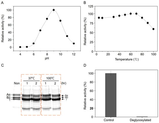
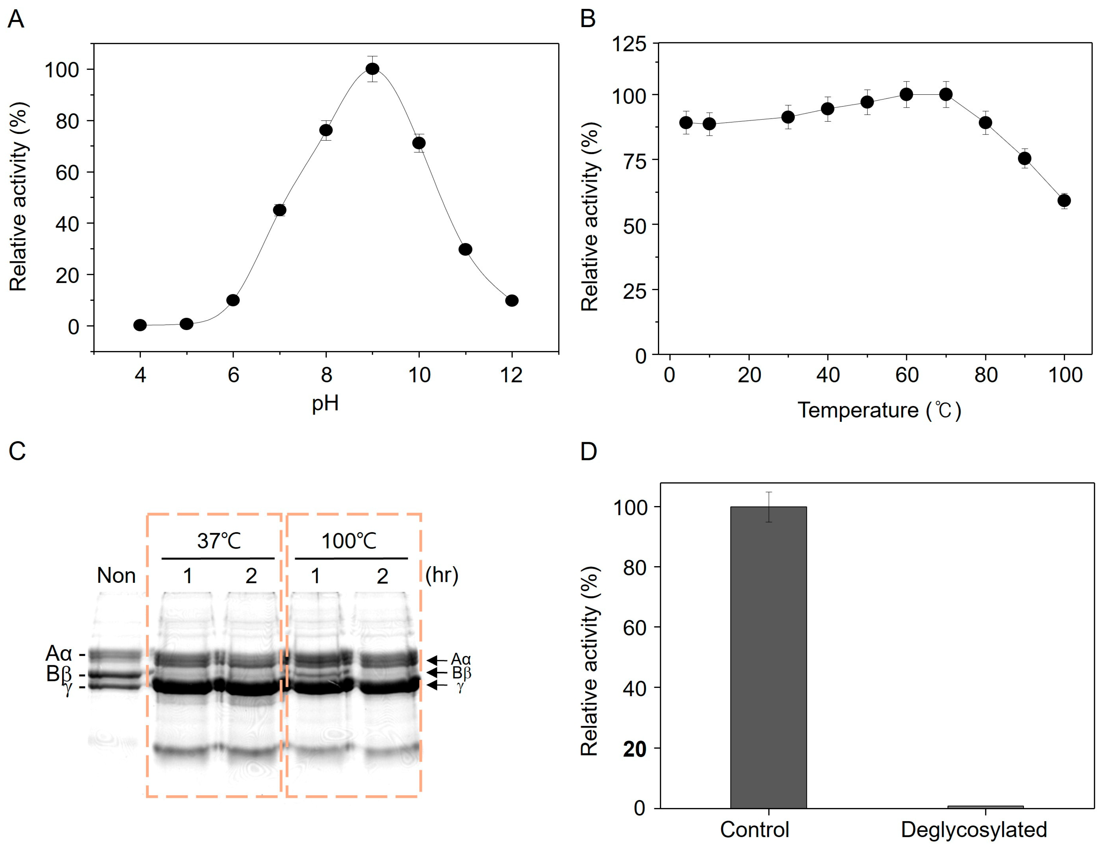
TLSP displayed maximal activity near pH 9.0 and maintained high activity over an alkaline range of approximately pH 8–10 (Figure 5A). After 30 min pre-incubation at increasing temperatures, TLSP retained measurable activity throughout the tested range; notably, approximately 59% of activity remained after exposure to 100 °C (Figure 5B). Enzyme pre-incubated at 100 °C also retained the ability to cleave the fibrinogen β-chain (Figure 5C). N-glycosidase F treatment was accompanied by loss of detectable Boc-VPR-pNA-hydrolyzing activity under the conditions tested (Figure 5D). However, this endpoint assay does not establish whether the loss of activity resulted from altered folding, active-site accessibility, stability, or another structural effect.
Figure 5.
pH dependence, heat resistance, and effect of N-glycosidase F treatment on TLSP. (A) Relative activity across pH 4–12, with maximal activity near pH 9.0. (B) Residual activity after temperature pre-incubation. (C) Preservation of fibrinogen β-chain cleavage after exposure to 100 °C. (D) Loss of detectable Boc-VPR-pNA-hydrolyzing activity after N-glycosidase F treatment. The original protein gel images are provided in Supplementary File S1.
4. Discussion
The biochemical data characterize TLSP as a glycosylated snake-venom serine protease with a distinctive combination of thrombin-like amidolytic preference, preferential β-fibrinogenolysis, absence of fibrin-clotting activity, and marked thermal resistance. The N-terminal sequence begins with VVGG, a motif commonly observed in mature snake-venom serine proteases, and the sequence identities reported for VLβF, calobin, and contortrixobin are substantially greater than that with human α-thrombin. Together with inhibition by aprotinin and partial inhibition by PMSF, these findings support classification of TLSP within the snake-venom serine-protease family [13].
The marked difference in apparent molecular mass between reducing and non-reducing SDS-PAGE conditions should be interpreted cautiously. Because no native molecular-mass or oligomeric-state analysis was performed, the observed migration difference cannot be attributed specifically to subunit dissociation or proteolytic processing and may instead reflect disulfide-dependent conformational effects or anomalous electrophoretic migration.
TLSP should nevertheless not be interpreted as a functional equivalent of thrombin. Although Boc-VPR-pNA was its preferred chromogenic substrate, TLSP showed much lower relative activity toward another thrombin-directed substrate, S-2238, and displayed substantial activity toward Boc-LGR-pNA and S-2288. The substantial activity toward several non-thrombin-directed chromogenic substrates indicates that TLSP has relatively broad active-site tolerance toward small synthetic substrates. Therefore, preference for Boc-VPR-pNA should not be interpreted as evidence of strict thrombin-like specificity. More importantly, TLSP preferentially degraded the fibrinogen β-chain and failed to induce fibrin-polymer formation. This phenotype resembles β-fibrinogenolytic venom serine proteases such as halystase and contortrixobin [13] and illustrates why chromogenic-substrate preference alone is insufficient to establish physiological thrombin-like function [8,14,15].
The cleavage of plasminogen and prothrombin after prolonged incubation should be interpreted cautiously. These experiments demonstrate proteolysis under the conditions tested, but do not establish physiological activation, cleavage-site specificity, or kinetic relevance. The TLSP-generated prothrombin fragments lacked thrombin activity, and TLSP did not degrade preformed or factor XIIIa-cross-linked fibrin. Consequently, the available experiments support proteolysis of these substrates but do not support prothrombin activation or plasmin-like fibrinolysis [3,16,17].
Thrombin is a multifunctional serine protease involved in hemostasis and other cellular responses [18]. Although coagulation-associated proteolysis can intersect with growth-factor signaling, including thrombin-mediated pro-HGFA activation [19,20,21], these pathways were not investigated in the present study.
A particularly notable feature of TLSP was its resistance to heat. Approximately 59% of amidolytic activity remained after exposure to 100 °C, and heat-treated enzyme retained β-fibrinogenolytic activity. TLSP was also PAS-positive and showed a molecular-mass shift after N-glycosidase F treatment. Previous studies of venom fibrinogenases have suggested that covalently attached carbohydrate can influence stability and enzyme properties [22,23]. N-glycosidase F treatment was accompanied by loss of detectable amidolytic activity, but the available data do not establish the underlying mechanism [24]. Importantly, the completeness of deglycosylation was not independently verified beyond the observed electrophoretic mobility shift [24]. Additional experiments would be required to determine whether N-linked glycans influence folding, active-site accessibility, solubility, stability, or other structural properties of TLSP [25].
The marked reduction in TLSP activity by β-mercaptoethanol and DTT suggests that disulfide bonds may be important for maintaining the catalytically active conformation of TLSP. However, because these reducing agents were tested at a relatively high concentration (500 mM), the present data do not establish a direct causal relationship between disulfide bonds and TLSP thermostability. Further structural studies are required to determine whether disulfide-bond stabilization contributes to its marked thermal resistance.
Limitations and Future Directions
The principal limitation of the present dataset is that molecular identification is based on a short N-terminal sequence rather than a complete protein sequence, peptide mass fingerprint, LC-MS/MS profile, or corresponding gene sequence. The exact molecular identity of TLSP therefore remains unresolved. Accordingly, the present study should be interpreted as a biochemical characterization of the purified TLSP preparation rather than as the establishment of a novel molecular entity. In addition, although most quantitative assays were performed in duplicate and kinetic measurements were performed in three independent experiments, the original source records do not consistently provide measures of variation or inferential statistical analyses for all quantitative assays. Future work should confirm TLSP identity by contemporary mass-spectrometric or sequence-based methods, repeat key quantitative assays with predefined biological and technical replicates, and directly test the contribution of glycosylation to folding and thermal stability. These steps would strengthen structure–function interpretation while preserving the distinctive biochemical phenotype documented here.
Although preferential cleavage of the fibrinogen β-chain was observed by SDS-PAGE, reliable densitometric quantification was limited by overlapping electrophoretic bands. Therefore, the relative degradation of the α- and β-chains could not be quantitatively determined.
The thermal-stability analysis was based on residual enzyme activity after a 30-min pre-incubation and did not assess reversibility of heat-induced changes, aggregation or precipitation, or complete thermal-inactivation kinetics. Therefore, the present data demonstrate marked heat resistance under the tested conditions but do not define its structural or thermodynamic basis.
We also did not determine whether TLSP-treated fibrinogen retains the ability to form fibrin following subsequent thrombin addition. This should be addressed in future studies to further define the functional consequences of TLSP-mediated fibrinogenolysis.
Future studies may examine whether TLSP can process plasminogen-related growth factors or their precursors, including HGF/HGFA- or MSP-related substrates, and whether such proteolysis influences downstream cellular responses [21,26,27,28]. These possibilities were not examined in the present study and remain to be investigated.
5. Conclusions
TLSP purified from Gloydius ussuriensis venom is a glycosylated serine protease that preferentially cleaves the fibrinogen β-chain and shows a preference for Boc-VPR-pNA among the chromogenic substrates tested, but does not display thrombin-like fibrin-clotting activity. The enzyme also showed marked heat resistance under the experimental conditions and did not degrade preformed or factor XIIIa-cross-linked fibrin. TLSP is operationally defined by its purification characteristics and N-terminal amino acid sequence. However, the exact molecular identity of TLSP remains unresolved, and the possibility that it corresponds to a previously described venom serine protease, such as gussorbin, cannot be excluded. Furthermore, the structural basis of its glycosylation- and heat-related properties remains to be elucidated. These data define the biochemical phenotype of the purified TLSP preparation and provide a basis for future molecular identification and structure–function studies.
Supplementary Materials
The following supporting information can be downloaded at: https://www.mdpi.com/article/10.3390/biology15201892/s1, Figure S1: Original kinetic plot of TLSP toward Boc-VPR-pNA; Supplementary Materials File S1: Original images of the protein gels used in this study.
Author Contributions
Investigation, methodology, formal analysis, and data curation, E.-R.J.; data interpretation, visualization, writing—original draft preparation, writing—review and editing, and supervision of manuscript preparation, J.E.P. All authors have read and agreed to the published version of the manuscript.
Funding
The experimental work was supported by a research fund from Chosun University in 2008. Manuscript preparation and publication were supported by the National Research Foundation of Korea (NRF) funded by the Ministry of Science under grant RS-2026-25562669.
Institutional Review Board Statement
Not applicable.
Informed Consent Statement
Not applicable.
Data Availability Statement
The data presented in this study are available on request from the corresponding author.
Acknowledgments
The authors acknowledge the Korea Basic Science Institute (Seoul, Republic of Korea) for N-terminal amino acid sequence analysis. The authors also disclose that part of the biochemical characterization of TLSP was previously included in the 2009 Master’s thesis of E.-R. Jo at Chosun University. The present manuscript presents these experimental data in an integrated form with updated interpretation and discussion in the context of the current literature. During the preparation of this manuscript, the authors used ChatGPT 6 (OpenAI, San Francisco, CA, USA) for manuscript restructuring and scientific-language editing. The authors reviewed and edited all AI-assisted output and take full responsibility for the content of this publication.
Conflicts of Interest
The authors declare no conflicts of interest.
Abbreviations
TLSP, thermostable serine protease; SVTLE, snake-venom thrombin-like enzyme; PAS, periodic acid–Schiff; PMSF, phenylmethanesulfonyl fluoride; DFP, diisopropyl fluorophosphate; EDTA, ethylenediaminetetraacetic acid; EGTA, ethylene glycol-bis(2-aminoethylether)-N,N,N′,N′-tetraacetic acid; DTT, dithiothreitol; BSA, bovine serum albumin; t-PA, tissue-type plasminogen activator.
References
- Tasoulis, T.; Isbister, G.K. A current perspective on snake venom composition and constituent protein families. Arch. Toxicol. 2023, 97, 133–153. [Google Scholar] [CrossRef] [Scilit] [PubMed]
- Vidal, J.F.D.; Schwartz, M.F.; Garay, A.V.; Valadares, N.F.; Bueno, R.V.; Monteiro, A.C.L.; Freitas, S.M.; Barbosa, J.A.R.G. Exploring the Diversity and Function of Serine Proteases in Toxicofera Reptile Venoms: A Comprehensive Overview. Toxins 2024, 16, 428. [Google Scholar] [CrossRef] [Scilit] [PubMed]
- Kini, R.M. Serine proteases affecting blood coagulation and fibrinolysis from snake venoms. Pathophysiol. Haemost. Thromb. 2005, 34, 200–204. [Google Scholar] [CrossRef] [Scilit] [PubMed]
- Kini, R.M.; Koh, C.Y. Metalloproteases Affecting Blood Coagulation, Fibrinolysis and Platelet Aggregation from Snake Venoms: Definition and Nomenclature of Interaction Sites. Toxins 2016, 8, 284. [Google Scholar] [CrossRef] [Scilit] [PubMed]
- Green, D. Hematology products from snake venoms. Thromb. Res. 2025, 245, 109215. [Google Scholar] [CrossRef] [Scilit] [PubMed]
- Matsui, T.; Fujimura, Y.; Titani, K. Snake venom proteases affecting hemostasis and thrombosis. Biochim. Biophys. Acta 2000, 1477, 146–156. [Google Scholar] [CrossRef] [Scilit] [PubMed]
- Amiconi, G.; Amoresano, A.; Boumis, G.; Brancaccio, A.; De Cristofaro, R.; De Pascalis, A.; Di Girolamo, S.; Maras, B.; Scaloni, A. A novel venombin B from Agkistrodon contortrix contortrix: Evidence for recognition properties in the surface around the primary specificity pocket different from thrombin. Biochemistry 2000, 39, 10294–10308. [Google Scholar] [CrossRef] [Scilit] [PubMed]
- Castro, H.C.; Zingali, R.B.; Albuquerque, M.G.; Pujol-Luz, M.; Rodrigues, C.R. Snake venom thrombin-like enzymes: From reptilase to now. Cell. Mol. Life Sci. 2004, 61, 843–856. [Google Scholar] [CrossRef] [Scilit] [PubMed]
- Castro, H.C.; Silva, D.M.; Craik, C.; Zingali, R.B. Structural features of a snake venom thrombin-like enzyme: Thrombin and trypsin on a single catalytic platform? Biochim. Biophys. Acta 2001, 1547, 183–195. [Google Scholar] [CrossRef] [Scilit] [PubMed]
- Matsui, T.; Sakurai, Y.; Fujimura, Y.; Hayashi, I.; Oh-Ishi, S.; Suzuki, M.; Hamako, J.; Yamamoto, Y.; Yamazaki, J.; Kinoshita, M.; et al. Purification and amino acid sequence of halystase from snake venom of Agkistrodon halys blomhoffii, a serine protease that cleaves specifically fibrinogen and kininogen. Eur. J. Biochem. 1998, 252, 569–575. [Google Scholar] [CrossRef] [Scilit] [PubMed]
- Yang, Q.; Hu, X.J.; Xu, X.M.; An, L.J.; Yuan, X.D.; Su, Z.G. Cloning, expression and purification of gussurobin, a thrombin-like enzyme from the snake venom of Gloydius ussuriensis. Acta Biochim. Biophys. Sin. 2002, 34, 6–10. [Google Scholar] [PubMed]
- Park, H.S.; Moon, J.M.; Chun, B.J.; Min, Y.; Cho, Y.S.; Park, K.-H.; Lee, S.; Koo, Y. Comprehensive molecular characterization and comparison of venom proteins and transcripts in three Gloydius species from South Korea. Sci. Rep. 2026, 16, 12299. [Google Scholar] [CrossRef] [Scilit] [PubMed]
- Koh, Y.-S.; Chung, K.-H.; Kim, D.-S. Biochemical characterization of a thrombin-like enzyme and a fibrinolytic serine protease from snake (Agkistrodon saxatilis) venom. Toxicon 2001, 39, 555–560. [Google Scholar] [CrossRef] [Scilit] [PubMed]
- Markland, F.S.; Kettner, C.; Schiffman, S.; Shaw, E.; Bajwa, S.S.; Reddy, K.N.; Kirakossian, H.; Patkos, G.B.; Theodor, I.; Pirkle, H. Kallikrein-like activity of crotalase, a snake venom enzyme that clots fibrinogen. Proc. Natl. Acad. Sci. USA 1982, 79, 1688–1692. [Google Scholar] [CrossRef] [Scilit] [PubMed]
- Tan, N.H.; Fung, S.Y.; Yap, Y.H. Isolation and characterization of the thrombin-like enzyme from Cryptelytrops albolabris (white-lipped tree viper) venom. Comp. Biochem. Physiol. B Biochem. Mol. Biol. 2012, 161, 79–85. [Google Scholar] [CrossRef] [Scilit] [PubMed]
- Kini, R.M. The intriguing world of prothrombin activators from snake venom. Toxicon 2005, 45, 1133–1145. [Google Scholar] [CrossRef] [Scilit] [PubMed]
- Flemmig, M.; Melzig, M.F. Serine-proteases as plasminogen activators in terms of fibrinolysis. J. Pharm. Pharmacol. 2012, 64, 1025–1039. [Google Scholar] [CrossRef] [Scilit] [PubMed]
- Al-Amer, O.M. The role of thrombin in haemostasis. Blood Coagul. Fibrinolysis 2022, 33, 145–148. [Google Scholar] [CrossRef] [Scilit] [PubMed]
- Gailani, D.; Broze, G.J., Jr. Factor XI activation in a revised model of blood coagulation. Science 1991, 253, 909–912. [Google Scholar] [CrossRef] [Scilit] [PubMed]
- Shimomura, T.; Kondo, J.; Ochiai, M.; Naka, D.; Miyazawa, K.; Morimoto, Y.; Kitamura, N. Activation of the zymogen of hepatocyte growth factor activator by thrombin. J. Biol. Chem. 1993, 268, 22927–22932. [Google Scholar] [CrossRef] [Scilit]
- Fukushima, T.; Uchiyama, S.; Tanaka, H.; Kataoka, H. Hepatocyte Growth Factor Activator: A Proteinase Linking Tissue Injury with Repair. Int. J. Mol. Sci. 2018, 19, 3435. [Google Scholar] [CrossRef] [Scilit] [PubMed]
- Silva-Junior, F.P.; Guedes, H.L.; Garvey, L.C.; Aguiar, A.S.; Bourguignon, S.C.; Di Cera, E.; Giovanni-De-Simone, S. BJ-48, a novel thrombin-like enzyme from the Bothrops jararacussu venom with high selectivity for Arg over Lys in P1: Role of N-glycosylation in thermostability and active site accessibility. Toxicon 2007, 50, 18–31. [Google Scholar] [CrossRef] [Scilit] [PubMed]
- Brás-Costa, C.; Alencar Chaves, A.F.; Trevisan-Silva, D.; Menezes, M.C.; Rocha, M.M.T.; Cajado-Carvalho, D.; Andrade-Silva, D.; Serrano, S.M.T. Sialic acid-containing glycans play a role in the activity of snake venom proteases. Biochimie 2023, 204, 140–153. [Google Scholar] [CrossRef] [Scilit] [PubMed]
- Goettig, P. Effects of Glycosylation on the Enzymatic Activity and Mechanisms of Proteases. Int. J. Mol. Sci. 2016, 17, 1969. [Google Scholar] [CrossRef] [Scilit] [PubMed]
- Mukherjee, A.K. The Pro-Coagulant Fibrinogenolytic Serine Protease Isoenzymes Purified from Daboia russelii russelii Venom Coagulate the Blood through Factor V Activation: Role of Glycosylation on Enzymatic Activity. PLoS ONE 2014, 9, e86823. [Google Scholar] [CrossRef] [Scilit] [PubMed]
- Donate, L.E.; Gherardi, E.; Srinivasan, N.; Sowdhamini, R.; Aparicio, S.; Blundell, T.L. Molecular evolution and domain structure of plasminogen-related growth factors (HGF/SF and HGF1/MSP). Protein Sci. 1994, 3, 2378–2394. [Google Scholar] [CrossRef] [Scilit] [PubMed]
- Nakamura, T.; Sakai, K.; Nakamura, T.; Matsumoto, K. Hepatocyte growth factor twenty years on: Much more than a growth factor. J. Gastroenterol. Hepatol. 2011, 26, 188–202. [Google Scholar] [CrossRef] [Scilit] [PubMed]
- Wang, M.H.; Zhou, Y.Q.; Chen, Y.Q. Macrophage-Stimulating Protein and RON Receptor Tyrosine Kinase: Potential Regulators of Macrophage Inflammatory Activities. Scand. J. Immunol. 2002, 56, 545–553. [Google Scholar] [CrossRef] [Scilit] [PubMed]

Disclaimer/Publisher’s Note: The statements, opinions and data contained in all publications are solely those of the individual author(s) and contributor(s) and not of MDPI and/or the editor(s). MDPI and/or the editor(s) disclaim responsibility for any injury to people or property resulting from any ideas, methods, instructions or products referred to in the content. |
© 2026 by the authors. Licensee MDPI, Basel, Switzerland. This article is an open access article distributed under the terms and conditions of the Creative Commons Attribution (CC BY) license.