


Simple Summary
Sex determination and gonadal differentiation are critical for fish growth and development. Unlike humans, fish sex is influenced by both genes and environmental factors such as water temperature. Sebastes schlegelii, an economically important marine fish in northern China, exhibits sex-related growth performance, and water temperature can affect offspring sex ratio. Our previous study found that gonadotropin-releasing hormone (GnRH) is involved in high-temperature-induced sex differentiation, but the mechanism remains unknown. Using transcriptome analysis, we discovered that GnRH regulates sex differentiation through three key pathways: steroid biosynthesis, TGF-β signaling, and MAPK signaling. Several key genes involved in this process were also identified. These findings have important implications. First, they help scientists understand how water temperature influences sex determination in fish, providing insights for predicting climate change impacts on fishery resources. Second, by artificially manipulating these pathways and genes, it may be possible in the future to selectively produce more females (which grow faster) in aquaculture, thereby improving economic efficiency.
Abstract
Sex determination and gonadal differentiation are essential for vertebrate development and reproduction. Fish, with both genetic and environmental sex-determining mechanisms, serve as ideal models for exploring environmental effects on gonadal development. The hypothalamic–pituitary–gonadal (HPG) axis is involved in sex differentiation, but its role as a key mediator in temperature-dependent sex differentiation remains unclear. Sebastes schlegelii is an economically important marine fish species along the northern coast of China. It exhibits obvious sexual growth dimorphism and typical temperature-dependent sex differentiation, making it an excellent model for investigating the mechanism of sex differentiation. In this study, individuals of S. schlegelii at the sex differentiation stage were treated with a GnRH receptor antagonist and subjected to transcriptome sequencing analysis. The results revealed that GnRH may regulate sex differentiation through steroid biosynthesis, TGF-β and MAPK signaling pathways. We identified several key genes, including srd5, bmp8a, bmp2, fgf23, pdgfra, and egfr, which may affect gonadal differentiation by acting on core sex-determining genes and steroidogenesis. In conclusion, GnRH precisely mediates the process of sex differentiation by modulating the TGF-β signaling pathway, the MAPK signaling pathway, and local gonadal steroidogenic networks.
1. Introduction
In recent decades, climate warming driven by greenhouse gas emissions has continuously reshaped the pattern of global ecosystems, and marine organisms exhibit particularly significant responses to such changes [1]. The Sixth Assessment Report of the Intergovernmental Panel on Climate Change (IPCC) indicates that global ocean heat content shows an irreversible upward trend, with the upper ocean warming rate reaching 0.11 °C per decade, accompanied by frequent extreme thermal events and intensified seasonal fluctuations [2,3]. The uneven spatiotemporal distribution and amplified variation amplitude of seawater temperature not only directly drive population decline and affect genetic diversity, but also weaken the structural stability and service supply functions of marine ecosystems through trophic cascade effects [4,5,6,7]. As key nodes in marine food webs, fish undertake the dual functions of maintaining ecological balance and supporting fishery economic output, and their life-history traits are highly sensitive to changes in thermal environments [8,9,10]. Continuous warming profoundly interferes with the growth and development of fish through multiple pathways, such as metabolic remodeling, oxidative stress activation, and immune suppression, posing a severe challenge to the long-term survival of fish populations [11,12,13,14].
Sex determination and gonadal differentiation constitute the core biological basis for development and reproduction in vertebrates [15]. Fish exhibit extremely diverse sex determination mechanisms, which are mainly classified into two categories: genotypic sex determination (GSD) and environmental sex determination (ESD). Sex differentiation in most species arises from the combined effects of genetic and environmental factors [16,17]. Among sex determination systems, chromosomal sex determination (CSD) is the most common form, mainly including the XX/XY male heterogametic model and the ZZ/ZW female heterogametic model [18,19,20]. Furthermore, the sex-determining genes of most fish are not restricted to sex chromosomes; some species even lack morphologically differentiated sex chromosomes, rendering their sex differentiation processes readily regulated by environmental factors [21,22]. Among these, temperature-dependent sex determination (TSD) is widespread in taxa such as Pleuronectiformes and Perciformes [23]. Temperature effects on sex differentiation can influence population dynamics. On the one hand, water temperature during the larval stage can modulate the direction of gonadal differentiation to a certain extent, thereby affecting the sex ratio of offspring. On the other hand, male- or female-skewed sex ratios induced by extreme thermal events may reduce reproductive capacity and threaten population sustainability [24,25]. Meanwhile, the TSD mechanism also provides a technical breakthrough for unisexual breeding in aquaculture. Precise temperature control to manipulate sex can significantly improve aquaculture production and economic benefits, serving as an important bridge linking basic research to industrial application.
Sebastes schlegelii is an economically important marine fish species along the northern coast of China, with an annual output of approximately 10,000 tons. This species exhibits significant sexual growth dimorphism (females grow faster than males), and previous studies have shown that it undergoes feminization at 24 °C and masculinization at 27 °C [26]. These two characteristics provide a technical entry point for monosex aquaculture and also make it an ideal model for dissecting the mechanism underlying high-temperature-mediated sex differentiation. Previous studies have mostly focused on the direct effects of sex-related gene expression and sex steroid hormone synthesis. Recent studies have indicated that the fish hypothalamic–pituitary–gonadal axis (HPG axis) participates in the process of sex differentiation [26]. Gonadotropin-releasing hormone (GnRH) serves as the core regulatory factor of the HPG axis; it stimulates the pituitary to release follicle-stimulating hormone (FSH) and luteinizing hormone (LH), thereby regulating gonadal steroidogenesis and gametogenesis [27]. Gene knockout studies in zebrafish have confirmed that the deletion of GnRH3 can lead to male-skewed sex ratios in populations, revealing for the first time that a specific GnRH isoform is involved in sex determination [28]. However, the regulatory mechanism of GnRH in sex differentiation remains unclear, especially in S. schlegelii, for which no relevant research has been reported.
This study aimed to elucidate the regulatory mechanism of GnRH in sex differentiation of S. schlegelii, filling the gap in neuroendocrine regulation research of this species. Using GnRH receptor antagonist treatment combined with transcriptome and qRT-PCR analyses, we demonstrated for the first time that GnRH signaling pathway is essential for sustaining female development. The GnRH signaling pathway regulates sex differentiation together with the key steroidogenic enzyme srd5 via the TGF-β signaling pathway (bmp8a, bmp2) and MAPK signaling pathway (fgf23, pdgfra, egfr). This study not only clarifies the central role of the GnRH signaling pathway in integrating neuroendocrine and local gonadal signaling networks, but also provides theoretical basis and molecular targets for analyzing the dynamics of population sex ratio and developing monosex breeding techniques of S. schlegelii under climate warming.
2. Materials and Methods
2.1. Ethics Statement
All experiments were conducted in strict compliance with the experimental animal guidance policy as outlined by the Institute of Oceanology’s Animal Ethics Committee, Chinese Academy of Sciences (Permit No. IOCAS20230916PPFA0007).
2.2. Culture Protocol of Experimental Animals
Experimental larvae of S. schlegelii were obtained from Weihhai Shenghang Technology Co., Ltd. (Weihai, Shandong Province, China). On the first day after parturition, the larvae were reared in an environment of 18 °C ± 1 °C under a photoperiod of 14 h light/10 h dark. This water temperature of 18 °C ± 1 °C was maintained throughout the entire larval rearing and subsequent feeding trial period. The selection of this specific temperature range was based on two considerations. First, it falls within the suitable range for juvenile growth, as previous research has demonstrated that water temperatures of 16–20 °C are conducive for rearing this species, with optimal growth performance observed at 20 °C [29]. Second, this temperature range is representative of the ambient water temperature measured at the local aquaculture facility where the broodstock was maintained, ensuring consistency with practical production conditions. After 30 d of rearing, healthy and uniformly sized juvenile fish were randomly selected and transferred to six 140 L tanks at a density of 200 individuals per tank, with three tanks assigned as the control group and the other three as the experimental group. During the larval rearing period, a recirculating aquaculture system was used, with each tank equipped with independent water supply and aeration devices. One-third of the water volume was replaced daily with sand-filtered and aerated seawater. The experimental group was fed a diet supplemented with the GnRH antagonist Relugolix (Cat. No. HY-16474; MedChemExpress LLC, Monmouth Junction, NJ, USA) at a dosage of 3 mg/kg body weight, as recommended by the manufacturer’s instructions. qPCR was used to detect the expression of downstream genes to verify the treatment effect of Relugolix. The control group received the same basal diet. Feed was provided manually, and the feeding ration was adjusted weekly based on changes in body weight. The feeding ration was kept consistent between the control and experimental groups, and all other rearing conditions were identical except for the presence or absence of Relugolix in the diet. The experiment lasted for 90 days. At 42 days post-hatching, nine individual fish were collected from each experimental group (control group and Relugolix-treated group). Every three individuals were pooled as one biological replicate, yielding a total of three biological replicates per group for transcriptome sequencing. Fish remaining after the 42-day sampling were reared until the end of the experiment (90 days) for phenotypic sex identification. The tail of each fish was excised and frozen in liquid nitrogen for genetic sex identification. The remaining whole-fish tissues were stored separately in liquid nitrogen, Bouin’s fixative, and paraformaldehyde (PFA) fixative for other subsequent experimental analyses. Whole-fish samples were used instead of gonadal tissues because the juvenile fish used in this study were extremely small, making it impossible to isolate gonadal tissues by dissection.
2.3. Extraction of Genomic DNA and Total RNA, and Synthesis of cDNA
DNA was extracted for the identification of genetic sex in S. schlegelii, while cDNA was extracted for the detection of gene expression profiles. The TIANamp Marine Animal DNA Kit was utilized to extract DNA from fish tail tissues, while TRIzol Reagent (Invitrogen, Carlsbad, CA, USA) was employed for isolating total RNA. A NanoDrop 2000 spectrophotometer (Thermo Fisher, Waltham, MA, USA) was used to evaluate the quality and concentration of both DNA and RNA. The Evo M-MLV RT Kit (Accurate Biology, Changsha, China) facilitated the synthesis of cDNA. Both DNA and cDNA were preserved at a temperature of −20 °C.
2.4. PCR-Based Genetic Sex Identification
Genetic sex identification was performed following the method of Song et al. [30], DNA extracted from S. schlegelii tail tissues was used as a template, and PCR amplification was conducted using specific primers targeting single-stranded DNA fragments (Table S2). A total of 30 individuals were used for genetic sex identification in this experiment.
2.5. Transcriptome Sequencing
To explore the molecular differences and characteristics of S. schlegelii following Relugolix treatment, we performed transcriptome sequencing on juvenile fish from the control and experimental groups. Nine whole fish were collected in each experimental group. Every three individuals were pooled as one biological replicate, with a total of three biological replicates per group. Total RNA was extracted from each sample, reverse-transcribed into cDNA, and purified to obtain double-stranded DNA templates. After end repair, adapter ligation, and PCR amplification, sequencing libraries with an insert size of 350 bp were constructed and subjected to 150 bp paired-end sequencing on the Illumina NovaSeq 6000 platform (Gene Denovo Biotech Co., Guangzhou, China). Data quality control was conducted using FASTP (v0.20.0) to ensure clean reads. Reference genome alignment was performed using Hisat2 v2.1.0 and Bowtie2 v2.5.4, and gene expression levels were calculated with RSEM.
2.6. Screening, Enrichment and Expression Pattern Analysis of DEGs
To analyze the differential RNA expression between the two groups, the DESeq2 software (v1.38.3) was employed [31], with the screening criteria for differentially expressed genes (DEGs) set as a false discovery rate (FDR) < 0.05 and an absolute fold change ≥ 2. For gene expression quantification, raw read counts for each transcriptional region were calculated using the RSEM software (v1.3.3) [32] and used for subsequent differential expression analysis. Additionally, FPKM values were also calculated by RSEM for visualization of expression abundance across samples. Principal component analysis (PCA) was conducted with the R software package (v4.2.2) (http://www.r-project.org/), aiming to reveal the similarity among different biological samples based on the overall gene expression levels. Additionally, the clusterProfiler R package was utilized to perform Gene Ontology (GO) enrichment analysis and KEGG functional enrichment analysis, so as to predict the potential functions of DEGs. The threshold for significant enrichment of gene sets was defined as p < 0.05.
2.7. Gene Set Enrichment Analysis
To more comprehensively clarify the regulatory effect of specific pathways, we used gene set enrichment analysis (GSEA) software (v4.3.3) combined with the MSigDB database [33] to perform gene set enrichment analysis, and simultaneously conducted KEGG enrichment analysis to verify whether differentially expressed genes are enriched in specific pathways. After inputting the gene expression matrix, genes were sorted using the signal-to-noise ratio normalization method, and then enrichment scores and p-values were calculated according to the default parameters of the software. Specifically, based on the expression data of all genes, we selected the most widely used Signal2Noise as the gene sorting standard; by analyzing the specific positions of the target gene set in the sorting of all genes, we quantitatively scored the pathways and GO terms associated with the gene set, and then obtained the enrichment score (ES value). Subsequently, permutation tests were performed based on the gene set to calculate the significance p-value, and finally, the normalized enrichment score (NES value) and false discovery rate (FDR value) were obtained through the multiple test correction process.
2.8. Quantitative Real-Time PCR
To detect the mRNA expression levels of genes associated with key pathways identified through transcriptomic analysis, we used the Bio-Rad CFX96™ Real-Time Detection System (Bio-Rad, Hercules, CA, USA) and quantitative real-time polymerase chain reaction (qRT-PCR). The qRT-PCR primers were designed using the NCBI Primer-BLAST online tool (based on BLAST+ v2.15.0, http://www.ncbi.nlm.nih.gov/tools/primer-blast (accessed on 22 December 2025)). After verifying no cross-reactivity by BLAST, the primers were synthesized by Sangon Biotech (Shanghai, China) Co., Ltd. The primer sequences are detailed in Table S1. The total volume of the qRT-PCR reaction system was 20 μL, consisting of 10 μL SYBR Green I PCR Master Mix, 0.4 μL forward primer, 0.4 μL reverse primer, 7.2 μL deionized water, and 2 μL cDNA. The amplification procedure began with an initial denaturation step at 95 °C for 30 s, followed by 40 cycles of 95 °C for 5 s, gene-specific annealing temperature for 30 s, and 72 °C for 30 s for extension. Each sample had three technical replicates, and based on previous research findings, we selected btf3l4 as the reference gene [34]. The Pfaffl method was employed to quantify mRNA expression levels [35].
2.9. Data Analysis
Statistical analyses were conducted using GraphPad Prism version 9.0.0. Group comparisons were assessed by one-way Analysis of Variance (ANOVA). Results are expressed as mean ± standard error (SE), with significance set at p < 0.05.
3. Results
3.1. Transcriptome Sequencing Assembly and Analysis
To investigate the molecular expression changes of sex differentiation-related genes in S. schlegelii under Relugolix treatment, genetically female individuals were firstly screened for the experiment. Genetically female S. schlegelii without any treatment were set as the control group, and Relugolix-exposed genetically female individuals were designated as the experimental group. Reference-based transcriptomic analysis was performed on the S. schlegelii transcriptome. High-throughput sequencing was performed on the Illumina NovaSeq 6000 platform, generating a total of 3.164 × 10⁷ raw reads. After quality control and trimming using FASTP (v0.20.0), a total of 3.1509 × 10⁷ clean reads were obtained. The quality of the clean reads was assessed, with Q20 and Q30 values exceeding 98.72% and 96.29%, respectively, meeting the basic quality criteria (Q20, Q30 ≥ 90%). The clean reads were then aligned to the reference genome using HISAT2 v2.1.0 software (Table 1).
Table 1.
Transcriptome sequencing data of S. schlegelii in the control and experimental groups.
3.2. Transcriptome Profiling and Differentially Expressed Gene Identification
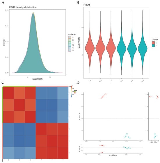
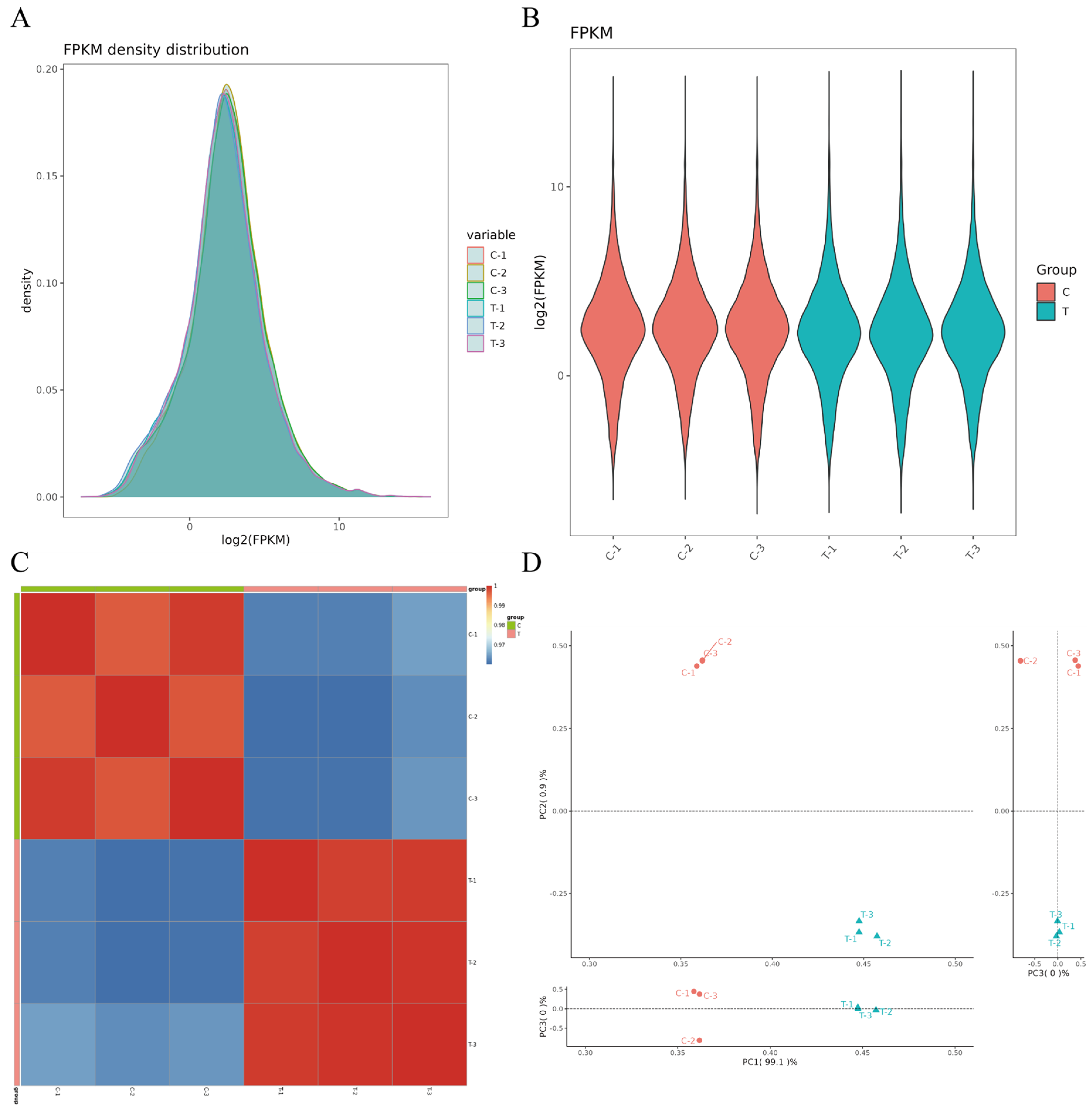
Based on the FPKM values of each gene in the assembled transcriptome, the gene expression distribution profile showed the expression level distribution of genes across six distinct samples (Figure 1A,B). Correlation analysis of gene expression levels among samples (Figure 1C) revealed high similarity between biological replicates within the same group, indicating high experimental reliability and reasonable sample selection. Meanwhile, principal component analysis (PCA) and Pearson correlation coefficient analysis demonstrated good reproducibility of samples within each group, suggesting that the sequencing data obtained were reliable and suitable for subsequent analysis (Figure 1D).
Figure 1.
FPKM-based transcriptomic gene expression and sample correlation analysis. Note: (A) Distribution of gene expression abundance. (B) Violin plot of gene expression. (C) Heatmap of correlations among three different sample groups, with the depth of color representing the magnitude of the correlation coefficient between two samples. Within the same color tone, the deeper the color, the stronger the correlation. (D) Principal component analysis (PCA) plot.
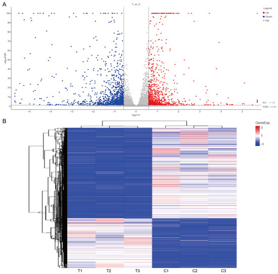
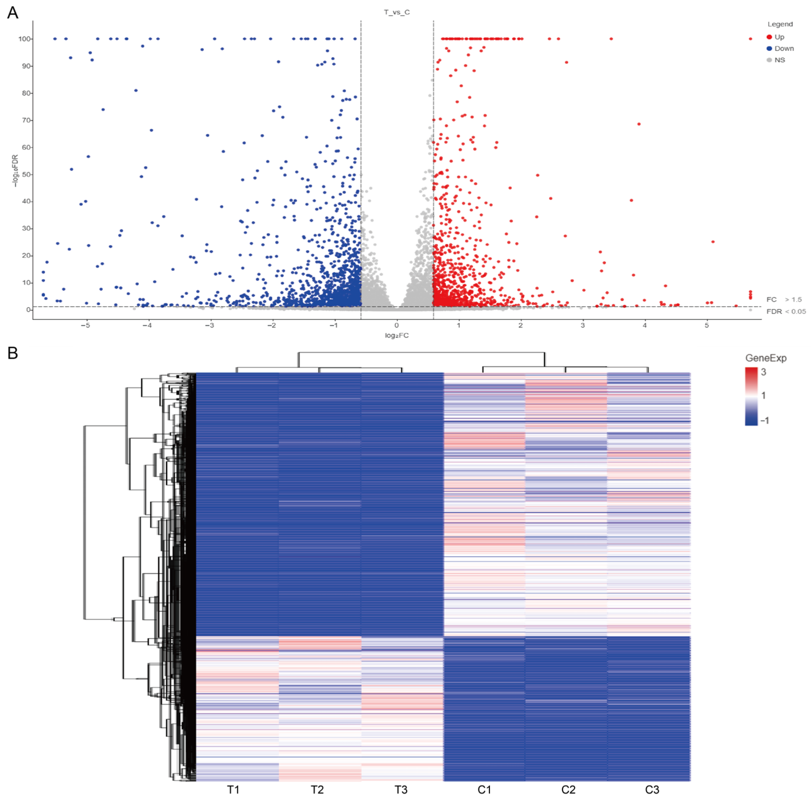
A total of 2538 differentially expressed genes (DEGs) were identified by differential expression analysis (screening criteria: FDR < 0.05 and |log2FC| > 1.5). Compared with the control group, 1047 genes were upregulated and 1491 genes were downregulated in the experimental group. The volcano plot illustrated the fold change and statistical significance of the DEGs (Figure 2A), and the clustering heatmap displayed the expression clustering of DEGs across all samples (Figure 2B).
Figure 2.
Volcano plot and cluster heatmap analysis of differentially expressed genes. Note: (A) Volcano plot of differentially expressed genes (DEGs) among different groups. Blue represents downregulated genes, red represents upregulated genes, and gray represents genes with no significant difference in expression. (B) Heatmap of cluster analysis for DEGs among different groups. The horizontal axis represents samples from different groups, and the vertical axis represents DEGs. The expression levels of DEGs in different samples are indicated by different colors: the redder the color, the higher the expression level; the bluer the color, the lower the expression level.
3.3. GO and KEGG Enrichment Analysis of Differentially Expressed Genes
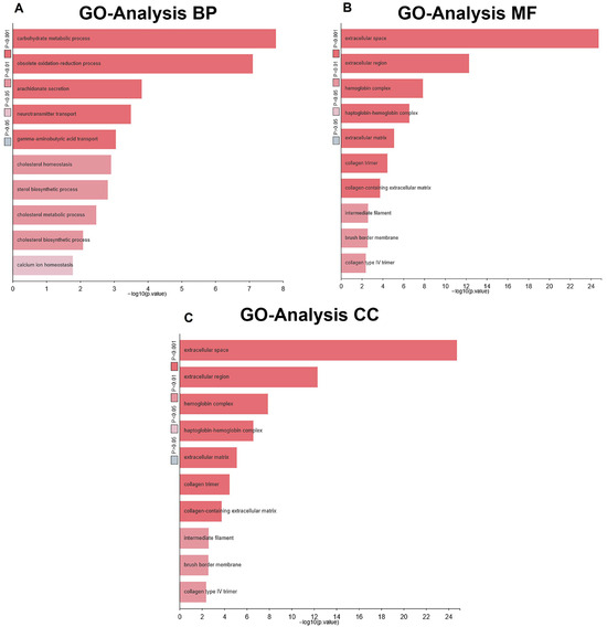
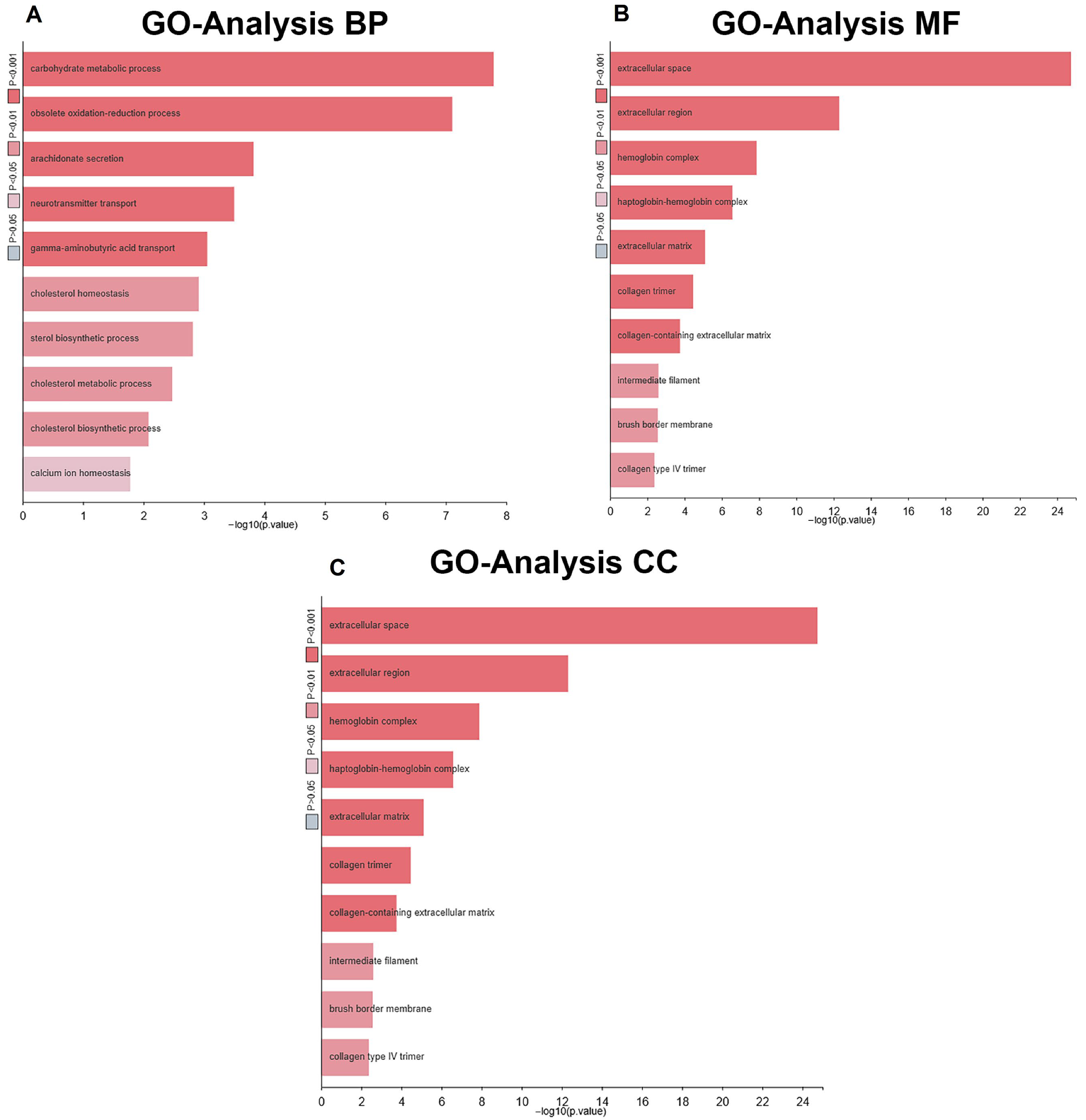
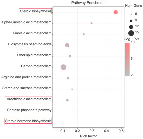
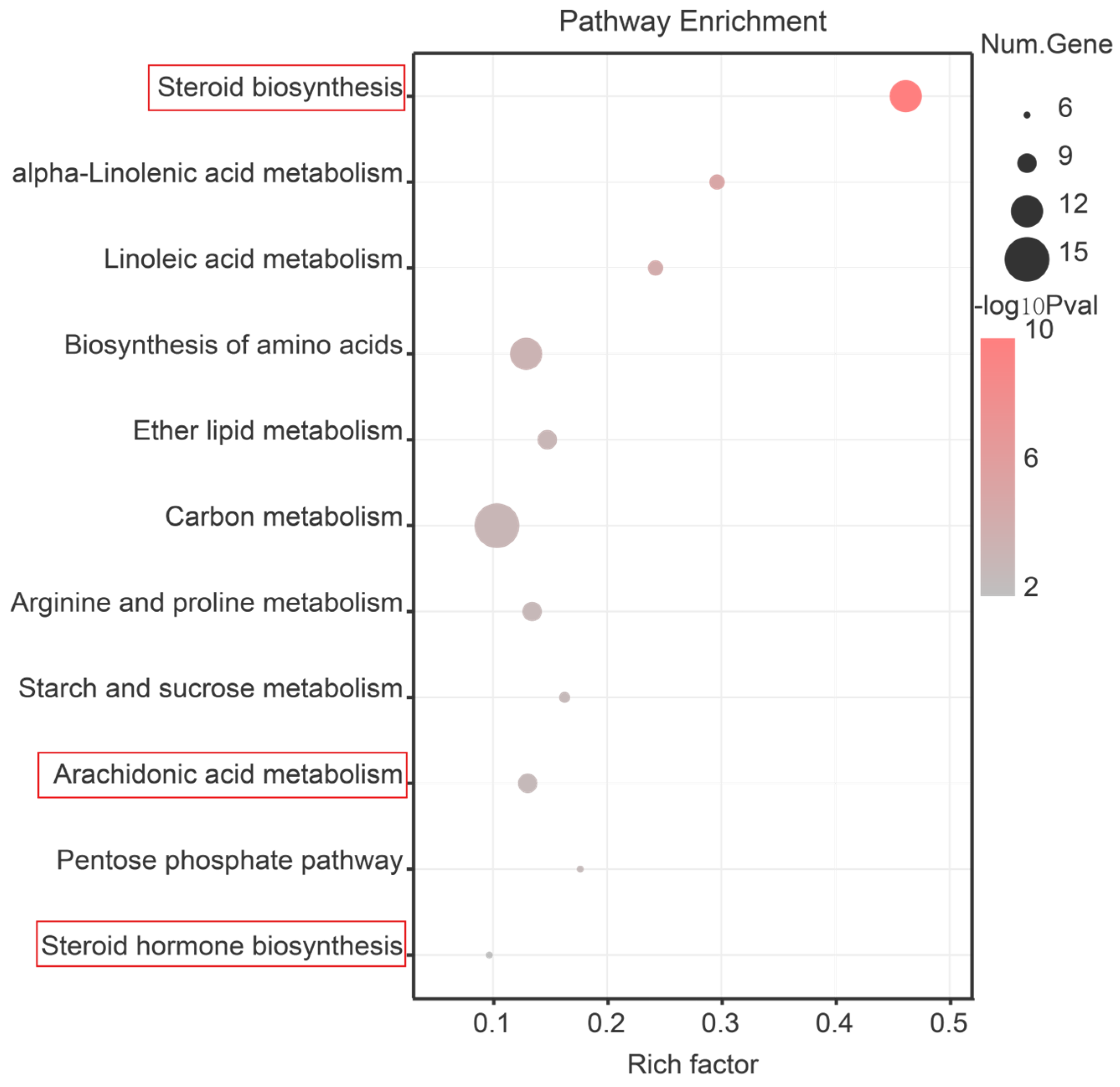
To identify the functional categories and pathways associated with the differentially expressed genes, GO functional annotation and KEGG pathway enrichment analysis were performed on the set of differentially expressed genes. GO analysis revealed that the differentially expressed genes were significantly enriched in all three main functional categories (Figure 3). In terms of Biological Process, pathways related to energy metabolism and lipid signaling were significantly enriched (FDR < 0.05), including carbohydrate metabolic process, oxidation–reduction process, and arachidonate secretion. In terms of Molecular Function, catalytic function terms were notably enriched, such as serine-type endopeptidase activity, serine-type peptidase activity, and hydrolase activity. In terms of Cellular Component, secretion-related compartments were enriched, including extracellular space, extracellular region, and hemoglobin complex. KEGG pathway enrichment analysis showed that the differentially expressed genes were significantly enriched in pathways closely related to sex differentiation, including steroid biosynthesis, arachidonic acid metabolism, and steroid hormone biosynthesis (Figure 4).
Figure 3.
GO classification of differentially expressed genes. Note: (A) Enrichment analysis of biological processes of DEGs among different groups. (B) Enrichment analysis of molecular functions of DEGs among different groups. (C) Enrichment analysis of cellular components of DEGs among different groups. The horizontal axis represents the −log10-transformed p value, and the vertical axis represents the enriched functional terms. The color of the bar indicates statistical significance: the closer the color is to red, the higher the significance; the closer the color is to gray, the lower the significance.
Figure 4.
KEGG enrichment analysis of differentially expressed genes. Note: The horizontal axis represents the enrichment factor (Rich Factor), which is the ratio of the input genes enriched to the target pathway to the annotated genes. The vertical axis represents the enriched pathway entries, and the color of the bubbles indicates the statistical significance.
3.4. Gene Set Enrichment Analysis of All Expressed Genes
To complement the traditional enrichment analysis and identify pathway-level trends that may not be detected by differentially expressed gene-based approaches, GSEA was performed based on a ranked list of all expressed genes according to their differential expression fold change. Unlike the KEGG enrichment analysis in Section 3.3, which was based on a pre-selected set of differentially expressed genes, GSEA evaluates the coordinated behavior of entire gene sets across the whole transcriptome.
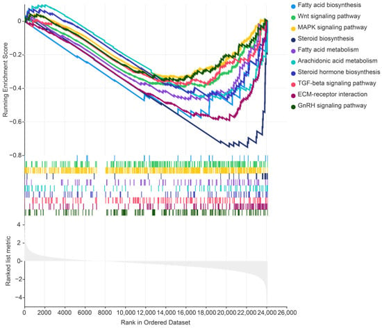
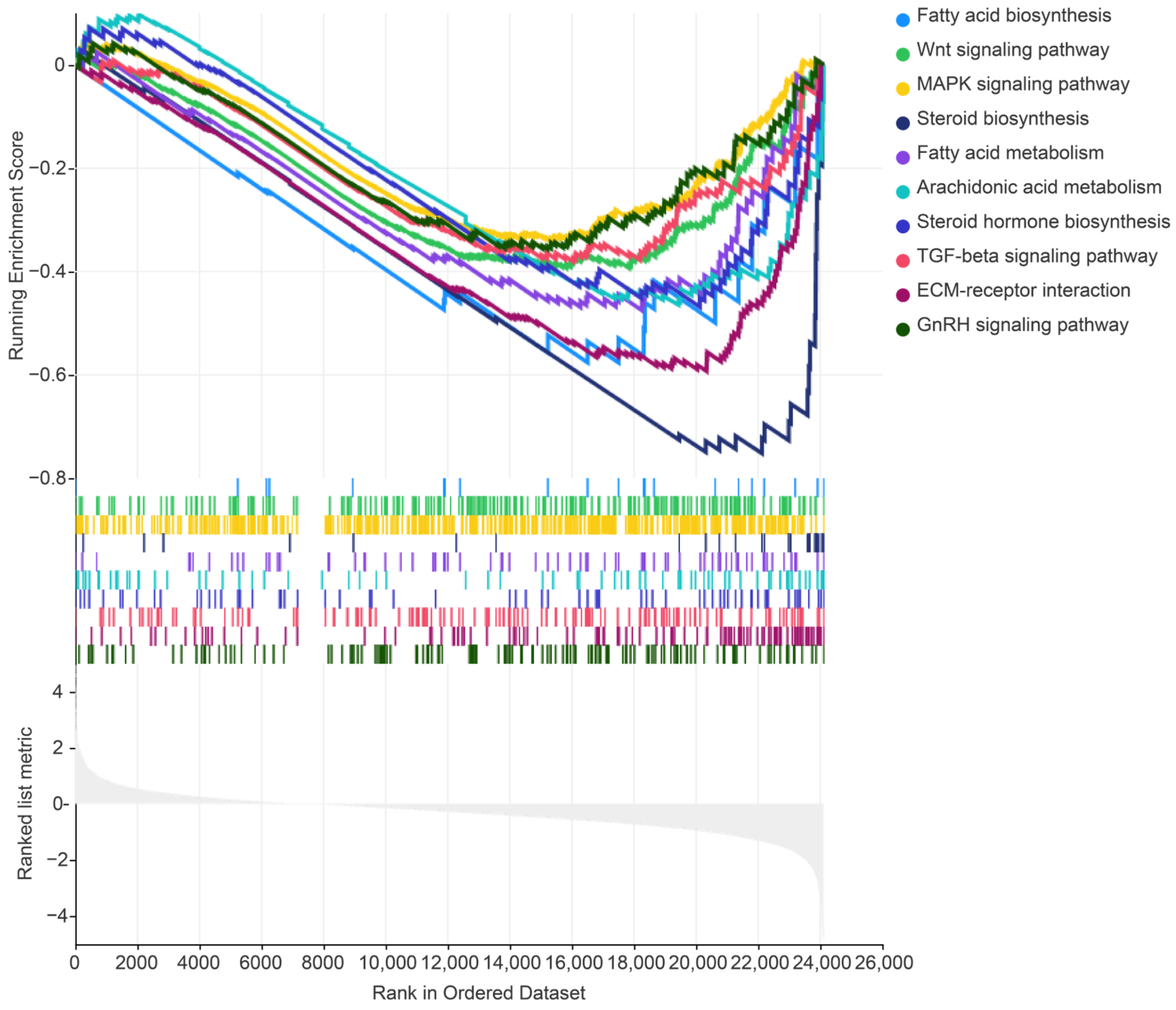
The GSEA results revealed that gene sets related to steroid hormone regulation, including the steroid biosynthesis pathway, steroid hormone biosynthesis pathway, and arachidonic acid metabolism pathway, were significantly enriched in the Relugolix-treated group (Figure 5). In addition, gene sets associated with upstream signaling regulation, such as the Wnt signaling pathway, MAPK signaling pathway, TGFβ signaling pathway, and GnRH signaling pathway, also showed enrichment trends. Notably, some pathways (e.g., steroid biosynthesis and arachidonic acid metabolism) were enriched in both methods, while others (e.g., Wnt and GnRH signaling pathways) were detected only by GSEA, highlighting the complementary nature of the two approaches.
Figure 5.
Gene set enrichment analysis. Note: The horizontal axis represents the ranking of genes. According to the gene ranking results, genes included in the KEGG entry are assigned positive scores, while those not included are assigned negative scores. The maximum value obtained from this scoring is defined as the maximum value of the entry. The positions of positive scoring are marked with vertical lines below the plot. The vertical axis denotes the cumulative enrichment score, where the peak value corresponds to the enrichment score of the gene set. The bar segment displays the distribution of ranking scores of all genes (genes are sorted in descending order of signal-to-noise ratio values).
3.5. qRT-PCR Validation of Transcriptomic Data
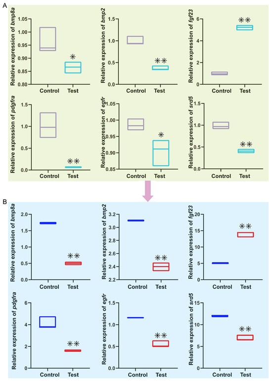
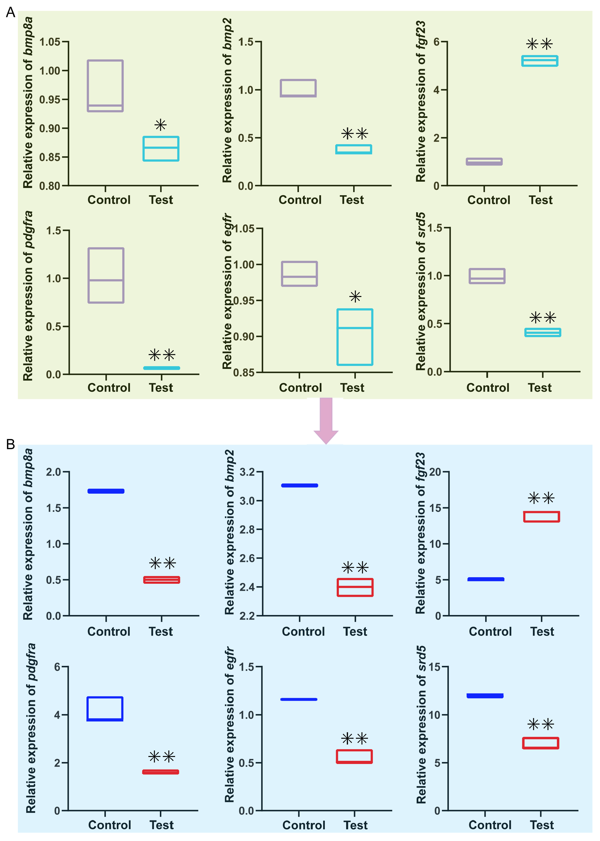
To verify the reliability of differentially expressed genes from the transcriptomic analysis, six genes (bmp8a, bmp2, fgf23, pdgfra, egfr, srd5) involved in pathways closely related to the experimental phenotype were selected for qRT-PCR validation. The results showed that the expression patterns obtained by qRT-PCR were consistent with those from the transcriptome (Figure 6), confirming the reliability of the transcriptomic data. In addition, differentially expressed genes from the steroid biosynthesis pathway, MAPK signaling pathway, and TGF-β signaling pathway were selected for validation. It was found that in the experimental group, the expression of bmp8a and bmp2 in the TGF-β signaling pathway was significantly downregulated. In the MAPK signaling pathway, fgf23 was significantly upregulated, whereas pdgfra and egfr were significantly downregulated. The steroid synthesis-related gene srd5 was also markedly downregulated.
Figure 6.
Analysis of the expression differences of target genes between the control group and the experimental group. Note: The x-axis represents different groups. The control group was the normal culture group at 18 °C, and the experimental group was the Relugolix-treated group at 18 °C. (A) Expression detected by qRT-PCR; (B) Expression in the transcriptome. * indicates a significant difference in expression level in the experimental group compared with the control group (p < 0.05), and ** indicates an extremely significant difference (p < 0.01).
4. Discussion
4.1. GnRH Is Involved in the Sex Differentiation of S. schlegelii
Comparative transcriptomic analysis was performed on gonads of genetic female S. schlegelii between the control group and the Relugolix-treated group. We revealed that differentially expressed genes were significantly enriched in the steroid biosynthesis pathway in the KEGG analysis, suggesting that GnRH signaling may participate in the masculinization process of genetic female S. schlegelii by regulating gonadal steroid hormone synthesis. Meanwhile, combined with GSEA and existing studies related to sex differentiation [28,36,37], we focused on the TGF-β signaling pathway, MAPK signaling pathway, Wnt signaling pathway, and calcium signaling pathway. Although these pathways did not show the most significant enrichment among the overall differentially expressed genes, they exhibited obvious enrichment trends in GSEA and were highly associated with vertebrate gonadal development, sex determination, and differentiation. This indirectly indicates their important potential roles in GnRH-mediated gonadal sex remodeling.
The results of GO functional enrichment analysis were generally consistent with the biological functions of the aforementioned pathways. In terms of biological processes, differentially expressed genes were mainly enriched in the carbohydrate metabolic process, oxidation–reduction process, and arachidonate secretion. Based on these observations, we speculate that Relugolix treatment may be associated with alterations in energy metabolism, redox homeostasis, and lipid signaling molecule secretion, which might provide a material and energy basis for gonadal structural remodeling and sex phenotype conversion [38,39,40]. This result is also consistent with the significant enrichment of the arachidonic acid pathway in the KEGG analysis. At the molecular function level, differentially expressed genes were enriched in serine-type endopeptidase activity, serine-type peptidase activity, and hydrolase activity. Previous studies have indicated that these enzymatic activities are closely related to extracellular matrix remodeling, growth factor maturation, and the cleavage and processing of signaling molecules [41,42,43]; accordingly, we speculate that they may play a role in regulating signal intensity and spatiotemporal distribution by influencing the activation and degradation of ligands and receptors in the TGF-β and MAPK pathways [41,42,43]. Cellular component analysis revealed that differentially expressed genes were mainly localized in the extracellular space, extracellular region, and hemoglobin complex. This distribution pattern may suggest that active extracellular signal communication, cellular microenvironment remodeling, and alterations in local oxygen supply and metabolic status occur in the gonad during sex remodeling, which is partially consistent with the extracellular signaling-dependent mode of action of the TGF-β and MAPK pathways [43,44,45].
4.2. Potential Mechanisms Underlying the Roles of Key Genes
Further screening and validation revealed that the key genes of the TGF-β signaling pathway, bmp8a and bmp2, were significantly downregulated after Relugolix treatment. The MAPK signaling pathway-related gene fgf23 was significantly upregulated, while pdgfra and egfr were significantly downregulated. Meanwhile, the expression of srd5, a key gene involved in steroid synthesis, was markedly decreased. Taken together, we hypothesize that GnRH signaling pathway may regulate the TGF-β and MAPK signaling pathways, thereby affecting steroid hormone biosynthesis and ultimately mediating the differentiation of genetic female S. schlegelii toward a male phenotype. As a key member of the TGF-β superfamily, bmp2 dynamically regulates FSH receptor expression and estradiol synthesis in ovarian granulosa cells via the Smad1/5/8 pathway, serving as a core factor maintaining folliculogenesis and granulosa cell differentiation [46]. In genetic female (XX) gonads, bmp2 acts as a downstream effector of the Wnt4-Rspo1-β-catenin pathway and cooperates with foxl2 to activate fst expression, thereby sustaining ovarian development and indirectly repressing male-related genes such as sox9 [47,48,49].
Based on the transcriptomic data from the present study, we speculate that the significant downregulation of bmp2 after blockade of the GnRH signaling pathway triggers a series of cascade reactions. On the one hand, the decreased expression of fst may affect the stability of foxl2, thereby weakening the function of upstream factors such as wnt4 and rspo1, and reducing β-catenin pathway activity, which might not favor the maintenance of the female differentiation pathway. On the other hand, the repression of sox9 may be subsequently relieved, accompanied by increased expression of male-related genes such as fgf9 and amh, which might, to some extent, shift the primordial gonad toward testicular development. Furthermore, previous studies have suggested that bmp8a may promote the proliferation of Sertoli cells in the testis [50]; its downregulation may reflect developmental timing disturbances caused by GnRH deficiency. In the present study, the significant upregulation of fgf23 and downregulation of pdgfra and egfr suggest that GnRH signaling pathway may be associated with specific branches of the FGF-FGFR-MAPK cascade. The downregulation of pdgfra and egfr may affect the survival signals of ovarian somatic cells [26,51,52], while the upregulation of fgf23 may influence the function of testicular interstitial cells through paracrine mechanisms [26,52]. These changes suggest that the MAPK signaling network may undergo a pro-male shift and may cooperate with the TGF-β pathway to influence gonadal development.
4.3. Limitations and Future Perspectives
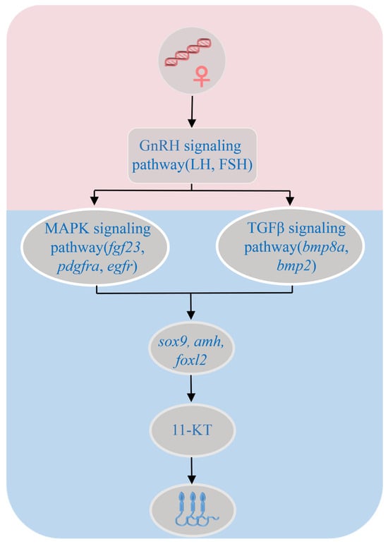
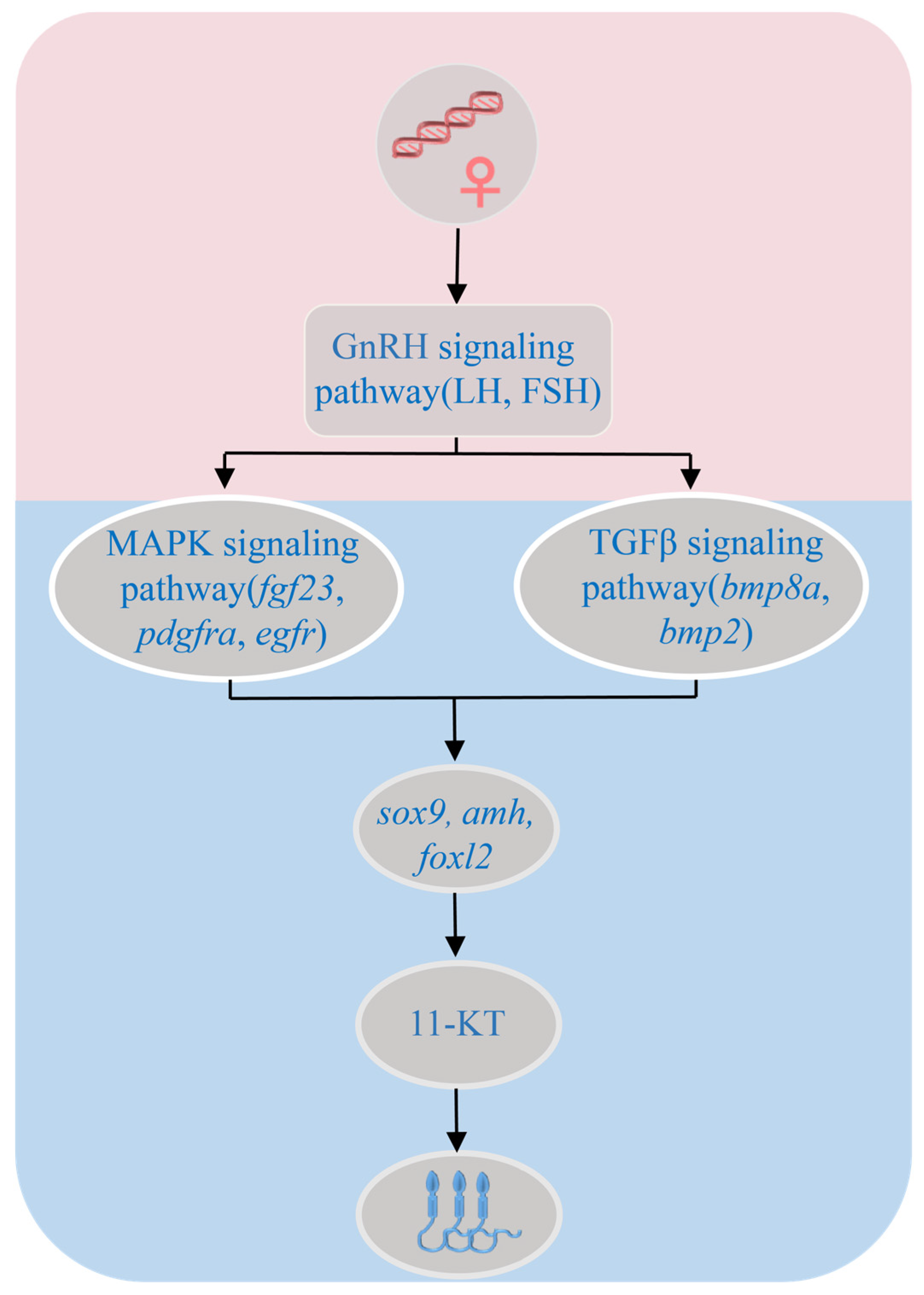
It should be emphasized that this study primarily employed transcriptome sequencing and gene set enrichment analysis to investigate changes in the gene expression profile following Relugolix treatment. We propose the following hypothesis: the GnRH signaling pathway may regulate the expression of key genes involved in sex differentiation and local gonadal steroidogenesis through the TGF-β and MAPK signaling pathways, thereby participating in the regulatory process of sex differentiation (Figure 7). However, this study lacks functional validation experimental evidence for these genes. The cellular localization of bmp2 and bmp8a during gonadal development in S. schlegelii remains unclear; the causal relationship between the downregulation of pdgfra and egfr and the reduction in ovarian somatic cell numbers is merely speculative, and has not been confirmed through experiments involving cell proliferation, apoptosis marker detection, and somatic cell counting. This hypothesis remains to be further verified by subsequent functional experiments. Additionally, although three biological replicates were set for each group in this study—meeting the basic requirements for transcriptome sequencing analysis—this still presents limitations in reflecting biological variation. Increasing the number of biological replicates would more accurately represent biological differences among samples. Due to current constraints, this study was unable to include additional replicate samples. Follow-up studies with expanded sample sizes are needed, combined with techniques such as in situ hybridization and immunohistochemistry, to systematically validate the roles of these genes in sex differentiation of S. schlegelii, thereby further elucidating the molecular mechanisms underlying the GnRH-mediated temperature-dependent sex determination regulatory network.
Figure 7.
Hypothetical model of GnRH receptor antagonism-induced masculinization in genetic female S. schlegelii based on transcriptomic data. The proposed relationships are correlative, not causal, and require functional validation.
5. Conclusions
In this study, transcriptome analysis combined with qRT-PCR verification clarified the regulatory role of GnRH signaling pathway in sex differentiation of S. schlegelii: GnRH signaling pathway can precisely mediate sex differentiation by regulating key genes associated with the TGF-β signaling pathway, MAPK signaling pathway, and steroid biosynthesis, and the expression changes in these key genes are somewhat correlated with gonadal development. Meanwhile, this study has certain limitations. Further functional experiments are needed to verify the specific roles and regulatory networks of individual genes, so as to improve the molecular mechanism of GnRH-mediated sex differentiation and provide theoretical support and experimental evidence for research on fish sex regulation and the optimization of monosex breeding techniques.
Supplementary Materials
The following supporting information can be downloaded at: https://www.mdpi.com/article/10.3390/biology15110857/s1, Table S1. Primers for qRT-PCR Analysis of Related Genes in S. schlegelii; Table S2. PCR primers for genetic sex identification in S. schlegelii.
Author Contributions
Conceptualization, J.H., Y.X. and J.L.; methodology, P.X. and J.H.; software, J.H. and P.X.; validation, J.H.; formal analysis, J.H.; investigation, J.H. and J.L.; data curation, J.H. and Y.X.; writing—original draft preparation, J.H.; writing—review and editing, J.L. and Y.X.; visualization, J.H.; supervision, Y.X. and J.L.; funding acquisition, J.L. All authors have read and agreed to the published version of the manuscript.
Funding
This work was supported by National Key Research and Development Program (2024YFD2400102), National Natural Science Foundation of China (No. 42276107; U24A20456), Natural Science Foundation of Shandong Province (ZR2024MC071), Shandong Province Key R&D Program (2023LZGCQY001), the Sino-Indonesia Joint Laboratory for Marine Sciences (SIMS).
Institutional Review Board Statement
All experiments were conducted in strict compliance with the experimental animal guidance policy as outlined by the Institute of Oceanology’s Animal Ethics Committee, Chinese Academy of Sciences (Permit No. IOCAS20230916PPFA0007, approval date: 16 September 2023).
Data Availability Statement
All generated data are presented in the article and Supplementary Materials.
Acknowledgments
We would like to thank Qinghua Liu and Xueying Wang for their help during the breeding period.
Conflicts of Interest
The authors declare no conflicts of interest.
References
- Zhang, Y.; Bian, W.; Chen, H.; Zhang, W.; Wang, C.; Qiu, J.; Sun, X.; Lin, J.; Yu, Y.; Feng, M. Contrasting size responses of loricate ciliates to long-term warming: Agglutinated forms decrease while hyaline forms increase. Mar. Biol. 2026, 173, 51. [Google Scholar] [CrossRef] [Scilit]
- Intergovernmental Panel on Climate Change (IPCC). Climate Change 2021—The Physical Science Basis; Intergovernmental Panel on Climate Change (IPCC): Geneva, Switzerland, 2023. [Google Scholar]
- Speizer, S.; Raymond, C.; Ivanovich, C.; Horton, R.M. Concentrated and Intensifying Humid Heat Extremes in the IPCC AR6 Regions. Geophys. Res. Lett. 2022, 49, e2021GL097261. [Google Scholar] [CrossRef] [Scilit]
- Lee, E.; Mcmanus, L.C. Rate of Temperature Increase and Genetic Diversity Drives Marine Metapopulation Persistence under Climate Change. Am. Nat. 2025, 206, 318–334. [Google Scholar] [CrossRef] [Scilit]
- Guillaume, A.S.; Joost, S.; Curpen, S.; Neelayya, D.D.; Harree-Somah, L.; Sadasing, O.; Saponari, L.; Dale, C.; Barret, L.; Andrews, N. Coral Genetic Structure in the Western Indian Ocean Mirrors Ocean Circulation and Thermal Stress History. Evol. Appl. 2026, 19, e70206. [Google Scholar] [CrossRef] [Scilit]
- Su, H.; Zhou, L.; Xie, P. What can we learn from the loss of sharks? Trends Ecol. Evol. 2025, 40, 531–533. [Google Scholar] [CrossRef] [Scilit] [PubMed]
- Hesselbarth, M.H.K.; Allgeier, J.E. High Fish Biomass and Low Nutrient Enrichment Synergistically Enhance Stability in a Seagrass Meta-Ecosystem. Conserv. Lett. 2024, 17, e13071. [Google Scholar] [CrossRef] [Scilit]
- Nagelkerken, I.; Allan, B.; Booth, D.; Donelson, J.; Mellin, C.; Ravasi, T.; Rummer, J. Climate Change Effects on Fishes. Ecology 2024. [Google Scholar] [CrossRef] [Scilit]
- Dahlke, F.T.; Wohlrab, S.; Butzin, M.; Prtner, H.O. Thermal bottlenecks in the life cycle define climate vulnerability of fish. Science 2020, 369, 65–70. [Google Scholar] [CrossRef] [Scilit]
- Andreassen, A.H.; Clements, J.C.; Morgan, R.; Spatafora, D.; Metz, M.; Sheim, E.R.; Pélabon, C.; Jutfelt, F. Evolution of warming tolerance alters physiology and life history traits in zebrafish. Nat. Clim. Chang. 2025, 15, 665–672. [Google Scholar] [CrossRef] [Scilit]
- Wang, J.; Li, L.; Zhao, S.; Wang, M.; Li, B.; Guo, H.; Han, D.; Zhu, X.; Zhang, L. Combined effects of prolonged temperature and hypoxia stress on growth, metabolism and physiological status of yellow catfish (Pelteobagrus fulvidraco). Aquac. Rep. 2025, 43, 102884. [Google Scholar] [CrossRef] [Scilit]
- Liu, Y.; Tian, C.; Yang, Z.; Huang, C.; Jiao, K.; Yang, L.; Duan, C.; Zhang, Z.; Li, G. Effects of Chronic Heat Stress on Growth, Apoptosis, Antioxidant Enzymes, Transcriptomic Profiles, and Immune-Related Genes of Hong Kong Catfish (Clarias fuscus). Animals 2024, 14, 1006. [Google Scholar] [CrossRef] [Scilit]
- Bai, S.; Li, T.; Kong, L.; Bi, B.; Hu, Q. Effects of Chronic Thermal Stress on the Physiology, Metabolism, Histology, and Gut Microbiota of Juvenile Schizothorax grahami. Animals 2025, 15, 2749. [Google Scholar] [CrossRef] [Scilit]
- Duan, C.; Zhou, D.; Feng, R.; Li, X.; Yang, L.; Li, X.; Li, G.; Chen, H.; Liao, Y.; Tian, C. Long-term thermal acclimation enhances heat resistance of Hong Kong catfish (Clarias fuscus) by modulating gill tissue structure, antioxidant capacity and immune metabolic pathways. Ecotoxicol. Environ. Saf. 2024, 284, 116930. [Google Scholar] [CrossRef] [Scilit]
- Dagmar, W.; Aitana, P.G.; Axel, N.; Marie-Christine, C. Gonadal sex determination in vertebrates: Rethinking established mechanisms. Development 2025, 152, dev204592. [Google Scholar] [CrossRef] [Scilit] [PubMed]
- Baroiller, J.F.O.; D’Cotta, H. The Reversible Sex of Gonochoristic Fish: Insights and Consequences. Sex. Dev. 2016, 10, 242–266. [Google Scholar] [CrossRef] [Scilit]
- Sarre, S.D.; Georges, A.; Quinn, A. The ends of a continuum: Genetic and temperature-dependent sex determination in reptiles. Bioessays 2004, 26, 639–645. [Google Scholar] [CrossRef] [Scilit] [PubMed]
- Palaiokostas, C.; Bekaert, M.L.; Khan, M.; Taggart, J.B.; Gharbi, K.; Mcandrew, B.J.; Penman, D.J. A novel sex-determining QTL in Nile tilapia (Oreochromis niloticus). Bmc Genom. 2015, 16, 171. [Google Scholar] [CrossRef] [Scilit]
- Matsuda, M.; Nagahama, Y.; Kobayashi, T.; Matsuda, C.; Hamaguchi, S. The sex determining gene of medaka: A Y-specific DM domain gene (DMY) is required for male development. Fish Physiol. Biochem. 2003, 28, 135–139. [Google Scholar] [CrossRef] [Scilit]
- Wei, J.; Chen, Y.; Wang, W. A High-Density Genetic Linkage Map and QTL Mapping for Sex and Growth-Related Traits of Large-Scale Loach (Paramisgurnus dabryanus). Front. Genet. 2019, 10, 1023. [Google Scholar] [CrossRef] [Scilit]
- Kitano, J.; Ansai, S.; Takehana, Y.; Yamamoto, Y. Diversity and Convergence of Sex Determination Mechanisms in Teleost Fish. Annu. Rev. Anim. Biosci. 2024, 12, 233–259. [Google Scholar] [CrossRef] [Scilit]
- Fang, J.; Li, G.; Luo, W.; Hu, Q. Understanding Genetic Regulation of Sex Differentiation in Hermaphroditic Fish. Animals 2025, 15, 119. [Google Scholar] [CrossRef] [Scilit] [PubMed]
- Baroiller, J.F.; D’Cotta, H. Environment and sex determination in farmed fish. Comp. Biochem. Physiol. C Toxicol. Pharmacol. 2001, 130, 399–409. [Google Scholar] [CrossRef] [Scilit]
- Feng, J.Y.; Wang, Z.H.; Wei, F.L.; Yu, M.; Ye, T.; Wu, C.N.; Liu, F.; Guo, D.D.; Lou, B.; Xie, Q.P. Effects of different temperatures on sex differentiation and critical thermosensitive period of little yellow croaker (Larimichthys polyactis). Aquac. Rep. 2025, 40, 102622. [Google Scholar] [CrossRef] [Scilit]
- Macnamara, R.; Mankiewicz, J.L.; Salger, S.A.; Stuart, K.; Borski, R.J.; Godwin, J.; Drawbridge, M. Temperature regulates sex determination and growth in the paralichthid flatfish California halibut. J. Exp. Zool. A Ecol. Integr. Physiol. 2024, 341, 811–821. [Google Scholar] [CrossRef] [Scilit]
- Rajendiran, P.; Jaafar, F.; Kar, S.; Sudhakumari, C.; Senthilkumaran, B.; Parhar, I.S. Sex Determination and Differentiation in Teleost: Roles of Genetics, Environment, and Brain. Biology 2021, 10, 973. [Google Scholar] [CrossRef] [Scilit]
- Yang, Y.Y.; Li, C.; Li, T.C.; Li, Y.F.; Xu, F.; Jiang, Z.B.; Zhou, H. Multidimensional Roles and Application Progress of GnRH during Fish Reproduction. Chin. Agric. Bull. 2025, 41, 154–164. [Google Scholar]
- Ke, F.; Xuefan, C.; Yanlong, S.; Binbin, T.; Ji, C.; Jing, W.; Shaojun, L.; Yonghua, S.; Zuoyan, Z.; Trudeau, V.L. Gnrh3 Regulates PGC Proliferation and Sex Differentiation in Developing Zebrafish. Endocrinology 2019, 1, bqz024. [Google Scholar] [CrossRef] [Scilit]
- Mizanur, R.M.; Yun, H.; Moniruzzaman, M.; Ferreira, F.; Kim, K.D.; Bai, S.C. Effects of feeding rate and water temperature on growth and body composition of juvenile Korean rockfish, Sebastes schlegeli (Hilgendorf 1880). Asian-Australas. J. Anim. Sci. 2014, 27, 690–699. [Google Scholar] [CrossRef] [Scilit] [PubMed]
- Song, W.; Xie, Y.; Sun, M.; Li, X.; He, Y. A duplicated amh is the master sex-determining gene for Sebastes rockfish in the Northwest Pacific. Open Biol. 2021, 11, 210063. [Google Scholar] [CrossRef] [Scilit]
- Love, M.I.; Huber, W.; Anders, S. Moderated estimation of fold change and dispersion for RNA-seq data with DESeq2. Genome Biol. 2014, 15, 550. [Google Scholar] [CrossRef] [Scilit] [PubMed]
- Dewey, C.N.; Bo, L. RSEM: Accurate transcript quantification from RNA-Seq data with or without a reference genome. BMC Bioinform. 2011, 12, 323. [Google Scholar] [CrossRef] [Scilit]
- Subramanian, A.; Tamayo, P.; Mootha, V.K.; Mukherjee, S.; Ebert, B.L.; Gillette, M.A.; Paulovich, A.; Pomeroy, S.L.; Golub, T.R.; Lander, E.S. Gene set enrichment analysis: A knowledge-based approach for interpreting genome-wide expression profiles. Proc. Natl. Acad. Sci. USA 2005, 102, 15545–15550. [Google Scholar] [CrossRef] [Scilit]
- Jin, C.F.; Song, W.H.; Wang, M.Y.; Qi, J.; Zhang, Q.Q.; He, Y. Transcriptome-wide identification and validation of reference genes in black rockfish (Sebastes schlegelii). J. Ocean Univ. China 2021, 20, 654–660. [Google Scholar] [CrossRef] [Scilit]
- Pfaffl, M.W. A new mathematical model for relative quantification in real-time RT-PCR. Nucleic Acids Res. 2001, 29, e45. [Google Scholar] [CrossRef] [Scilit] [PubMed]
- Shen, Z.G.; Wang, H.P. Molecular players involved in temperature-dependent sex determination and sex differentiation in Teleost fish. Genet. Sel. Evol. 2014, 46, 26. [Google Scholar] [CrossRef] [Scilit] [PubMed]
- Shuqing, Z.; Juan, L.; Zhilong, L.; Wenjing, T.; Deshou, W. Identification and Evolution of TGF-β Signaling Pathway Members in Twenty-Four Animal Species and Expression in Tilapia. Int. J. Mol. Sci. 2018, 19, 1154. [Google Scholar] [CrossRef] [Scilit]
- Pankhurst, N.W.; Munday, P.L. Effects of climate change on fish reproduction and early life history stages. Mar. Freshw. Res. 2011, 62, 1015. [Google Scholar] [CrossRef] [Scilit]
- Sakae, Y.; Oikawa, A.; Sugiura, Y.; Mita, M.; Nakamura, S.; Nishimura, T.; Suematsu, M.; Tanaka, M. Starvation causes female-to-male sex reversal through lipid metabolism in the teleost fish, medaka (Olyzias latipes). Biol. Open 2020, 9, bio050054. [Google Scholar] [CrossRef] [Scilit]
- Sakae, Y.; Tanaka, M. Metabolism and Sex Differentiation in Animals from a Starvation Perspective. Sex. Dev. 2021, 15, 168–178. [Google Scholar] [CrossRef] [Scilit]
- Liu, C.; Xu, P.; Lamouille, S.; Xu, J.; Derynck, R. TACE-mediated ectodomain shedding of the type I TGF-beta receptor downregulates TGF-beta signaling. Mol. Cell 2009, 35, 26–36. [Google Scholar] [CrossRef] [Scilit]
- Page-Mccaw, A.; Ewald, A.J.; Werb, Z. Matrix metalloproteinases and the regulation of tissue remodelling. Nat. Rev. Mol. Cell Biol. 2007, 8, 221–233. [Google Scholar] [CrossRef] [Scilit]
- Wang, Q.; Liu, K.; Feng, B.; Zhang, Z.; Wang, R.; Tang, L.; Shao, C. Transcriptome of Gonads From High Temperature Induced Sex Reversal During Sex Determination and Differentiation in Chinese Tongue Sole, Cynoglossus semilaevis. Front. Genet. 2019, 10, 1128. [Google Scholar] [CrossRef] [Scilit] [PubMed]
- Bravo, P.; Liu, Y.; Draper, B.W.; Marlow, F.L. Macrophage activation drives ovarian failure and masculinization in zebrafish. Sci. Adv. 2023, 9, 13. [Google Scholar] [CrossRef] [Scilit]
- Devlin, R.H.; Nagahama, Y. Sex determination and sex differentiation in fish: An overview of genetic, physiological, and environmental influences. Aquaculture 2002, 208, 191–364. [Google Scholar] [CrossRef] [Scilit]
- Pangas, S.A. Bone morphogenetic protein signaling transcription factor (SMAD) function in granulosa cells. Mol. Cell. Endocrinol. 2012, 356, 40–47. [Google Scholar] [CrossRef] [Scilit]
- Huang, S.S.; Ye, L.P.; Chen, H.L. Sex determination and maintenance: The role of DMRT1 and FOXL2. Asian J. Androl. 2017, 19, 619–624. [Google Scholar] [CrossRef] [Scilit]
- She, Z.Y.; Yang, W.X. Molecular mechanisms involved in mammalian primary sex determination. J. Mol. Endocrinol. 2014, 53, R21–R37. [Google Scholar] [CrossRef] [Scilit]
- Uehara, S.K.; Nishiike, Y.; Maeda, K.; Karigo, T.; Kuraku, S.; Okubo, K.; Kanda, S. Identification of the FSH-RH as the other gonadotropin-releasing hormone. Nat. Commun. 2024, 15, 5342. [Google Scholar] [CrossRef] [Scilit] [PubMed]
- Housh, M.J.; Telish, J.; Forsgren, K.L.; Lema, S.C. Fluctuating and Stable High Temperatures Differentially Affect Reproductive Endocrinology of Female Pupfish. Integr. Org. Biol. 2024, 6, obae003. [Google Scholar] [CrossRef] [Scilit]
- Miranda, L.A.; Strobl-Mazzulla, P.H.; Strüssmann, C.A.; Parhar, I.; Somoza, G.M. Gonadotropin-releasing hormone neuronal development during the sensitive period of temperature sex determination in the pejerrey fish, Odontesthes bonariensis. Gen. Comp. Endocrinol. 2003, 132, 444–453. [Google Scholar] [CrossRef] [Scilit]
- Fontaine, R.; Royan, M.R.; von Krogh, K.; Weltzien, F.A.; Baker, D.M. Direct and Indirect Effects of Sex Steroids on Gonadotrope Cell Plasticity in the Teleost Fish Pituitary. Front. Endocrinol. 2020, 11, 605068. [Google Scholar] [CrossRef] [Scilit]

Disclaimer/Publisher’s Note: The statements, opinions and data contained in all publications are solely those of the individual author(s) and contributor(s) and not of MDPI and/or the editor(s). MDPI and/or the editor(s) disclaim responsibility for any injury to people or property resulting from any ideas, methods, instructions or products referred to in the content. |
© 2026 by the authors. Licensee MDPI, Basel, Switzerland. This article is an open access article distributed under the terms and conditions of the Creative Commons Attribution (CC BY) license.