


Abstract
This study examines the colorimetric characteristics and possible biological effects of the quinoline-derived dye M1 1,4-bis((E)-2-(quinolin-2-yl)vinyl)benzene. The pi-conjugated structure and high planarity of M1 give it important semiconductor and optical properties, such as the modulation of its fluorescence properties depending on the synthesis method. These optical properties make this dye a candidate for use as a sensor for biological targets. Predictions were made on molecular targets such as receptors and enzymes involved in neurological and inflammatory processes. Docking studies indicated moderate to strong binding affinities (−10.3 to −7.1 kcal mol−1), supported mainly by hydrophobic pi interactions and low RMSD conformations. The predicted biological activity, together with the luminescent properties documented above, position M1 as a versatile dye with potential uses in biomedical applications and as a luminescent detection sensor. These results lay the groundwork for future experimental confirmation combining photophysical analysis and biological testing.
1. Introduction
M1 (1,4-bis((E)-2-(quinolin-2-yl)vinyl)benzene) is an organic compound with the molecular formula C28H20N2, featuring a nearly flat conjugated structure where a central benzene ring is connected via vinyl π bridges to lateral quinoline groups. This D–pi–A–pi–D type architecture, similar to the 1,4-bis[(E)-2-(9H-carbazole-9-yl)vinyl]benzene based on carbazole detailed by Lapkowski et al. [1], facilitates extended pi conjugation and effective intramolecular charge transfer throughout the molecule. This compound shows typical intrinsic fluorescence features associated with extended aromatic systems that include nitrogen-containing heterocycles. In closely related derivatives of 1,4-bis((E)-2-(1H-indol-3-yl)vinyl)benzene, Ma et al. found that cyanofunctionalization results in pronounced aggregation-induced emission and distinctive thermochromic behavior, highlighting how a rigid π-conjugated structure and heteroaromatic units can enhance AIE-type responses. These highly valued photophysical properties arise from extended conjugation and specific intermolecular interactions within the aggregated state, which also facilitate strong UV–Vis absorption bands in the visible region [2]. Our research group has reported that the synthesis of M1, M1mec, M1A, and M1Amec (2,2′-[2,5-bis(octoxy)-1,4-(phenyl-vinylene)]bisquinoline) can be achieved through a Knoevenagel-type condensation reaction between quinaldine and aromatic dialdehydes, terephthalaldehyde or 2,5-bis(octoxy)terephthalaldehyde, respectively. Both compounds have been synthesized using both a reflux methodology and a mechanosynthesis method, observing that the latter results in a high-photoluminescence quantum yield suitable for the development of electroluminescent diodes [3]. Its electroluminescent characteristics for M1Amec were verified, showing a peak brightness of 28 cd m−2 at a fairly low bias voltage of 7.4 V and a current density of 53 mA/cm2 [4]. There are research findings that suggest the fluorescence variations of the dye M1 in polypropylene (PP) and maleic anhydride-grafted polypropylene (PPgMA) films signify its potential use as an optical sensor for identifying structural changes in thermoplastics [5]. The identification of L-cysteine, an amino acid that contains sulfur and is essential for protein synthesis, glutathione, and other biological molecules [6,7], was made possible by M1 through the interaction between the two substances. A click reaction occurred, altering the optical characteristics of M1, since an increase in the concentration of L-cysteine leads to a reduction in the fluorescence of M1, demonstrating that aggregation-induced luminescence (AIL) is observed in solid states and polymer films, where molecular packing restricts intramolecular motions and enhances emission, whereas L-cysteine sensing operates in dilute homogeneous solution, where aggregation is minimal and the optical response arises from specific chemical interactions with the analyte. In the present study, we do not re-examine these regimes in detail but rather use the established photophysical behaviour of M1 as a starting point to explore new biological targets through in silico prediction and docking [8]. Other research has been conducted that examines variations of compounds featuring the quinoline structure, such as the pi-conjugated polymer PBQT, which contains units of bis-(2-ethylhexyloxy)-benzo[1,2-b′] bithiophene (BDT) interspersed with a quinoline–vinylene trimer. PBQT shows photochromic properties, changing its absorption from blue at 449 nm to burgundy at 545 nm, and its fluorescence emission changes from green at 513 nm to orange at 605 nm as a result of conformational photoisomerization transitioning from the trans to the cis form. This optical behavior can be utilized in optical sensors, security features, or optical switches such as M1 [9].
On another note, various research studies have shown that hydrazide/hydrazone quinoline derivatives exhibit a range of biological activities, such as antimalarial, antitubercular, anticancer, anti-inflammatory, and antimicrobial properties. Among these activities, the antibacterial function of hydrazide/hydrazone quinoline derivatives stands out. The synthetic adaptability of the quinoline ring has resulted in a broad spectrum of structurally diverse hydrazide/hydrazone quinoline derivatives, capable of targeting multiple bacterial sites, including DNA gyrase, glucosamine-6-phosphate synthase, enoyl ACP reductase, and 3-ketoacyl ACP reductase [10]. In addition, the potential of quinoline-based compounds, such as tetrahydroquinolines and phenyl-substituted quinolines, has been assessed for their ability to identify isoenzymes of human carbonic anhydrase I and II (hCAs I and II) as well as their inhibitory and antimicrobial effects on acetylcholinesterase (AChE) enzymes. The findings revealed that all synthesized compounds demonstrated strong inhibitory activities against all targets when compared to standard inhibitors, along with antifungal effects against yeast [11].
Since luminescent sensors used for detection (such as biomarkers like cysteine and RNA in cancer) require a conjugated pi structure for aggregation-induced emission, they must possess affinity for non-covalent bonds (like pi-pi interactions and hydrophobic forces) and adjustable quantum yield. Bioactive inhibitors require specific docking affinities [12]. Quinolic sensors identify ions with great sensitivity (LOD < 10−7 M) but identify few proteins or enzymes like GRM5 or ECE2; quinolic inhibitors exhibit antimicrobial/antienzyme activity with a Gibbs free energy of −8 kcal mol−1 in docking [13]. Consequently, this study explores the forecasting of molecular targets and the binding interactions of the fluorescent dye M1 with key biological proteins, linking its conjugated composition and interaction potential for possible applications as an optoluminescent sensor or as an inhibitor of bioactive substances. M1 meets requirements (high Φ in mechanosynthesis, cysteine detection through quenching, docking −10.3 to −7.1 kcal mol−1 compared to GRM5/ECE2/CHAT with predominant pi interactions), surpassing similar compounds due to its dual opto-bioactive function. This gap encourages forecasting targets and M1 docking to assess its dual potential and give rise to the following research inquiry: how are the predictive affinities of M1 for biological targets (GRM5, ECE2, CHAT) associated with its optical detection ability (for example, cysteine quenching), surpassing the limitations of current quinolic sensors? To provide answers, this study forecasts molecular targets using SwissTargetPrediction and assesses interactions through molecular docking.
2. Materials and Methods
The main capacity of M1 to be used as a colorimetric sensor in a variety of applications is due to its molecular arrangement and the way it interacts with other molecules of the same compound or of different species. The study of its optical properties shows that the compound has a broad absorption spectrum ranging from 250 to 420 nm with a maximum peak at 380 nm, while the emission spectrum has a broad spectrum extending from 450 to 650 nm with three peaks at 480, 520, and 540 nm. The fluorescence intensity depends on the organization and interactions of the intermolecular compound in the solid state. These properties are of interest due to the shifts in absorption and emission wavelengths when M1 interacts with certain compounds that show affinity with M1, such as cysteine [8]. The study of the type of affinity for forming second-order intermolecular bonds with compounds of biological interest is relevant for the identification of substances of biological interest such as those proposed in this study.
2.1. Molecular Targeting Prediction
The gathering and exploration of data were executed using the SwissTargetPrediction Tool (http://www.Swisstargetprediction.ch, accessed on 2 February 2026). This tool was chosen for its strong dual-scoring mechanism that assesses both 2D and 3D molecular similarities, which allows for precise identification of molecular targets for Homo sapiens as a biological entity. Therefore, the suggested compound can be uploaded even if there is no previous insight into its possible effects. As a result, the predicted target may hold significance if it shows a high resemblance to known ligands. The structure of M1 was illustrated using the SwissTargetPrediction platform, and its SMILES format was generated:
for Homo sapiens.
C(=C/C1=NC2=C(C=CC=C2)C=C1)\C1=CC=C(\C=C\C2=NC3=C(C=CC=C3)C=C2)C=C1,
2.2. Molecular Docking
To investigate the attraction and possible interactions between molecules, docking studies were performed using UCSF Chimera version 1.17.3 and AutoDock Tools version 1.17.5. This was carried out for the target Metabotropic glutamate receptor 5 (GRM5) with ID 4OO9 from Protein Data Bank (RCSB PDB), which belongs to the structure of the human class C GPCR metabotropic glutamate receptor 5 transmembrane domain in complex with the negative allosteric modulator mavoglurant, setting the Center Grid Box to coordinates −10, 0, 10, with XYZ dimensions of 40 for Chimera and 100 for AutoDock Tools. Similarly, the experiments included endothelin-converting enzyme 2 (ECE2, AlphaFold ID: AF-PODPD6-f1-model_v6) with Center Grid Box: 0, 5, 1.45 and XYZ dimensions 30 (Chimera) and 80 (AutoDock Tools) and choline acetylase (CHAT) PDB ID: 2FY2 (Structures of ligand bound human choline acetyltransferase provide insight into regulation of acetylcholine synthesis), Center Grid Box: 5, 0, 50, XYZ dimensions 30 (chimera) and XYZ dimensions. The affinity values recorded in Kcalmol−1 were evaluated using an ANOVA (α = 0.05) to compare the averages. In the end, the “out.pdbqt” files were reviewed in Discovery Studio 2024 to gather details about the interactions of the ligands, including their types and distances [14].
3. Results
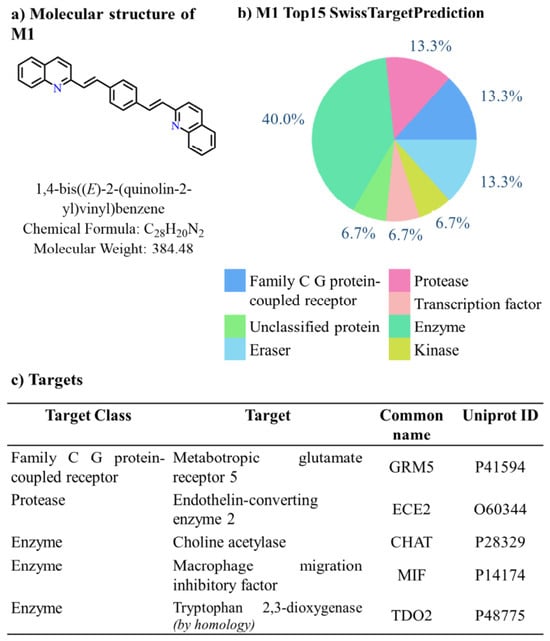
The SwissTargetPrediction server provided the target class probabilities from the array of possible molecular targets that M1 (Figure 1a) might interact with according to its molecular structure.
Figure 1.
(a) M1 molecular structure and IUPAC name; (b) top 15 molecular targets with potential biological activity and interaction with M1 predicted through SwissTargetPrediction; (c) list of targets belonging to the target classes most likely to interact.
M1 demonstrated a more likelihood of 13.3% for the Family C G protein-coupled receptor target category (see Figure 1b) with associated targets such as metabotropic glutamate receptor 5 (GRM5, Figure 1c) and for the protease class like endothelin-converting enzyme 2 (ECE2). Similarly, M1 indicated a 40.0% prediction for the enzyme class (refer to Figure 1b) featuring targets like choline acetylase (CHAT), macrophage migration inhibitory factor (MIF) and tryptophan 2,3-dioxygenase (TDO2).
Given the information above, the outcomes of the molecular docking analysis between M1 and metabotropic glutamate receptor 5 (GRM5), endothelin-converting enzyme 2 (ECE2) and choline acetylase (CHAT) are presented below.
3.1. Metabotropic Glutamate Receptor 5 (GRM5)
The metabotropic glutamate receptor 5 (mGluR5 or GRM5) is a G protein-coupled receptor (GPCR) belonging to group 1, which reacts to glutamate in a slow and modulatory manner. It activates pathways such as the release of intracellular calcium through Gq/11 and phospholipase C. This receptor is primarily found in postsynaptic neurons of the hippocampus, cortex, and basal ganglia, as well as in astrocytes and microglia, where it plays a role in regulating synaptic plasticity, neuroinflammation, and excitatory responses [15]. It has an extracellular domain shaped like a “Venus flytrap” for the orthosteric site of glutamate, linked to seven transmembrane helices that have allosteric sites for modulators. Its activation leads to the hydrolysis of phosphatidylinositol, the activation of PKC, and pathways such as ERK or mTOR, impacting LTP/LTD and learning processes. In glial cells, it modulates inflammatory responses, showing overexpression in neurodegeneration models. The mGlu5 has been suggested as a possible target for therapy in various neurodegenerative disorders. In early-stage models of these diseases, new allosteric modulators have demonstrated the ability to enhance cognitive function and lessen disease-associated damage [16]. For the treatment of neurodegenerative diseases such as Alzheimer’s or Parkinson’s, it is usually beneficial to reduce the activity of mGluR5 using negative allosteric modulators (NAMs), as this decreases issues like toxic aggregates, inflammation, and cognitive deficits in preclinical models. Targeting (positive allosteric modulators, PAMs) mGluR5 may have negative effects due to the potential for excitotoxicity and seizures, although certain PAMs exhibit advantages in conditions like schizophrenia or early stages [17].
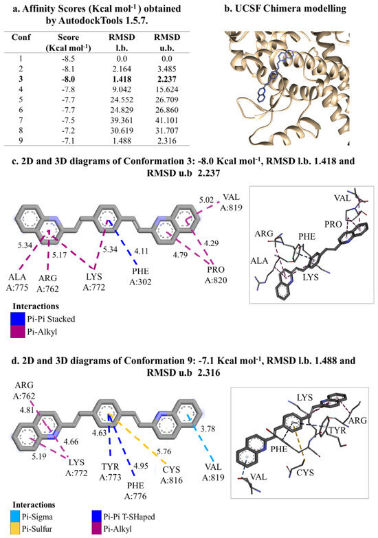
Based on the molecular docking assessment (Figure 2a), the affinity varies from −8.5 to −7.1 Kcal mol−1. An affinity measure of −8.5 Kcal mol−1 suggests a moderate to strong interaction, typically associated with micromolar (μM) affinities, while −7.1 Kcal mol−1 reflects a moderate level of binding, falling within the lower ranges of micromolar. These negative values represent advantageous binding free energies, with a lower number indicating improved stability for the ligand–protein complex (Figure 2b). Recent research has shown that approximately 50% of the natural metabolites assessed against viral proteases had measurements below −8 Kcal mol−1, hinting at their inhibitory capabilities. A 2023 study reported scores between −7.47 and −8.49 Kcal mol−1, which correspond to Ki values ranging from 3 μM to 0.94 nM in nanomolar; this relates to an estimated conversion between the binding free energy predicted by docking (more negative equals higher affinity) and the inhibition constant (Ki), which reflects how effective an inhibitor is. A lower Ki signifies a stronger inhibitor. Within the mentioned range, a score of −7.47 kcal mol−1 corresponds to approximately Ki 3 μM (moderate micromolar affinity), whereas −8.49 kcal mol−1 correlates with roughly Ki 0.94 nM (high nanomolar affinity) [18,19,20].
Figure 2.
(a) Molecular docking performed between M1 and the metabotropic glutamate receptor 5 (GRM5) utilizing AutodockTools version 1.5.7; (b) molecular docking with UCSF Chimera for modeling; (c) 2D and 3D diagram conformation 3: −8.0 Kcal mol−1 with intermolecular interactions and distances in Å was identified as the most stable structure; (d) 2D and 3D diagram conformation 8: −7.1 Kcal mol−1 with intermolecular interactions and distances in Å was determined to be the second most stable structure.
In this research, conformation 3 (Conf 3 in Figure 2c) is identified as the most stable model since it shows the smallest root mean square deviation (RMSD). A conformation might exhibit a low RMSD (indicating good geometry) but receive a poor score (indicating low predicted affinity), or the other way around; both factors are considered when selecting hits in screenings. In recent validations, RMSD validates poses, while scores determine potency [21,22]. Conf 3 has an affinity of −8.0 Kcal mol−1, with a lower-bound RMSD of 1.418 and an upper-bound RMSD of 2.237, featuring hydrophobic intermolecular interactions. The pi-pi stacking interaction, or parallel pi-pi stacking with phenylalanine (Phe) through the aromatic rings with 4.11 Å distance, refers to a non-covalent force that occurs between aromatic rings that are both parallel and offset (spacing of about 3.5–4 Å). This interaction is primarily stabilized by London dispersion and Pauli repulsion, alongside smaller contributions from electrostatic forces [23]. Generally, the energy range is about 1–2 kcal mol−1 per interaction in proteins, reaching up to 5–7 kcal mol−1 in supramolecular assemblies; it is primarily influenced by dispersion forces (approximately −3 to −5 kcal mol−1) opposing Pauli repulsion [23,24]. The intermolecular interaction between pi-alkyl and Alanine (Ala), Arginine (Arg), Lysine (Lys), Proline (Pro), and Valine (Val) is a non-covalent force occurring between an aromatic pi system (such as the rings of Phe, Tyr, or Trp in proteins) and a hydrophobic alkyl group (like the aliphatic chains of Leu, Ile, Val, or methyl groups). This interaction is stabilized by hydrophobic effects, London dispersion, and mutual polarization. It happens when the hydrogen atoms from a -CH3 group, -CH2 group, or an aliphatic chain engage with the pi electron cloud of an aromatic ring, usually at distances ranging from 3.5 to 5 Å. This is frequently observed at protein–ligand interfaces, where it aids in binding specificity without establishing strong directional bonds; the interaction strength is typically weak to moderate, around 0.5 to 2 kcal mol−1 for each contact [25].
The conformation Conf 8 shows an affinity of −7.1 Kcal mol−1, with a maximum RMSD of 1.488 and a peak RMSD of 2.316. It has the same hydrophobic intermolecular interactions as Conf 3, in addition to Pi-Sigma and Pi-Sulfur interactions (Figure 2d). The intermolecular pi-sigma interaction is a non-covalent force that occurs between the pi electron cloud of an aromatic ring (for example, Phe or Tyr) and a sigma C-H or C-C bond from an aliphatic or hydrophobic group. This interaction is defined by a perpendicular or semi-perpendicular arrangement, which is also known as the T-shaped variation. It involves engaging interactions between the sigma dipole (electron donor) and the pi quadrupole (electron acceptor), including dispersion and hydrophobic effects; the usual distance is between 3.5 and 4.5 Å, frequently observed in docking to stabilize ligands within aromatic pockets. The interaction strength shows a range of about 0.5 to 2.5 kcal mol−1 for each contact in docking analyses (like PLP and AutoDock). This is comparable to pi-alkyl interactions but is more directional due to the specific sigma bond [26,27,28].
The intermolecular interaction of pi-sulfur (or S⋯pi) refers to a type of non-covalent force that occurs between a sulfur atom (for example, found in Cys, Met, or ligands containing S) and the pi electron cloud of aromatic rings (such as Phe, Tyr, Trp, His), characterized by a typical perpendicular or parallel arrangement (with a distance of 3.5–4.5 Å); it is stabilized through dispersion, polarization, and electrostatic effects. The interaction strength is approximately 0.5 to 2 kcal mol−1 in docking and proteins (SAPT); in furan-disulfide heterodimers, it is around −2 to −3 kcal mol−1 due to S-S⋯pi interactions along with additive hydrogen bonds [29,30].
3.2. Endothelin-Converting Enzyme 2 (ECE2)
The endothelin-converting enzyme 2 (ECE2) is a zinc-dependent transmembrane metalloprotease (ECE/neprilysin family) consisting of isoforms ECE-2a and ECE-2b, which is active in an acidic pH range (5.0–5.5) and is present in neuroendocrine tissues (hypothalamus, midbrain, pituitary). It processes non-classic peptide precursors in acidic endolysosomes in a selective manner, producing endogenous opioid agonists such as BAM-12 (derived from prodynorphin) for κ-opioid receptors (KOR), managing their recycling, resensitization, and differential intracellular signaling based on brain regions. It also breaks down KOR inhibitors like PEN-LEN; its knockout influences cardiac development and modulates ET-1 within cells. Selective inhibition is beneficial in therapeutic settings because it reduces the recycling of KOR by endogenous peptides, which has potential implications for chronic pain and addiction (an ECE inhibitor decreases signaling); however, there is a risk associated with cancer (with ECE2 being highly pro-tumor) or opioid homeostasis. It is important not to inhibit broadly due to off-target effects. On the other hand, tracking its role as a predictive biomarker in lung adenocarcinoma (LUAD, elevated levels associated with lower survival rates, low immune B/dendritic cell infiltration, and m6A RNA modification) and being a differential modulator KOR in addiction/pain would be more advisable [31,32].
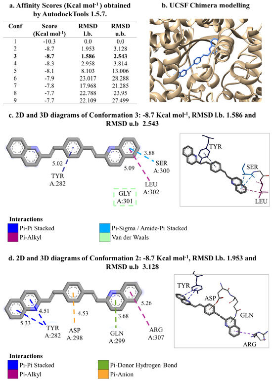
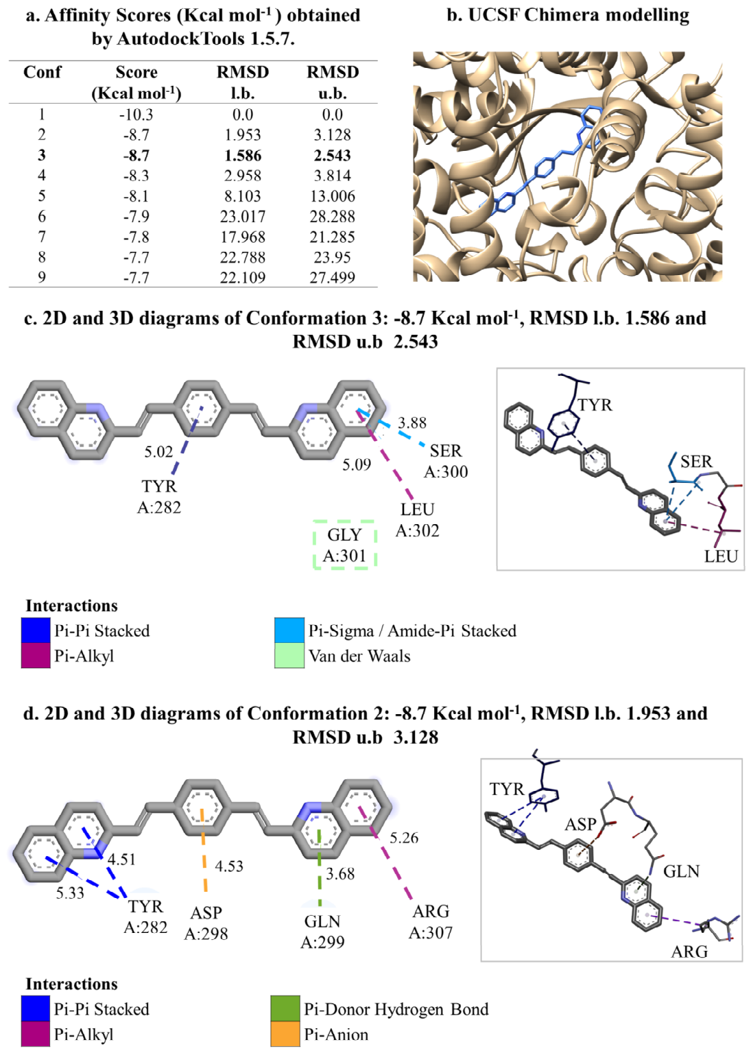
The molecular docking between M1 and ECE2 displayed a stronger binding affinity that ranges from −10.3 to −7.7 Kcal mol−1 (Figure 3a), indicating significant Gibbs free energy (Figure 3b). Based on AutodockTools version 1.5.7, the most likely conformation is Conf 3, which has −8.7 Kcal mol−1, a lower bound RMSD of 1.586, and an upper bound RMSD of 2.543 (Figure 3c). This structure shows hydrophobic interactions along with a weak hydrogen bond (Van der Waals with GLY A:301). In this instance, there is a Pi-Pi stacked interaction with tyrosine (TYR A:282), Pi-Alkyl with Leucine (LEU A:302), Pi-Sigma with Serine (SER A:300), or Amide-Pi Stacked, which pertains to non-covalent stacking between the pi system of the amide group in the protein backbone (C=O⋯N-H) and the hydroxyl group (-OH) of serine (Ser), even though technically amides are not pi-aromatic systems. It includes dipole–dipole interactions and hydrogen bonding with par-tial stacking of the CH2-Ser over the amide plane. It has approximately 0.5 kcal mol−1 of H-bonding between the OH and O=C in amides and is weaker compared to the pi-aromatic-amide due to the lower polarizability of Ser in contrast to aromatic rings [33,34].
Figure 3.
(a) Molecular docking performed between M1 and the endothelin-converting enzyme 2 (ECE2) utilizing AutodockTools version 1.5.7; (b) molecular docking using UCSF Chimera for modeling; (c) 2D and 3D diagrams of conformation 3: −8.7 Kcal mol−1 with intermolecular interactions and distances in Å, which was identified as the most stable structure; (d) 2D and 3D diagrams of conformation 8: −8.7 Kcal mol−1 with intermolecular interactions and distances in Å, which was determined to be the second most stable structure.
The second most likely conformation is Conf 2 with −8.7 Kcal mol−1, a lower-bound RMSD of 1.953, and an upper-bound RMSD of 3.128 (Figure 3d). This conformation also exhibits hydrophobic interactions such as Pi-Pi Stacked with TYR A:282 and Pi-Alkyl with arginine (ARG A:307). Additionally, it shows the pi–donor hydrogen bond interaction with glutamine (GLN A:299), which is an unusual hydrogen bond where the side amide group of glutamine (GLN, -CH2-CH2-C(O)-NH2) serves as a donor (N-H⋯pi), directing its hydrogen towards the pi electron cloud of an aromatic ring (Tyr, Phe, Trp, or ligand), stabilized by a quadrupole pi-acceptor and mutual polarization. It displays an intensity of around 2 to 2.5 kcal mol−1 in docking (AutoDock/PLANTS); this is stronger than pi-alkyl (approximately 1 kcal mol−1 more due to H-bond directionality) yet weaker than standard O-H⋯O hydrogen bonds [35]. Finally, the pi-anion interaction with aspartic acid (ASP A:298) is a non-covalent electrostatic interaction between the carboxylate anion (Asp-COO−) and the electropositive area of an aromatic ring edge (Phe, Tyr, Trp or pi ligand), which is reinforced by the favorable anion–pi quadrupole interaction. Its average interaction strength is approximately −4 kcal mol−1 in proteins [36].
3.3. Choline Acetylase (CHAT)
Choline acetyltransferase, abbreviated to ChAT or choline acetylase, is the transferase enzyme responsible for producing acetylcholine from choline and acetyl-CoA, as illustrated by the following reaction: ACh + CoA ↔ holine + acetyl-CoA. It serves as a distinct marker for cholinergic neurons found in both the central and peripheral nervous systems. It synthesizes ACh in cholinergic nerve terminals (not rate-limiting; restricted by choline uptake via CHT1), which is crucial for neuromuscular transmission, memory, focus, and the cholinergic anti-inflammatory pathway (vagal pathway). The primate-specific 82-kDa nuclear ChAT reduces neuroinflammation and Aβ in Alzheimer’s disease; circulating extracellular ChAT dampens cytokines (TNF) through an anti-inflammatory mechanism. Monitoring its presence can serve as a crucial biomarker for cholinergic integrity in Alzheimer’s disease (low early ChAT levels), dementias, and schizophrenia; PET tracers are being developed for early diagnosis. High levels of the protective 82-kDa protein are found in Alzheimer’s models, and the circulating form reacts to inflammation. Routine inhibition is not advised under new ACh treatments as it worsens cognitive deficits in Alzheimer’s; inhibitors studied as PET probes are not meant for therapy (as focus remains on AChE inhibitors). There is indirect therapeutic potential through the upregulation of ChAT [37,38,39,40].
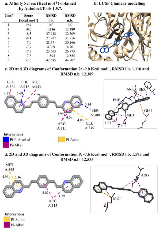
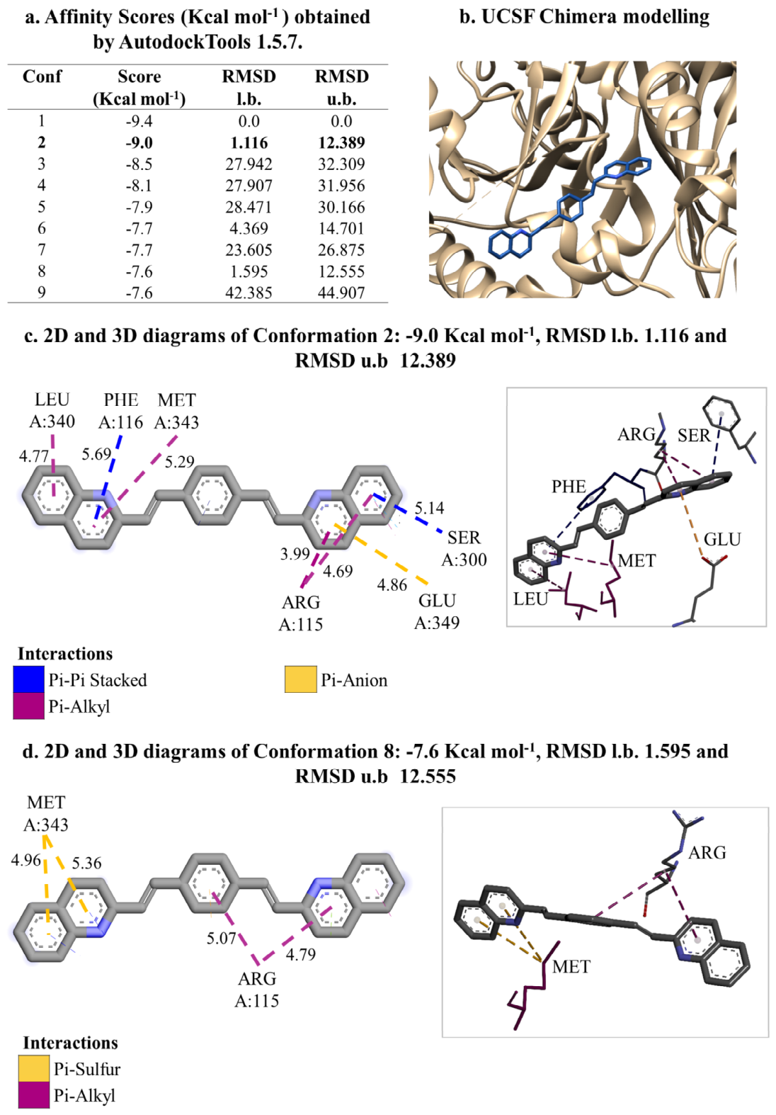
The interaction between M1 and CHAT showed an even greater binding strength compared to ECE2, with values between −9.4 and −7.6 Kcal mol−1 (Figure 4a), reflecting notable Gibbs free energy (Figure 4b). It benefits molecular interactions by enhancing the binding affinity and stability of complexes obtained by molecular docking, and Gibbs free energy leads to more spontaneous and stronger interactions between ligands and receptors [41]. According to AutodockTools version 1.5.7, the most probable conformation is Conf 2, which registers at −9.0 Kcal mol−1, with a lower RMSD of 1.116 and an upper RMSD of 12.389 (Figure 4c). This conformation exhibits similar intermolecular interactions, including pi-pi stacked interactions with phenylalanine (PHE A:116 and A:212) at distances of 5.69 and 5.14 Å; as well as pi-alkyl interactions with leucine (LEU A:340) at 4.77 Å and methionine (MET A:343) at 5.29 Å and arginine (ARG A:115, 3.99 and 4.69 Å); and pi-anion to glutamic acid (GLU A:349, 4.86 Å).
Figure 4.
(a) Molecular docking performed between M1 and the choline acetyltransferase (CHAT) utilizing AutodockTools version 1.5.7; (b) molecular docking using UCSF chimera for modeling; (c) 2D and 3D diagrams of conformation 3: −9.0 Kcal mol−1 with intermolecular interactions and distances in Å, which was identified as the most stable structure; (d) 2D and 3D diagrams of conformation 8: −7.6 Kcal mol−1 with intermolecular interactions and distances in Å, which was determined to be the second most stable structure.
The second most likely conformation is Conf 8, which has an energy of −7.6 Kcal mol−1, showing a lower RMSD of 1.595 and a higher RMSD of 12.555 (Figure 4c). This conformation displays comparable intermolecular interactions, featuring pi-sulfur interactions with methionine (MET A:343) at distances of 4.96 and 5.36 Å, as well as pi-alkyl interactions with arginine (ARG A:115) at 5.07 and 4.79 Å.
The ANOVA analysis showed that the average affinities of the conformations for the three targets are statistically similar (p > 0.05), meaning that compound M1 has the same Gibbs free energy with all three targets, suggesting a high likelihood of inhibitory activity.
4. Conclusions
SwissTargetPrediction identified GRM5, ECE2, and CHAT as the principal Homo sapiens targets of M1, with a preferential distribution toward enzymes/proteases (40%) and Family C GPCRs (13.3%). Docking analyses revealed consistently favorable binding energies (−10.3 to −7.1 kcal·mol−1), which were statistically comparable across targets (ANOVA, p > 0.05) and stabilized predominantly by pi-driven hydrophobic interactions. Low RMSD values confirmed stable binding conformations. These computational findings align with the intrinsic optoluminescent properties of M1, including its extended pi-conjugation, aggregation-induced luminescence, L-cysteine sensing capability, and electroluminescent output (28 cd m−2 at 7.4 V). Together, the results define M1 as a multifunctional scaffold with dual optoluminescent and pharmacological potential. While experimental validation remains necessary, M1 emerges as a promising multi-target neuromodulatory candidate that bridges materials science and computational drug design. It is worth mentioning that predictive in silico results need experimental validation, including IC50 enzyme tests, kinetic studies, MD simulations over 100 ns, and off-target selectivity assessed through kinase/protease panels. It should be noted that the docking simulations were carried out using the neutral form of M1, which matches the π-conjugated structure experimentally characterized in films and its solid state. Under physiological conditions, however, quinoline moieties can undergo partial protonation, giving rise to charged species with potentially different binding modes and affinities. A comprehensive analysis of protonation equilibria (including pKa determination and multi-state docking or MD simulations) is beyond the scope of this initial target-fishing study but will be addressed in future work, and it should be considered when extrapolating these in silico predictions to biological environments. In summary, M1 stands out as a promising option for multi-target inhibition in neurology, broadening its applications from optoluminescent sensor to computational pharmacology.
Author Contributions
Conceptualization, Z.G.-S., R.A.V.-G. and C.Á.-J.; methodology, Z.G.-S., R.A.V.-G. and C.Á.-J.; software, Z.G.-S., R.A.V.-G. and C.Á.-J.; validation, Z.G.-S., R.A.V.-G. and C.Á.-J.; formal analysis, Z.G.-S., R.A.V.-G. and C.Á.-J.; investigation, Z.G.-S., R.A.V.-G. and C.Á.-J.; resources, Z.G.-S., R.A.V.-G. and C.Á.-J.; data curation, Z.G.-S., R.A.V.-G., C.Á.-J., O.A.A.-S. and E.C.-V.; writing—original draft preparation, Z.G.-S., R.A.V.-G., C.Á.-J., O.A.A.-S., E.C.-V. and M.A.V.-R.; writing—review and editing, Z.G.-S., R.A.V.-G., C.Á.-J., O.A.A.-S., E.C.-V. and M.A.V.-R.; visualization, Z.G.-S., R.A.V.-G., C.Á.-J., O.A.A.-S., E.C.-V. and M.A.V.-R.; supervision, Z.G.-S., R.A.V.-G., C.Á.-J., O.A.A.-S., E.C.-V. and M.A.V.-R.; project administration, Z.G.-S., R.A.V.-G., C.Á.-J., O.A.A.-S., E.C.-V. and M.A.V.-R.; funding acquisition, not applicable. All authors have read and agreed to the published version of the manuscript.
Funding
This research received no external funding.
Institutional Review Board Statement
Not applicable.
Informed Consent Statement
Not applicable.
Data Availability Statement
The original contributions presented in this study are included in the article. Further inquiries can be directed to the corresponding author.
Acknowledgments
The authors express their sincere gratitude to the Secretaría de Ciencias, Humanidades, Tecnología e Innovación (SECIHTI) for its financial support (CVU 1034082) of the educational Ph.D. program and the research conducted in materials science for this project and the CITNOVA project DDCTI-FH-DCI-138.
Conflicts of Interest
The authors declare no conflicts of interest.
Abbreviations
The following abbreviations are used in this manuscript:
| M1 | (1,4-bis((E)-2-(quinolin-2-yl)vinyl)benzene) |
| GRM5 | Metabotropic glutamate receptor 5 |
| ECE2 | Endothelin-converting enzyme 2 |
| CHAT | Acetylcholinesterase |
| RMSD | Root mean square deviation |
| ANOVA | Analysis of variance |
References
- Lapkowski, M.; Zak, J.; Karon, K.; Marciniec, B.; Prukala, W. The mixed carbon–nitrogen conjugation in the carbazole based polymer; the electrochemical, UVVis, EPR, and IR studies on 1,4 bis[(E)2-(9H-carbazol-9-yl)vinyl] benzene. Electrochim. Acta 2011, 56, 4105–4111. [Google Scholar] [CrossRef] [Scilit]
- Ma, Y.; Li, Y.; Chen, L.; Xiong, Y.; Yin, G. Cyano-functionalized 1,4-bis((E)-2-(1H-indol-3-yl)vinyl) benzenes with aggregation induced emission characteristic and diverse thermochromic behaviour. Dye. Pigment. 2016, 126, 194–201. [Google Scholar] [CrossRef] [Scilit]
- Gutiérrez, A.R.; Vázquez, R.A.; Coreño, O.; Coreño, J.; Castruita, G.; Moggio, I.; Arías, E.; Maldonado, J.L.; Ramos, G.; Martínez, M. Synthesis and mechanosynthesis of phenylenedivinylenebisquinolines for optoelectronics. Superf. Vacío 2010, 23, 73–79. [Google Scholar]
- Gutiérrez, A.; Vázquez, R.; Moggio, I.; Arias, E.; Coreño, O.; Maldonado, J.; Ramos-Ortíz, G.; Rodríguez, O.; Jiménez-Barrera, R. Mechanosynthesis of a phenylenedivinylidenebisquinoline. Optical 2015, morphological and electroluminescence properties. J. Mol. Struct. 2015, 1086, 138–145. [Google Scholar] [CrossRef] [Scilit]
- Vazquez-Velazquez, A.R.; Vazquez-Garcia, R.Á.; Hernandez-Bucio, G.; Gonzalez-Gonzalez, V.A.; Moggio, I.; Vazquez-Rodriguez, S. Phenylvinilbisquinolines as fluorescent markers in functionalized polypropylene films. Polym. Bull. 2020, 77, 1781–1792. [Google Scholar] [CrossRef] [Scilit]
- Caballero Cerbon, D.A.; Gebhard, L.; Dokuyucu, R.; Ertl, T.; Härtl, S.; Mazhar, A.; Weuster-Botz, D. Challenges and Advances in the Bioproduction of L-Cysteine. Molecules 2024, 29, 486. [Google Scholar] [CrossRef] [Scilit] [PubMed]
- EFSA Panel on Additives and Products or Substances used in Animal Feed (FEEDAP); Bampidis, V.; Azimonti, G.; Bastos, M.d.L.; Christensen, H.; Durjava, M.; Dusemund, B.; Kouba, M.; López-Alonso, M.; Puente, S.L.; et al. Assessment of the feed additive consisting of L-cystine for all animal species for the renewal of its authorisation (Bretagne Chimie Fine [BCF Life Sciences]). EFSA J. 2024, 22, e8800. [Google Scholar] [CrossRef] [Scilit] [PubMed]
- Islas-Rodríguez, N.; Muñoz, R.; Vázquez-García, R.A.; Rodríguez, J.A.; Reyes-Pérez, M.; Hernández-Ortiz, O.J. Selective colorimetric detection of cysteine based on phenylvinylbisquinoline for its potential implementation in optoelectronic sensors. J. Mater. Sci. Mater. Electron. 2023, 34, 1116. [Google Scholar] [CrossRef] [Scilit]
- Hernández-Ortiz, O.J.; Castro-Monter, D.; Lugo, V.R.; Moggio, I.; Arias, E.; Reyes-Valderrama, M.I.; Veloz-Rodríguez, M.A.; Vázquez-García, R.A. Synthesis and Study of the Optical Properties of a Conjugated Polymer with Configurational Isomerism for Optoelectronics. Materials 2023, 16, 2908. [Google Scholar] [CrossRef] [Scilit]
- Verma, S.; Lal, S.; Narang, R.; Sudhakar, K. Quinoline hydrazide/hydrazone derivatives: Recent insights on antibacterial activity and mechanism of action. ChemMedChem 2023, 18, e202200571. [Google Scholar] [CrossRef] [Scilit]
- Koçyiğit, Ü.M.; Ökten, S.; Cakmak, O.; Burhan, G.; Ataş, M.; Taslimi, P.; Gülçin, İ. Arylated quinoline and tetrahydroquinolines: Synthesis, characterization and their metabolic enzyme inhibitory and antimicrobial activities. ChemistrySelect 2022, 7, e202203469. [Google Scholar] [CrossRef] [Scilit]
- Chen, Y.; Shoichet, B.K. Molecular docking and ligand specificity in fragment-based inhibitor discovery. Nat. Chem. Biol. 2009, 5, 358–364. [Google Scholar] [CrossRef] [Scilit]
- Mohasin, M.; Khan, S.A. A Review on Quinoline-Based Derivatives as Selective Colorimetric and Fluorescent Chemosensors for Cu2+ and Fe3+ Ions Detection. J. Fluoresc. 2026, 36, 2391–2412. [Google Scholar] [CrossRef] [Scilit] [PubMed]
- Hernandez-Hernandez, K.M.; Mendoza-Huizar, L.H. A computational study on the binding and interactions of flavonoids with gliadin using molecular docking. Pädi Basic Sci. Eng. Sci. Bull. ICBI 2024, 12, 13–18. [Google Scholar] [CrossRef] [Scilit]
- Wang, J.; He, Y.; Chen, X.; Huang, L.; Li, J.; You, Z.; Huang, Q.; Ren, S.; He, K.; Schibli, R.; et al. Metabotropic glutamate receptor 5 (mGluR5) is associated with neurodegeneration and amyloid deposition in Alzheimer’s disease: A [18F] PSS232 PET/MRI study. Alzheimer’s Res. Ther. 2024, 16, 9. [Google Scholar] [CrossRef] [Scilit]
- Budgett, R.F.; Bakker, G.; Sergeev, E.; Bennett, K.A.; Bradley, S.J. Targeting the type 5 metabotropic glutamate receptor: A potential therapeutic strategy for neurodegenerative diseases? Front. Pharmacol. 2022, 13, 893422. [Google Scholar] [CrossRef] [Scilit]
- Abd-Elrahman, K.S.; Hamilton, A.; Albaker, A.; Ferguson, S.S. mGluR5 contribution to neuropathology in Alzheimer mice is disease stage-dependent. ACS Pharmacol. Transl. Sci. 2020, 3, 334–344. [Google Scholar] [CrossRef] [Scilit] [PubMed]
- Konakanchi, S.; Vadluri, R.; Anumula, K.S.; Narashimulu Banothu, D.; Krishna, T.M. Antiproliferative, molecular docking, and bioavailability studies of diarylheptanoids isolated from stem bark of Garuga pinnata Rox B. 3 Biotech 2023, 13, 208. [Google Scholar] [CrossRef] [Scilit] [PubMed]
- Alam, W.; Khan, H.; Jan, M.S.; Darwish, H.W.; Daglia, M.; Elhenawy, A.A. In vitro 5-LOX inhibitory and antioxidant potential of isoxazole derivatives. PLoS ONE 2024, 19, e0297398. [Google Scholar] [CrossRef] [Scilit]
- Jablonský, M.; Štekláč, M.; Majova, V.; Gall, M.; Matúška, J.; Pitoňák, M.; Bučinský, L. Molecular docking and machine learning affinity prediction of compounds identified upon softwood bark extraction to the main protease of the SARS-CoV-2 virus. Biophys. Chem. 2022, 288, 106854. [Google Scholar] [CrossRef] [Scilit]
- Ma, Z.; Ajibade, A.; Zou, X. Docking strategies for predicting protein-ligand interactions and their application to structure-based drug design. Commun. Inf. Syst. 2024, 24, 199. [Google Scholar] [CrossRef] [Scilit]
- Tan, L.H.; Kwoh, C.K.; Mu, Y. RmsdXNA: RMSD prediction of nucleic acid-ligand docking poses using machine-learning method. Brief. Bioinform. 2024, 25, bbae166. [Google Scholar] [CrossRef] [Scilit] [PubMed]
- Carter-Fenk, K.; Liu, M.; Pujal, L.; Loipersberger, M.; Tsanai, M.; Vernon, R.M.; Forman-Kay, J.D.; Head-Gordon, M.; Heidar-Zadeh, F.; Head-Gordon, T. The energetic origins of pi–pi contacts in proteins. J. Am. Chem. Soc. 2023, 145, 24836–24851. [Google Scholar] [CrossRef] [Scilit]
- Hermann, J.; Alfe, D.; Tkatchenko, A. Nanoscale π–π stacked molecules are bound by collective charge fluctuations. Nat. Commun. 2017, 8, 14052. [Google Scholar] [CrossRef] [Scilit]
- McGaughey, G.B.; Gagné, M.; Rappé, A.K. π-stacking interactions: Alive and well in proteins. J. Biol. Chem. 1998, 273, 15458–15463. [Google Scholar] [CrossRef] [Scilit] [PubMed]
- Terefe, E.M.; Ghosh, A. Molecular docking, validation, dynamics simulations, and pharmacokinetic prediction of phytochemicals isolated from Croton dichogamus against the HIV-1 reverse transcriptase. Bioinform. Biol. Insights 2022, 16, 11779322221125605. [Google Scholar] [CrossRef] [Scilit] [PubMed]
- Cabral, M.B.; Dela Cruz, C.J.; Sato, Y.; Oyong, G.; Rempillo, O.; Galvez, M.C.; Vallar, E. In silico approach in the evaluation of pro-inflammatory potential of polycyclic aromatic hydrocarbons and volatile organic compounds through binding affinity to the human toll-like receptor 4. Int. J. Environ. Res. Public Health 2022, 19, 8360. [Google Scholar] [CrossRef] [Scilit]
- Xiao, Y.; Woods, R.J. Protein–ligand CH−π interactions: Structural informatics, energy function development, and docking implementation. J. Chem. Theory Comput. 2023, 19, 5503–5515. [Google Scholar] [CrossRef] [Scilit]
- Tauer, T.P.; Derrick, M.E.; Sherrill, C.D. Estimates of the ab initio limit for sulfur−π interactions: The H2S−benzene dimer. J. Phys. Chem. A 2005, 109, 191–196. [Google Scholar] [CrossRef] [Scilit]
- Shen, F.; Wang, L.; Chen, R.; Li, W.; Li, Y.; Feng, Y.; Lu, T.; Caminati, W.; Lesarri, A.; Feng, G. Unraveling the S–S⋯π interactions in furan–disulfide heterodimers: Insights from microwave spectroscopy and ab initio computations. Phys. Chem. Chem. Phys. 2025, 27, 20802–20809. [Google Scholar] [CrossRef] [Scilit]
- Zhang, Y.H.; Zeng, J.; Liu, X.S.; Gao, Y.; Kui, X.Y.; Liu, X.Y.; Zhang, Y.; Pei, Z.J. ECE2 is a prognostic biomarker associated with m6A modification and involved in immune infiltration of lung adenocarcinoma. Front. Endocrinol. 2022, 13, 1013238. [Google Scholar] [CrossRef] [Scilit]
- Gupta, A.; Gomes, I.; Sierra, S.; Osman, A.; Devi, L.A. Endothelin-converting enzyme 2 differentially regulates kappa opioid receptor trafficking and function. BioRxiv 2025, 202503. [Google Scholar] [CrossRef] [Scilit]
- DeFrees, K.; Kemp, M.T.; ElHilali-Pollard, X.; Zhang, X.; Mohamed, A.; Chen, Y.; Renslo, A.R. An empirical study of amide–heteroarene π-stacking interactions using reversible inhibitors of a bacterial serine hydrolase. Org. Chem. Front. 2019, 6, 1749–1756. [Google Scholar] [CrossRef] [Scilit]
- Krone, M.W.; Travis, C.R.; Lee, G.Y.; Eckvahl, H.J.; Houk, K.N.; Waters, M.L. More Than π–π–π Stacking: Contribution of Amide−π and CH−π Interactions to Crotonyllysine Binding by the AF9 YEATS Domain. J. Am. Chem. Soc. 2020, 142, 17048–17056. [Google Scholar] [CrossRef] [Scilit]
- Wakchaure, P.D.; Ganguly, B. Unraveling the role of π-Stacking interactions in Ligand Binding to the Thiamine Pyrophosphate Riboswitch with High-level Quantum Chemical calculations and Docking Study. J. Phys. Chem. B 2022, 126, 1076–1084. [Google Scholar] [CrossRef] [Scilit] [PubMed]
- Philip, V.; Harris, J.; Adams, R.; Nguyen, D.; Spiers, J.; Baudry, J.; Howell, E.E.; Hinde, R.J. A survey of aspartate−phenylalanine and glutamate-phenylalanine interactions in the protein data bank: Searching for anion-π pairs. Biochemistry 2011, 50, 2939–2950. [Google Scholar] [CrossRef] [Scilit] [PubMed]
- Gabalski, A.H.; Tynan, A.; Tsaava, T.; Li, J.H.; Lee, D.; Hepler, T.D.; Hide, D.; George, S.; Iñiguez, C.E.B.; Thompson, D.A.; et al. Circulating extracellular choline acetyltransferase regulates inflammation. J. Intern. Med. 2024, 295, 346–356. [Google Scholar] [CrossRef] [Scilit] [PubMed]
- Kumar, R.; Kumar, A.; Långström, B.; Darreh-Shori, T. Discovery of novel choline acetyltransferase inhibitors using structure-based virtual screening. Sci. Rep. 2017, 7, 16287. [Google Scholar] [CrossRef] [Scilit]
- Sheng, L.; Bhalla, R. Biomarkers and target-specific small-molecule drugs in Alzheimer’s diagnostic and therapeutic research: From amyloidosis to tauopathy. Neurochem. Res. 2024, 49, 2273–2302. [Google Scholar] [CrossRef] [Scilit]
- AlQot, H.E.; Rylett, R.J. Primate-specific 82-kDa choline acetyltransferase attenuates progression of Alzheimer’s disease-like pathology in the APPNL-GF knock-in mouse model. Sci. Rep. 2024, 14, 27614. [Google Scholar] [CrossRef] [Scilit] [PubMed]
- Owoloye, A.J.; Ligali, F.C.; Enejoh, O.A.; Musa, A.Z.; Aina, O.; Idowu, E.T.; Oyebola, K.M. Molecular docking, simulation and binding free energy analysis of small molecules as Pf HT1 inhibitors. PLoS ONE 2022, 17, e0268269. [Google Scholar] [CrossRef] [Scilit]
Disclaimer/Publisher’s Note: The statements, opinions and data contained in all publications are solely those of the individual author(s) and contributor(s) and not of MDPI and/or the editor(s). MDPI and/or the editor(s) disclaim responsibility for any injury to people or property resulting from any ideas, methods, instructions or products referred to in the content. |
© 2026 by the authors. Licensee MDPI, Basel, Switzerland. This article is an open access article distributed under the terms and conditions of the Creative Commons Attribution (CC BY) license.