


Abstract
Background/Objectives: Globally, the management of infections has been complicated greatly by the rapid emergence of antibiotic resistance among bacterial pathogens, particularly Escherichia coli (E. coli). This bacterium is commonly found in a wide range of vertebrate hosts, including livestock, which can serve as important sources of specific pathogenic or multidrug-resistant strains. Cross-contamination can occur from farm to food, posing public health concerns. Methods: This cross-sectional study examined the antibiotic resistance and virulence gene profiles of E. coli isolated from farm animals, food processing facilities (including meat and contact surfaces), and the surrounding environment (wastewater). Results: Out of 383 samples, 230 samples (60.1%) were positive for E. coli (95% CI: 55.1–64.9). The prevalence rates showed significant variation across different sources, with positive rates of 72.3% (180/249) in animal sources, 33.7% (28/83) in food sources, and 43.1% (22/51) in environmental sources. Over 80% of the isolates across all sources carried the sheA virulence gene, which is associated with hemolytic activity in E. coli. Multidrug resistance (MDR) was commonly observed, with rates of 61.1% in animal samples, 57.1% in food sources, and 50.0% in environmental samples. The E. coli isolates exhibited high levels of antibiotic resistance, particularly to streptomycin (64.9%), ampicillin (58.0%), and tetracycline (57.6%). The most common resistance gene pattern was tetA-strA-blaTEM (22.6%). Conclusions: These findings indicate widespread occurrence of antibiotic-resistant and virulence gene-carrying E. coli strains across the farm-to-food continuum, underscoring the urgent need for enhanced antimicrobial stewardship and surveillance programs to mitigate transmission from food-producing animals and reduce public health complications.
1. Introduction
Escherichia coli is a Gram-negative bacterium, belonging to the family Enterobacteriaceae, and it naturally inhabits the gastrointestinal tract of warm-blooded animals. Although most E. coli strains are beneficial, contributing to digestion and maintaining gut microbiota balance, some strains emerge as pathogenic, posing significant health threats to animals and humans [1,2]. Pathogenic E. coli strains are broadly classified into two groups: (i) diarrheagenic, including adherent-invasive E. coli (AIEC), enteroinvasive E. coli (EIEC), enterotoxigenic E. coli (ETEC), enteropathogenic E. coli (EPEC), enterohemorrhagic/Shiga toxin-producing E. coli (EHEC/STEC), diffusely adherent E. coli (DAEC), and enteroaggregative E. coli (EAEC) [3], which cause diarrhea in both animals and humans [4]; and (ii) extraintestinal, comprising uropathogenic E. coli (UPEC), septicemia-associated E. coli (SePEC), and meningitis-associated E. coli (MNEC) [5], which cause extraintestinal infections such as sepsis [6].
Pathogenic E. coli infections are often treated with antibiotics, but the emergence of antibiotic-resistant strains, fueled by the frequent use of antibiotics, complicates effective treatment, leading to treatment failure, prolonged illness, or death. Globally, antibiotic-resistant E. coli poses significant public health threats due to its proficiency in acquiring and spreading resistance genes [7,8,9]. A major contributor to this resistance is the indiscriminate use of antibiotics in animal production aimed at boosting food production [10,11]. Studies indicate that antibiotics administered to animals are often incompletely metabolized, leading to environmental contamination through excretion, which further promotes antibiotic resistance [12,13]. This complicates the management of E. coli infections. Consequently, resistant E. coli strains can spread through cross-contamination from animals to food of animal origin (meat), the environment, and humans, threatening public health globally [14,15]. The World Health Organization [15] reported that in the United States, E. coli outbreaks primarily originated from contaminated animal-based foods (69%), water (18%), and animals (14%). Other reports have also linked human antibiotic-resistant E. coli to livestock, animal-based food, and the environment [16,17,18,19].
Farm animals, meat processing facilities (slaughterhouses and processing plants), and the environment are interconnected, forming essential components of the One Health framework. Consequently, bacteria from livestock can easily cross-contaminate other areas. This may escalate the resistance situation, as resistant E. coli in the environment can also be transmitted to other E. coli strains or other bacterial species in food, animals, and humans through horizontal gene transfer mechanisms, including conjugation, transformation, transduction, and vesiduction [20], posing risks to human health. Although E. coli may be considered part of the normal gut flora, its role as an indicator of hygiene or fecal contamination and the potential emerging antimicrobial resistance (AMR) and virulence strains as a zoonotic threat [3,5] make it significantly relevant in the modern One Health context. Despite the growing number of studies examining interconnected sources [21,22,23], most have focused on a single animal–food continuum. Comprehensive investigations that simultaneously capture antibiotic-resistant and pathogenic E. coli across multiple interconnected animal, food, and environmental sources within a unified framework remain limited. This knowledge gap underscores the necessity for a comprehensive, coordinated, and multi-sectoral investigation.
Therefore, this study aimed to identify and characterize antibiotic resistance and virulence determinants in E. coli from animals, meat processing facilities, and the surrounding environment. The data can guide or inform policies on antibiotic use in animal farms, contribute to the global reduction in diseases caused by antibiotic-resistant E. coli from animal sources, and improve public health.
2. Results
2.1. Prevalence of Escherichia coli from Various Sources
Prevalence of E. coli across the sources varied significantly (p < 0.001). Overall, a prevalence rate of 60.1% (230/383) (95% CI: 55.1–64.9) was recorded. The highest prevalence was observed in animal sources (72.3%), followed by environmental sources (43.1%), while food sources showed the lowest prevalence (33.7%) (Table 1). Among animal-derived samples, the overall prevalence was 72.3%, with rectal swabs yielding a prevalence of 68.9% (144/209; 95% CI: 62.2–74.7). Within this group, sheep (88.9%) and broilers (76.7%) exhibited relatively high prevalence rates (Table 1). Animal fecal samples showed the highest prevalence (90.0%; 36/40; 95% CI: 77.0–96.0), particularly in pigs (93.3%). In food sources, E. coli was detected in 33.7% of samples (28/83; 95% CI: 24.4–44.4), with higher prevalence in meat samples (41.9%) compared to food contact surfaces (9.5%) (Table 1). Among meat types, broiler meat showed the highest contamination (48.6%), followed by pork (40.0%) and beef (25.0%). Environmental samples, represented by wastewater, showed a prevalence of 43.1% (22/51; 95% CI: 30.5–56.7) (Table 1).
Table 1.
Prevalence of Escherichia coli among different sources.
2.2. Virulence Gene Profiles of Escherichia coli from Various Sources
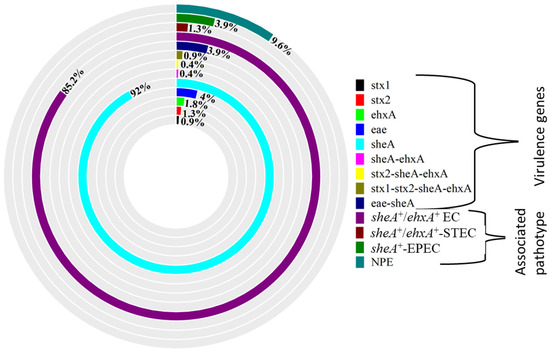
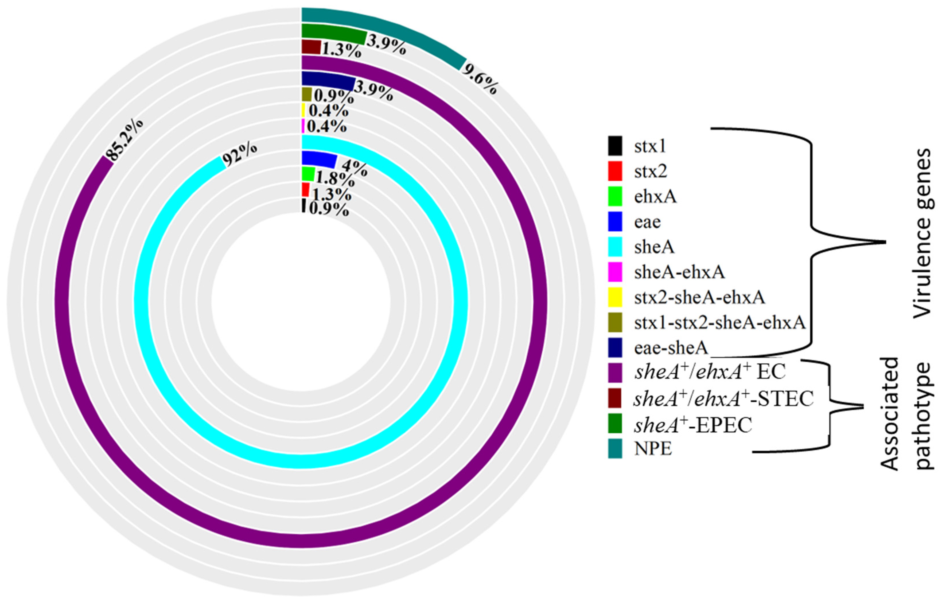
The sheA gene was the most frequently detected virulence marker among the E. coli isolates (92.0%), followed by the eae gene (4.0%), while the stx1 gene was the least detected (0.9%) (Figure 1). Thirteen isolates harbored multiple virulence genes, with the following combinations observed: eae-sheA, sheA-ehxA, stx1-stx2-sheA-ehxA, and stx2-sheA-ehxA (Figure 1). Based on virulence gene profiles (Figure 1), isolates were assigned to four pathotypes: (i) non-pathogenic E. coli (NPE); (ii) sheA/ehxA-positive E. coli (sheA+/ehxA+ EC), comprising isolates harboring sheA, ehxA, or both; (iii) sheA/ehxA-positive Shiga toxin-producing E. coli (sheA+/ehxA+-STEC), defined by stx2-sheA-ehxA and stx1-stx2-sheA-ehxA; and (iv) sheA-positive enteropathogenic E. coli (sheA+-EPEC), defined by eae-sheA (Figure 1). The majority of isolates were sheA+/ehxA+ EC (85.2%), followed by NPE (9.6%), sheA+-EPEC (3.9%), and sheA+/ehxA+-STEC (1.3%) (Figure 1). No isolates harbored bfp, lt, virF, hlyA, or eaeA (O157:H7) genes.
Figure 1.
The doughnut diagram illustrates the virulence genes and pathotypes of E. coli. NPE: non-pathogenic E. coli, sheA+/ehxA+ EC: sheA/ehxA-positive E. coli, sheA+/ehxA+-STEC: sheA/ehxA-positive Shiga toxin-producing E. coli, sheA+-EPEC: sheA-positive enteropathogenic E. coli.
2.3. Pathotype Distribution of Escherichia coli Strains from Various Sources
As shown in Table 2, the pathotype distribution varied among the sources. Based on the PCR analysis, the isolates from animal sources encompassed all identified pathotypes, with sheA+/ehxA+ EC being predominant (85.0%, 95% CI: 79.5–89.5) and non-pathogenic E. coli (NPE) accounting for 9.4% (95% CI: 5.5–15.1) (Table 2). Most E. coli isolates from food sources were sheA+/ehxA+ EC (85.7%, 95% CI: 70.5–94.3), with a small proportion classified as sheA+-EPEC (7.1%) and NPE (7.1%) (Table 2). Similarly, the majority of environmental isolates were sheA+/ehxA+ EC (86.4%, 95% CI: 68.5–95.9), while other isolates were NPE (13.6%, 95% CI: 3.1–33.6) (Table 2).
Table 2.
Distribution of Escherichia coli pathotypes among different sources.
2.4. Antibiotic Resistance Profile of Escherichia coli
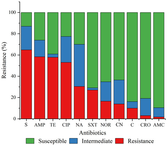
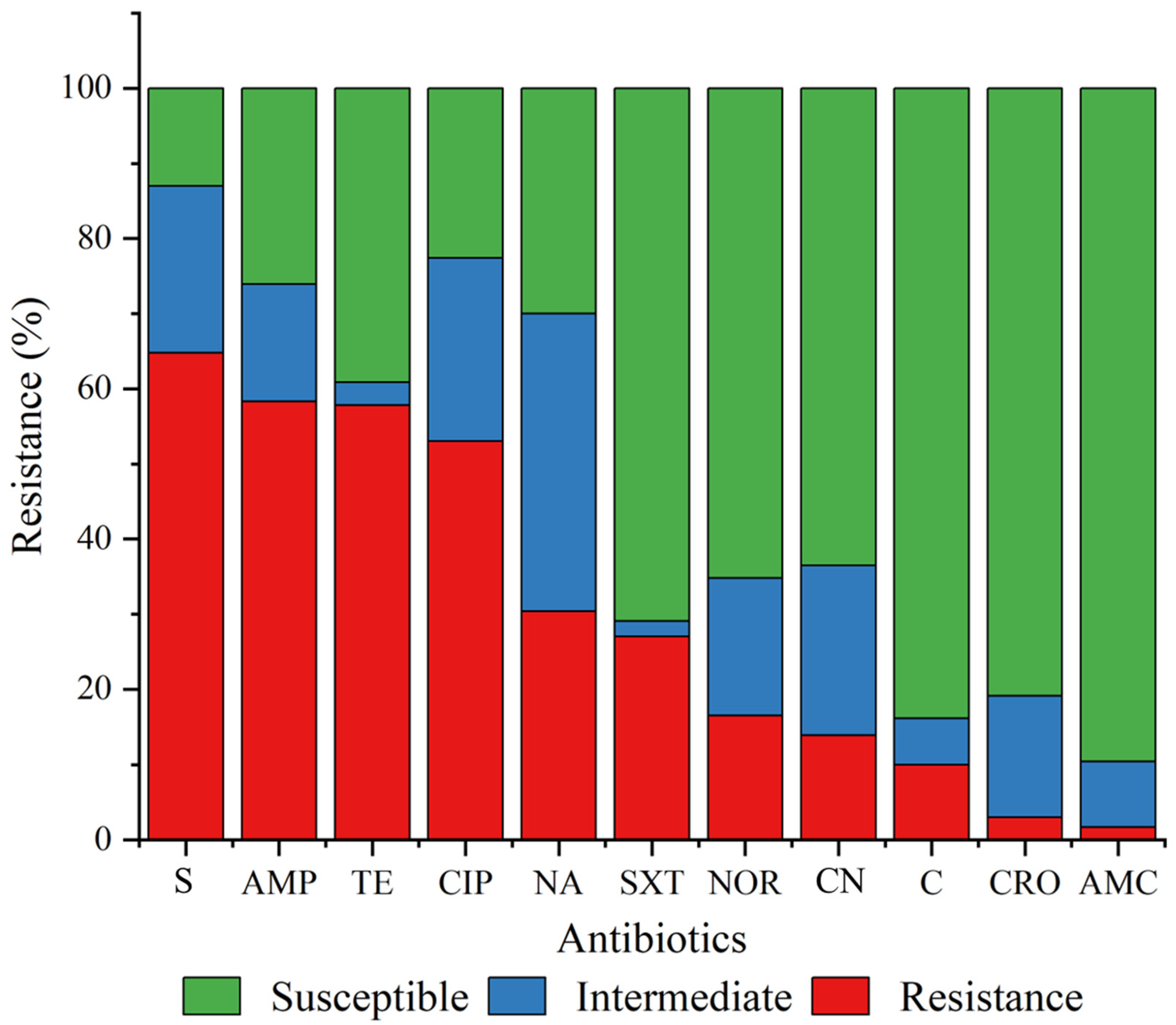
All E. coli isolates showed resistance to at least one of the tested antibiotics. The resistance exceeded 50.0% for streptomycin, ampicillin, tetracycline, and ciprofloxacin. The lowest resistance was observed for amoxicillin-clavulanic acid (1.7%) (Figure 2).
Figure 2.
Antibiotic resistance profile of Escherichia coli from various sources. AMP: ampicillin, S: streptomycin, SXT: trimethoprim-sulfamethoxazole, NA: nalidixic acid, AMC: amoxicillin-clavulanic acid, TE: tetracycline, C: chloramphenicol, CN: gentamicin, CIP: ciprofloxacin, CRO: ceftriaxone, NOR: norfloxacin.
2.4.1. Antibiotic Resistance Profiles Across Different Sources
The E. coli isolates exhibited varying antibiotic resistance, with the environmental isolates showing the highest resistance. However, this was not statistically significant (p = 0.315). Resistance rates of 28.0%, 28.6%, and 31.8% were observed among the isolates from animals, food sources, and the environment, respectively (Table 3). The isolates from animal sources were generally resistant to streptomycin (61.7%) and ciprofloxacin (60.0%). Similarly, food isolates showed high resistance to streptomycin (75.0%) and ampicillin (57.1%) (Table 3). Among the environmental isolates, 77.3% exhibited resistance to streptomycin. However, no resistance to amoxicillin-clavulanic acid was observed among the food and environmental E. coli isolates (Table 3).
Table 3.
Antibiotic resistance profile of Escherichia coli from various sources.
2.4.2. Multidrug Resistance Patterns in Relation to E. coli Pathotypes and Sources
Multidrug resistance (MDR) was observed across isolates from all sources, though the differences were not statistically significant (p = 0.850). Specifically, MDR was detected in 61.1% of isolates from animal sources, 57.1% from food sources, and 45.5% from environmental samples (Table 4). MDR among sheA+-EPEC isolates from animal sources ranged between 50% and 100%, while all sheA+-EPEC isolates from food sources exhibited MDR. Additionally, over half of the sheA+/ehxA+ EC isolates from all sources were found to be multidrug-resistant (Table 4). All isolates demonstrated varying levels of MDR (Table S1), with over 65% of isolates exhibiting resistance to three to four antibiotic classes (R3–4AC).
Table 4.
Prevalence of multidrug-resistant Escherichia coli from various sources.
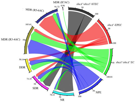
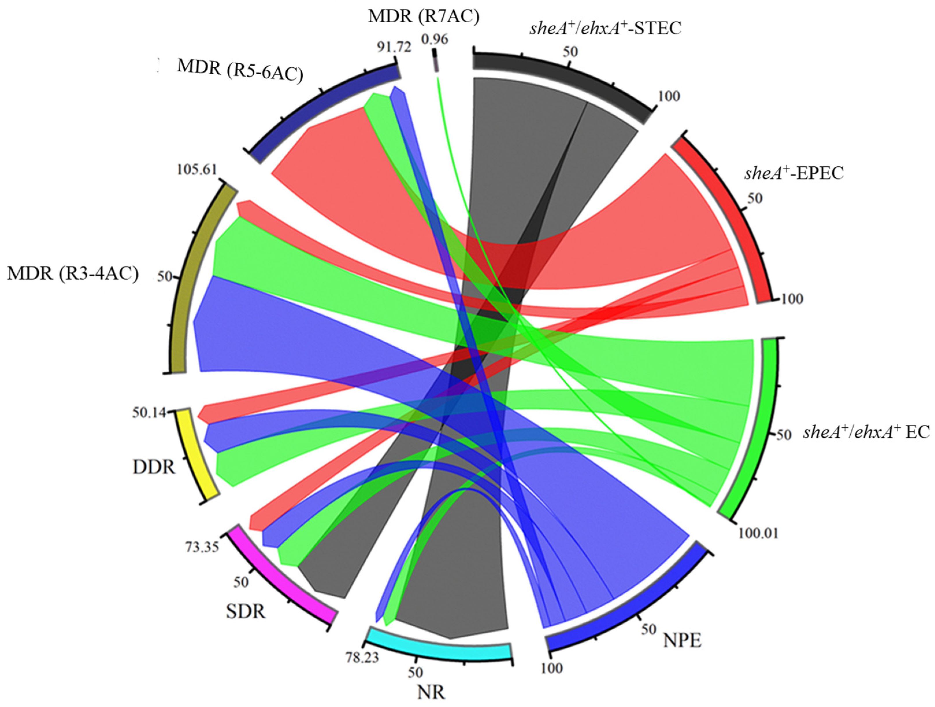
Resistance profiles also varied across E. coli pathotypes. Based on network associations, all isolates exhibited multidrug resistance (MDR), with sheA+/ehxA+ EC predominantly exhibiting resistance to three to four antibiotic classes, while some sheA+/ehxA+ EC isolates were resistant to all seven antibiotic classes tested. sheA+-EPEC pathotypes were primarily associated with resistance to five to six antibiotic classes, while NPE strains mainly showed resistance to three to four antibiotic classes. In contrast, sheA+/ehxA+-STEC pathotypes were more commonly linked to single-drug resistance (SDR) (Figure 3).
Figure 3.
The chord diagram illustrates the association of E. coli pathotype with antibiotic resistance levels. NPE: non-pathogenic E. coli, sheA+/ehxA+ EC: sheA/ehxA-positive E. coli, sheA+/ehxA+-STEC: sheA/ehxA-positive Shiga toxin-producing E. coli, sheA+-EPEC: sheA-positive enteropathogenic E. coli. NR: non-resistance; SDR: single-drug resistance; DDR: double-drug resistance; MDR (R3–4AC): multidrug resistance (resistance to 3–4 antibiotic classes); MDR (R5–6AC): multidrug resistance (resistance to 5–6 antibiotic classes); MDR (R7AC): multidrug resistance (resistance to 7 antibiotic classes).
2.5. Antibiotic Resistance Gene Profiles of Escherichia coli from Various Sources
Various antibiotic resistance genes were identified among the isolates from different sources, with the strA gene recorded as the most prevalent. In total, 29 resistance gene patterns were observed, with tetA-strA-blaTEM (22.6%) showing the most common in all investigated sources (Table 5).
Table 5.
Antibiotic-resistant gene profiles of Escherichia coli.
3. Discussion
This study investigated the prevalence of E. coli in animals, food sources (meat and contact surfaces), and the environment (wastewater) and its antibiotic resistance. The findings revealed a high prevalence of E. coli across all the sources investigated, with animal sources recording the highest values. This is concerning, as it may impact animal production. Also, cross-contamination could occur through contact with animals or through the consumption of contaminated meat [15]. However, sheep and broilers showed the highest prevalence of E. coli within the animal group. This could be attributed to variations in farm management practices across different farms. E. coli resides in animals, and its presence in the meat or meat processing facilities in the current study may suggest cross-contamination from animal sources, which could occur due to biosecurity or hygiene issues. Across meat types, the highest prevalence was recorded in broilers meat, possibly reflecting differences in farming practices and meat processing conditions. However, the prevalence reported in this study is lower than that previously reported in food from animal sources (meat: 72%) in Thailand [24], in broilers in the UAE (82.2%) [25], and in the farm-to-food continuum in Canada (95.6–97.4%) [21]. Methods of transportation of animals from farms to slaughterhouses, lairage conditions (overcrowding in slaughterhouse pens and fecal shedding, which leads to fecal buildup), slaughter processes, and poor biosecurity measures in slaughterhouses could facilitate the cross-transmission of bacteria from animals or meat to humans [20,26], posing a potential threat to public health.
Although E. coli is generally regarded as part of the normal flora in the gastrointestinal tract of animals, the emerging virulent trains, particularly in food-producing animals, pose a significant health concern [3,5]. These strains may acquire and exchange genetic elements in the environment and across different sources, which can influence their genetic profile. PCR analysis in the present study indicated a relatively high prevalence of E. coli isolates carrying hemolysin-associated genes (sheA and ehxA), with the sheA gene detected more frequently than ehxA, and with no detection of hlyA. This pattern is consistent with findings reported by Lorenz et al. [27], who observed a high occurrence of sheA (93.8%) and ehxA (69.2%) genes in food, animals, and humans in the USA, with the hlyA gene rarely recovered. The presence of the sheA gene has been linked to hemolytic potential in some E. coli strains [28,29]; however, phenotypic confirmation (e.g., hemolysis assay) was not performed in this study, and therefore functional expression cannot be inferred. This demands further investigation. Other virulence-associated genes, including eae, stx1, and stx2, were detected at lower frequencies. Their presence in E. coli isolates from livestock and meat may be of interest from an epidemiological perspective, as these genes may serve as indicators of genetic elements associated with strains of clinical relevance [30], and in some cases they were observed alongside the sheA gene. This is consistent with other studies that reported multiple virulence genes in E. coli [31,32]. This could potentially increase the pathogenicity of these strains; however, this remains uncertain without further functional characterization. The similarity of virulence genes recovered from the sources in the current study suggests that these E. coli strains may be interrelated or originated from a common source, indicating a potential for zoonotic transmission from animals to meat, and ultimately to humans. While some strains appeared to have no virulence genes, it is worth noting that they could harbor other virulence genes that were not identified in the current study. A further limitation is the use of a single isolate per sample, which may not capture within-sample diversity and could lead to underestimation of gene prevalence.
The emergence of antibiotic resistance in E. coli has made it a significant One Health pathogen worldwide. The current study assessed antibiotic-resistant E. coli from animals, food sources (meat and contact surfaces), and the environment (wastewater), which revealed a high prevalence (28–31.8%), particularly against streptomycin, ampicillin, and tetracycline, in all sources. Interestingly, resistance to ciprofloxacin was higher than that observed for nalidixic acid, which contrasts with the typical stepwise pattern of quinolone resistance. This discrepancy may be explained by the presence of additional resistance mechanisms, such as plasmid-mediated quinolone resistance determinants or efflux pumps, as well as selective pressure from the use of fluoroquinolones in animal production [33]. Furthermore, specific mutation patterns in quinolone resistance-determining regions (QRDRs) may differentially affect susceptibility to first- and second-generation quinolones [34]. Similar previous studies in Thailand have reported high streptomycin- and tetracycline-resistant E. coli [35,36]. This could be attributed to these antibiotics being widely used for animal production [10,11,35]. This induces antibiotic resistance over time due to selective pressure. However, the resistance reported in the current study is lower than that reported in previous studies, including those conducted in the farm-to-food continuum in South Africa (ampicillin: 100%, tetracycline: 100%) [22] and in broilers in the UAE (ampicillin: 100%, tetracycline: 72.6%, and ciprofloxacin: 88.4%) [25]. All isolates from food sources (meat and contact surfaces) exhibited susceptibility to amoxicillin-clavulanic acid. This conforms with Abou-Jaoudeh et al. [37], who reported low (13.9%) amoxicillin-clavulanic acid-resistant E. coli in chicken meat in Lebanon. This broadens the available treatment options for infections caused by antibiotic-resistant E. coli from animal sources. The resistance recorded in animals was lower than in previous reports in livestock (70–78.3%) [35,38]. This could be attributed to differences in farm management practices, such as variations in antibiotic use across different jurisdictions. High antibiotic resistance was also recorded in the environmental (wastewater) isolates. Wastewater can harbor diverse microbes, including antibiotic-resistant E. coli strains. A previous study in Ireland identified wastewater as a reservoir for carbapenem-resistant bacteria [39]. The current results indicate similar resistance characteristics exhibited by isolates from different sources, highlighting their interrelatedness and a potential risk of cross-contamination among animals, food (meat and contact surfaces), and the environment, thereby posing a public health threat.
Multidrug resistance (MDR) is usually associated with bacteria harboring multiple resistance genes that encode for different antibiotic resistance, an increase in gene expression, efflux pumps of antibiotics [40], or with other non-genetic factors. Animals and animal feces recorded the highest MDR in the current study. This could be attributed to the indiscriminate use of antibiotics in animal production [12,13]. This could induce antibiotic resistance in the environment by selective pressure and its subsequent potential effect on animal production and the ripple effect on human health. The MDR recorded from the animals in this study is higher than the MDR (23.9%) reported by Prapasawat and Intarapuk [35] in Thailand. However, a similar study along the pork production chain in central Thailand reported a higher prevalence of MDR (87.3%) [23]. This is directly linked to variations in antibiotic application for animal production in different jurisdictions and farms [41]. The current findings may negatively impact human health through direct contact with farm animals harboring these MDR E. coli or via the animal-based food chain. The MDR in the food sources is higher than the multidrug resistance (49.23%) reported in meat in Bangladesh [42]. This discrepancy could also be ascribed to antibiotic usage at farms at different locations. However, the major concern is the high level of MDR and resistance to five to six antibiotic classes recorded among the supposedly non-pathogenic E. coli strains across all sources, resistance to seven antibiotic classes among the sheA+/ehxA+ EC, and resistance to five to six antibiotic classes among the sheA+-EPEC pathotypes in the current study. This implies that the treatment options for bacterial strains from these sources are gradually diminishing. This, therefore, demands public sensitization, including veterinary services, and farmers’ awareness of the consequences of the indiscriminate use of antibiotics for animal farming, as this could negatively impact animal and human health.
The continuous application of antibiotics exerts selective pressure on bacteria, driving the development of various resistance mechanisms over time, including genotypic resistance. This study assessed the genotypic resistance of E. coli isolates. The highest resistance gene pattern observed in all the sources was tetA-strA-blaTEM. These genes encode resistance to tetracyclines, aminoglycosides (streptomycin), and penicillin (e.g., ampicillin) or cephalosporins (ceftriaxone). This could be attributed to the widespread use of these antibiotics in animal production [35], which exerts selective pressure for the expression of these resistance genes in the bacteria. This may have contributed to the increased phenotypic resistance observed in this study. For instance, the blaTEM gene in E. coli encodes the enzyme beta-lactamase that hydrolyzes the beta-lactam rings in beta-lactam antibiotics [43,44]. The tetA gene encodes an inducible efflux pump where a complex of tetracycline and divalent metallic ions is translocated out of the cell, reducing the concentration of tetracycline in the bacterial cell and protecting the ribosomes from the action of tetracycline antibiotics. In contrast, the strA gene mediates enzymatic modification of aminoglycosides. Consequently, antibiotics such as aminoglycosides (e.g., streptomycin) become ineffective [43]. The presence of these resistance gene patterns demonstrates the mechanisms by which E. coli from these current studied sources resist these antibiotics. This reduces treatment options and poses the risk of these genes being transferred to other non-resistant E. coli. This could have significant public health implications, demanding strict measures regarding the application of these antibiotics for animal production, as resistant E. coli in the environment can transmit resistance genes to other E. coli (through vertical gene transmission) or to other bacteria in food, animals, or humans (horizontal gene transfer) (through conjugation, transformation, transduction, and vesiduction) [20].
4. Materials and Methods
4.1. Interconnected Sampling Sites, Study Design, and Sample Collection
The research is a cross-sectional study in which a multistage random sampling technique was used to collect samples from designated sites to analyze the prevalence and distribution of antibiotic resistance and virulence genes in E. coli along the farm-to-fork continuum. Permission was obtained from the management of each area prior to sampling. Sampling was done from July 2023 to December 2023.
A total of 383 samples were randomly collected from farm animals, food processing facilities (slaughterhouses or meat processing plants), and the surrounding environments (wastewaters). To ensure the interconnectedness of these sectors, only animal farms linked (closed proximity and farms that supply animals to the selected processing facilities) to meat processing facilities were selected in this study. Likewise, only wastewater sources that were traceable to meat processing facilities under the investigation were included in the study.
A total of 249 samples from animal sources were randomly collected from farm animals. Rectal swabs were obtained from chicken, ducks, turkeys, guinea fowls, goats, and sheep using sterile cotton swabs gently inserted into the cloaca or anus. Fecal samples were collected from pigs and cattle. All samples were aseptically collected from farm animals in Bangkok, Chiang Mai, and Nakhon Ratchasima, Thailand, and each was individually stored in a sterile zip-lock plastic bag.
A total of 83 food-related samples were randomly collected from meat processing facilities in Bangkok, Prachinburi, and Saraburi for analysis. These included chicken, pork, and beef meat samples (approximately 100 g each) and surface swabs. Surface samples were obtained by swabbing contact areas (approximately 10 cm2 per surface) with sterile cotton swabs moistened with sterile saline. The sampling sites within the processing environments included utensils (chopping boards, weighing scales, pans, and butcher knives) and tables in slaughterhouses. All samples were aseptically collected and individually stored in sterile zip-lock bags. In addition, a total of 51 wastewater samples (~500 mL) linked to the meat processing facilities were aseptically collected in sterile bottles.
All samples were clearly labeled, stored in an ice box containing ice packs, and transported promptly to the laboratory for bacteriological analysis. These samples were processed in the Laboratory of Bacteriophage and Food Safety, Department of Biotechnology, Faculty of Agro-Industry, Kasetsart University, Thailand.
4.2. Sample Preparation and Isolation of Escherichia coli
Sample preparation and isolation followed an approach proposed by Feng et al. [45] with some modifications. Briefly, swab samples were immersed in 9 mL of buffered peptone water (BPW) (#CM1040, OXOID Ltd., Basingstoke, UK) in sterile test tubes. For solid samples (meat and animal feces/droppings), 25 g of each sample was transferred into sterile zip-lock bags containing 225 mL of BPW. Liquid samples (wastewater) were prepared by inoculating 25 mL of each homogenized sample into 225 mL of BPW in sterile zip-lock bags. All samples were homogenized and incubated at 37 °C for 24 h. Subsequently, a loopful of the enriched mixture was streaked onto Eosin Methylene Blue (EMB) agar plates (#M022, Himedia Laboratories Pvt. Ltd., Nagpur, Maharashtra, India) and incubated at 37 °C for 24 h. Colonies exhibiting a characteristic metallic green sheen on EMB were considered presumptive E. coli. Selected colonies were re-streaked on nutrient agar and incubated at 37 °C for an additional 24 h to obtain pure cultures for further biochemical confirmation. E. coli ATCC® 25922 (American Type Culture Collection, Manassas, VA, USA) was used as a positive control strain for the E. coli isolation.
4.3. Biochemical Identification of Escherichia coli
Biochemical confirmation of the presumptive E. coli followed Feng et al. [45] with slight modifications: The presumptive E. coli isolates were confirmed using the Triple Sugar Ion (TSI) agar test (#CM0277, OXOID Ltd., Basingstoke, UK), indole test, the O-Nitrophenyl-β-D-Galactopyranoside (ONPG) test as described by Feng et al. [45], and further confirmed using the LIA agar test (#M377, Himedia Laboratories Pvt. Ltd., Maharashtra, India) and citrate test (#M099, Himedia Laboratories Pvt. Ltd., Maharashtra, India). E. coli ATCC® 25922 was used as a positive control strain for the biochemical confirmations. After this, E. coli was further confirmed through PCR according to an approach described by Azimi et al. [46], using specific primer for the uidA gene (Table S2; Figure S1). The E. coli O157:H7 strain from Wiriyaprom et al. [47] was used as a positive control, while the PCR reaction without a DNA sample served as a negative control.
4.4. Antibiotic Susceptibility Testing of Escherichia coli
Antibiotic susceptibility was tested using the Kirby–Bauer disk diffusion method following the Laboratory Standards Institute (CLSI) guidelines. Mueller–Hinton agar (CM0337, OXOID Ltd., Basingstoke, UK) plates were inoculated with 24 h tryptic soy broth (#M011, Himedia Laboratories Pvt. Ltd., Maharashtra, India), E. coli cultures adjusted to a 0.5 McFarland standard, and antibiotic disks applied and incubated at 37 °C for 18 h [48,49]. Antibiotics, including ampicillin (10 µg), streptomycin (10 µg), trimethoprim-sulphamethoxazole (25 µg), nalidixic acid (30 µg), amoxicillin-clavulanic acid (30 µg), tetracycline (30 µg), chloramphenicol (30 µg), gentamicin (10 µg), ciprofloxacin (5 µg), ceftriaxone (30 µg), and norfloxacin (10 µg), were used. Besides being recommended for E. coli in the CLSI guidelines [49], these antibiotics are commonly used in animal production [31]; therefore, they were carefully selected for this study. After incubation, the inhibition zones were recorded and interpreted using the CLSI breakpoints [50]. E. coli ATCC® 25922 was used as a quality control strain in this study. Isolates exhibiting resistance to one and two antibiotic classes were identified as single-drug resistant (SDR) and double-drug resistant (DDR), respectively, while isolates exhibiting resistance to three or more antibiotic classes were identified as multidrug-resistant strains.
4.5. Determination of Virulence and Antibiotic Resistance Genes Among Escherichia coli
4.5.1. DNA Extraction
DNA of the E. coli isolates was extracted using a genomic DNA Prep kit (#GD 141-050, BIOFACTTM, BIOFACT Co., Ltd., Daejeon, Republic of Korea) for PCR analysis. The DNA was suspended in 100 µL of DNA hydration buffer, and 2 µL of the eluted DNA was used as the template for each PCR reaction.
4.5.2. Detection of Virulence Genes
Specific primers for representative virulence genes (Table S2) were used for the PCR following Azimi et al. [46] with slight modifications. Each 25 µL PCR reaction contained a PCR mix (2.5 µL of 10× PCR buffer, 2 µL of 2.5 mM dNTP, and 0.2 µL of 5U Taq polymerase) (#W1301, Wizbiosolution Inc., Seongnam, Gyeonggi-do, Republic of Korea), 16.3 µL of nucleotide-free deionized water, 2 µL of the DNA template, and 1 µL of 0.25 µM of each primer (forward and reverse). PCR was run using a thermocycler (BIORAD T100TM Thermal cycler, Bio-Rad Laboratories Ltd., Hercules, CA, USA) with gene-specific cycling conditions as follows: initial denaturation for 15 min at 95 °C for sheA and ehxA genes only [27,51], 5 min at 95 °C for all the remaining genes, followed by 25 cycles for sheA and ehxA genes only, 35 cycles for all the remaining genes, each cycle consisting of denaturation for 5 min at 94–95 °C, annealing for 30 s at 54–60 °C, extension for 1 min at 72 °C, and a final extension step for 5 min at 72 °C. The E. coli (O157:H7) and F1.1 strains from Wiriyaprom et al. [47] were used as quality control strains in this study, while a PCR reaction without a DNA sample served as a negative control. All experiments were done in triplicate.
4.5.3. Detection of Antibiotic Resistance Genes
The detection of representative antibiotic resistance genes for aminoglycosides (strA, aac3-(IV)), tetracycline (tetA), beta-lactam (blaTEM), sulfonamides (sul1, sul2, sul3), and phenicol (catI, catII, and floR) was performed through PCR [52,53,54,55,56,57] with slight modifications using specific primers (Table S3). The PCR mixture as described above was used. The PCR was run with a thermocycler (BIORAD T100TM Thermal cycler, Bio-Rad Laboratories Ltd., CA, USA) using the following conditions: initial denaturation for 5 min at 94–95 °C, followed by 30–35 cycles, each consisting of denaturation at 98 °C for 1 min for sul2 and sul3 genes only, 30–60 s at 94–95 °C for all the remaining genes, annealing for 30–60 s at 50–62.5 °C, extension for 1 min at 72 °C, and a final extension step for 5 min at 72 °C. Multidrug-resistant clinical E. coli strain (EC3.18) served as a positive control for resistance gene detection, while a PCR reaction without a DNA sample served as a negative control. All experiments were done in triplicate.
4.5.4. Visualization of Amplicons
All the amplified samples (amplicons) were analyzed through gel electrophoresis with a 1.5% agarose gel (#PC0701, Vivantis Technology Sdn Bhd, Subang Jaya, Selangor, Malaysia) pre-stained with 1% SYBR green (SYBRTM Green I nucleic acid gel, InvitrogenTM Thermo Fisher ScientificTM, Waltham, MA, USA), and the bands were observed using a gel documentation system (iBrightTM CL1500 imaging system, InvitrogenTM Thermo Fisher ScientificTM, Waltham, MA, USA). It was then compared with a standard-sized molecular marker (DNA ladder) (#NL1407, Vivantis Technology Sdn Bhd, Subang Jaya, Selangor, Malaysia) of 100 bp increments.
4.6. Statistical and Data Analysis
The data were scrutinized for completeness and analyzed using SPSS (IBM® SPSS® Statistic version 25, SPSS Inc., Chicago, IL, USA). The chi-square test was used to assess significant differences at the 95% confidence level, and a p-value of ≤0.05 was considered statistically significant. The graphs were generated using ORIGIN PRO® v.2024b Graphing and Analysis (OriginLab Corporation, Northampton, MA, USA).
5. Conclusions
A high prevalence of E. coli was observed across all the sources, with over 80% of the isolates identified as sheA+/ehxA+ EC. The E. coli isolates exhibited significant antibiotic resistance, with the highest resistance recorded for streptomycin, ampicillin, and tetracycline. Additionally, most of the E. coli isolates were multidrug-resistant. The most prevalent antibiotic resistance gene pattern was tetA-strA-blaTEM. Alarmingly, both virulence gene-carrying E. coli and NPE strains displayed MDR, often carrying multiple resistance genes. These constitute a possibility for zoonotic infection that could lead to complications, demanding an urgent need for effective antimicrobial stewardship policies to regulate the use of antibiotics in animal production, and robust systems to reduce resistance and virulence genes of E. coli from farm to fork. Future works should focus on the whole genome sequencing of these E. coli isolates.
Supplementary Materials
The following supporting information can be downloaded at: https://www.mdpi.com/article/10.3390/antibiotics15050455/s1, Figure S1: Gel image of uidA gene for E. coli identification; Table S1: Multidrug resistance patterns of E. coli from various sources; Table S2: Primer lists for virulence genes; Table S3: Primer lists for antibiotic-resistant genes.
Author Contributions
Conceptualization, D.Y.Y., M.Y., W.P. and K.V.; methodology, D.Y.Y., W.W., K.C., M.Y., W.P. and K.V.; investigation, D.Y.Y., W.W., N.V., K.C., V.C. and W.P.; resources, M.Y. and K.V.; data curation, D.Y.Y. and W.P.; writing—original draft, D.Y.Y. and W.P.; writing—review and editing, M.Y., W.P. and K.V.; visualization, D.Y.Y. and W.P.; supervision, M.Y., W.P. and K.V.; project administration, W.P.; funding acquisition, M.Y., W.P. and K.V. All authors have read and agreed to the published version of the manuscript.
Funding
This work is funded by the Kasetsart’s Agro-Industry Scholarship 2023, Faculty of Agro-Industry, Kasetsart University (to D.Y.Y.), the Faculty of Medicine, Prince of Songkla University, grant no. 64-007-3 (to M.Y.), the Walailak University under the New Researcher Development scheme, contract number WU69210 (to W.P.), and the Kasetsart University Research and Development Institute, KURDI, grant no. FF(KU-SRIU)12.68 (to K.V.).
Institutional Review Board Statement
This study was conducted under the permission of Animal Care and Use for Science and Technology Research with the approval number MACUC013A/2566 provided by the Maejo University, Animal Care and Use Committee, Maejo University, Thailand.
Informed Consent Statement
Not applicable.
Data Availability Statement
All data generated or analyzed during this study are included in this published article and its Supplementary Materials files.
Acknowledgments
The authors acknowledged the Department of Biotechnology, Faculty of Agro-Industry, Kasetsart University, and the Faculty of Animal Science and Technology, Maejo University, for providing facilities to conduct this work.
Conflicts of Interest
The authors declare no conflicts of interest.
References
- Hayer, S.S.; Rovira, A.; Olsen, K.; Johnson, T.J.; Vannucci, F.; Rendahl, A.; Perez, A.; Alvarez, J. Prevalence and trend analysis of antimicrobial resistance in clinical Escherichia coli isolates collected from diseased pigs in the USA between 2006 and 2016. Transbound. Emerg. Dis. 2020, 67, 1930–1941. [Google Scholar] [CrossRef] [Scilit]
- Khairy, R.M.; Mohamed, E.S.; Abdel Ghany, H.M.; Abdelrahim, S.S. Phylogenic classification and virulence genes profiles of uropathogenic E. coli and diarrhegenic E. coli strains isolated from community acquired infections. PLoS ONE 2019, 14, e0222441. [Google Scholar] [CrossRef] [Scilit] [PubMed]
- Barbau-Piednoir, E.; Denayer, S.; Botteldoorn, N.; Dierick, K.; De Keersmaecker, S.C.; Roosens, N.H. Detection and discrimination of five E. coli pathotypes using a combinatory SYBR® Green qPCR screening system. Appl. Microbiol. Biotechnol. 2018, 102, 3267–3285. [Google Scholar] [CrossRef] [Scilit]
- Chekole, W.S.; Adamu, H.; Sternberg-Lewrein, S.; Magnusson, U.; Tessema, T.S. Occurrence of Escherichia coli pathotypes in diarrheic calves in a low-income setting. Pathogens 2022, 12, 42. [Google Scholar] [CrossRef] [Scilit]
- Erjavec, M.S. Introductory Chapter: The Versatile Escherichia coli. In The Universe of Escherichia coli; IntechOpen: London, UK, 2019; pp. 3–17. [Google Scholar]
- Owrangi, B.; Masters, N.; Kuballa, A.; O’Dea, C.; Vollmerhausen, T.L.; Katouli, M. Invasion and translocation of uropathogenic Escherichia coli isolated from urosepsis and patients with community-acquired urinary tract infection. Eur. J. Clin. Microbiol. Infect. Dis. 2018, 37, 833–839. [Google Scholar] [CrossRef] [Scilit]
- Murray, C.J.; Ikuta, K.S.; Sharara, F.; Swetschinski, L.; Aguilar, G.R.; Gray, A.; Han, C.; Bisignano, C.; Rao, P.; Wool, E.; et al. Global burden of bacterial antimicrobial resistance in 2019: A systematic analysis. Lancet 2022, 399, 629–655. [Google Scholar] [CrossRef] [Scilit]
- Reygaert, W.C. An overview of the antimicrobial resistance mechanisms of bacteria. AIMS Microbiol. 2018, 4, 482–501. [Google Scholar] [CrossRef] [Scilit]
- Tagliabue, A.; Rappuoli, R. Changing priorities in vaccinology: Antibiotic resistance moving to the top. Front. Immunol. 2018, 9, 1068. [Google Scholar] [CrossRef] [Scilit]
- Malijan, G.M.; Howteerakul, N.; Ali, N.; Siri, S.; Kengganpanich, M.; Nascimento, R.; Booton, R.D.; Turner, K.M.; Cooper, B.S.; Meeyai, A.; et al. A scoping review of antibiotic use practices and drivers of inappropriate antibiotic use in animal farms in WHO Southeast Asia region. One Health 2022, 15, 100412. [Google Scholar] [CrossRef] [Scilit] [PubMed]
- United States Food and Drug Administration. 2020 Summary Report on Antimicrobials Sold or Distributed for Use in Food-Producing Animals; Food and Drug Administration: Silver Spring, MD, USA, 2019. [Google Scholar]
- Food and Agriculture Organization of the United Nations. Antimicrobial Resistance. 2024. Available online: https://www.fao.org/antimicrobial-resistance/background/what-is-it/en/ (accessed on 25 September 2025).
- Liu, C.; Tan, L.; Zhang, L.; Tian, W.; Ma, L. A review of the distribution of antibiotics in water in different regions of China and current antibiotic degradation pathways. Front. Environ. Sci. 2021, 9, 692298. [Google Scholar] [CrossRef] [Scilit]
- Muloi, D.; Ward, M.J.; Pedersen, A.B.; Fevre, E.M.; Woolhouse, M.E.; van Bunnik, B.A. Are food animals responsible for transfer of antimicrobial-resistant Escherichia coli or their resistance determinants to human populations? A systematic review. Foodborne Pathog. Dis. 2018, 15, 467–474. [Google Scholar] [CrossRef] [Scilit]
- World Health Organization. Global E. coli Outbreaks-Food Safety Alliance. 2019. Available online: https://safefoodalliance.com/food-safety/global-e-coli-outbreaks/ (accessed on 25 September 2025).
- Boonyasiri, A.; Tangkoskul, T.; Seenama, C.; Saiyarin, J.; Tiengrim, S.; Thamlikitkul, V. Prevalence of antibiotic resistant bacteria in healthy adults, foods, food animals, and the environment in selected areas in Thailand. Pathog. Glob. Health 2014, 108, 235–245. [Google Scholar] [CrossRef] [Scilit]
- European Food Safety Authority. The European Union summary report on antimicrobial resistance in zoonotic and indicator bacteria from humans, animals and food in 2021–2022. EFSA J. 2024, 22, e8583. [Google Scholar]
- Millstein, S. Experts: Poor Farming Practices Cause Most Toxic E. coli Outbreaks. Sentient. Broadcast Version by F. Ross for Texas News Service Reporting for the Sentient-Public News Service Collaboration. 2024. Available online: https://www.publicnewsservice.org/2024-12-31/rural-farming/experts-poor-farming-practices-cause-most-toxic-e-coli-outbreaks/a94367-1 (accessed on 25 September 2025).
- Noenchat, P.; Srithong, T.; Lertpiriyasakulkit, S.; Nhoonoi, C.; Saksangawong, C.; Sornplang, P. Prevalence and antimicrobial resistance profile of Escherichia coli O157: H7 isolated from chickens and chicken meats from local poultry slaughterhouses in Nakhon Ratchasima Province, Thailand. Vet. Integr. Sci. 2024, 22, 769–785. [Google Scholar] [CrossRef] [Scilit]
- Herawati, C.; Endayani, H.; Indragiri, S.; Kristanti, I.; Wahyuni, N.T.; Dani, A.H.; Hikmawati, I. Sanitary Hygiene and Behavior of Food Handlers in the Presence of Escherichia coli Bacteria. J. Pure Appl. Microbiol. 2023, 17, 2098–2103. [Google Scholar] [CrossRef] [Scilit]
- Medrano, H.; Hill, S.; Boulianne, M.; Cereno, T.; Deckert, A.E.; Charlebois, A.; Gow, S.P.; McDonald, K.; Reid-Smith, R.J.; Agunos, A. Widespread dissemination of Salmonella, Escherichia coli and Campylobacter resistant to medically important antimicrobials in the poultry production continuum in Canada. PLoS ONE 2025, 20, e0319086. [Google Scholar] [CrossRef] [Scilit]
- Abdalla, S.E.; Bester, L.A.; Abia, A.L.K.; Allam, M.; Ismail, A.; Essack, S.Y.; Amoako, D.G. Genomic Insights of Antibiotic-Resistant Escherichia coli Isolated from Intensive Pig Farming in South Africa Using ‘Farm-to-Fork’ Approach. Antibiotics 2025, 14, 446. [Google Scholar] [CrossRef] [Scilit]
- Prapasawat, W.; Siriphap, A.; Wiriyasirivaj, S.; Intarapuk, A.; Pachanon, R.; Nakajima, C.; Suzuki, Y.; Suthienkul, O. Antimicrobial resistance, extended-spectrum β-lactamase determinants, and virulence gene profiles of Escherichia coli along the pork production chain in central Thailand. Vet. World 2026, 19, 52–64. [Google Scholar] [CrossRef] [Scilit] [PubMed] [PubMed Central]
- Ngamwongsatit, N.; Chaturongakul, S.; Aunpad, R. Development and validation of an efficient multiplex PCR assay for simultaneous detection of six common foodborne pathogens and hygiene indicators. Foodborne Pathog. Dis. 2023, 20, 222–229. [Google Scholar] [CrossRef] [Scilit] [PubMed]
- Khalifa, H.O.; Mohammed, T.; Elbediwi, M.; Abdalla, A.; Mohamed, M.-Y.I.; Ramadan, H.; Lakshmi, G.B.; Ghazawi, A.; Habib, I. Prevalence and genetic basis of extended-spectrum β-lactamase-producing Escherichia coli carriage in broiler farms in the United Arab Emirates. Front. Vet. Sci. 2025, 12, 1714381. [Google Scholar] [CrossRef] [Scilit] [PubMed]
- Mannion, C.; Fanning, J.; McLernon, J.; Lendrum, L.; Gutierrez, M.; Duggan, S.; Egan, J. The role of transport, lairage and slaughter processes in the dissemination of Salmonella spp. in pigs in Ireland. Food Res. Int. 2012, 45, 871–879. [Google Scholar] [CrossRef] [Scilit]
- Lorenz, S.C.; Son, I.; Maounounen-Laasri, A.; Lin, A.; Fischer, M.; Kase, J.A. Prevalence of hemolysin genes and comparison of ehxA subtype patterns in Shiga toxin-producing Escherichia coli (STEC) and non-STEC strains from clinical, food, and animal sources. Appl. Environ. Microbiol. 2013, 79, 6301–6311. [Google Scholar] [CrossRef] [Scilit]
- Lai, X.H.; Arencibia, I.; Johansson, A.; Wai, S.N.; Oscarsson, J.; Kalfas, S.; Sundqvist, K.G.; Mizunoe, Y.; Sjostedt, A.; Uhlin, B.E. Cytocidal and apoptotic effects of the ClyA protein from Escherichia coli on primary and cultured monocytes and macrophages. Infect. Immun. 2000, 68, 4363–4367. [Google Scholar] [CrossRef] [Scilit]
- Ludwig, A.; Von Rhein, C.; Bauer, S.; Huttinger, C.; Goebel, W. Molecular analysis of cytolysin A (ClyA) in pathogenic Escherichia coli strains. J. Bacteriol. 2004, 186, 5311–5320. [Google Scholar] [CrossRef] [Scilit] [PubMed]
- Hua, Y.; Zhang, J.; Jernberg, C.; Chromek, M.; Hansson, S.; Frykman, A.; Xiong, Y.; Wan, C.; Matussek, A.; Bai, X. Molecular characterization of the enterohemolysin gene (ehxA) in clinical Shiga toxin-producing Escherichia coli isolates. Toxins 2021, 13, 71. [Google Scholar] [CrossRef] [Scilit] [PubMed]
- Ortiz, Y.; Cerino, B.; Moreno, M.; Yañez, E.; Heredia, N.; Dávila-Aviña, J.; Quezada, T.; Calle, A.; García, S. Diarrheagenic Escherichia coli with multidrug resistance in cattle from Mexico. J. Food Prot. 2024, 87, 100257. [Google Scholar] [CrossRef] [Scilit]
- Tutija, J.F.; Ramos, C.A.; Lemos, R.A.; Santos, A.A.; Reckziegel, G.H.; Freitas, M.G.; Leal, C.R. Molecular and phenotypic characterization of Escherichia coli from calves in an important meat-producing region in Brazil. J. Infect. Dev. Ctries. 2022, 16, 1030–1036. [Google Scholar] [CrossRef] [Scilit]
- Jacoby, G.A.; Strahilevitz, J.; Hooper, D.C. Plasmid-mediated quinolone resistance. Microbiol. Spectr. 2015, 2, 475–503. [Google Scholar]
- Ruiz, J. Mechanisms of resistance to quinolones: Target alterations, decreased accumulation and DNA gyrase protection. J. Antimicrob. Chemother. 2003, 51, 1109–1117. [Google Scholar] [CrossRef] [Scilit]
- Prapasawat, W.; Intarapuk, A. Prevalence of antimicrobial resistance and integrons in Escherichia coli isolated from feces of dairy goats in Nong Chok, Bangkok, Thailand. Vet. Integr. Sci. 2021, 19, 223–236. [Google Scholar] [CrossRef] [Scilit]
- Rodroo, J.; Intanon, M.; Kreausukon, K.; Kongkaew, A.; Bender, J.; Awaiwanont, N. Occurrence of extended-spectrum beta-lactamase producing E. coli in broiler farm workers and the farm environment in Chiang Mai-Lamphun, Thailand. Vet. Integr. Sci. 2021, 19, 23–35. [Google Scholar] [CrossRef] [Scilit]
- Abou-Jaoudeh, C.; Khalil, J.; El-Hayek, E.; Abou-Khalil, R. Food safety control in poultry industry: Prevalence and antimicrobial susceptibility of Escherichia coli isolated from raw chicken and the potential use of Origanum essential oils as alternative to antibiotics. Br. Poult. Sci. 2024, 65, 494–501. [Google Scholar] [CrossRef] [Scilit]
- Silva, A.; Silva, V.; Pereira, J.E.; Maltez, L.; Igrejas, G.; Valentão, P.; Falco, V.; Poeta, P. Antimicrobial resistance and clonal lineages of Escherichia coli from food-producing animals. Antibiotics 2023, 12, 1061. [Google Scholar] [CrossRef] [Scilit] [PubMed]
- Cahill, N.; O’Connor, L.; Mahon, B.; Varley, Á.; McGrath, E.; Ryan, P.; Cormican, M.; Brehony, C.; Jolley, K.A.; Maiden, M.C.; et al. Hospital effluent: A reservoir for carbapenemase-producing Enterobacterales? Sci. Total Environ. 2019, 672, 618–624. [Google Scholar] [CrossRef] [Scilit]
- Nikaido, H. Multidrug resistance in bacteria. Annu. Rev. Biochem. 2009, 78, 119–146. [Google Scholar] [CrossRef] [Scilit]
- Barton, M.D. Impact of antibiotic use in the swine industry. Curr. Opin. Microbiol. 2014, 19, 9–15. [Google Scholar] [CrossRef] [Scilit]
- Rahman, M.M.; Husna, A.; Elshabrawy, H.A.; Alam, J.; Runa, N.Y.; Badruzzaman, A.T.M.; Banu, N.A.; Al Mamun, M.; Paul, B.; Das, S.; et al. Isolation and molecular characterization of multidrug-resistant Escherichia coli from chicken meat. Sci. Rep. 2020, 10, 21999. [Google Scholar] [CrossRef] [Scilit] [PubMed]
- Dowling, A.; O’Dwyer, J.; Adley, C. Antibiotics: Mode of action and mechanisms of resistance. Antimicrob. Res. Nov. Bio Knowl. Educ. Programs 2017, 1, 536–545. [Google Scholar]
- Gelalcha, B.D.; Kerro Dego, O. Extended-spectrum beta-lactamases producing Enterobacteriaceae in the USA dairy cattle farms and implications for public health. Antibiotics 2022, 11, 1313. [Google Scholar] [CrossRef] [Scilit]
- Feng, P.; Weagant, S.D.; Jinneman, K. BAM Chapter 4A: Diarrheagenic Escherichia coli; United States Food and Drug Administration: Silver Spring, MD, USA, 2020. [Google Scholar]
- Azimi, T.; Azimi, L.; Fallah, F.; Pourmand, M.R.; Ostadtaghizadeh, A.; Abai, M.R.; Rahimi Foroushani, A. Detection and characterization of Enterobacteriaceae family members carried by commensal Rattus norvegicus from Tehran, Iran. Arch. Microbiol. 2021, 203, 1321–1334. [Google Scholar] [CrossRef] [Scilit] [PubMed]
- Wiriyaprom, R.; Ngasaman, R.; Kaewnoi, D.; Prachantasena, S. Prevalence and virulent gene profiles of sorbitol non-fermenting Shiga toxin-producing Escherichia coli isolated from goats in southern Thailand. Trop. Med. Infect. Dis. 2022, 7, 357. [Google Scholar] [CrossRef] [Scilit]
- Chanthavong, V.; Vigad, N.; Pelyuntha, W.; Yamik, D.Y.; Vongkamjan, K.; Yingkajorn, M.; Chaisowwong, W.; Tippaya, K.; Tadee, P.; Chukiatsiri, K. Effectiveness of a single-dose phage cocktail on the reduction of multidrug-resistant Escherichia coli in suckling piglets. Vet. Microbiol. 2025, 302, 110395. [Google Scholar] [CrossRef] [Scilit]
- Luangmanyvongkhao, V.; Vigad, N.; Pelyuntha, W.; Vongkamjan, K.; Chukiatsiri, K. Lytic activity of novel bacteriophages recovered from pig farm sewage against multidrug-resistant Escherichia coli. Vet. Int. Sci. 2026, 24, e2026037-1-14. [Google Scholar] [CrossRef] [Scilit]
- Clinical and Laboratory Standard Institute. CLSI M100TM Performance Standards for Antimicrobial Susceptibility Testing, 34th ed.; Clinical and Laboratory Standard Institute: Wayne, PA, USA, 2024; pp. 58–69. [Google Scholar]
- Kerényi, M.; Allison, H.E.; Bátai, I.; Sonnevend, A.; Emody, L.; Plaveczky, N.; Pál, T. Occurrence of hlyA and sheA genes in extraintestinal Escherichia coli strains. J. Clin. Microbiol. 2005, 43, 2965–2968. [Google Scholar] [CrossRef] [Scilit] [PubMed]
- Maynard, C.; Fairbrother, J.M.; Bekal, S.; Sanschagrin, F.; Levesque, R.C.; Brousseau, R.; Masson, L.; Lariviere, S.; Harel, J. Antimicrobial resistance genes in enterotoxigenic Escherichia coli O149: K91 isolates obtained over a 23-year period from pigs. Antimicrob. Agents Chemother. 2003, 47, 3214–3221. [Google Scholar] [CrossRef] [Scilit]
- Gow, S.P.; Waldner, C.L.; Harel, J.; Boerlin, P. Associations between antimicrobial resistance genes in fecal generic Escherichia coli isolates from cow-calf herds in western Canada. Appl. Environ. Microbiol. 2008, 74, 3658–3666. [Google Scholar] [CrossRef] [Scilit]
- Boerlin, P.; Travis, R.; Gyles, C.L.; Reid-Smith, R.; Lim, N.J.H.; Nicholson, V.; McEwen, S.A.; Friendship, R.; Archambault, M. Antimicrobial resistance and virulence genes of Escherichia coli isolates from swine in Ontario. Appl. Environ. Microbiol. 2005, 71, 6753–6761. [Google Scholar] [CrossRef] [Scilit]
- Jiang, H.; Cheng, H.; Liang, Y.; Yu, S.; Yu, T.; Fang, J.; Zhu, C. Diverse mobile genetic elements and conjugal transferability of sulfonamide resistance genes (sul1, sul2, and sul3) in Escherichia coli isolates from Penaeus vannamei and pork from large markets in Zhejiang, China. Front. Microbiol. 2019, 10, 1787. [Google Scholar] [CrossRef] [Scilit] [PubMed]
- Nong, F.; Zhang, P.; Meng, J.; Xie, Q.; Li, Y.; Pan, Y.; Zhao, Y.; Liu, H. Characterization of Shiga-toxin producing Escherichia coli (STEC) isolated from retail raw meats in Southeast China. Food Control 2021, 126, 108061. [Google Scholar] [CrossRef] [Scilit]
- Bonyadian, M.; Barati, S.; Mahzounieh, M.R. Phenotypic and genotypic characterization of antibiotic-resistant in Escherichia coli isolates from patients with diarrhea. Iran. J. Microbiol. 2019, 11, 220–224. [Google Scholar] [CrossRef] [Scilit]

Disclaimer/Publisher’s Note: The statements, opinions and data contained in all publications are solely those of the individual author(s) and contributor(s) and not of MDPI and/or the editor(s). MDPI and/or the editor(s) disclaim responsibility for any injury to people or property resulting from any ideas, methods, instructions or products referred to in the content. |
© 2026 by the authors. Licensee MDPI, Basel, Switzerland. This article is an open access article distributed under the terms and conditions of the Creative Commons Attribution (CC BY) license.