


Abstract
Background/Objectives: Integrons are genetic platforms that allow bacteria to acquire antimicrobial resistance (AMR) genes, making them a focal point for many AMR studies and surveillance programs. This study investigated how the prevalence of integrons (intI and attI genes) in third-generation cephalosporin-resistant E. coli (3GCR-Ec) varied across three different sources (i.e., healthy children, domestic animals and urinary tract infections). The study aimed to determine how different classes of AMR genes vary among 3GCR-Ec with integrons present versus those where integrons are absent. Methods: We analyzed 3GCR-Ec isolates collected from semirural parishes of Eastern Quito, Ecuador, that included: (1) 3GCR-Ec from healthy children (n = 946), (2) 3GCR-Ec from domestic animal species (n = 673), and 3GCR-Ec from patients with urinary tract infections (UTIs) (n = 138). Genomic analyses were performed for all 1757 sequences to determine how the presence and absence of integrons was associated with AMR gene carriage. Results: Among the total sequences of 3GCR-Ec evaluated across all datasets, nearly one-third (31%) were integron-negative. 3GCR-Ec from UTI patients, however, had a higher percentage containing integrons (79%). Across all sets of 3GCR-EC, integron-positive isolates carried an average of 10.3 (±3.0 SD) AMR genes versus 4.8 (±2.5 SD) AMR genes in integron-negative isolates. This study found that between 21% to 33% of 3GCR-Ec across the three different sources lacked integrons but maintained the ability to carry diverse classes of AMR genes, including beta-lactams, aminoglycosides, tetracyclines, and multidrug resistance mechanisms (e.g., general-purpose efflux pumps). Conclusions: While integrons were associated with greater AMR genes on average, the study highlights that solely relying on integrons for tracking drug-resistant bacteria misses a substantive portion of AMR that is present in integron-negative strains.
1. Introduction
Integrons are genetic elements that serve as platforms for genetic exchange. They have core features that include an integrase gene (intI), a recombination site (attI) and a promoter (Pc). These genetic elements allow integrons to capture and express external genes as part of gene cassettes recombined into the attI site [1,2]. Integrons enable bacterial evolution through the addition, subtraction, collection, and recombination of open reading frames in cassettes [3]. Complete integrons typically consist of several consecutively ordered genetic components, beginning with an integrase gene (intI), followed by a variable array of gene cassettes, each flanked by a recombination site (attC) [4].
The ability of integrons to acquire and mobilize antimicrobial resistance (AMR) genes is considered a major contributor to the emergence of multidrug-resistant bacteria [5,6]. Consequently, integrons have been widely adopted as a proxy for anthropogenic pollution and commonly used as an indicator in AMR surveillance [7,8]. They are considered important markers of AMR in microbial communities [9], and are also used to estimate antimicrobial concentrations in environmental reservoirs such as wastewater [10]. Researchers have shown the undeniable role that integrons play in the evolution of AMR and have highlighted the need to better understand the mechanistic behavior of integrons, as well as the implications that more multidrug-resistant bacteria pose to humans [11,12]. Others, however, have found that AMR genes are often present in non-integron-containing isolates [13,14].
Orthogonally, third-generation cephalosporin-resistant Escherichia coli (3GCR-Ec) have emerged as a critical concern to public health. Third-generation cephalosporins, such as ceftriaxone, cefotaxime, and ceftazidime, are frequently prescribed due to their established safety profile and clinical utility. The widespread and sometimes inappropriate use of these broad-spectrum antibiotics in clinical settings has been a significant driver of bacterial evolution and the proliferation of resistance [15], some of which commonly circulates in communities [16]. 3GCR-Ec isolates commonly harbor AMR genes, such as extended spectrum beta-lactamases (ESBLs), rendering them resistant to numerous currently available antibiotics [17,18]. This can pose a therapeutic challenge for patients suffering from life-threatening conditions such as sepsis and meningitis. Additionally, research has highlighted that high-risk E. coli strains are commonly transmitted between humans and animals, complicating control strategies [19]. In 2021, an estimated 1.14 million deaths were attributable to antibiotic-resistant bacterial infections; most occurred in low- and middle-income countries [20]. With limited treatment options, cephalosporin-resistant and ESBL-producing bacterial infections result in longer and more costly hospital stays, increased severity of illness, and increased risk of mortality.
In a previous study [21], we found extensive overlap between isolates from children, dogs, and chickens in 222 households in peri-urban communities of Quito, Ecuador, sampled across five timepoints. Among them, ST10, ST155, ST117, ST2847, ST162, ST38, and ST354 were dominant. Moreover, ESBL genes (blaCTX-M-15 blaCTX-M-27, and blaCTX-M-3) were of significantly higher prevalence in human isolates compared to both dog and chicken isolates. To better understand the role of integrons in clinically relevant bacterial populations, we analyzed the prevalence of integron carriage and AMR genes across three distinct sets of 3GCR-Ec previously collected from healthy children, domestic animals, and patients with urinary tract infections (UTIs) in semirural parishes of Eastern Quito, Ecuador.
2. Results
2.1. Integron Prevalence in 3GCR-Ec
A total of 946 3GCR-Ec isolates were obtained from healthy children living in a semi-rural region of Ecuador where commercial and domestic livestock production is common [21]. Out of 946 isolates, 631 (66.7%) were integron-positive. The integron-positive group harbored a total of 6350 resistance genes, corresponding to an average of 10.1 (±2.9 SD) AMR genes per isolate. Conversely, 315 isolates (33.3%) were integron-negative and collectively harbored 1446 resistance genes, with an average of 4.6 (±2.3 SD) AMR genes per isolate (Table 1).
Table 1.
Counts and percentages of integron carriage and antimicrobial resistance genes for third-generation cephalosporin-resistant E. coli from healthy children, domestic animals and urinary tract infections. p-values were calculated using the Mann–Whitney test for the integron-positive vs. negative isolates. (Note. SD = standard deviation).
We analyzed 673 isolates collected from domestic animals (e.g., dogs, chickens, pigs, ducks, cows, and geese) within the same communities as the healthy children (Table 1). Among the animal isolates, 467 were found to be integron-positive (69.4%) and averaged 10.6 (±3.2 SD) AMR genes per isolate. The remaining 206 isolates were integron-negative (30.6%) and had an average of 5.1 (±2.8 SD) AMR genes per isolate.
Integron carriage was highest in the 138 clinical 3GCR-Ec isolates from patients with a urinary tract infection (UTI) (Table 1). In this group of isolates, 109 contained integrons (79.0%). These isolates averaged 10.3 (±2.3 SD) AMR genes per isolate. The 29 integron-negative isolates (21.0%) possessed an average of 5.5 (±2.6 SD) AMR genes per isolate. Overall, the integron-negative isolates consistently had fewer AMR genes than the integron-positive group.
2.2. AMR Genes in Integron-Positive and Integron-Negative Isolates
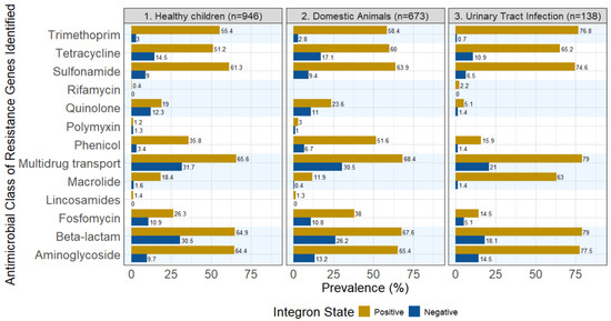
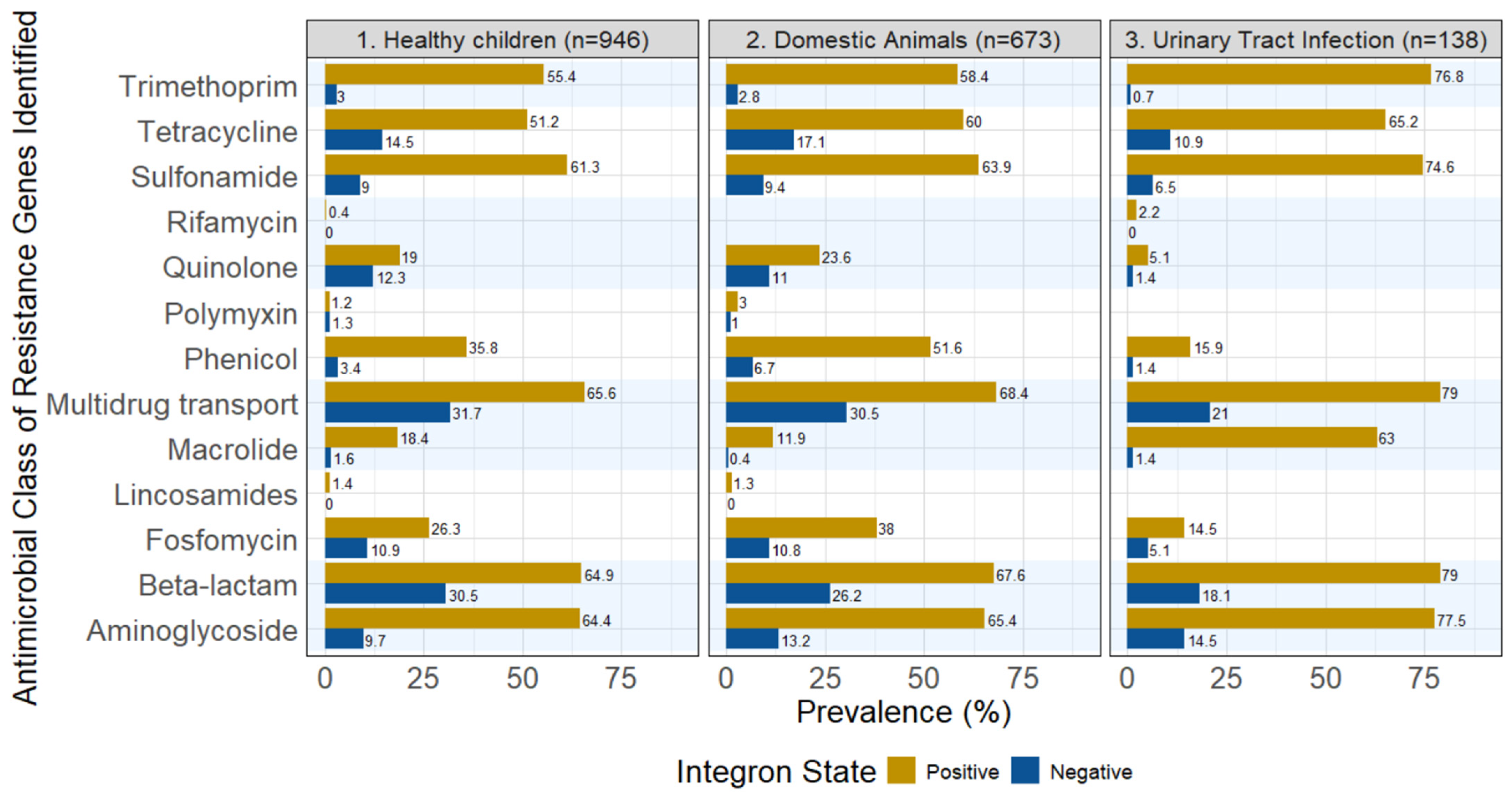
Figure 1 summarizes the number of AMR genes present in integron-positive versus integron-negative isolates. These genes conferred resistance to major drug classes, including first-line treatments (e.g., beta-lactamases and quinolones) and critically important antimicrobials such as polymyxin, alongside multidrug efflux pump genes (e.g., mdf, mdt, nor, and oqx). We grouped the resistance genes into 13 classes of AMR; nearly all classes were found in our isolates, whether they were integron-positive or integron-negative, with the exceptions of rifamycin and lincosamides, which were found with very low prevalences in integron-positive isolates, and absent in integron-negative isolates. Notably, the prevalence of polymyxin resistance was slightly higher in the integron-negative group in the healthy children dataset.
Figure 1.
Prevalence of AMR genes aggregated by antimicrobial classes across the three sampling sources. All isolates were 3GCR-Ec. Multidrug transport is a summary class of efflux pump genes such as mdf, mdt, nor, and oqx. Supplementary Tables S1–S3 contain the data presented in the figure.
The diversity of AMR genes in 3GCR-Ec from domestic animals (Figure 1) was similar to those found in 3GCR-Ec from healthy children, with comparable proportions across most classes.
For the 3GCR-Ec from UTI patients, resistance genes for 11 antimicrobial classes, out of 13, were detected; polymyxin and lincosamides were not detected. Of the classes detected, the dominant classes were trimethoprim, tetracycline, sulfonamide, multidrug transport (efflux pumps), beta-lactamase and aminoglycoside, although they tended to have a higher prevalence than the isolates from healthy children and domestic animals. Similar to the other 3GCR-Ec, AMR carriage was still found in integron-negative isolates in most classes, except for rifamycin; their prevalence compared to integron-positive isolates was also significantly lower. On the other hand, resistance to macrolides was much higher in the clinical UTI isolates than in the isolates from healthy children and domestic animals. For example, the integron-positive group had a macrolide resistance gene prevalence of 63% in 3GCR-Ec from UTI patients versus 18% in 3GCR-Ec from children and 11% in 3GCR-Ec from domestic animals. For the integron-negative isolates, the prevalence ranged from 0.4% in 3GCR-Ec from domestic animals to 1.6% in 3GCR-Ec from children. AMR genes were not identified for the lincosamide and polymyxin classes and quinolone and rifamycin AMR genes were found at a very low prevalence (See Supplemental Table S3 for data).
2.3. Beta-Lactamase Genes in Integron-Positive and Integron-Negative Isolates
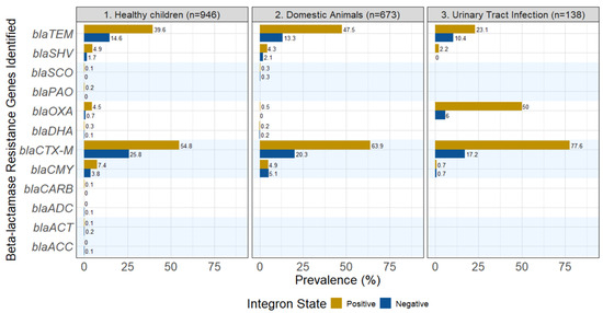
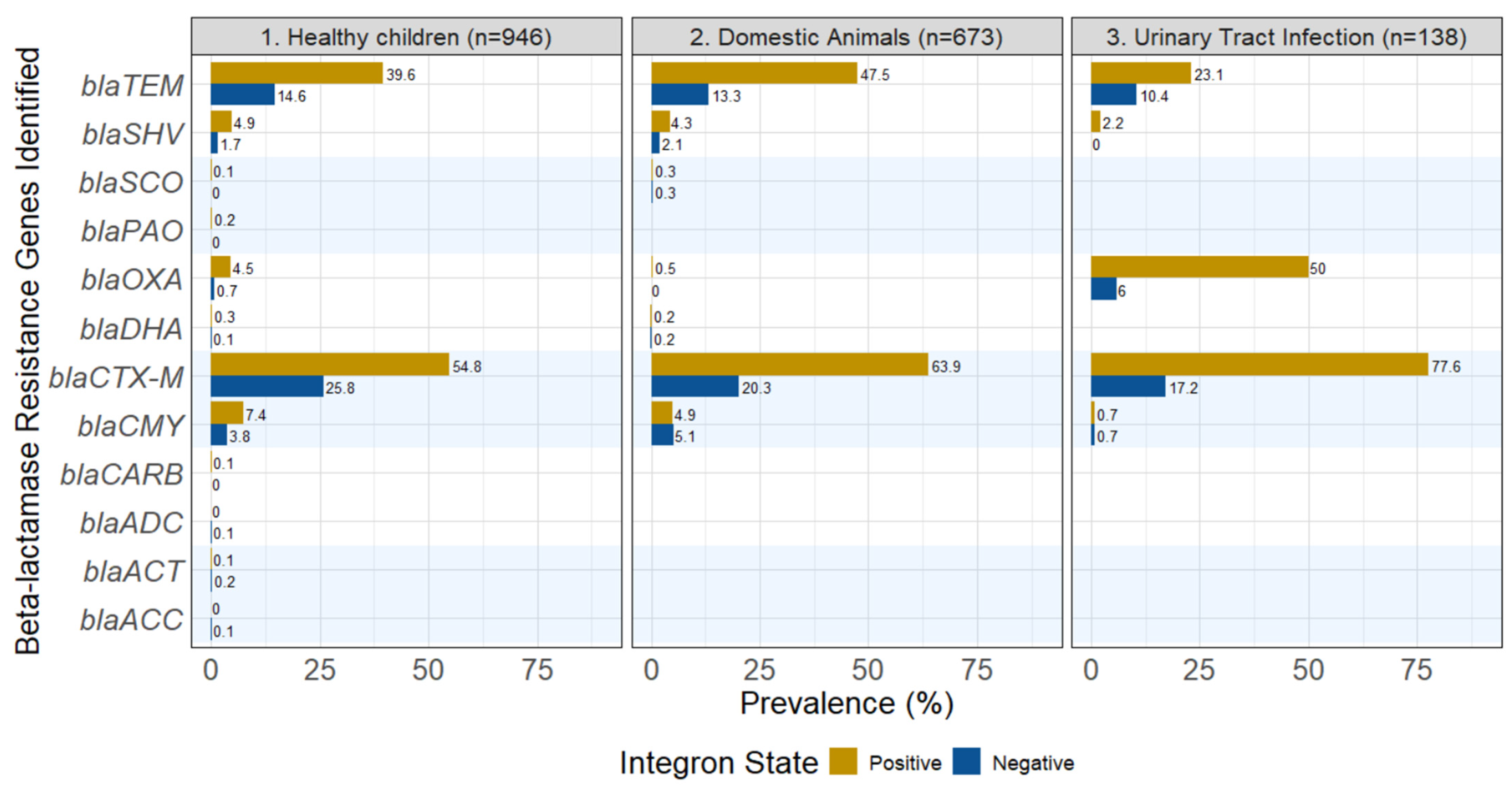
The prevalence of different beta-lactamase genes varied greatly across the 3GCR-Ec isolates from healthy children. The dominant beta-lactamase gene type was blaCTX-M, showing up in 54.8% of the integron-positive isolates from healthy children. The same blaCTX-M genes were still present in integron-negative isolates, but at lower levels (25.8%). Other frequent beta-lactamase genes included blaTEM, blaCMY and blaSHV. Their prevalences were higher in the integron-positive isolates from the children, but even in the absence of integrons, they were still present: 14.6% for blaTEM, 3.8% for blaCMY, and 1.7% for blaSHV. An additional eight beta-lactamase genes were found in a small proportion of isolates from healthy children (Figure 2).
Figure 2.
Prevalence of specific beta-lactamase genes for integron-positive and integron-negative 3GCR-Ec from healthy children, domestic animals and patients with urinary tract infections.
Among the isolates from domestic animals, the prevalences of various beta-lactamase genes were similar to those observed in children. Again, blaCTX-M was the most dominant resistance gene (63.9% for integron-positive isolates and 20.3% for integron-negative isolates), with blaTEM, blaCMY and blaSHV following as second, third and fourth in prevalence. Integron-positive isolates had higher prevalences, and integron-negative isolates had nearly one-third to one-half of the beta-lactamase genes as those found in integron-positive isolates.
In the 3GCR-Ec from UTI patients, blaCTX-M was again the most dominant resistance gene (77.6% for integron-positive isolates and 17.2% for integron-negative isolates). In contrast to the other isolates described above, integron-positive 3GCR-Ec from UTIs commonly had blaOXA (50.0% in integron-positive isolates versus 6.0% in integron-negative isolates). The blaTEM gene was the third most common beta-lactamase for integron-positive isolates, but second among integron-negative isolates (Figure 2). The beta-lactamase genes, blaSHV and blaCMY, were also identified in 3GCR-Ec from UTI patients, but at lower prevalences.
2.4. Sequence-Type Distribution in Integron-Positive and Integron-Negative Isolates
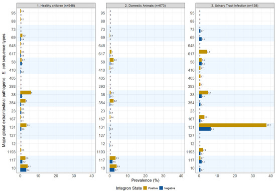
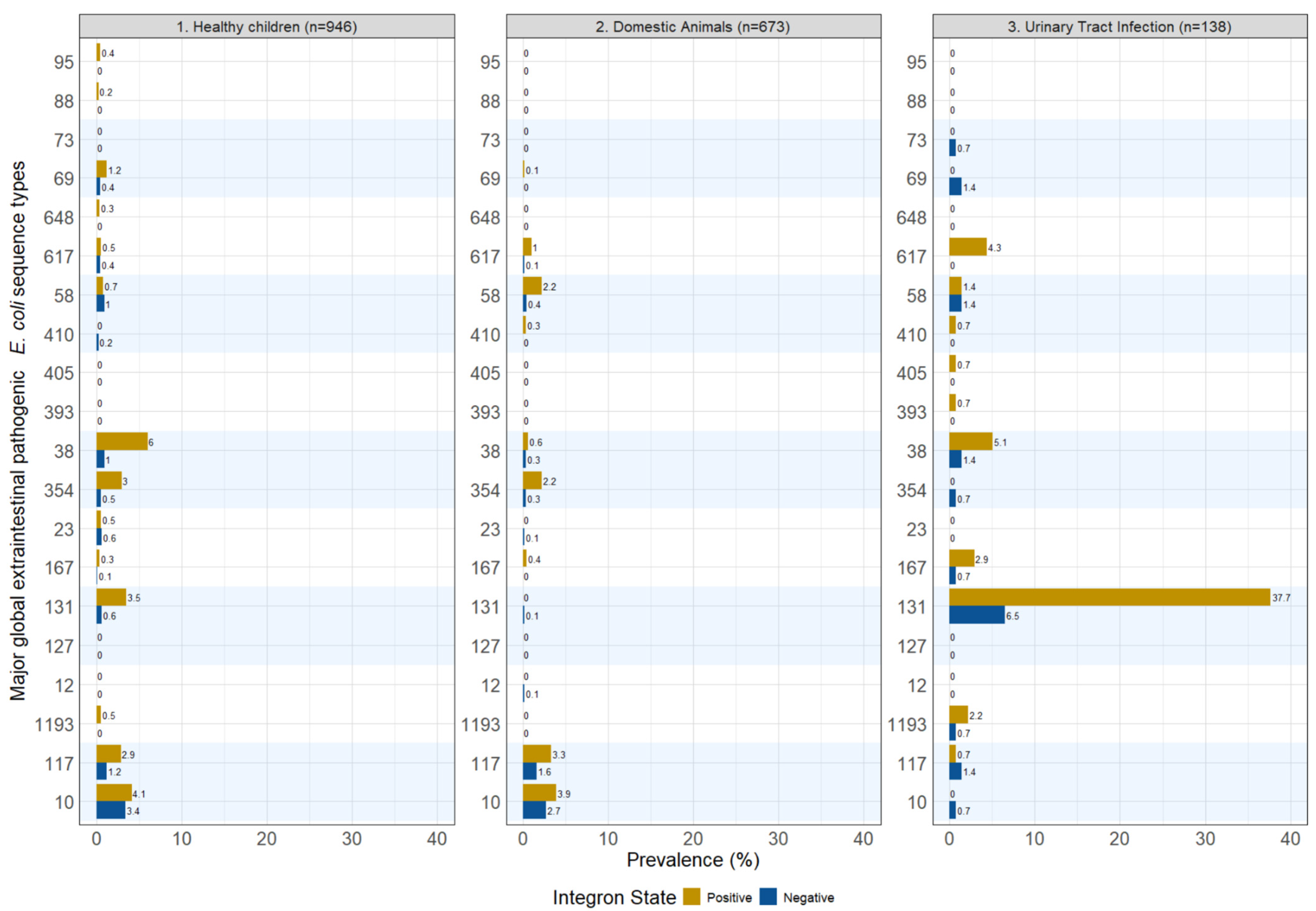
We compared integron carriage by known extraintestinal pathogenic E. coli sequence types (STs) (Figure 3). Specifically, we presented integron carriage among the top twenty global extraintestinal pathogenic E. coli sequence types based on a systematic review of the most prevalent extraintestinal pathogenic Escherichia coli (ExPEC) lineages identified in studies of extraintestinal infections [22]. Overall, the top twenty STs had a low prevalence across our sampled populations. ST131 was the most notable ST, commanding a total of 44.2% from our UTI patient cohort; 37.7% were integron-positive and 6.5% were integron-negative. Among healthy children, ST131 had 3.5% and 0.6% integron-positive and integron-negative prevalence, respectively. ST131 was essentially zero among the domestic animal isolates. ST38 had a similar distribution pattern as that of ST131. Across all of the isolates in each dataset, integron-positive ST38 was present in 5.1% of the UTI patient samples, 6% of the healthy children samples, and 0.6% among the domestic animal samples; integron-negative ST38 prevalence dropped to 1.4%, 1% and 0.3%, respectively, across these datasets. Two other sequence types, ST10 and ST117, were more prevalent among healthy children and domestic animals, but less so among patients with UTIs. Integron-positive ST 10 had prevalences of 4.1% and 3.4% in patients with UTIs and domestic animals, respectively, but was negative in the healthy children set. Some STs were seen at higher prevalence in their integron-negative state, including: ST69, ST73, ST354 and ST117 for patients with UTIs. Many of the top 20 global ExPEC lineages were absent in the three datasets. There were a total of 156 unique STs found among the healthy children, 144 STs among the domestic animals, and 34 STs among patients with UTIs.
Figure 3.
Presence and absence of integrons among the top 20 ExPEC Sequence Types for 3GCR-Ec isolated from healthy children, domestic animals and patients with urinary tract infections [22]. Note: The prevalence in this figure is a percentage of the total within the corresponding sample type. They do not add to 100%, as many more sequence types were present beyond the top 20 ExPEC lineages reported in this figure.
3. Discussion
Escherichia coli is notorious for horizontal gene transfer, an important pathogen in local infections, and the principal cause of UTIs and bacterial bloodstream infections [23]. Integrons are common genetic motifs frequently found in many bacteria, including E. coli. The prevalence of integrons in bacterial populations of South America has not been widely studied. In 2024, however, Solis et al. [24] reported finding integrons in almost one third of the E. coli isolates they collected from Andean countries, which included Venezuela, Peru, Ecuador, Colombia and Bolivia. The researchers highlighted that the prevalence of integrons within E. coli populations found in Ecuador was the highest, at nearly 40%. Our study also adds to this literature, and we found integron prevalence within 3GCR-Ec isolates averaging 69% across the three sources studied.
This present analysis of 3GCR-Ec isolates supports the notion that integrons are indeed important genetic elements associated with a higher burden of AMR genes in resistant E. coli. The data also highlight that integron-negative isolates constitute a significant source of AMR genes, containing an average of 4.8 (±2.5 SD) AMR genes. This value is lower than the average for those of integron-positive isolates, which contained an average of 10.3 (±3.0 SD) AMR genes, but not an insignificant number.
Prior studies have established that integron prevalence is source-dependent, suggesting a distinct function for integrons in host–pathogen co-evolutionary dynamics [25]. Furthermore, specific sequence types of E. coli lineages are well-known for their global dissemination and association with antimicrobial resistance [26]. Our findings from the diverse 3GCR-Escherichia coli samples collected in the semirural parishes near Quito, Ecuador, support the notion that integron-positive E. coli are indeed important strains associated with a higher burden of resistance genes. However, our data also highlight that integron-negative isolates constituted a significant source of antimicrobial resistance genes. While integron-positive isolates averaged a high number of resistance genes (approximately 10 per isolate), the integron-negative isolates were far from benign. The integron-negative isolates carried an average of nearly five AMR genes per isolate. Thus, diverse resistance genes in integron-negative isolates can drive multidrug resistance, complicating clinical treatment [27,28].
We observed a high diversity of AMR gene classes in both integron-positive and integron-negative strains across all three populations of 3GCR-Ec. The widespread occurrence of multidrug efflux pump genes (e.g., mdf, mdt, nor, and oqx) was also a concern. The detection of fosfomycin resistance is also of medical significance, although it is rarely prescribed in Ecuador for UTIs [29,30].
Integron-negative E. coli isolates accounted for approximately 30% of the total AMR genes detected in the beta-lactamase and multidrug resistance (efflux pump) categories. For example, integron-negative isolates were responsible for around 15% of the total AMR genes detected for both tetracycline and quinolone resistance; this is a small fraction, but not a completely insignificant contribution.
There were strong similarities between the 3GCR-Ec from healthy children and the domestic animals in terms of the prevalence of integrons, AMR genes, and more specifically, the prevalence of beta-lactamase genes. This finding highlights that human and domestic animal E. coli populations may be frequently exchanged due to interactions between humans, their animals, and their environments [31,32].
There were some cases where antimicrobial resistance to specific classes seemed unrelated to whether the isolate was integron-positive or negative. For instance, resistance to polymyxin, while low in overall prevalence (approximately 1%), was detected with similar frequency in both integron-positive and integron-negative isolates. In contrast, quinolone resistance in integron-positive isolates was nearly half that of integron-negative isolates (19% vs. 12% in 3GCR-Ec from children and 23% vs. 11% in 3GCR-Ec from domestic animals). These two classes of antimicrobials are also non-trivial, as both quinolones and polymyxins are classified as Highest Priority Critically Important Antimicrobials (HPCIA) by the World Health Organization [33]; thus, the presence of those resistance genes is concerning.
While integron presence was associated with more beta-lactamase genes on average, they were not essential for beta-lactamase gene carriage [34]. After removing all integron-positive isolates, there would still remain a prevalence of blaCTX-M in 25.8% of the isolates from healthy children, and 17.2% of the isolates from patients with UTIs. The case for blaTEM, while lower, would still account for 14.6% and 10.4% of 3GCR-Ec from children and UTI patients, respectively. These numbers, while lower, are not low enough to be medically ignored for their contribution to human health. Interestingly, although blaCTX-M genes are transferred between bacteria by mobile genetic elements, like transposable elements [35], reports have detected these genes within the integron’s variable region (the cassette) [36,37].
While there were specific differences in the AMR classes and prevalence of integrons found between the isolates in healthy humans versus domestic animals, on the aggregate, we found them to be very similar. This is likely due to frequent exchange of bacterial populations and their resistance genes between humans and animals living in proximity.
In terms of integron carriage by sequence types known to cause disease globally, we found that integron-positive isolates were more prevalent across most STs. This finding aligns with the distribution of integron carriage across various resistance genes discussed above. The most notable sequence type was ST131 in the UTI patient dataset. ST131 is the most frequent ST among the top 20 global ExPEC lineages as reported by a systematic review by Manges et al. [22]. In our UTI patient cohort, 37.7% and 6.5% of the isolates are attributed to the integron-positive and integron-negative isolates, respectively, totaling 44.2%.
Several key pandemic sequence types—specifically ST69, ST73, ST354 and ST10—were present, but were exclusively integron-negative isolates in our UTI patient dataset. We note that ST69 is the second-highest among the top 20 ExPEC STs, and no integron-positive isolates were found among our UTI patients. This study showed that integrons are often present in many multidrug-resistant lineages like ST131, but absent in many pandemic lineages.
There were several limitations to this study. While healthy children and domestic animal isolates were collected at the same community concurrently, UTI isolates were collected during a different time period. The temporal gap between the collection of UTI isolates (2014–2015) and subsequent samples (2019–2021) is a limitation. Given the rapid evolution of AMR, the older isolates may reflect a baseline resistance profile that has since shifted due to changing antibiotic stewardship practices or the emergence of new clones in the intervening five years. Additionally, we did not determine the genomic location of AMR genes nor whether these genes were present within integron-associated gene cassettes. The relative positioning of AMR genes with respect to integrons warrants further investigation, as analyses would provide valuable mechanistic insights into the role of integrons in shaping AMR gene repertoires. However, the primary objective of this study was to assess how the presence or absence of integrons within an isolate influences AMR gene prevalence across different populations of 3GCR-Ec. The study showed that there was discriminatory power with the use of integrons, but that important AMR genes were still found in integron-negative isolates. Future studies may also benefit by further elucidating whether certain farm animals, or pets, may confer the same or different AMR risks.
In this study, we analyzed sequence data from three sets of 3GCR-Ec derived from healthy children, domestic animals and patients with a UTI to better understand how integrons are associated with antimicrobial resistance gene carriage. Studies have shown that E. coli lineages that cause UTIs, versus commensal lineages, may experience higher selective pressure as they are exposed more often to antibiotic treatments [38]. The carriage of integrons in the UTI isolates was likely associated with this selective pressure. Our findings indicate that while integrons may be associated with a higher prevalence of AMR genes, important resistance determinants are also common in integron-negative isolates. This highlights the need for future analyses that integrate integron carriage with bacterial lineage and AMR gene profiles to better understand the mechanisms driving AMR dissemination in commensal and pathogenic E. coli populations.
4. Materials and Methods
4.1. 3GCR-Ec from Healthy Children
A total of 1699 fecal samples from 600 children were collected from seven semirural parishes east of Quito, Ecuador, as part of an existing study conducted by researchers at the Universidad San Francisco de Quito (USFQ), Ecuador [21]. A total of 376 households were involved, with repeated sample collection spanning over 3 years (2019–2021). 3GCR and ESBL-producing E. coli from the samples were determined phenotypically: Samples were cultured on MacConkey agar (Difco, Sparks, MA, USA) with 2 mg/L of ceftriaxone and incubated for 18 h at 37 °C [39]. Up to 2 ceftriaxone-resistant isolates phenotypically matching E. coli were selected from each plate. The identity of presumptive E. coli colonies was confirmed by culture on Chromocult coliform agar (Merck KGaA, Darmstadt, Germany), at 37 °C for 24 h, through its β-D-glucuronidase activity, followed by the multi-substrate API RapiD-20E identification system (bioMérieux, Marcy l’Etoile, France) using a cut-off of 95%. Confirmed E. coli were preserved at −80 °C in trypticase soy broth medium (Difco, Sparks, MA, USA) containing 20% glycerol. 3GCR-EC isolates for each sample were thawed and regrown on MacConkey agar at 37 °C for 18–24 h for evaluation of antibiotic susceptibility by the disk diffusion method (Kirby–Bauer test) on Mueller–Hinton agar (Difco, Sparks, MA, USA). Presumptive E. coli colonies were confirmed on Chromocult coliform agar (Merck, Darmstadt, Germany). DNA was extracted from the isolates using Wizard Genomic DNA Purification (Promega, Madison, WI, USA) kits and DNEasy Blood & Tissue Kits (QIAGEN, Hilden, Germany) according to the manufacturer’s protocol. Confirmed 3GCR-E. coli isolates were sequenced at the University of Minnesota Genomic Center using Illumina MiSeq or NovaSeq with Nextera XT libraries (San Diego, CA, USA).
4.2. 3GCR-Ec from Domestic Animals
The study included 1871 domestic animal fecal samples from the same households in semirural parishes of Ecuador as described above. The sampling area, approximately 320 km2, includes both commercial food animal operations and small-scale “backyard” animal production. Samples were cultured and DNA extraction was done as described in Section 4.1 above. Animals sampled included chickens, pigs, guinea pigs, cattle, sheep and goats. For a detailed breakdown of their composition, please see [21].
4.3. 3GCR-Ec from Patients with a Urinary Tract Infection
A total of 149 ceftriaxone-resistant E. coli isolates were collected from nine clinics east of Quito, Ecuador, serving the same general community as the above healthy children and domestic animals. As previously described [40], samples were taken from patients with a UTI over a 12-month period between May 2014 and May 2015. Samples were sent to the USFQ team for bacterial culture and analysis. Each bacterial isolate was cultured on Chromocult agar (Merck, Darmstadt, Germany). E. coli were identified through β-D-glucuronidase activity and preserved at −80 °C in Brain Heart Infusion (BHI) medium, containing 20% glycerol. On each plate, a single bacterial colony was selected and inoculated into 3 mL of trypticase soy broth (TSB) at 37 °C for 18–24 h. DNA from the bacterial isolates was extracted with the QIAGEN DNeasy Blood & Tissue kit using the manufacturer’s instructions. QC was done using the NanoDrop One (Thermo Scientific, Madison, WI, USA). Whole genome sequencing was performed using Illumina NextSeq 2000 (San Diego, CA, USA).
4.4. Whole Genome Sequencing and Bioinformatics Analyses
De novo assemblies were performed using SPAdes v3.15 and contigs were joined into a scaffold with a fixed gap size of 100 bp [41]. Integron carriage was identified using Integron Finder 2.0.2, using linear topology, a distance threshold of 4000 bp, a calin threshold of 2, and a minimum and maximum attC size of 40 and 200 bp, respectively [2,42]. An isolate was classified as integron-positive if it contained either a complete or partial integron, and negative if no integron was detected. The rationale for this method was that some researchers might perform integron detection without regard to whether the integron was complete or partial; thus, we matched on the lowest common denominator. AMR genes were determined by ABRicate 1.0.1 [43] using the ResFinder database version 4 which covers 13 classes of AMR genes [7,44] using default parameters. Data analysis was performed using RStudio 2025.09.2 [45].
To determine the sequence type (ST) for each isolate, species identity was first confirmed via ribosomal multi-locus sequence typing (rMLST) v0.1.8 [46,47]. Confirmed E. coli isolates were then characterized using traditional MLST to assign specific STs (Seeman MLST v 2.19.0) [48,49]. Integron carriage for each isolate was ascertained. The prevalence of specific lineages was calculated by dividing the number of isolates of a given ST and integron status by the total number of isolates within each respective dataset (healthy children, domestic animals, and patients with urinary tract infections).
5. Conclusions
Integrons play a significant role in the evolution of E. coli and the dissemination of antimicrobial resistance genes, underscoring the need for continued study. This analysis of three distinct E. coli populations from healthy children, domestic animals and UTI patients demonstrated that a substantial proportion of E. coli isolates (31%) were integron-negative yet collectively harbored a significant burden of AMR (an average of 4.8 AMR genes per isolate). While integrons are pivotal in the evolution and dissemination of resistance in E. coli, the findings highlight the limitations of using them as a sole proxy for AMR. To more accurately track AMR across the One Health interface, surveillance programs should adopt a broader genomic strategy that accounts for diverse mobilization elements relevant to clinical outcomes.
Supplementary Materials
The following supporting information can be downloaded at https://www.mdpi.com/article/10.3390/antibiotics15050427/s1. Table S1: Prevalence of AMR genes for Healthy children; Table S2: Prevalence of AMR genes for Domestic Animals; Table S3: Prevalence of AMR genes for Urinary Tract Infection (UTI) patients.
Author Contributions
Conceptualization, J.G., N.W., M.P. and T.H.; methodology, J.G., H.K.A., L.S., N.W., M.P. and G.T.; software, H.K.A., L.S., G.T., N.W. and T.H.; formal analysis, T.H., N.W., M.P. and J.G.; investigation, J.G., H.K.A., L.S., G.T. and T.H.; data curation, H.K.A., L.S., G.T., T.J., J.G., N.W., M.P. and T.H.; writing—original draft preparation, T.H. and M.P.; writing—review and editing, T.H., J.G., L.S. and N.W.; visualization T.H. and J.G.; project administration, G.T. and L.S.; funding acquisition, J.G. All authors have read and agreed to the published version of the manuscript.
Funding
This study was funded by the National Institute of Allergy and Infectious Diseases of the National Institutes of Health (NIH) under Award Numbers R01AI135118 and 1R01AI167989-01.
Institutional Review Board Statement
Methods and procedures that collect the data in the upstream studies were reviewed and approved by their respective Institutional Review Boards. The healthy children and domestic animals datasets were reviewed and approved by the Office for Protection of Human Subjects at the University of California, Berkeley (IRB# 2019-02-11803), with an approval date of 16 April 2019, the Bioethics Committee at the Universidad San Francisco de Quito (#2017-178M), and the Ecuadorian Health Ministry (#MSPCURI000243-3). Formal written consent was obtained from each primary caregiver of children enrolled in the study prior to participation. The clinical UTI patient’s dataset was approved by the USFQ bioethics committee (code 2015-167T); the approval date was 12 April 2023.
Informed Consent Statement
Not applicable.
Data Availability Statement
All sequences were deposited in NCBI under the Bioproject accession Nos. PRJNA861272 and PRJNA1168579.
Acknowledgments
This research used the Savio computational cluster resource provided by the Berkeley Research Computing program at the University of California, Berkeley (supported by the UC Berkeley Chancellor, Vice Chancellor for Research, and Chief Information Officer). This publication made use of the PubMLST website (https://pubmlst.org/) developed by Keith Jolley Wellcome Open Res. 2018 Sep 24:3:124 and sited at the University of Oxford. The development of that website was funded by the Wellcome Trust. Finally, special thanks to the reviewers who greatly improved the manuscript with their constructive comments.
Conflicts of Interest
The authors declare no conflicts of interest. The funders had no role in the design of the study; in the collection, analyses, or interpretation of data; in the writing of the manuscript; or in the decision to publish the results.
References
- Cambray, G.; Guerout, A.-M.; Mazel, D. Integrons. Annu. Rev. Genet. 2010, 44, 141–166. [Google Scholar] [CrossRef] [Scilit]
- Néron, B.; Littner, E.; Haudiquet, M.; Perrin, A.; Cury, J.; Rocha, E.P. IntegronFinder 2.0: Identification and Analysis of Integrons across Bacteria, with a Focus on Antibiotic Resistance in Klebsiella. Microorganisms 2022, 10, 700. [Google Scholar] [CrossRef] [Scilit] [PubMed]
- Escudero, J.A.; Loot, C.; Nivina, A.; Mazel, D. The Integron: Adaptation on Demand. Microbiol. Spectr. 2015, 3, 10–1128. [Google Scholar] [CrossRef] [Scilit]
- Boucher, Y.; Labbate, M.; Koenig, J.E.; Stokes, H. Integrons: Mobilizable Platforms That Promote Genetic Diversity in Bacteria. Trends Microbiol. 2007, 15, 301–309. [Google Scholar] [CrossRef] [Scilit] [PubMed]
- Suhartono, S.; Savin, M.C.; Gbur, E.E. Transmissible Plasmids and Integrons Shift Escherichia coli Population toward Larger Multiple Drug Resistance Numbers. Microb. Drug Resist. 2018, 24, 244–252. [Google Scholar] [CrossRef] [Scilit]
- Mathai, E.; Grape, M.; Kronvall, G. Integrons and Multidrug Resistance among Escherichia coli Causing Community-Acquired Urinary Tract Infection in Southern India. Apmis 2004, 112, 159–164. [Google Scholar] [CrossRef] [Scilit] [PubMed]
- Gillings, M.R.; Gaze, W.H.; Pruden, A.; Smalla, K.; Tiedje, J.M.; Zhu, Y.-G. Using the Class 1 Integron-Integrase Gene as a Proxy for Anthropogenic Pollution. ISME J. 2015, 9, 1269–1279. [Google Scholar] [CrossRef] [Scilit]
- Barraud, O.; Laval, L.; Le Devendec, L.; Larvor, E.; Chauvin, C.; Jouy, E.; Le Bouquin, S.; Vanrobaeys, Y.; Thuillier, B.; Lamy, B.; et al. Integrons from Aeromonas Isolates Collected from Fish: A Global Indicator of Antimicrobial Resistance and Anthropic Pollution. Aquaculture 2023, 576, 739768. [Google Scholar] [CrossRef] [Scilit]
- Bhat, B.A.; Mir, R.A.; Qadri, H.; Dhiman, R.; Almilaibary, A.; Alkhanani, M.; Mir, M.A. Integrons in the Development of Antimicrobial Resistance: Critical Review and Perspectives. Front. Microbiol. 2023, 14, 1231938. [Google Scholar] [CrossRef] [Scilit]
- Tavares, R.D.; Fidalgo, C.; Rodrigues, E.T.; Tacão, M.; Henriques, I. Integron-Associated Genes Are Reliable Indicators of Antibiotic Resistance in Wastewater despite Treatment-and Seasonality-Driven Fluctuations. Water Res. 2024, 258, 121784. [Google Scholar] [CrossRef] [Scilit]
- Souque, C.; Escudero, J.A.; MacLean, R.C. Integron Activity Accelerates the Evolution of Antibiotic Resistance. Elife 2021, 10, e62474. [Google Scholar] [CrossRef] [Scilit]
- Ali, N.; Ali, I.; Din, A.U.; Akhtar, K.; He, B.; Wen, R. Integrons in the Age of Antibiotic Resistance: Evolution, Mechanisms, and Environmental Implications: A Review. Microorganisms 2024, 12, 2579. [Google Scholar] [CrossRef] [Scilit]
- Racewicz, P.; Majewski, M.; Biesiada, H.; Nowaczewski, S.; Wilczyński, J.; Wystalska, D.; Kubiak, M.; Pszczoła, M.; Madeja, Z.E. Prevalence and Characterisation of Antimicrobial Resistance Genes and Class 1 and 2 Integrons in Multiresistant Escherichia coli Isolated from Poultry Production. Sci. Rep. 2022, 12, 6062. [Google Scholar] [CrossRef] [Scilit]
- Kim, E.; Nealon, N.J.; Murray, K.A.; Jardine, C.; Magnuson, R.; Rao, S. Integron-Mediated Antimicrobial Resistance and Virulence Factors in Salmonella Typhimurium Isolated from Poultry. Animals 2024, 14, 3483. [Google Scholar] [CrossRef] [Scilit]
- Ghatani, R.; Pongener, N.; Visi, V. Inappropriate Use of Third-Generation Cephalosporins and Antimicrobial Resistance in Hospital Settings: A Systematic Review and Meta-Analysis. Int. J. Med. Biomed. Stud. 2025, 9, 32–42. [Google Scholar] [CrossRef] [Scilit]
- Salinas, L.; Loayza, F.; Cárdenas, P.; Saraiva, C.; Johnson, T.J.; Amato, H.; Graham, J.P.; Trueba, G. Environmental Spread of Extended Spectrum Beta-Lactamase (ESBL) Producing Escherichia coli and ESBL Genes among Children and Domestic Animals in Ecuador. Environ. Health Perspect. 2021, 129, 027007. [Google Scholar] [CrossRef] [Scilit] [PubMed]
- Bezabih, Y.M.; Sabiiti, W.; Alamneh, E.; Bezabih, A.; Peterson, G.M.; Bezabhe, W.M.; Roujeinikova, A. The Global Prevalence and Trend of Human Intestinal Carriage of ESBL-Producing Escherichia coli in the Community. J. Antimicrob. Chemother. 2021, 76, 22–29. [Google Scholar] [CrossRef] [Scilit] [PubMed]
- Belley, A.; Morrissey, I.; Hawser, S.; Kothari, N.; Knechtle, P. Third-Generation Cephalosporin Resistance in Clinical Isolates of Enterobacterales Collected between 2016–2018 from USA and Europe: Genotypic Analysis of β-Lactamases and Comparative in Vitro Activity of Cefepime/Enmetazobactam. J. Glob. Antimicrob. Resist. 2021, 25, 93–101. [Google Scholar] [CrossRef] [Scilit]
- Abdullahi, I.N.; Trabelsi, I. Guts of Healthy Humans, Livestock, and Pets Harbor Critical-Priority and High-Risk Escherichia coli Clones. Epidemiol. Health 2025, 47, e2025013. [Google Scholar] [CrossRef] [Scilit]
- Naghavi, M.; Vollset, S.E.; Ikuta, K.S.; Swetschinski, L.R.; Gray, A.P.; Wool, E.E.; Aguilar, G.R.; Mestrovic, T.; Smith, G.; Han, C.; et al. Global Burden of Bacterial Antimicrobial Resistance 1990–2021: A Systematic Analysis with Forecasts to 2050. Lancet 2024, 404, 1199–1226. [Google Scholar] [CrossRef] [Scilit]
- Amato, H.K.; Loayza, F.; Salinas, L.; Paredes, D.; Garcia, D.; Sarzosa, S.; Saraiva-Garcia, C.; Johnson, T.J.; Pickering, A.J.; Riley, L.W.; et al. Risk Factors for Extended-Spectrum Beta-Lactamase (ESBL)-Producing E. Coli Carriage among Children in a Food Animal-Producing Region of Ecuador: A Repeated Measures Observational Study. PLoS Med. 2023, 20, e1004299. [Google Scholar] [CrossRef] [Scilit]
- Manges, A.R.; Geum, H.M.; Guo, A.; Edens, T.J.; Fibke, C.D.; Pitout, J.D. Global Extraintestinal Pathogenic Escherichia coli (ExPEC) Lineages. Clin. Microbiol. Rev. 2019, 32, 10–1128. [Google Scholar] [CrossRef] [Scilit] [PubMed]
- Han, N.; Peng, X.; Zhang, T.; Qiang, Y.; Li, X.; Zhang, W. Rapid Turnover and Short-Term Blooms of Escherichia coli in the Human Gut. J. Bacteriol. 2024, 206, e00239-23. [Google Scholar] [CrossRef] [Scilit] [PubMed]
- Solis, M.N.; Loaiza, K.; Torres-Elizalde, L.; Mina, I.; Šefcová, M.A.; Larrea-Álvarez, M. Detecting Class 1 Integrons and Their Variable Regions in Escherichia coli Whole-Genome Sequences Reported from Andean Community Countries. Antibiotics 2024, 13, 394. [Google Scholar] [CrossRef] [Scilit]
- Sunde, M.; Simonsen, G.S.; Slettemeås, J.S.; Böckerman, I.; Norström, M. Integron, Plasmid and Host Strain Characteristics of Escherichia coli from Humans and Food Included in the Norwegian Antimicrobial Resistance Monitoring Programs. PLoS ONE 2015, 10, e0128797. [Google Scholar] [CrossRef] [Scilit] [PubMed]
- Wyrsch, E.R.; Bushell, R.N.; Marenda, M.S.; Browning, G.F.; Djordjevic, S.P. Global Phylogeny and F Virulence Plasmid Carriage in Pandemic Escherichia coli ST1193. Microbiol. Spectr. 2022, 10, e02554-22. [Google Scholar] [CrossRef] [Scilit]
- Olsen, N.S.; Riber, L. Metagenomics as a Transformative Tool for Antibiotic Resistance Surveillance: Highlighting the Impact of Mobile Genetic Elements with a Focus on the Complex Role of Phages. Antibiotics 2025, 14, 296. [Google Scholar] [CrossRef] [Scilit]
- Akunne, O.Z.; Emmanuel, B.N.; Saidu, U.F.; Akinnawo, A.S.; Adeyemi, N.A.; Egoh, L.C.; Muhammad, M.A.; Ade-adekunle, O.A.; Peter, D.A.; Freitas, A.A. Antibiotic Resistance: The Growing Threats and Potential Solutions. Am. J. Pharmacother. Pharm. Sci. 2025, 4, 13. [Google Scholar] [CrossRef] [Scilit]
- Freedman, S.B.; Xie, J.; Neufeld, M.S.; Hamilton, W.L.; Hartling, L.; Tarr, P.I.; Alberta Provincial Pediatric Enteric Infection Team (APPETITE); Nettel-Aguirre, A.; Chuck, A.; Lee, B.; et al. Shiga Toxin–Producing Escherichia coli Infection, Antibiotics, and Risk of Developing Hemolytic Uremic Syndrome: A Meta-Analysis. Clin. Infect. Dis. 2016, 62, 1251–1258. [Google Scholar] [CrossRef] [Scilit]
- Ikeda, K.; Ida, O.; Kimoto, K.; Takatorige, T.; Nakanishi, N.; Tatara, K. Effect of Early Fosfomycin Treatment on Prevention of Hemolytic Uremic Syndrome Accompanying Escherichia coli O157: H7 Infection. Clin. Nephrol. 1999, 52, 357–362. [Google Scholar]
- Walas, N.; Müller, N.F.; Parker, E.; Henderson, A.; Capone, D.; Brown, J.; Barker, T.; Graham, J.P. Application of Phylodynamics to Identify Spread of Antimicrobial-Resistant Escherichia coli between Humans and Canines in an Urban Environment. Sci. Total Environ. 2024, 916, 170139. [Google Scholar] [CrossRef] [Scilit]
- Loayza, F.; Graham, J.P.; Trueba, G. Factors Obscuring the Role of E. Coli from Domestic Animals in the Global Antimicrobial Resistance Crisis: An Evidence-Based Review. Int. J. Environ. Res. Public Health 2020, 17, 3061. [Google Scholar] [CrossRef] [Scilit]
- World Health Organization (WHO). List of Medically Important Antimicrobials: A Risk Management Tool for Mitigating Antimicrobial Resistance Due to Non-Human Use; World Health Organization (WHO): Geneva, Switzerland, 2024.
- Tavares, R.D.; Tacao, M.; Henriques, I. Integrons Are Key Players in the Spread of Beta-Lactamase-Encoding Genes. Int. J. Antimicrob. Agents 2025, 65, 107421. [Google Scholar] [CrossRef] [Scilit]
- Lartigue, M.-F.; Poirel, L.; Aubert, D.; Nordmann, P. In Vitro Analysis of IS Ecp1B-Mediated Mobilization of Naturally Occurring β-Lactamase Gene Bla CTX-M of Kluyvera Ascorbata. Antimicrob. Agents Chemother. 2006, 50, 1282–1286. [Google Scholar]
- Casella, T.; Rodríguez, M.M.; Takahashi, J.T.; Ghiglione, B.; Dropa, M.; Assuncao, E.; Nogueira, M.L.; Lincopan, N.; Gutkind, G.; Nogueira, M.C.L. Detection of blaCTX-M-Type Genes in Complex Class 1 Integrons Carried by Enterobacteriaceae Isolated from Retail Chicken Meat in Brazil. Int. J. Food Microbiol. 2015, 197, 88–91. [Google Scholar] [CrossRef] [Scilit]
- Arduino, S.M.; Roy, P.H.; Jacoby, G.A.; Orman, B.E.; Pineiro, S.A.; Centron, D. Bla CTX-M-2 Is Located in an Unusual Class 1 Integron (In35) Which Includes Orf513. Antimicrob. Agents Chemother. 2002, 46, 2303–2306. [Google Scholar] [CrossRef] [Scilit]
- Guilcazo, D.; López, L.; Calderón, D.; Vasquez, K.; Chávez, C.; Price, L.B.; Graham, J.P.; Eisenberg, J.; Trueba, G. Comparative Analysis of ESBL Phenotypes and Antimicrobial Resistance in Escherichia coli Associated with Urinary Tract Infections and in Commensal Strains. Microb. Drug Resist. 2025, 31, 380–383. [Google Scholar] [CrossRef] [Scilit]
- Lautenbach, E.; Bilker, W.B.; Tolomeo, P.; Maslow, J.N. Impact of Diversity of Colonizing Strains on Strategies for Sampling Escherichia coli from Fecal Specimens. J. Clin. Microbiol. 2008, 46, 3094–3096. [Google Scholar] [CrossRef] [Scilit]
- Guilcazo, D.; Salinas, L.; Chavez, C.; Vasquez, K.; Mendez, G.I.; Price, L.B.; Graham, J.P.; Eisenberg, J.N.; Trueba, G. Tracking Bla CTX-M Transmission through Transposable Elements in Uropathogenic and Commensal E. Coli. Future Microbiol. 2025, 20, 287–293. [Google Scholar]
- Prjibelski, A.; Antipov, D.; Meleshko, D.; Lapidus, A.; Korobeynikov, A. Using SPAdes de Novo Assembler. Curr. Protoc. Bioinform. 2020, 70, e102. [Google Scholar] [CrossRef] [Scilit]
- Cury, J.; Jové, T.; Touchon, M.; Néron, B.; Rocha, E.P. Identification and Analysis of Integrons and Cassette Arrays in Bacterial Genomes. Nucleic Acids Res. 2016, 44, 4539–4550. [Google Scholar] [CrossRef] [Scilit]
- Seemann, T. ABRicate; GitHub: San Francisco, CA, USA; Available online: https://github.com/tseemann/abricate (accessed on 10 January 2026).
- Bortolaia, V.; Kaas, R.S.; Ruppe, E.; Roberts, M.C.; Schwarz, S.; Cattoir, V.; Philippon, A.; Allesoe, R.L.; Rebelo, A.R.; Florensa, A.F.; et al. ResFinder 4.0 for Predictions of Phenotypes from Genotypes. J. Antimicrob. Chemother. 2020, 75, 3491–3500. [Google Scholar] [CrossRef] [Scilit]
- Posit Team. RStudio: Integrated Development Environment for R; Posit Software; PBC: Boston, MA, USA, 2024. [Google Scholar]
- Jolley, K.A.; Bliss, C.M.; Bennett, J.S.; Bratcher, H.B.; Brehony, C.; Colles, F.M.; Wimalarathna, H.; Harrison, O.B.; Sheppard, S.K.; Cody, A.J.; et al. Ribosomal Multilocus Sequence Typing: Universal Characterization of Bacteria from Domain to Strain. Microbiology 2012, 158, 1005–1015. [Google Scholar] [CrossRef] [Scilit]
- Jolley, K.A.; Maiden, M.C. BIGSdb: Scalable Analysis of Bacterial Genome Variation at the Population Level. BMC Bioinform. 2010, 11, 595. [Google Scholar] [CrossRef] [Scilit]
- Wirth, T.; Falush, D.; Lan, R.; Colles, F.; Mensa, P.; Wieler, L.H.; Karch, H.; Reeves, P.R.; Maiden, M.C.; Ochman, H.; et al. Sex and Virulence in Escherichia coli: An Evolutionary Perspective. Mol. Microbiol. 2006, 60, 1136–1151. [Google Scholar] [CrossRef] [Scilit]
- Seemann, T. MLST; GitHub: San Francisco, CA, USA; Available online: https://github.com/tseemann/mlst (accessed on 10 January 2026).

Disclaimer/Publisher’s Note: The statements, opinions and data contained in all publications are solely those of the individual author(s) and contributor(s) and not of MDPI and/or the editor(s). MDPI and/or the editor(s) disclaim responsibility for any injury to people or property resulting from any ideas, methods, instructions or products referred to in the content. |
© 2026 by the authors. Licensee MDPI, Basel, Switzerland. This article is an open access article distributed under the terms and conditions of the Creative Commons Attribution (CC BY) license.