


Abstract
Exosomal miRNAs are emerging as attractive liquid-biopsy biomarkers because of their stability, specificity, and association with cancer progression. Microfluidic technologies can improve multiplex exosomal miRNAs analysis by reducing sample consumption, organizing parallel reactions, integrating extracellular-vesicle enrichment and lysis, and enabling on-chip signal decoding. This review critically compares three implementation routes-spatial encoding, bead-based encoding, and in situ detection-with emphasis on the experimentally demonstrated multiplexing level rather than theoretical coding capacity. Representative studies are compared by analyte scope, microfluidic function, target number, limit of detection, sample type, and clinical validation. Studies using total circulating miRNAs, cell or tissue lysates, non-miRNA exosomal cargoes, or in vivo imaging are explicitly identified as technology-transfer examples rather than direct demonstrations of microfluidic multiplex exosomal miRNA profiling. The remaining challenges include harmonized reporting, scalable on-chip sample preparation, robust decoding, and validation in independent clinical cohorts.
1. Introduction
MiRNAs are a class of small, non-coding RNA molecules that are widely present in eukaryotes. Mature miRNAs are derived from hairpin precursor molecules of 70–100 nucleotides in length and are ultimately processed into single-stranded RNAs of 19–25 bases. More than 2600 human mature miRNAs have been reported so far [1]. As post-transcriptional regulators, miRNAs are widely involved in key biological processes and play critical roles in various aspects, including gene expression, individual development, cell proliferation, differentiation and apoptosis, biological evolution, and disease occurrence in eukaryotes [2]. It primarily exerts regulatory effects through two mechanisms: the first involves partial complementary pairing of the miRNA with its target, thereby inhibiting protein translation; the second involves complete binding to the target mRNA, guiding the RNA-induced silencing complex to cleave the mRNA molecule, resulting in mRNA degradation. Therefore, miRNA can play a role in intercellular communication and regulate genes by inhibiting translation or degrading mRNA.
Exosomes are small membrane vesicles with a diameter of approximately 40–150 nm, secreted by cells. They are primarily derived from multivesicular bodies formed by the inward budding of endosomal membranes, which upon fusion with the plasma membrane, release exosomes into the extracellular matrix. High-throughput sequencing has shown that exosomes can carry a variety of bioactive substances, including proteins, lipids, mitochondrial DNA, and numerous non-coding RNAs (miRNA, lncRNA, etc.) [3,4]. Under the influence of their surface molecules, exosomes transport these cargoes toward recipient cells. Once attached to target cells, exosomes either induce signaling through receptor-ligand interactions or are internalized through phagocytosis, thereby altering various physiological and pathological processes of the recipient cells [5]. Due to differences in both the content and types of miRNAs they carry, exosomal miRNAs derived from different cell types participate in distinct biological processes, thereby producing different biological effects. They have demonstrated significant value in early diagnosis, disease mechanism investigation, and prognosis evaluation, and are considered promising liquid biopsy biomarkers, making them indispensable tools in both basic medical research and clinical practice [6]. However, conventional detection methods are often limited by labor-intensive procedures, low throughput, and insufficient multiplexing capability. Recent advances in microfluidic technologies have enabled sensitive, rapid, and multiplexed analysis of exosomal miRNAs.
Recent reviews have broadly covered exosomal miRNAs as cancer biomarkers and in situ exosomal RNA sensing [7,8]. In contrast, this review focuses on how encoding architecture and microfluidic integration jointly determine multiplex performance. Spatial encoding identifies each miRNA by the location of its capture probe or reaction compartment, whereas bead-based encoding identifies the target through a physical or optical barcode on the probe-bearing particle. In situ detection addresses a complementary aspect of multiplexing by delivering probes to vesicle-associated miRNAs, allowing multiple targets to be measured without prior RNA extraction. For each approach, we identify the role of microfluidics, from fabrication of assay carriers to vesicle capture, sample routing, reaction partitioning, and signal readout. We compare the number of distinct miRNAs measured together in EV-derived samples with the nominal coding capacity and interpret detection limits in light of the material tested, sample volume, and clinical validation. Spatial encoding, bead-based encoding, and in situ detection are then compared using analytical and translational criteria relevant to exosomal miRNA profiling.
2. Spatial Encoding Strategy
In recent years, microfluidics-based spatial encoding strategies have demonstrated significant advantages in exosomal miRNA detection. In particular, their integration with technologies such as CRISPR and digital PCR has led to substantial advances in multiplexed detection, analytical sensitivity, and clinical applicability. By designing probe arrays with defined spatial arrangements or partitioning microfluidic channels into distinct regions, this approach assigns unique positional signals to different miRNA targets, thereby enabling the simultaneous detection of multiple targets within a single sample. Such position-encoded detection systems not only markedly increase detection throughput but also effectively minimize cross-interference among multiple targets through spatial signal separation, providing a powerful platform for the highly sensitive and highly specific analysis of exosomal miRNAs.
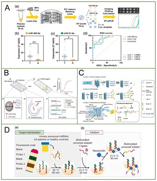
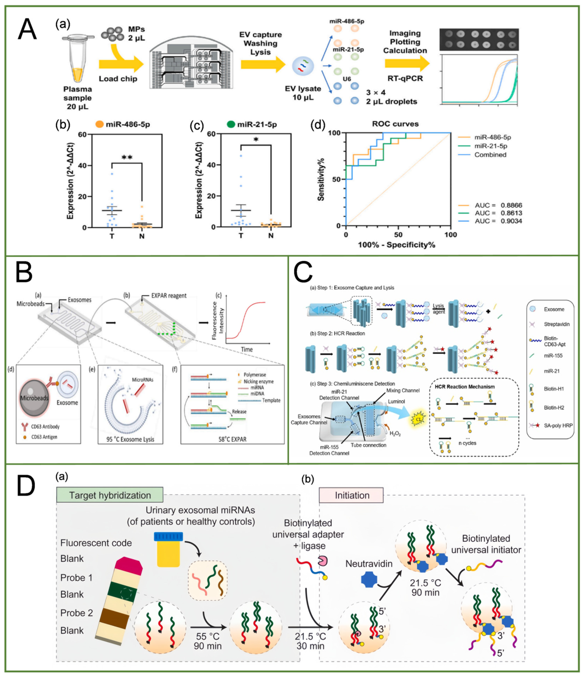
In EV-miRNA detection, reverse transcription-quantitative polymerase chain reaction (RT-qPCR), a gold-standard technique for RNA quantification, is often limited by laborious sample preparation and time-consuming multistep procedures. To overcome these limitations, Tong et al. developed a miniaturized digital microfluidic (DMF) workstation for automated and integrated EV-miRNA detection based on RT-qPCR [9] (Figure 1A). Within a 40 × 40 mm droplet manipulation area, the platform integrated EV capture, washing, lysis, template distribution, and 12 parallel RT-qPCR reaction sites. Unlike conventional workflows that require prior off-chip EV isolation, this platform enabled the direct capture of EVs from clinical plasma samples on-chip using Fe3O4@TiO2 magnetic particles, followed by washing and lysis. The resulting miRNA-containing lysate was subsequently distributed among spatially separated reaction sites for parallel analysis. Using this approach, the authors detected two NSCLC-associated EV-miRNAs, miR-21-5p and miR-486-5p, with U6 serving as an internal reference. The reported limit of detection (LOD) for the on-chip qPCR assay was 2 copies/μL. This study demonstrated the potential of digital microfluidics for integrating EV pretreatment with spatially distributed parallel nucleic acid detection. However, the actual multiplexing capacity of this approach depends on the number of independently addressable reaction sites and how reactions are allocated to individual targets. Although the workstation provided 12 parallel reaction sites, only two disease-associated EV-miRNAs were evaluated in the clinical samples. Expanding the target panel may therefore require additional reaction sites or more efficient reaction allocation, potentially increasing the structural and fluidic complexity of the device.
Figure 1.
Multiplex miRNA detection methods based on spatial encoding strategy. (A) Illustration of the on-chip EV-miRNA sample preparation and RT-qPCR detection protocol. (Reprinted with permission from ref. [9]. Copyright (2025) Elsevier.) (B) Schematic diagram of the on-chip exosomal miRNA analysis assay. (Reprinted with permission from ref. [10]. Copyright (2022) Elsevier.) (C) Schematic illustration of the multiplexed detection of exosomal microRNAs by microfluidic chemiluminescence method for breast cancer diagnosis. (Reprinted with permission from ref. [11]. Copyright (2025) Elsevier.) (D) Schematic illustration of urinary exosomal miRNA hybridization and HCR initiation on hydrogel microparticles. (Reprinted with permission from ref. [12]. Copyright (2021) Elsevier.).
Qian et al. developed a portable microfluidic platform for the multiplexed detection of exosomal miRNAs based on an on-chip exponential amplification reaction (EXPAR) assay [10] (Figure 1B). The system consisted of two interconnected microfluidic chips, one for extracellular vesicle (EV) enrichment and lysis and the other for miRNA detection. In this study, EVs derived from murine macrophage (J774.1) culture supernatants were directly introduced into the EV-processing chip without prior off-chip isolation. On-chip EV capture and enrichment were achieved, with a capture efficiency of approximately 50% and an enrichment factor exceeding 200-fold. The released miRNAs were subsequently transferred to the downstream detection chip without an additional RNA purification step. For multiplexed detection, the detection chip incorporated eight independent reaction chambers preloaded with target-specific single-stranded DNA (ssDNA) templates. Two chambers were assigned to each target, enabling the detection of four exosomal miRNAs: miR-127, miR-146b, miR-210, and miR-223. In this spatially partitioned design, target identity was determined by the position of the corresponding reaction chamber rather than by optical barcodes or bead-based encoding. Hybridization of the target miRNA with its complementary template initiated EXPAR at 58 °C, and the resulting amplification products were monitored using SYBR Green fluorescence. The platform enabled quantitative detection of all four miRNAs over a concentration range of 100 fM to 100 nM, although a formally determined limit of detection (LOD) was not reported. Furthermore, fluorescence signals were acquired using a custom-built portable reader equipped with an ESP32 microcontroller and a CMOS camera, rather than a commercial flow cytometer. By integrating on-chip EV enrichment with spatially separated isothermal amplification, this platform demonstrated the potential of microfluidics for portable multiplexed exosomal miRNA analysis. Nevertheless, the number of experimentally validated targets was limited to four, and the EV capture efficiency leaves room for further improvement. In addition, the platform was evaluated using macrophage-derived EVs rather than clinical samples. Increasing the density of reaction chambers, improving fluidic automation, and validating the platform with clinical samples will therefore be important steps toward its practical diagnostic application.
Peng et al. developed a microfluidic chemiluminescence (CL) platform for the multiplexed detection of exosomal miRNAs by integrating exosome capture, lysis, hybridization chain reaction (HCR), and CL detection within a single chip [11] (Figure 1C). The device consisted of three functional units: a CD63 aptamer-modified ship-shaped channel incorporating Y-shaped arrays for exosome capture, two parallel ship-shaped channels for target-specific miRNA detection, and a serpentine channel for mixing luminol and H2O2. Regarding sample preparation, exosomes derived from cell culture supernatants were pre-isolated by ultracentrifugation in the model experiments, whereas clinical plasma samples could be directly introduced into the chip, allowing on-chip exosome capture via CD63 aptamers without prior isolation. Subsequently, the captured exosomes were lysed on-chip, and the released miRNAs were specifically captured by corresponding hairpin probes immobilized in the detection channels. The detection signals were further enhanced through HCR amplification and streptavidin-conjugated poly-horseradish peroxidase (poly-HRP)-catalyzed CL reactions. Using this strategy, the platform simultaneously detected two breast cancer-associated exosomal miRNAs, miR-21 and miR-155, with limits of detection (LODs) of 0.39 and 0.49 fM, respectively. The method was further evaluated using 15 clinical plasma samples, comprising 9 breast cancer patients and 6 healthy volunteers, and revealed significantly elevated levels of both miRNAs in the patient group. Unlike bead-based encoding systems that require the identification of individual particle codes, this platform distinguished the two targets through spatially separated reaction channels and directly read out the resulting CL signals using an ultra-weak chemiluminescence analyzer. This study demonstrates the potential of microfluidics for integrating exosome enrichment and downstream multiplex miRNA analysis, although further optimization is needed to improve capture efficiency and automate signal acquisition across multiple channels.
Spatial encoding can be achieved not only through fixed detection sites on a microfluidic chip but also by compartmentalizing probe regions within individual microparticles. Kim et al. developed a hydrogel microparticle platform integrating intraparticle spatial encoding with hybridization chain reaction (HCR) for multiplexed detection of urinary exosomal miRNAs [12] (Figure 1D). Microfluidics enabled the fabrication of these spatially compartmentalized carriers: poly(ethylene glycol) diacrylate (PEGDA) microparticles were produced by stop-flow lithography within a 40-μm-high polydimethylsiloxane (PDMS) microchannel. Complementary DNA probes targeting different miRNAs were immobilized in separate regions, alongside a rhodamine-containing shape-code region and inert blank regions that facilitated particle identification and probe-region assignment. Target identity was thus determined primarily by the spatial position of each probe region, while its fluorescence intensity provided the quantitative readout. Before analysis, exosomes were isolated from urine using an aqueous two-phase system, and exosomal total RNA was extracted using TRIzol for subsequent hybridization and HCR signal amplification. The assay therefore measured extracted exosomal miRNAs rather than detecting them in situ within intact vesicles. Using three particle types, each containing four probe regions, the platform supported 12-plex screening of candidate miRNAs. Subsequently, miR-6090 and miR-3665 were selected for duplex validation in urine samples from 19 prostate cancer patients and 19 controls, with 600 μL of urine used per sample. Based on synthetic miRNA standards and a signal-to-noise threshold of 3, the estimated limits of detection ranged from 1 to 10 amol for miR-6090 and 10 to 100 amol for miR-3665.
The principal contribution of microfluidics was the controlled fabrication of encoded microparticles with defined spatial compartments, whereas downstream hybridization and HCR were performed off-chip in microtubes. Fluorescence images were acquired using an inverted fluorescence microscope and analyzed with custom MATLAB-R2025a code for region identification and signal quantification; neither a dedicated microfluidic decoding device nor a commercial flow cytometer was used. Spatial compartmentalization allowed multiple targets to share universal HCR reagents and a common fluorescent reporter, providing a scalable strategy for multiplexed exosomal miRNA analysis. Nevertheless, prior exosome isolation, RNA extraction, repeated washing, and approximately 4 h of HCR amplification increased procedural complexity and assay time, while the small clinical cohort limited assessment of diagnostic generalizability. Furthermore, the markedly separated PSA distributions of the patient and control groups, together with the lack of diagnostically challenging samples, such as those within the PSA gray zone, leave the assay’s discriminatory performance in routine clinical screening and its incremental value over PSA testing to be established.
3. Bead-Based Encoding Strategy
Beads are spherical particles made of polymeric or composite materials, with sizes at the micrometer or nanometer scale. Bead-based encoding strategies assign unique barcodes to beads functionalized with target-specific recognition elements., enabling high-throughput detection through recognition of the bead-encoded information. Their application in exosomal microRNA detection is an emerging and rapidly developing field.
3.1. Physically Encoded Microspheres
Among various bead-encoding strategies, physical encoding provides a means of assigning distinct identities to individual microparticles based on their intrinsic physical characteristics, such as size, shape, density, and internal structure. In particular, geometrical encoding enables target-specific identification by introducing distinguishable structural patterns into microparticles, thereby facilitating multiplexed detection through the differentiation of individual bead populations.
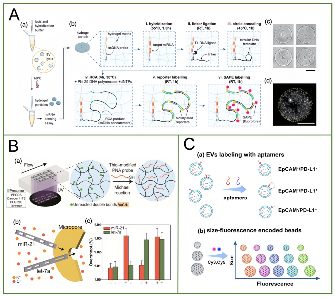
Al Sulaiman et al. developed a multiplexed EV-derived miRNA detection platform based on graphically encoded hydrogel microparticles [13] (Figure 2A). PEG-based hydrogel microparticles with distinct geometric patterns were fabricated in PDMS microfluidic channels using stop-flow lithography (SFL). Each microparticle was functionalized with sequence-specific single-stranded DNA capture probes, while its graphical code, consisting of a central triangle surrounded by three squares in different spatial arrangements, enabled target identification. The microparticle codes were decoded through bright-field microscopy, whereas fluorescence microscopy was used to quantify the target-dependent signals, thereby enabling multiplexed detection. Importantly, the microfluidic system was primarily employed for microparticle fabrication rather than microparticle decoding, which relied on microscopy imaging rather than a commercial flow cytometer. Regarding sample preparation, EVs were first isolated from serum by polyethylene glycol (PEG) precipitation and subsequently lysed to release their miRNA cargo for capture by the functionalized microparticles. Thus, this platform does not enable in situ detection of intact EVs. By incorporating Phi29 DNA polymerase-mediated rolling circle amplification (RCA), the platform enabled multiplexed detection of miR-21, miR-19b, and let-7a, achieving a limit of detection (LOD) of 2.3 zmol for synthetic miR-21, approximately three orders of magnitude lower than that of the non-amplified assay. This study highlights the potential of graphically encoded hydrogel microparticles for sensitive and multiplexed quantification of EV-derived miRNAs.
Although this platform enables multiplexed detection of EV-derived miRNAs through graphical encoding and enhances analytical sensitivity by incorporating rolling circle amplification (RCA), several limitations should be acknowledged. First, the assay involves multiple sequential steps, including hybridization, ligation, amplification, and washing. In particular, the relatively prolonged RCA reaction may increase the overall assay time and compromise detection throughput. In addition, PEG-based precipitation may lead to the co-isolation of non-EV components, potentially affecting the purity of the isolated EV fraction. Finally, despite its high analytical sensitivity for synthetic miRNA targets, the clinical utility of this platform remains to be established through validation in larger clinical cohorts.
Using a similar geometrical encoding strategy, Jang et al. developed an amplification-free and label-free platform for multiplexed detection of exosomal miRNAs based on geometrically encoded hydrogel microparticles [14] (Figure 2B). In this approach, stop-flow lithography (SFL) was performed in a microfluidic channel to fabricate hydrogel microparticles with distinct internal geometrical patterns. These differences in particle geometry served as physical identifiers, enabling particle discrimination without relying on fluorescent signals. By designing different patterns using predefined photomasks, the encoding system theoretically provided more than 64 distinguishable combinations. For sample analysis, EVs were first isolated from plasma using a polymer-based precipitation method and subsequently lysed with Triton X-100 to release their intravesicular miRNAs. The released miRNAs were then captured through hybridization with sequence-specific peptide nucleic acid (PNA) probes immobilized on differently encoded microparticles. The authors further developed a custom micropore-based resistive-pulse sensing device. As the microparticles were driven individually through the micropore under pressure, the resulting electrical current signals were used to identify their geometrical codes, while target binding-induced changes in the electrical signals enabled quantitative detection. Using this approach, the platform simultaneously detected two EV-derived miRNAs, miR-21 and let-7a, with limits of detection (LODs) of 481 and 551 fM, respectively. Notably, although the platform offered a theoretical encoding capacity of more than 64 combinations through geometrical pattern design, this figure represents the potential number of distinguishable codes rather than the number of targets experimentally detected. In the reported study, only two miRNA targets were simultaneously detected. Furthermore, clinical validation was limited to 10 breast cancer patients and 10 healthy controls, and the encoding and detection performance of the platform with a larger target panel has yet to be established. Therefore, although the integration of graphical encoding with micropore-based resistive-pulse sensing offers a promising approach to label-free EV-miRNA profiling, its practical multiplexing capability and clinical applicability require further validation using larger target panels and clinical cohorts.
3.2. Optically Encoded Microspheres
Although encoding methods such as physical encoding [15,16], graphical encoding [17], and magnetic encoding continue to be explored, fluorescence-based optical encoding is more widely pursued by researchers due to its flexible design, ease of integration, and good compatibility with the most commonly used decoding platform, flow cytometry [18,19].
Organic dyes [20] and quantum dots (QDs) [21] are commonly used fluorescent materials for the preparation of fluorescence-encoded beads. However, barcode systems based on organic dyes still face challenges in effectively enhancing their encoding capacity, including the limited availability of dyes, broad emission spectra, spectral overlap, and the need for multiple excitation lasers [22,23]. Compared with organic dyes, QDs, as another ideal fluorescent element for barcoding, possess superior optical properties, including narrower emission spectra, tunable emission wavelengths, and high fluorescence quantum yields [24,25].
However, during the preparation of encoded microspheres, both conventional organic dyes and quantum dots exhibit a pronounced aggregation-caused quenching (ACQ) effect at high concentrations [26]. In recent years, a novel class of fluorescent materials, AIEgens, has been reported. Compared with conventional organic dyes and quantum dots, AIEgens not only exhibit the unique aggregation-induced emission effect, but also possess excellent optical properties, including high brightness, outstanding photostability, low background interference, and good biocompatibility, which has attracted great interest in the development of various novel chemical and biosensors [27,28]. Therefore, AIEgens also represent ideal candidates for the construction of optically encoded microspheres to establish a miRNA detection platform. Zou et al. developed a suspension array based on aggregation-induced emission luminogen (AIEgen)-encoded microspheres for simultaneous detection of three gastric cancer-associated miRNAs (let-7b-5p, miR-16-5p, and miR-19b-3p) [29]. Using Shirasu porous glass membrane emulsification, the authors prepared three bead populations with distinct hexaphenylsilole (HPS) fluorescence intensities, each carrying a target-specific capture probe. After hybridization, a commercial CytoFLEX flow cytometer identified each bead population by its HPS barcode and quantified the corresponding miRNA using the median fluorescence intensity of the streptavidin–R-phycoerythrin reporter. In assays with synthetic targets, the platform discriminated single-base mismatches and achieved triplex limits of detection of 0.43 nM for let-7b-5p, 0.76 nM for miR-16-5p, and 0.60 nM for miR-19b-3p. Its relevance to microfluidic profiling of exosomal miRNAs lies in the optical encoding principle, which could be adapted to such assays, although validation in this study was limited to synthetic miRNAs analyzed by conventional flow cytometry.
3.3. Combinatorial Encoded Microspheres
To significantly enhance encoding capacity and avoid potential code insufficiency in high-throughput multiplex analyses, several combinatorial encoding strategies have been developed. Magnetofluorescent combinatorial encoding is one such approach, which is realized by co-doping quantum dots and magnetic nanoparticles into polymer beads or by co-assembling them on the outer surface of polymer beads. Although the amplification of fluorescence intensity and magnetization can enhance encoding capacity, along with the additional advantage of rapid magnetic separation, their practical applications are limited by the complex decoding process across different devices. Combining quantum dots with photonic crystals (PhCs) to construct multiplex barcodes is another effective strategy [30]. PhCs have periodically ordered structures that can be used to tune the reflection peaks as encoding elements [31].
Pei et al. developed a triple-encoding platform based on target-fusion-encoded beads (TFEBs) for the multiplexed in situ profiling of miRNAs in distinct extracellular vesicle (EV) subpopulations [32] (Figure 2C). In this study, EVs were first isolated and enriched from plasma by ultracentrifugation. Aptamers were used to recognize EV surface proteins, while mesoporous polystyrene beads of three different sizes (1, 2, and 3 μm) served as encoding carriers. The beads were further encoded using different Cy3/Cy5 fluorescence ratios and loaded with FAM-labeled molecular beacons targeting specific miRNAs. Hybridization between the aptamer-modified EVs and bead-bound probes induced targeted membrane fusion, allowing intravesicular miRNAs to enter the beads and generate a FAM fluorescence signal. The platform integrates bead size, Cy3/Cy5 fluorescence ratios, and target-responsive FAM signals to distinguish EV subpopulations, identify miRNA targets, and assess their relative expression levels. In the ovarian cancer study, CA125 and CD24 were used as EV surface markers to identify three subpopulations (CA125-positive, CD24-positive, and double-positive EVs), while 11 miRNAs were simultaneously analyzed, yielding 33 subpopulation-specific miRNA features. The combined miRNA model achieved an AUC of 0.995 for distinguishing ovarian cancer patients from healthy controls, with a validation accuracy of 78.6% for ovarian cancer subtype classification. Notably, bead decoding was performed using a commercial flow cytometer (BD FACSVerse) and FlowJo software-v11, rather than a custom microfluidic device. Thus, this method is an encoding-bead-based multiplexed EV analysis platform coupled with flow cytometry, not a microfluidic detection system. Although it enables the in situ analysis of EV miRNA cargo without conventional RNA extraction, it still requires prior EV isolation and specialized flow cytometry equipment. In addition, the analytical limit of detection (LOD) was not explicitly reported in the main text.
Feng et al. combined optical and physical encoding principles to develop a fluorescence–size dual-encoded microsphere [33]. Two sizes of mesoporous silica microspheres (2 μm and 3 μm) and two fluorescent dyes (Cy3 and Cy5) were first selected, and then five different ratios of Cy3 and Cy5 were modified onto the microspheres to establish a library of fluorescence–size dual-encoded microspheres. Next, target-specific molecular beacons (MBs) were incorporated into individually encoded microspheres with liposomes participating in the assembly process. Liposomes thereafter formed a supporting lipid bilayer on microsphere surfaces to achieve membrane fusion with tumor-derived extracellular vesicles. Once fusion occurred, miRNAs released from EVs could interact with internal MBs and produce fluorescent signals. Signal readout for barcode decoding and miRNA quantification was implemented using a commercial flow cytometer. Finally, a flow cytometer was used to decode the fluorescence–size information and read out the miRNA signals. Microspheres with different encodings were loaded with different molecular beacons, and this strategy effectively increased the throughput of miRNA detection. The researchers measured the expression levels of six miRNAs (miR-21, miR-16, miR-10b, miR-155, miR-1246, and miR-196a) using both qPCR and the aforementioned multiplex encoded microsphere strategy. Correlation analysis (correlation = 0.9482, p = 0.004) indicated that the miRNA expression levels measured by the multiplex encoded microsphere strategy were highly consistent with those measured by qPCR, validating the reliability of the multiplex encoded microsphere strategy for miRNA detection in extracellular vesicles. Furthermore, to evaluate the clinical potential of the multiplex encoded fusion microsphere strategy, Plasma was collected from 20 pancreatic cancer patients, 6 pancreatitis patients, and 10 healthy individuals, where only 2 μL of each sample was consumed per single detection run. The expression levels of the six aforementioned miRNAs were measured in these 36 samples. For each individual miRNA, expression levels in pancreatic cancer patients were higher than in healthy individuals, and the diagnostic accuracy based on the combined six extracellular vesicle miRNAs (98%) was higher than that of any single extracellular vesicle miRNA (e.g., the accuracy of miR-155 was 73%). These results indicate that multiplexed detection offers an advantage in improving the diagnostic accuracy of pancreatic cancer and can effectively enhance diagnostic performance.
Figure 2.
A multiplex miRNA detection method based on microsphere encoding. (A) Schematic of EV lysis followed by miRNA capture and detection in encoded hydrogel microparticles. (Reprinted with permission from ref. [13]. Copyright (2022) John Wiley and Sons.) (B) Schematic illustration of microfluidic-assisted fabrication of PNA-functionalized hydrogel barcodes and their application in the electrical multiplexed detection of miR-21 and let-7a. (Reprinted with permission from ref. [14]. Copyright (2026) American Chemical Society.) (C) Schematic illustration of aptamer-based labeling of distinct EV subpopulations and the preparation of triple-encoded fusion beads (TFEBs) with different sizes, Cy3/Cy5 fluorescence ratios, and functionalized probes. (Reprinted with permission from ref. [32]. Copyright (2026) American Chemical Society.).
4. In Situ Multiplex Strategies
Exosomal miRNAs not only reflect the biological status of tumor cells but also hold considerable promise as non-invasive biomarkers for early cancer diagnosis [7]. Accordingly, exosomal RNA detection methods based on different principles have rapidly developed over the past decades. Traditional detection methods, such as real-time quantitative PCR [34], Northern blotting [35], and microarrays [36], demonstrate good detection performance but their broader application remains constrained by factors such as relatively high cost and labor-intensive procedures [8]. In recent years, various methods based on microfluidics, localized surface plasmon resonance [37], surface-enhanced Raman scattering (SERS) [38], electrochemistry [39], and fluorescence [40] have been employed to detect exosomal miRNAs, with each technique offering distinct advantages. However, most of these methods require lysis of the exosomes to extract the internal RNA for detection. This process is prone to RNA degradation and loss, and it is susceptible to false-positive signals, reducing detection sensitivity and accuracy, thereby limiting their clinical applicability. The development of in situ detection technologies has provided new approaches for the analysis of exosomal miRNAs. In contrast, in situ detection does not require exosome lysis and can directly detect miRNAs within or on the surface of intact exosomes. This approach simplifies the analytical procedure while reducing sample loss, thereby improving detection reliability and offering a potentially more convenient and cost-effective strategy for exosomal miRNA analysis.
4.1. Fluorescent Probes
Fluorescence-based methods have been widely used for exosomal miRNA detection because of their high sensitivity, relatively simple signal readout, and compatibility with nucleic acid probes and signal amplification techniques. Various fluorescent strategies have been developed to improve the sensitivity and specificity of exosomal miRNA analysis. MBs are nanoscale oligonucleotide probes with a characteristic hairpin structure, consisting of a fluorophore at one end and a quencher at the other. Owing to their target-responsive fluorescence properties, MBs have been widely used for in situ detection, demonstrating notable advantages, particularly in live-cell RNA imaging and exosomal miRNA analysis. The loop region of an MB is designed to contain a sequence complementary to the target miRNA, when the MB is delivered into exosomes, it spontaneously hybridizes with the target RNA, resulting in spatial separation of the fluorophore and quencher and consequently generating a detectable fluorescence signal. This mechanism enables MBs to serve as effective probes for the specific detection of exosomal miRNAs [41].
Conventional MBs can directly detect miRNAs within intact exosomes; however, their performance may be compromised by unintended fluorescence quenching and fluorescence FRET arising from molecular collisions and high-frequency Brownian motion in confined space [42,43]. DNA nanostructure-based probes offers a potential strategy to overcome limitations by providing structural stability and spatial control over probe organization [44]. Based on this concept, Mao et al. developed a cubic DNA nanocage-based three-dimensional molecular beacon (ncMB) for in situ fluorescence detection of exosomal miRNAs [43]. Exosomes were first isolated from the culture supernatants of U251 glioma cells and normal HEB cells by differential centrifugation followed by ultracentrifugation. The isolated exosomes were then incubated with ncMB probes, enabling the probes to enter the vesicles and recognize their intraluminal miRNAs without disrupting the vesicular membranes. By designing distinct ncMB probes targeting miR-21 and miR-221 and labeling them with different fluorophores, the authors demonstrated dual-target detection. The cubic DNA nanocage provided steric hindrance and electrostatic repulsion, thereby reducing unintended interactions between fluorophores and quenchers. This architecture effectively suppressed unexpected fluorescence quenching during single-target detection and undesired FRET during dual-target detection, resulting in a fluorescence recovery signal more than 9.5-fold higher than that obtained with conventional molecular beacons. The reported limits of detection for miR-21 and miR-221 were 3.39 and 2.91 nM, respectively, in the solution-based assays; separate LOD values for the exosomal assays were not reported. It should be noted that this study did not involve microfluidic technology; all exosome processing and miRNA detection were performed using conventional centrifugation-based isolation and fluorescence detection methods.
Lee et al. developed a molecular beacon (MB)-based strategy for the simultaneous in situ fluorescent detection of multiple miRNAs in intact exosomes [41]. Using pre-isolated MCF-7 cell-derived exosomes, the authors detected miR-21, miR-27a, and miR-375 with differently fluorophore-labeled MBs. Upon hybridization with their complementary target miRNAs, the hairpin probes opened, separating the fluorophores from the quenchers and restoring fluorescence. This enabled multiplexed detection based on distinct fluorescence signals without exosome lysis or RNA extraction. The assay was completed within approximately 1h and remained applicable in the presence of human serum. Notably, multiplexing relied on fluorescence discrimination rather than microfluidic channels, encoded microspheres, or flow cytometric decoding. Despite these advantages, the method has several limitations. Its multiplexing capacity may be restricted by spectral overlap and fluorescence crosstalk. Detection sensitivity also depends on probe entry into exosomes and hybridization efficiency. Moreover, exosome isolation was not integrated with the detection process, and the study did not explicitly report a numerical limit of detection (LOD). Further validation using clinical samples is therefore needed.
Microfluidic technology provides additional opportunities to combine exosome enrichment, probe recognition, and fluorescence detection within a compact system. Qian et al. developed an agarose-based microfluidic chip for exosome concentration and in situ detection of exosomal miR-21 [45]. Although this platform was designed for single-target detection, it demonstrated the feasibility of integrating exosome handling and fluorescence-based miRNA analysis within a microfluidic device. They integrated the CHA strategy into the microfluidic chip. First, exosome-containing samples were mixed with hairpin probes (H1 and H2). miR-21 within the exosomes hybridized with H1 to form a single-stranded sticky end, which subsequently hybridized with H2 to form an H1–H2 duplex. After being released from the duplex, miR-21 can cyclically trigger the formation of additional H1–H2 duplexes, enabling single miR-21 molecules to induce exponential amplification of a large number of fluorescently labeled duplexes. The resulting mixture was then transferred to the microfluidic chip for fluorescence detection. Due to the capillary effect and high water permeability of the agarose gel, the target exosomes loaded at the inlet were automatically enriched at the closed end of the microchannel. The entire detection process required only 2.5 h. Experimental results showed that the fluorescence signal from exosomes derived from MCF-7 cancer cells was significantly higher than that from exosomes derived from HUVEC normal cells. The researchers employed the CHA strategy to achieve exponential amplification of the fluorescence signal, while integrating microfluidic technology to enrich exosomes. This approach provides a novel method for clinical cancer screening, enabling early diagnosis and dynamic monitoring in a simple, rapid, and highly sensitive manner.
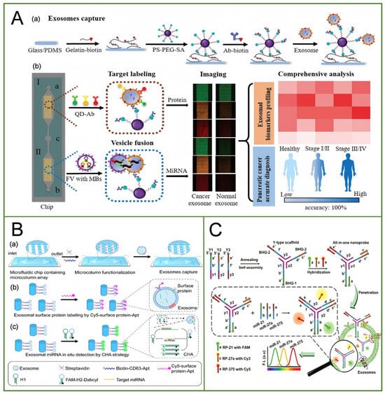
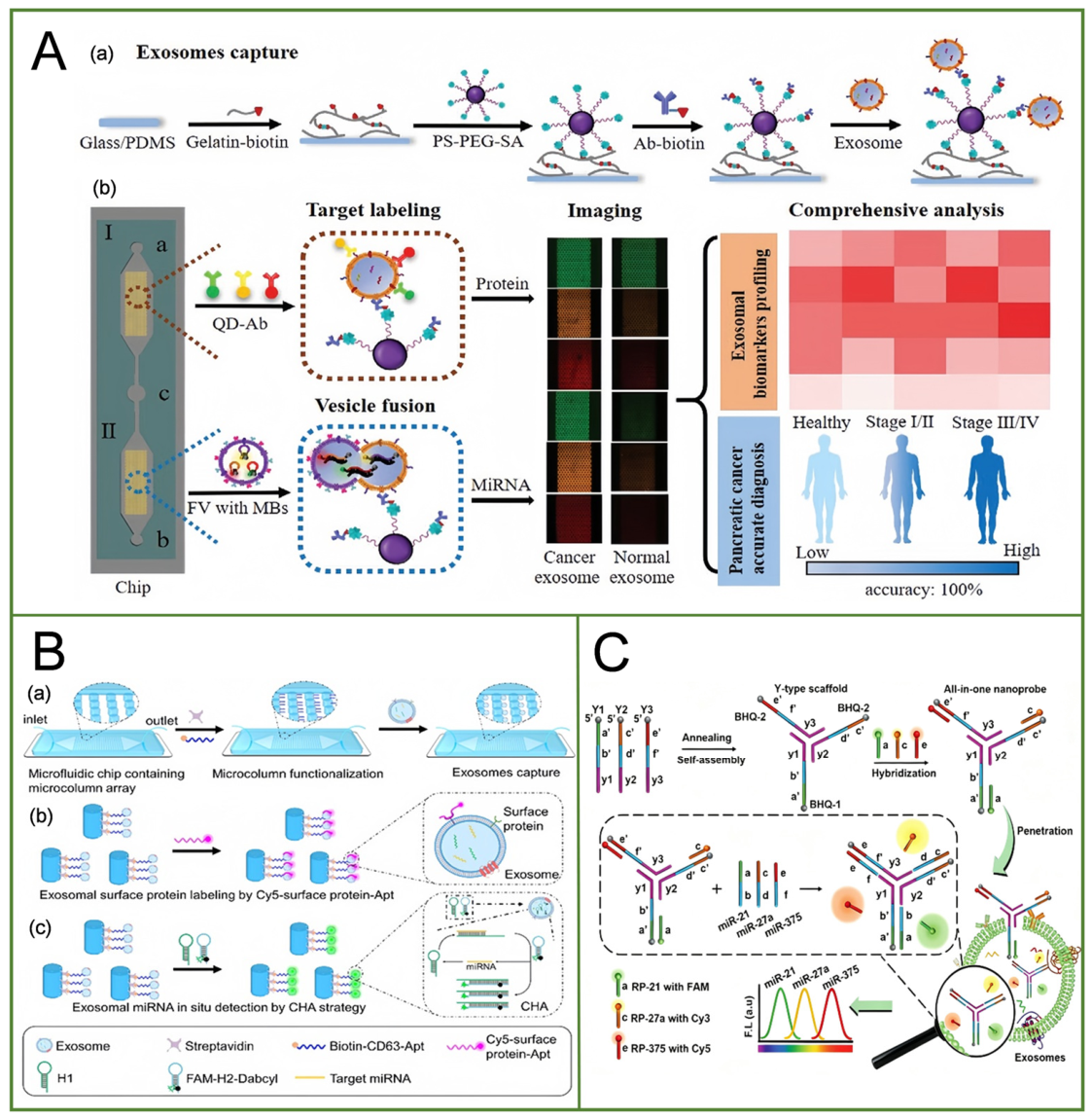
To overcome the limitations of conventional exosomal miRNA analysis requiring vesicle lysis and RNA extraction, Zhou et al. reported a 3D microfluidic chip enabling direct in situ multiplex detection of exosomal miRNAs [46] (Figure 3A). The chip employed anti-CD63-modified Y-shaped micropost arrays for exosome capture, followed by the introduction of virus-mimicking fusogenic vesicles carrying three engineered molecular beacons. These fusogenic vesicles efficiently fused with captured exosomes and released molecular beacons for fluorescence activation upon binding with target miRNAs, enabling simultaneous detection of miR-451a, miR-21, and miR-10b without disrupting exosomal integrity. The method achieved detection limits of 14, 22, and 20 exosomes μL−1 for the three miRNAs, respectively, and showed approximately 45-fold improved sensitivity compared with previous reports. In clinical plasma samples from 30 pancreatic cancer patients and 10 benign controls, the combined analysis of CD81-normalized EphA2, miR-451a, miR-21, and miR-10b achieved an area under the ROC curve (AUC) of 1.0 and enabled accurate cancer diagnosis and staging with an overall accuracy of 100%.
Figure 3.
A multiplex miRNA detection method based on the in situ strategy. (A) Schematic of microfluidic chip-based exosome capture and in situ multiplex detection of exosomal protein biomarkers and miRNAs. Exosome or clinical samples were injected into “c.” After exosomes were captured in the chip, three detected Abs modified with different QDs were injected into “a”, FVs with three MBs were injected into “b.” (Reprinted with permission from ref. [46]. Copyright (2020) John Wiley and Sons.) (B) Schematic illustration of capturing exosomes and simultaneous multiplexed in situ detection of exosomal proteins and miRNAs on the microfluidic chip. (Reprinted with permission from ref. [47]. Copyright (2023) Royal Society of Chemistry.) (C) Schematic representation of the in situ multiplex detection of serum exosomal microRNAs using all-in-one biosensor for breast cancer diagnosis. (Reprinted with permission from ref. [48]. Copyright (2020) Royal Society of Chemistry.).
Chen et al. developed a boat-shaped microfluidic chip integrated with a PDMS micropillar array for the simultaneous in situ multiplexed fluorescent detection of exosomal surface proteins and intravesicular miRNAs [47] (Figure 3B). In this platform, exosomes were specifically captured by CD63 aptamers immobilized on the inner walls of the microchannels. The micropillar array increased the effective surface area of the channels, thereby enhancing the contact between exosomes and the functionalized interface and improving capture efficiency. Based on this design, four parallel detection channels were incorporated to independently detect two exosomal surface proteins (EGFR and EpCAM) and two intravesicular miRNAs (miRNA-21 and miRNA-200). Specifically, EGFR and EpCAM were recognized using Cy5-labeled specific aptamers, whereas miRNA detection relied on streptolysin O (SLO) to facilitate the entry of hairpin probes into exosomes, followed by catalytic hairpin assembly (CHA) for enzyme-free signal amplification. This strategy enabled the in situ analysis of intravesicular nucleic acids without the need for RNA extraction. Notably, the platform employed spatial compartmentalization through multiple microfluidic channels to achieve independent detection of different targets, thereby minimizing signal interference between different fluorescent detection systems. Thus, multiplexing was achieved through spatial separation rather than fluorescently encoded microspheres coupled with flow cytometric decoding. Furthermore, for clinical plasma sample analysis, diluted plasma could be directly introduced into the chip, where exosomes were captured in situ via CD63 aptamers for subsequent multiplexed detection, eliminating the need for conventional pre-isolation of exosomes. The limits of detection (LODs) for EGFR, EpCAM, miRNA-21, and miRNA-200 were 18, 25, 83, and 35 exosomes/μL, respectively, with an overall LOD of 83 exosomes/μL for multiplexed detection. By integrating exosome capture, in situ recognition of distinct classes of biomarkers, and signal amplification into a single microfluidic platform, this study provides a promising strategy for multiplexed exosome analysis and liquid biopsy.
Nevertheless, this design has several limitations. First, multiplexed detection primarily relies on spatial compartmentalization across parallel microfluidic channels, with each target assigned to a separate channel. It therefore does not achieve single-channel multiplexed target identification and decoding based on fluorescent encoding, which may constrain further increases in detection throughput due to limitations in chip architecture and channel number. Second, exosome capture depends on CD63-mediated affinity recognition, and heterogeneity in CD63 expression among exosomes from different sources may result in the insufficient capture of certain exosome subpopulations, thereby limiting comprehensive exosome profiling. In addition, in situ miRNA detection requires SLO-mediated probe entry into exosomes, making the assay dependent on membrane permeabilization efficiency and reaction conditions. Finally, the clinical validation involved only 11 plasma samples, and the clinical applicability of this platform requires further evaluation using larger cohorts. Therefore, developing more flexible fluorescent encoding strategies that enable high-throughput identification and decoding of multiple targets within a single detection region, while preserving the capability for in situ exosome analysis, remains an important direction for the further development of microfluidic platforms for exosome detection.
Wang et al. developed an integrated multiplexed fluorescent biosensor based on a DNA three-way junction (TWJ) structure for the simultaneous in situ detection of exosomal miR-21, miR-27a, and miR-375 [48] (Figure 3C). The sensor employed a competitive strand displacement mechanism, in which three fluorescent reporter strands labeled with FAM, Cy3, and Cy5 were incorporated into the DNA nanostructure. Upon entering exosomes, the sensor hybridized with the corresponding target miRNAs, releasing the fluorescent reporter strands and restoring fluorescence signals, thereby enabling multiplexed target discrimination. Streptolysin O (SLO) was used to facilitate sensor entry into exosomes. The reported limits of detection (LODs) were 0.116, 0.125, and 0.287 μg/mL, respectively, expressed as exosome mass concentrations. The platform was further evaluated using clinical serum samples, demonstrating its potential for distinguishing breast cancer patients from healthy controls. Despite these advantages, the method has several limitations. First, sensor delivery relies on SLO-mediated membrane permeabilization, making detection dependent on the efficiency of probe entry. Second, multiplexing capacity may be constrained by spectral overlap and fluorescence crosstalk among different fluorophores. Furthermore, exosome isolation and multiplexed detection have not been integrated into a microfluidic platform, leaving room for improvement in sample processing and assay integration.
Ma et al. developed EValarm, a membrane fusion-based fluorescence assay for the simultaneous detection of miR-21-5p, miR-125b-5p, and miR-155-5p within plasma extracellular vesicles (EVs) [49]. A microfluidic chip was used to prepare anionic liposomes encapsulating target-specific pairs of DNA hairpin probes (H1 and H2). The probe-loaded liposomes were added directly to diluted plasma, eliminating the need for prior EV isolation or RNA extraction. Fusion of the liposomal and EV membranes brought the probes into contact with intravesicular miRNAs. Each target initiated a catalytic hairpin assembly reaction that separated the fluorophore on H2 from its quencher and released the target to initiate further reaction cycles, thereby amplifying the fluorescence signal. The three targets were distinguished by their respective fluorescence channels (FAM, ROX, and Cy5), and endpoint signals were measured using a commercial fluorescence microplate reader. Thus, microfluidics served specifically to fabricate the probe carriers, while multiplexed readout relied on spectral discrimination.
The reported limits of detection for the individual probe systems were 72.2 fM for miR-21-5p, 0.016 fM for miR-125b-5p, and 50.3 fM for miR-155-5p. These values were obtained by varying the target miRNA concentration and do not establish target-specific detection limits in clinical plasma. In a separate calibration using serially diluted, purified EVs, the membrane fusion assay achieved a particle-based detection limit of 6.1 × 10⁷ particles mL−1. Although EValarm simplifies plasma analysis by avoiding vesicle isolation, its bulk fluorescence readout measures the aggregate signal for each target across the EV population. Consequently, detection of all three miRNAs in the same sample does not establish their co-occurrence within individual vesicles.
4.2. SERS
Surface-enhanced Raman scattering (SERS) is a highly sensitive spectroscopic technique that enhances Raman signals through nanostructured substrates. Its high sensitivity and narrow spectral bands provide opportunities for the detection of low-abundance miRNAs and potentially enable multiplexed analysis using distinct Raman reporters. Jiang et al. reported a SERS-based strategy for the in situ detection of exosomal miR-10b in serum samples using SERS [50]. Au@DTNB nanoparticles modified with locked nucleic acids entered exosomes and assembled with the target miRNA to induce the formation of ‘hot spots’, generating SERS signals. Subsequently, Fe3O4@TiO2 nanoparticles were added, and exosomes were introduced to enrich exosomes through the affinity interaction between TiO2 and the biological membrane. The assembly of hot spots within exosomes, combined with subsequent exosome aggregation, produced a dual enhancement of the Raman signal, thereby improving detection sensitivity. Throughout the detection process, neither capture antibodies nor ultracentrifugation pretreatment were required. Based on this platform, the target miRNA can be directly identified in situ, with a detection limit of 0.21 fM. This method successfully distinguished pancreatic ductal adenocarcinoma patients from normal controls with an accuracy of 99.6%. However, this approach focused on a single exosomal miRNA target and did not incorporate microfluidic integration, highlighting the need for further development toward multiplexed, microfluidic-assisted in situ detection.
In recent years, advances in microfluidic technology have further expanded the application of SERS in the in situ detection of miRNAs. Dai et al. developed this platform, in which CHA was used as a signal amplification strategy and combined with the rapid enrichment and separation capabilities of magnetic beads to construct a SERS-based microfluidic chip for the detection of miR-122-3p and miR-150-5p in the serum of healthy individuals and colorectal cancer patients [51]. In this platform, CHA amplification and MB-mediated aggregation and separation were used to induce AuNC aggregation, thereby enhancing the SERS signal and enabling highly sensitive, specific, and quantitative detection of miR-122-3p and miR-150-5p. The designed pump-free microfluidic system adopted a closed-channel configuration, which effectively reduced exposure to external contaminants and oxidative reactions, thereby improving the reproducibility of detection results. Compared with conventional systems that rely on syringe or peristaltic pumps, this chip integrates a fluidic control system to achieve a fully automated detection process. This design not only eliminated the need for large external equipment but also simplified operational procedures, significantly enhancing overall detection efficiency. Moreover, the microchannels have very small cross-sectional areas, substantially reducing the sample volume required for testing, which is advantageous for the analysis of precious clinical samples and high-throughput screening applications. This method was simple and rapid, with the entire detection process within 5 min. Experimental results showed that the expression levels of miR-122-3p and miR-150-5p in the serum of colorectal cancer (CRC) patients were significantly higher than those in healthy individuals (* p < 0.05). These findings indicate that SERS-based microfluidic technology can facilitate early diagnosis and prognostic monitoring of CRC, providing a new technological platform for clinical biomarker detection. However, its applicability to intravesicular miRNA detection remains to be demonstrated.
5. Summary and Discussion
Tumor-derived exosomal miRNAs are stable and disease-relevant liquid-biopsy candidates, but the comparison in Table 1 shows that the field remains methodologically heterogeneous. Only a subset of the cited studies directly combines exosomal targets, experimentally demonstrated multiplex detection, and a substantive microfluidic function. Spatial platforms provide the clearest route to integrated processing, as illustrated by droplet routing, on-chip extracellular-vesicle preparation, and parallel reaction compartments. Bead-based platforms offer the largest theoretical code spaces, but many rely on off-chip bead synthesis and conventional flow cytometry; therefore, a microsphere-based encoding library with more than 64 distinguishable codes should not be equated with the experimentally validated simultaneous detection of only two extracellular vesicle-derived miRNAs. In situ strategies reduce losses associated with RNA extraction and can combine intravesicular miRNAs with surface proteins, although probe delivery and membrane accessibility remain important sources of variability.
Table 1.
Analytical performance of representative multiplex miRNA platforms discussed in this review.
In terms of multiplexing capability, spatial encoding and microsphere-based encoding exhibit distinct technical characteristics. Spatial encoding assigns each target to a specific channel, droplet, microwell, or spatial location, thereby avoiding the need to discriminate multiple targets within the same signal dimension and reducing potential signal cross-talk. However, its multiplexing capacity is generally constrained by the number of independently addressable spatial units. Increasing the number of targets may therefore require additional reaction regions or more complex chip architectures. In comparison, microsphere-based encoding assigns identification information to individual microspheres. By combining particle size, fluorescence, and other encoding parameters, the coding capacity can be expanded across multiple dimensions. In particular, combinatorial strategies such as fluorescence-size and structure-fluorescence encoding introduce additional independent coding dimensions and therefore increase the theoretical number of distinguishable codes. However, a higher coding capacity does not necessarily translate into improved clinical detection performance, because more complex encoding systems also increase the requirements for signal decoding, calibration, and batch-to-batch consistency. Therefore, the performance of multiplex detection platforms should not be evaluated solely by the number of targets that can theoretically be encoded but should also consider the actual number of detectable targets, signal cross-talk, decoding accuracy, operational complexity, and reproducibility in clinical samples.
In situ detection provides another approach to addressing sample-processing limitations in exosomal miRNA analysis. Compared with methods requiring exosome lysis and nucleic acid release, in situ detection can potentially reduce sample loss during RNA extraction and preserve exosome-associated information to some extent. Recent studies have incorporated molecular beacons, nanoprobes, hybridization chain reactions, surface-enhanced Raman scattering, and microfluidic enrichment into in situ exosomal miRNA detection, with some platforms further enabling multiplex analysis and combined profiling of exosomal surface proteins and intravesicular miRNAs. However, in situ detection also involves an important technical trade-off. Probes need to efficiently interact with intravesicular targets without disrupting exosome integrity, whereas the intact membrane can restrict probe delivery and target accessibility. Therefore, improving probe delivery efficiency alone may not directly result in higher detection accuracy, and probe stability, nonspecific adsorption, and background signals also need to be considered. In addition, the analytical sensitivity reported by different studies is affected by multiple factors, including sample type, sample pretreatment, signal amplification strategy, and assay configuration. Therefore, reported limits of detection should not be used as the sole criterion for comparing the overall performance of different platforms. Overall, microfluidic multiplex exosomal miRNA detection has progressed from single-target analysis toward multiplex profiling, but most current studies remain at the stage of methodological validation or evaluation in limited clinical cohorts. Further efforts are needed to improve assay standardization and reproducibility and to validate the diagnostic value of multiplex exosomal miRNA profiling in larger clinical cohorts with independent validation, thereby facilitating the transition of these technologies from analytical methods to clinically applicable detection tools.
Funding
This work was supported by the National Natural Science Foundation of China (822770014 and 82570028), 2025 Zhejiang Provincial College Students’ Science and Technology Innovation Activity Plan (Xinmiao Talent Plan, 2025R421A024) and Scientific Research Foundation of Zhejiang Shuren University (2024R006).
Institutional Review Board Statement
Not applicable.
Informed Consent Statement
Not applicable.
Data Availability Statement
No new data were created or analyzed in this study. Data sharing is not applicable to this article.
Acknowledgments
During the preparation of this manuscript and the conduct of this study, the authors used ChatGPT-5 and Doubao Large Language Model 2.0 Expert Edition to polish the manuscript and organize the content of cited references. The authors have thoroughly reviewed, revised, and refined all content generated by these artificial intelligence tools. The overall conceptualization of the manuscript, core research design, and finalization of its content were independently completed by the authors, who assume full responsibility for all research content, viewpoints expressed, and compliance with academic standards and ethical requirements.
Conflicts of Interest
The authors declare no conflicts of interest.
References
- Kozomara, A.; Birgaoanu, M.; Griffiths-Jones, S. miRBase: From microRNA sequences to function. Nucleic Acids Res. 2019, 47, D155–D162. [Google Scholar] [CrossRef] [Scilit]
- Yang, G.; Zheng, Z.; Zhang, X. Research progress in protective effect of miRNA on neonatal hypoxic-ischemic brain injury. J. Jilin Univ. (Med. Ed.) 2025, 51, 1423–1428. [Google Scholar]
- Ferguson, S.W.; Nguyen, J. Exosomes as therapeutics: The implications of molecular composition and exosomal heterogeneity. J. Control. Release 2016, 228, 179–190. [Google Scholar] [CrossRef] [Scilit]
- Vlassov, A.V.; Magdaleno, S.; Setterquist, R.; Conrad, R. Exosomes: Current knowledge of their composition, biological functions, and diagnostic and therapeutic potentials. Biochim. Biophys. Acta (BBA)-Gen. Subj. 2012, 1820, 940–948. [Google Scholar] [CrossRef] [Scilit]
- Ludwig, N.; Whiteside, T.L.; Reichert, T.E. Challenges in exosome isolation and analysis in health and disease. Int. J. Mol. Sci. 2019, 20, 4684. [Google Scholar] [CrossRef] [Scilit]
- Liu, F.; Yang, J.; Jiang, J.; Bian, H.; Nan, G. Exosomal microRNA and cardiovascular disease. Chem. Life 2025, 45, 598–607. [Google Scholar]
- Preethi, K.A.; Selvakumar, S.C.; Ross, K.; Jayaraman, S.; Tusubira, D.; Sekar, D. Liquid biopsy: Exosomal microRNAs as novel diagnostic and prognostic biomarkers in cancer. Mol. Cancer 2022, 21, 54. [Google Scholar] [CrossRef] [Scilit]
- Sun, Z.; Wu, Y.; Gao, F.; Li, H.; Wang, C.; Du, L.; Dong, L.; Jiang, Y. In situ detection of exosomal RNAs for cancer diagnosis. Acta Biomater. 2023, 155, 80–98. [Google Scholar] [CrossRef] [Scilit]
- Tong, Z.; Xu, X.; Shen, C.; Yang, D.; Li, Y.; Li, Q.; Yang, W.; Xu, F.; Wu, Z.; Zhou, L.; et al. All-in-one multiple extracellular vesicle miRNA detection on a miniaturized digital microfluidic workstation. Biosens. Bioelectron. 2025, 270, 116976. [Google Scholar] [CrossRef] [Scilit]
- Qian, J.; Zhang, Q.; Liu, M.; Wang, Y.; Lu, M. A portable system for isothermal amplification and detection of exosomal microRNAs. Biosens. Bioelectron. 2022, 196, 113707. [Google Scholar] [CrossRef] [Scilit]
- Peng, J.; Li, B.; Ma, Z.; Qiu, Z.; Hu, H.; Jiang, Y.; Gao, D. A microfluidic-based chemiluminescence biosensor for sensitive multiplex detection of exosomal microRNAs based on hybridization chain reaction. Talanta 2025, 281, 126838. [Google Scholar] [CrossRef] [Scilit]
- Kim, J.; Shim, J.S.; Han, B.H.; Kim, H.J.; Park, J.; Cho, I.-J.; Kang, S.G.; Kang, J.Y.; Bong, K.W.; Choi, N. Hydrogel-based hybridization chain reaction (HCR) for detection of urinary exosomal miRNAs as a diagnostic tool of prostate cancer. Biosens. Bioelectron. 2021, 192, 113504. [Google Scholar] [CrossRef] [Scilit]
- Al Sulaiman, D.; Juthani, N.; Doyle, P.S. Quantitative and multiplex detection of extracellular vesicle-derived microRNA via rolling circle amplification within encoded hydrogel microparticles. Adv. Healthc. Mater. 2022, 11, 2102332. [Google Scholar] [CrossRef] [Scilit]
- Jang, W.; Song, C.-W.; Hong, J.; Lim, S.Y.; Moon, D.G.R.M.; Roh, H.Y.; Park, K.H.; Han, C.-S.; Bong, K.W. Amplification-Free and Label-Free Multiplexed Profiling of Extracellular Vesicle-Derived MicroRNA via Micropore Sensing Based on PNA-Functionalized Hydrogel Barcodes. ACS Sens. 2026, 11, 1129–1136. [Google Scholar] [CrossRef] [Scilit]
- Fuja, T.; Hou, S.; Bryant, P. A multiplex microsphere bead assay for comparative RNA expression analysis using flow cytometry. J. Biotechnol. 2004, 108, 193–205. [Google Scholar] [CrossRef] [Scilit]
- Vaino, A.R.; Janda, K.D. Euclidean shape-encoded combinatorial chemical libraries. Proc. Natl. Acad. Sci. USA 2000, 97, 7692–7696. [Google Scholar] [CrossRef] [Scilit]
- Xu, W.; Chen, C.; Ma, X.; Yuan, L.; Liu, S.; Zheng, K.; Li, J. Digitally encoded silica microparticles for multiplexed nucleic acid detection. Chem. Commun. 2017, 53, 5866–5869. [Google Scholar] [CrossRef] [Scilit]
- Zhu, Y.; Xu, H.; Chen, K.; Fu, J.; Gu, H. Encoding through the host–guest structure: Construction of multiplexed fluorescent beads. Chem. Commun. 2014, 50, 14041–14044. [Google Scholar] [CrossRef] [Scilit]
- Huang, K.; Idris, N.M.; Zhang, Y. Engineering of lanthanide-doped upconversion nanoparticles for optical encoding. Small 2016, 12, 836–852. [Google Scholar] [CrossRef] [Scilit]
- Sarma, D.; Carl, P.; Climent, E.; Schneider, R.J.; Rurack, K. Multifunctional polystyrene core/silica shell microparticles with antifouling properties for bead-based multiplexed and quantitative analysis. ACS Appl. Mater. Interfaces 2019, 11, 1321–1334, Correction in ACS Appl. Mater. Interfaces 2019, 11, 11028. https://doi.org/10.1021/acsami.9b03348. [Google Scholar] [CrossRef] [Scilit]
- Han, M.; Gao, X.; Su, J.Z.; Nie, S. Quantum-dot-tagged microbeads for multiplexed optical coding of biomolecules. Nat. Biotechnol. 2001, 19, 631–635. [Google Scholar] [CrossRef] [Scilit]
- Yang, M.; Liu, Y.; Jiang, X. Barcoded point-of-care bioassays. Chem. Soc. Rev. 2019, 48, 850–884. [Google Scholar] [CrossRef] [Scilit]
- Wang, G.; Leng, Y.; Dou, H.; Wang, L.; Li, W.; Wang, X.; Sun, K.; Shen, L.; Yuan, X.; Li, J.; et al. Highly efficient preparation of multiscaled quantum dot barcodes for multiplexed hepatitis B detection. ACS Nano 2013, 7, 471–481. [Google Scholar] [CrossRef] [Scilit]
- Resch-Genger, U.; Grabolle, M.; Cavaliere-Jaricot, S.; Nitschke, R.; Nann, T. Quantum dots versus organic dyes as fluorescent labels. Nat. Methods 2008, 5, 763–775. [Google Scholar] [CrossRef] [Scilit]
- Zhang, J.; Shikha, S.; Mei, Q.; Liu, J.; Zhang, Y. Fluorescent microbeads for point-of-care testing: A review. Microchim. Acta 2019, 186, 361. [Google Scholar] [CrossRef] [Scilit]
- Li, Y.; Shao, A.; Wang, Y.; Mei, J.; Niu, D.; Gu, J.; Shi, P.; Zhu, W.; Tian, H.; Shi, J. Tumor Bioimaging: Morphology-Tailoring of a Red AIEgen from Microsized Rods to Nanospheres for Tumor-Targeted Bioimaging. Adv. Mater. 2016, 28, 3224. [Google Scholar] [CrossRef] [Scilit]
- Mei, J.; Leung, N.L.C.; Kwok, R.T.K.; Lam, J.W.Y.; Tang, B.Z. Aggregation-induced emission: Together we shine, united we soar! Chem. Rev. 2015, 115, 11718–11940. [Google Scholar] [CrossRef] [Scilit]
- Mei, J.; Hong, Y.; Lam, J.W.Y.; Qin, A.; Tang, Y.; Tang, B.Z. Aggregation-induced emission: The whole is more brilliant than the parts. Adv. Mater. 2014, 26, 5429–5479. [Google Scholar] [CrossRef] [Scilit]
- Zou, D.; Wu, W.; Zhang, J.; Ma, Q.; Fan, S.; Cheng, J.; Li, D.; Niu, J.; Qian, X.; Li, W.; et al. Multiplex detection of miRNAs based on aggregation-induced emission luminogen encoded microspheres. RSC Adv. 2019, 9, 39976–39985. [Google Scholar] [CrossRef] [Scilit]
- Li, J.; Zhao, X.-W.; Zhao, Y.-J.; Gu, Z.-Z. Quantum-dot-coated encoded silica colloidal crystals beads for multiplex coding. Chem. Commun. 2009, 45, 2329–2331. [Google Scholar] [CrossRef] [Scilit]
- Zhao, Y.; Zhao, X.; Pei, X.; Hu, J.; Zhao, W.; Chen, B.; Gu, Z. Multiplex detection of tumor markers with photonic suspension array. Anal. Chim. Acta 2009, 633, 103–108. [Google Scholar] [CrossRef] [Scilit]
- Pei, H.; Kuang, T.; Dai, Y.; Li, J.; Zheng, L.; Tan, Q.; Qu, F. Triple-Encoding Platform for Multiplexed In Situ Profiling of Extracellular Vesicle miRNAs Enables Precision Diagnosis of Ovarian Cancer. ACS Nano 2026, 20, 28548–28564. [Google Scholar] [CrossRef] [Scilit]
- Feng, J. Construction of Fusion Beads and Interfaces and Their Application in UItrasensitive Detection of MicroRNA in Extracellular Vesicles. Master’s Thesis, Yangzhou University, Yangzhou, China, 2023. [Google Scholar]
- Benes, V.; Castoldi, M. Expression profiling of microRNA using real-time quantitative PCR, how to use it and what is available. Methods 2010, 50, 244–249. [Google Scholar] [CrossRef] [Scilit]
- Válóczi, A.; Hornyik, C.; Varga, N.; Burgyán, J.; Kauppinen, S.; Havelda, Z. Sensitive and specific detection of microRNAs by northern blot analysis using LNA-modified oligonucleotide probes. Nucleic Acids Res. 2004, 32, e175. [Google Scholar] [CrossRef] [Scilit]
- Hu, J.; Yue, X.; Liu, J.; Kong, D. Construction of an miRNA-gene regulatory network in colorectal cancer through integrated analysis of mRNA and miRNA microarrays. Mol. Med. Rep. 2018, 18, 5109–5116. [Google Scholar] [CrossRef] [Scilit]
- Joshi, G.K.; Deitz-McElyea, S.; Liyanage, T.; Lawrence, K.; Mali, S.; Sardar, R.; Korc, M. Label-free nanoplasmonic-based short noncoding RNA sensing at attomolar concentrations allows for quantitative and highly specific assay of microRNA-10b in biological fluids and circulating exosomes. ACS Nano 2015, 9, 11075–11089. [Google Scholar] [CrossRef] [Scilit]
- Lee, J.U.; Kim, W.H.; Lee, H.S.; Park, K.H.; Sim, S.J. Quantitative and specific detection of exosomal miRNAs for accurate diagnosis of breast cancer using a surface-enhanced Raman scattering sensor based on plasmonic head-flocked gold nanopillars. Small 2019, 15, 1804968. [Google Scholar] [CrossRef] [Scilit]
- Luo, L.; Wang, L.; Zeng, L.; Wang, Y.; Weng, Y.; Liao, Y.; Chen, T.; Xia, Y.; Zhang, J.; Chen, J. A ratiometric electrochemical DNA biosensor for detection of exosomal MicroRNA. Talanta 2020, 207, 120298. [Google Scholar] [CrossRef] [Scilit]
- Zhang, J.; Guan, M.; Ma, C.; Liu, Y.; Lv, M.; Zhang, Z.; Gao, H.; Zhang, K. Highly effective detection of exosomal miRNAs in plasma using liposome-mediated transfection CRISPR/Cas13a. ACS Sens. 2023, 8, 565–575. [Google Scholar] [CrossRef] [Scilit]
- Lee, J.H.; Kim, J.A.; Jeong, S.; Rhee, W.J. Simultaneous and multiplexed detection of exosome microRNAs using molecular beacons. Biosens. Bioelectron. 2016, 86, 202–210. [Google Scholar] [CrossRef] [Scilit]
- Lee, J.H.; Kim, J.A.; Kwon, M.H.; Kang, J.Y.; Rhee, W.J. In situ single step detection of exosome microRNA using molecular beacon. Biomaterials 2015, 54, 116–125. [Google Scholar] [CrossRef] [Scilit]
- Mao, D.; Zheng, M.; Li, W.; Xu, Y.; Wang, C.; Qian, Q.; Li, S.; Chen, G.; Zhu, X.; Mi, X. Cubic DNA nanocage-based three-dimensional molecular beacon for accurate detection of exosomal miRNAs in confined spaces. Biosens. Bioelectron. 2022, 204, 114077. [Google Scholar] [CrossRef] [Scilit]
- Fu, X.; Ke, G.; Peng, F.; Hu, X.; Li, J.; Shi, Y.; Kong, G.; Zhang, X.-B.; Tan, W. Size-selective molecular recognition based on a confined DNA molecular sieve using cavity-tunable framework nucleic acids. Nat. Commun. 2020, 11, 1518. [Google Scholar] [CrossRef] [Scilit]
- Qian, C.; Xiao, Y.; Wang, J.; Li, Y.; Li, S.; Wei, B.; Du, W.; Feng, X.; Chen, P.; Liu, B.-F. Rapid exosomes concentration and in situ detection of exosomal microRNA on agarose-based microfluidic chip. Sens. Actuators B Chem. 2021, 333, 129559. [Google Scholar] [CrossRef] [Scilit]
- Zhou, S.; Hu, T.; Han, G.; Wu, Y.; Hua, X.; Su, J.; Jin, W.; Mou, Y.; Mou, X.; Li, Q.; et al. Accurate cancer diagnosis and stage monitoring enabled by comprehensive profiling of different types of exosomal biomarkers: Surface proteins and miRNAs. Small 2020, 16, 2004492. [Google Scholar] [CrossRef] [Scilit]
- Chen, Y.; Gao, D.; Zhu, Q.; Chu, B.; Peng, J.; Wang, J.; Liu, L.; Jiang, Y. Rapid exosome isolation and in situ multiplexed detection of exosomal surface proteins and microRNAs on microfluidic platform. Analyst 2023, 148, 2387–2394. [Google Scholar] [CrossRef] [Scilit]
- Wang, H.; He, D.; Wan, K.; Sheng, X.; Cheng, H.; Huang, J.; Zhou, X.; He, X.; Wang, K. In situ multiplex detection of serum exosomal microRNAs using an all-in-one biosensor for breast cancer diagnosis. Analyst 2020, 145, 3289–3296. [Google Scholar] [CrossRef] [Scilit]
- Ma, J.Y.; Wang, X.; Cai, Y.; Wang, G.; Lu, M.; Feng, K.; Zhao, Y.; Wu, X.; Zhang, X.; Wu, H.; et al. Anionic Liposomes as Optimal Membrane Fusion Carriers Enabling in Situ Multiplexed Detection of Extracellular Vesicle MicroRNAs. Adv. Sci. 2026, 13, e19758. [Google Scholar] [CrossRef] [Scilit]
- Jiang, S.; Li, Q.; Wang, C.; Pang, Y.; Sun, Z.; Xiao, R. In situ exosomal MicroRNA determination by target-triggered SERS and Fe3O4@TiO2-based exosome accumulation. ACS Sens. 2021, 6, 852–862. [Google Scholar] [CrossRef] [Scilit]
- Dai, C.; Tan, M.; Liu, Z.; Shi, J.; Wang, F.; Cao, X.; Shen, K.; Yang, H.; Wei, W. Rapid and highly sensitive detection of miRNAs in serum of colorectal cancer patients by surface-enhanced Raman spectroscopy microfluidic chip based on catalytic hairpin assembly. Chin. Sci. Bull. 2025, 70, 600–611. [Google Scholar]

Disclaimer/Publisher’s Note: The statements, opinions and data contained in all publications are solely those of the individual author(s) and contributor(s) and not of MDPI and/or the editor(s). MDPI and/or the editor(s) disclaim responsibility for any injury to people or property resulting from any ideas, methods, instructions or products referred to in the content. |
© 2026 by the authors. Licensee MDPI, Basel, Switzerland. This article is an open access article distributed under the terms and conditions of the Creative Commons Attribution (CC BY) license.