


Abstract
Marine ranches are open ecological-production integrated systems, and as intensive coastal aquaculture expands, the threat of pathogenic diseases is increasingly severe. Pathogen monitoring in these environments faces three major practical challenges: dispersed sampling, low pathogen loads, and strong environmental interference (high salinity, high turbidity, and high organic content), which limit the effectiveness of conventional laboratory methods. This review therefore explores the potential and bottlenecks of microfluidic biosensing technologies for in situ pathogen monitoring in marine ranches. Using a structured literature search and title/abstract evidence mapping, we summarize recent advances in pathogen characteristics, continuous-flow/droplet/digital microfluidic platforms, and AI-based early warning systems. A complementary bibliometric scan of the Web of Science Core Collection (44,892 records) tracks publication growth in this field. Comparisons between reported environmental pathogen concentrations (e.g., culturable Vibrio communities at 103–105 CFU·mL−1 in production water) and analytical sensitivity in buffer (<1 copy/μL) show that buffer LOD alone is insufficient to establish field applicability. The key bottlenecks include matrix tolerance, sample-processing throughput, and validation depth. Among the 20 evaluated platforms, two reported on-site or field demonstrations, but neither constitutes multi-center prospective diagnostic validation. Consequently, four key deployment barriers for microfluidic monitoring in marine ranch settings can be identified: (1) mismatch between sampling volume and chip interface; (2) matrix inhibition affecting quantitative accuracy; (3) difficulty in distinguishing pathogen viability states; and (4) failure of mechanisms such as electrowetting in seawater. To address these issues, this paper proposes a three-step implementation pathway: developing validated multiplex detection panels; launching regional early-warning pilot projects; and finally, establishing a data-driven closed-loop monitoring ecosystem. The case study focuses on the South China Sea, while the framework is applicable to warm-water regions worldwide.
1. Introduction
Marine ranches can be defined at the practical level as open, human-managed, composite ecological production systems. In contrast to land-based pond or recirculating aquaculture, where water quality is controlled ex situ, and to fixed open-sea cage farming, a marine ranch is spatially open and hydrodynamically connected to surrounding waters, so pathogens disperse freely with currents and sediments and representative sampling becomes intrinsically difficult. Unlike the idealized marine aquaculture ecosystem depicted in Figure 1, which possesses multiple ecological functions and services, the artificially constructed ecological structure of marine ranches struggles to fully replicate the inherent multi-scale self-organizing complexity of natural habitats. Their ecological benefits are highly dependent on key parameters such as habitat structural complexity, species composition, hydrodynamic conditions, and environmental carrying capacity [1,2,3]. Once ecological thresholds are exceeded—such as through excessive habitat engineering or overstocking—the system’s returns will exhibit diminishing marginal gains and may intensify local ecological and biosecurity pressures [4]. In this system, human interventions, including habitat creation, stock enhancement, cage farming, feeding management, harvesting operations, and environmental regulation, are continuously coupled with natural hydrodynamic processes, food web dynamics, and microbial cycles. This human–nature coupling fundamentally influences disease monitoring strategies: compared to closed recirculating water systems, marine ranch boundaries are permeable, and flow paths are highly heterogeneous; compared to natural ecosystems, the human-controlled stocking densities and periodic production activities significantly increase host contact frequencies, repeatedly creating opportunities for pathogen introduction and transmission. Therefore, the core objective of monitoring is not limited to clinical case diagnosis but rather to quantitatively characterize ecological stress levels at an early stage of pathogen exposure, with sufficiently high spatiotemporal resolution, thereby enabling scientific, timely, and proportionate management responses.
Figure 1.
Schematic Diagram of the Core Characteristics of an Ideal Hybrid Marine Ranching Ecosystem. Labels (A)–(F) denote the six functional panels; the central circle summarizes the overall goal (functional autonomy and dynamic balance) and the resulting outcome (ecosystem self-maintenance and repair).
The South China Sea presents a challenging testing environment: warm temperatures shorten microbial reproduction times, while typhoons, rainfall, water stratification, and tidal exchanges alter seawater salinity, turbidity, suspended solids content, and their distribution. Regional surveys indicate that Vibrio species, viruses, and parasites are the primary pathogens in diseased marine fish in China and Southeast Asia [5]; meanwhile, pathogen samples collected from the South China Sea exhibit diverse virulence genes and extensive multidrug resistance [6]. Pathogen signals are often hidden within seemingly healthy or wild fish populations; metagenomic sequencing of gill tissues from South China Sea fish has detected microbial communities with potential pathogenicity, confirming the value of non-culture monitoring while also highlighting its interpretive challenges [7]. Aquaculture further reshapes coastal microbiomes and resistance profiles [8]. As shown in Figure 2, the large-scale development of marine aquaculture and cage farming is a double-edged sword: it enhances resource efficiency through improved strains, high-nutrient feeds, intelligent monitoring systems, and shortened production cycles; yet it simultaneously intensifies ecological pressures, leading to habitat homogenization, nutrient imbalances, organic waste accumulation, reduced dissolved oxygen levels, benthic degradation, and loss of biodiversity [9,10,11,12]. Collectively, these findings underscore the need for detection methods capable of distinguishing between pathogen presence and actual disease risk, while preserving critical information on bacterial abundance, viability, genotypes, host condition, and environmental context. Under high-density cultivation conditions, pathogen loads and annual outbreak risks increase significantly, emphasizing the necessity of real-time quantitative pathogen monitoring.
Figure 2.
Ecological and environmental impacts of intensive marine aquaculture. Gray arrows indicate the progression from traditional marine ranching to industrialized intensive aquaculture and, consequently, to environmental load and disease risk.
Traditional diagnostic methods remain essential for aquatic pathogen detection—but face major field limitations. Culture confirms viable bacteria and enables antimicrobial testing, yet takes days and misses VBNC cells. Histopathology locates lesions but requires complex fixation and sectioning. Microscopy detects parasites effectively but lacks objectivity and reproducibility at low pathogen loads. qPCR offers high sensitivity and specificity, but its extraction–amplification–detection steps demand multiple instruments and manual handling [13,14,15]. None of these methods is readily deployable as a standalone on-site diagnostic solution for marine ranches.
Microfluidic biosensors enable precise manipulation of sample volumes spanning picoliters to microliters, integrating sample pretreatment, pathogen enrichment, nucleic acid amplification, immunoassays, washing, signal generation, and readout within a miniaturized, portable device—effectively addressing the gap in rapid on-site detection. Recent reviews have validated that this technology facilitates rapid pathogen detection via multiple modalities, including nucleic acid-based, immunological, metabolic, electrochemical, and optical approaches [16,17,18,19]. Compared with traditional laboratory methods, microfluidic technology offers low sample and reagent consumption, rapid mass and heat transfer, short reaction times, miniaturization, parallelization, flexible signal readout and compatibility with automated operation [20]. Microfluidic systems have enabled rapid and multiplexed detection of waterborne pathogens in coastal water and aquaculture samples [21,22], demonstrating their potential for decentralized “sample-in, result-out” monitoring [23]. It holds particular promise for in situ analysis of dispersed samples from turbid, high-salinity and organic-rich seawater environments, such as marine ranches in the South China Sea.
Although the literature has systematically highlighted the advantages of microfluidic biosensors in terms of detection sensitivity, functional integration, and field applicability, three conceptual boundaries urgently need clarification to support scientific evaluation, reliable validation, and practical application.
- (1)
- “Microfluidics” refers solely to fluid manipulation at the microscale and does not inherently possess biosensing functionality; strictly speaking, a biosensor must simultaneously incorporate a specific biorecognition element, quantifiable and reproducible signal transduction, and a calibrated output response.
- (2)
- While hydrodynamic droplet generation, ddPCR, and electrode-addressable digital microfluidics (DMF) share a common microfluidic foundation, they differ fundamentally in driving principles (pressure-driven vs. electrowetting), discretization mechanisms (passive splitting vs. active addressing), throughput strategies (continuous flow vs. parallel arrays), and integration modes (closed-chip vs. open-interface).
- (3)
- High sensitivity limits of detection (LOD) achieved in buffer systems cannot be extrapolated to environmental matrices; their performance in real seawater, tissue homogenates, or field samples must undergo multi-level validation—including matrix spiking recovery, tolerance testing for interferents (e.g., humic acid, salt ions, proteins), and parallel on-site comparisons with gold-standard methods—before serving as a scientific basis for risk early warning and control decisions.
Building on these recent reviews [16,17,18,19], the present study advances research in five distinct aspects compared to previous work:
- (1)
- Using measured pathogen loads in production water (rather than buffer LODs) as the benchmark for sensor performance (Section 3.5);
- (2)
- Providing a unified scoring system for 20 platforms based on criteria including WOAH validation stages and technology readiness levels (Section 4.4);
- (3)
- Employing traceable and reproducible literature search methods (Section 2);
- (4)
- Explicitly identifying the pre-sampling interface from “macroscopic water bodies to microscopic sensing” (rather than molecular recognition) as the core deployment bottleneck;
- (5)
- Proposing a staged, three-phase implementation pathway (Section 7).
To our knowledge, no prior review has integrated pathogen load benchmarking with a validation and grading framework for aquaculture biosensing.
As shown in Figure 3a, the logical pathway of this review begins with intensive aquaculture and pathogen pressure in marine ranches, identifies diagnostic bottlenecks, and then focuses on the evolution of microfluidic biosensing, validation frameworks, and data-driven monitoring. First, marine ranches are defined as eco-productive coupled systems, and detection requirements are clarified based on pathogen biology and measured environmental loads. Second, continuous-flow, droplet-based, and digital microfluidic platforms are compared under a unified quantitative framework, covering sample processing, molecular recognition, matrix tolerance, and quality control. The analysis centers on three core questions: which pathogen–host–matrix combinations are most valuable for monitoring? Which functions have been experimentally validated and correspond to which WOAH validation stage? What validation systems and data infrastructure are required to achieve AI-driven “micro-scale measurements → ecosystem risk decision-making”? Finally, deployment challenges and four measurable milestones are proposed. Publication activity in microfluidic pathogen detection has remained high over the past years, with annual record counts consistently above 3000 and a peak in 2025 (Figure 3b), indicating a field in a phase of rapid engineering translation.
Figure 3.
Overview framework of microfluidic pathogen detection and the recent literature statistics. (a) Research logic pathway of this review; (b) publication trends in microfluidic pathogen detection (based on a thematic search of the Web of Science Core Collection, combining platform terms “microfluidic biosensor,” “digital microfluidics,” “droplet-based microfluidics,” “droplet microfluidics,” and “lab-on-a-chip” with pathogen detection terms; search conducted in September 2026; complete result set n = 44,892). The curve shows the absolute number of records per year from 2021 to 2026, while the bar chart indicates the proportion of each year’s results relative to the total dataset; data for 2026 are partial (as of September), representing 42% of the full record window.
2. Materials and Methods: Literature Search and Evidence Mapping
2.1. Databases, Search Period, and Query Design
A structured literature search was conducted in October 2026 using PubMed (title/abstract fields) and OpenAlex (combined title/abstract searches), principally covering January 2021–October 2026. The preserved exports, dated 11 October 2026, contained 474 records. Foundational publications before 2021—such as early integrated microfluidic systems or classic electrowetting-on-dielectric (EWOD) models—were retained where they established theoretical or technical paradigms. The retained query labels were organized around three concept groups:
- (1)
- Platform-related terms including “microfluidic,” “lab-on-a-chip,” “biosensor,” “digital microfluidics,” “electrowetting,” “droplet,” and “paper-based”;
- (2)
- Application-related terms focusing on aquaculture entities such as “aquaculture,” “marine ranching,” “fish,” “shrimp,” “shellfish,” “oyster,” and “seawater”;
- (3)
- Objective-related terms targeting core research goals, namely “pathogen,” “disease,” “detection,” and “early warning.”
Additional query labels covered CRISPR/Cas-mediated nucleic acid detection, isothermal amplification (LAMP, RPA, and RAA), droplet digital PCR (ddPCR), and machine-learning models for aquaculture-disease early warning. The complete database-specific Boolean strings and all filter settings for the PubMed/OpenAlex searches were not retained; therefore, no reconstructed strings are presented as the original searches. The Supplementary Search Audit instead reports the preserved query labels, record counts, raw exports, metadata corrections, deduplication map, and title/abstract decisions. As a separate analysis, a bibliometric trend scan (Figure 3b) was conducted in the Web of Science Core Collection in September 2026. Platform terms—“microfluidic biosensors,” “digital microfluidics,” “droplet microfluidics,” “droplet-based microfluidics,” and “lab-on-a-chip”—were combined with pathogen-detection keywords, yielding 44,892 records. This Web of Science corpus was independent and was not merged with the PubMed/OpenAlex evidence-mapping set.
2.2. Inclusion and Exclusion Criteria for Literature
This study employs clear, tiered screening criteria to ensure the scientific rigor, relevance, and comparability of the evidence base. The inclusion criteria are as follows:
- (1)
- Peer-reviewed journal articles published between January 2021 and October 2026; foundational works published prior to 2021 that are widely cited as establishing theoretical or technical paradigms (e.g., the first integrated microfluidic pathogen detection system, classic EWOD actuation models, or early CRISPR-based sensing validation studies) will be retained;
- (2)
- Studies must substantially focus on pathogen detection in aquaculture or marine environments, including: ① direct detection methods based on microfluidic or biosensor platforms; ② universal technological platforms with potential for cross-scenario application (such as digital microfluidics, droplet generation and manipulation, and CRISPR-Cas-mediated specific recognition); ③ molecular detection protocols developed and validated specifically for aquaculture pathogens (including ddPCR, CRISPR/Cas-assisted detection, LAMP, RPA, and lateral flow assays); ④ artificial intelligence/machine learning models trained on real-world aquaculture monitoring data for disease risk prediction or early warning;
- (3)
- Systematic reviews or scoping reviews whose analytical frameworks, evidence synthesis, or technical evaluations directly support the aforementioned research directions.
Exclusion criteria were grouped into five categories: (E1) aquaculture research that did not address pathogen-detection pathways (e.g., vaccine development, immune mechanisms, genetic breeding, or metagenomic sequencing without a detection application); (E2) studies confined to microfluidic device design, fabrication, or fluid simulation without pathogen-target recognition, signal transduction, or quantitative detection; (E3) non-aquaculture applications without a clear technology-transfer pathway; (E4) non-peer-reviewed literature (including preprints, conference abstracts, book chapters, and database entries) and non-English publications; and (E5) non-pathogen targets, water-quality-only or general bibliometric records, and other out-of-scope records.
2.3. Literature Screening Process
The preserved PubMed and OpenAlex exports contained 474 records (PubMed n = 212; OpenAlex n = 262). A reconstructed metadata and title/abstract relevance audit was performed to provide an auditable supplement to this narrative Review. The complete screening workflow and record-level disposition are summarized in Figure 4.
Figure 4.
Reconstructed literature-search and title/abstract evidence-mapping audit for this narrative Review. The 238 potentially relevant records comprise 128 core and 110 methodological-transfer records and do not represent full-text systematic inclusion. The audit is based on preserved exports. See Section 2 and Supplementary Materials File S1 for record-level decisions and limitations.
Deduplication used verified DOI or normalized-title exact matches, supplemented by one manually verified preprint–journal version merge; fuzzy similarity alone was not used to delete records. This yielded 383 unique records and removed 91 duplicates or version duplicates. Individual title/abstract review classified 128 records as directly relevant to aquaculture/marine pathogen detection (INCLUDE_CORE), 110 as methodological-transfer evidence only (INCLUDE_TRANSFER), and 145 as excluded (E1 = 19; E2 = 9; E3 = 36; E4 = 53; E5 = 28). The 238 potentially relevant records represent title/abstract-level evidence mapping, not full-text inclusion or mandatory citation. Studies reporting quantitative performance metrics (e.g., limit of detection, analysis time, matrix, and validation evidence) were further extracted into Table 2 to support cross-technology comparison.
Table 2.
Quantitative comparison of microfluidic and biosensing platforms for aquaculture-relevant pathogen detection.
The PRISMA 2020 statement [24] was used only as a reference for identification and screening terminology; this Review does not claim PRISMA compliance or Systematic Review methodology. Screening was a single-reviewer reconstructed title/abstract relevance audit. No dual independent screening, Cohen’s κ, full-text systematic eligibility assessment, risk-of-bias assessment, or meta-analysis was performed. The Supplementary Search Audit provides the record-level decisions, exclusion reasons, metadata-verification status, and unresolved items; unresolved metadata were not silently corrected or assigned guessed DOIs. The bibliometric corpus shown in Figure 3b remained independent of this screening process.
3. Priority Pathogens in Warm-Water Marine Ranching: Implications for Biosensor Design
3.1. Surveillance Targets
From a practical application standpoint, pathogen monitoring in marine ranches should not aim for “complete genomic coverage”—that is, indiscriminately cataloging every organism mentioned in scattered scientific literature—but instead focus on core pathogens with clear epidemiological significance to ensure that monitoring resources are proportionate to actual disease control outcomes.
This study systematically evaluates candidate pathogens based on the following criteria: disease burden (quantifying their actual impact on aquaculture production, economic losses, and ecosystem health); transmission routes (identifying dominant modes such as waterborne, contact, or vertical transmission); preclinical detectability (whether they have a sufficiently long incubation period allowing for early warning); host range breadth (whether restricted to a single economically important species or posing cross-host dissemination risks); zoonotic potential (whether they represent a public health threat); and association strength with antimicrobial resistance (whether they carry transferable resistance genes or frequently co-occur with resistant bacteria). Crucially, the seventh criterion—whether a positive result necessarily triggers clearly defined, timely, and feasible intervention measures (such as emergency medication, isolation, or trace-back investigation)—must be met. Based on these considerations, a three-tiered, differentiated monitoring framework is proposed:
Level 1 monitoring targets high-abundance, rapidly multiplying, highly pathogenic bacterial indicator groups, prioritizing pathogenic species within the genus Vibrio; detection results directly trigger standardized emergency response protocols.
Level 2 monitoring covers viral pathogens posing significant threats to industry stability and biosecurity; initial positive screenings must immediately initiate transport controls, physical isolation of affected populations, and multi-methodological tiered confirmation.
Level 3 monitoring addresses parasites (e.g., Cryptocaryon irritans) and emerging/re-emerging pathogens, utilizing environmental DNA (eDNA) technology for broad-spectrum screening, but only incorporating risk assessment when infection-active morphological units (such as invasive larvae, mature cysts, or intact free virus particles) are detected—ensuring accurate life-stage interpretation.
Additionally, syndrome-based indicators—including total microbial load in water, host serum lysozyme/heat shock protein 70 (HSP70) as immune and stress markers, and key physicochemical parameters like temperature, dissolved oxygen, and ammonia nitrogen—can provide systemic health warnings. However, these signals are inherently non-specific and can only indicate risk trends; they must never replace molecular or culture-based confirmation of specific target pathogens.
3.2. Bacterial Pathogens: Vibrio Genus as Indicator Genus
As shown in Figure 5a, Vibrio is the dominant bacterial indicator genus in marine ranching aquaculture. Vibrio harveyi was detected in 88.76% of diseased farmed fish, compared to only 36.27% in asymptomatic control groups—a significant quantitative difference supporting its application as a microbial risk indicator [5,25]. This genus also includes Vibrio parahaemolyticus (whose clinical isolates often carry virulence factors TLH, TDH, and Hly), Vibrio alginolyticus, and Vibrio vulnificus; due to overlapping ecological niches, a multiplex detection strategy is required [26,27,28].
Figure 5.
General pathogenic mechanisms of the priority pathogens and the resulting biosensor design constraints: (a) Vibrio spp. (bacterial indicator genus); (b) rhabdoviruses (VHSV/IHNV); (c) nervous necrosis virus (NNV); (d) Singapore grouper iridovirus (SGIV); (e) Cryptocaryon irritans.
For sensor design, the severity of vibriosis is a regulated, density-dependent phenotype: quorum sensing coordinates the expression of hemolysins, proteases, secretion systems, and outer membrane vesicles, while salinity, temperature, dissolved oxygen, and host stress jointly modulate bacterial abundance and host susceptibility [25]. Therefore, species identification alone cannot accurately assess toxin production burden. An informative bacterial detection panel should include species/pathotype-specific genes, total viable cell counts, and key virulence or resistance-determining factors; only when test results are correlated with environmental covariates do they provide meaningful decision-making reference value.
3.3. Viral Pathogens: Infection Progression and Monitoring Time Window
Fish viruses are small in size (20–200 nm), obligate intracellular parasites, often carried asymptomatically for extended periods, and currently no antiviral drugs are available for field use [29,30]. This study focuses on four representative viruses: the RNA viruses IHNV, VHSV, NNV [31], and the DNA virus SGIV [30]. Their infection progression (invasion → colonization → dissemination → shedding) occurs over a well-defined timeline, enabling identification of optimal analytes for preclinical detection (e.g., viral RNA, capsid proteins), the most sensitive sample types (gills, kidneys, brain, environmental eRNA), and the ideal sampling time window.
As shown in Figure 5b, the rhabdoviruses VHSV and IHNV—enveloped single-stranded RNA viruses—can cause acute hemorrhagic systemic disease with mortality rates approaching 100% in susceptible populations. They remain high-priority targets for biosecurity control during live fish transport, despite currently not being dominant in tropical aquaculture farms in southern China [32,33,34,35,36,37,38]. Their replication process—local invasion, viremia, followed by dissemination to endothelial and hematopoietic tissues—dictates the preferred tissue sample matrices (gills, kidneys) and optimal sampling time window in monitoring design.
Figure 5c shows nervous necrosis virus (NNV), a non-enveloped betanodavirus with a bisegmented RNA genome and particle size of 25–30 nm, which causes viral nervous necrosis in larval and juvenile stages of fish, resulting in cumulative mortality close to 100%. It is commonly found in groupers, sea bass, and mackerel species [39,40,41,42]. The virus exhibits strong neurotropism, making brain and eye tissues the primary samples for sampling, detection, and diagnosis. Although environmental RNA (eRNA) isolated from water can reflect population-level viral shedding, its appearance lags behind tissue infection and thus cannot serve as a standalone early warning signal [43]. Nevertheless, NNV is amenable to integration with microfluidic detection technologies due to its conserved nucleic acid targets that can be amplified without cell culture. Reports indicate that an integrated magnetic bead/molecular beacon chip can detect NNV and iridovirus in infected fish within 30 min without amplification [21]. Additionally, a CRISPR/Cas12a-RT-RAA assay targeting the capsid protein and RNA-dependent RNA polymerase genes enables detection within 30 min, suitable for rapid screening in hatcheries [44].
SGIV (Singapore grouper iridovirus), belonging to the genus Ranavirus, depicted in Figure 5d, induces systemic infection in its host, characterized by splenomegaly and hemorrhage. The SGIV-HN strain has caused up to 83.33% mortality in susceptible groupers [45,46]. Its major capsid protein (MCP) gene serves as a standard conserved target for sensitive nucleic acid testing in water, mucus, and tissues [47]. Similarly, a positive SGIV test result only indicates the presence of the virus or prior exposure, without distinguishing between inactivated particles, non-infectious viral particles, or established systemic infection. Therefore, monitoring strategies should be stage-specific: qualitative screening during exposure risk periods, quantitative monitoring during the incubation phase, and comprehensive assessment combining clinical signs, pathological changes, and viral load at the onset of disease [46].
3.4. Parasitic Pathogens: Staged Life Cycles and Environmental DNA Monitoring
As shown in Figure 5e, Cryptocaryon irritans, the primary cause of marine white spot disease in tropical and subtropical mariculture [48], cycles through host-associated trophonts, encysted dividing tomonts, and free-swimming infective theronts [49,50]. Only tomonts and theronts are accessible to water sampling, and because theronts survive for only hours, microscopy frequently misses them; their continuous eDNA release, however, extends the detectable window. A ddPCR assay quantifying C. irritans eDNA reliably detects 0.5 copies/µL and provides 7–10 days of pre-symptomatic warning, outperforming qPCR in sensitivity and inhibitor tolerance [51]; coupling ddPCR with microfluidic sample preparation reduces the total assay time to under one hour. Water temperature governs stage-transition rates, and water flow controls theront dispersal and eDNA distribution, so these parameters must accompany any quantitative eDNA result [49,50].
3.5. Analytical Requirements for Biosensing-Based Detection of Pathogens
The three aforementioned pathogen target categories exhibit systematic differences in what biosensors need to detect: bacteria require consideration of their active quantity along with virulence and resistance profiles; viruses necessitate analytes and matrices matched to the stage of infection; and parasites demand interpretation of environmental DNA based on their life cycle dynamics. Table 1 translates these biological constraints into analytical requirements, including target analyte class, sample matrix, expected environmental load, activity correlation, and compatible microfluidic pathways.
As shown in Table 1, the analytical sensitivity of sensors must be well-matched to environmental pathogen loads. Sensor performance evaluation should be based on actual pathogen concentrations present in real aquaculture water bodies, rather than theoretical detection limits (LODs) measured in clean buffer solutions, as the latter may severely overestimate field applicability. In laboratory settings, total culturable Vibrio counts are commonly used as a benchmark metric because they represent a currently widely adopted and operationally feasible risk indicator in aquaculture facilities. However, pathogenicity is highly strain-specific, and accurate quantification of species or virulence genes at environmentally relevant concentrations (<105 CFU·mL−1) remains limited by method sensitivity, matrix interference, and throughput bottlenecks, making routine monitoring unfeasible at present; species identification should therefore be strictly confined to secondary targeted validation procedures. Empirical data clearly support this requirement: in a comprehensive monitoring study across the entire production cycle of sea bass (Dicentrarchus labrax) farms, concentrations of suspected culturable Vibrionaceae bacteria in farm water ranged from 2.28 × 10³ to 1.74 × 10⁵ CFU·mL−1 [52]; meanwhile, concurrent monitoring at 18 typical marine aquaculture sites in Dongshan Bay, southern China, reported live Vibrio concentrations of 5.65 × 10⁴ to 5.75 × 10⁵ CFU·L−1 (approximately 56.5–575 CFU·mL−1) [53]. These studies differed in sampling design, target definitions, and reporting units; their values therefore should not be interpreted as directly comparable universal thresholds. Instead, they illustrate site-specific detection requirements and the need to validate analytical range against the intended matrix and monitoring objective. Furthermore, pathogenic threshold experiments provide critical constraints: immersion infection with Vibrio harveyi at a dose of 3.6 × 10⁷ CFU·mL−1 can cause 100% mortality in Japanese horse mackerel within 72 h [25]. Accordingly, early-warning systems should define site- and target-specific quantitative ranges rather than assume a universal threshold. Where the monitoring objective requires detection below the range reliably measurable from a microlitre-scale chip aliquot, upstream concentration coupled with large-volume sample intake becomes necessary (detailed later in this paper).
Table 1.
Detection Requirements versus Technical Capabilities for Priority Pathogens in South China Sea Marine Ranching.
Currently, conventional diagnostics for marine ranches primarily rely on three approaches: (1) culture-based biochemical identification—the traditional gold standard—which takes 48 to 72 h and cannot detect viable but non-culturable (VBNC) cells; (2) PCR/qPCR techniques, which reduce detection time to 2–4 h but require laboratory facilities, specialized personnel, and cold-chain transport of reagents; and (3) antigen-detection immunoassays (such as ELISA and lateral flow dipsticks), which provide low-cost results within 15 to 30 min, yet typically have detection limits of only 104–105 CFU·mL−1 (bacteria) or 105–106 genome copies·mL−1 (viruses), with known cross-reactivity among related strains. For example, lateral flow dipsticks targeting nervous necrosis virus enable rapid on-site screening but are only effective at viral loads present in visibly diseased fish [54,58]. Microfluidic nucleic acid testing holds promise in bridging the gap between sensitivity and speed of existing methods: it can detect outbreak windows at population levels of 103 to 105 colony-forming units per milliliter (CFU·mL−1)—a range unattainable by immunoassays—while maintaining sub-hour turnaround times that are difficult to achieve with conventional lab-based PCR [54].
In summary, an ideal platform should deliver quantitative, multidimensional, and contextually relevant diagnostic outcomes—not merely answering “yes or no,” but also “how much, whether alive, whether pathogenic, and what the risk level is.” Future systems should integrate pathogen detection, host biomarker analysis, and real-time environmental monitoring to support closed-loop health management in marine ranches.
4. Microfluidic Detection of Pathogenic Microorganisms
4.1. Definition and Detection Modes of Microfluidic Biosensors
This review adopts the strict definition of a “biosensor” as a device integrating a specific biorecognition element, a quantifiable and reproducible signal transduction mechanism, and a calibrated output response. Three microfluidic modes are distinguished based on their fluid driving principles:
- (1)
- Continuous flow systems propel samples through fixed microchannels via external pressure or capillary action, offering defined residence times but lacking reconfigurability after fabrication;
- (2)
- Droplet-based (segmented flow) microfluidics employs hydrodynamic forces to partition reactants into water-in-oil emulsions, enabling digital counting statistics (e.g., ddPCR), although control over individual droplets is limited once formed;
- (3)
- Digital microfluidics (DMFs), on the other hand, manipulates discrete droplets on electrode arrays using dielectrophoretic wetting (EWOD), where applied voltages modulate local surface wettability according to the Young–Lippmann relationship, thereby enabling software-defined droplet routing, merging, and splitting without fixed channels [63].
The effective analytical sensitivity of any of these detection methods is determined by
where represents the number of target molecules or cells captured from volume . Accordingly, microfluidic sensors can enhance sensitivity either by pre-enrichment (e.g., immunomagnetic beads, membrane filtration) to reduce the effective processing volume, followed by surface chemistry optimization to improve capture efficiency [64,65], or by integrating sample preparation with detection on-chip to avoid losses during transfer. A reported approach demonstrated an enzyme-free, multicolor colorimetric detection using custom gold nanoparticles in paper-based microfluidic systems, providing a solution compatible with this detection framework and eliminating the need for cold chain storage [66].
Among current detection methods, optical detection remains dominant. Quantum dots (QDs) offer stable fluorescence, narrow emission peaks, and tunable wavelengths, making them ideal for simultaneous multi-target detection in microfluidics [67]. Upconversion nanoparticles (UCNPs) utilize near-infrared excitation to circumvent background autofluorescence in aqueous samples and have been applied in portable devices [68]. Surface-enhanced Raman scattering (SERS) amplifies Raman signals by 106–108 times via nanostructured surfaces, enabling label-free, fingerprint-level identification [69,70]. When coupled with filtration membranes or electrophoresis, SERS simplifies sample preparation and supports field-based multiplex screening. For laboratory confirmation, SERS or high-resolution fluorescence are preferred, while paper-based chips (μPADs) incorporating UCNPs/QDs are suitable for initial screening in aquaculture farms [71]. Electrochemiluminescence (ECL) offers low background, wide dynamic range (6–7 orders of magnitude), and minimal reagent consumption [72]. Impedance and amperometric methods require no labeling and are well-suited for settings with limited cold-chain infrastructure [64]. Screen-printed electrodes (SPEs) have already achieved low-cost, disposable, and microfluidics-compatible mass production [73].
At the molecular biology level, isothermal amplification techniques such as LAMP are increasingly replacing PCR. For example, LAMP (60–65 °C) can detect 102 copies within 30 min, while RPA (37–42 °C) completes amplification in under 20 min—truly room-temperature, low-power operations ideal for off-grid fish farms [74]. Both methods reduce reliance on temperature control and enable battery operation lasting over 8 h. As a next-generation molecular diagnostic technology, CRISPR-Cas systems, particularly Cas12a/Cas13a, enable single-nucleotide-specific recognition guided by gRNA and amplify signals via trans-cleavage activity [75]. When combined with LAMP/RPA, detection limits reach 1–10 aM. Direct detection methods can quantify IHNV/VHSV RNA in undiluted samples within <40 min [76]. gRNA can be rapidly redesigned, allowing adaptation to new viral strains or emerging pathogens within days.
Matrix effects during detection. Environmental and biological matrices impair detection performance through three distinct mechanisms, each with measurable thresholds. ① Enzyme inhibition: Humic substances completely inhibit isothermal amplification (LAMP) at concentrations as low as 9 µg·mL−1; even at sub-inhibitory levels, detection time increases from 13.6 to 19.1 min. In contrast, qPCR can tolerate approximately fivefold higher loads (~50 µg·mL−1) [77]; high salinity further reduces the efficiency of RPA in untreated seawater and impairs the recognition capability of PAM-dependent Cas12a [78]. ② Recovery loss: Target molecules adsorb onto particulates or container/channel surfaces, leading to reduced recovery rates. In a shellfish aquaculture water workflow, extraction recovery measured with a pre-extraction spike-in control averaged 30.8% and ranged from 12.96% to 41.56% [62], demonstrating that recovery must be quantified for the intended matrix. ③ Physical interference: Suspended solids clog microchannels and passivate capture interfaces, while macromolecular adsorption hinders droplet actuation on DMF surfaces [79,80]. Corresponding mitigation strategies address these three pathways: using dilution, buffer exchange, and additives (such as BSA and betaine) to counteract inhibition; applying lubricating coatings without surfactants to minimize adsorption [81]; and employing upstream filtration and replaceable filter cartridges to prevent clogging. Since inhibition thresholds vary by several orders of magnitude across different detection chemistries, claims of “matrix tolerance” across platforms are meaningful only when both inhibitory concentration and limit of detection (LOD) are reported—practices already adopted in comparative CRISPR literature involving aqueous matrices [82].
4.2. Continuous Flow Microfluidic Detection
A fixed-channel continuous-flow microfluidic system delivers samples and reagents into predefined microchannels through pressure-driven flow, capillary action, centrifugal force, electrokinetic forces, or other externally applied physical forces. Its core advantage lies in the modular and orderly integration of detection processes—key steps such as enrichment, washing, cell lysis, nucleic acid amplification, and signal transduction can be synergistically performed within a highly integrated microchip, enabling precise control over reaction dwell times and significantly reducing reagent consumption [17]. The “fixed-channel continuous-flow microfluidic system” defined here also includes chip configurations that distribute samples to stationary reaction chambers via channel networks; it should be specifically noted that these devices do not involve emulsification of aqueous phases in oil and thus lack the capability for droplet generation and dynamic compartmentalization, fundamentally distinguishing them from microfluidic systems based on water-in-oil (W/O) emulsions.
Fixed-channel continuous-flow microfluidic systems have demonstrated significant analytical potential in the field of aquaculture pathogen detection. As shown in Figure 6a, an early integrated method by Su et al. utilized magnetic beads, capture probes, and fluorescent molecular beacons to detect two viruses in infected fish within 30 min without amplification [42]. To our knowledge, this was among the earliest demonstrations of an integrated sample-to-signal workflow for aquatic pathogen detection; its generalizability to multiplexed targets and field matrices nevertheless remains to be established. Figure 6b illustrates a portable real-time loop-mediated isothermal amplification (LAMP) platform developed by Zhong et al., specifically designed for Vibrio splendidus, achieving a detection sensitivity of approximately 20 CFU per reaction and supporting parallel operation of up to four chips, significantly enhancing throughput [55]. Figure 6c presents a dual-sample loading chip-based LAMP architecture that successfully enabled synchronous parallel detection of ten aquatic bacterial and viral pathogens [83]. Furthermore, Hu et al. validated a triple LAMP chip (Figure 6d) capable of simultaneously detecting decapod iridescent virus 1 (DIV1), white spot syndrome virus (WSSV), and Enterocytozoon hepatopenaei (EHP). This method completed the entire testing process within 40 min, with detection limits of 10 copies/reaction for DIV1 and EHP, and 100 copies/reaction for WSSV. It was validated using 94 clinical samples from Pacific white shrimp (Litopenaeus vannamei), demonstrating strong clinical applicability [61]. These studies collectively confirm the feasibility of this technological approach for rapid, multiplexed, and field-deployable diagnostics. However, it should be noted that such systems currently lack fully autonomous capabilities for seawater environmental monitoring—routine applications still rely on upstream nucleic acid extraction, lyophilized or pre-mixed reagents, and manual sample loading under standardized conditions, falling short of end-to-end unattended operation from raw water samples to results.
Figure 6.
Applications for Continuous-Flow Microfluidic Detection for Aquaculture Pathogen Testing. (a) Functionalized magnetic microspheres combined with molecular beacons for detection of nervous necrosis virus (NNV) and iridovirus (IV). Reproduced with permission from Ref. [42] Copyright (2014) Elsevier. (b) Real-time LAMP platform for detecting Vibrio splendidus. Reproduced with permission from Ref. [55] Copyright (2020) Royal Society of Chemistry (Great Britain). (c) Optimized dual-sample chip LAMP platform for parallel detection of ten aquatic bacterial and viral pathogens. Reproduced with permission from Ref. [83] Copyright (2020) John Wiley & Sons Ltd. (d) Triplex LAMP chip for detection of DIV1, WSSV, and Enterocytozoon hepatopenaei. Reproduced with permission from Ref. [61] Copyright (2022) John Wiley & Sons Ltd. Arrows and colored labels in the panels indicate operational workflows and detection readouts.
Although several of the above platforms were validated on shrimp pathogens (WSSV, IHHNV, EHP, AHPND/EMS), they remain directly instructive for finfish marine ranching for three reasons. First, the sample matrices are shared or harsher: shrimp pond water combines high salinity with high organic loads, so assays that function there meet a superset of the matrix challenges of marine cage or ranching water. Second, shrimp health management operates under the same constraints—no laboratory on site, decisions within hours, juvenile stocking gates—which is why shrimp aquaculture has driven much of the CRISPR-based on-farm detection literature, from the first shrimp-pathogen CRISPR assay [84] to multiplexed chip implementations [61]. Third, the multiplexing architectures demonstrated for shrimp pathogens transfer directly to fish-pathogen panels once primers/crRNAs are redesigned, a re-targeting exercise measured in days rather than months [75]. Shrimp-derived platforms are therefore treated throughout this review as transferable engineering evidence, while fish-specific validation is flagged explicitly where it exists.
Continuous-flow microfluidic architectures also provide an effective platform for integrating biological detection with functional phenotypic analysis. However, this architecture presents several key engineering and application limitations [7,17,85].
Firstly, fixed fluid pathways are designed for specific testing protocols, making it difficult to adapt to practical needs such as changes in pathogen profiles or quality control standards, severely limiting reconfigurability;
Second, integrated components, including pumps, valves, tubing, and connectors, not only increase system dead volume but also significantly raise risks of liquid leakage and cross-contamination;
Third, and most fundamentally, microlitre-scale chips process only minute aliquots (typically <10 µL) of environmental water, which cannot represent the distribution of pathogens present at 103–105 CFU·mL−1 (Section 3.5). A macro-to-micro interface is therefore mandatory: tangential-flow or hollow-fiber ultrafiltration can concentrate hundreds of milliliters to liters of water into sub-milliliter volumes (102–104-fold volume reduction) and is established for microbial recovery from environmental water [86], while immunomagnetic capture provides target-selective enrichment at the 10–100 mL scale. Critically, enrichment and extraction recoveries are matrix-dependent and must be reported separately for each matrix and workflow (in a shellfish aquaculture water workflow, mean extraction recovery was 30.8%, with a range of 12.96–41.56% [62]); any LOD quoted without the upstream recovery and volume-reduction factor is not comparable across platforms;
Fourth, suspended particulates and dissolved organic matter in water can easily clog microchannels and lead to passivation or deactivation of capture interfaces and sensing elements;
Fifth, conventional molecular assays cannot distinguish the active status of nucleic acids, potentially detecting non-infectious genetic material from dead or lysed cells unless vitality-dependent pretreatments (e.g., selective nuclease digestion) or phenotypic readout modules (e.g., metabolic activity staining) are introduced.
In summary, while continuous-flow microfluidic systems excel in deterministic, time-controlled laboratory-based detection, their rigid flow design, heavy reliance on sample preprocessing, and limited robustness against real-world environmental matrices collectively restrict their broad deployment in field, on-site, and resource-limited settings. It is precisely for these reasons that droplet-separation strategies have emerged—achieving reaction independence through spatial isolation and enabling absolute digital counting based on Poisson statistics, thereby systematically overcoming the bottlenecks.
4.3. Droplet Microfluidic Detection
Hydrodynamic droplet microfluidics enables the partitioning of aqueous reaction systems into discrete picolitre- to nanoliter-scale droplets through precise fluid manipulation within microfluidic chips, with these droplets stably encapsulated and directionally transported by an immiscible continuous phase (e.g., fluorinated oil or silicone oil). Each droplet serves as a physically isolated, volume-controllable independent reaction unit, conferring analytical advantages including high throughput, minimal cross-contamination, and robust reproducibility. In digital polymerase chain reaction (dPCR), the number of positive and negative droplets is quantified via fluorescence imaging, and the initial concentration of target molecules is derived through inverse calculation based on the Poisson distribution model, thereby achieving absolute quantification without dependence on external standard curves [87]. This partitioning strategy effectively dilutes sample matrix components, significantly mitigating matrix inhibition effects and competitive interference between templates during amplification. Nevertheless, quantitative biases may still arise under conditions such as excessive concentrations of potent inhibitors in samples, insufficient uniformity in droplet generation, or low signal-to-noise ratios in detection [88]. Consequently, methodological validation—encompassing evaluations of linear range, precision, anti-interference capability, and limit of detection—is particularly imperative for typical aquaculture samples (e.g., seawater, sediment extracts, and fish tissue homogenates) [87,89]. It is of particular note that such hydrodynamic droplet systems should be explicitly distinguished from electrode-addressable digital microfluidics (DMF): the former leverages chip microstructures and fluid dynamic parameters (e.g., flow rate ratios, interfacial tension) to achieve batch droplet generation and passive transport, whereas the latter implements active manipulation of individual droplets (e.g., splitting, merging, displacement) via programmable electrode arrays based on the principle of dielectrophoretic wetting. These two approaches exhibit fundamental differences in terms of driving mechanisms, system integration complexity, and applicable analytical scenarios.
Numerous applied studies have demonstrated the practical utility of droplet microfluidic technology. As illustrated in Figure 7a, Tsang et al. quantified environmental DNA (eDNA) of Cryptocaryon irritans at a seawater aquaculture farm in Hong Kong, correlating parasite abundance with environmental variables and providing a paradigm of non-invasive monitoring [51]. In Figure 7b, Zhao et al. established a ddPCR-based detection assay for carp herpesvirus (not shrimp-infecting viruses), achieving an analytical limit of detection (LOD) of 0.52 copies/μL, whereas the LOD of real-time PCR in the same study was 50.12 copies/μL; notably, viral DNA was detected in skin mucus as early as 1 day post-infection [60].
Figure 7.
Application of Droplet Microfluidic Detection in Aquaculture Pathogen Testing. (a) Quantitative environmental DNA detection of Cryptocaryon irritans. Reproduced with permission from Ref. [51] Copyright (2021) MDPI-AG. (b) ddPCR detection process for carp herpesvirus. Reproduced with permission from Ref. [60] Copyright (2024). Elsevier. (c) Vibrio parahaemolyticus and Vibrio genus detection. Reproduced with permission from Ref. [62] Copyright (2025). Ciesielski et al. (d) Single-tube droplet FISH enables absolute bacterial counting without amplification. Reproduced with permission from Ref. [90] Copyright (2022). MDPI-AG.
For shellfish aquaculture (Figure 7c), Ciesielski et al. developed a suite of detection assays targeting Vibrio parahaemolyticus, Perkinsus marinus and the broader Vibrio genus, evaluating longitudinally collected environmental water samples from 12 coastal sites and aquaculture farm peripheries [62]. These studies support the sensitive detection of environment- and host-associated factors, yet no single study suffices to substantiate that droplet platforms can predict disease outbreaks or enhance aquaculture production outcomes.
The application scope of droplet technology is also extending beyond conventional thermocycling-based ddPCR. A pump-free, stepwise emulsification chip can generate tens of thousands of droplets within 40 min with a coefficient of variation (CV) below 5% and perform duplex digital LAMP assays; however, this proof-of-concept targeted Escherichia coli DNA and bacteriophage λ, rather than aquaculture pathogens [91]. Similarly, the single-tube droplet FISH method depicted in Figure 7d enables amplification-free absolute bacterial enumeration, but it only quantifies total cells and has not been validated in marine matrices [90]. Such studies should be interpreted as evidence of transferable engineering technologies.
Droplet microfluidic partitioning cannot address upstream bottlenecks such as sample collection, enrichment, and droplet generation. Traditional droplet generation relies on an oil phase, surfactants, precise flow rate control, and detection equipment capable of counting or classifying large numbers of partitioned units [87]. Inconsistent droplet size, coalescence, evaporation, improper threshold settings, or insufficient effective partitioning units all reduce quantitative accuracy [87,89]. The process becomes rigid after emulsification, making it difficult to integrate filtration, extraction, washing, and waste disposal [87]. Therefore, reports must follow the dMIQE2020 guidelines by providing information on partitioning volume, number of acceptor units, threshold values, control samples, pre-analytical recovery rates, and uncertainty [89]. Although droplet systems enable reaction isolation and digital quantification, they lack reconfigurable sample-to-result processing capability—this is precisely why digital microfluidic (DMF) chips exist.
4.4. Biosensing Applications in Marine Environments
Quantitative comparisons in Table 2 show that deployment evidence remains limited and heterogeneous. Of the 20 platforms, two reported on-site or field demonstrations: the paper microfluidic platform was field-validated in diverse aquatic environments and compared with PCR (R2 = 0.98) [57], and the portable CRISPR platform was demonstrated with live-bacteria-spiked natural seawater [92]. A separate ddPCR study analyzed field-collected aquaculture water in the laboratory and therefore provides field-matrix evidence, not evidence of on-site device operation [62]. Neither these demonstrations nor the other matrix or clinical validations in Table 2 constitute multi-center prospective diagnostic validation. The remaining studies relied on laboratory workflows, experimentally infected or clinical specimens, or matrix-specific analytical tests. Accordingly, the WOAH stages and TRLs in Table 2 are conservative author assessments rather than formal certifications. Most aquaculture demonstrations remain at early analytical or matrix-validation stages. The four-stage WOAH framework is used here as an evaluation benchmark: ① analytical performance (precision, accuracy, LOD/LOQ); ② diagnostic sensitivity and specificity (relative to reference methods); ③ multi-center reproducibility (across sites, operators, and reagent lots); and ④ prospective practicality (stability, robustness, and decision-support value in real-world settings).
5. Limitations and Improvements via Digital Microfluidics
Continuous-flow and droplet-based fluidic platforms target different stages of diagnostic workflows, yet both face gaps in translational application. Continuous-flow devices enable efficient sequential processing but suffer from fixed channels that are prone to clogging, dead volumes, cross-contamination, and hardware-dependent path control. Droplet systems offer isolation and digital counting capabilities yet typically require emulsification equipment and have limited control over individual partitions after generation. Both platforms analyze samples at the microliter scale, meaning low-abundance detection still relies on upstream concentration steps, and any concentrated sample must be tested for residual matrix inhibition [17,42]. Therefore, emerging digital microfluidics (DMFs) technology is not intended to replace upstream concentration or digital partitioning techniques but rather serves as a programmable integration between them.
5.1. Advantages and Prospects of Digital Microfluidics (DMF)
In dielectric-layer-based electrowetting-on-dielectric (EWOD)-driven digital microfluidic (DMF) systems, voltage applied to patterned electrodes modulates local surface wettability, enabling precise manipulation of discrete droplets within open or closed electrode arrays. Through software-defined operational sequences, the system achieves nanoliter-to-microliter droplet handling—including dispensing, transport, merging, mixing, splitting, incubation, and directional guidance—without relying on fixed microchannel networks [63]. This allows a single electrode array to execute diverse diagnostic assays or trigger sample redistribution upon quality control failure. Such spatial addressing capability constitutes a core advantage over hydrodynamic droplet microfluidics, where droplets are constrained to predefined channel pathways.
In microbial DMF detection, Lu et al. integrated cell lysis, recombinase polymerase amplification (RPA), and CRISPR/Cas12a-based target recognition into a DMF platform for automated Staphylococcus aureus detection; however, this work employed food/clinical bacterial models rather than marine field samples [93]. For aquaculture-specific applications, Sukthang et al. implemented colorimetric loop-mediated isothermal amplification (LAMP) on an EWOD device for early mortality syndrome (EMS) detection in shrimp, achieving a limit of detection (LOD) of 102 copies of prepared plasmid templates with sensitivity comparable to benchtop LAMP assays [94]. While this study validated compatibility between the LAMP assay and EWOD platform, diagnostic performance remains unevaluated in naturally infected shrimp tissues or pond water samples. Thus, DMF should be positioned as a preliminary proof-of-concept engineering direction for aquaculture, rather than a validated diagnostic tool for marine ranching.
This paper posits that a marine environment-adapted sample-to-answer integrated architecture for pathogen detection in real-world marine ranches must incorporate two coupled operational scales:
Front-end preprocessing scale. Tens to hundreds of milliliters of water samples or appropriate volumes of tissue homogenate are processed via membrane filtration, centrifugation, or affinity capture to enrich target pathogens. Captured targets are then washed to remove interfering matrices and transferred into low-volume buffers compatible with downstream amplification and EWOD operations. Only these concentrated, matrix-reduced fractions are introduced into the DMF chip, where automated workflows (magnetic minbead-based nucleic acid purification, lysis, nucleic acid amplification, sequence-specific recognition, and optical/electrochemical readout) are executed. Hybrid systems combining EWOD with magnetic, optical, thermal, or pressure-driven modules are more technically feasible than single-actuation mechanisms for comprehensive operation coverage [79]. This front-end preprocessing module forms the essential macro-to-micro interface required for the continuous-flow and droplet-based microfluidic platforms described in Section 4.
Paper-based microfluidic platforms, as a low-infrastructure alternative, operate without external pumps or power sources and have demonstrated robust and reliable performance in aquaculture field applications. For instance, a multiplex paper-based platform integrating isothermal amplification with CRISPR/Cas12a detection achieved a detection limit of 1 CFU·mL−1, completed the entire “sample-in–result-out” process within 60 minutes, and incurred a per-test reagent cost as low as £2.5 [57]. Another field-deployable paper-based device developed for Vibrio vulnificus integrates mechanical lysis, nucleic acid amplification, and colorimetric detection into a single disposable cartridge [98]. Meanwhile, the “single-use, pump-free, portable paper lab” model has been successfully applied in primary diagnostics in resource-limited settings for over a decade, supported by mature engineering foundations and extensive field validation [99]. In contrast, digital microfluidics (DMF) based on electrowetting-on-dielectric (EWOD) offers software-defined fluidic reconfiguration, multi-step closed-loop sensing, and precise droplet manipulation; however, its deployment requires dedicated driving circuits and stable power supply. Paper-based platforms, on the other hand, sacrifice programmable fluid routing in favor of exceptional ease-of-use, disposability, and environmental robustness—though their automation is limited by structural design, and quantitative capabilities typically rely on semi-quantitative optical readouts, making dynamic feedback control challenging. Thus, these two technologies are not mutually exclusive but rather complementary solutions tailored to distinct application scenarios: paper-based platforms are ideal for frontline aquaculture personnel conducting rapid screening and early risk warning, while DMF platforms suit high-reliability automated monitoring stations requiring remote data transmission and networked decision-making responses. For intelligent biosecurity monitoring systems in large-scale marine ranches, technical architectures must balance broad coverage with high reliability. Therefore, a tiered integration of paper-based and DMF platforms represents the most practically viable strategy today.
Full-workflow quality control scaled. DMF chips should integrate no-template controls (NTCs), positive controls, and internal process/inhibition controls, with controls tracked throughout the entire workflow. For bacterial targets, if management decisions rely on viable cell load, nucleic acid testing must be complemented by viability assessment methods. For RNA viruses, extraction and reverse transcription (RT) efficiency controls are mandatory. In environmental DNA (eDNA) monitoring, results should be reported as molecular signals with explicitly stated sampling volumes and recovery rates, avoiding direct equating of signal intensity to infection load.
5.2. Implementation Challenges and Verification Strategies of DMF Technology
EWOD actuation follows the Young–Lippmann relation,
where is the capacitance per unit area of the dielectric stack [100]. Two hard limits follow for seawater operation. First, conventional EWOD devices typically require actuation voltages around 100 V, and the thin dielectric is exposed to breakdown and electrolysis when the liquid is conductive; seawater (~50 mS·cm−1) aggravates both, so dielectric integrity, not surface energy, is usually the binding constraint [101]. Second, biomolecular fouling progressively raises the contact-angle hysteresis until droplets stall; mitigation options include electro-dewetting actuation at ±2.5 V for biological samples [101], surfactant-free PCR on slippery liquid-infused porous surfaces [81], and PCB-based EWOD stacks that integrate resistive heating and impedance sensing for closed-loop control [102]. Recent reviews of DMF for molecular diagnostics converge on the same bottleneck list—dielectric reliability, biofouling, and cartridge-level integration [103,104]—which is why operational validation must be conducted in real seawater and tissue matrices rather than inferred from buffer performance.
To facilitate translational applications, ideal detection chips may involve transferring prototype coatings and electrode stack architectures to reproducible manufacturing processes using thermoplastic materials, printed circuit boards (PCBs), or active-matrix technologies. Dried or lyophilized reagents should undergo both real-time and accelerated stability studies under the temperature and humidity conditions anticipated during storage and deployment. Prior to unattended operation, instruments should be programmed to detect missing, stalled, merged, or evaporated droplets, with automated workflow adjustments or result invalidation triggered accordingly. Adopting a modular architecture—equipped with replaceable front-end concentration units and disposable assay cartridges—enables independent validation and replacement of these functional components.
The key lies in ensuring that the application validation of DMF in marine ranching is closely tied to specific purposes: healthy population screening, clinical diagnosis, or environmental trend monitoring. According to the WOAH Manual of Diagnostic Tests for Aquatic Animals, testing methods must undergo systematic validation—including optimization and standardization, analytical and diagnostic performance (sensitivity/specificity, repeatability, threshold determination, applicability, etc.), with methods submitted to the Manual required to meet at least validation level 2 [105]. Therefore, detection limits based solely on buffer solutions hold no practical value for decision-making. Validation should proceed in three stages:
- Evaluate nucleic acid recovery rate, matrix inhibition, cross-contamination, and robustness using real seawater and tissue matrices;
- Assess diagnostic performance by comparing against a reference method using naturally infected and clearly negative samples in a blinded fashion;
- Conduct external validation across different locations, seasons, operators, reagent batches, and typical environmental conditions.
Only after completing these three levels of validation can prospective utility studies be initiated to evaluate impacts on response speed, false positive rates, mortality, and medication use, along with cost-effectiveness analysis.
Given the stringent requirements of the entire verification process regarding data reliability, method robustness, and regulatory compliance, positioning DMF technology as a high-confidence data acquisition layer within an AI-driven marine ranch ecological early-warning system not only aligns with the fundamental principles of evidence-based decision-making and regulatory access but also demonstrates clear technical feasibility and scalability.
5.3. Differentiation of Pathogen Activity: Distinguishing Infectious Pathogens from Residual Nucleic Acids
In reality, nucleic acid testing alone cannot distinguish between viable infectious microorganisms and dead or lysed cells—a limitation that is particularly pronounced in pathogen activity detection within marine ranching environments, where disinfectants and environmental degradation leave behind substantial amounts of non-infectious DNA. Three complementary strategies are now emerging to address this challenge: ① Membrane-impermeable dye approach: Propidium monoazide (PMA) and its improved derivative PMAxx selectively block amplification of cells with compromised membranes; PMA-qPCR has achieved detection of 14 genomic copies per gram in seafood matrices using a 4 µg·mL−1 dye treatment [106], while PMAxx-qPCR enables quantification of live Vibrio bacteria at 10.5 CFU·mL−1 [107]. ② Quantification strategy for viable but non-culturable (VBNC) Vibrio: Droplet digital PCR targeting intact membrane cells allows counting of VBNC Vibrio cholerae, which are entirely missed by conventional culture methods [108], directly reflecting the dormant state of marine Vibrio populations. ③ RNA-based activity detection: RNA degrades faster than DNA in the environment, so combined measurement of eDNA and eRNA can approximate transcriptional activity and has been successfully applied to track pathogen dynamics in recirculating Atlantic salmon aquaculture systems [109]. It should be noted that molecular counts do not equate to culturable microbial loads. In 4 m deep aquaculture tanks, species-specific qPCR detects only 0.48–3.36 log·mL−1 genomic equivalents, whereas culturable bacterial numbers are several orders of magnitude higher [110]; thus, any threshold must clearly define its quantitative basis. The DMF platform is highly suitable for integrating dye treatment, incubation, and amplification onto an addressable array. However, engineering challenges remain regarding dye penetration into aggregates and biofilms, as well as optimization across different sample matrices. Therefore, pretreatment of samples and mitigation of matrix interference remain critical issues requiring attention and resolution.
6. Outlook: Building an AI and Data-Driven Monitoring Ecosystem for Marine Ranches
As noted earlier, artificial intelligence (AI) can only generate value when biosensors are able to provide reliable detection data. In the absence of stable sampling, quality control, metadata, and external validation, highly sensitive detection methods will produce significant uncertainty rather than actionable intelligence. Therefore, a digital ecosystem should be built upon measurement governance and organized hierarchically, prioritizing detection data, followed by contextual data, models, and ultimately management interventions [111]. The overall architecture of this digital twin and AI ecosystem is illustrated in Figure 8.
Figure 8.
Digital Twin and AI Ecosystem Architecture Diagram for Marine Ranches.
6.1. Digital Twin Framework for Marine Ranching: From On-Site Detection to Data Standardization
A rigorous digital twin system for marine ranches should integrate quality-controlled test results with hydrodynamic transport, environmental persistence, host exposure, and management records; dynamically updating predictions and reporting uncertainties as new observational data arrive, rather than simply outputting a single risk score.
Each result must document target analyte and assay version, sample type and volume, concentration and extraction factor, sampling location and time, kit and reagent lot numbers, raw or minimized signal, calibration information, control results, inhibition flags, limit of detection/quantification, operator or instrument ID, and details of manual intervention. For digital PCR, additional fields such as partition volume, effective number of partitions, threshold setting method, and uncertainty must be included [89]. These metadata enable differentiation between genuine biological changes and technical artifacts (e.g., failed extraction, reagent degradation, or software updates). Test data should be linked—rather than merely merged—with environmental variables (temperature, salinity, dissolved oxygen, turbidity, rainfall, current) and host/operational variables (species, batch, biomass, stocking density, activity, treatments, clinical signs, mortality), enabling meaningful analysis. The system architecture must employ persistent identifiers, controlled vocabularies, versioned units, and machine-readable source metadata compliant with FAIR principles [112]; missing values and sampling intensity must be explicitly labeled, as absence of measurement does not equate to a negative result.
A digital twin is a synchronized virtual representation of a physical aquaculture farm, supporting state estimation, prediction, and scenario simulation by integrating field measurements with mechanistic or data-driven models. Applications in aquaculture are still in early stages: water quality, fish growth, cage structures, and robotic operations have been demonstrated feasible [113], while a full-scale cage study has even assimilated remote sensing data into a structure-hydrodynamics model for real-time monitoring [114]. However, existing efforts focus primarily on infrastructure modeling, and no digital twin system dedicated to pathogen monitoring has yet been developed.
6.2. AI Model Construction and Governance
The complexity of marine ranch ecosystem models can only be enhanced when data is sufficient and objectives are clearly defined. Level one: establish quality control rules, control charts, and predefined upgrade thresholds; level two: transparent statistical or tree-based models for calibrating risk estimates; level three: employ time-series models (e.g., LSTM) only when dense longitudinal data justifies their use; level four: integrate data-driven models incorporating hydrodynamic or epidemiological constraints. EEMD-MLR-LSTM has been applied to predict water quality in marine salmon farms [115], while pruned decision trees have been used to classify farming mortality rates [116]; however, neither approach has validated pathogen outbreak prediction using microfluidic detection. When evaluating AI models in the field of disease detection at marine ranches, three detrimental patterns must be guarded against. First, at the data level: when observational data exhibit spatial and temporal autocorrelation, random partitioning of training and test sets can overestimate model performance; therefore, evaluation should employ cross-validation methods that block by site and by time to ensure models are always tested on unseen sites and future time periods [117]. Second, at the standard level: reporting should adhere to TRIPOD+AI guidelines regarding threshold values, calibration, and clinical utility; if artificial intelligence is used to guide interventions, CONSORT-AI standards must also be followed [118,119]. Third, at the mechanistic level: seasonal variations in pathogens, abrupt changes in water quality, and updates to reagents or firmware can alter input data distributions; thus, deployed models require continuous drift monitoring and predefined conditions for retraining [120]. Recent aquaculture research illustrates both this potential and its limits. Xie et al. combined 429 cryptocaryoniasis outbreak events with 17 environmental predictors; on their test set, the random-forest model achieved 98.61% sensitivity and XGBoost achieved an F1 score of 0.9388 [121]. However, open-sea cage validation comprised only 12 events, with a best accuracy of 91.67%, and the inputs were outbreak and environmental data rather than microfluidic biosensor measurements [121]. These results support further prospective evaluation but do not establish marine-ranch deployment readiness. Other studies have predicted SAV spread among Norwegian salmon farms [122], while a recent review traced the evolution of the field from traditional models to multimodal large models [123]; nevertheless, no study has validated data-driven outbreak prediction based on microfluidic biosensor data. Moreover, no report has comprehensively presented safety assessment frameworks or mitigation strategies addressing these issues, which underscores the significance of AI model deployment discussed in this paper’s outlook. For rare outbreak events, accuracy alone is insufficient; reports must include sensitivity, specificity, prevalence-adjusted predicted values, calibration status, lead time of alerts, monthly false alarm frequency at each monitoring point, and quantification of uncertainty. The operational workflow follows perception → quality control → inference → recommendation → human decision-making → outcome logging. Automated quality control can flag invalid results or trigger resampling, while biological interventions must be carried out by trained personnel. Alert messages must display pathogen signals, quality control outcomes (pass/fail), environmental context, uncertainty, and detailed information about threshold exceedances. Recommended actions must be reversible and proportionate, such as resampling, confirmatory testing, flow restriction, or targeted clinical examination. This design directly addresses the issue of asymmetric error costs: false negatives delay prevention and control, whereas false positives lead to overtreatment, erode trust, and exacerbate antibiotic misuse; thus, high sensitivity is not always optimal. Thresholds must be jointly set by operators and aquatic veterinarians and adjusted only through traceable change control processes. Alert outcomes (accept or reject) must be fed back into the system for auditing and recalibration, and unsupervised online learning is not recommended in this context.
Governance must clearly define ownership, access rights, data retention, permitted secondary use, and responsibilities among farms, laboratories, technology providers, and regulatory agencies. Firmware, testing protocols, preprocessing code, and model outputs must be version-controlled; training and validation datasets should be fully archived; and cybersecurity, incident response, rollback, and auditing procedures must be implemented. The National Institute of Standards and Technology (NIST) Artificial Intelligence Risk Management Framework provides a general governance—map—measure—manage structure for lifecycle risk management [124]. Although not specifically designed for aquaculture, it serves as a robust governance reference.
Prior to deployment, detection methods, models, thresholds, and recommended actions must be locked through prospective evaluation. A phased, multi-site strategy should be adopted, first assessing availability and workflow integration, followed by comparing diagnostic consistency, epidemiological impact, operational efficiency, and economic effectiveness against current practices. Key evaluation metrics include concordance with reference methods, detection time, lead time before clinical disease onset, monthly false positive rates per site, mortality, antimicrobial usage, labor input, system downtime, and cost per valid result. The DECIDE-AI principles support early real-world evaluation of AI-assisted decision-making systems, including human factors and error analysis [125]; WOAH requirements remain the gold standard for evaluating diagnostic validity [105]. Until these prospective studies are completed, related claims must be strictly limited to decision support and early warning potential—not implying autonomous disease control [126].
7. Conclusions
This review integrates biological traits of key pathogens in South China Sea marine ranches—pathogenic Vibrio, RNA/DNA viruses, and Cryptocaryon irritans—with the capabilities and limits of microfluidic biosensors. It highlights their differences in infection mode, abundance, survival, tissue tropism, and environmental persistence. No single analyte, sample type, or platform enables universal disease risk assessment. Continuous-flow systems enable rapid, multiplexed detection; droplet platforms support absolute quantification and rare-target analysis; digital microfluidics programmatically orchestrates enrichment, lysis, amplification, CRISPR detection, readout, and QC. Together, they shorten the sampling-to-decision cycle. Yet real-world utility hinges not on buffer-based LODs, but on recovery from seawater/tissue, contamination control, reproducibility, and agreement with reference methods. Molecular results must therefore be interpreted contextually by integrating pathogen viability, virulence/resistance markers, host status, and environmental drivers.
This paper recommends validated microfluidic detection methods as a high-confidence sensing layer within the digital ecosystem of marine aquaculture. Standardized metadata and quality control labels should link results to hydrodynamics, water quality, behavior, biomass, clinical symptoms, and operational conditions. As longitudinal data accumulates, artificial intelligence models can evolve from rule-based foundational systems to spatiotemporal prediction and digital twin systems, enabling uncertainty-aware early warnings, scenario simulations, and tiered recommendations, while retaining final decision-making authority with certified professionals.
Looking ahead, the following research priorities will determine the feasibility of this vision:
First, microfluidic biosensors require the development of marine environment-adapted sampling and anti-fouling interfaces to enhance recovery efficiency and long-term stability of rare targets in high-salinity, turbid, and organically enriched matrices.
Second, multiplexed detection and activity-dependent assays should distinguish between mere molecular presence and viable, pathogenic, or actively replicating pathogens. Field validation must advance beyond analytical characterization to include blinded diagnostic comparisons, external multi-site evaluations, and prospective pragmatic studies—these studies should quantify warning lead time, false positive burden, mortality rates, antimicrobial usage, operational downtime, and cost per actionable result.
Finally, constructing an engineering-practical AI digital twin system necessitates interoperable FAIR (Findable, Accessible, Interoperable, Reusable) data standards, version-controlled models, cybersecurity protocols, drift monitoring mechanisms, and auditable human-in-the-loop feedback to ensure the digital twin remains traceable and controllable. Progress in these domains is expected to transform pathogen monitoring in marine aquaculture from sporadic diagnostics to earlier, spatially resolved risk management, while mitigating overstatement of current evidence supporting autonomous disease control.
Supplementary Materials
The following supporting information can be downloaded at: https://www.mdpi.com/article/10.3390/bios16110656/s1, The search and title/abstract evidence-mapping audit is provided as Supplementary Materials File S1, including the raw records, preserved query log, metadata verification, deduplication map, record-level screening decisions, exclusion reasons, unresolved metadata, and change log.
Author Contributions
Conceptualization, W.W.; methodology, W.W.; validation, R.P. and Y.L.; formal analysis, W.W.; investigation, X.W. and Y.W.; resources, R.P.; data curation, W.W.; writing—original draft preparation, W.W.; writing—review and editing, Y.W. and W.W.; supervision, J.Y.; project administration, J.Y.; funding acquisition, Y.W., X.W. and J.Y. All authors have read and agreed to the published version of the manuscript.
Funding
This research was funded by National Natural Science Foundation of China (Grant No. 52501431), Liaoning Province Science and Technology Project (Grant No. 2025JH2/101330030) and “Characteristic Innovation” Project of the Department of Education of Guangdong Province: Mechanism Study on the Influence of Microscopic Fluid-Solid Interface Forces on the Power Generation Performance of LS-TENG (Grant No. 2023KTSCX045).
Institutional Review Board Statement
Not applicable.
Informed Consent Statement
Not applicable.
Data Availability Statement
The record-level literature search and title/abstract screening data supporting this review are provided in Supplementary Materials File S1. No new experimental data were created or analyzed in this study.
Acknowledgments
This research supported by the Open Fund of Guangdong Provincial Key Laboratory of Intelligent Equipment for South China Sea Marine Ranching (Grant NO. 2023B1212030003). During the preparation of this manuscript, the authors used ChatGPT (ChatGPT-5.6 sol, OpenAI) for language polishing and Gemini (Gemini nano banana 2, Google) for draft figure generation (schematic elements in Figure 2, Figure 3 and Figure 5). All AI-assisted text and figure elements were reviewed, verified against the cited primary literature, and edited by the authors, who take full responsibility for the integrity and accuracy of the content. No AI tool was used to generate, analyze, or fabricate research data; all quantitative values cited are drawn from the referenced sources.
Conflicts of Interest
The authors declare no conflicts of interest.
References
- Zhou, X.; Zhao, X.; Zhang, S.; Lin, J. Marine Ranching Construction and Management in East China Sea: Programs for Sustainable Fishery and Aquaculture. Water 2019, 11, 1237. [Google Scholar] [CrossRef] [Scilit]
- Yan, J.; Chen, Y.; Cao, Y.; Sun, J.; Wen, B.; Gao, X.; Wang, G.; Gong, L.; Liu, H.; Li, Q.; et al. Marine ranching enhances ecosystem stability and biological carbon sequestration potential: Insights from Ecopath with Ecosim model simulation of 30-year ecological path of a national marine ranching in China. Front. Mar. Sci. 2025, 12, 1583896. [Google Scholar] [CrossRef] [Scilit]
- Wang, G.; Guan, X.; Shi, Y. Simulation study on the artificial ecosystem of marine ranching at Dalian Zhangzi island. Appl. Ecol. Environ. Res. 2021, 19, 525–548. [Google Scholar] [CrossRef] [Scilit]
- Qin, M.; Nie, Y. Impact of marine ranching demonstration areas on regional ecological efficiency—Trial evidence based on the SCM. Mar. Pollut. Bull. 2024, 205, 116636. [Google Scholar] [CrossRef] [Scilit]
- Zhu, Z.; Duan, C.; Dong, C.; Weng, S.; He, J. Epidemiological situation and phylogenetic relationship of Vibrio harveyi in marine-cultured fishes in China and Southeast Asia. Aquaculture 2020, 529, 735652. [Google Scholar] [CrossRef] [Scilit]
- Deng, Y.; Xu, L.; Chen, H.; Liu, S.; Guo, Z.; Cheng, C.; Ma, H.; Feng, J. Prevalence, virulence genes, and antimicrobial resistance of Vibrio species isolated from diseased marine fish in South China. Sci. Rep. 2020, 10, 14329. [Google Scholar] [CrossRef] [Scilit]
- Zrihan, S.; Itay, P.; Kroin, Y.; Davidovich, N.; Wosnick, N.; Tchernov, D.; Koh, X.P.; Lau, S.C.K.; Morick, D. Monitoring Fish Bacterial Pathogens of Wild Fish Species From the South China Sea by Applying Next-Generation Sequencing on Gill Tissue. J. Fish Dis. 2025, 48, e14050. [Google Scholar] [CrossRef] [Scilit]
- He, L.-X.; He, L.-Y.; Gao, F.-Z.; Zhang, M.; Chen, J.; Jia, W.-L.; Ye, P.; Jia, Y.-W.; Hong, B.; Liu, S.-S.; et al. Mariculture affects antibiotic resistome and microbiome in the coastal environment. J. Hazard. Mater. 2023, 452, 131208. [Google Scholar] [CrossRef] [Scilit]
- Clifford, C.C.; Heffernan, J.B. Artificial Aquatic Ecosystems. Water 2018, 10, 1096. [Google Scholar] [CrossRef] [Scilit]
- Swenson, W.; Wilson, D.S.; Elias, R. Artificial ecosystem selection. Proc. Natl. Acad. Sci. USA 2000, 97, 9110–9114. [Google Scholar] [CrossRef] [Scilit]
- Wang, Z.; Feng, J.; Lozano-Montes, H.M.; Loneragan, N.R.; Zhang, X.; Tian, T.; Wu, Z. Estimating ecological carrying capacity for stock enhancement in marine ranching ecosystems of Northern China. Front. Mar. Sci. 2022, 9, 936028. [Google Scholar] [CrossRef] [Scilit]
- Wang, Z.; Yao, L.; Yu, J.; Chen, P.; Li, Z.; Yang, W. Evaluation of the ecological carrying capacity of Wailingding marine ranching in Zhuhai, China by high-resolution remote sensing. Front. Mar. Sci. 2024, 11, 1354407. [Google Scholar] [CrossRef] [Scilit]
- MacAulay, S.; Ellison, A.R.; Kille, P.; Cable, J. Moving towards improved surveillance and earlier diagnosis of aquatic pathogens: From traditional methods to emerging technologies. Rev. Aquac. 2022, 14, 1813–1829. [Google Scholar] [CrossRef] [Scilit]
- Johny, T.K.; Swaminathan, T.R.; Sood, N.; Pradhan, P.K.; Lal, K.K. A panoptic review of techniques for finfish disease diagnosis: The status quo and future perspectives. J. Microbiol. Methods 2022, 196, 106477. [Google Scholar] [CrossRef] [Scilit]
- Kumar, G.; Eble, J.E.; Gaither, M.R. A practical guide to sample preservation and pre-PCR processing of aquatic environmental DNA. Mol. Ecol. Resour. 2020, 20, 29–39. [Google Scholar] [CrossRef] [Scilit]
- Rossi, C.S.; Coulon, F.; Ma, S.; Zhang, Y.S.; Yang, Z. Microfluidics for Rapid Detection of Live Pathogens. Adv. Funct. Mater. 2023, 33, 2212081. [Google Scholar] [CrossRef] [Scilit]
- Xu, G.; Cai, G.; Liang, L.; Cheng, J.; Song, L.; Sun, R.; Shen, F.; Liu, B.; Feng, S.; Zhang, J. Recent Advances in Microfluidics-Based Monitoring of Waterborne Pathogens: From Isolation to Detection. Micromachines 2025, 16, 462. [Google Scholar] [CrossRef] [Scilit]
- Xing, G.; Zhang, W.; Li, N.; Pu, Q.; Lin, J.-M. Recent progress on microfluidic biosensors for rapid detection of pathogenic bacteria. Chin. Chem. Lett. 2022, 33, 1743–1751. [Google Scholar] [CrossRef] [Scilit]
- Oon, Y.-L.; Oon, Y.-S.; Ayaz, M.; Deng, M.; Li, L.; Song, K. Waterborne pathogens detection technologies: Advances, challenges, and future perspectives. Front. Microbiol. 2023, 14, 1286923. [Google Scholar] [CrossRef] [Scilit]
- Jaywant, S.A.; Arif, K.M. A Comprehensive Review of Microfluidic Water Quality Monitoring Sensors. Sensors 2019, 19, 4781. [Google Scholar] [CrossRef] [Scilit]
- Jin, J.; Duan, L.; Fu, J.; Chai, F.; Zhou, Q.; Wang, Y.; Shao, X.; Wang, L.; Yan, M.; Su, X.; et al. A real-time LAMP-based dual-sample microfluidic chip for rapid and simultaneous detection of multiple waterborne pathogenic bacteria from coastal waters. Anal. Methods 2021, 13, 2710–2721. [Google Scholar] [CrossRef] [Scilit]
- Xu, L.; Yao, F.; Zhang, C.; Wan, X.; Shi, C.; Shen, F. A field-deployable microfluidic platform for point-of-care testing of aquaculture biosecurity. npj Sci. Food 2026, 10, 347. [Google Scholar] [CrossRef] [Scilit]
- Yang, C.; Gan, X.; Zeng, Y.; Xu, Z.; Xu, L.; Hu, C.; Ma, H.; Chai, B.; Hu, S.; Chai, Y. Advanced design and applications of digital microfluidics in biomedical fields: An update of recent progress. Biosens. Bioelectron. 2023, 242, 115723. [Google Scholar] [CrossRef] [Scilit]
- Page, M.J.; McKenzie, J.E.; Bossuyt, P.M.; Boutron, I.; Hoffmann, T.C.; Mulrow, C.D.; Shamseer, L.; Tetzlaff, J.M.; Akl, E.A.; Brennan, S.E.; et al. The PRISMA 2020 statement: An updated guideline for reporting systematic reviews. BMJ 2021, 372, n71. [Google Scholar] [CrossRef] [Scilit]
- Zhang, X.-H.; He, X.; Austin, B. Vibrio harveyi: A serious pathogen of fish and invertebrates in mariculture. Mar. Life Sci. Technol. 2020, 2, 231–245. [Google Scholar] [CrossRef] [Scilit]
- Vandeputte, M.; Kashem, M.A.; Bossier, P.; Vanrompay, D. Vibrio pathogens and their toxins in aquaculture: A comprehensive review. Rev. Aquac. 2024, 16, 1858–1878. [Google Scholar] [CrossRef] [Scilit]
- Thaotumpitak, V.; Odoi, J.O.; Anuntawirun, S.; Jeamsripong, S. Meta-Analysis and Systematic Review of Phenotypic and Genotypic Antimicrobial Resistance and Virulence Factors in Vibrio parahaemolyticus Isolated from Shrimp. Antibiotics 2024, 13, 370. [Google Scholar] [CrossRef] [Scilit]
- Cui, X.; Zhou, H.; Wang, Z.; Yang, J.; Lu, Z.; Shi, C.; Hu, A.; Li, R.; Bie, X. Establishment and application of a multiplex real-time PCR assay coupled with propidium monoazide for the simultaneous detection of viable Vibrio parahaemolyticus, Vibrio vulnificus and Vibrio alginolyticus. Food Control 2024, 158, 110251. [Google Scholar] [CrossRef] [Scilit]
- Volpe, E.; Errani, F.; Mandrioli, L.; Ciulli, S. Advances in Viral Aquatic Animal Disease Knowledge: The Molecular Methods’ Contribution. Biology 2023, 12, 466. [Google Scholar] [CrossRef] [Scilit]
- Guo, C.; He, J.; Xu, X.; Weng, S.; He, J. Megalocytivirus: A Review of Epidemiology, Pathogenicity, Immune Evasion, and Prevention Strategies. Rev. Aquac. 2025, 17, e70025. [Google Scholar] [CrossRef] [Scilit]
- Ahmadivand, S.; Palić, D.; Weidmann, M. Molecular Epidemiology of Novirhabdoviruses Emerging in Iranian Trout Farms. Viruses 2021, 13, 448. [Google Scholar] [CrossRef] [Scilit]
- Zhang, W.; Sun, B.; Huang, H.; Chen, J.; Liang, Y.; Li, W.; Peng, Q.; Zhang, P.; Zhan, Z.; Yang, X.; et al. Conserved use of tetraspanin CD9 as an entry receptor by rhabdoviruses spanning multiple genera. Proc. Natl. Acad. Sci. USA 2026, 123, e2530369123. [Google Scholar] [CrossRef] [Scilit]
- Bearzotti, M.; Delmas, B.; Lamoureux, A.; Loustau, A.-M.; Chilmonczyk, S.; Bremont, M. Fish Rhabdovirus Cell Entry Is Mediated by Fibronectin. J. Virol. 1999, 73, 7703–7709. [Google Scholar] [CrossRef] [Scilit]
- Liu, H.; Liu, Y.; Liu, S.; Pang, D.-W.; Xiao, G. Clathrin-Mediated Endocytosis in Living Host Cells Visualized through Quantum Dot Labeling of Infectious Hematopoietic Necrosis Virus. J. Virol. 2011, 85, 6252–6262. [Google Scholar] [CrossRef] [Scilit]
- Ke, Q.; Weaver, W.; Pore, A.; Gorgoglione, B.; Wildschutte, J.H.; Xiao, P.; Shepherd, B.S.; Spear, A.; Malathi, K.; Stepien, C.A.; et al. Role of Viral Hemorrhagic Septicemia Virus Matrix (M) Protein in Suppressing Host Transcription. J. Virol. 2017, 91, e00279-17. [Google Scholar] [CrossRef] [Scilit]
- Biacchesi, S.; Mérour, E.; Chevret, D.; Lamoureux, A.; Bernard, J.; Brémont, M. NV Proteins of Fish Novirhabdovirus Recruit Cellular PPM1Bb Protein Phosphatase and Antagonize RIG-I-Mediated IFN Induction. Sci. Rep. 2017, 7, 44025. [Google Scholar] [CrossRef] [Scilit]
- Wang, Z.-X.; Zhou, Y.; Lu, L.-F.; Lu, X.-B.; Ni, B.; Liu, M.-X.; Guan, H.-X.; Li, S.; Zhang, Y.-A.; Ouyang, S. Infectious hematopoietic necrosis virus N protein suppresses fish IFN1 production by targeting the MITA. Fish Shellfish Immunol. 2020, 97, 523–530. [Google Scholar] [CrossRef] [Scilit]
- Gorgoglione, B.; Ringiesn, J.L.; Pham, L.H.; Shepherd, B.S.; Leaman, D.W. Comparative effects of Novirhabdovirus genes on modulating constitutive transcription and innate antiviral responses, in different teleost host cell types. Virol. J. 2020, 17, 110. [Google Scholar] [CrossRef] [Scilit]
- Chen, Y.-Y.; Wu, C.-L.; Hsu, C.-W.; Wang, C.-H.; Su, C.-R.; Huang, C.-J.; Chen, H.-R.; Chau, L.-K.; Wang, S.-C. Trace Determination of Grouper Nervous Necrosis Virus in Contaminated Larvae and Pond Water Samples Using Label-Free Fiber Optic Nanoplasmonic Biosensor. Biosensors 2022, 12, 907. [Google Scholar] [CrossRef] [Scilit]
- Nallala, V.S.; Makesh, M.; Radhika, K.; Kumar, T.S.; Raja, P.; Subburaj, R.; Kailasam, M.; Vijayan, K. Characterization of red-spotted grouper nervous necrosis virus isolated from ovarian fluids of asymptomatic wild Asian seabass, Lates calcarifer. Aquaculture 2021, 542, 736846. [Google Scholar] [CrossRef] [Scilit]
- Leiva-Rebollo, R.; Labella, A.M.; Gémez-Mata, J.; Castro, D.; Borrego, J.J. Fish Iridoviridae: Infection, vaccination and immune response. Vet. Res. 2024, 55, 88. [Google Scholar] [CrossRef] [Scilit]
- Su, Y.-C.; Wang, C.-H.; Chang, W.-H.; Chen, T.-Y.; Lee, G.-B. Rapid and amplification-free detection of fish pathogens by utilizing a molecular beacon-based microfluidic system. Biosens. Bioelectron. 2015, 63, 196–203. [Google Scholar] [CrossRef] [Scilit]
- Nguyen, N.-T.T.; Minami, J.; Martinez, M.A.; Knuckey, R.M.; Zenger, K.R.; Jerry, D.R.; Condon, K. Red-spotted Grouper Nervous Necrosis Virus (RGNNV) detection in giant grouper (Epinephelus lanceolatus) using destructive and non-destructive samples is influenced by infectious dose, challenge route, and infection outcome. Aquaculture 2026, 623, 744129. [Google Scholar] [CrossRef] [Scilit]
- Gao, J.; Huang, S.; Jiang, J.; Miao, Q.; Zheng, R.; Kang, Y.; Tang, W.; Zuo, H.; He, J.; Xie, J. Dual-CRISPR/Cas12a-assisted RT-RAA visualization system for rapid on-site detection of nervous necrosis virus (NNV). Anal. Chim. Acta 2025, 1335, 343469. [Google Scholar] [CrossRef] [Scilit]
- Song, W.J.; Qin, Q.W.; Qiu, J.; Huang, C.H.; Wang, F.; Hew, C.L. Functional Genomics Analysis of Singapore Grouper Iridovirus: Complete Sequence Determination and Proteomic Analysis. J. Virol. 2004, 78, 12576–12590. [Google Scholar] [CrossRef] [Scilit]
- Wei, J.; Huang, Y.; Zhu, W.; Li, C.; Huang, X.; Qin, Q. Isolation and identification of Singapore grouper iridovirus Hainan strain (SGIV-HN) in China. Arch. Virol. 2019, 164, 1869–1872. [Google Scholar] [CrossRef] [Scilit]
- Liu, Y.; Tran, B.N.; Wang, F.; Ounjai, P.; Wu, J.; Hew, C.L. Visualization of Assembly Intermediates and Budding Vacuoles of Singapore Grouper Iridovirus in Grouper Embryonic Cells. Sci. Rep. 2016, 6, 18696. [Google Scholar] [CrossRef] [Scilit]
- Zhou, L.; Zhou, R.; Xie, X.; Yin, F. Characteristics and risk assessment of cryptocaryoniasis in large yellow croaker (Larimichthys crocea) at different densities in industrialized aquaculture. Aquaculture 2024, 582, 740501. [Google Scholar] [CrossRef] [Scilit]
- Colorni, A.; Burgess, P. Cryptocaryon irritans Brown 1951, the Cause of ‘White Spot Disease’ in Marine Fish: An Update. Aquar. Sci. Conserv. 1997, 1, 217–238. [Google Scholar] [CrossRef] [Scilit]
- Mo, Z.-Q.; Li, Y.-W.; Wang, H.-Q.; Wang, J.-L.; Ni, L.-Y.; Yang, M.; Lao, G.-F.; Luo, X.-C.; Li, A.-X.; Dan, X.-M. Comparative transcriptional profile of the fish parasite Cryptocaryon irritans. Parasites Vectors 2016, 9, 630. [Google Scholar] [CrossRef] [Scilit]
- Tsang, H.H.; Domingos, J.A.; Westaway, J.A.F.; Kam, M.H.Y.; Huerlimann, R.; Gomes, G.B. Digital Droplet PCR-Based Environmental DNA Tool for Monitoring Cryptocaryon irritans in a Marine Fish Farm from Hong Kong. Diversity 2021, 13, 350. [Google Scholar] [CrossRef] [Scilit]
- Mougin, J.; Roquigny, R.; Flahaut, C.; Bonnin-Jusserand, M.; Grard, T.; Le Bris, C. Abundance and spatial patterns over time of Vibrionaceae and Vibrio harveyi in water and biofilm from a seabass aquaculture facility. Aquaculture 2021, 542, 736862. [Google Scholar] [CrossRef] [Scilit]
- Xu, W.; Lin, W.; Wang, Z.; Gao, Y.; Luo, Y.; Grossart, H.-P.; Guo, Y.; Gao, Q.; Huang, L.; Luo, Z. Disentangling the abundance and structure of Vibrio communities in a semi-enclosed Bay with mariculture (Dongshan Bay, Southern China). Comput. Struct. Biotechnol. J. 2021, 19, 4381–4393. [Google Scholar] [CrossRef] [Scilit]
- Loo, K.-Y.; Law, J.W.-F.; Tan, L.T.-H.; Pusparajah, P.; Letchumanan, V.; Lee, L.-H. Diagnostic techniques for rapid detection of Vibrio species. Aquaculture 2022, 561, 738628. [Google Scholar] [CrossRef] [Scilit]
- Zhong, R.; Liu, S.; Wang, X.; Zhang, G.; Gong, N.; Wang, M.; Sun, Y. A real-time isothermal amplification based portable microfluidic system for simple and reliable detection of Vibrio splendidus. Anal. Methods 2020, 12, 2985–2994. [Google Scholar] [CrossRef] [Scilit]
- Fang, J.; Chen, Q.; Ran, M.; Chen, R.; Chen, W.; Cui, J.; Wang, J.; Zhong, K.; Shi, L.; Lu, C.; et al. RPA-CRISPR/Cas12a-coupled microfluidic biosensor enabling on-site, sensitive quantification of Vibrio parahaemolyticus. Biosens. Bioelectron. 2026, 296, 118327. [Google Scholar] [CrossRef] [Scilit]
- Pan, Y.; Yang, Z. Paper Microfluidic Platform Using Multiplexed Isothermal Amplification and CRISPR/Cas12a for Aquatic Pathogen Detection. ACS Sens. 2026, 11, 5893–5908. [Google Scholar] [CrossRef] [Scilit]
- Shyam, K.U.; Jeong, H.-N.; Oh, M.-J.; Kim, C.-S.; Kim, W.-S. Development of a lateral flow immuno-chromatic strip assay for the detection of nervous necrosis virus (NNV, RGNNV genotype). Aquaculture 2020, 520, 734944. [Google Scholar] [CrossRef] [Scilit]
- Rong, F.; Wang, H.; Tang, X.; Xing, J.; Sheng, X.; Chi, H.; Zhan, W. The development of RT-RPA and CRISPR-Cas12a based assay for sensitive detection of infectious hematopoietic necrosis virus (IHNV). J. Virol. Methods 2024, 326, 114892. [Google Scholar] [CrossRef] [Scilit]
- Zhao, Y.; Xu, D.; Ke, F.; Zhou, Y.; Li, M.; Gui, L. A droplet digital PCR method (ddPCR) for sensitive detection and quantification of Carassius auratus herpesvirus (CaHV). Water Biol. Secur. 2024, 3, 100252. [Google Scholar] [CrossRef] [Scilit]
- Hu, K.; Li, Y.; Wang, F.; Liu, J.; Li, Y.; Zhao, Q.; Zheng, X.; Zhu, N.; Yu, X.; Fang, S.; et al. A loop-mediated isothermal amplification-based microfluidic chip for triplex detection of shrimp pathogens. J. Fish Dis. 2023, 46, 137–146. [Google Scholar] [CrossRef] [Scilit]
- Ciesielski, M.; Clerkin, T.; Funnell, N.; Ben-Horin, T.; Noble, R.T. A suite of ddPCR assays targeting microbial pathogens for improved management of shellfish aquaculture. Appl. Environ. Microbiol. 2025, 91, e0214924. [Google Scholar] [CrossRef] [Scilit]
- Choi, K.; Ng, A.H.C.; Fobel, R.; Wheeler, A.R. Digital Microfluidics. Annu. Rev. Anal. Chem. 2012, 5, 413–440. [Google Scholar] [CrossRef] [Scilit]
- Jagannath, A.; Cong, H.; Hassan, J.; Gonzalez, G.; Gilchrist, M.D.; Zhang, N. Pathogen detection on microfluidic platforms: Recent advances, challenges, and prospects. Biosens. Bioelectron. X 2022, 10, 100134. [Google Scholar] [CrossRef] [Scilit]
- Chen, G.; Yan, J.; Liang, J.; Zheng, J.; Wang, J.; Pang, H.; Wang, X.; Weng, Z.; Wang, W. Advances in Wettability-Engineered Open Planar-Surface Droplet Manipulation. Micromachines 2025, 16, 893. [Google Scholar] [CrossRef] [Scilit]
- Pinheiro, T.; Marques, A.C.; Carvalho, P.; Martins, R.; Fortunato, E. Paper Microfluidics and Tailored Gold Nanoparticles for Nonenzymatic, Colorimetric Multiplex Biomarker Detection. ACS Appl. Mater. Interfaces 2021, 13, 3576–3590. [Google Scholar] [CrossRef] [Scilit]
- Xue, L.; Jiang, F.; Xi, X.; Li, Y.; Lin, J. Lab-on-chip separation and biosensing of pathogens in agri-food. Trends Food Sci. Technol. 2023, 137, 92–103. [Google Scholar] [CrossRef] [Scilit]
- Somvanshi, S.B.; Ulloa, A.M.; Zhao, M.; Liang, Q.; Barui, A.K.; Lucas, A.; Jadhav, K.; Allebach, J.P.; Stanciu, L.A. Microfluidic paper-based aptasensor devices for multiplexed detection of pathogenic bacteria. Biosens. Bioelectron. 2022, 207, 114214. [Google Scholar] [CrossRef] [Scilit]
- Cialla-May, D.; Bonifacio, A.; Markin, A.; Markina, N.; Fornasaro, S.; Dwivedi, A.; Dib, T.; Farnesi, E.; Liu, C.; Ghosh, A.; et al. Recent advances of surface enhanced Raman spectroscopy (SERS) in optical biosensing. TrAC Trends Anal. Chem. 2024, 181, 117990. [Google Scholar] [CrossRef] [Scilit]
- Abuhelwa, M.; Singh, A.; Liu, J.; Almalaysha, M.; Carlson, A.V.; Trout, K.E.; Morey, A.; Kinzel, E.; Channaiah, L.H.; Almasri, M. Fiber optics-based surface enhanced Raman Spectroscopy sensors for rapid multiplex detection of foodborne pathogens in raw poultry. Microsyst. Nanoeng. 2024, 10, 199. [Google Scholar] [CrossRef] [Scilit]
- Li, W.; Xu, Z.; He, Q.; Pan, J.; Zhao, Y.; Liu, W.; Li, D. Immunoassay-based biosensing for aquaculture pathogen detection: A tiered and application-driven analytical framework. Aquaculture 2026, 621, 744013. [Google Scholar] [CrossRef] [Scilit]
- Nazari-Vanani, R.; Negahdary, M. Recent advances in electrochemical aptasensors and genosensors for the detection of pathogens. Environ. Res. 2024, 243, 117850. [Google Scholar] [CrossRef] [Scilit]
- Wang, W.; Zhao, P.; Wang, C.; Xu, A.; Ma, W.; Wang, G.; Han, Z.; Lu, Y.; Yan, J.; Peng, R. Heavy Metal Ion Detection by Carbonized Metal–Organic–Framework (MOF-C) Nanocomposite-Modified Electrochemical Sensors. Chemosensors 2026, 14, 40. [Google Scholar] [CrossRef] [Scilit]
- Shang, Y.; Xing, G.; Lin, H.; Sun, Y.; Chen, S.; Lin, J.-M. Development of nucleic acid extraction and real-time recombinase polymerase amplification (RPA) assay integrated microfluidic biosensor for multiplex detection of foodborne bacteria. Food Control 2024, 155, 110047. [Google Scholar] [CrossRef] [Scilit]
- Han, X.; Song, D.; Xu, W.; Lu, L.; Zhu, A.; Long, F. CRISPR/Cas12a powered air-displacement enhanced evanescent wave fluorescence fiber-embedded microfluidic biochip for nucleic acid amplification-free detection of Escherichia coli O157:H7. J. Hazard. Mater. 2024, 469, 134037. [Google Scholar] [CrossRef] [Scilit]
- Xing, G.; Shang, Y.; Wang, X.; Lin, H.; Chen, S.; Pu, Q.; Lin, L. Multiplexed detection of foodborne pathogens using one-pot CRISPR/Cas12a combined with recombinase aided amplification on a finger-actuated microfluidic biosensor. Biosens. Bioelectron. 2023, 220, 114885. [Google Scholar] [CrossRef] [Scilit]
- Nwe, M.K.; Jangpromma, N.; Taemaitree, L. Evaluation of molecular inhibitors of loop-mediated isothermal amplification (LAMP). Sci. Rep. 2024, 14, 5916. [Google Scholar] [CrossRef] [Scilit]
- Ali, N.B.; Xiao, Y.; Jin, K.; Wang, Y.; Zhao, Y. Microfluidic-Integrated CRISPR-Cas Biosensor for Marine Pollutant and Pathogen Monitoring: A Review. Biosensors 2026, 16, 483. [Google Scholar] [CrossRef] [Scilit]
- Tong, Z.; Shen, C.; Li, Q.; Yin, H.; Mao, H. Combining sensors and actuators with electrowetting-on-dielectric (EWOD): Advanced digital microfluidic systems for biomedical applications. Analyst 2023, 148, 1399–1421. [Google Scholar] [CrossRef] [Scilit]
- Xu, X.; Cai, L.; Liang, S.; Zhang, Q.; Lin, S.; Li, M.; Yang, Q.; Li, C.; Han, Z.; Yang, C. Digital microfluidics for biological analysis and applications. Lab Chip 2023, 23, 1169–1191. [Google Scholar] [CrossRef] [Scilit]
- Kalyani, N.; Pastor, M.C.; Pezzarossa, L.; Moller-Hansen, I.; Tanev, G.; Dimaki, M.; Borodina, I.; Madsen, J.; Svendsen, W.E. Antifouling slippery liquid infused porous surface for surfactant-free PCR on digital microfluidics platform. Talanta 2025, 282, 127001. [Google Scholar] [CrossRef] [Scilit]
- Wu, Y.; Shen, F.; Hu, Q.; Yu, Z.; Ye, Z.; Ding, X. Recent advances in integrated CRISPR/Cas biosensing for aquatic food safety: Overcoming the matrix interference and the difficulties in point-of-care testing. Crit. Rev. Food Sci. Nutr. 2026, 66, 6206–6227. [Google Scholar] [CrossRef] [Scilit]
- Zhou, Q.; Lu, J.; Su, X.; Jin, J.; Li, S.; Zhou, Y.; Wang, L.; Shao, X.; Wang, Y.; Yan, M.; et al. Simultaneous detection of multiple bacterial and viral aquatic pathogens using a fluorogenic loop-mediated isothermal amplification-based dual-sample microfluidic chip. J. Fish Dis. 2021, 44, 401–413. [Google Scholar] [CrossRef] [Scilit]
- Chaijarasphong, T.; Thammachai, T.; Itsathitphaisarn, O.; Sritunyalucksana, K.; Suebsing, R. Potential application of CRISPR-Cas12a fluorescence assay coupled with rapid nucleic acid amplification for detection of white spot syndrome virus in shrimp. Aquaculture 2019, 512, 734340. [Google Scholar] [CrossRef] [Scilit]
- Lehnert, T.; Gijs, M.A.M. Microfluidic systems for infectious disease diagnostics. Lab Chip 2024, 24, 1441–1493. [Google Scholar] [CrossRef] [Scilit]
- Aw, T.G.; Rose, J.B. Detection of pathogens in water: From phylochips to qPCR to pyrosequencing. Curr. Opin. Biotechnol. 2012, 23, 422–430. [Google Scholar] [CrossRef] [Scilit]
- Xu, D.; Zhang, W.; Li, H.; Li, N.; Lin, J.-M. Advances in droplet digital polymerase chain reaction on microfluidic chips. Lab Chip 2023, 23, 1258–1278. [Google Scholar] [CrossRef] [Scilit]
- Sumon, M.A.A.; Meregildo-Rodriguez, E.D.; Lee, P.; Dinh-Hung, N.; Larson, E.T.; Permpoonpattana, P.; Van Doan, H.; Jung, W.; Linh, N.V. Droplet digital PCR for fish pathogen detection and quantification: A systematic review and meta-analysis. J. Fish Dis. 2024, 47, e14019. [Google Scholar] [CrossRef] [Scilit]
- The dMIQE Group; Huggett, J.F. The Digital MIQE Guidelines Update: Minimum Information for Publication of Quantitative Digital PCR Experiments for 2020. Clin. Chem. 2020, 66, 1012–1029. [Google Scholar] [CrossRef] [Scilit]
- Kao, Y.-T.; Calabrese, S.; Borst, N.; Lehnert, M.; Lai, Y.-K.; Schlenker, F.; Juelg, P.; Zengerle, R.; Garstecki, P.; von Stetten, F. Microfluidic One-Pot Digital Droplet FISH Using LNA/DNA Molecular Beacons for Bacteria Detection and Absolute Quantification. Biosensors 2022, 12, 237. [Google Scholar] [CrossRef] [Scilit]
- Wu, C.; Liu, L.; Ye, Z.; Gong, J.; Hao, P.; Ping, J.; Ying, Y. TriD-LAMP: A pump-free microfluidic chip for duplex droplet digital loop-mediated isothermal amplification analysis. Anal. Chim. Acta 2022, 1233, 340513. [Google Scholar] [CrossRef] [Scilit]
- Kim, N.; Collins, D.S.; Donghia, N.M.; Miller, B.S.; Sallum, H.M.; Lybbert, S.R.; Perini, E.; Niemi, J.B.; Collins, J.J.; Nguyen, P.Q. A field-deployable CRISPR-based biosensing platform for monitoring marine ecosystems. Nat. Sustain. 2026, 9, 51–64. [Google Scholar] [CrossRef] [Scilit]
- Lu, L.; Zhang, H.; Lin, F.; Zhou, L.; Zhu, Z.; Yang, C. Sensitive and automated detection of bacteria by CRISPR/Cas12a-assisted amplification with digital microfluidics. Sens. Actuators B Chem. 2023, 381, 133409. [Google Scholar] [CrossRef] [Scilit]
- Sukthang, K.; Kampeera, J.; Sriprachuabwong, C.; Kiatpathomchai, W.; Pengwang, E.; Tuantranont, A.; Wechsatol, W. Sensitivity Validation of EWOD Devices for Diagnosis of Early Mortality Syndrome (EMS) in Shrimp Using Colorimetric LAMP–XO Technique. Sensors 2021, 21, 3126. [Google Scholar] [CrossRef] [Scilit]
- Wang, L.; Wu, Y.; Ying, W.; Sun, B.; Lin, Z.; Wang, W.; Chen, J. Field-adaptable RAA–CRISPR/LbCas12a assay for specific detection of Karenia brevis in natural seawater. Mar. Environ. Res. 2026, 216, 107909. [Google Scholar] [CrossRef] [Scilit]
- Huang, F.; Shan, J.; Liang, K.; Yang, M.; Zhou, X.; Duan, X.; Jia, X.; Zhao, H.; Qin, Q.; Wang, Q. A new method to detect red spotted grouper neuro necrosis virus (RGNNV) based on CRISPR/Cas13a. Aquaculture 2022, 555, 738217. [Google Scholar] [CrossRef] [Scilit]
- Luo, X.-X.; Huang, H.-L.; Liu, Q.-Q.; Kwok, H.-Y.; Ma, H.-T.; Jiang, H.-B.; Yang, R. Ultra-highly specific and visual detection of Vibrio pathogens in Porphyra using RPA-coupled CRISPR-LbaCas12a-LFD system. Aquaculture 2026, 616, 743697. [Google Scholar] [CrossRef] [Scilit]
- Han, W.; Lee, G.; Park, S.-H.; Lee, S.G.; Kim, Y.-M.; Jung, W.-K.; Shin, J.H. Field-Deployable Paper-Based Detection of Vibrio vulnificus via Mechanically Automated Sequential RPA/CRISPR-Cas12a. ACS Sens. 2026. [Google Scholar] [CrossRef] [Scilit]
- Costa, M.N.; Veigas, B.; Jacob, J.M.; Santos, D.S.; Gomes, J.; Baptista, P.V.; Martins, R.; Inácio, J.; Fortunato, E. A low cost, safe, disposable, rapid and self-sustainable paper-based platform for diagnostic testing: Lab-on-paper. Nanotechnology 2014, 25, 094006. [Google Scholar] [CrossRef] [Scilit]
- Mugele, F.; Baret, J.-C. Electrowetting: From basics to applications. J. Phys. Condens. Matter 2005, 17, R705–R774. [Google Scholar] [CrossRef] [Scilit]
- Li, J.; Ha, N.S.; Liu, T.; van Dam, R.M.; Kim, C.-J. Ionic-surfactant-mediated electro-dewetting for digital microfluidics. Nature 2019, 572, 507–510. [Google Scholar] [CrossRef] [Scilit]
- Chowdury, M.A.; Nagesh, G.; Cho, E.H.; Wang, Q.L.; Bhawya; Altabbaa, A.; Rose, L.; Rondeau-Gagné, S.; Kim, C.-J.; Ahamed, M.J. Integrated heating & sensing for PCB EWOD chips on a digital microfluidics cloud platform. Lab Chip 2025, 25, 4436–4445. [Google Scholar] [CrossRef] [Scilit]
- Thai, D.A.; Liu, Y. Nucleic acid amplification tests in digital microfluidics: The promise of next-generation point-of-care diagnostics. Microsyst. Nanoeng. 2025, 11, 155. [Google Scholar] [CrossRef] [Scilit]
- Coelho, B.; Veigas, B.; Fortunato, E.; Martins, R.; Águas, H.; Igreja, R.; Baptista, P.V. Digital Microfluidics for Nucleic Acid Amplification. Sensors 2017, 17, 1495. [Google Scholar] [CrossRef] [Scilit]
- Template for the Validation Report Form for Tests Recommended in the Aquatic Manual—WOAH—World Organisation for Animal Health. Available online: https://www.woah.org/en/document/template-for-the-validation-report-form-for-tests-recommended-in-the-aquatic-manual/ (accessed on 15 August 2026).
- Zhong, Q.; Tian, J.; Wang, B.; Wang, L. PMA based real-time fluorescent LAMP for detection of Vibrio parahaemolyticus in viable but nonculturable state. Food Control 2016, 63, 230–238. [Google Scholar] [CrossRef] [Scilit]
- Cao, X.; Zhao, L.; Zhang, J.; Chen, X.; Shi, L.; Fang, X.; Xie, H.; Chang, Y.; Wang, L. Detection of viable but nonculturable Vibrio parahaemolyticus in shrimp samples using improved real-time PCR and real-time LAMP methods. Food Control 2019, 103, 145–152. [Google Scholar] [CrossRef] [Scilit]
- Zhao, S.; Lu, X.; Zhang, J.; Kan, B. Absolute Quantification of Viable but Nonculturable Vibrio cholerae Using Droplet Digital PCR with Oil-Enveloped Bacterial Cells. Microbiol. Spectr. 2022, 10, e0070422. [Google Scholar] [CrossRef] [Scilit]
- Krishna, D.; Petersen, P.E.; Dahl, M.M.; Egholm, I.; Jørgensen, L.v.G.; Christiansen, D.H. Environmental DNA/RNA for non-invasive early detection and monitoring of pathogen dynamics in Atlantic salmon (Salmo salar) recirculating aquaculture systems (RAS). Aquaculture 2026, 611, 743060. [Google Scholar] [CrossRef] [Scilit]
- Da Fonseca Ferreira, A.; Roquigny, R.; Grard, T.; Le Bris, C. Temporal and Spatial Dynamics of Vibrio harveyi: An Environmental Parameter Correlation Investigation in a 4-Metre-Deep Dicentrarchus labrax Aquaculture Tank. Microorganisms 2024, 12, 1104. [Google Scholar] [CrossRef] [Scilit]
- Gonzalez, A.; August, T.; Bailey, S.; Bobiwash, K.; Boersch-Supan, P.H.; Burgess, N.D.; Daru, B.H.; Elphick, C.S.; Freckleton, R.P.; Frick, W.F.; et al. From data to decisions: Toward a Biodiversity Monitoring Standards Framework. Proc. Natl. Acad. Sci. USA 2026, 123, e2519347123. [Google Scholar] [CrossRef] [Scilit]
- Wilkinson, M.D.; Dumontier, M.; Aalbersberg, I.J.; Appleton, G.; Axton, M.; Baak, A.; Blomberg, N.; Boiten, J.W.; da Silva Santos, L.B.; Bourne, P.E.; et al. The FAIR Guiding Principles for scientific data management and stewardship. Sci. Data 2016, 3, 160018. [Google Scholar] [CrossRef] [Scilit]
- Føre, M.; Alver, M.O.; Alfredsen, J.A.; Rasheed, A.; Hukkelås, T.; Bjelland, H.V.; Su, B.; Ohrem, S.J.; Kelasidi, E.; Norton, T.; et al. Digital Twins in intensive aquaculture—Challenges, opportunities and future prospects. Comput. Electron. Agric. 2024, 218, 108676. [Google Scholar] [CrossRef] [Scilit]
- Su, B.; Bjørnson, F.O.; Tsarau, A.; Endresen, P.C.; Ohrem, S.J.; Føre, M.; Fagertun, J.T.; Klebert, P.; Kelasidi, E.; Bjelland, H.V. Towards a holistic digital twin solution for real-time monitoring of aquaculture net cage systems. Mar. Struct. 2023, 91, 103469. [Google Scholar] [CrossRef] [Scilit]
- Eze, E.; Kirby, S.; Attridge, J.; Ajmal, T. Aquaculture 4.0: Hybrid neural network multivariate water quality parameters forecasting model. Sci. Rep. 2023, 13, 16129. [Google Scholar] [CrossRef] [Scilit]
- Gkikas, D.C.; Gkikas, M.C.; Theodorou, J.A. Fostering Sustainable Aquaculture: Mitigating Fish Mortality Risks Using Decision Trees Classifiers. Appl. Sci. 2024, 14, 2129. [Google Scholar] [CrossRef] [Scilit]
- Roberts, D.R.; Bahn, V.; Ciuti, S.; Boyce, M.S.; Elith, J.; Guillera-Arroita, G.; Hauenstein, S.; Lahoz-Monfort, J.J.; Schröder, B.; Thuiller, W.; et al. Cross-validation strategies for data with temporal, spatial, hierarchical, or phylogenetic structure. Ecography 2017, 40, 913–929. [Google Scholar] [CrossRef] [Scilit]
- Collins, G.S.; Moons, K.G.M.; Dhiman, P.; Riley, R.D.; Beam, A.L.; Van Calster, B.; Ghassemi, M.; Liu, X.; Reitsma, J.B.; van Smeden, M.; et al. TRIPOD+AI statement: Updated guidance for reporting clinical prediction models that use regression or machine learning methods. BMJ 2024, 385, e078378, Correction in BMJ 2024, 385, q902. https://doi.org/10.1136/bmj.q902. [Google Scholar] [CrossRef] [Scilit]
- Liu, X.; Rivera, S.C.; Moher, D.; Calvert, M.J.; Denniston, A.K.; The SPIRIT-AI and CONSORT-AI Working Group. Reporting guidelines for clinical trial reports for interventions involving artificial intelligence: The CONSORT-AI extension. Nat. Med. 2020, 26, 1364–1374. [Google Scholar] [CrossRef] [Scilit]
- Lu, J.; Liu, A.; Dong, F.; Gu, F.; Gama, J.; Zhang, G. Learning under Concept Drift: A Review. IEEE Trans. Knowl. Data Eng. 2018, 31, 2346–2363. [Google Scholar] [CrossRef] [Scilit]
- Xie, X.; Zhang, B.; Wang, X.; Jiang, Y.; Buchmann, K.; Zhou, S.; Li, Y.; Yin, F.; Galindo-Villegas, J. A machine learning-driven early warning system for cryptocaryoniasis in marine aquaculture. Parasites Vectors 2025, 18, 490. [Google Scholar] [CrossRef] [Scilit]
- Öhlschuster, M.; Comiskey, D.; Kavanagh, M.; Kickinger, F.; Scaldaferri, C.; Sigler, M.; Nilsen, P. On the prediction of SAV transmission among Norwegian aquaculture sites. Prev. Vet. Med. 2024, 224, 106095. [Google Scholar] [CrossRef] [Scilit]
- Liu, S.; Du, Z.; Wang, G.; Zhang, P.; Xu, W.; Yu, J.; Li, D. From Traditional Machine Learning Models to Multimodal Large Models: A Review of Aquaculture. Rev. Aquac. 2026, 18, e70111. [Google Scholar] [CrossRef] [Scilit]
- Tabassi, E. Artificial Intelligence Risk Management Framework (AI RMF 1.0); NIST Technical Report NIST AI 100-1; National Institute of Standards and Technology: Gaithersburg, MD, USA, 2023. [Google Scholar] [CrossRef] [Scilit]
- Vasey, B.; Nagendran, M.; Campbell, B.; Clifton, D.A.; Collins, G.S.; Denaxas, S.; Denniston, A.K.; Faes, L.; Geerts, B.; Ibrahim, M.; et al. Reporting guideline for the early-stage clinical evaluation of decision support systems driven by artificial intelligence: DECIDE-AI. Nat. Med. 2022, 28, 2218. [Google Scholar] [CrossRef]
- Rahman, M.A.; Akter, S.; Ashrafudoulla, M.; Lee, H.R.; Lee, G.; Ghimire, A.; Jung, S.; Yoon, H.; Ha, S. AI-Driven Farm-To-Fork Biofilm Detection and Control in Aquatic Foods: From Industry 4.0 to Industry 5.0. Compr. Rev. Food Sci. Food Saf. 2026, 25, e70462. [Google Scholar] [CrossRef] [Scilit]
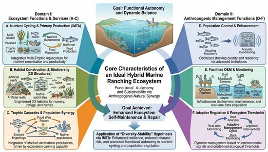
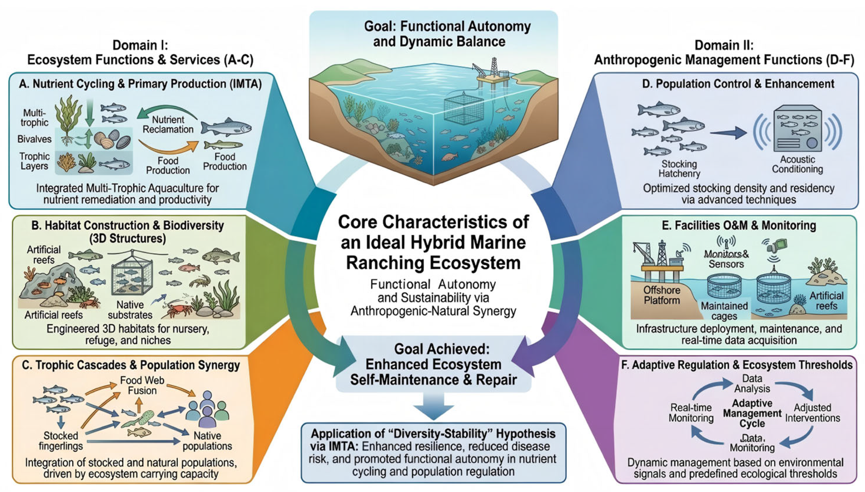
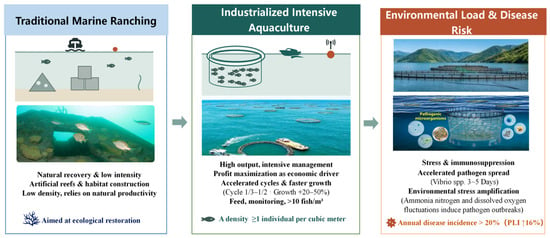
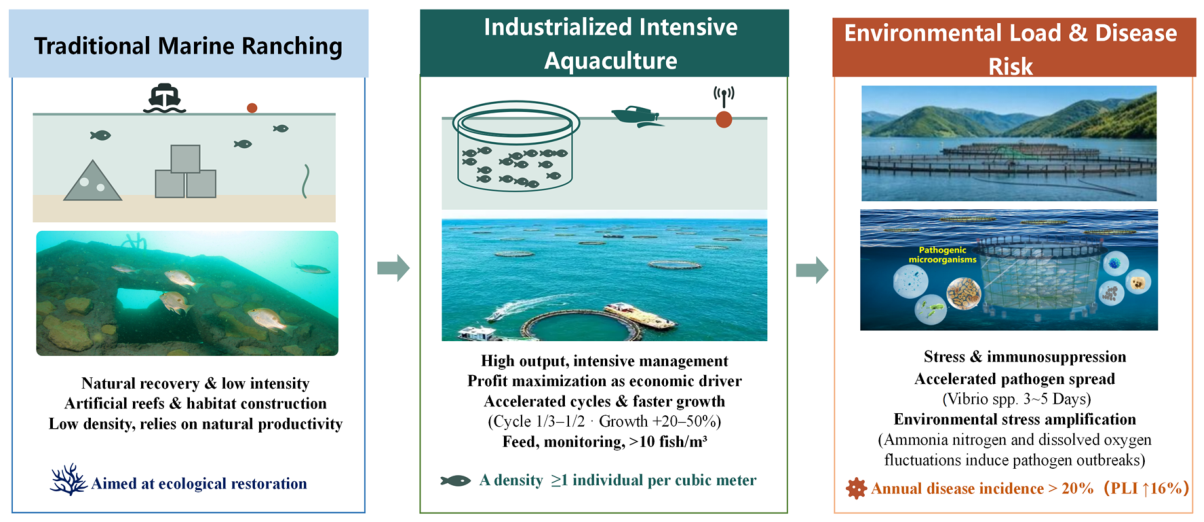
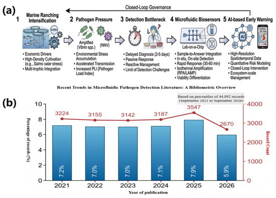
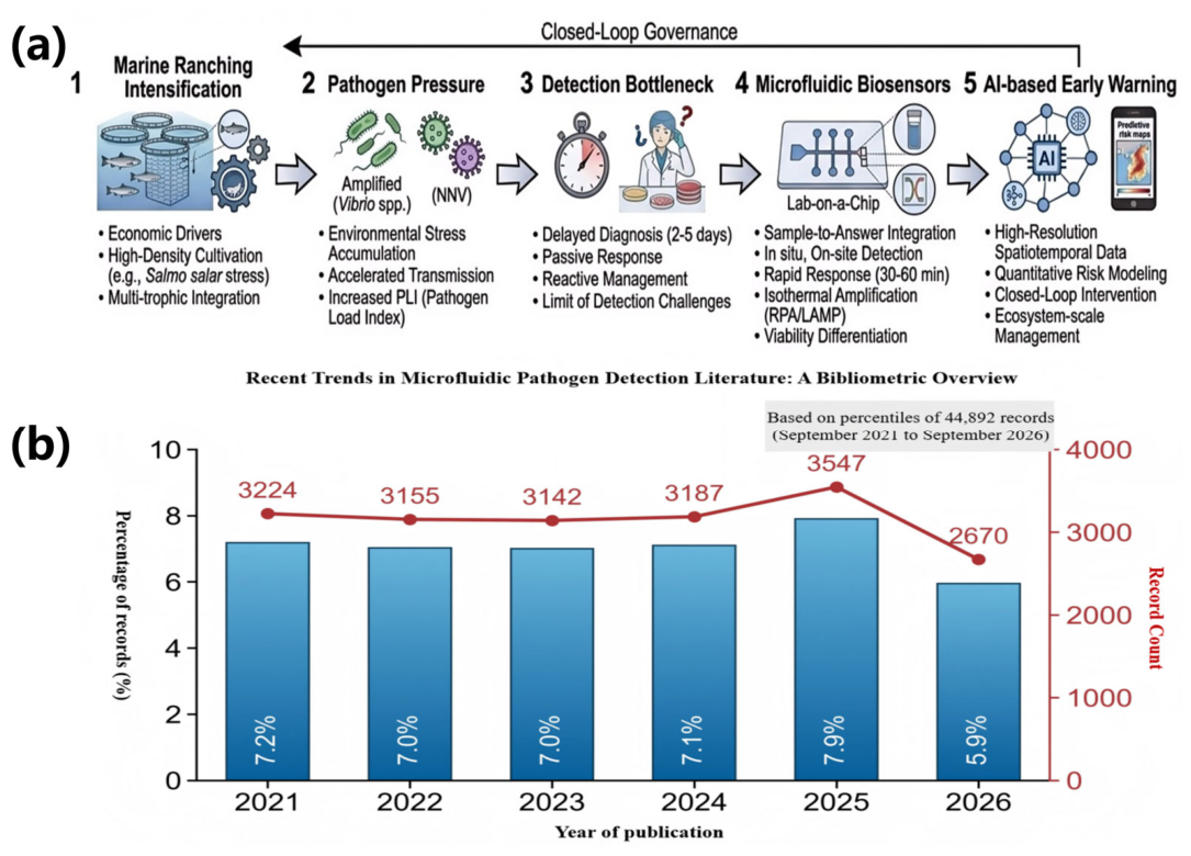
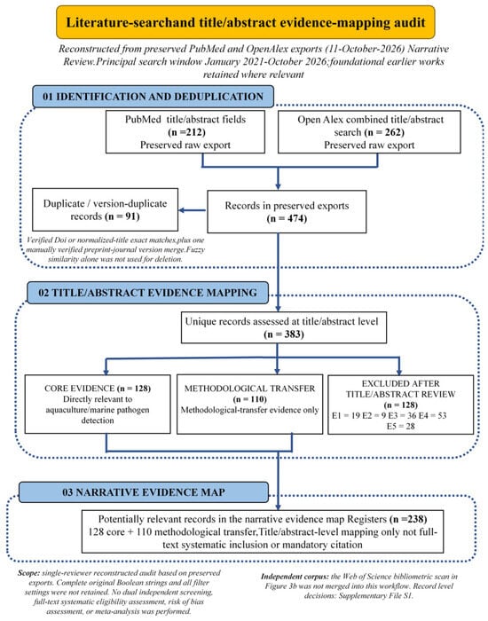
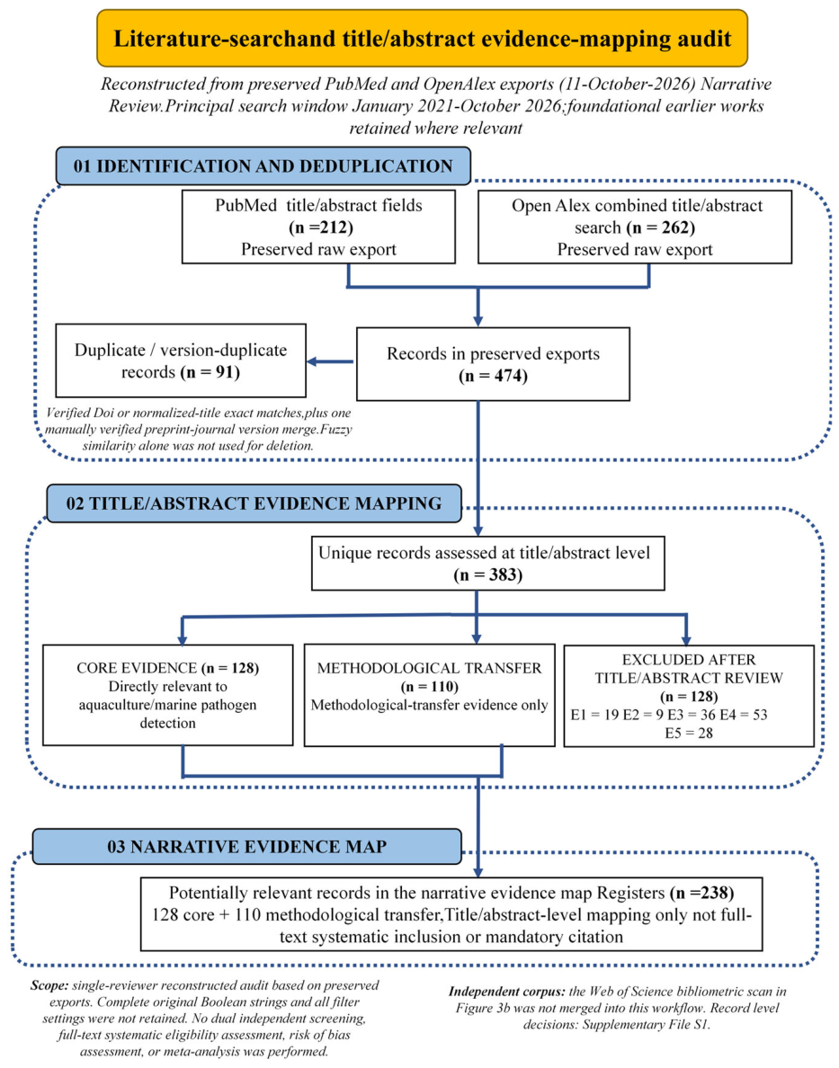
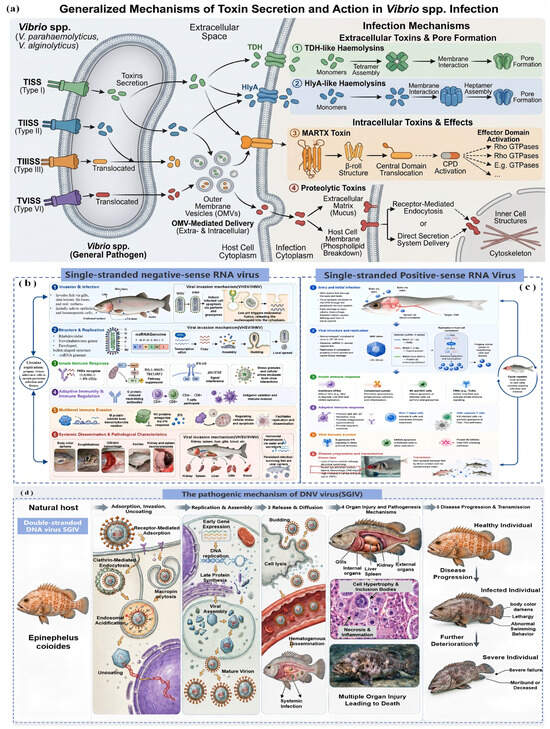
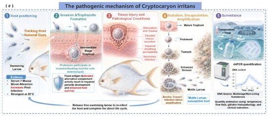
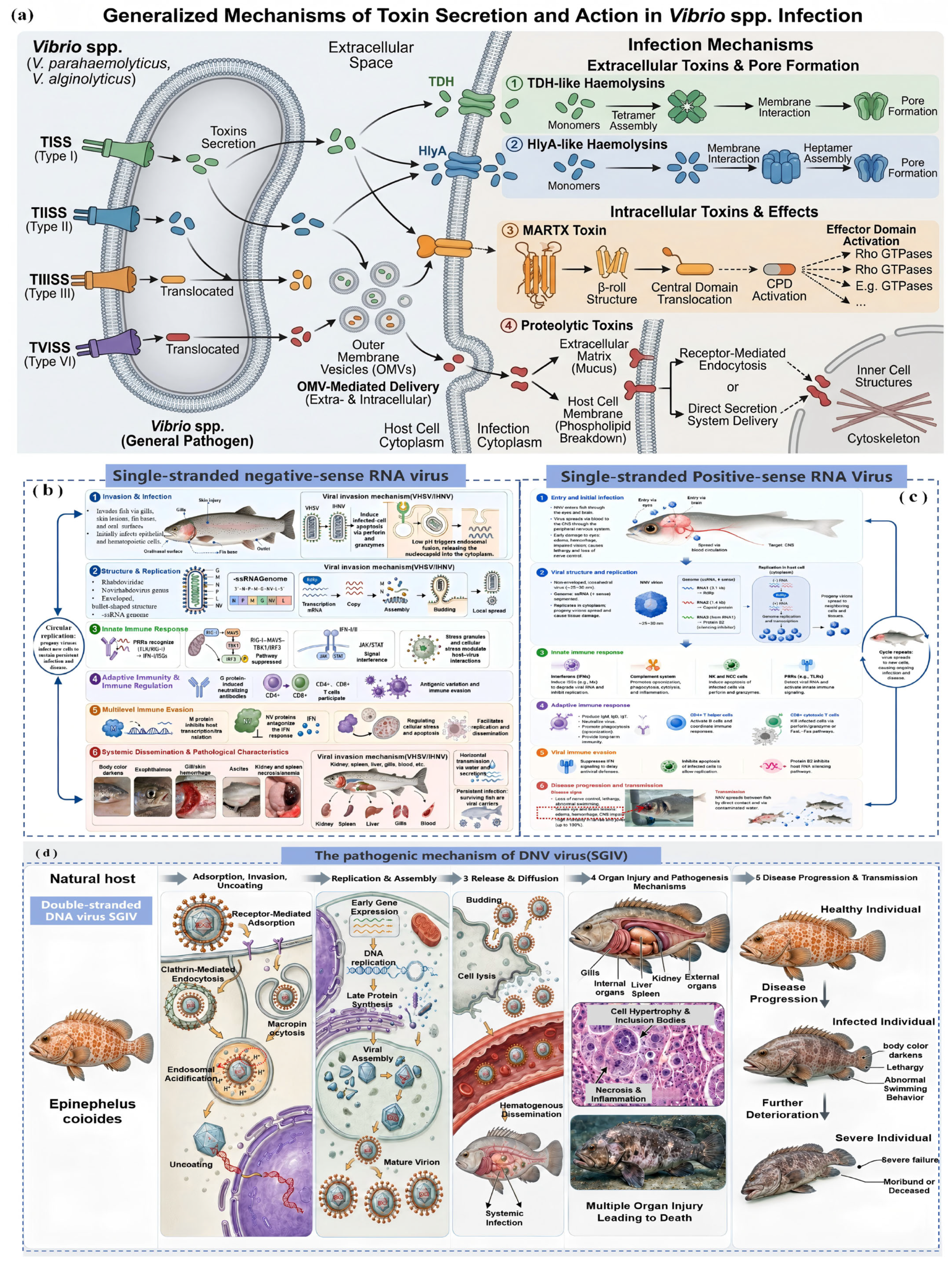
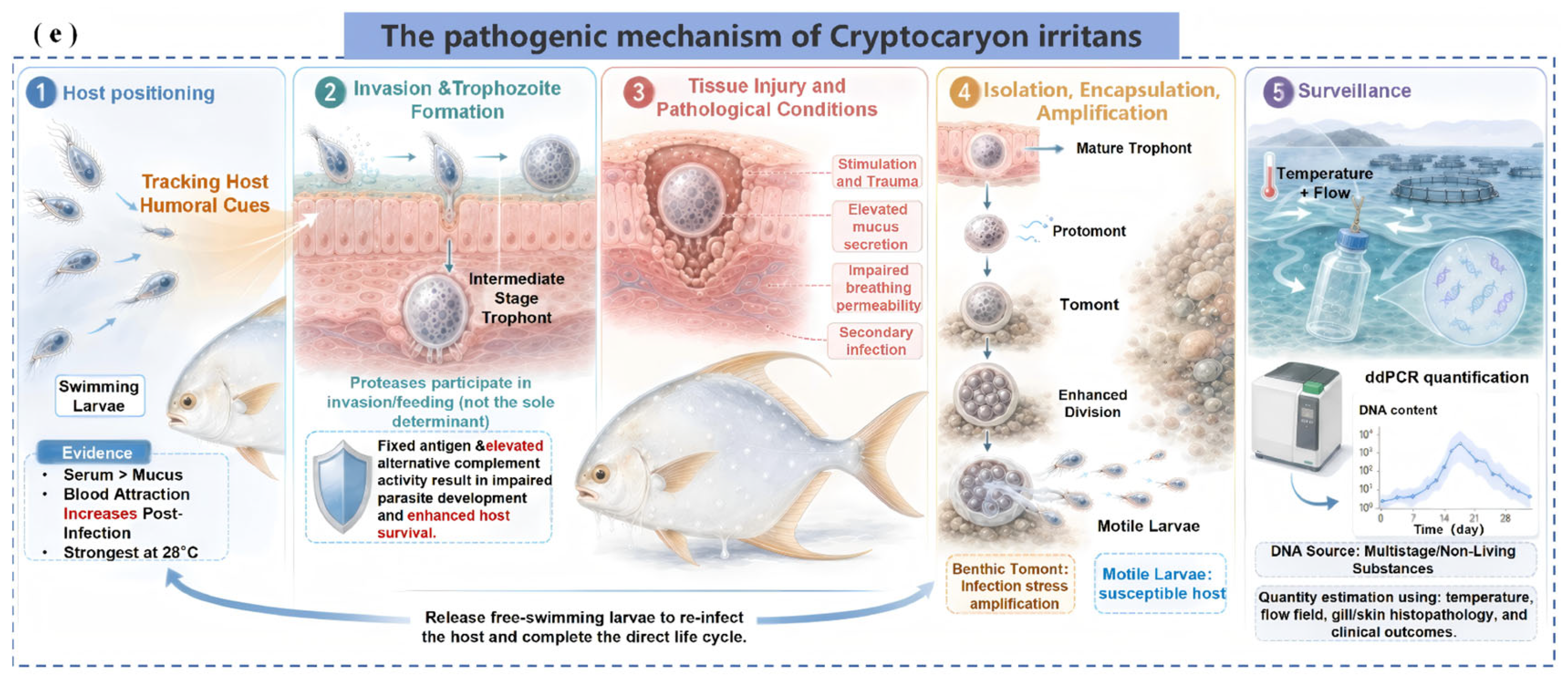
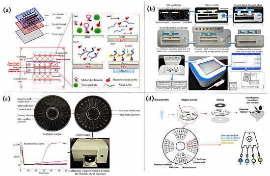
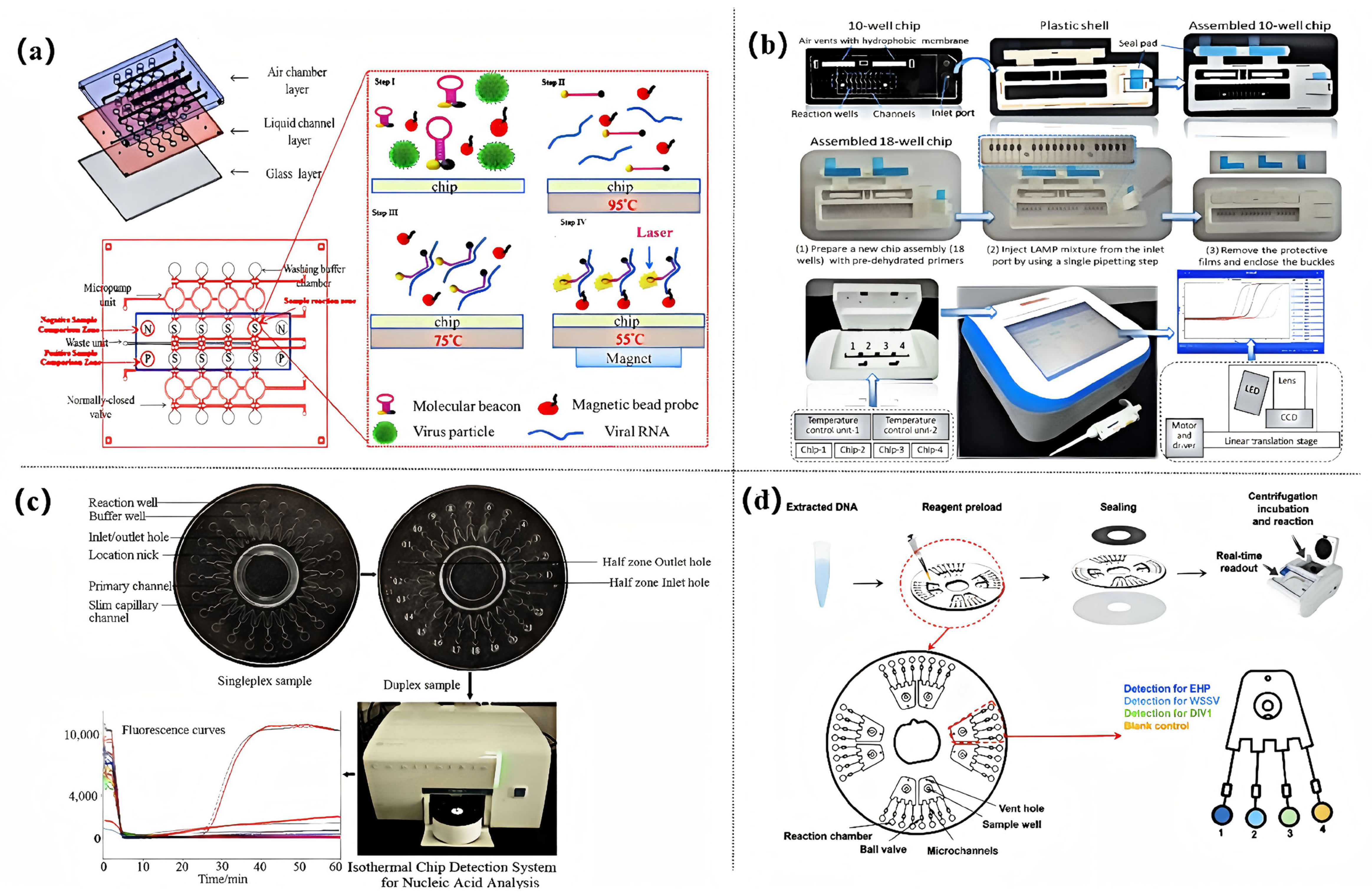
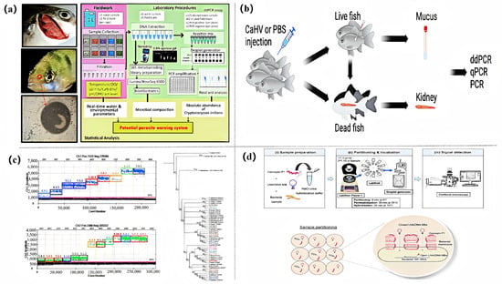
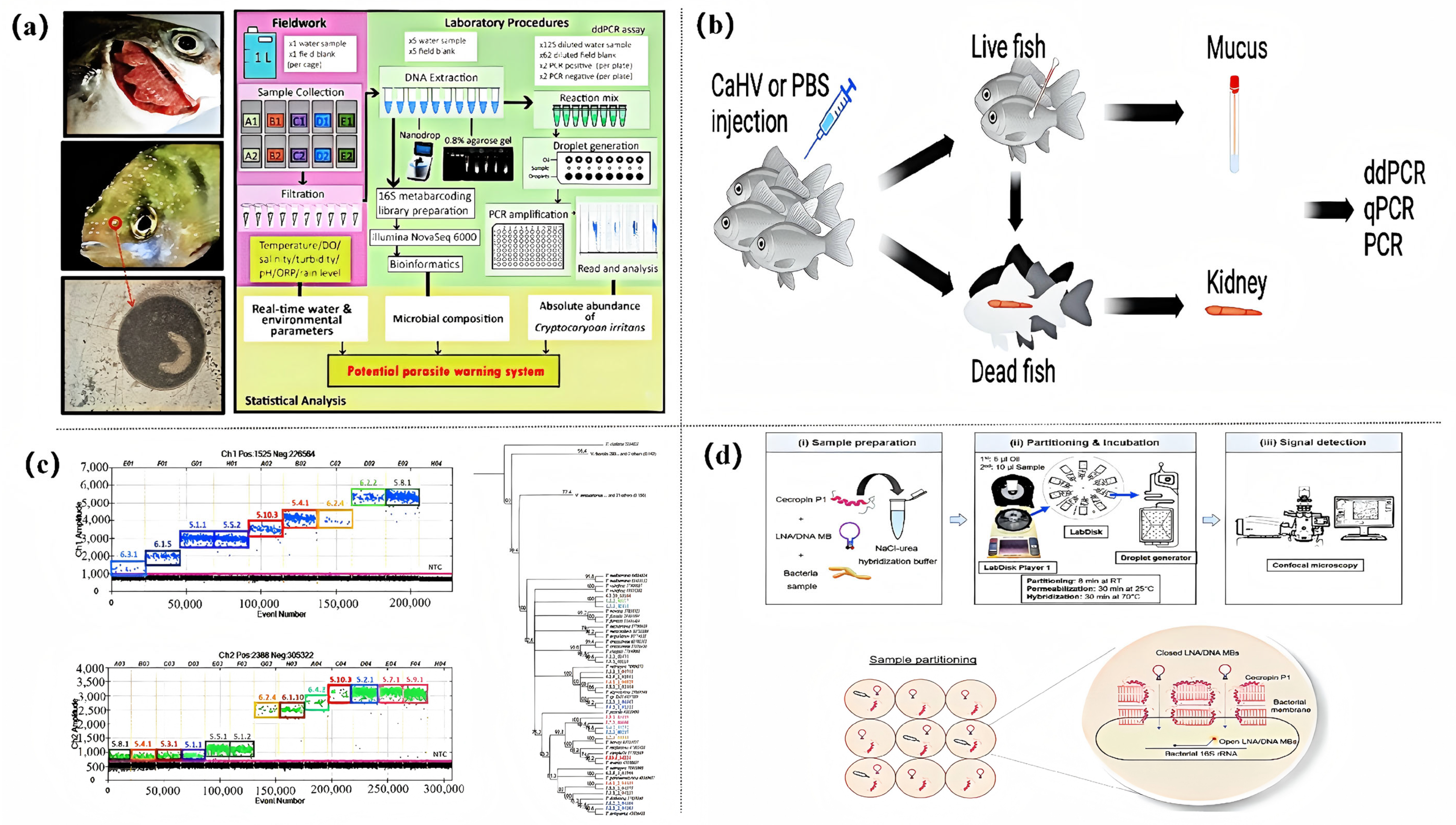
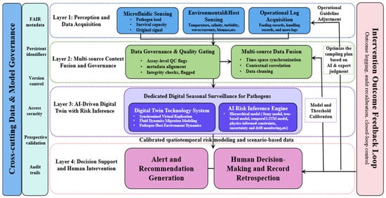
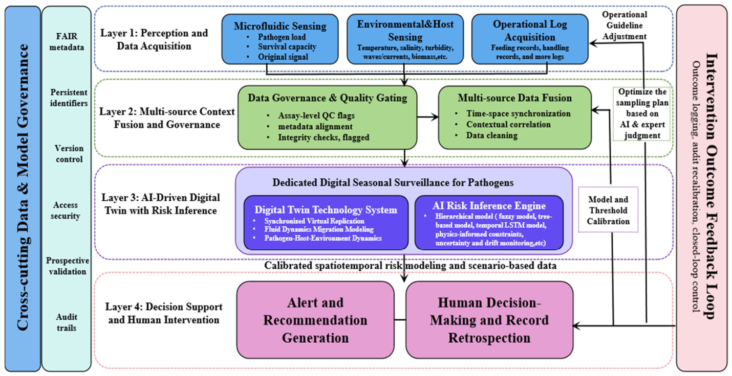

Disclaimer/Publisher’s Note: The statements, opinions and data contained in all publications are solely those of the individual author(s) and contributor(s) and not of MDPI and/or the editor(s). MDPI and/or the editor(s) disclaim responsibility for any injury to people or property resulting from any ideas, methods, instructions or products referred to in the content. |
© 2026 by the authors. Licensee MDPI, Basel, Switzerland. This article is an open access article distributed under the terms and conditions of the Creative Commons Attribution (CC BY) license.