


Abstract
Background/Objectives: Non-carious cervical lesions (NCCLs) are common defects in adults that can lead to dentin hypersensitivity and aesthetic concerns, for which composite resin restorations currently represent the gold standard of care. However, evidence regarding the long-term clinical superiority of high-filled injectable composites and Er,Cr:YSGG laser-based cavity preparation remains limited. The present study aimed to compare the 1-year clinical performance of two different surface preparation protocols (Er,Cr:YSGG laser vs. conventional bur preparation with phosphoric acid etching) and two composite resin types (high-filled injectable vs. conventional paste-type) in the restoration of NCCLs. Methods: In this prospective, split-mouth, randomized controlled clinical trial, a total of 168 NCCLs in 27 patients were restored. Lesions were randomly allocated to four groups according to the combination of surface preparation (Er,Cr:YSGG laser or phosphoric acid etching) and high-filled injectable composite (G-ænial Universal Injectable) or paste-type composite (G-ænial Anterior). The same universal adhesive system was used in all cases. Clinical evaluations were performed by a blinded examiner at 1 week, 6 months, and 12 months, using the FDI World Dental Federation criteria. Results: At the 1-year follow-up, 25 patients and 150 restorations were available for evaluation, corresponding to a recall rate of 98.22%. High clinical acceptability was observed in all groups with respect to aesthetic, functional, and biological parameters. Retention was 100% in the acid-etched paste-type composite group and ranged from 94.7% to 97.4% in the remaining groups, with no statistically significant differences among groups (p > 0.05). A transient increase in postoperative sensitivity was detected in the laser groups at the 1-week evaluation (p = 0.026); however, sensitivity scores declined to zero in all groups at 6 months and 1 year. Conclusions: High-filled injectable composites demonstrated 1-year clinical performance comparable to that of conventional paste-type composites in the restoration of NCCLs. Er,Cr: YSGG laser-based cavity conditioning produced outcomes similar to conventional phosphoric acid etching with respect to retention, marginal adaptation, and biological compatibility. The early increase in laser-related postoperative sensitivity was transient and did not compromise long-term clinical success. Taken together, the ease of application and favorable clinical performance of injectable composites indicate that these materials constitute a reliable alternative for the restoration of non-carious cervical lesions.
1. Introduction
Non-carious cervical lesions (NCCLs) are pathological defects characterized by a loss of hard dental tissue at the enamel–cementum junction that occurs independently of bacterial caries. Their prevalence in adults has been reported to reach approximately 46.7% and to increase significantly with advancing age [1]. Today, increased life expectancy, the preservation of a greater number of natural teeth, acidic dietary habits, and the growing prevalence of parafunctional habits have led to a higher incidence and greater clinical significance of NCCLs. These factors collectively contribute to a substantial clinical burden, particularly with respect to dentin hypersensitivity, aesthetic concerns, the need for restorative interventions, and potential pulpal and periodontal complications [2,3]. Therefore, elucidating the pathogenesis and optimizing the clinical management of NCCLs, particularly in the context of an aging population, is a priority in contemporary preventive and restorative dentistry [4].
Clinically evident dentin hypersensitivity, aesthetically disturbing cervical defects, progressive loss of tooth structure, subgingival margins that complicate plaque control, and deep or extensive lesions that increase the risk of pulpal exposure or fracture constitute the primary indications for restorative treatment [3,4]. However, the cavity morphology—typically characterized by a wedge-shaped configuration lacking macromechanical retention—the presence of sclerotic dentin, and the location of the cervical/subgingival margins make adhesive bonding and isolation challenging, often reducing long-term restoration survival. When the cervical margin is located in dentin rather than enamel, the reduced presence of enamel prisms and the prevalence of sclerotic dentin hinder hybrid layer formation and render the bonded interface more susceptible to hydrolytic and mechanical degradation. This situation has been associated with increased rates of marginal breakdown, microleakage, and retention loss, thereby underscoring the need for adhesive protocols specifically optimized for dentin-located cervical margins to ensure favorable long-term clinical performance [5,6]. Consequently, both the adhesive protocol and the material properties of the composite resin gain critical importance [7,8].
Composite resins used in conjunction with adhesive systems are widely employed for the restoration of NCCLs, owing to their favorable aesthetic properties, satisfactory mechanical performance, and ability to establish micromechanical bonding with enamel and dentin substrates. [9]. In clinical practice, conventional etch-and-rinse and self-etch adhesive strategies are most frequently employed for bonding in NCCLs [10]. Etch-and-rinse systems generally provide strong enamel bonding following phosphoric acid etching but may be more technique-sensitive when applied to sclerotic dentin [11]. In contrast, self-etch adhesives simplify the clinical procedure and are less dependent on dentin etching, which may be advantageous in deep cervical areas where enamel is limited. Because marginal integrity and retention in NCCLs largely depend on the quality of the adhesive interface, a thorough understanding of the indications and limitations of both etch-and-rinse and self-etch approaches is essential when selecting an adhesive protocol for these lesions [12]. High-filled flowable/injectable composite resins are designed to provide improved flow and cavity adaptation owing to their low viscosity, while their increased filler content aims to enhance mechanical strength and abrasion resistance compared with conventional flowable composites [13,14]. Short- and medium-term clinical trials have reported comparable retention rates and, in many cases, superior marginal adaptation relative to conventional paste-type composite resins [13,15]. However, the number of well-designed, long-term randomized clinical trials that directly demonstrate the superiority of these new-generation materials over conventional paste-type composites remains limited [10].
Cavity surface preparation also plays a critical role in the success of NCCL restorations. In this context, erbium lasers such as Er,Cr:YSGG have been proposed to provide potential clinical advantages in cavity preparation, including minimally invasive removal of hard tissues and favorable modification of the dentin substrate to enhance bonding [16]. Although conventional bur preparation and acid etching aim to enhance micromechanical retention, the formation of a smear layer and the limited effectiveness of etching on sclerotic dentin do not always provide an ideal bonding substrate [4]. Therefore, laser systems such as Er,Cr:YSGG, which provide smear layer-free, irregular, high-energy surfaces, have come to the forefront. Microleakage and bonding performance on Er,Cr:YSGG laser-prepared surfaces have been reported to be comparable to, or in some protocols even superior to, those obtained with conventional bur and acid-etch techniques. However, other investigations have found no clear advantage or have reported increased microleakage when the laser is used as a sole conditioning method [17,18]. Although it has been reported that supplementary acid etching, particularly following laser conditioning, may enhance adhesive infiltration and marginal integrity, the evidence regarding the actual impact of laser-based surface preparation on the long-term retention and marginal adaptation of restorations in NCCLs remains heterogeneous and highly dependent on the specific clinical protocols employed [18,19].
The current literature predominantly examines, separately, the performance of high-fill injectable composites versus paste-type composites and the effectiveness of Er,Cr:YSGG laser roughening compared with conventional bur–acid protocols in the treatment of NCCLs. Randomized split-mouth studies that directly evaluate these two variables concurrently within the same clinical design remain quite limited [15,20]. In this context, the aim of this split-mouth, randomized, controlled clinical trial is to compare the 1-year clinical performance of two different cavity preparation protocols (Er,Cr:YSGG laser roughening and conventional bur preparation followed by phosphoric acid etching) and two different composite resin types (high-filled injectable composite and paste-type composite) in non-carious cervical lesions. Accordingly, the null hypotheses are as follows:
H01:
There is no difference in the 1-year clinical performance of NCCL restorations prepared using Er,Cr:YSGG laser conditioning compared with those prepared using conventional bur–acid etching protocols.
H02:
There is no difference in the 1-year clinical performance of restorations placed with injectable composite resin versus paste-type composite resin in NCCLs restored under the same cavity preparation protocols.
2. Materials and Methods
2.1. Study Design and Ethical Approval
This research was designed as a prospective, split-mouth, randomized, controlled clinical trial with a 1-year clinical follow-up period. The study protocol was approved by the Interventional Ethics Committee of the Faculty of Medicine, Van Yüzüncü Yıl University (decision no. 15, dated 6 August 2019) prior to the commencement of patient enrollment. The research was conducted in accordance with the ethical principles outlined in the 1964 Declaration of Helsinki and its subsequent revisions. The study was reported with reference to the CONSORT guidelines and was registered at ClinicalTrials.gov on 19 September 2019 (registration no. NCT04095520). Before inclusion in the study, all participants were thoroughly informed about the study objectives, treatment procedures, and potential risks, and written informed consent was obtained from all patients.
2.2. Participants/Eligibility Criteria
The study included patients over 18 years of age, in the permanent dentition stage, who presented with four or multiples of four non-carious cervical lesions. This requirement ensured that, within the split-mouth design comprising four experimental groups, each patient received one restoration per group, thereby enabling balanced intra-individual comparisons and appropriate random allocation of lesions across the four treatment protocols. Eligible patients had no pulpal or endodontic pathology or percussion sensitivity in the teeth to be treated, had at least one occlusal contact with both antagonist and proximal adjacent teeth, and were expected to attend scheduled follow-up appointments for periodic evaluations.
Individuals were excluded if they presented with systemic conditions that could adversely affect healing or oral health (e.g., uncontrolled diabetes, immunosuppressive disorders, ongoing chemotherapy or radiotherapy, or severe cardiovascular disease); were pregnant, suspected of being pregnant, or breastfeeding; had a known allergy to dental materials; or had diagnosed periodontal disease or poor oral hygiene; were thought to experience excessive occlusal loading on specific teeth due to severe dental crowding; had carious lesions in the cervical region or had previously received desensitizing agents or fluoride treatment in this region; or were undergoing active orthodontic treatment.
In selecting patients with non-carious cervical lesions for inclusion, care was taken to choose wedge-shaped defects located in either the maxilla or mandible, with lesion depth not exceeding 3 mm as measured with a periodontal probe. Lesions classified as grade 4 according to the University of North Carolina index and those presenting sclerotic dentin within the cavity were excluded from the study [21].
2.3. Sample Size
The G*Power software (G*Power, version 3.0.10, Düsseldorf, Germany; http://www.psycho.uni-duesseldorf.de/aap/projects/gpower) (accessed on 10 July 2019) was used to calculate the required sample size. To detect differences between the study groups, a power analysis was performed assuming a moderate effect size (f = 0.25), 80% statistical power (1−β = 0.80), and a 5% significance level (α = 0.05). Based on these parameters, it was determined that a minimum of 35 restorations per group, and 140 restorations in total, would be sufficient.
To compensate for potential dropouts during the follow-up period and to preserve the planned statistical power, a larger number of lesions was initially evaluated, and the predefined inclusion and exclusion criteria were applied. Following this evaluation, a total of 168 restorations of non-carious cervical lesions in 27 patients were deemed eligible and included in the study, and all clinical assessments were performed on these restorations. Thus, the initially calculated minimum sample size was achieved, and the final sample was further strengthened by accounting for possible losses to follow-up.
2.4. Randomization and Blinding
After enrollment, patients and their non-carious cervical lesions were randomly allocated to one of four experimental groups according to a split-mouth design. Randomization was performed using a random number table generated with the Research Randomizer Program (http://www.randomizer.org/form.htm) (accessed on 25 January 2021) [22]. This method ensured a balanced allocation of lesions in each patient’s mouth to one of four treatment groups, which were defined according to the cavity preparation protocol (acid etching or laser etching) and the type of restorative material used (injectable or paste-type composite).
In the blinding procedure, clinical evaluations of the restorations were performed by a single examiner who was not involved in the restorative procedures and was blinded to the group assignments. Furthermore, participants were not informed about which etching protocol or type of composite material had been used for each tooth. Thus, a double-blind evaluation was ensured, as neither the patients nor the clinical examiner had any knowledge of the specific restorative material or surface preparation protocol applied to the individual teeth.
2.5. Clinical Application Protocol
2.5.1. Cavity Preparation
Before initiating the restorative procedures, all patients received oral hygiene instruction. Subsequently, all teeth scheduled for treatment were cleaned with pumice and rubber cups to remove the salivary pellicle and dental plaque, and no local anesthesia was administered during the procedure.
In the acid-etching group, a conservative enamel bevel was prepared along the cervical enamel margin and 34.5% phosphoric acid gel (Vococid®, VOCO GmbH, Cuxhaven, Germany) was applied to the enamel and dentin surfaces for 20 s. This was followed by rinsing each tooth with pressurized water for at least 15 s and gentle air-drying. Cotton rolls and a saliva ejector were used to prevent salivary contamination, and the etched surfaces were examined for the presence of a characteristic chalky-white appearance.
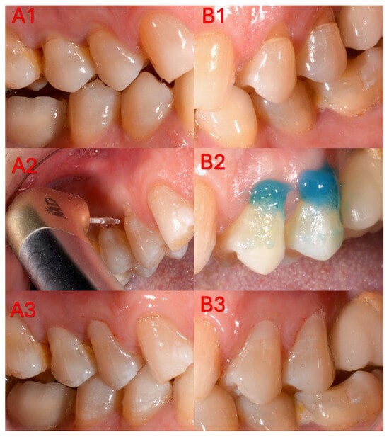
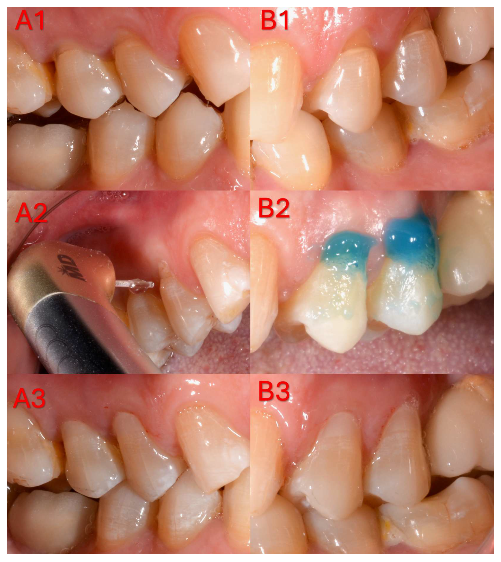
In the laser group, cavity roughening was performed using an Er,Cr:YSGG hard tissue laser (WaterLase® iPlus, Biolase Technologies Inc., Santa Ana, CA, USA) with a wavelength of 2.78 µm. The procedure was carried out with a 600-µm-diameter sapphire tip (MGG6–6 mm) and a gold handpiece, at 2.25 W output power, 50 Hz repetition rate, 140 µs pulse duration, and 60% air and 30% water cooling. The optical tip was moved along the cavity surface at a right angle and within the working distance recommended by the manufacturer for approximately 10–15 s, without contacting the tooth structure [23]. Each pulse delivered 45 mJ of energy, corresponding to an energy density of approximately 15.91 J/cm2. To prevent contamination of the tip during the procedure, it was periodically cleaned with alcohol-soaked gauze, and laser irradiation was discontinued once the surface exhibited an opaque white appearance (Figure 1).
Figure 1.
Clinical stages of a representative non-carious cervical lesion restored using two different surface conditioning protocols. (A) Laser-etched group: (A1) baseline clinical photograph before treatment; (A2) cavity surface conditioning performed with Er,Cr: YSGG laser irradiation; (A3) final appearance after composite resin restoration. (B) Acid-etched group: (B1) baseline clinical photograph before treatment; (B2) enamel and dentin etching with 34.5% phosphoric acid gel; (B3) final appearance after composite resin restoration.
2.5.2. Adhesives and Restoration
In all cavities, a single-bottle, single-step adhesive procedure was standardized by applying a universal adhesive agent (G-Premio BOND, GC Dental Products Corp, Aichi, Japan) in accordance with the manufacturer’s instructions. Subsequently, non-carious cervical lesions were restored with either a microhybrid conventional paste-type composite (G-ænial Anterior, GC Dental Products Corp, Aichi, Japan) or a high-filled injectable composite resin (G-ænial Universal Injectable, GC Dental Products Corp, Aichi, Japan). The materials were placed in a single increment, following the manufacturer’s recommendations, and adapted carefully along the cavity walls to prevent void or bubble formation. The type, composition, filler content, and lot numbers of the restorative and adhesive materials used are detailed in Table 1.
Table 1.
Restorative and adhesive materials used in this study.
Composite resins were polymerized using an LED curing unit (Elipar S10, 3M ESPE, St. Paul, MN, USA) emitting light in the 430–480 nm wavelength range, at an irradiance of approximately 1200 mW/cm2. For each restoration, the light guide was positioned perpendicular to the composite surface and activated for the exposure time recommended by the manufacturer. The output intensity of the curing unit was checked with an LED radiometer (LED Radiometer, SDI Dental Limited, Victoria, Australia) before each use to ensure that it remained within the standard operating range.
Finishing and polishing procedures were completed in the same session in which the restorations were placed. For this purpose, multi-step disc systems (Sof-Lex, 3M ESPE Dental Products, St. Paul, MN, USA), diamond-impregnated two-step flexible discs (CLEARFIL Twist DIA, EVE Ernst Vetter GmbH, Keltern, Germany), and a two-step disc–rubber system (Enhance® and PoGo®, Dentsply DeTrey GmbH, Konstanz, Germany) were used. The abrasive content, particle size distribution, and lot numbers of the polishing systems employed are presented in Table 2. Following restoration, all patients received brief counseling regarding potential etiological factors associated with NCCLs and were advised on general measures to minimize mechanical and chemical stresses on the cervical tooth structure.
Table 2.
Polishing system used in this study.
2.6. Clinical Evaluation (FDI Criteria)
Clinical check-ups were conducted at three time points—baseline (week 1), 6 months, and 12 months—and at each visit the clinical evaluation criteria of the FDI World Dental Federation were applied (Table A1 in Appendix A) [24]. Based on these criteria, the restorations were evaluated under three main domains: aesthetic, functional, and biological. The aesthetic evaluation considered surface gloss, surface discoloration, color stability, translucency, and anatomical form. The functional evaluation focused on fractures and retention, marginal adaptation, wear, and the patient’s satisfaction with the restoration. The biological evaluation encompassed post-operative sensitivity, recurrent caries, erosion/abfraction, tooth integrity, periodontal response, adjacent mucosa, and oral and general health status. Each restoration was scored on the FDI scale from 1 to 5 within these subdomains; scores of 1 to 3 were regarded as clinically acceptable, whereas scores of 4 or 5 were classified as clinically unacceptable for the respective criterion. The clinical acceptability rate (CAR) was calculated as the percentage of restorations exhibiting FDI scores between 1 and 3 for the respective parameter at each recall visit.
2.7. Statistical Analysis
All statistical analyses were performed using IBM SPSS Statistics software (version 23.0, IBM Corp., Armonk, NY, USA). The Fleiss kappa test was employed to evaluate the agreement of measurements over time. Pearson’s chi-square test was used for intergroup comparisons of categorical variables; when expected cell counts were insufficient, Fisher’s exact test was applied, and Yates’ continuity correction was used when appropriate. Survival analysis was conducted using the Kaplan–Meier method, and differences between survival curves were evaluated with the log-rank test. For the purposes of this analysis, failure was defined as complete loss of the restoration (failure = score 5 according to the FDI “Fractures and Retention” criterion). Categorical variables were summarized as frequency (percentage). A p-value of <0.05 was considered to indicate statistical significance.
3. Results
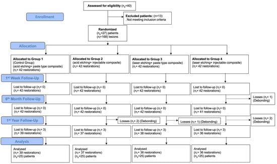
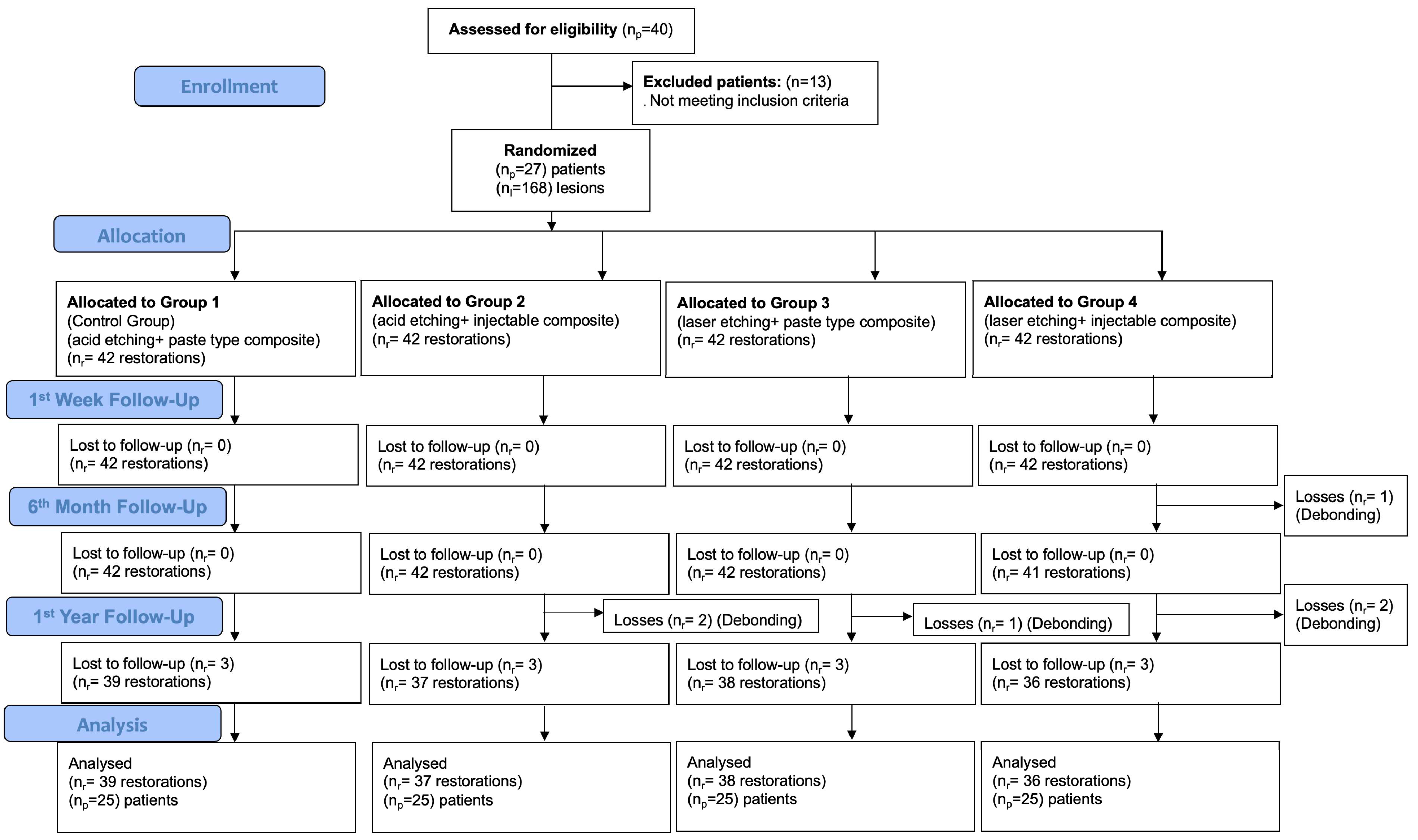
Initially, a total of 27 patients and 168 non-carious cervical lesions (NCCLs) were restored in the study. After the one-year follow-up period, 25 patients attended their recall appointments, and 150 restorations were clinically evaluated. Accordingly, the patient recall rate was calculated as 98.22% (Figure 2). At the one-year follow-up, two patients discontinued participation in the study due to relocation to other cities.
Figure 2.
Flowchart of the study.
Table 3 presents the distribution of restored NCCLs according to dental arch, tooth type, etching method, and composite material. In the maxillary arch, the majority of the 51 lesions restored following acid etching were located in premolar and incisor teeth, whereas in the mandibular arch, a substantial proportion of the 33 lesions was situated in the premolar region. Similarly, the 84 lesions treated with laser etching were evenly distributed among canines, premolars, and incisors in both the maxillary and mandibular arches. At baseline, no statistically significant differences were observed between the groups with respect to the distribution of the teeth included or the dental arch in which they were located (p > 0.05).
Table 3.
Distribution of NCCLs restored in the study.
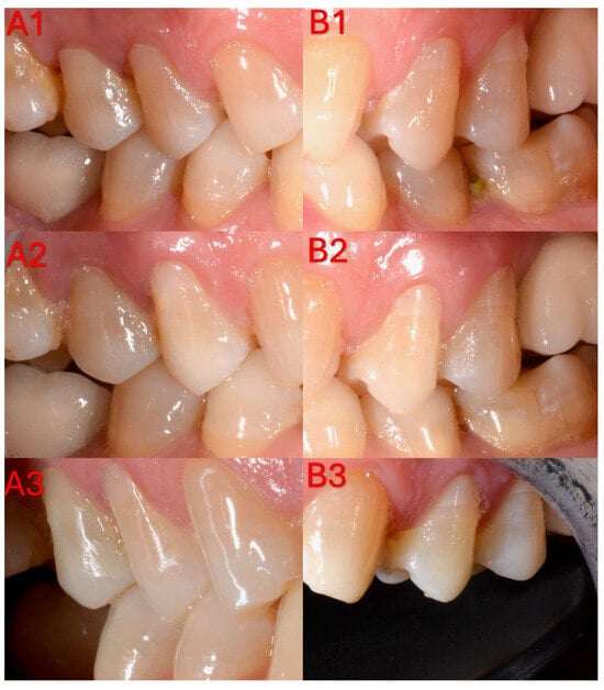
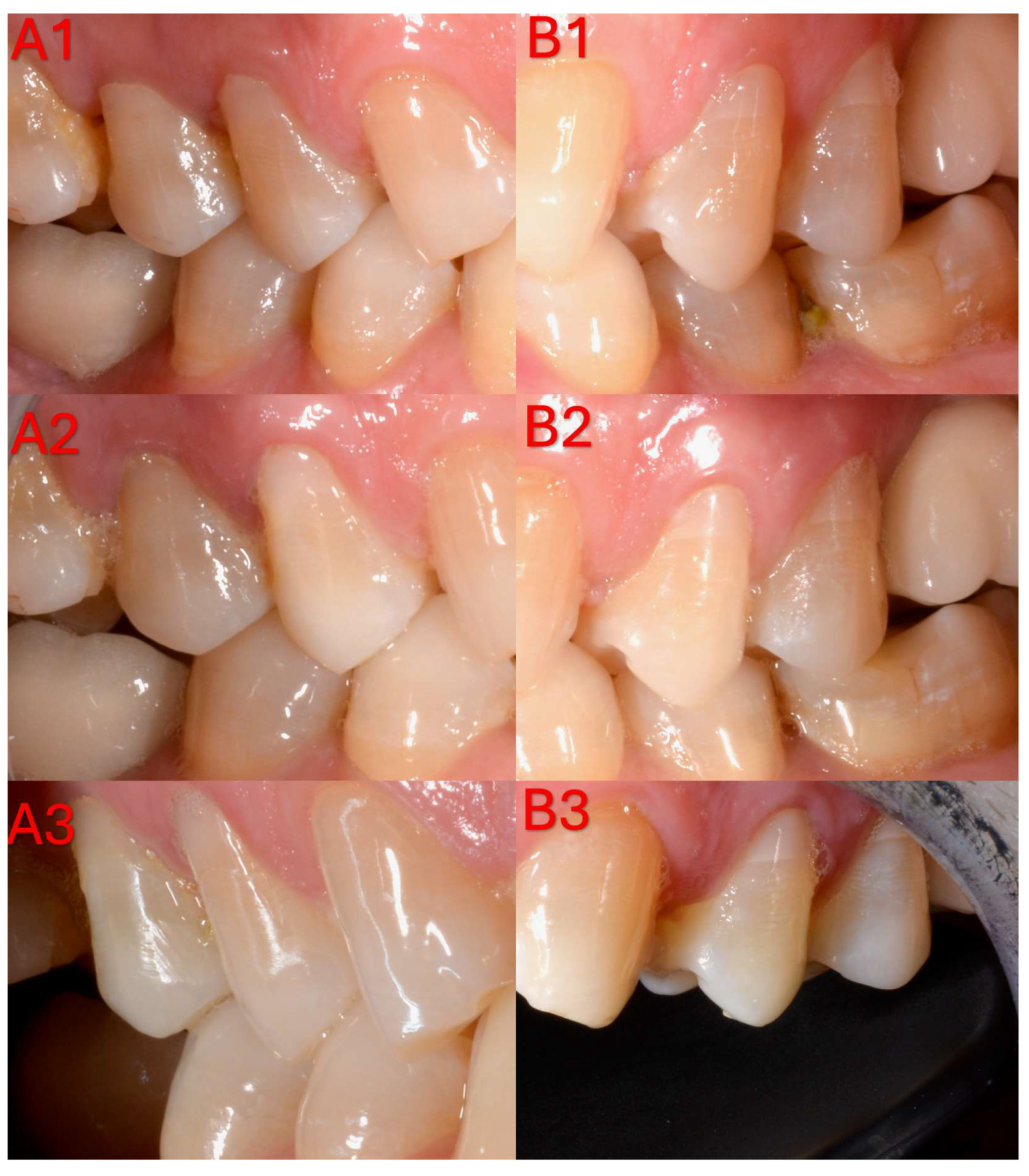
According to the FDI criteria, high clinical acceptability rates were obtained in all groups over the 1-year follow-up period with respect to aesthetic, functional, and biological parameters (Table 4, Table A4 and Table A5) (Figure 3).
Table 4.
Distributions of values measured at different times depending on the etching method and composite used.
Figure 3.
Representative clinical follow-up photographs of non-carious cervical lesion restorations evaluated according to the FDI World Dental Federation criteria at different time points. (A) Laser-etched group: (A1), 1-week recall (baseline); (A2), 6-month follow-up; (A3), 12-month follow-up. (B) Acid-etched group: (B1), 1-week recall (baseline); (B2), 6-month follow-up; (B3), 12-month follow-up.
Examination of the aesthetic criteria revealed that clinically acceptable scores (1–2) predominated in all groups for surface gloss, surface staining, color stability/translucency, and anatomical form, and that 100% clinical acceptability was maintained at the 1-week, 6-month, and 1-year evaluations. Although the frequency of score 2 increased over time, these changes were not statistically significant (p > 0.05).
With respect to fractures and retention, regarded as the most critical parameters within the functional criteria, the retention rate in the paste-type composite group following acid etching was 100% at all evaluation time points. After 1 year of follow-up, the retention rates were 94.8% in the injectable composite group following acid etching, 94.73% in the injectable composite group following laser etching, and 97.4% in the paste-type composite group following laser etching. Despite these losses, no statistically significant differences in retention were detected among the groups at 1 week, 6 months, or 1 year (p = 1.00, p = 0.62, p = 0.74, respectively). Although an increase in score 2 was observed over time for marginal adaptation and wear, these changes were not statistically significant (p = 0.362).
Clinically acceptable scores were maintained in all groups throughout the 1-year follow-up period with respect to the biological criteria (post-operative sensitivity, recurrent caries, tooth integrity, periodontal response, adjacent mucosa, and oral/general health), yielding a clinical acceptability rate of 100%. A transient increase in post-operative sensitivity scores was observed at the 1-week follow-up only in the laser-etching groups (p = 0.026); however, sensitivity scores decreased to 0 in all groups at the 6-month and 1-year evaluations. These findings indicate that, according to the FDI criteria, both composite types and both surface-conditioning methods demonstrated generally comparable and successful clinical performance over the 1-year observation period (Table 4).
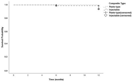
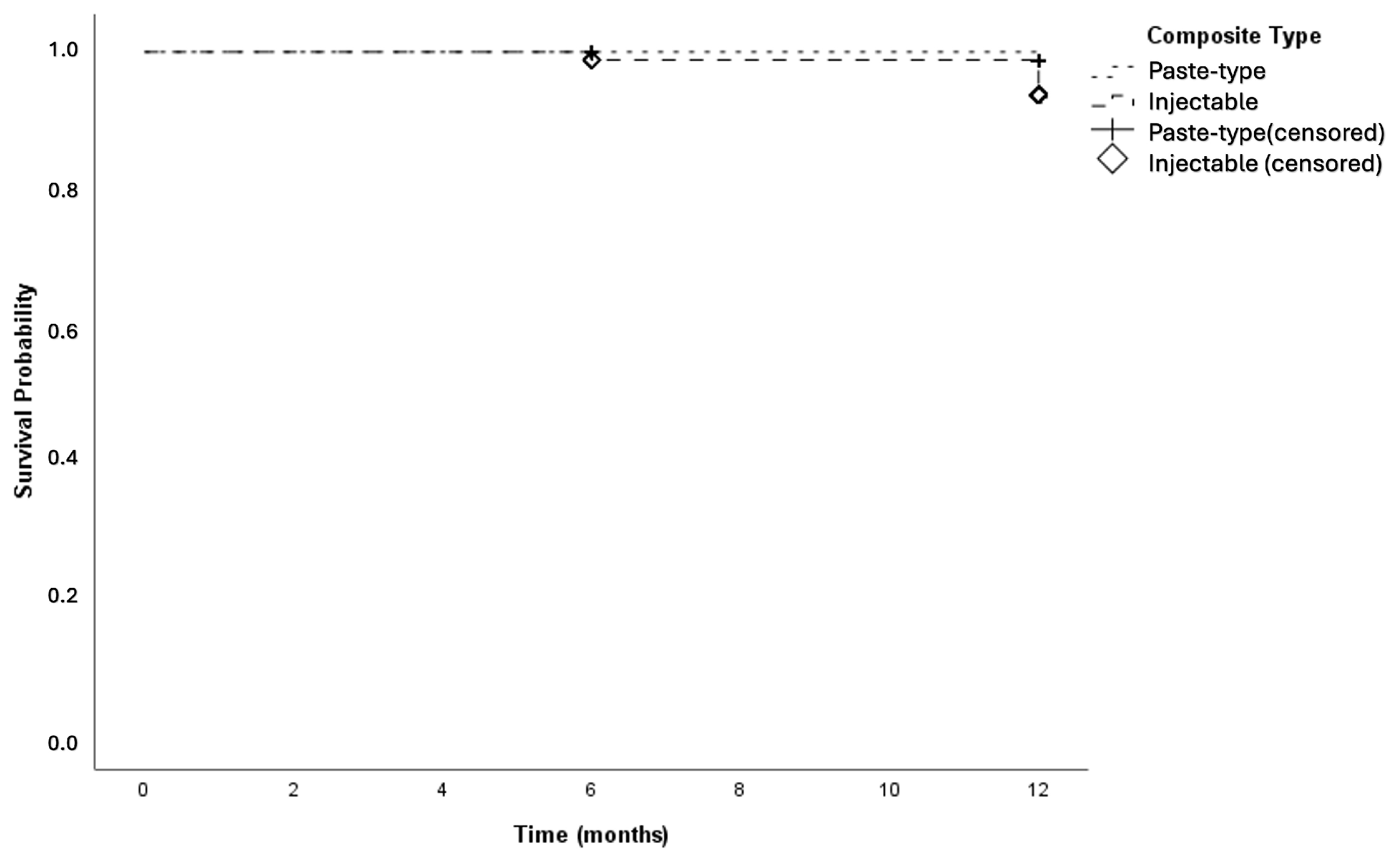
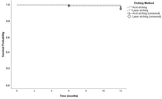
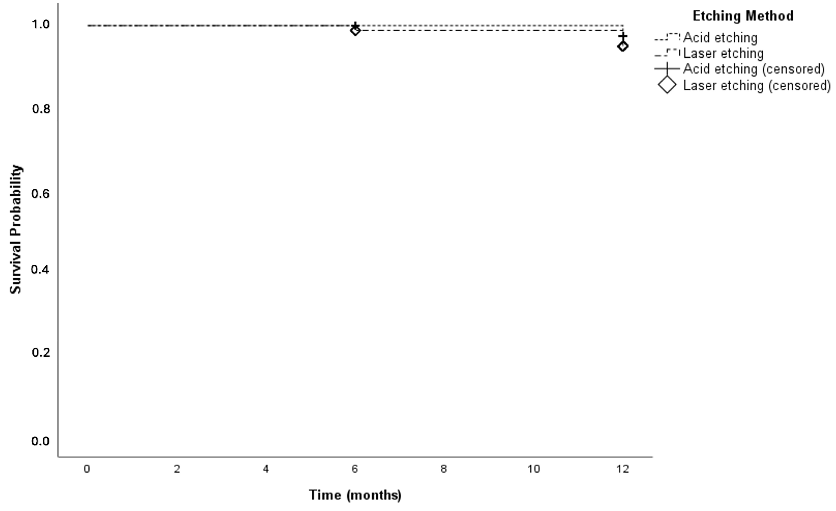
Kaplan–Meier survival analysis demonstrated 12-month cumulative survival rates of 98.7% for the paste-type composite and 93.7% for the high-filled injectable composite, with no statistically significant difference observed between the two composite materials (p = 0.096) (Figure 4). Similarly, for the cavity preparation methods, the 12-month cumulative survival rates were 97.4% in the acid-etching group and 95.0% in the Er,Cr:YSGG laser-etched group with no statistically significant difference in survival (p = 0.402) (Figure 5).
Figure 4.
Kaplan–Meier survival curves of restorations according to composite type.
Figure 5.
Kaplan–Meier survival analysis based on the etching protocol.
4. Discussion
Evaluating the restorative success of NCCLs using multidimensional criteria—such as retention, marginal adaptation, and biological compatibility—is regarded as fundamental for the objective assessment of clinical performance [25]. The findings of the present study demonstrated that the 1-year clinical performance of the high-filled injectable composite and the conventional paste-type composite, when applied under different surface preparation protocols, was comparable, and that both materials achieved high success according to the FDI criteria. With respect to retention, statistically similar outcomes were obtained, with a 100% retention rate in the paste-type composite group following acid etching and rates ranging from 94.7% to 97.4% in the remaining groups (p > 0.05). The observation that changes in the aesthetic and functional sub-criteria remained within clinically acceptable limits supports the acceptance of the null hypotheses for both the surface preparation method (H01) and the composite type (H02). Nonetheless, the transient increase in post-operative sensitivity observed at the 1-week follow-up in the laser-etching groups (p = 0.026) represents a noteworthy exception with respect to the biological criteria.
Several factors are considered critical for retention success, including the reduced etchability of sclerotic dentin [2], which complicates hybrid layer formation, and the increase in stress accumulation in the cervical region due to occlusal loading, thereby challenging the bonding interface [3]. Indeed, randomized clinical trials employing universal adhesives have demonstrated that the degree of dentinal sclerosis and the internal angle morphology of the lesion can increase the risk of retention loss [26], whereas occlusal wear and the presence of antagonistic teeth may contribute to the etiopathogenesis and functional load profile of NCCLs [12]. The 1-year retention rates observed in the present study, when compared with the first-year data reported by Peumans et al.—who documented retention rates of approximately 86–87% at 3 years in NCCLs restored using two different application modes of a universal adhesive—similarly reflect a pattern of high early retention and a limited number of lost restorations [12]. Moreover, a similar trend was observed in the multimodal adhesive study by Manarte-Monteiro et al., which reported an overall 1-year retention rate of 97.5% across different adhesive application modes, thereby corroborating that composite-based NCCL restorations generally remain stable in the short term despite variations in adhesive protocols and materials [27]. Similarly, a study comparing pre-heated and non-heated composites reported retention rates of 90–97% over a period of up to 24 months, further supporting the short-term clinical success of composite-based NCCL restorations despite variations in restorative technique or material viscosity [28]. In recent years, numerous clinical studies and systematic reviews have reported survival or retention rates generally exceeding 80% for composite-based NCCL restorations over 2–5 years of follow-up, despite variability in universal adhesive formulations, application times, and cavity preparation protocols, thereby underscoring that the adhesive protocol is a more critical determinant of clinical outcome than the material type [29,30,31]. However, in the 7.7-year follow-up study by Lührs et al., the overall retention rate was reported as 82.8%, whereas retention loss increased to 27.8% in lesions that were merely cleaned without any cavity preparation, indicating that there are also studies in the literature reporting comparatively lower retention rates [32]. This discrepancy is thought to be attributable to factors such as the increasing prominence of fatigue and hydrolytic degradation at the adhesive interface over longer follow-up periods, as well as the adverse effect of omitting selective roughening on highly sclerotic dentin surfaces on the quality of the hybrid layer.
The favorable clinical performance of injectable composites observed in the present study can be attributed to the balanced combination of superior cavity adaptation, resulting from their low viscosity, and the mechanical stability conferred by their high filler content. High-filled flowable/injectable materials, owing to their thixotropic behavior, effectively wet cavity walls—even in cavities with limited macromechanical retention and irregular surfaces—thereby reducing the formation of voids and bubbles and consequently enhancing marginal adaptation. Consistent with these findings, Hançer Sarıca et al. reported that G-ænial Universal Injectable provided superior adaptation to cavity walls and margins in Class II restorations and reduced the risk of void formation in challenging areas [33]. Similarly, Basheer et al. demonstrated that in high-filled flowable composites such as G-ænial Universal FLO and G-ænial Universal Injectable, a lower elastic modulus and increased flowability significantly reduce microleakage by decreasing polymerization shrinkage stresses [34]. Another study demonstrated that resin composites with reduced viscosity, when applied using an injection technique, produced fewer interfacial defects at the gingival wall compared with the placement of the same material using a spatula, thereby highlighting that the combination of injection and low viscosity enhances internal adaptation [35]. These findings suggest that the favorable marginal adaptation and high retention rates observed in NCCL restorations over the 1-year follow-up in the present study are consistent with the rheological properties of the injectable composite. Conversely, the absence of significant differences between the injectable composite and the conventional paste-type composite with respect to retention, fracture, wear, and anatomical form can be attributed to the fact that both materials exhibit similar mechanical properties due to their high filler content. The literature indicates a strong correlation between the inorganic filler fraction of composite systems and their elastic modulus and flexural strength, and it has been reported that high-filled flowable/nanohybrid composites can demonstrate mechanical performance equivalent or comparable to that of certain hybrid paste composites [36]. In a 2-year Class II clinical trial, Hançer Sarıca et al. compared G-ænial Universal Injectable with conventional and bulk-fill composites and found no significant differences among the materials for most FDI criteria, attributing these findings to their high filler content and consequent similarity in mechanical behavior [33]. Consistent with these findings, the principal advantage of the injectable composite in the present study was observed in terms of cavity adaptation and ease of application, whereas the absence of significant differences in mechanically oriented outcomes—such as retention and fracture—when compared with a paste-type composite of similar filler content can be regarded as an expected result.
In the present study, the higher level of postoperative sensitivity observed at the first-week evaluation following laser roughening can be attributed to both pulpal thermal effects and the morphological alterations induced by the laser on dentinal tubules. In vitro investigations employing hard tissue lasers (particularly Er:YAG, Er,Cr:YSGG, and high-power diode lasers) have shown that, under certain parameters, these devices can produce melting, recrystallization, and narrowing or obliteration of tubule orifices on the dentin surface, in addition to a short-term temperature increase and a limited pulpal inflammatory response [37]. Meng et al. reported that TNF-α and HSP-70 expression increased significantly on day 1 following 980-nm diode laser application, that inflammation gradually subsided between days 7 and 14, and that the intrapulpal temperature rise remained below the critical threshold [38]. These findings suggest that early pulpal inflammation and increased neural excitability may manifest clinically as heightened sensitivity during the first week. Conversely, it has been reported that hard tissue lasers, rather than completely occluding dentinal tubules, can under certain parameters expose tubule orifices, increase surface roughness, or produce only partial occlusion; such effects may initially facilitate the hydrodynamic mechanism and thereby exacerbate dentinal sensitivity [39]. Dündar et al. also reported that Er,Cr:YSGG laser irradiation significantly modifies the dentin surface by altering tubule patency and surface roughness, noting that this effect may initially be unfavorable with respect to sensitivity but may, in the longer term, promote tubule obliteration and thus yield more favorable outcomes [40]. In the present study, the reduction in sensitivity observed over time in the laser group, in line with the aforementioned literature, can be attributed to the resolution of the initial, limited thermal and inflammatory pulpal response, the maturation of laser-induced tubule plugs, and the formation of secondary or tertiary dentin, collectively resulting in decreased dentin permeability.
The finding that marginal adaptation values in the laser-prepared cavity group were comparable to those obtained with conventional methods appears consistent with the wetting and adaptation advantages of injectable composites arising from their low viscosity. Low viscosity facilitates superior wetting of the cavity walls, particularly in the cervical region, and enables the material to spread along the walls without leaving voids; this, in turn, can be associated with higher initial proportions of “continuous margins” in the restorations [41]. Low-viscosity resins used in cervical cavities have been reported to demonstrate high marginal adaptation and retention rates over 2–3 years of follow-up [32]. Both clinical and laboratory studies have indicated that, in NCCL models, a thin layer of low-viscosity resin can function as a “stress-absorbing interlayer,” distributing polymerization shrinkage stresses and reducing marginal gaps, particularly at the gingival margin [42]. The favorable marginal integrity observed in the laser group in the present study is consistent with this stress-distributing effect and supports the notion that injectable composites can be safely used on laser-prepared surfaces. However, it has also been shown that bulk-fill materials placed in large volumes in a single increment are associated with increased overall polymerization shrinkage and more pronounced marginal deterioration after aging, although differences among various placement techniques tend to diminish following thermomechanical loading [43]. The minimal intergroup differences in marginal adaptation observed during long-term follow-up in the present study suggest that, rather than conferring a permanent standalone advantage, the injectable composite provides clinically adequate and predictable marginal integrity when used with an appropriate layering protocol.
The literature indicates that enamel and dentin surfaces prepared with erbium lasers exhibit a smear-free, high-energy morphology conducive to micromechanical retention; when combined with appropriate adhesive protocols, these surfaces can provide bond strengths comparable to conventional acid etching and yield clinically acceptable outcomes with respect to marginal adaptation and marginal discoloration [44]. Moreover, laser-based cavity preparation has been reported to yield outcomes that are at least comparable to, and in some cases superior to, conventional bur preparation with respect to marginal adaptation and color stability in Class V restorations [19]. The stable marginal parameters observed in the laser group in the present study are consistent with these reports and support the conclusion that the combination of laser-prepared surfaces with injectable composites represents a clinically safe and predictable option with respect to marginal integrity and color stability.
Several limitations should be taken into account when interpreting the findings of the present study. First, although care was taken to allocate restorations in multiples of four per patient, restorations were not assigned to predefined quadrants or jaws. Therefore, perfect split-mouth standardization at the quadrant level could not be achieved. Also the 1-year follow-up period is insufficient to fully characterize the long-term clinical behavior of parameters that may exhibit progressive changes over time, such as marginal adaptation and color stability. In addition, the single-center design limits the generalizability of the results to other clinical environments and more heterogeneous patient populations. Future investigations with larger sample sizes and multicenter designs, incorporating follow-up periods of 3–5 years, are warranted to more comprehensively elucidate both the long-term performance of laser-etched surfaces and the clinical behavior of next-generation injectable composites in patients with varying risk profiles.
5. Conclusions
Based on the 1-year follow-up results of this randomized controlled clinical trial, the following conclusions can be drawn:
- High-filled injectable composites demonstrated clinical performance comparable to that of conventional paste-type composites in the restoration of non-carious cervical lesions.
- The cavity roughening protocol performed with an Er,Cr:YSGG laser produced outcomes comparable to those achieved with conventional phosphoric acid etching, with similar 1-year retention rates, marginal adaptation scores, and overall FDI-based clinical acceptability in NCCL restorations.
- The increase in post-operative sensitivity observed in the laser-roughening groups during the first week was transient and did not adversely affect long-term clinical success.
Author Contributions
Conceptualization, A.D. and B.Ü.D.; methodology, A.D.; software, A.D.; validation, B.Ü.D.; formal analysis, B.Ü.D.; investigation, A.D.; resources, A.D.; data curation, B.Ü.D.; writing—original draft preparation, B.Ü.D.; writing—review and editing, A.D.; visualization, A.D.; supervision, B.Ü.D. project administration, A.D.; funding acquisition, A.D., B.Ü.D. All authors have read and agreed to the published version of the manuscript.
Funding
This research was funded by Van Yuzuncu Yil University Scientific Research Projects Coordination Unit, grant number TSA-2019-8446.
Institutional Review Board Statement
The study was conducted in accordance with the Declaration of Helsinki and approved by the Interventional Ethics Committee of the Faculty of Medicine, Van Yüzüncü Yıl University (decision no. 15, dated 6 August 2019).
Informed Consent Statement
Informed consent was obtained from all subjects involved in the study.
Data Availability Statement
Data are contained within the article.
Acknowledgments
We would like to thank Naci Murat for his invaluable assistance with the statistical analysis of this study, as well as the patients who generously participated and thereby made this research possible.
Conflicts of Interest
The authors declare no conflicts of interest.
Appendix A
Table A1.
FDI Criteria and Scoring System for Esthetic Properties [24].
Table A2.
FDI Criteria and Scoring System for Functional Properties [24].
Table A3.
FDI Criteria and Scoring System for Biological Properties [24].
Table A4.
Distributions of esthetic property values measured at different time points according to the etching method and type of composite used.
Table A5.
Distributions of functional and biological property scores measured at different time points according to the etching method and composite type used.
References
- Teixeira, D.N.R.; Thomas, R.; Soares, P.; Cune, M.; Gresnigt, M.; Slot, D. Prevalence of Noncarious Cervical Lesions Among Adults: A Systematic Review. J. Dent. 2020, 95, 103285. [Google Scholar] [CrossRef] [Scilit]
- Patano, A.; Malcangi, G.; De Santis, M.; Morolla, R.; Settanni, V.; Piras, F.; Inchingolo, A.; Mancini, A.; Inchingolo, F.; Dipalma, G.; et al. Conservative Treatment of Dental Non-Carious Cervical Lesions: A Scoping Review. Biomedicines 2023, 11, 1530. [Google Scholar] [CrossRef] [Scilit] [PubMed]
- Goodacre, C.; Roberts, E.; Muñoz, C. Non-carious cervical lesions (NCCLs): Morphology and progression, prevalence, etiology, pathophysiology, and clinical guidelines for restoration. J. Prosthodont. 2023, 32, e1–e18. [Google Scholar] [CrossRef] [Scilit] [PubMed]
- Peumans, M.; Politano, G.; Van Meerbeek, B. Treatment of noncarious cervical lesions: When, why, and how. Int. J. Esthet. Dent. 2020, 15, 16–42. [Google Scholar] [PubMed]
- Amin, F.; Fareed, M.; Zafar, M.S.; Khurshid, Z.; Palma, P.; Kumar, N. Degradation and Stabilization of Resin-Dentine Interfaces in Polymeric Dental Adhesives: An Updated Review. Coatings 2022, 12, 1094. [Google Scholar] [CrossRef] [Scilit]
- Mokeem, L.; Garcia, I.; Melo, M. Degradation and Failure Phenomena at the Dentin Bonding Interface. Biomedicines 2023, 11, 1256. [Google Scholar] [CrossRef] [Scilit]
- Karimi, Z. The challenges of composite restorations of non-carious cervical lesions. Dent. Update 2025, 52, 340–346. [Google Scholar] [CrossRef] [Scilit]
- Hamdi, K.; Zaeneldin, A.; Samaha, A.H.; Hamama, H. Surface Pretreatments for Enhancing Bond Strength of Resin Composite to Sclerotic Dentin in Non-Carious Cervical Lesions: A Systematic Review. J. Dent. 2025, 163, 106123. [Google Scholar] [CrossRef] [Scilit]
- Szesz, A.; Parreiras, S.; Martini, E.; Reis, A.; Loguércio, A. Effect of flowable composites on the clinical performance of non-carious cervical lesions: A systematic review and meta-analysis. J. Dent. 2017, 65, 11–21. [Google Scholar] [CrossRef] [Scilit]
- Josic, U.; Maravic, T.; Mazzitelli, C.; Radovic, I.; Jaćimović, J.; Del Bianco, F.; Florenzano, F.; Breschi, L.; Mazzoni, A. Is clinical behavior of composite restorations placed in non-carious cervical lesions influenced by the application mode of universal adhesives? A systematic review and meta-analysis. Dent. Mater. 2021, 37, e503–e521. [Google Scholar] [CrossRef] [Scilit]
- Oz, F.; Ergin, E.; Canatan, S. Twenty-four-month clinical performance of different universal adhesives in etch-and-rinse, selective etching and self-etch application modes in NCCL—A randomized controlled clinical trial. J. Appl. Oral Sci. 2019, 27, e20180358. [Google Scholar] [CrossRef] [Scilit] [PubMed]
- Peumans, M.; Vandormael, S.; Coster, I.; Munck, J.; Meerbeek, B. Three-year Clinical Performance of a Universal Adhesive in Non-Carious Cervical Lesions. J. Adhes. Dent. 2023, 25, b4186751. [Google Scholar] [CrossRef] [Scilit] [PubMed]
- Tzimas, K.; Pappa, E.; Fostiropoulou, M.; Papazoglou, E.; Rahiotis, C. Highly Filled Flowable Composite Resins as Sole Restorative Materials: A Systematic Review. Materials 2025, 18, 3370. [Google Scholar] [CrossRef] [Scilit] [PubMed]
- Rajabi, H.; Denny, M.; Karagiannopoulos, K.; Petridis, H. Comparison of Flexural Strength and Wear of Injectable, Flowable and Paste Composite Resins. Materials 2024, 17, 4749. [Google Scholar] [CrossRef] [Scilit]
- Zhang, H.; Wang, L.; Hua, L.; Guan, R.; Hou, B. Randomized controlled clinical trial of a highly filled flowable composite in non-carious cervical lesions: 3-year results. Clin. Oral Investig. 2021, 25, 5955–5965. [Google Scholar] [CrossRef] [Scilit]
- Moharam, L.; Salem, H.; Abdou, A.; Afifi, R. Outcome of Er, Cr:YSGG laser and antioxidant pretreatments on bonding quality to caries-induced dentin. BMC Oral Health 2025, 25, 66. [Google Scholar] [CrossRef] [Scilit]
- Dua, D.; Dua, A.; Anagnostaki, E.; Poli, R.; Parker, S. Effect of different types of adhesive systems on the bond strength and marginal integrity of composite restorations in cavities prepared with the erbium laser—A systematic review. Lasers Med. Sci. 2021, 37, 19–45. [Google Scholar] [CrossRef] [Scilit]
- Zhang, Y.; Chen, W.; Zhang, J.; Li, Y. Does Er,Cr:YSGG reduce the microleakage of restorations when used for cavity preparation? A systematic review and meta-analysis. BMC Oral Health 2020, 20, 269. [Google Scholar] [CrossRef] [Scilit]
- Heyder, M.; Sigusch, B.; Hoder-Przyrembel, C.; Schuetze, J.; Kranz, S.; Reise, M. Clinical effects of laser-based cavity preparation on class V resin-composite fillings. PLoS ONE 2022, 17, e0270312. [Google Scholar] [CrossRef] [Scilit]
- Pappa, E.; Gkavela, G.; Sampri, I.; Masouras, K.; Rahiotis, C.; Kakaboura, A. A 2-Year Randomized Clinical Trial of Three Bonding Techniques in Non-Carious Cervical Lesions. Medicina 2024, 60, 1005. [Google Scholar] [CrossRef] [Scilit]
- Heymann, H.O.; Sturdevant, J.R.; Bayne, S.; Wilder, A.D.; Sluder, T.B.; Brunson, W.D. Examining tooth flexure effects on cervical restorations: A two-year clinical study. J. Am. Dent. Assoc. 1991, 122, 41–47. [Google Scholar] [CrossRef] [Scilit]
- Gurgan, S.; Koc Vural, U.; Kutuk, Z.B.; Cakir, F.Y. Does a new formula have an input in the clinical success of posterior composite restorations? A chat study. Clin. Oral Investig. 2021, 25, 1715–1727. [Google Scholar] [CrossRef] [Scilit] [PubMed]
- Kalender, B.; Lausch, J.; Gutknecht, N.; Gorucu, J. Surface conditioning of artificial caries lesions with Er, Cr:YSGG laser before resin infiltration: An in vitro study. Lasers Dent. Sci. 2024, 8, 16. [Google Scholar] [CrossRef] [Scilit]
- Hickel, R.; Peschke, A.; Tyas, M.; Mjör, I.; Bayne, S.; Peters, M.; Hiller, K.A.; Randall, R.; Vanherle, G.; Heintze, S.D. FDI World Dental Federation—Clinical criteria for the evaluation of direct and indirect restorations. Update and clinical examples. J. Adhes. Dent. 2010, 12, 259–272. [Google Scholar] [CrossRef] [Scilit] [PubMed]
- Ñaupari-Villasante, R.; Carpio-Salvatierra, B.; De Freitas, A.; De Paris Matos, T.; Núñez, A.; Tarden, C.; Barceleiro, M.; Reis, A.; Loguércio, A. Influence of different viscosity and chemical composition of flowable composite resins: A 48-month split-mouth double-blind randomized clinical trial. Dent. Mater. 2024, 40, 1798–1807. [Google Scholar] [CrossRef] [Scilit]
- Manarte-Monteiro, P.; Domingues, J.; Teixeira, L.; Gavinha, S.; Manso, M. Universal Adhesives and Adhesion Modes in Non-Carious Cervical Restorations: 2-Year Randomised Clinical Trial. Polymers 2021, 14, 33. [Google Scholar] [CrossRef] [Scilit]
- Manarte-Monteiro, P.; Domingues, J.; Teixeira, L.; Gavinha, S.; Manso, M. Multi-Mode adhesives performance and success/retention rates in NCCLs restorations: Randomised clinical trial one-year report. Biomater. Investig. Dent. 2019, 6, 43–53. [Google Scholar] [CrossRef] [Scilit]
- Favoreto, M.; Carneiro, T.; Ñaupari-Villasante, R.; Cordeiro, D.; Cochinski, G.; Nascimento, T.V.P.M.D.; Matos, T.; Bandeca, M.C.; Reis, A.; Loguércio, A. Clinical performance of preheating thermoviscous composite resin for non-carious cervical lesions restoration: A 24-month randomized clinical trial. J. Dent. 2024, 144, 104930. [Google Scholar] [CrossRef] [Scilit]
- Yurdagüven, G.Y.; Doğruer, I.; Çam, M.; Ulukapı, H.; Aykor, A.A. Clinical evaluation of different solvent type and HEMA-content of universal adhesive systems in non-carious cervical lesions: A two-year double-blind split-mouth randomized clinical trial. BMC Oral Health 2025, 25, 1661. [Google Scholar] [CrossRef] [Scilit]
- Omoto, E.; Santos, P.D.; Shinohara, M.; De Andrade Carvalho, P.R.M.; Catelan, A.; Fagundes, T. Clinical performance of different adhesion strategies in non-carious cervical lesion restorations: A four-year randomized clinical trial. J. Dent. 2025, 153, 105529. [Google Scholar] [CrossRef] [Scilit]
- Fernández, E.; Gil, A.C.; Caviedes, R.; Díaz, L.; Bersezio, C. Clinical Longevity of Direct Dental Restorations: An Umbrella Review of Systematic Reviews. J. Esthet. Restor. Dent. 2025; online ahead of print. [CrossRef] [Scilit]
- Lührs, A.; Jacker-Guhr, S.; Günay, H.; Herrmann, P. Composite restorations placed in non-carious cervical lesions—Which cavity preparation is clinically reliable? Clin. Exp. Dent. Res. 2020, 6, 558–567. [Google Scholar] [CrossRef] [Scilit]
- Sarıca, S.H.; Arslan, S.; Balkaya, H. Comparison of the 2-year clinical performances of class II restorations using different restorative materials. Clin. Oral Investig. 2025, 29, 128. [Google Scholar] [CrossRef] [Scilit] [PubMed]
- Basheer, R.; Hasanain, F.; Abuelenain, D. Evaluating flexure properties, hardness, roughness and microleakage of high-strength injectable dental composite: An in vitro study. BMC Oral Health 2024, 24, 546. [Google Scholar] [CrossRef] [Scilit] [PubMed]
- Andrade, A.C.M.; Trennepohl, A.A.; Moecke, S.; Borges, A.; Torres, C. Viscosity modulation of resin composites versus hand application on internal adaptation of restorations. Clin. Oral Investig. 2022, 26, 4847–4856. [Google Scholar] [CrossRef] [Scilit] [PubMed]
- Leprince, J.; Palin, W.; Vanacker, J.; Sabbagh, J.; Devaux, J.; Leloup, G. Physico-mechanical characteristics of commercially available bulk-fill composites. J. Dent. 2014, 42, 993–1000. [Google Scholar] [CrossRef] [Scilit]
- Zhuang, H.; Liang, Y.; Xiang, S.; Li, H.; Dai, X.; Zhao, W. Dentinal tubule occlusion using Er:YAG Laser: An in vitro study. J. Appl. Oral Sci. 2021, 29, e20200266. [Google Scholar] [CrossRef] [Scilit]
- Meng, Y.; Huang, F.; Wang, S.; Huang, X.; Lu, Y.; Li, Y.; Dong, Y.; Pei, D. Evaluation of dentinal tubule occlusion and pulp tissue response after using 980-nm diode laser for dentin hypersensitivity treatment. Clin. Oral Investig. 2023, 27, 4843–4854. [Google Scholar] [CrossRef] [Scilit]
- Rodriguez, C.G.B.; Manna, M.P.N.C.; Tarrilo, F.E.A.; Zezell, D.; Eduardo, C. In vitro study of the effects of diode laser on dentin hypersensitivity and evaluation of intra-pulpal temperature variation. Lasers Med. Sci. 2025, 40, 66. [Google Scholar] [CrossRef] [Scilit]
- Dündar, A.; Yılmaz, K.; Barutçugil, Ç.; Özyurt, Ö.K. Effect of laser applications with different agents on dentin surface roughness, bacterial adhesion and tubule occlusion. Clin. Oral Investig. 2025, 29, 353. [Google Scholar] [CrossRef] [Scilit]
- Scepanovic, D.; Par, M.; Attin, T.; Tauböck, T. Marginal Adaptation of Flowable vs Sonically Activated or Preheated Resin Composites in Cervical Lesions. J. Adhes. Dent. 2022, 24, b3032461. [Google Scholar] [CrossRef] [Scilit]
- De Oliveira Correia, A.M.; Tribst, J.P.M.; Matos, F.; Platt, J.; Caneppele, T.; Borges, A. Polymerization shrinkage stresses in different restorative techniques for non-carious cervical lesions. J. Dent. 2018, 76, 68–74. [Google Scholar] [CrossRef] [Scilit]
- Dietschi, D.; Askari, M.; Rossier, I.; Caseiro, L.; Krejci, I.; Leprince, J.; Di Bella, E.; Ardu, S. Marginal Adaptation of In Vitro Class II Restorations Made Out of Bulk or Conventional Composite Using Single- or Multi-Layered Techniques. Materials 2023, 16, 6325. [Google Scholar] [CrossRef] [Scilit]
- Jiang, T.; Gong, Q.-W.; Liu, Y.; Zhang, P.-P.; Zhang, L. Effect of erbium family laser etching on shear bond strength of enamel surfaces: A meta-analysis-PRISMA. Medicine 2022, 101, e30990. [Google Scholar] [CrossRef] [Scilit]

Disclaimer/Publisher’s Note: The statements, opinions and data contained in all publications are solely those of the individual author(s) and contributor(s) and not of MDPI and/or the editor(s). MDPI and/or the editor(s) disclaim responsibility for any injury to people or property resulting from any ideas, methods, instructions or products referred to in the content. |
© 2026 by the authors. Licensee MDPI, Basel, Switzerland. This article is an open access article distributed under the terms and conditions of the Creative Commons Attribution (CC BY) license.