


Abstract
Regeneration of the dental, oral, and craniofacial (DOC) complex is challenging because mineralized, soft, vascular, and neural tissues lie side by side, and defects are often large and irregular. Three-dimensional (3D) bioprinting enables fabrication of patient-specific, cell-laden constructs and may offer new regenerative options for complex oral cases. This narrative review summarizes current bioprinting technologies, their applications across DOC tissues, and the barriers to clinical adoption. Inkjet, laser-assisted, light-based, extrusion, and co-axial platforms differ in speed, resolution, cell viability, and cost, while intraoperative bioprinting allows constructs to be printed directly into the defect during surgery. Hydrogel-based bioinks, often loaded with cells or growth factors, support cell delivery and guided differentiation. Across the dentin–pulp complex, periodontium, gingiva, salivary glands, peripheral nerves, cartilage, and craniofacial bone, preclinical studies report that bioprinted constructs can recreate tissue microarchitecture, enhance vascularization, and support partial functional repair. Whole-tooth and load-bearing bone regeneration remain constrained by spatial patterning, vascularization, and mechanical demands. Most evidence remains preclinical, and translation is limited by cost, fragmented workflows, biological and mechanical mismatch at tissue interfaces, and unclear regulatory pathways. Standardized fidelity and viability metrics, improved bioinks, and point-of-care workflows will be needed before bioprinting becomes part of routine oral regenerative care.
1. Introduction
Regenerative strategies for the dental, oral, and craniofacial (DOC) complex must contend with a uniquely heterogeneous landscape: highly mineralized tissues such as enamel and dentin, which lie immediately adjacent to soft tissues and nerves. When defects arise from trauma, oncologic resection, or congenital conditions, large irregular volumes with demanding vascular requirements are created. Grafts and biologics are now routinely used in these defects, and cell-based therapies have been tested clinically, yet restoring the original architecture and function of multiple adjacent tissues remains a challenge [1,2]. Three-dimensional (3D) bioprinting offers a path to fabricate patient-specific constructs with biologically active materials. Current bioprinting platforms for DOC regeneration include inkjet-based deposition, laser-assisted transfer, light-based printing, extrusion printing, and emerging intraoperative approaches that enable point-of-care fabrication. Each modality presents distinct trade-offs between speed, cell viability, print size, and mechanical integrity, and each benefits from the expanding palette of bioinks, particularly hydrogel systems and composite formulations that support cell delivery, growth-factor presentation, and gradient architectures [3].
Oral and underlying tissues such as the periodontium, alveolar bone, salivary glands, and nerves are critical targets for regenerative techniques, as these tissues are important for structure, vascularization, and innervation. Additionally, these tissues are manipulated in routine surgical and restorative treatments with a risk of iatrogenic damage. Clinical defects in these tissues can be viewed as a tissue engineering problem, in which the surgical site functions as a tissue reactor for matrix, cells, and biologic signaling [2]. Bioprinting offers a way to deliver these components with spatial precision. For injured or missing oral tissues, advances in bioprinting promise to yield a chairside regenerative treatment option for patients with complex cases.
In recent years, the applications for 3D bioprinting have grown rapidly [3]. In the dentin–pulp complex, tailored hydrogels and human dental pulp stem cells (hDPSCs) are being leveraged to recreate tubule-scale features and to guide odontogenic differentiation [4,5,6]. In the periodontium, printed scaffolds and pro-angiogenic bioinks seek to re-establish the orientation and function of periodontal ligament (PDL) fibers while addressing concomitant alveolar bone loss [7,8,9]. Salivary gland engineering is incorporating structurally adaptable matrices and organoid platforms [10], whereas peripheral nerve repair in the maxillofacial region is exploring scaffold-free constructs and aligned fibrillar hydrogels to promote axonal growth [11,12]. Beyond these oral tissues, cartilage of the temporomandibular joint (TMJ) and nasal tissues, and craniofacial bone are under active investigation, with composite and vascularized constructs pushing toward functional restoration [13,14,15,16].
Despite these advances, translation to routine clinical use remains constrained by cost and infrastructure needs, the challenge of matching biological and mechanical properties across interfaces, and evolving regulatory and quality-control frameworks [2,17]. This review surveys current DOC-focused bioprinting technologies, summarizes limitations of the technology, and outlines pathways toward advancing clinical translation. Future advances, including workflow digitization, automation, and point-of-care manufacturing, hold promise to accelerate clinical adoption [17]. Compared to recent reviews of bioprinting for dental alveolar tissues [3], clinical translation in oral and maxillofacial reconstruction [18], and craniofacial reconstruction [19], this review relates each printing modality, including intraoperative and imaging-guided printing, to specific DOC tissue targets, separates cell-laden bioprinting from other printing approaches, and summarizes the experimental model and level of evidence for key studies (Section 3).
This narrative review is based on searches of PubMed and ClinicalTrials.gov for articles published up to October 2026, without a lower date limit, supplemented by the reference lists of recent reviews. Search terms combined “bioprinting”, “three-dimensional printing”, or “intraoperative bioprinting” with tissue terms, including “dental pulp”, “dentin”, “periodontal”, “gingiva”, “salivary gland”, “facial nerve”, “cartilage”, “temporomandibular joint”, and “craniofacial bone”. Representative studies were selected for relevance to DOC tissues rather than for exhaustive coverage. Recent reviews were used to frame each topic, and primary studies were used for the tissue-specific findings.
2. Bioprinting Technologies
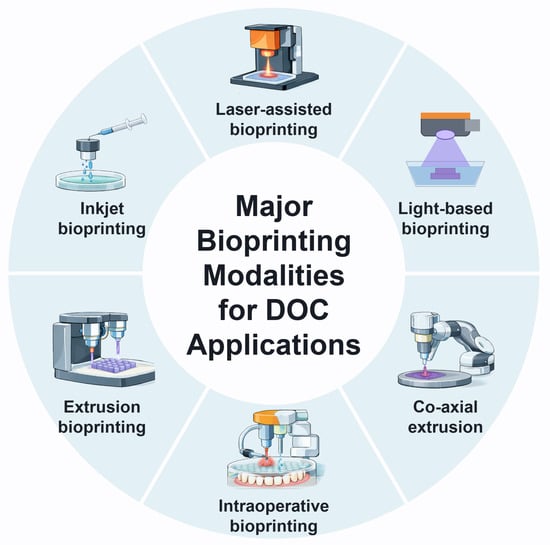
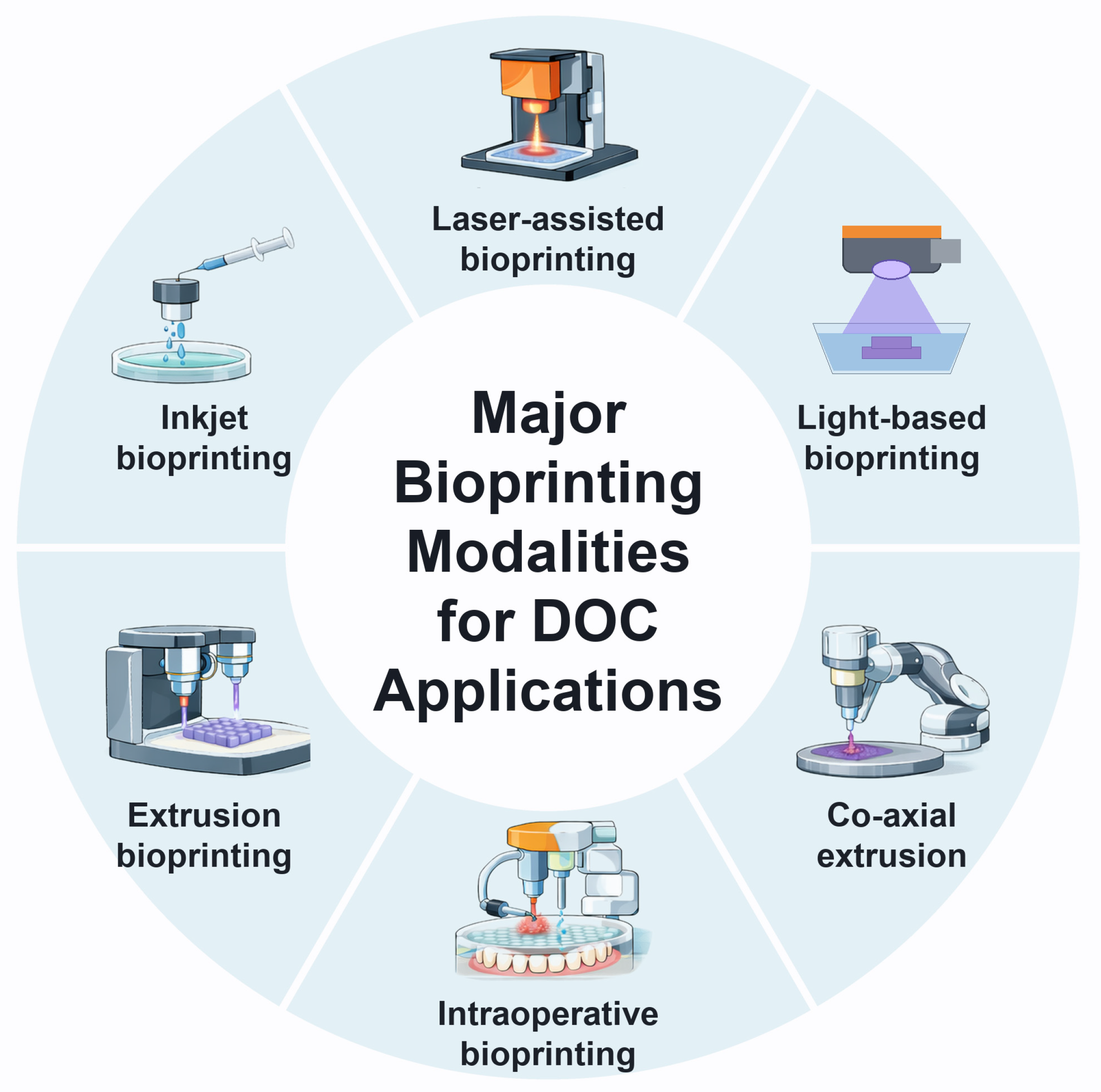
Bioprinting represents a relatively new field and a new approach to using 3D printing technology to offer benefits in the field of tissue engineering and regenerative medicine. 3D printing, in this context, refers to a process known as additive manufacturing, where a printer can form 3D objects using computer-aided design (CAD) software, which can in turn incorporate information from 3D scanners [20]. Bioprinting specifically refers to the layer-by-layer deposition of living cells, biomaterials, and bioactive molecules, collectively termed bioinks, to build tissue-like constructs [21]. In this review, printing of acellular scaffolds, molds, or anatomical models is not considered bioprinting and is identified explicitly as acellular printing. Currently, there is a wide variety of 3D printing techniques in DOC regenerative medicine, in terms of both materials and techniques (Figure 1), each having specific advantages and limitations (Table 1) [3]. The goal is for printed constructs to be used in functional applications in clinical medicine, drug discovery or delivery, and in vitro disease studies. The major bioprinting methods currently available are discussed in this section.
Figure 1.
Major bioprinting modalities for dental, oral, and craniofacial (DOC) applications. Co-axial printing is an extrusion variant, and intraoperative printing is a setting. Created by the authors; the icons and illustrations were generated with the assistance of generative artificial intelligence.
Approaches to DOC tissue fabrication fall into four groups. Direct bioprinting deposits cell-laden bioinks by inkjet, laser-assisted, extrusion, or light-based mechanisms. A second group prints acellular scaffolds that are seeded with cells afterwards. A third group prints acellular molds, templates, or anatomical models that guide casting, surgery, or tissue formation. A fourth group uses non-printing approaches, such as electrospinning, to make scaffolds. Co-axial bioprinting is a variant of extrusion in which a bioink and a crosslinking solution flow together to form fibers and tubes. Intraoperative bioprinting is a setting rather than a distinct mechanism in which extrusion, droplet-based, laser-based, or light-based printing is performed directly in the defect during surgery.
2.1. Inkjet-Based Bioprinting
Inkjet bioprinting deposits droplets of bioink of controlled volume onto a surface. Various techniques are used to adapt this technology for use in 3D printing, including thermal inkjet bioprinting, piezoelectric inkjet bioprinting, electrostatic inkjet bioprinting and electrohydrodynamic inkjet bioprinting [22].
These techniques rely on a process called microdropletization, where the inkjet heads deposit droplets of roughly 10 to 100 µm in diameter at high speed. Given the speed and size of the process, 3D inkjet printing has high throughput, where droplets are ejected on demand from individual nozzles and output scales with the number of nozzles [22]. Additionally, this process is “non-contact” where droplets are deposited, not placed onto a surface. This process means that there is a ‘drop-on-demand’ property of inkjet printing, where the process is highly controllable. However, despite the advantages of this process, there are also disadvantages to this method. For example, the ink must be deposited in liquid form, as a solid ink would clog and be unable to “microdropletize”. Given the low viscosity that these materials must have, this means that crosslinking is often attempted to strengthen the printed structure. Some crosslinkers are toxic to cellular viability. Cell damage in inkjet printing arises mainly from the thermal or mechanical stress of droplet ejection and impact, rather than from the nozzle shear that dominates extrusion printing [23], and inks are limited to low viscosities of approximately 3.5 to 12 mPa·s [21]. Additionally, the low viscosity is maintained through a lower concentration of the bioink, which can cause failed geometry of 3D-printed scaffolds. This can serve to explain why inkjet printing is often combined with other forms of bioprinting to add strength and integrity to the 3D structure [22,24,25].
2.2. Laser-Assisted and Light-Based Bioprinting
Laser-assisted bioprinting is an adaptation of laser-induced forward transfer (LIFT), a technique first described in 1986 for metal transfer [26] and adapted to transfer living cells in the early 2000s [27]. There are five main components of this system: a pulsed laser beam, a focusing system, a ribbon donor structure made up of an energy-absorbing layer, liquid bioink and a receiving substrate. In LIFT, a laser pulse heats the absorbing layer and propels the bioink from the donor onto the substrate. This has the potential to produce highly precise constructs, and given the laser-guided design, the print heads are open, eliminating the clogging issue that is commonly seen with nozzle-based bioprinting. Because there is no nozzle, the cells are not exposed to nozzle shear stress, which supports high cell viability [21,28]. However, the jet formation involves high pressure and rapid temperature changes that are not yet fully understood. Additionally, laser-assisted bioprinting is costly, highly complex, and slow, which limits large-scale fabrication [21,28].
Light-based bioprinting uses light to crosslink photosensitive bioinks in place rather than to transfer them. Digital light processing (DLP) cures an entire layer at once, allowing fine features at higher speed than nozzle-based methods, and has been used to print periodontal patches with oriented PDL fibers [8]. Volumetric bioprinting goes further, solidifying a whole cell-laden construct within seconds through tomographic light projection [29], and recent context-aware systems use real-time imaging to adapt the printed geometry to existing tissue [30]. Resolution on the order of 10 to 50 µm has been reported for digital light processing [31]. Light-based printing requires photocrosslinkable and sufficiently transparent inks, and exposure to photoinitiators and radicals can reduce cell viability [31,32].
Table 1.
Mechanisms, advantages, and limitations of bioprinting modalities for dental, oral, and craniofacial (DOC) applications.
2.3. Extrusion-Based Bioprinting
Extrusion-based bioprinting combines a fluid-dispensing system and an automated robotic system that will extrude material onto a platform. The continuous force of the extrusion will print the material into uninterrupted cylindrical lines, instead of a single droplet. Because the system is robotically controlled, the 3D shape and structure can be precisely controlled. One of the main advantages of a system like this is the compatibility with various printable materials. For example, this system can print hydrogels, copolymers and cell spheroids. This makes the system adaptable to many applications. A major disadvantage of this system is that the encapsulated cells are exposed to greater shear stresses, meaning that cell viability can be reduced during printing [21,33]. Reported resolution ranges from about 100 µm to the millimeter scale and viability from about 40% to 80%, and shear stress during extrusion through the nozzle is the main cause of cell loss [21,33,34]. Recent hybrid approaches combine extrusion with imaging-guided volumetric printing, allowing channels to be printed automatically around extruded cell-laden structures and improving cell function [30].
Co-axial bioprinting is a variant of extrusion-based bioprinting. In this technique, the bioink and the crosslinking solution are dispensed using co-axial needle assemblies, enabling the bioinks to be deposited as a liquid which will then set. Because the bioink is dispensed as a liquid and set by the crosslinker on contact, a wide range of components can be mixed evenly into the ink before it gels. However, the downside of this liquid formation of the bioink is that there are possible complications for the bioink stability upon removal of the inks from the setting bath. Co-axial printing is mainly used to produce hollow fibers and tubes, such as the salivary gland microtubes discussed in Section 3.4 [35], and the crosslinking solution (for example, calcium chloride for alginate) is what sets the bioink. A support bath is needed only when the filament does not gel quickly enough to hold its shape [36,37].
2.4. Intraoperative Bioprinting
Intraoperative bioprinting refers to using 3D printing at the time of a procedure and printing directly into the site of a defect or injury to aid healing [38]. This is an extremely promising new field of bioprinting that offers immediate scanning and printing of biomaterials that are specifically sized and shaped for the injury being repaired. Intraoperative bioprinting is a setting rather than a printing mechanism, and extrusion, droplet-based, laser-based, or light-based systems can be used [38]. Thermoplastics such as polycaprolactone are not currently suitable for printing in a patient because of their solvents and melting temperatures [38]. This type of bioprinting is currently limited to animal models, including recent large animal studies [39], but has been applied to both non-load-bearing sites and composite tissue remodeling for craniomaxillofacial reconstruction. Preclinical studies have reported that intraoperative bioprinting using bioinks can work for both hard and soft tissue reconstruction. In rats, an acellular osteogenic ink printed into 5-mm calvarial defects was reported to give approximately 80% bone coverage area at 6 weeks on micro-computed tomography (micro-CT) [40].
3. Three-Dimensional Bioprinting of Tissues
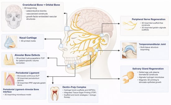
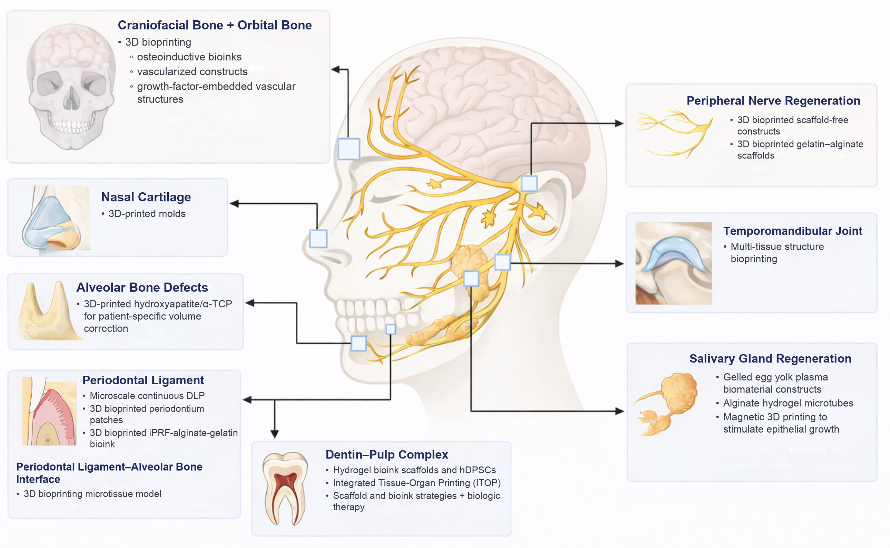
The key studies discussed in this section are summarized in Table 2, with the printing approach, experimental model, and level of evidence for each, and the tissue targets are shown in Figure 2. Studies of cell-laden bioprinting are distinguished from printing of acellular scaffolds, molds and anatomical models, and from non-printing approaches.
Figure 2.
Dental, oral, and craniofacial (DOC) tissue targets for three-dimensional (3D) bioprinting. DLP, digital light processing; hDPSCs, human dental pulp stem cells; iPRF, injectable platelet-rich fibrin; ITOP, integrated tissue–organ printing; α-TCP, α-tricalcium phosphate. Printed molds, as shown for nasal cartilage, are acellular. Created by the authors; the icons and illustrations were generated with the assistance of generative artificial intelligence.
3.1. Teeth and Pulp
Dental tissues pose unique challenges for 3D bioprinting due to their dual nature of hardness and softness. The intricacy lies in developing a scaffold capable of providing a supportive environment for depositing cells and tissues in their respective complex locations. Modern 3D bioprinting utilizes bioinks that are tailored for use with various dental alveolar tissues, including dental pulp, dentin, PDL, bone, gingiva, and the ambitious endeavor of whole-tooth regeneration [3,41]. Bioinks should ideally be biocompatible and printable, with a degradation rate that approximates that of new tissue formation [42,43]. Hydrogels, defined as 3D polymeric networks with high water content, are widely used bioinks because their tunable properties can mimic the extracellular matrix, although they typically trade printability against mechanical strength [42,43]. In the context of tooth regeneration, natural hydrogels such as collagen, gelatin, fibrin, hyaluronic acid, alginate, agarose, and gelatin methacryloyl (GelMA) are commonly employed [3].
Collagen-based scaffolds enhance periodontal and bone tissue regeneration, supporting dental pulp repair due to their biocompatibility and ability to promote cell attachment [44,45]. Gelatin functions in drug delivery systems and tissue regeneration applications, serving as a versatile biomaterial for dental pulp regeneration, endodontics, and oral wound healing [44]. Fibrin is a natural hydrogel used in tissue engineering [46] and has been used with hDPSCs to support odontogenic differentiation in vitro [4]. Hyaluronic acid is applied in periodontal disease treatment, dental pulp regeneration, and oral wound healing, serving in tissue engineering scaffolds and drug delivery systems [44,47]. Alginate is a biocompatible, ionically crosslinkable polymer with tunable mechanics, but its low bioactivity and rapid degradation limit its use alone [48]; in dental and craniofacial applications, it has mainly been used experimentally, for example, as a bioink component for cells and growth factors [5,7,9]. Agarose is widely used to form hydrogels for wound healing and tissue engineering, although its specific dental applications are less extensively documented [49]. GelMA serves as a versatile platform for regenerative endodontics and dental pulp regeneration, functioning as an injectable vehicle for delivery of stem cells, growth factors, and bioactive substances for tissue repair, with its photocrosslinkable properties allowing for in situ fabrication of patient-specific hydrogels [50,51].
Material selection depends on the demands of the intended construct. Soft, vascularized tissues such as pulp and gingiva tolerate low-stiffness hydrogels such as fibrin and GelMA, whereas bone reconstruction requires reinforcement with a mineral phase or a stiffer framework to bear load, and the printed architecture must allow nutrient diffusion and vascular ingrowth [42,43,52]. Co-printing of vascular channels with multiple cell types in hydrogel matrices has been demonstrated [53].
Pulpal and whole-tooth regeneration remain related but biologically distinct goals in 3D bioprinting. Pulpal regeneration primarily targets vascularized soft-tissue repair within existing dentin architecture, whereas whole-tooth regeneration requires coordinated reconstruction of enamel, dentin, cementum, and pulp across multiple length scales [41]. Ameloblasts are lost after tooth eruption, so enamel cannot repair itself, which is a major limit for whole-tooth strategies [54]. This limits but does not rule out cellular approaches, because ameloblast-like cells derived from induced pluripotent stem cells and dental epithelial organoids express ameloblast markers and enamel matrix proteins in vitro [54]. Recent bioink strategies (including fibrinogen/gelatin/hyaluronic acid/glycerol formulations) support hDPSC viability and odontogenic differentiation for dentin–pulp applications [4]. For whole-tooth efforts, key bottlenecks remain spatial patterning, epithelial–mesenchymal coordination, and angiogenesis. Notably, blocking uterine sensitization-associated gene-1 (USAG-1) with an antibody rescued tooth formation in mouse models of congenital tooth agenesis [55]. Because this is a pharmacologic rather than a printing strategy, it could complement printed constructs, although such a combination has not been tested.
Current research involving the 3D bioprinting of dentin and pulp has reported odontogenic differentiation and scaffolds compatible with hDPSCs, mostly in vitro. An early study created a fibrin-based bioink scaffold compatible with hDPSCs aimed at regenerating the dentin–pulp complex [4]. Notably, odontogenic differentiation was achieved through altering fibrinogen concentrations in the scaffolding, suggesting the possibility of augmenting site-specific regeneration. The crosslinked hydrogel had pore sizes of about 2 to 4 µm on scanning electron microscopy, of a similar order of magnitude to human dentinal tubules, whose diameter varies from the dentino-enamel junction to the pulp, although these are hydrogel pores rather than true tubules [4]. The study was conducted in vitro. In another study, an acellular ink of collagen, hydroxyapatite, and alginate was printed as an in vitro dentin model with tubule-like pores for testing occlusive agents against dentin hypersensitivity, and polystyrene microspheres narrowed the pores of the scaffold much as they did in natural dentin [5]. This is a testing platform rather than a regenerative scaffold. A third study with similar aims used 3D bioink scaffolding made of bone morphogenetic protein (BMP) peptide-tethering bioink materials. The study created a BMP-GelMA bioink formulation using an integrated tissue–organ printing (ITOP) system. The bioink formulation provided a microenvironment suitable for accelerated odontogenic differentiation of hDPSCs [6].
Modern 3D bioprinting for whole-tooth and pulp regeneration is still at an early stage. The studies presented demonstrate the available 3D bioprinting strategies combined with novel bioink formulations that address the commonly met roadblocks to oral regenerative medicine. Moving forward, future research should further the implementation of in vivo studies and work toward reproducible methods. As advancements in bioink formulations progress and the precision of scaffolding improves, there will be greater potential for compartmentalized growth, leading to the eventual possibility of whole-tooth regeneration.
3.2. Periodontal and Alveolar Bone Regeneration
Periodontal regeneration includes both hard and soft tissue regeneration. Restoring critical-size alveolar bone volume defects is a niche where the specificity of bioprinting can excel. Anderson et al. printed acellular, patient-specific hydroxyapatite and alpha-tricalcium phosphate (α-TCP) scaffolds from cone-beam computed tomography data; the scaffolds had higher compressive strength than an alginate and beta-tricalcium phosphate hybrid, were cytocompatible with alveolar bone marrow stem cells in vitro, and fit defect models within 0.27 mm of the digital design [7]. In vivo bone regeneration was not assessed. A PDL-alveolar bone microtissue model using GelMA has also been developed to better simulate and study periodontal tissue engineering in vitro [56].
Current research utilizing 3D bioink for PDL fiber and periodontal defect regeneration is limited by the intricate nature of the PDL fiber network and angiogenic needs. A recent study introduced biomimetic periodontium patches (BPPs) for PDL regeneration with the use of microscale continuous DLP 3D bioprinting [8]. In rat and beagle periodontal defect models, the oriented patches were reported to regenerate periodontium with region-specific PDL fiber orientation that tolerated orthodontic tooth movement, compared with randomly arranged and untreated controls [8]. Similarly, a periodontal module combining a DLP-printed ligament layer and an extrusion-printed bone layer, made with a dental follicle-derived decellularized extracellular matrix bioink, restored aligned PDL fibers and the bone–ligament interface in a canine critical-size defect [57]. Another challenge associated with PDL regeneration involves the necessity for localized angiogenic properties to meet the blood supply requirements. A recent study achieved an increase in the local angiogenic supply with the use of a 3D bioink preparation of injectable platelet-rich fibrin (iPRF)-alginate-gelatin [9]. The bioink-based iPRF treatment proved to have sustained release of growth factors and angiogenic properties. Furthermore, in a subcutaneous implantation model in nude mice, the bioink showed good biocompatibility, increased vascularization, and reduced macrophage infiltration at later time points relative to an alginate-gelatin control [9]. Because the mice are immunodeficient and the implantation site is ectopic, these findings do not directly show oral soft tissue regeneration or periodontal repair.
3.3. Periodontal Plastic Surgery and Soft Tissue Regeneration
In contrast to bone regeneration, soft tissue regeneration is generally less shape-specific and favors biologic wound healing principles. In a beagle study (six dogs, three groups, two-month follow-up), implant-site grafts of a bioprinted acellular dermal matrix (ADM), gelatin, and alginate scaffold carrying gingival fibroblasts yielded significantly more keratinized and attached gingiva than the same scaffold without cells (an increase in keratinized gingiva width of 1.58 to 1.93 mm versus 0.67 to 1.30 mm), with higher expression of collagen types I and III and vascular endothelial growth factor A [58]. These preclinical findings need confirmation in larger and longer studies.
3.4. Salivary Glands
Salivary glands are heterogeneous tissues with specific geometries, but there have been recent advances in structural and adjunctive stem cell use in bioprinting for salivary gland regeneration. In vitro, gelled egg yolk plasma could be printed and supported culture of salivary acinar cells and fibroblasts, but function and in vivo performance were not assessed [59]. Pre-formed alginate microtubes supported epithelial–mesenchymal self-organization and cavitation of mouse salivary cells in vitro, although this platform was made with a co-axial microfluidic device rather than printed [60]. Microfluidic co-axial printing produced thin cell-laden hydrogel fibers and patent hollow tubes with viable salivary gland cells, an early step toward microscale epithelial units [35]. An in vitro mouse study showed that a bioprinted sweat gland-like microenvironment could redirect mammary progenitor cells toward sweat gland markers, which is indirect evidence for salivary tissue [61]. Magnetic 3D bioprinting of human dental pulp stem cells generated innervated, amylase-producing salivary gland-like organoids that stimulated epithelial and neuronal growth when transplanted into damaged salivary gland tissue ex vivo [10].
Functional salivary gland replacement requires polarized acinar and ductal epithelium arranged as a ductal network that supports unidirectional saliva flow, together with innervation and vascularization [62,63]. To date, bioprinted salivary constructs have shown cell viability, organization, and marker expression in vitro or ex vivo, but restoration of salivary flow in vivo has not been shown [10,35]. In our opinion, current extrusion and co-axial platforms can approximate ductal geometry, but they cannot yet reproduce polarized secretory acini, connection to a ductal outflow, and neurovascular integration in the same construct.
3.5. Peripheral Nerves
The peripheral nervous system innervates the oral and underlying tissues and communicates with the pulp. The connection of both the local and systemic nervous systems adds complexity to regenerative attempts of nerves in the DOC region. Gingiva-derived mesenchymal stem cells show promise in a bioprinted scaffold-free construct for functional recovery in a rat segmental facial nerve defect [11]. A bioprinted gelatin and alginate scaffold with rat Schwann cells showed good viability and higher neurotrophic factor expression than two-dimensional culture in vitro, and the cells persisted after four weeks of implantation, but nerve regeneration was not assessed [64]. Although not bioprinted, an aligned electrospun fibrin nanofiber hydrogel used as a conduit filler restored a 10-mm rat sciatic nerve gap to a degree approaching an autologous graft [12], which illustrates the value of aligned microarchitecture that printing could reproduce.
3.6. Craniofacial and Maxillofacial Bone, Cartilage, and Skin
Cartilage is formed by chondroblasts and chondrocytes. In the orofacial complex, cartilage is found predominantly in the TMJ and nose. When these structures require repair, surgeons may select from autologous or allogeneic cartilage and alloplastic implants. Each material type has its set of unique challenges, and all struggle with adaptability due to differences in topographies between the implantable material and the recipient site. As early as 2015, researchers used computed tomography (CT) scans of cadaveric lower lateral cartilage to 3D print patient-shaped molds, in which poly(glycolic acid)/poly-L-lactic acid scaffolds seeded with human chondrocytes were cultured and then implanted subcutaneously in nude mice, yielding engineered cartilage with alar length and width matching native cartilage, although the thickness differed [13]. The cells were seeded onto the scaffolds rather than bioprinted. Since then, the development of hydrogels for chondrogenic engineering has been extensively studied due to their physical properties mimicking native cartilage and topographical adaptability [65,66,67]. Recently, image-guided volumetric printing automatically aligned a cell-laden cartilage layer onto a bone construct to produce an osteochondral model [30]. Another study identifies bioprinting as transforming TMJ reconstruction by enabling complex, multi-tissue structures with cellular viability, though these approaches remain in preclinical stages requiring extensive validation before clinical translation [15].
Bone remains one of the most difficult DOC targets because successful regeneration requires both vascularization and restoration of site-specific mechanical function. In practice, calvarial defects are relatively low-load compared with maxillo-mandibular defects, where cyclic masticatory loading imposes higher structural demands, so material architecture and reinforcement requirements should be defect-site specific [38,52,68]. Most of the bone data discussed here come from calvarial models. Novel techniques have been developed to print osteoinductive bioinks for 3D bone bioprinting in the treatment of craniomaxillofacial bone defects [68]. Biomaterials have also been developed incorporating growth factors into bioprinted vascular structures in a single step [16,69]. Exosomes are currently being evaluated to circumvent the shortcomings of growth factor embedding and improve targeted therapeutic delivery [70]. The printing performance, degradability, and osteoconductivity/osteoinductivity of bioprinting materials such as hyaluronic acid, alginate, collagen, silk fibroin, GelMA, poly(ethylene glycol) (PEG) hydrogels, and others are being studied extensively in cartilaginous and bone engineering applications, with the goal of expanding a surgeon’s armamentarium [52]. Printed anatomical models have also been used clinically to plan orbital wall reconstruction and pre-shape implants, which is a non-biological use of 3D printing [71]. Outside the DOC region, bioprinting has been investigated for the cornea [14] and retina [72], and these applications are not discussed further.
Chronic wounds and trauma often require restoration of human skin in addition to cartilage and bone. Unfortunately, this area of study is still considered in the early stages of research and requires additional investigation prior to clinical trials [73,74]. Fabrication of functional living skin utilizing a biomimetic hydrogel bioink was tested in a large animal model with limited success [75] but is still preferred over conventional hydrogels in skin regeneration due to improved mechanical strength. A key challenge is recreating hair follicles and sweat glands at the dermis and epidermis levels [74]. Investigations on growth factors involved in the wound healing process will improve the development of bioprinted skin grafts, especially in life-threatening wounds such as burns [76]. Skin is included here because craniofacial trauma and burns frequently involve it.
Table 2.
Key preclinical studies of bioprinting and related printing approaches for dental, oral, and craniofacial (DOC) tissues, with the experimental model and level of evidence.
4. Limitations and Challenges
While 3D bioprinting holds immense potential for transforming DOC regenerative medicine, several fundamental challenges must still be addressed before these technologies can be integrated into routine clinical care [18]. Current progress highlights both the promise of 3D bioprinting and the complexity of bringing it closer to real-world clinical translation. The main barriers can be grouped into cost and resource constraints, physiological and mechanical limitations, and regulatory and quality control considerations, each of which is discussed below, followed by future directions for the field.
4.1. Cost and Resource Barriers
One of the most immediate challenges is cost. Advanced printers, sterile enclosures, consumables, and trained personnel require substantial capital and operating expense [18]. High-resolution extrusion and laser-assisted systems demand precise calibration, which further increases the expense [28]. Furthermore, bioinks incorporating growth factors or living cells are costly to formulate, store, validate, and maintain [77]. For many clinicians and institutions, these costs can be a barrier to early adoption [17,19]. As a result, adoption is currently limited to research or tertiary centers with dedicated regenerative programs.
Efficient workflow integration remains another major hurdle. The process from imaging and CAD modeling to bioprinting and post-processing is still fragmented and time-consuming, adding to the human cost of 3D bioprinting [78]. Automating these steps through robotics or artificial intelligence (AI) could enable adaptive print parameter optimization, real-time error correction, and predictive modeling of tissue outcomes [79]. Digital integration will be critical for moving from isolated prototypes toward reproducible, clinically relevant prints.
Clinical adoption requires cultural and logistical change, not just technical capability. Most clinics lack the infrastructure or personnel to support on-site fabrication, and procedural workflows are not yet optimized for intraoperative bioprinting. Cost-sharing models, centralized fabrication labs, and standardized shelf-stable bioink kits could lower financial barriers [19,77]. Education and training will also be key factors in driving adoption [19]. Initial clinical applications may emerge first in academic oral and maxillofacial surgery, periodontology, or endodontics programs, as these settings already have the cross-disciplinary teams, research infrastructure, and surgical expertise needed to adopt new regenerative technologies. This is our expectation rather than an established finding.
4.2. Physiological and Mechanical Limitations
Another major limitation lies in recapitulating the intricate biological and mechanical environment of native tissues. The DOC region is exceptionally diverse, ranging from the mineralized enamel-dentin interface to the soft, vascularized pulp, gingiva, and neural structures. Reproducing this gradient of tissue stiffness, cellular composition, and immune compatibility remains an unsolved engineering challenge [18]. Balancing mechanical integrity with cellular viability is often a trade-off, as even small mismatches between material degradation and tissue remodeling rates can compromise long-term stability [80].
Because these are barrier tissues exposed to the outside environment, the host immune response to the graft also needs to be considered. Balancing the prevention of both graft infection and immune rejection is an important variable that future biomaterial designs must address [19]. Standardized degradation profiles and immunomodulatory biomaterials are promising avenues toward reducing these gaps [2,57].
The oral environment adds specific barriers. Intraoral printing requires bioinks that solidify rapidly and remain stable in a saliva-moistened, dynamic environment, together with ergonomic, sterile delivery systems that fit the limited intraoral access [18]. Intraoral surgical sites also carry a risk of postoperative infection, and oral bacteria can colonize graft materials [81]. Reconstructed jaw tissue must withstand repeated masticatory loading, with maximum molar bite forces of roughly 450 to 600 N in healthy adults [82], so load-bearing performance cannot be inferred from calvarial models. Dental pulp, exfoliated deciduous teeth, and periodontal ligament stem cells are accessible cell sources [83,84]. Cell-laden bioinks also require sterilization methods that do not damage their components [85] and have a limited shelf life; alginate-methylcellulose bioinks stored at 4 °C for four weeks kept their printability but lost human cell viability and function [86]. Finally, defect designs and healing times vary widely among large-animal studies of alveolar bone, which makes comparison across studies difficult [87].
4.3. Regulatory and Quality Control Considerations
Regulatory oversight of bioprinted constructs is incompletely harmonized rather than absent [19,78]. In the United States, the guidance of the U.S. Food and Drug Administration (FDA) on additively manufactured medical devices does not address biological, cellular, or tissue-based products, and directs such questions to the Center for Biologics Evaluation and Research [88]; its 2021 discussion paper on point-of-care printing likewise excludes bioprinting and is not guidance [89]. Constructs that contain living cells blur the line between medical devices and biologic therapies [19,90]. A construct containing living cells and a bioink would generally not qualify for regulation solely as a human cell, tissue, or cellular and tissue-based product (HCT/P) under Section 361 of the Public Health Service Act, which requires minimal manipulation and no combination with another article, and would instead be regulated as a drug, device, biologic, or combination product [91]. In the European Union, such constructs may fall within the tissue-engineered or combined advanced therapy medicinal product categories, and the hospital exemption is implemented differently among Member States [92,93]. Consensus standards are emerging, notably ASTM International standard F3659-24, a guide for bioinks that the FDA recognized in 2025 [94]. Classification, risk assessment, and quality control nonetheless remain open questions [95]. Questions also remain regarding the classification of intraoperative bioprinting and the ethical considerations of using autologous versus allogeneic cells. Until regulatory bodies provide clearer guidance for bioprinted constructs, translational efforts will continue to face uncertainty in study design, manufacturing compliance, and patient safety standards.
Consistency and reproducibility are persistent concerns that should be addressed before regulatory approval can be obtained [19]. Small variations in bioink composition, crosslinking parameters, or printing speed can alter the final biological performance [77,96]. Although ASTM F3659-24 provides a guide for bioinks [94], there are no widely adopted standardized metrics for evaluating print fidelity, cell viability, or functional integration, making it difficult to compare outcomes across laboratories [18,96]. Shared benchmarks, outcome reporting guidelines, and validated reference materials are therefore needed so that results can be compared across centers and clinical translation can proceed with confidence [19,96].
Human experience in the DOC region is limited, and most of it involves acellular printed implants rather than cell-laden bioprinting. Examples are patient-specific polycaprolactone scaffolds for ear reconstruction in five patients with microtia followed for one year [97], and patient-specific 3D-printed implants for zygomatic and orbital reconstruction described in a recent review [19]. For cell-laden constructs, a scoping review of trial registries found 11 bioprinting trials, of which four planned to implant bioprinted autologous tissue (blood vessel, trachea, external ear, and wound dressing); the only one in the DOC region was a phase 1/2 trial of AuriNovo, a bioprinted collagen hydrogel construct containing the patient’s own auricular chondrocytes, for ear reconstruction in microtia [98]. That trial enrolled two patients and was terminated for a company decision that was not related to safety, and, to our knowledge, no outcome data have been published [99]. Printing in the operating room adds requirements for sterility, process validation, and quality control at the point of care, which the FDA has so far addressed only in a discussion paper [89]. Autologous cells require a donor-site biopsy and a manufacturing interval before implantation, whereas allogeneic cells raise questions of immune compatibility and donor screening.
4.4. Future Directions
Ultimately, progress in 3D bioprinting depends on sustained collaboration between engineers, biologists, clinicians, regulatory specialists, and industry partners. No single discipline can address the challenges of bioprinting alone. Academic medical centers are well positioned to serve as incubators for translational progress, as they bring together preclinical animal models, regulatory consultation, and surgical expertise. As the field matures, interdisciplinary communication will determine not only the speed but also the safety and equity with which bioprinting enters the clinic [19].
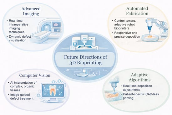
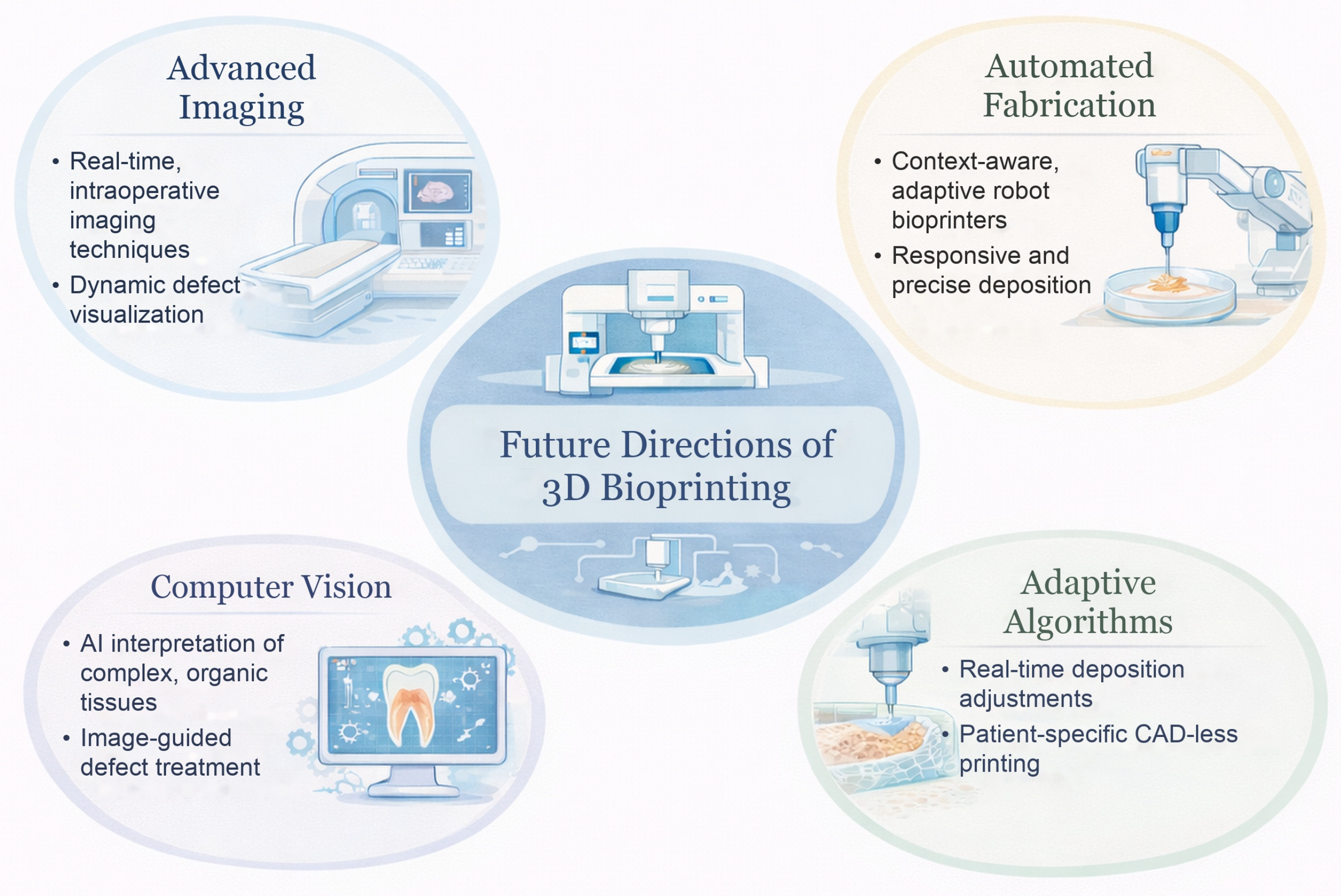
A key future direction in DOC bioprinting is the convergence of advanced imaging, computer vision, and adaptive fabrication (Figure 3). Recent work in context-aware volumetric bioprinting demonstrates how real-time imaging and computational interpretation of biological structures can guide deposition without reliance on static, preoperative CAD models. Such approaches enable printed geometries to adapt dynamically to existing tissue features, improving spatial fidelity and functional integration. This paradigm is particularly relevant to DOC regeneration, where defect morphology is irregular and often intraoperatively defined. As image-guided feedback and adaptive algorithms mature, these systems may enable more precise, patient-specific, and intraoperative regenerative strategies in oral and maxillofacial surgery [30].
Figure 3.
Future directions for bioprinting in dental, oral, and craniofacial (DOC) scenarios. Created by the authors; the icons and illustrations were generated with the assistance of generative artificial intelligence.
5. Conclusions
3D bioprinting for DOC regeneration is transitioning from proof-of-concept constructs to increasingly biomimetic, application-specific solutions. Across teeth and pulp, periodontal tissues, salivary glands, nerves, cartilage, bone, and skin, the field has increasingly combined spatial precision with bioactive and cell-laden materials to address microarchitectural fidelity, vascularization, and guided differentiation. Clinical adoption, however, will depend on reproducible manufacturing, predictable performance in vivo, and clear regulatory pathways in addition to improved printing capabilities. Most of the evidence to date remains preclinical, and intraoperative bioprinting has been demonstrated only in animal models. With coordinated effort among engineers, clinicians, and regulators, the field has the potential to deliver patient-specific constructs that restore form and function across DOC tissues, beyond tertiary medical centers and into routine clinical care.
Author Contributions
Conceptualization, Y.-C.C., J.W., Z.G. and D.T.W.; investigation, Y.-C.C., J.W., Z.G., P.H., R.R. and D.T.W.; writing—original draft preparation, Y.-C.C., J.W., Z.G., P.H., R.R., J.P., M.C. and K.W.; writing—review and editing, Y.-C.C., J.W., Z.G., P.H., R.R., J.P., M.C., K.W. and D.T.W.; visualization, Y.-C.C. and J.W.; supervision, D.T.W.; project administration, D.T.W. All authors have read and agreed to the published version of the manuscript.
Funding
This research received no external funding.
Institutional Review Board Statement
Not applicable.
Informed Consent Statement
Not applicable.
Data Availability Statement
No new data were created or analyzed in this study. Data sharing is not applicable to this article.
Acknowledgments
Claude (Opus 5.5; Anthropic) was used for grammatical editing of the manuscript. All ideas presented in this manuscript are the original work of the authors. Generative AI tools were also used to create the icons and illustrations in Figure 1, Figure 2 and Figure 3; the authors assembled and reviewed all figures.
Conflicts of Interest
The authors declare no conflicts of interest.
Abbreviations
The following abbreviations are used in this manuscript:
| 3D | Three-dimensional |
| α-TCP | Alpha-tricalcium phosphate |
| ADM | Acellular dermal matrix |
| AI | Artificial intelligence |
| BMP | Bone morphogenetic protein |
| BPP | Biomimetic periodontium patch |
| CAD | Computer-aided design |
| CT | Computed tomography |
| DLP | Digital light processing |
| FDA | U.S. Food and Drug Administration |
| DOC | Dental, oral, and craniofacial |
| GelMA | Gelatin methacryloyl |
| HCT/P | Human cells, tissues, and cellular and tissue-based products |
| hDPSC | Human dental pulp stem cell |
| iPRF | Injectable platelet-rich fibrin |
| ITOP | Integrated tissue–organ printing |
| LIFT | Laser-induced forward transfer |
| PDL | Periodontal ligament |
| PEG | Poly(ethylene glycol) |
| TMJ | Temporomandibular joint |
| USAG-1 | Uterine sensitization-associated gene-1 |
References
- Hazrati, P.; Alanazi, A.; Alrmali, A.E.; Galindo-Fernandez, P.; Kassem, H.; Kaigler, D. Clinical stem cell therapy in oral and craniofacial bone regeneration: A systematic review and meta-analysis. Front. Bioeng. Biotechnol. 2026, 14, 1677400. [Google Scholar] [CrossRef] [Scilit] [PubMed]
- Latimer, J.; Wu, D.T.; Yilmaz, B.T.; Cha, J.K.; Gouveia, Z.; Yao, Y.; Giannobile, W. Biological Principles for Success in Alveolar Bone, Soft Tissue, and Periodontal Regeneration. J. Periodontal Res. 2026, 1–24. [Google Scholar] [CrossRef] [Scilit] [PubMed]
- Ostrovidov, S.; Ramalingam, M.; Bae, H.; Orive, G.; Fujie, T.; Shi, X.; Kaji, H. Bioprinting and biomaterials for dental alveolar tissue regeneration. Front. Bioeng. Biotechnol. 2023, 11, 991821. [Google Scholar] [CrossRef] [Scilit] [PubMed]
- Han, J.; Kim, D.S.; Jang, H.; Kim, H.R.; Kang, H.W. Bioprinting of three-dimensional dentin-pulp complex with local differentiation of human dental pulp stem cells. J. Tissue Eng. 2019, 10, 2041731419845849. [Google Scholar] [CrossRef] [Scilit] [PubMed]
- Naseri, S.; Cooke, M.E.; Rosenzweig, D.H.; Tabrizian, M. 3D Printed In Vitro Dentin Model to Investigate Occlusive Agents against Tooth Sensitivity. Materials 2021, 14, 7255. [Google Scholar] [CrossRef] [Scilit] [PubMed]
- Park, J.H.; Gillispie, G.J.; Copus, J.S.; Zhang, W.; Atala, A.; Yoo, J.J.; Yelick, P.C.; Lee, S.J. The effect of BMP-mimetic peptide tethering bioinks on the differentiation of dental pulp stem cells (DPSCs) in 3D bioprinted dental constructs. Biofabrication 2020, 12, 035029. [Google Scholar] [CrossRef] [Scilit] [PubMed]
- Anderson, M.; Dubey, N.; Bogie, K.; Cao, C.; Li, J.; Lerchbacker, J.; Mendonça, G.; Kauffmann, F.; Bottino, M.C.; Kaigler, D. Three-dimensional printing of clinical scale and personalized calcium phosphate scaffolds for alveolar bone reconstruction. Dent. Mater. 2022, 38, 529–539. [Google Scholar] [CrossRef] [Scilit] [PubMed]
- Ma, Y.; Yang, X.; Chen, Y.; Zhang, J.; Gai, K.; Chen, J.; Huo, F.; Guo, Q.; Guo, W.; Gou, M.; et al. Biomimetic Periodontium Patches for Functional Periodontal Regeneration. Adv. Healthc. Mater. 2023, 12, e2202169. [Google Scholar] [CrossRef] [Scilit] [PubMed]
- Yi, K.; Li, Q.; Lian, X.; Wang, Y.; Tang, Z. Utilizing 3D bioprinted platelet-rich fibrin-based materials to promote the regeneration of oral soft tissue. Regen. Biomater. 2022, 9, rbac021. [Google Scholar] [CrossRef] [Scilit] [PubMed]
- Adine, C.; Ng, K.K.; Rungarunlert, S.; Souza, G.R.; Ferreira, J.N. Engineering innervated secretory epithelial organoids by magnetic three-dimensional bioprinting for stimulating epithelial growth in salivary glands. Biomaterials 2018, 180, 52–66. [Google Scholar] [CrossRef] [Scilit] [PubMed]
- Zhang, Q.; Nguyen, P.D.; Shi, S.; Burrell, J.C.; Cullen, D.K.; Le, A.D. 3D bio-printed scaffold-free nerve constructs with human gingiva-derived mesenchymal stem cells promote rat facial nerve regeneration. Sci. Rep. 2018, 8, 6634. [Google Scholar] [CrossRef] [Scilit] [PubMed]
- Du, J.; Liu, J.; Yao, S.; Mao, H.; Peng, J.; Sun, X.; Cao, Z.; Yang, Y.; Xiao, B.; Wang, Y.; et al. Prompt peripheral nerve regeneration induced by a hierarchically aligned fibrin nanofiber hydrogel. Acta Biomater. 2017, 55, 296–309. [Google Scholar] [CrossRef] [Scilit] [PubMed]
- Xu, Y.; Fan, F.; Kang, N.; Wang, S.; You, J.; Wang, H.; Zhang, B. Tissue engineering of human nasal alar cartilage precisely by using three-dimensional printing. Plast. Reconstr. Surg. 2015, 135, 451–458. [Google Scholar] [CrossRef] [Scilit] [PubMed]
- Kim, H.; Park, M.N.; Kim, J.; Jang, J.; Kim, H.K.; Cho, D.W. Characterization of cornea-specific bioink: High transparency, improved in vivo safety. J. Tissue Eng. 2019, 10, 2041731418823382. [Google Scholar] [CrossRef] [Scilit] [PubMed]
- Baecher, H.; Maheta, B.; Lottner, L.-M.; Panayi, A.C.; Knoedler, S.; Guastaldi, F.; Mano, J.F.; Koerdt, S.; Rendenbach, C.; Heiland, M.; et al. Current clinical and translational challenges in temporomandibular joint reconstruction. Front. Bioeng. Biotechnol. 2025, 13, 1590021. [Google Scholar] [CrossRef] [Scilit] [PubMed]
- Leucht, A.; Volz, A.C.; Rogal, J.; Borchers, K.; Kluger, P.J. Advanced gelatin-based vascularization bioinks for extrusion-based bioprinting of vascularized bone equivalents. Sci. Rep. 2020, 10, 5330. [Google Scholar] [CrossRef] [Scilit] [PubMed]
- Barootchi, S.; Tavelli, L.; Sabri, H.; Giannobile, W.V.; Kaigler, D. Emerging, personalized, and minimally invasive technologies for periodontal and peri-implant soft and hard tissue regenerative therapy. J. Periodontol. 2026, 97, S103–S139. [Google Scholar] [CrossRef] [Scilit] [PubMed]
- Dixit, S.; Shayeb, M.A.; Kathayat, G.; Rokaya, D. Clinical translation of 3D bioprinting in oral and maxillofacial reconstruction: Recent progress and future directions. J. Oral Biol. Craniofac. Res. 2026, 16, 101401. [Google Scholar] [CrossRef] [Scilit] [PubMed]
- Najafi, N.; Vartanian, K.B.; Eskandar, T.; Ghookas, K.; Rostomian, E.; Agrawal, D.K. Bioprinting for craniofacial reconstruction: A review of advancements, clinical use, and challenges. J. Craniomaxillofac. Surg. 2025, 53, 2255–2269. [Google Scholar] [CrossRef] [Scilit] [PubMed]
- Palmara, G.; Frascella, F.; Roppolo, I.; Chiappone, A.; Chiadò, A. Functional 3D printing: Approaches and bioapplications. Biosens. Bioelectron. 2021, 175, 112849. [Google Scholar] [CrossRef] [Scilit] [PubMed]
- Zhang, J.; Wehrle, E.; Rubert, M.; Müller, R. 3D Bioprinting of Human Tissues: Biofabrication, Bioinks, and Bioreactors. Int. J. Mol. Sci. 2021, 22, 3971. [Google Scholar] [CrossRef] [Scilit] [PubMed]
- Li, X.; Liu, B.; Pei, B.; Chen, J.; Zhou, D.; Peng, J.; Zhang, X.; Jia, W.; Xu, T. Inkjet Bioprinting of Biomaterials. Chem. Rev. 2020, 120, 10793–10833. [Google Scholar] [CrossRef] [Scilit] [PubMed]
- Cui, X.; Dean, D.; Ruggeri, Z.M.; Boland, T. Cell damage evaluation of thermal inkjet printed Chinese hamster ovary cells. Biotechnol. Bioeng. 2010, 106, 963–969. [Google Scholar] [CrossRef] [Scilit] [PubMed]
- Choi, W.S.; Ha, D.; Park, S.; Kim, T. Synthetic multicellular cell-to-cell communication in inkjet printed bacterial cell systems. Biomaterials 2011, 32, 2500–2507. [Google Scholar] [CrossRef] [Scilit] [PubMed]
- Dou, R.; Saunders, R.E.; Mohamet, L.; Ward, C.M.; Derby, B. High throughput cryopreservation of cells by rapid freezing of sub-μl drops using inkjet printing--cryoprinting. Lab Chip 2015, 15, 3503–3513. [Google Scholar] [CrossRef] [Scilit] [PubMed]
- Bohandy, J.; Kim, B.F.; Adrian, F.J. Metal deposition from a supported metal film using an excimer laser. J. Appl. Phys. 1986, 60, 1538–1539. [Google Scholar] [CrossRef] [Scilit]
- Barron, J.; Wu, P.; Ladouceur, H.; Ringeisen, B. Biological Laser Printing: A Novel Technique for Creating Heterogeneous 3-dimensional Cell Patterns. Biomed. Microdevices 2004, 6, 139–147. [Google Scholar] [CrossRef] [Scilit] [PubMed]
- Chang, J.; Sun, X. Laser-induced forward transfer based laser bioprinting in biomedical applications. Front. Bioeng. Biotechnol. 2023, 11, 1255782. [Google Scholar] [CrossRef] [Scilit] [PubMed]
- Bernal, P.N.; Delrot, P.; Loterie, D.; Li, Y.; Malda, J.; Moser, C.; Levato, R. Volumetric Bioprinting of Complex Living-Tissue Constructs within Seconds. Adv. Mater. 2019, 31, e1904209. [Google Scholar] [CrossRef] [Scilit] [PubMed]
- Florczak, S.; Größbacher, G.; Ribezzi, D.; Longoni, A.; Gueye, M.; Grandidier, E.; Malda, J.; Levato, R. Adaptive and context-aware volumetric printing. Nature 2025, 645, 108–114. [Google Scholar] [CrossRef] [Scilit] [PubMed]
- Xu, H.Q.; Liu, J.C.; Zhang, Z.Y.; Xu, C.X. A review on cell damage, viability, and functionality during 3D bioprinting. Mil. Med. Res. 2022, 9, 70. [Google Scholar] [CrossRef] [Scilit] [PubMed]
- Lim, K.S.; Galarraga, J.H.; Cui, X.; Lindberg, G.C.J.; Burdick, J.A.; Woodfield, T.B.F. Fundamentals and Applications of Photo-Cross-Linking in Bioprinting. Chem. Rev. 2020, 120, 10662–10694. [Google Scholar] [CrossRef] [Scilit] [PubMed]
- Cui, X.; Li, J.; Hartanto, Y.; Durham, M.; Tang, J.; Zhang, H.; Hooper, G.; Lim, K.; Woodfield, T. Advances in Extrusion 3D Bioprinting: A Focus on Multicomponent Hydrogel-Based Bioinks. Adv. Healthc. Mater. 2020, 9, e1901648. [Google Scholar] [CrossRef] [Scilit] [PubMed]
- Blaeser, A.; Duarte Campos, D.F.; Puster, U.; Richtering, W.; Stevens, M.M.; Fischer, H. Controlling Shear Stress in 3D Bioprinting is a Key Factor to Balance Printing Resolution and Stem Cell Integrity. Adv. Healthc. Mater. 2016, 5, 326–333. [Google Scholar] [CrossRef] [Scilit] [PubMed]
- Yin, Y.; Vázquez-Rosado, E.J.; Wu, D.; Viswananthan, V.; Farach, A.; Farach-Carson, M.C.; Harrington, D.A. Microfluidic coaxial 3D bioprinting of cell-laden microfibers and microtubes for salivary gland tissue engineering. Biomater. Adv. 2023, 154, 213588. [Google Scholar] [CrossRef] [Scilit] [PubMed]
- Costantini, M.; Colosi, C.; Święszkowski, W.; Barbetta, A. Co-axial wet-spinning in 3D bioprinting: State of the art and future perspective of microfluidic integration. Biofabrication 2018, 11, 012001. [Google Scholar] [CrossRef] [Scilit] [PubMed]
- Gao, Q.; He, Y.; Fu, J.Z.; Liu, A.; Ma, L. Coaxial nozzle-assisted 3D bioprinting with built-in microchannels for nutrients delivery. Biomaterials 2015, 61, 203–215. [Google Scholar] [CrossRef] [Scilit] [PubMed]
- Wu, Y.; Ravnic, D.J.; Ozbolat, I.T. Intraoperative Bioprinting: Repairing Tissues and Organs in a Surgical Setting. Trends Biotechnol. 2020, 38, 594–605. [Google Scholar] [CrossRef] [Scilit] [PubMed]
- Yeo, M.; Gupta, D.; Derman, I.D.; Yildirim, S.; Singh, Y.P.; Gerhard, E.M.; Rizk, E.; Neuberger, T.; Simon, S.; Ozbolat, I.T. Intraoperative Bioprinting for Craniomaxillofacial Bone Reconstruction in Rats and Sheep. Small Sci. 2025, 5, 2400621. [Google Scholar] [CrossRef] [Scilit] [PubMed]
- Moncal, K.K.; Gudapati, H.; Godzik, K.P.; Heo, D.N.; Kang, Y.; Rizk, E.; Ravnic, D.J.; Wee, H.; Pepley, D.F.; Ozbolat, V.; et al. Intra-Operative Bioprinting of Hard, Soft, and Hard/Soft Composite Tissues for Craniomaxillofacial Reconstruction. Adv. Funct. Mater. 2021, 31, 2010858. [Google Scholar] [CrossRef] [Scilit] [PubMed]
- Hazrati, P.; Mirtaleb, M.H.; Boroojeni, H.S.H.; Koma, A.A.Y.; Nokhbatolfoghahaei, H. Current Trends, Advances, and Challenges of Tissue Engineering-Based Approaches of Tooth Regeneration: A Review of the Literature. Curr. Stem Cell Res. Ther. 2024, 19, 473–496. [Google Scholar] [CrossRef] [Scilit] [PubMed]
- Hospodiuk, M.; Dey, M.; Sosnoski, D.; Ozbolat, I.T. The bioink: A comprehensive review on bioprintable materials. Biotechnol. Adv. 2017, 35, 217–239. [Google Scholar] [CrossRef] [Scilit] [PubMed]
- Unagolla, J.M.; Jayasuriya, A.C. Hydrogel-based 3D bioprinting: A comprehensive review on cell-laden hydrogels, bioink formulations, and future perspectives. Appl. Mater. Today 2020, 18, 100479. [Google Scholar] [CrossRef] [Scilit] [PubMed]
- Alsalhi, A. Applications of selected polysaccharides and proteins in dentistry: A review. Int. J. Biol. Macromol. 2024, 260, 129215. [Google Scholar] [CrossRef] [Scilit] [PubMed]
- Paczkowska-Walendowska, M.; Kulawik, M.; Kwiatek, J.; Bikiaris, D.; Cielecka-Piontek, J. Novel Applications of Natural Biomaterials in Dentistry—Properties, Uses, and Development Perspectives. Materials 2025, 18, 2124. [Google Scholar] [CrossRef] [Scilit] [PubMed]
- Taghipour, Y.D.; Hokmabad, V.R.; Del Bakhshayesh, A.R.; Asadi, N.; Salehi, R.; Nasrabadi, H.T. The Application of Hydrogels Based on Natural Polymers for Tissue Engineering. Curr. Med. Chem. 2020, 27, 2658–2680. [Google Scholar] [CrossRef] [Scilit] [PubMed]
- Chelu, M.; Popa, M.; Calderón Moreno, J.M. Next-Generation Natural Hydrogels in Oral Tissue Engineering. Pharmaceutics 2025, 17, 1256. [Google Scholar] [CrossRef] [Scilit] [PubMed]
- Wu, D.T.; Munguia-Lopez, J.G.; Cho, Y.W.; Ma, X.; Song, V.; Zhu, Z.; Tran, S.D. Polymeric Scaffolds for Dental, Oral, and Craniofacial Regenerative Medicine. Molecules 2021, 26, 7043. [Google Scholar] [CrossRef] [Scilit] [PubMed]
- Ijaz, F.; Tahir, H.M.; Ali, S.; Ali, A.; Khan, H.A.; Muzamil, A.; Manzoor, H.H.; Qayyum, K.A. Biomolecules based hydrogels and their potential biomedical applications: A comprehensive review. Int. J. Biol. Macromol. 2023, 253, 127362. [Google Scholar] [CrossRef] [Scilit] [PubMed]
- Huang, L.; Chen, X.; Yang, X.; Zhang, Y.; Qiu, X. GelMA-based hydrogel biomaterial scaffold: A versatile platform for regenerative endodontics. J. Biomed. Mater. Res. B Appl. Biomater. 2024, 112, e35412. [Google Scholar] [CrossRef] [Scilit] [PubMed]
- Dal-Fabbro, R.; Daghrery, A.; Anselmi, C.; Soares, I.P.M.; Reis-Prado, A.H.D.; Oliveira, P.H.C.; Bottino, M.C. Recent Advances in Injectable Hydrogel Biotherapeutics for Regenerative Dental Medicine. Macromol. Biosci. 2025, 25, e00096. [Google Scholar] [CrossRef] [Scilit] [PubMed]
- Badhe, R.V.; Chatterjee, A.; Bijukumar, D.; Mathew, M.T. Current advancements in bio-ink technology for cartilage and bone tissue engineering. Bone 2023, 171, 116746. [Google Scholar] [CrossRef] [Scilit] [PubMed]
- Kolesky, D.B.; Truby, R.L.; Gladman, A.S.; Busbee, T.A.; Homan, K.A.; Lewis, J.A. 3D bioprinting of vascularized, heterogeneous cell-laden tissue constructs. Adv. Mater. 2014, 26, 3124–3130. [Google Scholar] [CrossRef] [Scilit] [PubMed]
- Hermans, F.; Hasevoets, S.; Vankelecom, H.; Bronckaers, A.; Lambrichts, I. From Pluripotent Stem Cells to Organoids and Bioprinting: Recent Advances in Dental Epithelium and Ameloblast Models to Study Tooth Biology and Regeneration. Stem Cell Rev. Rep. 2024, 20, 1184–1199. [Google Scholar] [CrossRef] [Scilit] [PubMed]
- Murashima-Suginami, A.; Kiso, H.; Tokita, Y.; Mihara, E.; Nambu, Y.; Uozumi, R.; Tabata, Y.; Bessho, K.; Takagi, J.; Sugai, M.; et al. Anti-USAG-1 therapy for tooth regeneration through enhanced BMP signaling. Sci. Adv. 2021, 7, eabf1798. [Google Scholar] [CrossRef] [Scilit] [PubMed]
- Vurat, M.T.; Şeker, Ş.; Lalegül-Ülker, Ö.; Parmaksiz, M.; Elçin, A.E.; Elçin, Y.M. Development of a multicellular 3D-bioprinted microtissue model of human periodontal ligament-alveolar bone biointerface: Towards a pre-clinical model of periodontal diseases and personalized periodontal tissue engineering. Genes Dis. 2022, 9, 1008–1023. [Google Scholar] [CrossRef] [Scilit] [PubMed]
- Yang, X.; Ma, Y.; Wang, X.; Yuan, S.; Huo, F.; Yi, G.; Zhang, J.; Yang, B.; Tian, W. A 3D-Bioprinted Functional Module Based on Decellularized Extracellular Matrix Bioink for Periodontal Regeneration. Adv. Sci. 2023, 10, e2205041. [Google Scholar] [CrossRef] [Scilit] [PubMed]
- Liu, P.; Li, Q.; Yang, Q.; Zhang, S.; Yi, K.; Zhang, G.; Tang, Z. Evaluation of the effect of 3D-bioprinted gingival fibroblast-encapsulated ADM scaffolds on keratinized gingival augmentation. J. Periodontal Res. 2023, 58, 564–574. [Google Scholar] [CrossRef] [Scilit] [PubMed]
- Charbonneau, A.M.; Kinsella, J.M.; Tran, S.D. 3D Cultures of Salivary Gland Cells in Native or Gelled Egg Yolk Plasma, Combined with Egg White and 3D-Printing of Gelled Egg Yolk Plasma. Materials 2019, 12, 3480. [Google Scholar] [CrossRef] [Scilit] [PubMed]
- Jorgensen, M.; Ramesh, P.; Toro, M.; Evans, E.; Moskwa, N.; Zhang, X.; Sharfstein, S.T.; Larsen, M.; Xie, Y. Alginate Hydrogel Microtubes for Salivary Gland Cell Organization and Cavitation. Bioengineering 2022, 9, 38. [Google Scholar] [CrossRef] [Scilit] [PubMed]
- Wang, R.; Wang, Y.; Yao, B.; Hu, T.; Li, Z.; Liu, Y.; Cui, X.; Cheng, L.; Song, W.; Huang, S.; et al. Redirecting differentiation of mammary progenitor cells by 3D bioprinted sweat gland microenvironment. Burns Trauma 2019, 7, 29. [Google Scholar] [CrossRef] [Scilit] [PubMed]
- Klangprapan, J.; Souza, G.R.; Ferreira, J.N. Bioprinting salivary gland models and their regenerative applications. BDJ Open 2024, 10, 39. [Google Scholar] [CrossRef] [Scilit] [PubMed]
- Rose, S.C.; Larsen, M.; Xie, Y.; Sharfstein, S.T. Salivary Gland Bioengineering. Bioengineering 2023, 11, 28. [Google Scholar] [CrossRef] [Scilit] [PubMed]
- Wu, Z.; Li, Q.; Xie, S.; Shan, X.; Cai, Z. In vitro and in vivo biocompatibility evaluation of a 3D bioprinted gelatin-sodium alginate/rat Schwann-cell scaffold. Mater. Sci. Eng. C 2020, 109, 110530. [Google Scholar] [CrossRef] [Scilit] [PubMed]
- Liu, Y.; Wang, M.; Luo, Y.; Liang, Q.; Yu, Y.; Chen, F.; Yao, J. Enhancing Stem Cell Therapy for Cartilage Repair in Osteoarthritis-A Hydrogel Focused Approach. Gels 2021, 7, 263. [Google Scholar] [CrossRef] [Scilit] [PubMed]
- Zhu, S.; Li, Y.; He, Z.; Ji, L.; Zhang, W.; Tong, Y.; Luo, J.; Yu, D.; Zhang, Q.; Bi, Q. Advanced injectable hydrogels for cartilage tissue engineering. Front. Bioeng. Biotechnol. 2022, 10, 954501. [Google Scholar] [CrossRef] [Scilit] [PubMed]
- Gao, F.; Xu, Z.; Liang, Q.; Li, H.; Peng, L.; Wu, M.; Zhao, X.; Cui, X.; Ruan, C.; Liu, W. Osteochondral Regeneration with 3D-Printed Biodegradable High-Strength Supramolecular Polymer Reinforced-Gelatin Hydrogel Scaffolds. Adv. Sci. 2019, 6, 1900867. [Google Scholar] [CrossRef] [Scilit] [PubMed]
- Chimene, D.; Miller, L.; Cross, L.M.; Jaiswal, M.K.; Singh, I.; Gaharwar, A.K. Nanoengineered Osteoinductive Bioink for 3D Bioprinting Bone Tissue. ACS Appl. Mater. Interfaces 2020, 12, 15976–15988. [Google Scholar] [CrossRef] [Scilit] [PubMed]
- Goker, M.; Derici, U.S.; Gokyer, S.; Parmaksiz, M.G.; Kaya, B.; Can, A.; Yilgor, P. Spatial Growth Factor Delivery for 3D Bioprinting of Vascularized Bone with Adipose-Derived Stem/Stromal Cells as a Single Cell Source. ACS Biomater. Sci. Eng. 2024, 10, 1607–1619. [Google Scholar] [CrossRef] [Scilit] [PubMed]
- Amondarain, M.; Gallego, I.; Puras, G.; Saenz-Del-Burgo, L.; Luzzani, C.; Pedraz, J.L. The role of microfluidics and 3D-bioprinting in the future of exosome therapy. Trends Biotechnol. 2023, 41, 1343–1359. [Google Scholar] [CrossRef] [Scilit] [PubMed]
- Oh, T.S.; Jeong, W.S.; Chang, T.J.; Koh, K.S.; Choi, J.W. Customized Orbital Wall Reconstruction Using Three-Dimensionally Printed Rapid Prototype Model in Patients With Orbital Wall Fracture. J. Craniofac. Surg. 2016, 27, 2020–2024. [Google Scholar] [CrossRef] [Scilit] [PubMed]
- Solberg, A.; Robles-Anda, N.; Pasquier, E.; Dorado Monreal, H.; Ladero, M.; Gallardo, M.E.; Chinga-Carrasco, G. 3D Printing of Chitosan Scaffolds and Films with Varying Roughness for Cultivation of Human Retinal Progenitor Cells. ACS Omega 2025, 10, 45342–45352. [Google Scholar] [CrossRef] [Scilit] [PubMed]
- Perez-Valle, A.; Del Amo, C.; Andia, I. Overview of Current Advances in Extrusion Bioprinting for Skin Applications. Int. J. Mol. Sci. 2020, 21, 6679. [Google Scholar] [CrossRef] [Scilit] [PubMed]
- Zhang, M.; Zhang, C.; Li, Z.; Fu, X.; Huang, S. Advances in 3D skin bioprinting for wound healing and disease modeling. Regen. Biomater. 2023, 10, rbac105. [Google Scholar] [CrossRef] [Scilit] [PubMed]
- Zhou, F.; Hong, Y.; Liang, R.; Zhang, X.; Liao, Y.; Jiang, D.; Zhang, J.; Sheng, Z.; Xie, C.; Peng, Z.; et al. Rapid printing of bio-inspired 3D tissue constructs for skin regeneration. Biomaterials 2020, 258, 120287. [Google Scholar] [CrossRef] [Scilit] [PubMed]
- Tan, S.H.; Ngo, Z.H.; Sci, D.B.; Leavesley, D.; Liang, K. Recent Advances in the Design of Three-Dimensional and Bioprinted Scaffolds for Full-Thickness Wound Healing. Tissue Eng. Part B Rev. 2022, 28, 160–181. [Google Scholar] [CrossRef] [Scilit] [PubMed]
- Chakraborty, S.; Islam, F.; Desai, S.S.; Bhattarai, N.; Azad, M.A. Scaling strategies for bioink manufacturing in bone and cartilage bioprinting: Challenges, innovations, and future perspectives. Bioprinting 2026, 58, e00489. [Google Scholar] [CrossRef] [Scilit]
- Taylor, S.; Mueller, E.; Jones, L.R.; Makela, A.V.; Ashammakhi, N. Translational Aspects of 3D and 4D Printing and Bioprinting. Adv. Healthc. Mater. 2024, 13, e2400463. [Google Scholar] [CrossRef] [Scilit] [PubMed]
- Silva Robazzi, J.V.; Derman, I.D.; Gupta, D.; Haugh, L.; Singh, Y.P.; Pal, V.; Yilmaz, Y.O.; Liu, S.; Dias, A.L.; Andrade Flauzino, R.; et al. The Synergy of Artificial Intelligence and 3D Bioprinting: Unlocking New Frontiers in Precision and Tissue Fabrication. Adv. Funct. Mater. 2026, 36, e09530. [Google Scholar] [CrossRef] [Scilit] [PubMed]
- Ballard, A.; Patush, R.; Perez, J.; Juarez, C.; Kirillova, A. Bioprinting: Mechanical Stabilization and Reinforcement Strategies in Regenerative Medicine. Tissue Eng. Part A 2024, 30, 387–408. [Google Scholar] [CrossRef] [Scilit] [PubMed]
- Popowski, W.; Koseski, D.; Domanowska, D.; Zalewska, M.; Popowska, M. Bacterial colonization of bone substitute materials used in oral surgery: Mechanisms, clinical implications, and preventive strategies—A narrative review. Front. Microbiol. 2025, 16, 1715632. [Google Scholar] [CrossRef] [Scilit] [PubMed]
- Anitua, E.; Ochoa, L.; Larrazabal, N.; Eguía, A. Maximum bite forces in healthy adults with complete natural dentition: A systematic review. J. Prosthodont. 2026, 1–20. [Google Scholar] [CrossRef] [Scilit] [PubMed]
- Huang, G.J.; Gronthos, S.; Shi, S. Mesenchymal Stem Cells Derived from Dental Tissues vs. Those from Other Sources: Their Biology and Role in Regenerative Medicine. J. Dent. Res. 2009, 88, 792–806. [Google Scholar] [CrossRef] [Scilit] [PubMed]
- Miura, M.; Gronthos, S.; Zhao, M.; Lu, B.; Fisher, L.W.; Robey, P.G.; Shi, S. SHED: Stem cells from human exfoliated deciduous teeth. Proc. Natl. Acad. Sci. USA 2003, 100, 5807–5812. [Google Scholar] [CrossRef] [Scilit] [PubMed]
- O’Connell, C.D.; Onofrillo, C.; Duchi, S.; Li, X.; Zhang, Y.; Tian, P.; Lu, L.; Trengove, A.; Quigley, A.; Gambhir, S.; et al. Evaluation of sterilisation methods for bio-ink components: Gelatin, gelatin methacryloyl, hyaluronic acid and hyaluronic acid methacryloyl. Biofabrication 2019, 11, 035003. [Google Scholar] [CrossRef] [Scilit] [PubMed]
- Windisch, J.; Reinhardt, O.; Duin, S.; Schütz, K.; Rodriguez, N.J.N.; Liu, S.; Lode, A.; Gelinsky, M. Bioinks for Space Missions: The Influence of Long-Term Storage of Alginate-Methylcellulose-Based Bioinks on Printability as well as Cell Viability and Function. Adv. Healthc. Mater. 2023, 12, 2300436. [Google Scholar] [CrossRef] [Scilit] [PubMed]
- Shanbhag, S.; Kampleitner, C.; Sanz-Esporrin, J.; Lie, S.; Gruber, R.; Mustafa, K.; Sanz, M. Regeneration of alveolar bone defects in the experimental pig model: A systematic review and meta-analysis. Clin. Oral Implants Res. 2024, 35, 467–486. [Google Scholar] [CrossRef] [Scilit] [PubMed]
- U.S. Food and Drug Administration. Technical Considerations for Additive Manufactured Medical Devices: Guidance for Industry and Food and Drug Administration Staff; U.S. Food and Drug Administration: Silver Spring, MD, USA, 2017. Available online: https://www.fda.gov/media/97633/download (accessed on 13 October 2026).
- U.S. Food and Drug Administration. 3D Printing Medical Devices at the Point of Care: Discussion Paper; U.S. Food and Drug Administration: Silver Spring, MD, USA, 2021. Available online: https://www.fda.gov/media/154729/download (accessed on 13 October 2026).
- Larsson, L.; Decker, A.M.; Nibali, L.; Pilipchuk, S.P.; Berglundh, T.; Giannobile, W.V. Regenerative Medicine for Periodontal and Peri-implant Diseases. J. Dent. Res. 2016, 95, 255–266. [Google Scholar] [CrossRef] [Scilit] [PubMed]
- U.S. Government Publishing Office. Code of Federal Regulations, Title 21, Part 1271 (Sections 1271.10 and 1271.20) and Part 3 (Section 3.2). Available online: https://www.ecfr.gov (accessed on 13 October 2026).
- European Parliament and Council of the European Union. Regulation (EC) No 1394/2007 of 13 November 2007 on advanced therapy medicinal products. Off. J. Eur. Union 2007, L 324, 121–137. [Google Scholar]
- Hills, A.; Awigena-Cook, J.; Genenz, K.; Ostertag, M.; Butler, S.; Eggimann, A.V.; Hubert, A. An assessment of the hospital exemption landscape across European Member States: Regulatory frameworks, use and impact. Cytotherapy 2020, 22, 772–779.e1. [Google Scholar] [CrossRef] [Scilit] [PubMed]
- ASTM F3659-24; Standard Guide for Bioinks Used in Bioprinting. ASTM International: West Conshohocken, PA, USA, 2024. Available online: https://www.astm.org/f3659-24.html (accessed on 13 October 2026).
- Mladenovska, T.; Choong, P.F.; Wallace, G.G.; O’Connell, C.D. The Regulatory Challenge of 3D Bioprinting. Regen. Med. 2023, 18, 659–674. [Google Scholar] [CrossRef] [Scilit] [PubMed]
- Schwab, A.; Levato, R.; D’Este, M.; Piluso, S.; Eglin, D.; Malda, J. Printability and Shape Fidelity of Bioinks in 3D Bioprinting. Chem. Rev. 2020, 120, 10850–10877. [Google Scholar] [CrossRef] [Scilit] [PubMed]
- Kim, M.; Kim, Y.J.; Kim, Y.S.; Roh, T.S.; Lee, E.-J.; Shim, J.-H.; Kang, E.H.; Kim, M.J.; Yun, I.S. One-Year Results of Ear Reconstruction with 3D Printed Implants. Yonsei Med. J. 2024, 65, 456–462. [Google Scholar] [CrossRef] [Scilit] [PubMed]
- Briones, Y.; Pascua, B.; Tiangco, N.; Crisostomo, I.; Casiguran, S.; Remenyi, R. Assessing the Landscape of Clinical and Observational Trials Involving Bioprinting: A Scoping Review. 3D Print. Med. 2025, 11, 5. [Google Scholar] [CrossRef] [Scilit] [PubMed]
- 3DBio Therapeutics. AuriNovo for Auricular Reconstruction. ClinicalTrials.gov Identifier: NCT04399239. Available online: https://clinicaltrials.gov/study/NCT04399239 (accessed on 15 October 2026).
Disclaimer/Publisher’s Note: The statements, opinions and data contained in all publications are solely those of the individual author(s) and contributor(s) and not of MDPI and/or the editor(s). MDPI and/or the editor(s) disclaim responsibility for any injury to people or property resulting from any ideas, methods, instructions or products referred to in the content. |
© 2026 by the authors. Licensee MDPI, Basel, Switzerland. This article is an open access article distributed under the terms and conditions of the Creative Commons Attribution (CC BY) license.