


Abstract
Bovine chymosin B (Chn) selectively cleaves the Phe105–Met106 bond of κ-casein (κ-CN), initiating milk coagulation. The molecular organization of the enzyme–substrate complex immediately preceding cleavage remains incompletely characterized. Here, AlphaFold3-based structural modeling, molecular docking, replicated molecular dynamics (MD), and quantum mechanics/molecular mechanics (QM/MM) calculations were combined to characterize the pre-reactive Chn/κ-CN complex. Ten independent 300 ns MD trajectories were generated for each modeled system. Chn predominantly sampled an open Tyr77 conformation near 300°, while substrate binding was stabilized by a network of contacts involving the His-Pro region of κ-CN. The Phe105–Met106 scissile region remained locally mobile relative to the Asp34/Asp216 catalytic dyad, indicating a dynamically organized rather than rigid pre-reactive geometry. QM/MM refinement revealed a hydrogen-bond network involving Asp34, Asp216, the scissile peptide region, and several water molecules. An exploratory one-dimensional relaxed scan along the O(water)···C(carbonyl) coordinate revealed a non-monotonic potential-energy response, with a low-energy region at approximately 2.6–2.9 Å and an energetic increase upon further compression to 2.0 Å. These results provide an atomistic multiscale model of the pre-reactive Chn/κ-CN complex and identify structural features that may contribute to substrate recognition and catalytic organization in bovine chymosin.
1. Introduction
Chymosin (EC 3.4.23.4) is a pepsin-like aspartic protease expressed predominantly in the stomach of young mammals [1,2,3]. In the present study, the term bovine chymosin B (Chn) is used unless otherwise specified. Its primary physiological substrate is κ-casein (κ-CN), in which Chn selectively cleaves the Phe105–Met106 peptide bond. Hydrolysis of this bond destabilizes casein micelles and initiates milk coagulation [1,4,5,6]. The high substrate selectivity of Chn is also relevant to its physiological role in neonatal digestion. By promoting milk coagulation while maintaining relatively low overall proteolytic activity, Chn contributes to prolonged retention of milk nutrients in the stomach and limits extensive degradation of milk proteins [4,7,8].
The combination of high milk-clotting activity and relatively low nonspecific proteolytic activity also underlies the extensive technological use of Chn as a milk-clotting enzyme in cheese manufacture [1,9,10,11].
Chn is synthesized as preprochymosin and subsequently converted to the active mature enzyme through removal of the signal and propeptide sequences [12,13,14].
There are two known genetic variants of Chn, A and B, which differ by one amino acid residue at position 244: Asp244 in Chn A is replaced by Gly244 in Chn B [15]. Chn B predominates in commercial rennet preparations. The Asp244Gly substitution alters the physicochemical properties of the enzyme: compared with Chn A, Chn B is less active, shows increased thermal stability, and is more stable at pH < 3.5 [16]. The pH optima of Chn A and Chn B are 4.2 and 3.7, respectively [17].
Several crystal structures of bovine chymosin have been determined by X-ray diffraction [18,19,20,21]. In addition, the Protein Data Bank (PDB) includes the three-dimensional structure of Chn in complex with the inhibitor norstatin, also determined by X-ray diffraction (PDB ID: 1CZI) [22].
No experimentally determined three-dimensional structure of full-length bovine κ-CN is currently available in the PDB, which may be related to the rheomorphic nature of this protein [5,15,23].
Chn is a monomeric globular protein consisting of 323 amino acid residues [14]. The residue numbering used in this study follows that of the X-ray structure deposited as PDB ID: 1CMS [24]. The spatial organization of Chn is characteristic of aspartic proteases, among which a high degree of structural homology is observed [25,26]. Chn has a pseudosymmetric bi-lobal structure with a fold known as a “β-barrel” [27]. The enzyme molecule (dimensions: ≈40 × 60 × 65 Å) consists of two domains, the N- and C-terminal domains, separated by a cavity (or cleft) approximately 2.5 Å wide [18]. The cavity contains a substrate-binding site and two catalytic aspartate residues, Asp34 and Asp216, which are oriented toward each other (“face-to-face”) and form part of the conserved Asp-Thr-Gly motifs (Figure 1). The position of catalytic aspartates is stabilized by a network of hydrogen bonds, where the key role is played by two Thr residues, whose interaction is called “the fireman’s grip” [19,25]. The enzyme contains three disulfide bridges (Cys47–Cys52, Cys207–Cys211, and Cys250–Cys283) and a cis-proline residue (Pro25) [16].
Figure 1.
Depiction of the Chn/κ-CN complex [28]. Catalytic Asp34 and Asp216 are shown in green; the α-helical region located in close proximity to the binding pocket is shown in purple; the β-hairpin flap, which can slightly change its conformation, is shown in light blue; Tyr77, which plays a key role in the transition from one form of the enzyme to another, is shown in blue. Disulfide bridges (Cys47–Cys52, Cys207–Cys211, and Cys250–Cys283) are colored yellow. The histidine–proline cluster of κ-CN is shown in red.
In the immediate vicinity of the active site of Chn, residues 71–82 form a β-hairpin containing Tyr77. X-ray crystallographic studies have shown that the Tyr77-containing flap can adopt two alternative conformational states, referred to as the open (active) and closed (self-inhibited) states [29,30]. Notably, the Val111Phe mutant structure PDB ID 3CMS contains two alternative conformations of the Tyr77-containing flap, corresponding to the open and self-inhibited arrangements discussed in the literature. PDB IDs 1CMS and 4CMS predominantly represent the self-inhibited arrangement, in which Tyr77 occupies part of the substrate-binding region. In the self-inhibited form (Chn-SI), the side chain of Tyr77 occupies part of the substrate-binding cleft, whereas in the open form (Chn-Act), the Tyr77 side chain is displaced from the cleft, leaving the substrate-binding region accessible [29,30]. For the present simulations, PDB ID 4AA8 was used as the representative self-inhibited starting structure, whereas PDB ID 4AUC was used as the representative open starting structure. It is assumed that the P8–P4 fragment of κ-CN, comprising the His98–Pro99–His100–Pro101–His102 sequence and referred to as the histidine–proline (His–Pro) cluster, can act as an allosteric effector promoting conversion of the closed form of Chn into the open form [17]. Earlier kinetic analysis of bovine κ-CN-derived tryptic fragments spanning residues 98–111 and 98–112 showed that inclusion of the His–Pro-rich 98–102 segment markedly improved overall substrate properties toward Chn [31]. According to [30], preliminary incubation of Chn with a pentapeptide reproducing the His–Pro cluster increased its proteolytic activity toward the κ-CN (f103–109) peptide by approximately 189-fold.
A number of works deal with the study of the mechanism of catalytic cleavage of the amide bond by aspartic proteases [2,3,18,19,20,21,24,25,26,27,32,33].
It has been proposed that hydrolysis of the scissile peptide bond proceeds through nucleophilic attack on the peptide carbonyl carbon by a water molecule [2,34,35,36]. However, there is no consensus on the precise details of the hydrolytic mechanism [2,17,29,32].
For example, the authors of [37] proposed the reaction mechanism in which the carbonyl bond (C=O) of Asp34, together with the hydrolyzable bond Phe105–Met106, are protonated and simultaneously subjected to nucleophilic attack by a water molecule, which is activated by deprotonated Asp216 (Figure 2).
Figure 2.
Proposed mechanism of chymosin-catalyzed hydrolysis of the Phe105–Met106 peptide bond in κ-casein: (I) activation of a water molecule and nucleophilic attack on the peptide carbonyl group; (II) formation of the tetrahedral intermediate; (III) decomposition of the tetrahedral intermediate and cleavage of the peptide bond [26].
According to this proposed mechanism, the hydrolysis process begins with proton transfer from a water molecule to the Asp34/Asp216 catalytic dyad (Figure 2, structure I). Simultaneously, a proton migrates from the dyad to the carbonyl oxygen of Phe105 of the cleaved peptide bond Phe105–Met106 (Figure 2, structure II). Proton migration is the driving force for the bond formation between the substrate and water and leads to the formation of the intermediate tetrahedral oxyanion (II) [37,38]. The resulting oxyanion is hydrolyzed by an acid–base catalysis mechanism, with simultaneous proton transfer from the intermediate compound (II) to the aspartate dyad and from this dyad to the nitrogen atom of the substrate, forming hydrolysis products (III). A similar mechanism has been previously described in [24,28,29,30].
The role of water molecules in the structure and functioning of aspartic proteases is also discussed in [39,40].
The model of κ-CN and Chn interaction is studied in [41], using molecular docking, conformational search algorithms and molecular dynamics (MD). This model suggests that Lys111 and Lys112 residues of κ-CN interact with the N-terminal domain of Chn, replacing a water molecule, and the histidine–proline cluster of the substrate binds to the C-terminal domain of the enzyme.
Previous studies have proposed that the allosteric activator of Chn may be a fragment of homologous κ-CN; if this hypothesis is correct, it could help explain species-dependent differences in the hydrolysis of heterologous κ-CNs [42,43]. Although the process of allosteric activation of Chn is widely discussed in the literature [2,19,28,29,30,41,44], this mechanism has not been elucidated at the molecular level due to the complexity of its detailed description.
The role of the N- and C-terminal sequences of the Chn-sensitive region of κ-CN (residues 96–119) [45], including the His-Pro cluster, in the proposed regulation of Chn activity remains incompletely understood. In particular, it is unclear how the open and self-inhibited conformations of Chn are populated under solution conditions and to what extent substrate binding contributes to stabilization of the open state. The molecular organization of the Chn/κ-CN complex immediately preceding cleavage of the Phe105–Met106 bond also remains insufficiently characterized. In particular, the positioning and mobility of the scissile region relative to the Asp34/Asp216 catalytic dyad, as well as the local organization of water molecules within the catalytic microenvironment, require further atomistic characterization. A detailed description of these structural features may help to clarify the molecular basis of Chn substrate recognition and specificity.
In the present study, we applied a multiscale computational strategy combining AlphaFold3-based structural modeling, molecular docking, replicated molecular dynamics simulations, and QM/MM calculations to characterize the pre-reactive Chn/κ-CN complex. Particular attention was paid to the conformational behavior of Tyr77, the contribution of the His-Pro cluster to stabilization of the enzyme–substrate complex, the positioning and mobility of the Phe105–Met106 region relative to the catalytic dyad, and the organization of the hydrogen-bonded water network within the active site. QM/MM refinement was used to examine the local catalytic microenvironment, while an exploratory one-dimensional relaxed scan along the O(water)···C(carbonyl) coordinate was employed to probe the energetic response of the active-site environment to progressive water approach toward the scissile carbonyl group. The resulting multiscale model provides a structural framework for interpreting substrate recognition and pre-reactive organization in the homologous bovine Chn/κ-CN system.
2. Materials and Methods
2.1. Protein and Peptide Preparation
Three starting models of bovine chymosin were considered: the X-ray structures representing the self-inhibited and open conformations (PDB IDs: 4AA8 and 4AUC, respectively) [46,47] and a structure predicted using AlphaFold3 (Chn-Fold) [48]. The X-ray structures differ in the conformation of the flexible loop comprising residues 72–79, which contains Tyr77 and is associated with the self-inhibited (Chn-SI) and open (Chn-Act) states of the enzyme.
The AlphaFold3 model was included as an independent sequence-based structural reference that was not constrained by a particular crystallographic state or by the presence of a co-crystallized ligand. It was not used as a substitute for the available experimental structures; rather, comparison of Chn-Fold with the Chn-SI and Chn-Act models allowed us to assess whether the conformational behavior observed during MD simulations depended on the starting structural representation.
The experimental chymosin structures were prepared for subsequent calculations by defining the Cys47–Cys52, Cys207–Cys211, and Cys250–Cys283 disulfide bonds, adding hydrogen atoms, removing crystallographic water molecules and, in the case of PDB ID 4AUC, the co-crystallized inhibitor Pepstatin A, and performing restrained structural optimization using the OPLS3e force field at pH 6.5 [49].
The amino acid sequence of bovine κ-CN (NP776719.1) was obtained from the GenBank database. The Chn-sensitive region comprising 24 amino acid residues (residues 96–119) was chosen as the substrate model [45,50]. To predict the three-dimensional structure of the selected fragment, the AlphaFold3 methodology was used, implemented in the web application (https://alphafoldserver.com, accessed on 15 July 2026) [48]. The predicted local distance difference test (pLDDT) score provided by AlphaFold3 was used as a per-residue confidence metric for the predicted structure. Higher pLDDT values indicate greater confidence in the local geometry of the predicted model [48,51].
2.2. Molecular Docking
Molecular docking of the Chn-sensitive κ-CN fragment was performed using the representative structure of the most populated cluster obtained from the selected Chn-Fold MD-10 trajectory as the receptor. The κ-CN peptide model generated with AlphaFold3 was used as the ligand. Docking calculations were carried out using the PIPER protein–protein docking protocol [52]. During pose generation, the receptor and peptide were treated as rigid bodies, and the ligand was systematically rotated and translated relative to the receptor. The resulting docking solutions were evaluated using the PIPER scoring function and their geometric compatibility with the chymosin catalytic cleft.
A positional constraint was applied to retain the Phe105–Met106 region of κ-CN within 7 Å of the Asp34/Asp216 catalytic dyad [1,50]. The PIPER pose energy was used as an initial ranking parameter but was not treated as the sole criterion for pose selection. For each docking solution, the minimum distance between the backbone carbonyl oxygen of Phe105 and the carboxylate oxygen atoms of Asp34 and Asp216 was evaluated, together with the orientation of the Phe105–Met106 scissile region and its accommodation within the catalytic cleft. Pose 2 was selected because it uniquely provided simultaneous close proximity of the Phe105 carbonyl oxygen to both catalytic Asp residues, with minimum O···O distances of 2.885 Å to Asp34 and 2.890 Å to Asp216. Other poses showed asymmetric positioning toward one catalytic Asp residue or larger distances to both residues (Table S2). Accordingly, pose 2 was selected as a working geometric starting model for subsequent MD simulations; this selection does not imply that it is the uniquely correct or energetically most stable configuration of the Chn–κ-CN complex.
2.3. Molecular Dynamics Simulations
Molecular dynamics (MD) simulations were performed for three models of bovine chymosin: Chn-Act, Chn-SI, and Chn-Fold. In addition, the selected Chn-Fold + κ-CN docking complex was subjected to MD simulations to characterize the stability and conformational dynamics of the enzyme–substrate complex. All simulations were performed using the Desmond molecular dynamics engine within the Maestro environment [53,54].
Each system was placed in an explicit-solvent box with a minimum buffer distance of 15 Å between the solute and the box boundaries. The systems were solvated using the TIP3P water model and supplemented with NaCl to a concentration of 0.15 M. Protein structures were prepared at pH 6.5 prior to MD simulations. Structure preparation was performed using the Protein Preparation Wizard in Schrödinger 2022-4. Fixed protonation states were retained throughout the conventional MD simulations. In the Chn component, Asp34, Asp216, and Asp279 were represented in their deprotonated carboxylate forms. His55, His76, His146, His181, and His292 were neutral and protonated at Nε2. In the κ-CN fragment, His98, His100, and His102 of the His–Pro cluster were represented in the doubly protonated, positively charged state, whereas Asp115 and Glu118 were deprotonated and Lys111, Lys112, and Lys116 were protonated. The same protonation scheme was used in all 10 independent Chn-Fold + κ-CN MD replicas. All remaining titratable side chains followed the fixed protonation pattern of the prepared production systems: Asp/Glu residues were deprotonated, Lys/Arg residues were protonated, and Tyr residues were neutral.
The protonation pattern used here represents one fixed protonation model generated for the systems prepared at pH 6.5, corresponding to milk-like conditions. This model was used consistently in all conventional MD simulations to characterize the organization and dynamics of the Chn–κ-CN complex. It does not represent the full range of possible acid–base states of the catalytic Asp34/Asp216 dyad or other titratable groups, which may vary with local environment and pH. Accordingly, the present simulations are interpreted as a structural model of the pre-reactive complex under the selected protonation scheme rather than as a comprehensive description of all catalytically relevant protonation states. No constant-pH, alternative-protonation, or acidic-pH control simulations were performed.
Before production MD, each system was subjected to the standard Desmond NPT relaxation protocol. Relaxation started with 100 ps of Brownian dynamics in the NVT ensemble at 10 K using small integration time steps (1, 1, and 3 fs) and harmonic positional restraints of 50 kcal mol−1 Å−2 on solute heavy atoms. This was followed by 12 ps of restrained NVT dynamics at 10 K, 12 ps of restrained NPT dynamics at 10 K, and a further 12 ps of restrained NPT equilibration at the target simulation temperature. Finally, a 24 ps unrestrained NPT equilibration stage was performed. Thus, the total relaxation/equilibration period was 160 ps. No separate standalone energy-minimization stage was specified in the saved Desmond protocol.
Production simulations were carried out in the NPT ensemble at 310 K and 1.01325 bar using the OPLS3e force field [49]. The Martyna–Tobias–Klein (MTK) method was used for temperature and pressure control, with thermostat and barostat relaxation times of 1.0 and 2.0 ps, respectively. Pressure was applied isotropically. A multiple-time-step integration scheme with time-step settings of 2, 2, and 6 fs was used, together with a 9.0 Å nonbonded cutoff. No positional restraints were applied during production MD. Trajectory coordinates were saved every 20 ps and energy data every 1.2 ps. Each production trajectory was propagated for 300 ns.
For each modeled system, 10 independent 300 ns MD trajectories were generated using independently randomized initial velocities, providing a total production sampling time of 3.0 μs per system.
For the Chn-Fold + κ-CN complex, enzyme–substrate contacts were additionally evaluated across all 10 independent trajectories. Minimum heavy-atom distances were calculated for selected residue pairs in every trajectory frame. A residue pair was considered to be in contact when the minimum interatomic distance between non-hydrogen atoms was ≤4.0 Å, and contact occupancy was calculated as the percentage of frames satisfying this criterion. This distance-based criterion defines residue proximity and should not be interpreted as a hydrogen-bond criterion. The proximity of the Phe105 scissile region to the catalytic dyad was evaluated separately by monitoring the minimum distance between the Phe105 carbonyl oxygen and the carboxylate oxygen atoms of Asp34 and Asp216.
For the Chn-Fold system, the time-averaged RMSD and the corresponding standard deviation were calculated separately for each of the 10 independent trajectories. The MD-10 trajectory, which showed the lowest mean RMSD (2.123 Å) together with moderate conformational fluctuations (σ = 0.19 Å), was selected for subsequent structural clustering. RMSD-based clustering was performed using the Trajectory Clustering tool implemented in Maestro/Desmond and was based on the protein Cα atoms. The 300 ns MD-10 trajectory was partitioned into 10 clusters. For each cluster, the representative frame was defined as the structure with the lowest average RMSD relative to the other members of that cluster. The representative structure of the most populated MD-10 cluster was subsequently used as the receptor for molecular docking of the κ-CN peptide. Because the number of clusters was predefined, no separate RMSD cutoff was applied.
For the Chn-Fold + κ-CN complex, RMSD characteristics were likewise evaluated separately for the 10 independent trajectories. In addition, the distance between the carbonyl oxygen of Phe105 and the carboxylate oxygen atoms of Asp34 and Asp216 was monitored throughout all 10 trajectories. For each frame, the minimum O(Phe105)···O(Asp) distance was determined separately for Asp34 and Asp216, and the minimum distance to either catalytic Asp residue was used to characterize the proximity of the scissile region to the catalytic dyad. Fractions of trajectory frames with minimum distances ≤4 Å and ≤5 Å were calculated for each independent simulation. To compare enzyme–substrate interactions among replicas, minimum heavy-atom distances were calculated for a predefined set of residue pairs throughout each trajectory. A residue pair was considered to be in contact when the minimum interatomic distance between non-hydrogen atoms was ≤4.0 Å. Contact occupancy was calculated as the percentage of trajectory frames satisfying this criterion and was determined separately for each of the 10 independent simulations. MD-5 was retained as a selected trajectory for detailed structural visualization and clustering, rather than as a statistically representative member of the complete MD ensemble. Ensemble-level conclusions regarding enzyme–substrate contacts and catalytic-site proximity were derived from all 10 independent trajectories, whereas clustering was performed using the selected 300 ns MD-5 trajectory. Clustering was performed using the Trajectory Clustering tool in Maestro/Desmond and was based on the positional RMSD of the κ-CN ligand. The trajectory was partitioned into 10 clusters, and the representative structure of the most populated MD-5 cluster was used for structural characterization of the pre-reactive complex. Because the number of clusters was predefined, no separate RMSD cutoff was applied.
2.4. Quantum Mechanics/Molecular Mechanics (QM/MM) Calculations
A frame from the selected 300 ns MD-5 trajectory of the Chn-Fold + κ-CN complex was chosen as the starting structure for QM/MM calculations. The frame was selected based on the open-state geometry of Tyr77 and the proximity of the Phe105 scissile region to the catalytic Asp34/Asp216 dyad. QM/MM calculations were performed using QSite implemented in Schrödinger 2022-4 [53], with Jaguar 11.8 (release 134) for the QM calculations. The B3LYP density functional [55,56,57] was used for the QM treatment, whereas the MM environment was described using Impact/OPLS2005 [58]. This frame was intentionally selected from the subset of MD-5 configurations displaying locally favorable catalytic-site geometry and should not be regarded as a typical or ensemble-average state of MD-5 or of the complete 10-replica MD ensemble. Consequently, the subsequent QM/MM results describe the energetic and structural response of this selected local configuration.
To characterize the distribution of water molecules in the catalytic region, water occupancy was analyzed around Phe105, Met106, Asp34, and Asp216 using the Volume Map plugin of VMD [59]. A water molecule located within a region of increased solvent occupancy was selected as the putative nucleophilic water.
For the one-dimensional relaxed QM/MM scan, the QM subsystem comprised the catalytic Asp34 and Asp216 side chains, the substrate backbone region containing the Phe105–Met106 scissile bond, and three explicit water molecules. The QM subsystem had a total charge of −2 and was described at the B3LYP/LACVP* level, whereas the remainder of the system was treated with the OPLS2005 force field. Four covalent QM/MM boundaries were treated using the QSite frozen-orbital scheme: the Cα–Cβ bonds of Asp34 and Asp216, the Cα–C(carbonyl) bond of Phe105, and the N–Cα bond of Ala107. No hydrogen-cap link atoms were used. No manual redistribution of MM boundary charges was introduced; the default QSite/Impact QM/MM electrostatic treatment was retained.
The scan coordinate was defined as the distance between the oxygen atom of the selected water molecule and the carbonyl carbon of Phe105, O(water)···C(carbonyl). Ten relaxed scan points were calculated from 4.789 to 2.000 Å with an interval of approximately 0.310 Å. At each point, the scan coordinate was constrained while the remaining optimized degrees of freedom were relaxed. Standard Jaguar geometry-convergence criteria were applied: 5.0 × 10⁻⁵ Hartree for the energy change; 6.5 × 10⁻⁴ and 5.0 × 10⁻⁴ Hartree bohr−1 for the maximum and RMS gradients, respectively; and 1.8 × 10⁻³ and 1.2 × 10⁻³ Å for the maximum and RMS displacements, respectively. All ten scan points used for analysis reached the geometry-optimization stopping criteria.
Selected scan-derived structures were additionally subjected to independent QM/MM refinement at the B3LYP/6-31G(d,p) level using a four-water QM model. These higher-level optimizations were used for structural characterization of the representative pre-reactive configuration and were not used to construct the potential-energy profile of the relaxed scan.
2.5. Use of Generative Artificial Intelligence
Generative artificial intelligence (GenAI) was used solely to assist in the preparation of the Graphical Abstract. OpenAI ChatGPT (GPT-4o, with image-generation capabilities; accessed on 20 October 2026) was used to generate and iteratively edit graphical elements based on author-provided molecular representations and scientific instructions. GenAI was not used to generate or modify scientific data, perform calculations or statistical analyses, or interpret the results. The final Graphical Abstract was reviewed and edited by the authors to ensure scientific accuracy and consistency with the manuscript.
3. Results
3.1. Analysis of Chymosin and Peptide Structures
It is assumed that Chn can exist in two conformations (forms): self-inhibited or active ones [28,29]. In the self-inhibited form, the Tyr77 residue covers the active site of the enzyme (torsion angle θN–Cα–Cβ–Cγ = 175°), hindering access of the Chn-sensitive region of κ-CN to the catalytic site. In contrast, in the active form (θ = 300°), Tyr77 does not obstruct the interaction of the substrate with the catalytic aspartates (Figure 3). Conformational flexibility of the active-site region has also been demonstrated crystallographically for the aspartic proteinase penicillopepsin [60].
Figure 3.
Alternative Tyr77 conformations in bovine Chn corresponding to the open (red) and self-inhibited (green) states. The bonds highlighted in yellow indicate the N–Cα–Cβ–Cγ atom sequence used to define the side-chain dihedral angle (θ) of Tyr77. Both conformations are represented in PDB ID 3CMS [19,61].
The bovine κ-CN sequence used in this study (NP776719.1) corresponds to a mature protein of 169 amino acid residues. For comparison, the primary structure of human κ-CN has also been characterized [62]. The 24-residue Chn-sensitive segment (residues 96–119), including the histidine–proline cluster and the Phe105–Met106 scissile bond, was used as the substrate model [45,50] (Figure 4).
Figure 4.
Chymosin-sensitive region of bovine κ-CN. The histidine–proline cluster is highlighted in bold black text; key amino acid residues are shown in bold green text; and carbon and nitrogen of the hydrolyzable amide bond are shown as C1 and N2.
AlphaFold3 was used to predict the three-dimensional structure of the Chn-sensitive fragment of bovine κ-CN. The resulting models were evaluated using the predicted local distance difference test (pLDDT) confidence metric. The pLDDT values ranged from 69.2 to 73.8 (Table S1), indicating moderate confidence in the predicted local structure of this short peptide fragment. Among the generated models, model No. 4 showed the highest pLDDT value and was therefore selected as the starting structure for subsequent calculations.
3.2. Molecular Dynamics Simulations of Chn-SI, Chn-Act, and Chn-Fold
Ten independent MD simulations were performed for each of the three bovine chymosin models: Chn-Act, Chn-SI, and Chn-Fold. The distribution of the Tyr77 torsion angle across the concatenated trajectory frames is shown in Figure 5, while the corresponding RMSD profiles are presented in Figure S1. In all three systems, RMSD values remained below 5 Å.
Figure 5.
Dynamic rearrangement of bovine chymosin in the self-inhibited conformation (a), open conformation (b), and AlphaFold 3-generated model (c), monitored by changes in the Tyr77 torsion angle across concatenated frames from 10 independent MD trajectories. Black points represent the torsion angle values for individual frames, red lines show the smoothed profiles, and red horizontal bars indicate the frequency distributions.
In the Chn-Fold and Chn-Act simulations, Tyr77 predominantly sampled the open-state torsion-angle region near 300°, which accounted for more than 90% of the analyzed frames. In simulations initiated from the Chn-SI structure, the self-inhibited-state region near 175° was populated for approximately 40% of the analyzed frames, whereas the remaining frames sampled the open-state region near 300°.
Under the present simulation conditions, the Chn-Fold model showed stable RMSD behavior and predominantly sampled the open-state Tyr77 conformation. The choice of Chn-Fold for subsequent docking was not intended to imply that the predicted model was superior to the available crystallographic structures. Rather, replicated MD simulations showed that Chn-Fold predominantly sampled the same open-state Tyr77 conformational region as Chn-Act, while providing an independently generated, ligand-free structural model relaxed under the solution conditions used in this study. Importantly, the raw AlphaFold3 prediction was not used directly for docking.
For each of the 10 Chn-Fold simulations (MD-1–MD-10), the time-averaged RMSD and the corresponding standard deviation σ(RMSD) were calculated (Figure 6). The MD-10 trajectory showed the lowest mean RMSD (2.123 Å) together with moderate conformational fluctuations (σ = 0.19 Å) and was therefore selected for downstream structural clustering. Clustering was performed using the MD-10 trajectory, and the representative structure of the most populated cluster was subsequently used as the receptor structure for molecular docking.
Figure 6.
Estimation of the conformational stability of bovine chymosin (Chn-Fold) using RMSD in an ensemble of MD simulations. The abscissa axis—the simulation number; and the ordinate axis—the ⟨RMSD⟩ (dark bars) and σ(RMSD) (light bars) values. The red box highlights MD-10, which exhibited the lowest mean RMSD (2.123 Å) and standard deviation (0.19 Å) among the ten simulations and was selected for further analysis.
3.3. Molecular Docking Results
Molecular docking of the bovine κ-CN peptide fragment was performed using the representative Chn-Fold structure obtained from the most populated cluster of the MD-10 trajectory. Docking poses were generated under the positional constraint that the Phe105–Met106 region of the substrate should remain within 7 Å of the Asp34/Asp216 catalytic dyad. Among the six docking solutions summarized in Table S2, pose 1 had the most favorable PIPER pose energy (−1030.287 kJ/mol), whereas pose 2 had a less favorable value (−738.611 kJ/mol). However, comparison of the catalytic-site geometry revealed a clear distinction between the poses. In pose 2, the backbone carbonyl oxygen of Phe105 was simultaneously positioned close to both catalytic residues, with minimum O···O distances of 2.885 Å to Asp34 and 2.890 Å to Asp216. In contrast, pose 1 showed an asymmetric geometry (2.264 and 5.031 Å to Asp34 and Asp216, respectively), while pose 3 showed larger corresponding distances of 3.321 and 4.109 Å. The remaining poses likewise displayed close approach predominantly to only one catalytic Asp residue or remained more distant from both residues. Therefore, pose 2 was selected for subsequent MD refinement based on the combined PIPER ranking and, importantly, its more balanced geometric positioning of the Phe105 carbonyl group relative to the Asp34/Asp216 catalytic dyad.
In the selected docking pose, the κ-CN peptide occupied the catalytic cleft, with the Phe105–Met106 region oriented toward Asp34 and Asp216 (Figure 7). The atom-specific distances shown in Figure 7 are 2.88 Å and 4.00 Å for the displayed Phe105–Asp34 and Phe105–Asp216 oxygen pairs, respectively. For the uniform comparison of all docking solutions in Table S2, the minimum distance to either carboxylate oxygen (OD1/OD2) of each catalytic Asp residue was used; for pose 2, these minimum distances were 2.885 Å to Asp34 and 2.890 Å to Asp216. Because proximity to the catalytic dyad was included as a positional constraint during docking, these distances should be interpreted as geometric characteristics of the selected starting model rather than as independent evidence of a catalytically competent configuration.
Figure 7.
The arrangement of two functional residues of the κ-CN peptide in the Chn-Fold catalytic cleft. The distances between atoms are given in angstroms. The dihedral angle Tyr77 (θN-Cα-Cβ-Cγ) is given in degrees. Hydrogen bridges are shown as a yellow dashed line. The carbon and nitrogen atoms of the cleavable amide bond are designated as C1 and N2, respectively.
In the selected Chn-Fold + κ-CN pose, Tyr77 adopted a torsion angle of approximately 300°, corresponding to the open-state geometry and leaving the S1 region accessible to the Phe105 side chain. The interpretation of the pre-reactive geometry was guided by established stereochemical concepts of peptide-bond hydrolysis by aspartic proteinases [63]. According to the Schechter–Berger nomenclature [64], Phe105 occupies the P1 position of the substrate and the corresponding enzyme pocket is designated S1. No direct π–π stacking interaction was observed between Tyr77 and Phe105 in this pose. Tyr77 also maintained contacts with neighboring residues, including Trp41, Ile75, and Gly80, which are consistent with retention of the open-state geometry. In addition, a hydrogen-bond interaction between Asp279 of chymosin and His100 of the κ-CN His-Pro cluster was observed (Table S3), which may contribute to positioning of the substrate peptide within the binding cleft.
An analysis of the Ramachandran angles [65,66] for the amino acid residues of both partners of the complex showed that the overwhelming majority of dihedral angles are located in the energetically allowed regions (Figure S2), which indicates the stereochemical correctness of the constructed model. The only mismatch is observed for the Gln280 residue (marked with a green circle in Figure S2), located in the loop region near the binding cleft. This deviation is located in a flexible loop region and does not substantially affect the overall stereochemical quality of the model. The resulting structure demonstrates a high degree of complementarity of the enzyme and substrate surfaces, characteristic of the homologous Chn-Fold + κ-CN system.
Thus, the selected docking pose provides a structurally plausible starting model of the Chn-Fold + κ-CN complex, in which the substrate occupies the catalytic cleft and the Phe105–Met106 region is positioned in the vicinity of the Asp34/Asp216 catalytic dyad. This model was therefore used as the starting structure for subsequent molecular dynamics simulations of the enzyme–substrate complex.
3.4. Molecular Dynamics Simulations of the Chn-Fold + κ-CN Complex
The selected docking pose of the Chn-Fold + κ-CN complex was used as the starting structure for 10 independent MD simulations. The RMSD profiles showed trajectory-dependent fluctuations followed by overall stabilization of the complex (Figure S3).
Analysis of the Tyr77 torsion angle showed that the enzyme predominantly retained the open-state geometry, with values mainly distributed around 280–300° (Figure 8). No persistent transition to the self-inhibited Tyr77 conformation was observed within the sampled trajectories. These results are consistent with stabilization of the open-state geometry in the substrate-bound complex under the present simulation conditions.
Figure 8.
Variation in the Tyr77 torsion angle in the Chn-Fold + κ-CN complex across concatenated frames from 10 independent MD trajectories. Black points represent the torsion angle values for individual frames, the red line shows the smoothed profile, and the red histogram indicates the frequency distribution of the torsion angle values.
For each of the 10 Chn-Fold + κ-CN simulations (MD-1–MD-10), the time-averaged RMSD and the corresponding standard deviation σ(RMSD) were calculated (Figure 9). In addition, the position of the Phe105 scissile region relative to the Asp34/Asp216 catalytic dyad was evaluated across all 10 trajectories. The minimum distance between the Phe105 carbonyl oxygen and either catalytic Asp residue reached 2.72–2.99 Å in all simulations, demonstrating that close approaches to the catalytic dyad were sampled in every replicate. However, the persistence of these geometries varied substantially among trajectories. The fraction of frames with the Phe105 carbonyl oxygen within 4 Å of either Asp34 or Asp216 ranged from 1.29% to 79.97%, with an ensemble-average occupancy of 21.73%. Using a 5 Å criterion, the corresponding values ranged from 7.41% to 99.09%, with an ensemble average of 48.15%. These results indicate pronounced replica-dependent dynamics of the scissile region and show that Phe105 does not remain locked in a single geometry relative to the catalytic dyad but repeatedly samples close approaches to Asp34 and/or Asp216. Residue-level contact analysis across all 10 independent trajectories further revealed pronounced replica-dependent variability of the enzyme–substrate interface. The residue pair with the highest mean contact occupancy was His76–His102 (71.9 ± 35.8% across the ten replicas). Other recurrent contacts included Thr219–Ala107 (36.1 ± 34.3%), Tyr77–Phe105 (35.5 ± 27.7%), Gln280–Pro110 (32.5 ± 33.8%), Asp34–His102 (26.9 ± 37.7%), and Ser132–His100 (22.9 ± 26.1%). Thus, the simulations did not support a single invariant interaction network common to all replicas; instead, the Chn–κ-CN interface was characterized by a dynamic and partially interchangeable set of residue contacts. MD-5 was retained for detailed structural visualization and clustering, whereas conclusions concerning the dynamics of the Phe105–Met106 region and the overall enzyme–substrate contact pattern were based on the ensemble of all 10 independent trajectories.
Figure 9.
Evaluation of the conformational stability of bovine chymosin (Chn-Fold) in complex with the κ-casein peptide using RMSD in an ensemble of MD simulations. The abscissa shows the simulation number, and the ordinate shows the ⟨RMSD⟩ (dark bars) and σ(RMSD) (light bars) values. The red box highlights MD-5, which exhibited a relatively low mean RMSD (2.614 Å) and small RMSD fluctuations (σ(RMSD) = 0.31 Å).
3.5. Enzyme–Peptide Interaction
Detailed structural inspection of the selected MD-5 trajectory identified multiple enzyme–substrate contacts involving the N-terminal, central, and C-terminal regions of the κ-CN fragment (Figure 10). These contacts are presented as a structural illustration of the MD-5 interaction pattern, whereas their occupancies across the complete ensemble of 10 trajectories are summarized in Table S4.
Figure 10.
Intermolecular interactions identified in the selected 300 ns MD-5 trajectory of the Chn-Fold + κ-CN complex. κ-CN residues are shown as molecular structures and chymosin residues as colored ellipsoids according to the legend. Black dotted lines indicate hydrogen bonds, and the red dotted line indicates a salt bridge.
The His292–Phe105 contact was examined separately because an earlier structure-based interaction annotation classified this pair as a steric clash. Trajectory-wide analysis did not support a persistent steric overlap. In the selected MD-5 trajectory, the mean and median minimum heavy-atom distances between His292 and Phe105 were 4.34 and 3.47 Å, respectively, and the shortest distance observed over the 15,003 analyzed frames was 2.75 Å. The pair was within 4.0 Å in 75.9% of MD-5 frames, indicating frequent proximity rather than a persistent steric clash. Moreover, the corresponding contact occupancy varied substantially among the 10 independent trajectories (0–75.9%; mean 19.2 ± 26.6%), demonstrating that this interaction is replica-dependent rather than an invariant structural feature of the complex.
In the N-terminal region of the κ-CN fragment, hydrogen-bond interactions involved His76 (Chn-Fold)–Pro99 (κ-CN), His76 (Chn-Fold)–His102 (κ-CN), Ser132 (Chn-Fold)–His100 (κ-CN), and Asp34 (Chn-Fold)–His102 (κ-CN). These interactions contribute to anchoring of the substrate peptide within the binding cleft.
Additional contacts were observed in the central and C-terminal regions of the substrate, including hydrogen bonds involving Thr219–Ala107, Ser220–Ile108, Gln280–Pro110, Glu118–Lys111, Thr115–Gln114, Cys52–Lys116, and Phe114–Lys116, together with a Glu118–Lys111 salt bridge (Figure 10). Together, these interactions form a multipoint substrate-anchoring network extending across the κ-CN fragment. The ensemble analysis of all 10 trajectories further showed that the Phe105 scissile region remains conformationally mobile relative to the catalytic dyad while repeatedly sampling close approaches to Asp34 and Asp216. Thus, substrate anchoring and local mobility of the scissile region coexist within the simulated complex.
3.6. MD Clustering Analysis
Structural clustering was performed using the selected 300 ns MD-5 trajectory of the Chn-Fold + κ-CN complex. The representative structure of the most populated cluster was selected for subsequent structural analysis and is shown in Figure 11. In this representative structure, several intermolecular hydrogen-bond contacts were observed between chymosin and the κ-CN peptide (Table S5).
Figure 11.
Representative structure of the most populated cluster obtained from the 300 ns MD-5 trajectory of the Chn-Fold + κ-CN complex. Chymosin is shown as a gray ribbon and the κ-CN peptide as a green ribbon. Selected chymosin residues are shown in yellow and κ-CN residues in green.
In the representative structure of the most populated MD-5 cluster, hydrogen-bond interactions were observed between His76 of chymosin and Pro99 of κ-CN, and between Gly78 of chymosin and His102 of κ-CN. These contacts contribute to stabilization of the N-terminal region of the peptide within the binding cleft. According to the Schechter–Berger nomenclature [64], Phe105 occupies the P1 position of the substrate, whereas Pro101 occupies the P5 position. The His102 and Pro99 residues are part of the His-Pro cluster of κ-CN. In this clustered structure, Phe105 remains conformationally mobile and does not form a persistent direct interaction with Asp34 or Tyr77, indicating that the scissile region is not rigidly fixed within the catalytic site.
On the C-terminal side of the scissile bond, Pro110 of κ-CN, corresponding to the P5′ position, forms a hydrogen-bond interaction with Gln280 of chymosin. This interaction may contribute to stabilization of the C-terminal region of the substrate within the binding cleft.
The representative structure of the most populated cluster obtained from the MD-5 trajectory was further analyzed to characterize the relative positioning of the substrate with respect to the Asp34/Asp216 catalytic dyad (Figure 12).
Figure 12.
Relative positions of key κ-CN residues and the Asp34/Asp216 catalytic dyad in the representative structure of the most populated cluster obtained from the 300 ns MD-5 trajectory.
In this representative clustered structure, Tyr77 adopts an open-state conformation with a torsion angle of 304.3°, leaving the catalytic cleft accessible to the substrate peptide. The κ-CN fragment occupies the binding cleft with the Phe105–Met106 region oriented toward the Asp34/Asp216 catalytic dyad.
The distance between the carbonyl oxygen O3 of Phe105 and the O4 atom of Asp34 is 7.05 Å. This distance is substantially larger than a typical hydrogen-bonding distance and indicates that Phe105 is not rigidly engaged with Asp34 in this representative structure. Instead, the scissile region remains spatially associated with the catalytic cleft while retaining local mobility relative to the catalytic dyad, consistent with a dynamically organized pre-reactive configuration.
3.7. QM/MM Refinement of the Catalytic Region
A separate frame from the selected MD-5 trajectory was used as the starting structure for the QM/MM calculations. This frame was chosen based on two geometric criteria relevant to the organization of the catalytic region: an open-state Tyr77 torsion angle and a short Phe105–Asp34 distance. The selected frame represents a geometrically favorable local configuration sampled during MD-5 rather than the dominant state of the trajectory. The resulting configuration of the Chn-Fold + κ-CN complex is shown in Figure 13a and was used to construct the QM/MM model.
Figure 13.
Starting configuration used for construction of the QM/MM model of the Chn-Fold + κ-CN complex. (a) Geometry of the selected MD-5 frame used as the starting point for QM/MM calculations; the selected putative nucleophilic water molecule is shown in purple. The Phe105 and Met106 residues of the substrate are shown in green, while the enzyme residues are shown in gray. (b) Volumetric water-occupancy map in the vicinity of Phe105, Met106, Asp34, and Asp216. The enzyme surface is shown in gray. Blue isosurfaces indicate regions of increased water occupancy, and the purple spheres indicate the scissile peptide-bond region. The red arrow marks the distance of 3.925 Å between the selected high-occupancy water region and the scissile-bond region.
A volumetric occupancy map of water molecules in the vicinity of Phe105, Met106, Asp34, and Asp216 is shown in Figure 13b. A high-occupancy water region was identified approximately 4 Å from the scissile peptide bond. A water molecule located within this region was selected as the initial putative nucleophilic water for construction of the QM/MM model and for the subsequent relaxed scan along the O(water)···C(carbonyl) coordinate.
A representative QM/MM-refined active-site configuration is shown in the left molecular inset of Figure 14. In this structure, the C1–N2 distance remained approximately 1.35 Å, consistent with an intact Phe105–Met106 amide bond. The local geometry was stabilized by a hydrogen-bond network involving the scissile peptide region, Asp34, Asp216, and nearby water molecules, consistent with a pre-reactive organization of the catalytic region.
Figure 14.
Relative QM/MM potential energy profile for the converged one-dimensional relaxed scan along the O(water)···C(carbonyl) coordinate. Energies are reported relative to the lowest sampled point at O4···C1 = 2.930 Å. The scan comprises 10 points from 4.789 to 2.000 Å and represents a constrained potential energy profile rather than a complete reaction pathway or activation free-energy profile. The molecular insets are provided as representative illustrations of the corresponding active-site geometries, whereas the quantitative scan values are indicated by the plotted points and labels. The green and red shaded regions highlight the geometries corresponding to the energy minimum and the scan endpoint, respectively. Yellow dashed lines indicate hydrogen-bond interactions within the catalytic site. The black dashed arrow on the left indicates the scan direction toward shorter O4···C1 distances, while the vertical dashed arrow indicates the energy difference of 13.51 kcal/mol between the minimum and the endpoint. Black arrows labeled “Catalytic water” identify the catalytic water molecule in the molecular insets.
The converged one-dimensional relaxed QM/MM scan covered O4···C1 distances from 4.789 to 2.000 Å in 10 points (Table S6). The resulting potential-energy profile was non-monotonic, with the lowest sampled energy at O4···C1 = 2.930 Å and a closely related low-energy region extending to 2.620 Å. Further shortening of the O4···C1 distance to 2.000 Å produced an energetic increase of 13.5 kcal mol−1 relative to the lowest sampled point at O4···C1 = 2.930 Å. Over the same interval, the C1–N2 bond increased from 1.357 to 1.379 Å (Δ = 0.022 Å). The scan therefore indicates a modest structural response of the scissile amide bond to enforced water approach, but does not demonstrate peptide-bond cleavage. The pronounced non-monotonicity of the profile may reflect differences between the optimized geometries at adjacent constrained scan points. However, the present results do not establish a continuous reaction pathway. Rather, it was used to probe the energetic response of the active-site model to enforced approach of the selected water molecule toward the scissile carbonyl.
The reported energies are QM/MM potential-energy differences along a single constrained O(water)···C(carbonyl) coordinate and do not include thermal or entropic contributions. Moreover, proton transfer was not included as an explicit reaction coordinate. Accordingly, the 13.5 kcal mol−1 value cannot be compared directly with an experimental activation free energy and is not interpreted as an activation barrier. No transition-state optimization or vibrational analysis was performed, and none of the sampled structures is assigned as a transition state or tetrahedral intermediate.
4. Discussion
In the present study, a multiscale computational approach was used to characterize the structural organization of the bovine Chn/κ-CN complex preceding cleavage of the Phe105–Met106 bond. Particular attention was paid to the conformational behavior of Tyr77, substrate anchoring within the binding cleft, and the local organization of the catalytic region.
The replicated MD simulations revealed substantial conformational plasticity of the Tyr77-containing flap. In simulations initiated from the Chn-Fold and Chn-Act structures, Tyr77 predominantly sampled the open-state torsion-angle region near 300°, accounting for more than 90% of the analyzed frames. In simulations initiated from the Chn-SI structure, the self-inhibited-state region near 175° was populated for approximately 40% of the analyzed frames, whereas the remaining frames sampled the open-state region. These results indicate that both conformational states are accessible under the present simulation conditions, with the open-state geometry being more frequently sampled in the systems investigated here.
This behavior is consistent with previous studies suggesting that chymosin can undergo interconversion between open and self-inhibited conformations [21,28,30,41,67]. At the same time, the present simulations should not be interpreted as a direct determination of the equilibrium populations of these states in solution, because the observed distributions depend on the starting structures, simulation protocol, and accessible timescale. Rather, the results demonstrate that the open Tyr77 configuration is dynamically accessible and can remain stable over the simulated trajectories. More broadly, previous studies have demonstrated the utility of combining molecular docking with MD simulations to characterize biomolecular complexes [68,69] and of applying QM/MM approaches to investigate local chemical reactivity in biological systems [70].
In the Chn-Fold + κ-CN simulations, Tyr77 remained predominantly within the open-state angular range, without a persistent transition to the self-inhibited geometry. The bound peptide formed a network of contacts involving residues of the His-Pro region, including interactions with His76, Ser132, Asp34, and other residues of the binding cleft. These observations are consistent with a contribution of substrate contacts to stabilization of the open-state geometry. However, the present simulations do not by themselves establish a unique allosteric mechanism by which the His-Pro cluster drives the Chn-SI-to-Chn-Act transition.
As a representative structural illustration, analysis of the selected 300 ns MD-5 trajectory and of the representative structure of its most populated cluster revealed a multipoint pattern of κ-CN anchoring within the chymosin binding cleft (Figure 10, Figure 11 and Figure 12). Contacts involving the N-terminal His-Pro region included interactions of Pro99 and His102 of κ-CN with residues His76, Gly78, Ser132, and Asp34 of chymosin, whereas the central and C-terminal regions of the substrate formed additional contacts with residues including Glu118 and Gln280. Together, these interactions stabilize several regions of the peptide simultaneously, while allowing the Phe105–Met106 scissile region to retain local conformational mobility.
In the representative structure of the most populated MD-5 cluster, the distance between the carbonyl oxygen of Phe105 and Asp34 was approximately 7.05 Å, indicating the absence of a direct hydrogen-bond interaction between these groups in this configuration (Figure 12). This value differs substantially from the 2.88 Å distance observed in the selected docking pose. However, because proximity of the Phe105–Met106 region to the Asp34/Asp216 dyad was included as a positional constraint during docking, this difference should not be interpreted simply as disruption of a pre-existing catalytic contact. Rather, the MD results indicate that the scissile region can move relative to the catalytic dyad while the peptide remains anchored through contacts elsewhere along the binding cleft.
In the selected docking configuration shown in Figure 7, the aromatic rings of Tyr77 and Phe105 were spatially separated and did not adopt a geometry compatible with direct π–π stacking. Thus, in this particular open-state configuration, Tyr77 does not directly anchor the Phe105 side chain. Instead, the open orientation of Tyr77 appears to maintain accessibility of the catalytic cleft while other enzyme–substrate contacts contribute to peptide positioning.
Taken together, the MD results support a model in which κ-CN binding is stabilized by a distributed network of contacts rather than by rigid fixation of the scissile bond itself. Such multipoint anchoring may help maintain the substrate within the binding cleft while preserving local flexibility of the Phe105–Met106 region, thereby providing a dynamically organized pre-reactive ensemble from which geometries more favorable for catalytic organization can be sampled.
QM/MM refinement of the catalytic region provided a more detailed description of the local organization of the selected pre-reactive configuration. The optimized structure revealed a hydrogen-bonded network involving Asp34, Asp216, the Phe105–Met106 region, and several nearby water molecules. Rather than identifying a single rigidly preorganized catalytic water molecule, the calculations indicate that solvent molecules form part of a dynamically organized hydrogen-bond network surrounding the scissile bond. Such an arrangement is compatible with proposed mechanisms of aspartic proteases in which catalytic organization involves concerted rearrangement of the substrate, catalytic aspartates, and active-site water molecules [37,71,72,73].
The exploratory one-dimensional QM/MM relaxed scan revealed a non-monotonic potential-energy response to progressive approach of the selected water molecule toward the carbonyl carbon of Phe105. The lowest sampled region occurred at O(water)···C(carbonyl) distances of approximately 2.6–2.9 Å, whereas further compression to 2.0 Å increased the QM/MM potential energy by approximately 13.5 kcal mol−1 relative to the lowest sampled point. The accompanying C1–N2 elongation was small (approximately 0.022 Å), indicating that the imposed water approach alone does not reproduce peptide-bond cleavage. Because the scan samples only one geometric coordinate and does not include an explicit proton-transfer coordinate, thermal contributions, or entropy, the resulting potential-energy difference cannot be equated with an experimental activation free energy. The scan should therefore be regarded as a qualitative probe of the energetic and structural response of the modeled active site rather than as a reaction free-energy profile.
5. Conclusions
This study combined AlphaFold3-based structural modeling, molecular docking, replicated molecular dynamics simulations, and QM/MM calculations to characterize the pre-reactive complex formed by bovine chymosin and the Chn-sensitive fragment of κ-casein. Replicated MD simulations showed that the open-state Tyr77 geometry is dynamically accessible under the simulated conditions and remains predominant in the Chn-Fold + κ-CN complex. Ensemble analysis of all 10 Chn-Fold + κ-CN trajectories revealed a dynamic and replica-dependent enzyme–substrate contact network, while the Phe105–Met106 scissile region retained local mobility relative to the Asp34/Asp216 catalytic dyad. MD-5 was used only for detailed structural visualization and clustering of a representative interaction pattern.
QM/MM refinement of a selected MD-5 configuration revealed an organized hydrogen-bond network involving Asp34, Asp216, the Phe105–Met106 region, and nearby water molecules. A converged one-dimensional relaxed QM/MM scan showed a non-monotonic potential-energy profile with a low-energy region at O(water)···C(carbonyl) distances of approximately 2.6–2.9 Å and an increase of approximately 13.5 kcal mol−1 upon further shortening to 2.0 Å. The associated C1–N2 elongation was limited to approximately 0.022 Å. Because this value represents a constrained QM/MM potential-energy difference rather than a free-energy barrier, the scan is interpreted only as evidence for the structural and energetic response of the pre-reactive active-site environment to water approach.
Overall, the results support a model in which bovine κ-CN recognition is achieved through distributed enzyme–substrate contacts, whereas the scissile Phe105–Met106 region remains dynamically organized rather than rigidly fixed before catalysis. The resulting multiscale model provides an atomistic framework for further investigation of chymosin substrate specificity and catalytic organization. A complete description of the hydrolysis pathway, including transition-state characterization and determination of the corresponding activation free energy, will require dedicated reaction-path calculations beyond the scope of the present study.
Supplementary Materials
The following supporting information can be downloaded at https://www.mdpi.com/article/10.3390/computation14110266/s1, Table S1: Prediction results: pLDDT values and coloring of the chain of κ-CN fragment at the N-C (N-term corresponds to red, C-term is blue) and pLDDT values (red color is worse prediction, blue color is the best, the color gradient indicates the prediction confidence); Figure S1: Fluctuation in the RMSD values of bovine chymosin for the (a) AlphaFold3 model, (b) Chn-Act model, and (c) Chn-SI model. The MD frame index is shown along the X-axis, and RMSD values (Å) are shown along the Y-axis; Table S2: Molecular docking results and catalytic-site geometry of the six selected docking poses; Table S3: Protein interaction analysis of the Chn-Fold + κ-CN complex; Figure S2: Ramachandran plot for Chn-Fold + κ-CN complex for optimal docking-position; Figure S3: RMSD profiles of the Chn-Fold + κ-CN complex across 10 independent MD trajectories; Table S4: Ensemble analysis of catalytic-site proximity and enzyme–substrate residue contacts across 10 independent 300 ns MD trajectories of the Chn-Fold + κ-CN complex; Table S5: Interactions in the representative structure of the most populated cluster obtained from the selected 300 ns MD-5 trajectory of the Chn-Fold + κ-CN complex; Table S6: Numerical data from the converged one-dimensional relaxed QM/MM scan along the O(water)···C(carbonyl) coordinate.
Author Contributions
Conceptualization, S.S.B. and M.G.I.; methodology, M.G.I. and E.M.K.; software, M.G.I.; validation, V.V.E., D.N.S. and E.Y.E.; formal analysis, M.G.I. and E.M.K.; investigation, M.G.I., E.M.K., S.S.B., V.V.E., D.N.S. and E.Y.E.; resources, V.V.E. and D.N.S.; data curation, M.G.I.; writing—original draft preparation, M.G.I.; writing—review and editing, S.S.B., E.M.K., V.V.E., D.N.S., E.Y.E. and A.Y.F.; visualization, M.G.I.; supervision, S.S.B. and A.Y.F.; project administration, S.S.B. and A.Y.F. All authors have read and agreed to the published version of the manuscript.
Funding
This work was supported by the Ministry of Science and Higher Education of the Russian Federation within the governmental order for the Synchrotron Radiation Facility “SKIF” at the Boreskov Institute of Catalysis (project No. FWUR-2024-0040), which covered the structural modeling and molecular docking studies. The molecular dynamics simulations and quantum mechanics/molecular mechanics (QM/MM) calculations were performed in accordance with the research plan of the Ufa Institute of Chemistry of the Ufa Federal Research Center of the Russian Academy of Sciences (project No. 125020601626-9).
Data Availability Statement
The original contributions presented in this study are included in the article/Supplementary Material. Further inquiries can be directed to the corresponding author.
Acknowledgments
The authors are grateful to the theoretical group ‘Quanta and Dynamics’ (https://monrel.ru). During the preparation of this manuscript, the authors used OpenAI ChatGPT with image-generation functionality (accessed 20 October 2026) for the purpose of generating and editing graphical elements of the Graphical Abstract. The authors reviewed and edited the output and take full responsibility for the content of this publication.
Conflicts of Interest
The authors declare no conflicts of interest.
Abbreviations
The following abbreviations are used in this manuscript:
| Chn | bovine chymosin B |
| κ-CN | κ-casein |
| Chn-Act | open (active) conformation of chymosin |
| Chn-SI | self-inhibited conformation of chymosin |
| Chn-Fold | AlphaFold3-generated model of bovine chymosin |
| MD | molecular dynamics |
| QM/MM | quantum mechanics/molecular mechanics |
| MCA | milk-clotting activity |
| PA | proteolytic activity |
| Ab | antibody |
| R | rennet |
| TS | thermal stability |
| Mm | molecular mass |
| ER | endoplasmic reticulum |
| XRD | X-ray diffraction |
| RMSD | root-mean-square deviation |
References
- Uniacke-Lowe, T.; Fox, P.F. Chymosin, Pepsins and Other Aspartyl Proteinases: Structures, Functions, Catalytic Mechanism and Milk-Clotting Properties. In Cheese; Elsevier: Amsterdam, The Netherlands, 2017; pp. 69–113. [Google Scholar] [CrossRef] [Scilit]
- Dunn, B.M. Structure and Mechanism of the Pepsin-Like Family of Aspartic Peptidases. Chem. Rev. 2002, 102, 4431–4458. [Google Scholar] [CrossRef] [Scilit]
- Davies, D.R. The Structure and Function of the Aspartic Proteinases. Annu. Rev. Biophys. Biophys. Chem. 1990, 19, 189–215. [Google Scholar] [CrossRef]
- Foltmann, B. Chymosin: A short review on foetal and neonatal gastric proteases. Scand. J. Clin. Lab. Investig. 1992, 52, 65–79. [Google Scholar] [CrossRef] [Scilit]
- Holt, C.; Carver, J.A.; Holland, B.R.; Janin, J. Invited Review: Caseins and the Casein Micelle: Their Biological Functions, Structures, and Behavior in Foods. J. Dairy Sci. 2013, 96, 6127–6146. [Google Scholar] [CrossRef] [Scilit] [PubMed]
- Mulvihill, D.M.; Ennis, M.P. Functional Milk Proteins: Production and Utilization. In Advanced Dairy Chemistry—1 Proteins; Fox, P.F., Ed.; Springer US: Boston, MA, USA, 2003; pp. 1175–1228. [Google Scholar] [CrossRef] [Scilit]
- Kageyama, T. Pepsinogens, progastricsins, and prochymosins: Structure, function, evolution, and development. Cell. Mol. Life Sci. 2002, 59, 288–306. [Google Scholar] [CrossRef] [Scilit]
- Borghesi, J.; Mario, L.C.; Rodrigues, M.N.; Favaron, P.O.; Miglino, M.A. Immunoglobulin Transport during Gestation in Domestic Animals and Humans—A Review. Open J. Anim. Sci. 2014, 4, 323–336. [Google Scholar] [CrossRef]
- Hurley, W.L.; Theil, P.K. Perspectives on Immunoglobulins in Colostrum and Milk. Nutrients 2011, 3, 442–474. [Google Scholar] [CrossRef] [Scilit]
- Yegin, S.; Dekker, P. Progress in the field of aspartic proteinases in cheese manufacturing: Structures, functions, catalytic mechanism, inhibition, and engineering. Dairy Sci. Technol. 2013, 93, 565–594. [Google Scholar] [CrossRef] [Scilit]
- Sharp, J.A.; Menzies, K.; Lefevre, C.; Nicholas, K.R. Milk | Milk of Monotremes and Marsupials. In Encyclopedia of Dairy Sciences; Elsevier: Amsterdam, The Netherlands, 2011; pp. 553–562. [Google Scholar] [CrossRef] [Scilit]
- Harboe, M.; Broe, M.L.; Qvist, K.B. The Production, Action and Application of Rennet and Coagulants. In Technology of Cheesemaking; Wiley-Blackwell: Oxford, UK, 2010; pp. 98–129. [Google Scholar] [CrossRef] [Scilit]
- Flamm, E.L. How FDA Approved Chymosin: A Case History. Nat. Biotechnol. 1991, 9, 349–351. [Google Scholar] [CrossRef] [Scilit] [PubMed]
- Foltmann, B.; Pedersen, V.B.; Kauffman, D.; Spire, J. The Primary Structure of Calf Chymosin. J. Biol. Chem. 1979, 254, 8447–8456. [Google Scholar] [CrossRef] [Scilit]
- Foltmann, B.; Pedersen, V.B.; Dixon, J.; Kaufman, S. The complete amino acid sequence of prochymosin. Proc. Natl. Acad. Sci. USA 1977, 74, 2321–2324. [Google Scholar] [CrossRef] [Scilit] [PubMed]
- Williams, M.G.; Wilsher, J.; Nugent, P.; Mills, A.; Dhanaraj, V.; Fabry, M.; Sedlacek, J.; Uusitalo, J.M.; Penttila, M.E.; Pitts, J.E.; et al. Mutagenesis, biochemical characterization and X-ray structural analysis of point mutants of bovine chymosin. Protein Eng. Des. Sel. 1997, 10, 991–997. [Google Scholar] [CrossRef] [Scilit] [PubMed]
- Martin, P.; Alais, C. Kinetic studies on the action of Mucor pusillus, Mucor miehei acid proteases and chymosins A and B on a synthetic chromophoric hexapeptide. Biochim. Biophys. Acta 1980, 612, 410–420. [Google Scholar] [CrossRef] [Scilit] [PubMed]
- Gilliland, G.L.; Winborne, E.L.; Nachman, J.; Wlodawer, A. The three-dimensional structure of recombinant bovine chymosin at 2.3 Å resolution. Proteins Struct. Funct. Genet. 1990, 8, 82–101. [Google Scholar] [CrossRef] [Scilit] [PubMed]
- Strop, P.; Newman, M.; Gilliland, G.L. Engineering enzyme subsite specificity: Preparation, kinetic characterization, and x-ray analysis at 2.0-Å resolution of Val111Phe site-mutated calf chymosin. Biochemistry 1990, 29, 9863–9871. [Google Scholar] [CrossRef] [Scilit] [PubMed]
- Newman, M.; Safro, M.; Frazao, C.; Khan, G.; Zdanov, A.; Tickle, I.; Blundell, T.; Andreeva, N. X-ray analyses of aspartic proteinases IV. J. Mol. Biol. 1991, 221, 1295–1309. [Google Scholar] [CrossRef] [Scilit]
- Groves, M.R.; Dhanaraj, V.; Badasso, M.; Nugent, P.; Pitts, J.E.; Hoover, D.J.; Blundell, T.L. A 2.3 Å resolution structure of chymosin complexed with a reduced bond inhibitor shows that the active site beta-hairpin flap is rearranged when compared with the native crystal structure. Protein Eng. Des. Sel. 1998, 11, 833–840. [Google Scholar] [CrossRef] [Scilit] [PubMed]
- Barkholt Pedersen, V.; Asbæk Christensen, K.; Foltmann, B. Investigations on the Activation of Bovine Prochymosin. Eur. J. Biochem. 1979, 94, 573–580. [Google Scholar] [CrossRef] [Scilit] [PubMed]
- Mohanty, A.K.; Mukhopadhyay, U.K.; Grover, S.; Batish, V.K. Bovine chymosin. Biotechnol. Adv. 1999, 17, 205–217. [Google Scholar] [CrossRef] [Scilit] [PubMed]
- Chitpinityol, S.; Crabbe, M.J.C. Chymosin and aspartic proteinases. Food Chem. 1998, 61, 395–418. [Google Scholar] [CrossRef] [Scilit]
- Metcalf, P.; Fusek, M. Two crystal structures for cathepsin D: The lysosomal targeting signal and active site. EMBO J. 1993, 12, 1293–1302. [Google Scholar] [CrossRef] [Scilit] [PubMed]
- Madala, P.K.; Tyndall, J.D.; Nall, T.; Fairlie, D.P. Update 1 of: Proteases Universally Recognize Beta Strands in Their Active Sites. Chem. Rev. 2010, 110, PR1–PR31. [Google Scholar] [CrossRef] [Scilit] [PubMed]
- Sørensen, J.; Palmer, D.S.; Qvist, K.B.; Schiøtt, B. Initial Stage of Cheese Production: A Molecular Modeling Study of Bovine and Camel Chymosin Complexed with Peptides from the Chymosin-Sensitive Region of κ-Casein. J. Agric. Food Chem. 2011, 59, 5636–5647. [Google Scholar] [CrossRef] [Scilit] [PubMed]
- Ansari, S.M.; Sharma, R.; Bhattacharyya, R.P. Allosteric-Activation Mechanism of Bovine Chymosin Revealed by Bias-Exchange Metadynamics and Molecular Dynamics Simulations. J. Phys. Chem. B 2016, 120, 10453–10462. [Google Scholar] [CrossRef] [Scilit] [PubMed]
- Andreeva, N.; Dill, J.; Gilliland, G.L. Can enzymes adopt a self-inhibited form? Results of X-ray crystallographic studies of chymosin. Biochem. Biophys. Res. Commun. 1992, 184, 1074–1081. [Google Scholar] [CrossRef] [Scilit] [PubMed]
- Gustchina, E.; Rumsh, L.; Ginodman, L.; Majer, P.; Andreeva, N. Post X-ray crystallographic studies of chymosin: The existence of two structural forms and the regulation of activity by the interaction with the histidine-proline cluster of κ-casein. FEBS Lett. 1996, 379, 60–62. [Google Scholar] [CrossRef] [Scilit] [PubMed]
- Visser, S.; Van Rooijen, P.J.; Slangen, C.J. Peptide substrates for chymosin (rennin). Isolation and substrate behaviour of two tryptic fragments of bovine κ-casein. Eur. J. Biochem. 1980, 108, 415–421. [Google Scholar] [CrossRef] [Scilit] [PubMed]
- Syme, C.D.; Blanch, E.W.; Holt, C.; Jakes, R.; Goedert, M.; Hecht, L.; Barron, L.D. A Raman optical activity study of rheomorphism in caseins, synucleins and tau. Eur. J. Biochem. 2002, 269, 148–156. [Google Scholar] [CrossRef] [Scilit] [PubMed]
- Holt, C.; Sawyer, L. Caseins as rheomorphic proteins: Interpretation of primary and secondary structures of the αS1-, β- and κ-caseins. J. Chem. Soc. Faraday Trans. 1993, 89, 2683–2692. [Google Scholar] [CrossRef] [Scilit]
- Pearl, L.H. The catalytic mechanism of aspartic proteinases. FEBS Lett. 1987, 214, 8–12. [Google Scholar] [CrossRef] [Scilit] [PubMed]
- Tang, J. Aspartic Proteases: Structure, Function, and Inhibition; Landes Bioscience: Georgetown, TX, USA, 2010; pp. 23–41. [Google Scholar]
- Szecsi, P.B. The aspartic proteases. Scand. J. Clin. Lab. Investig. 1992, 52, 5–22. [Google Scholar] [CrossRef] [Scilit]
- Veerapandian, B.; Cooper, J.B.; Šali, A.; Blundell, T.L.; Rosati, R.L.; Dominy, B.W.; Damon, D.B.; Hoover, D.J. Direct observation by X-ray analysis of the tetrahedral “intermediate” of aspartic proteinases. Protein Sci. 1992, 1, 322–328. [Google Scholar] [CrossRef] [Scilit] [PubMed]
- James, M.N.G.; Sielecki, A.R.; Hayakawa, K.; Gelb, M.H. Crystallographic analysis of transition state mimics binding to penicillopepsin: Difluorostatine- and difluorostatone-containing peptides. Biochemistry 1992, 31, 3872–3886. [Google Scholar] [CrossRef] [Scilit] [PubMed]
- James, M.N.G.; Hsu, I.-N.; Delbaere, L.T.J. Mechanism of acid protease catalysis based on the crystal structure of penicillopepsin. Nature 1977, 267, 808–813. [Google Scholar] [CrossRef] [Scilit] [PubMed]
- Prasad, B.V.L.S.; Suguna, K. Role of water molecules in the structure and function of aspartic proteinases. Acta Crystallogr. D Biol. Crystallogr. 2002, 58, 250–259. [Google Scholar] [CrossRef] [Scilit] [PubMed]
- Palmer, D.S.; Christensen, A.U.; Sørensen, J.; Celik, L.; Qvist, K.B.; Schiøtt, B. Bovine Chymosin: A Computational Study of Recognition and Binding of Bovine κ-Casein. Biochemistry 2010, 49, 2563–2573. [Google Scholar] [CrossRef] [Scilit] [PubMed]
- Langholm Jensen, J.; Mølgaard, A.; Navarro Poulsen, J.C.; Harboe, M.K.; Simonsen, J.B.; Lorentzen, A.M.; Hjernø, K.; van den Brink, J.M.; Qvist, K.B.; Larsen, S. Camel and bovine chymosin: The relationship between their structures and cheese-making properties. Acta Crystallogr. D Biol. Crystallogr. 2013, 69, 901–913. [Google Scholar] [CrossRef] [Scilit] [PubMed]
- Kappeler, S.R.; van den Brink, H.M.; Rahbek-Nielsen, H.; Farah, Z.; Puhan, Z.; Hansen, E.B.; Johansen, E. Characterization of recombinant camel chymosin reveals superior properties for the coagulation of bovine and camel milk. Biochem. Biophys. Res. Commun. 2006, 342, 647–654. [Google Scholar] [CrossRef] [Scilit] [PubMed]
- Visser, S.; Van Rooijen, P.J.; Schattenkerk, C.; Kerling, K.E.T. Peptide substrates for chymosin (Rennin) Kinetic studies with bovine κ-casein-(103–108)-hexapeptide analogues. Biochim. Biophys. Acta 1977, 481, 171–176. [Google Scholar] [CrossRef] [Scilit] [PubMed]
- Jensen, J.L.; Jacobsen, J.; Moss, M.L.; Rasmussen, F.; Qvist, K.B.; Larsen, S.; Brink, J.M.v.D. The function of the milk-clotting enzymes bovine and camel chymosin studied by a fluorescence resonance energy transfer assay. J. Dairy Sci. 2015, 98, 2853–2860. [Google Scholar] [CrossRef] [Scilit] [PubMed]
- Langholm Jensen, J.; Molgaard, A.; Navarro Poulsen, J.C.; van den Brink, J.M.; Harboe, M.; Simonsen, J.B.; Qvist, K.B.; Larsen, S. Bovine Chymosin at 1.8A Resolution; Worldwide Protein Data Bank: Piscataway, NJ, USA, 2012. [Google Scholar] [CrossRef]
- Langholm Jensen, J.; Navarro Poulsen, J.C.; Jacobsen, J.; van den Brink, J.M.; Qvist, K.B.; Larsen, S. Bovine Chymosin in Complex with Pepstatin A; Worldwide Protein Data Bank: Piscataway, NJ, USA, 2013. [Google Scholar] [CrossRef]
- Abramson, J.; Adler, J.; Dunger, J.; Evans, R.; Green, T.; Pritzel, A.; Ronneberger, O.; Willmore, L.; Ballard, A.J.; Bambrick, J.; et al. Accurate structure prediction of biomolecular interactions with AlphaFold 3. Nature 2024, 630, 493–500. [Google Scholar] [CrossRef] [Scilit] [PubMed]
- Shivakumar, D.; Harder, E.; Damm, W.; Friesner, R.A.; Sherman, W. Improving the Prediction of Absolute Solvation Free Energies Using the Next Generation OPLS Force Field. J. Chem. Theory Comput. 2012, 8, 2553–2558. [Google Scholar] [CrossRef] [Scilit] [PubMed]
- Sørensen, J.; Palmer, D.S.; Schiøtt, B. Hot-Spot Mapping of the Interactions between Chymosin and Bovine κ-Casein. J. Agric. Food Chem. 2013, 61, 7949–7959. [Google Scholar] [CrossRef] [Scilit] [PubMed]
- Mariani, V.; Biasini, M.; Barbato, A.; Schwede, T. lDDT: A local superposition-free score for comparing protein structures and models using distance difference tests. Bioinformatics 2013, 29, 2722–2728. [Google Scholar] [CrossRef] [Scilit] [PubMed]
- Kozakov, D.; Brenke, R.; Comeau, S.R.; Vajda, S. PIPER: An FFT-based protein docking program with pairwise potentials. Proteins Struct. Funct. Bioinform. 2006, 65, 392–406. [Google Scholar] [CrossRef] [Scilit] [PubMed]
- Schrödinger Release 2022-4: Maestro; Schrödinger, LLC: New York, NY, USA, 2022.
- Bowers, K.J.; Chow, E.; Xu, H.; Dror, R.O.; Eastwood, M.P.; Gregersen, B.A.; Klepeis, J.L.; Kolossvary, I.; Moraes, M.A.; Sacerdoti, F.D.; et al. Molecular dynamics---Scalable algorithms for molecular dynamics simulations on commodity clusters. In Proceedings of the 2006 ACM/IEEE Conference on Supercomputing (SC ’06); ACM Press: New York, NY, USA, 2006; p. 84. [Google Scholar] [CrossRef] [Scilit]
- Becke, A.D. Density-functional thermochemistry. III. The role of exact exchange. J. Chem. Phys. 1993, 98, 5648–5652. [Google Scholar] [CrossRef] [Scilit]
- Lee, C.; Yang, W.; Parr, R.G. Development of the Colle-Salvetti correlation-energy formula into a functional of the electron density. Phys. Rev. B 1988, 37, 785–789. [Google Scholar] [CrossRef] [Scilit] [PubMed]
- Miehlich, B.; Savin, A.; Stoll, H.; Preuss, H. Results obtained with the correlation energy density functionals of becke and Lee, Yang and Parr. Chem. Phys. Lett. 1989, 157, 200–206. [Google Scholar] [CrossRef] [Scilit]
- Banks, J.L.; Beard, H.S.; Cao, Y.; Cho, A.E.; Damm, W.; Farid, R.; Felts, A.K.; Halgren, T.A.; Mainz, D.T.; Maple, J.R.; et al. Integrated Modeling Program, Applied Chemical Theory (IMPACT). J. Comput. Chem. 2005, 26, 1752–1780. [Google Scholar] [CrossRef] [Scilit] [PubMed]
- Humphrey, W.; Dalke, A.; Schulten, K. VMD: Visual molecular dynamics. J. Mol. Graph. 1996, 14, 33–38. [Google Scholar] [CrossRef] [Scilit] [PubMed]
- James, M.N.; Sielecki, A.; Salituro, F.; Rich, D.H.; Hofmann, T. Conformational flexibility in the active sites of aspartyl proteinases revealed by a pepstatin fragment binding to penicillopepsin. Proc. Natl. Acad. Sci. USA 1982, 79, 6137–6141. [Google Scholar] [CrossRef] [Scilit] [PubMed]
- Newman, M.; Frazao, C.; Shearer, A.; Tickle, I.J.; Blundell, T.L. Engineering Enzyme Sub-Site Specificity: Preparation, Kinetic Characterization and X-Ray Analysis at 2.0-Angstroms Resolution of val111phe Site-Mutated Calf Chymosin; Worldwide Protein Data Bank: Piscataway, NJ, USA, 1992. [Google Scholar] [CrossRef]
- Brignon, G.; Chtourou, A.; Ribadeau-Dumas, B. Preparation and amino acid sequence of human κ-casein. FEBS Lett. 1985, 188, 48–54. [Google Scholar] [CrossRef] [Scilit] [PubMed]
- James, M.N.G.; Sielecki, A.R. Stereochemical analysis of peptide bond hydrolysis catalyzed by the aspartic proteinase penicillopepsin. Biochemistry 1985, 24, 3701–3713. [Google Scholar] [CrossRef] [Scilit] [PubMed]
- Schechter, I.; Berger, A. On the size of the active site in proteases. I. Papain. Biochem. Biophys. Res. Commun. 1967, 27, 157–162. [Google Scholar] [CrossRef] [Scilit] [PubMed]
- Ramachandran, G.N.; Ramakrishnan, C.; Sasisekharan, V. Stereochemistry of polypeptide chain configurations. J. Mol. Biol. 1963, 7, 95–99. [Google Scholar] [CrossRef] [Scilit] [PubMed]
- Mannige, R.V. An exhaustive survey of regular peptide conformations using a new metric for backbone handedness (h). PeerJ 2017, 5, e3327. [Google Scholar] [CrossRef] [Scilit] [PubMed]
- Kashparov, I.V.; Russ, A.V.; Andreeva, N.S. Molecular Dynamics Analysis of Chymosin in Solution and in a Crystalline Environment. Mol. Biol. 2002, 36, 754–759. [Google Scholar] [CrossRef] [Scilit]
- Kolybalov, D.S.; Kadtsyn, E.D.; Arkhipov, S.G. Computer Aided Structure-Based Drug Design of Novel SARS-CoV-2 Main Protease Inhibitors: Molecular Docking and Molecular Dynamics Study. Computation 2024, 12, 18. [Google Scholar] [CrossRef] [Scilit]
- Anjum, F.; Hazazi, A.; Alsaeedi, F.A.; Bakhuraysah, M.; Shafie, A.; Alshehri, N.A.; Hawsawi, N.; Ashour, A.A.; Banjer, H.J.; Alharthi, A.; et al. Identification and Dynamics Understanding of Novel Inhibitors of Peptidase Domain of Collagenase G from Clostridium histolyticum. Computation 2024, 12, 153. [Google Scholar] [CrossRef] [Scilit]
- Ando, T.; Tamura, K. Quantum Mechanics/Molecular Mechanics Simulations for Chiral-Selective Aminoacylation: Unraveling the Nature of Life. Computation 2024, 12, 238. [Google Scholar] [CrossRef] [Scilit]
- Polgár, L. The mechanism of action of aspartic proteases involves ‘push-pull’ catalysis. FEBS Lett. 1987, 219, 1–4. [Google Scholar] [CrossRef] [Scilit] [PubMed]
- Coates, L.; Erskine, P.T.; Wood, S.P.; Myles, D.A.A.; Cooper, J.B. A Neutron Laue Diffraction Study of Endothiapepsin: Implications for the Aspartic Proteinase Mechanism. Biochemistry 2001, 40, 13149–13157. [Google Scholar] [CrossRef] [Scilit] [PubMed]
- Northrop, D.B. Follow the Protons: A Low-Barrier Hydrogen Bond Unifies the Mechanisms of the Aspartic Proteases. Acc. Chem. Res. 2001, 34, 790–797. [Google Scholar] [CrossRef] [Scilit] [PubMed]
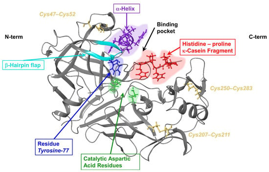
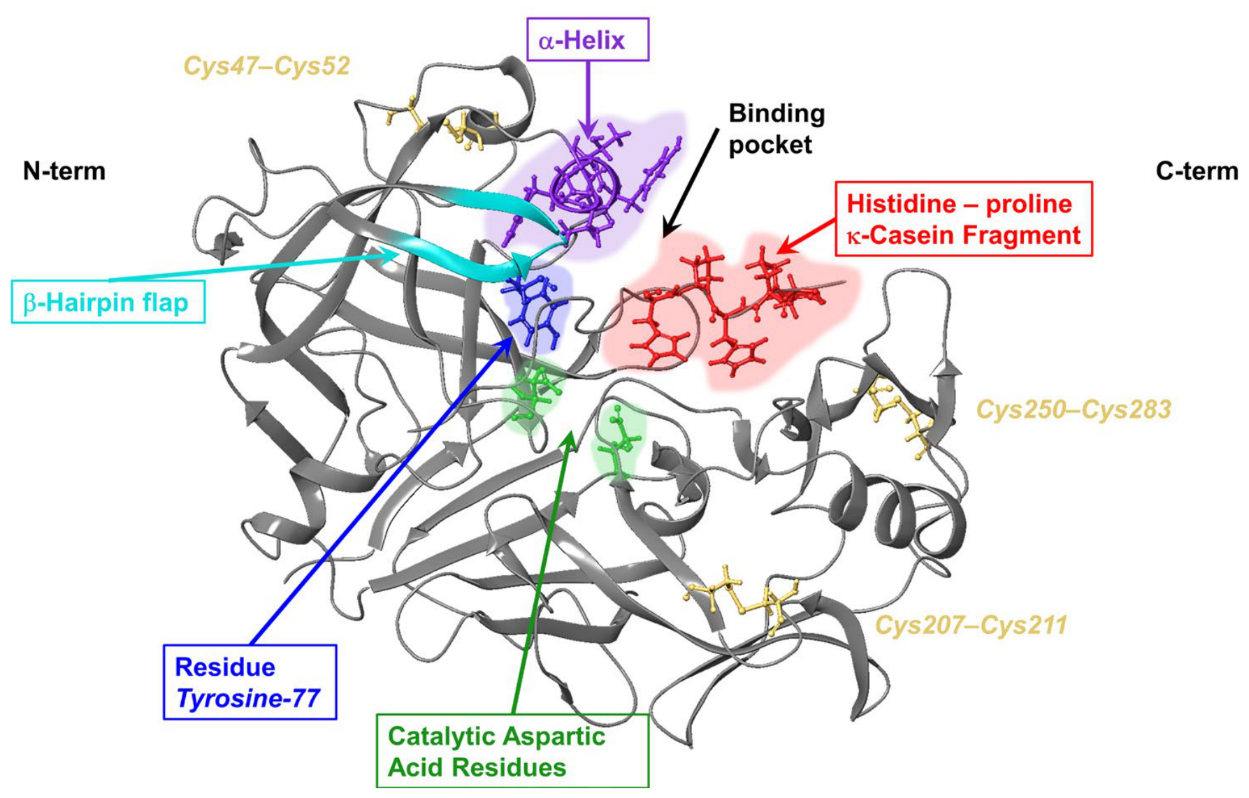
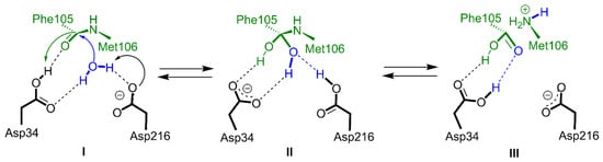
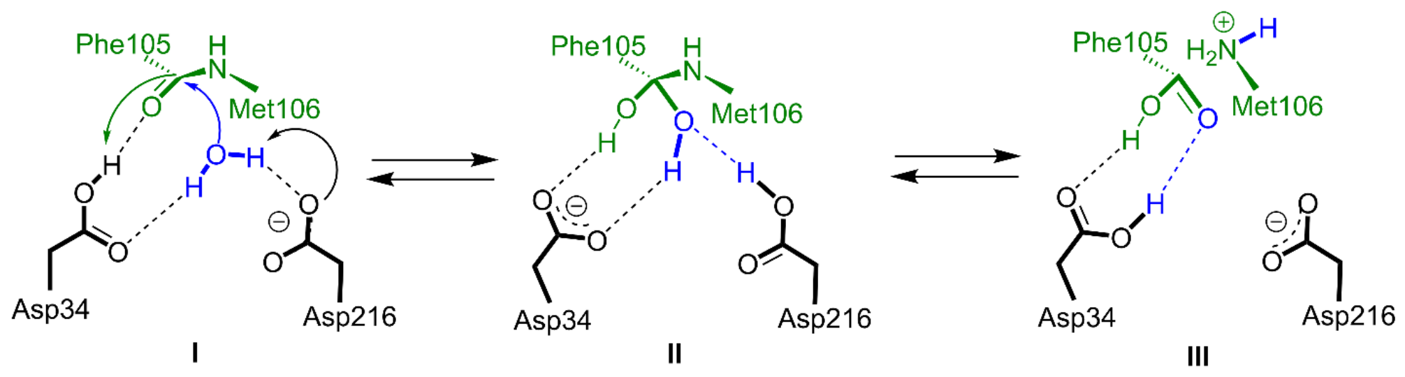
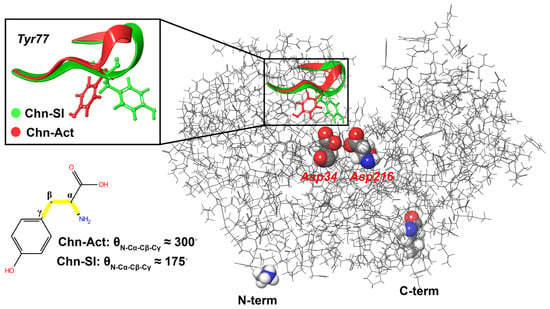
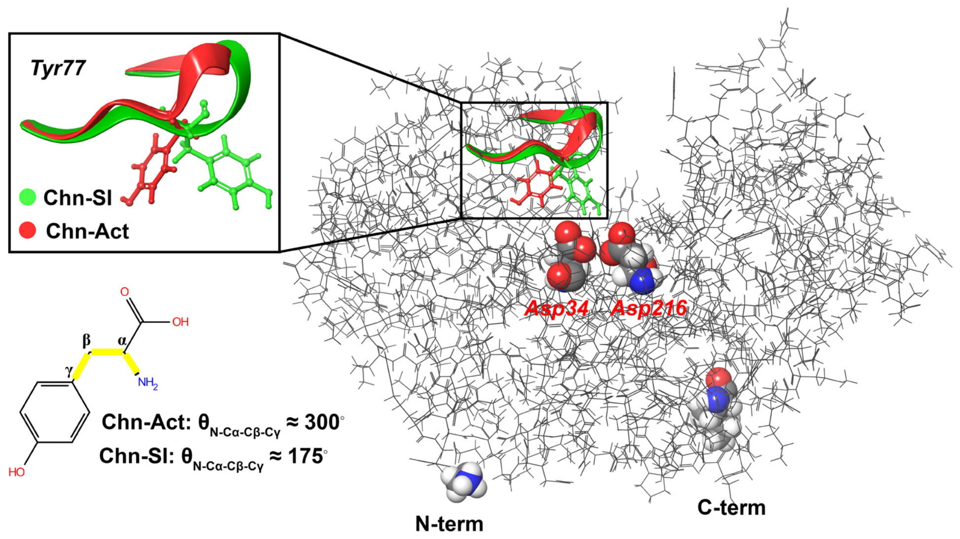
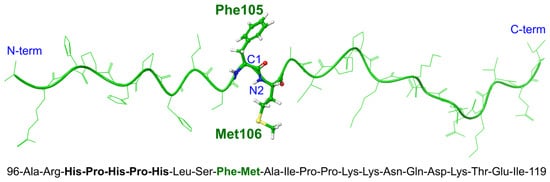
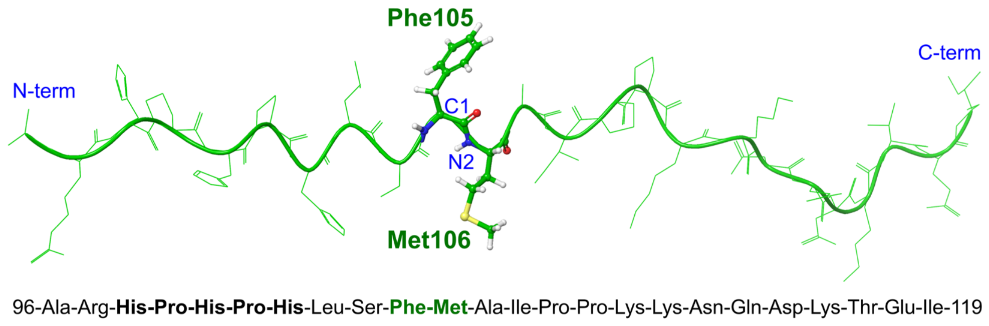
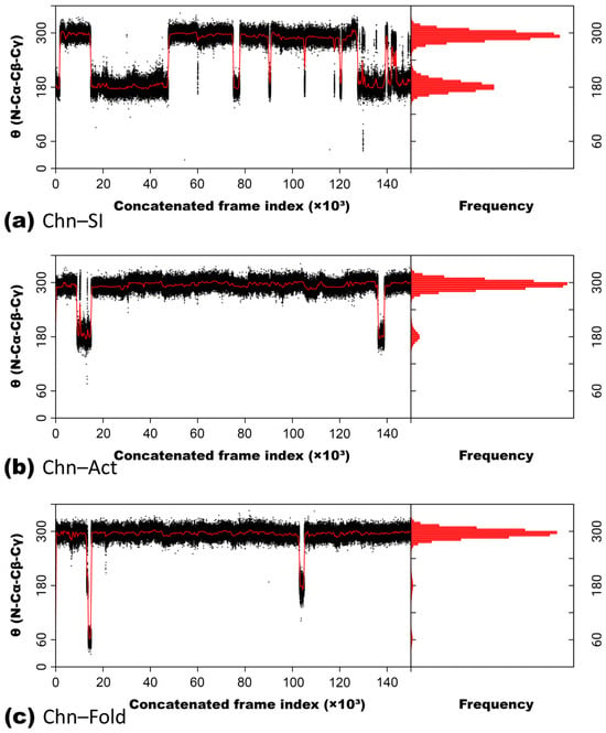
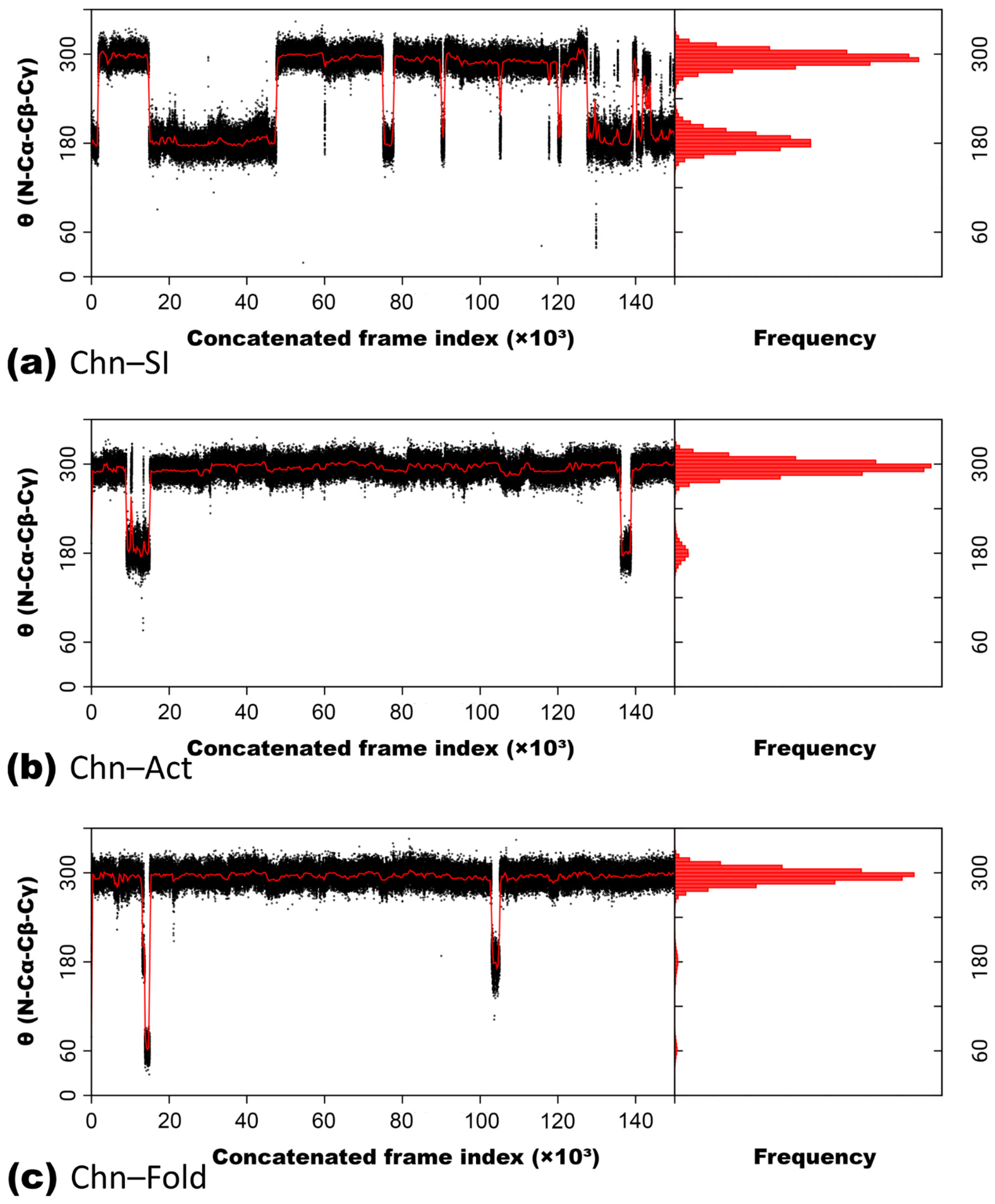
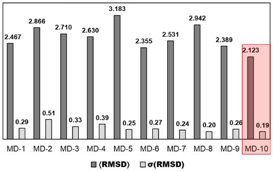
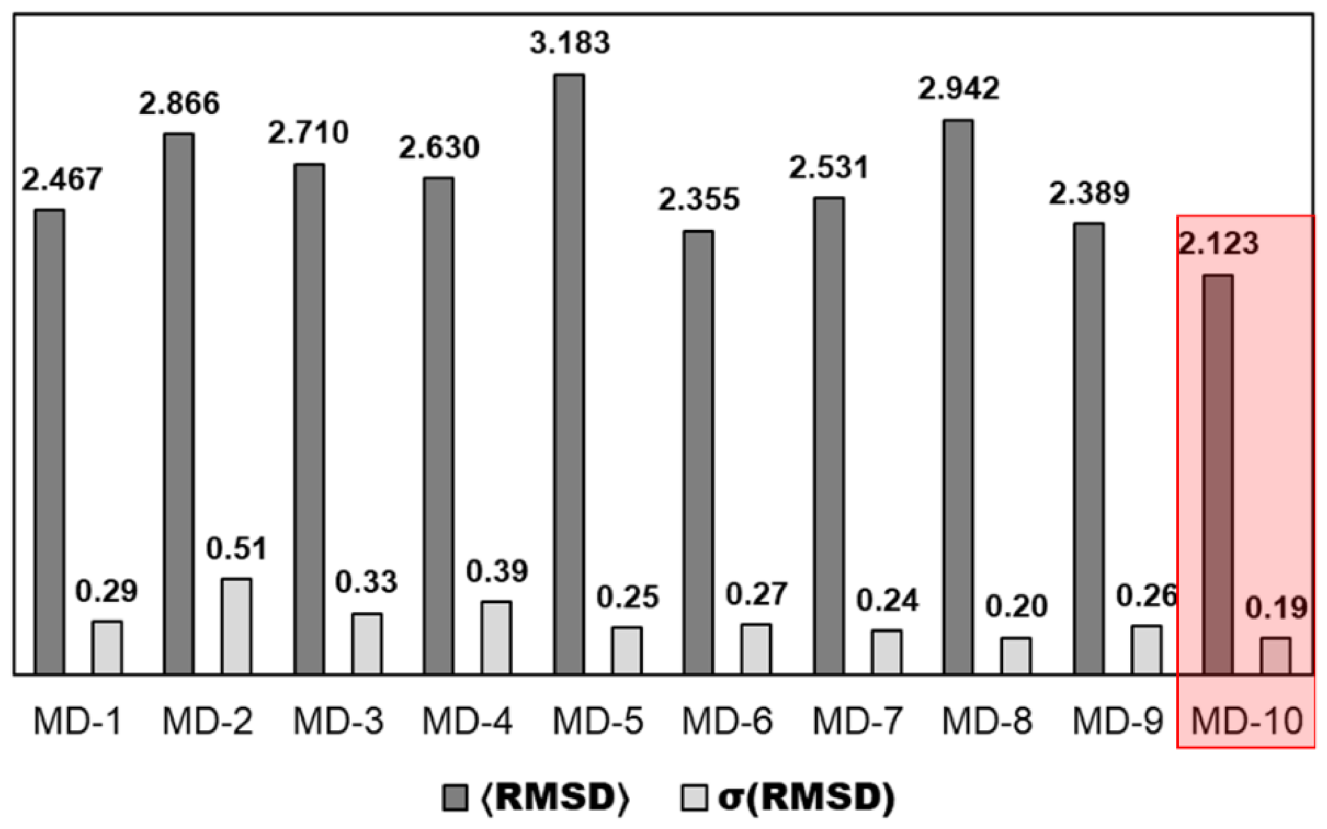
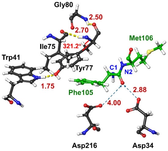
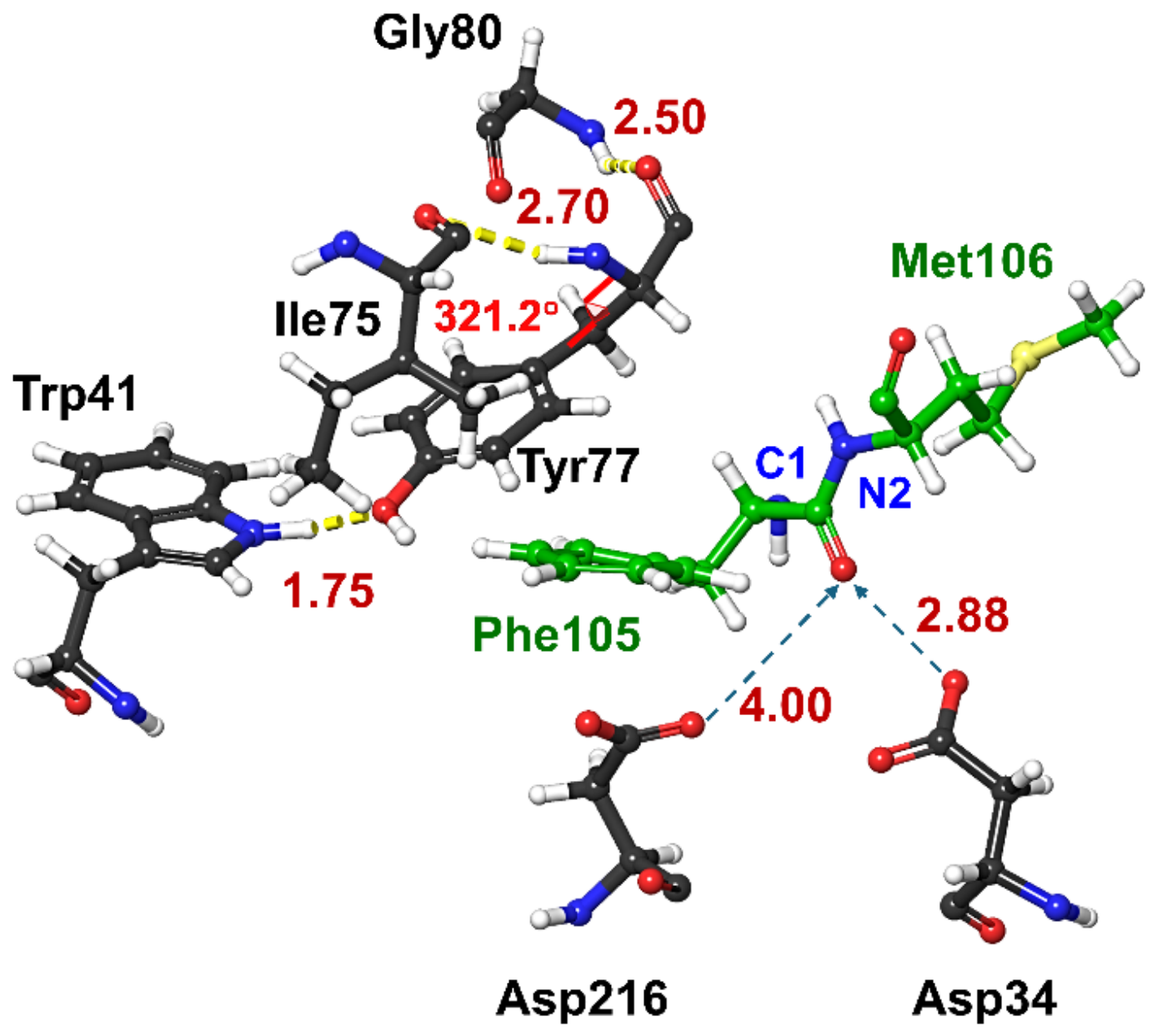
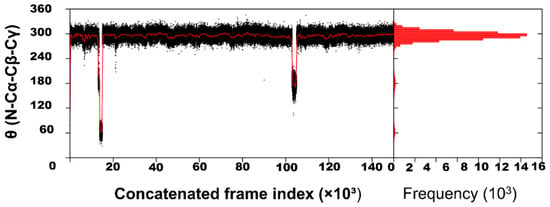
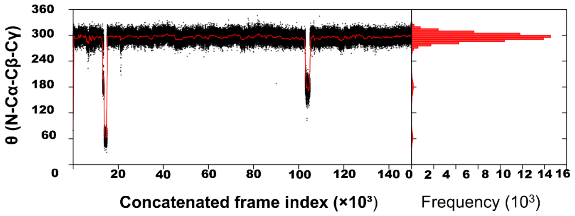
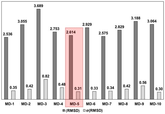
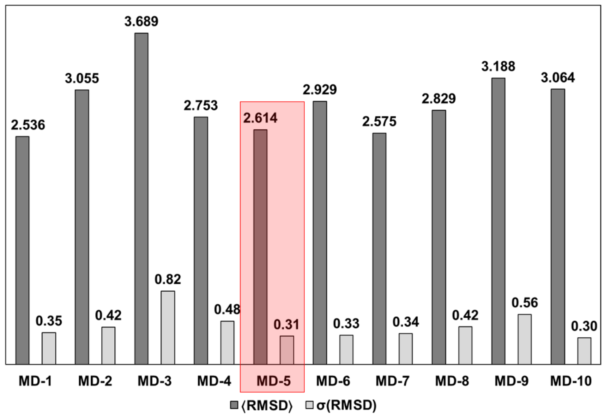
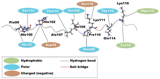
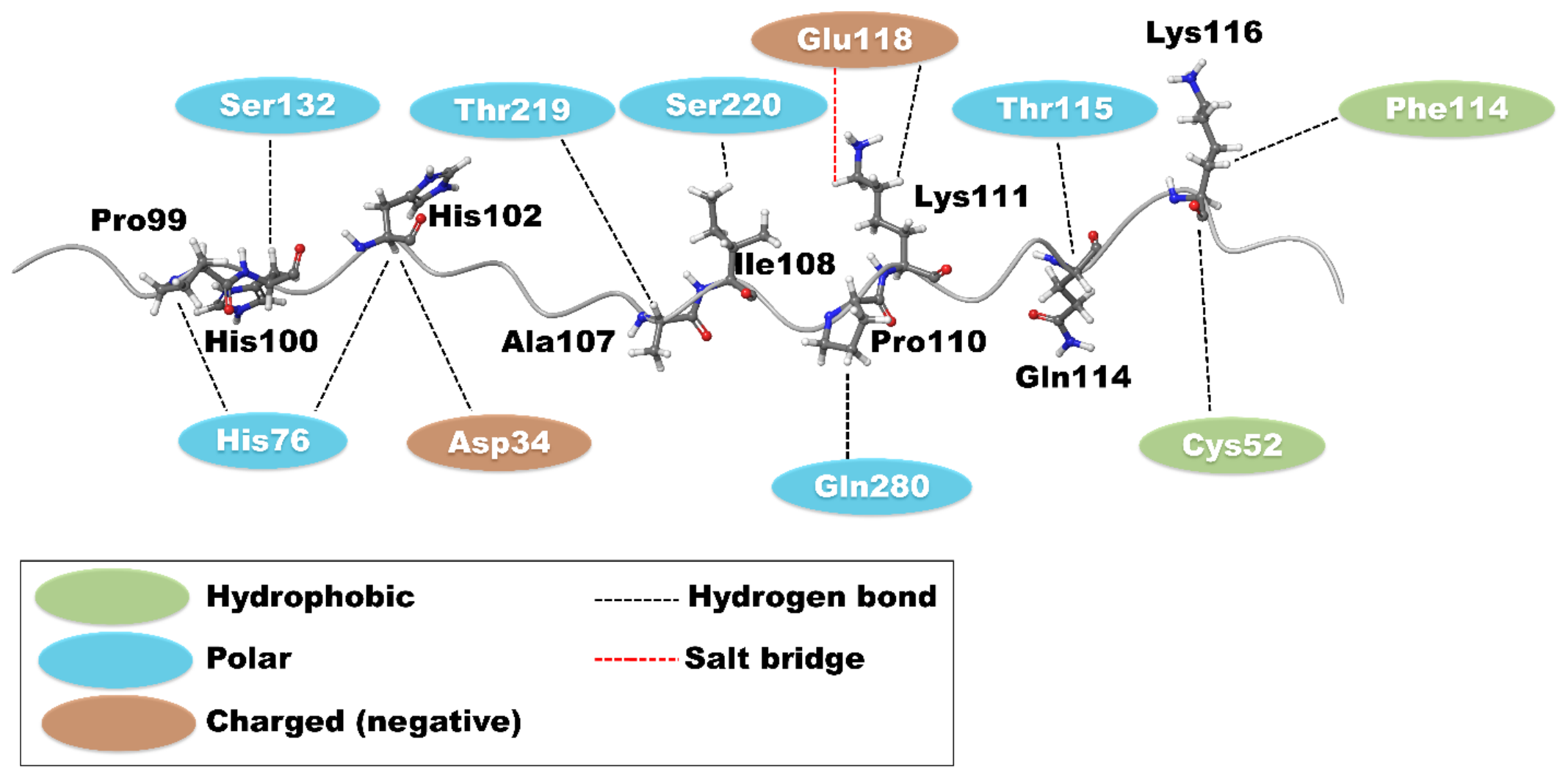
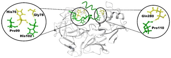
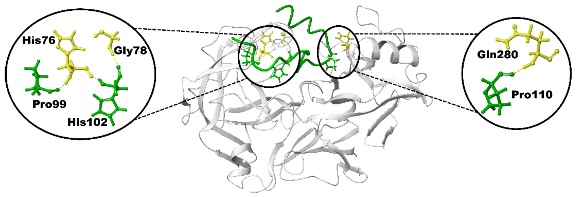
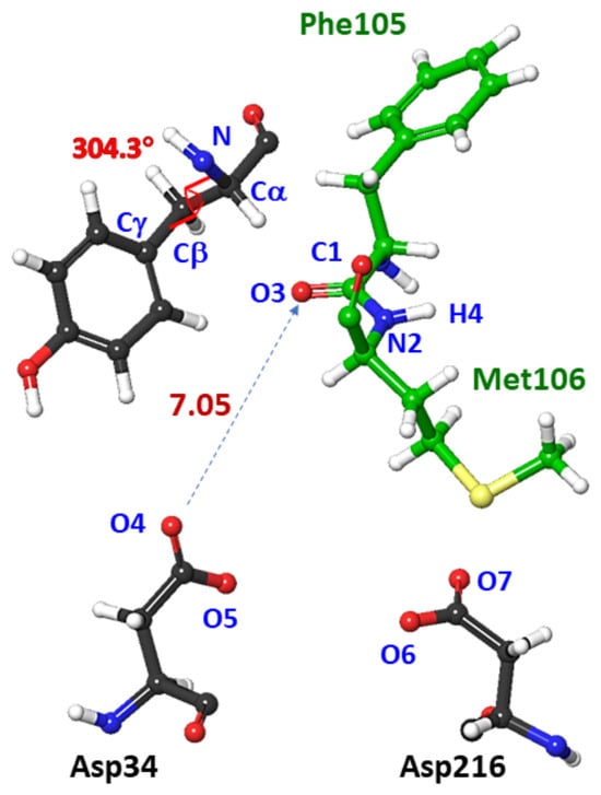
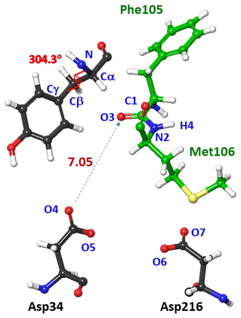
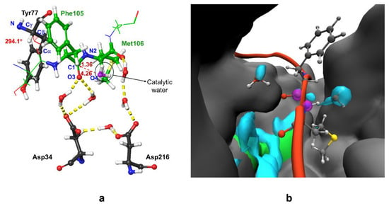
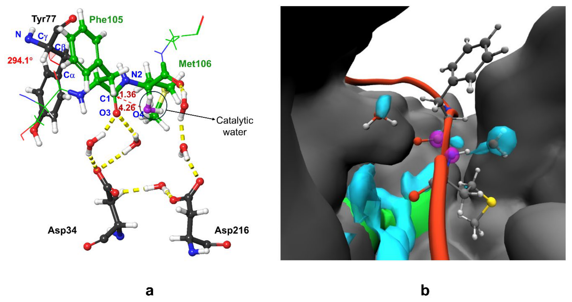
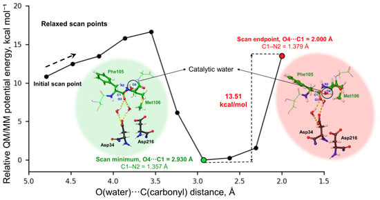
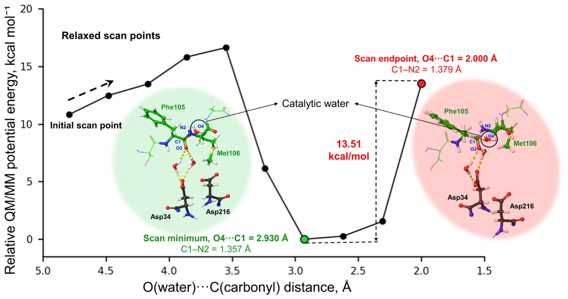
Disclaimer/Publisher’s Note: The statements, opinions and data contained in all publications are solely those of the individual author(s) and contributor(s) and not of MDPI and/or the editor(s). MDPI and/or the editor(s) disclaim responsibility for any injury to people or property resulting from any ideas, methods, instructions or products referred to in the content. |
© 2026 by the authors. Licensee MDPI, Basel, Switzerland. This article is an open access article distributed under the terms and conditions of the Creative Commons Attribution (CC BY) license.