


Abstract
Colletotrichum species are the causal agents of potato and tomato diseases, such as black dot and anthracnose. Several new species and species complexes were recently established. Thereby, a reassessment of the genus diversity is required. The study revealed two species, Colletotrichum coccodes and Colletotrichum nigrum, as Russia’s main disease agents of cultivated Solanaceae plants. Black dot and anthracnose in potato were caused exclusively by C. coccodes, whereas the same diseases in tomato, eggplant, and pepper were predominately caused by C. nigrum. However, one isolate of C. coccodes was also identified as an agent of the tomato disease. Five potentially hybrid isolates were discovered. Morphological examination and pathogenicity assessment revealed no significant differences between the two Colletotrichum species. All isolates were sensitive to the fungicides azoxystrobin, difenoconazole, and thiabendazole, which are currently used in agriculture. This is the first report of the occurrence of C. nigrum in Russia.
1. Introduction
Colletotrichum is a well-known causal agent of potato and tomato diseases such as black dot and anthracnose, dramatically damaging both underground and aboveground plant parts. Although several revisions of the genus Colletotrichum were recently published [1,2,3,4], it still remains taxonomically puzzling. Currently, 16 species complexes and 15 singleton species (e.g., C. coccodes and C. nigrum) are established within Colletotrichum [5]. Given the complicated systematics of the genus, the species identification is based on morphological features, combined with molecular data.
C. coccodes predominantly infects Solanaceae plants, including chilli fruit [6], potato tubers [7,8], tomato [9], sweet pepper [10,11,12], black nightshade [13], and eggplant [14]. Nevertheless, its wide host range is not limited to Solanaceae, since the species was reported to infect strawberry [15], pumpkin [16], or onion [17].
There are very few reports concerning C. nigrum. The species was first described as an agent of pepper anthracnose from Gloucester County, New Jersey, USA [18], but it can be associated with tomato and eggplant diseases [19,20] as well. Liu and colleagues [17,21] reviewed the species’ description, introduced a neotype to C. coccodes, selected an epitype to C. nigrum, and stated the ability of both species to induce anthracnose.
In Russia, the diversity within the genus is poorly described, due to the predominance of the morphological identification of causal agents. The review by Kotova and Kungurtseva [22] specified that C. coccodes is the only cause of potato and tomato black dot and anthracnose. Several studies in Russia investigated the diversity of Colletotrichum species on potato and tomato leaves using genetic markers [9,23,24] directly from the plant material, without isolating the species in axenic cultures. Kazartsev and colleagues [25] recently scrutinized the diversity of Colletotrichum species on several wild and cultivated plants (no potato or tomato plants were included into the analysis), using molecular and morphological approaches to identify the species. C. coccodes strains were isolated from Ambrosia artemisiifolia, Beta vulgaris, Brassica napus, Cannabis sativa, Galinsoga parviflora, and Portulaca oleracea. Poluektova and colleagues [26] analysed the glyceraldehyde-3-phosphate dehydrogenase and glutamine synthetase genes of four C. coccodes strains from Russian potato. To the best of our knowledge, these are the only molecular investigations of the genus in Russia to date.
This study focuses on the disease agents of several cultivated Solanaceae crops in Russia. Our research combines both morphological and molecular approaches to reveal the diversity within the genus Colletotrichum. To this end, four genetic markers were used: ITS1-5.8S-ITS2 region (ITS) as a well-established barcode, the glyceraldehyde-3-phosphate dehydrogenase gene intron (gaphd), considered the most reliable genetic marker for Colletotrichum species, the actin intron (act), and the glutamine synthetase intron (gs). To estimate the agricultural risks of the disease spread, we assessed the sensitivity to some fungicides that are officially used for tuber treatment, and the pathogenicity range towards the tomato fruit and potato tuber slices.
2. Materials and Methods
2.1. Sampling and Isolation of Cultures
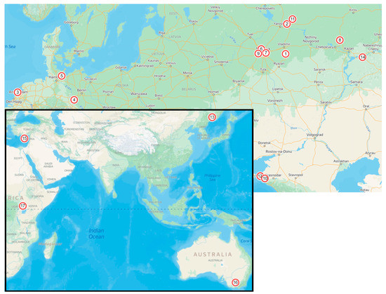
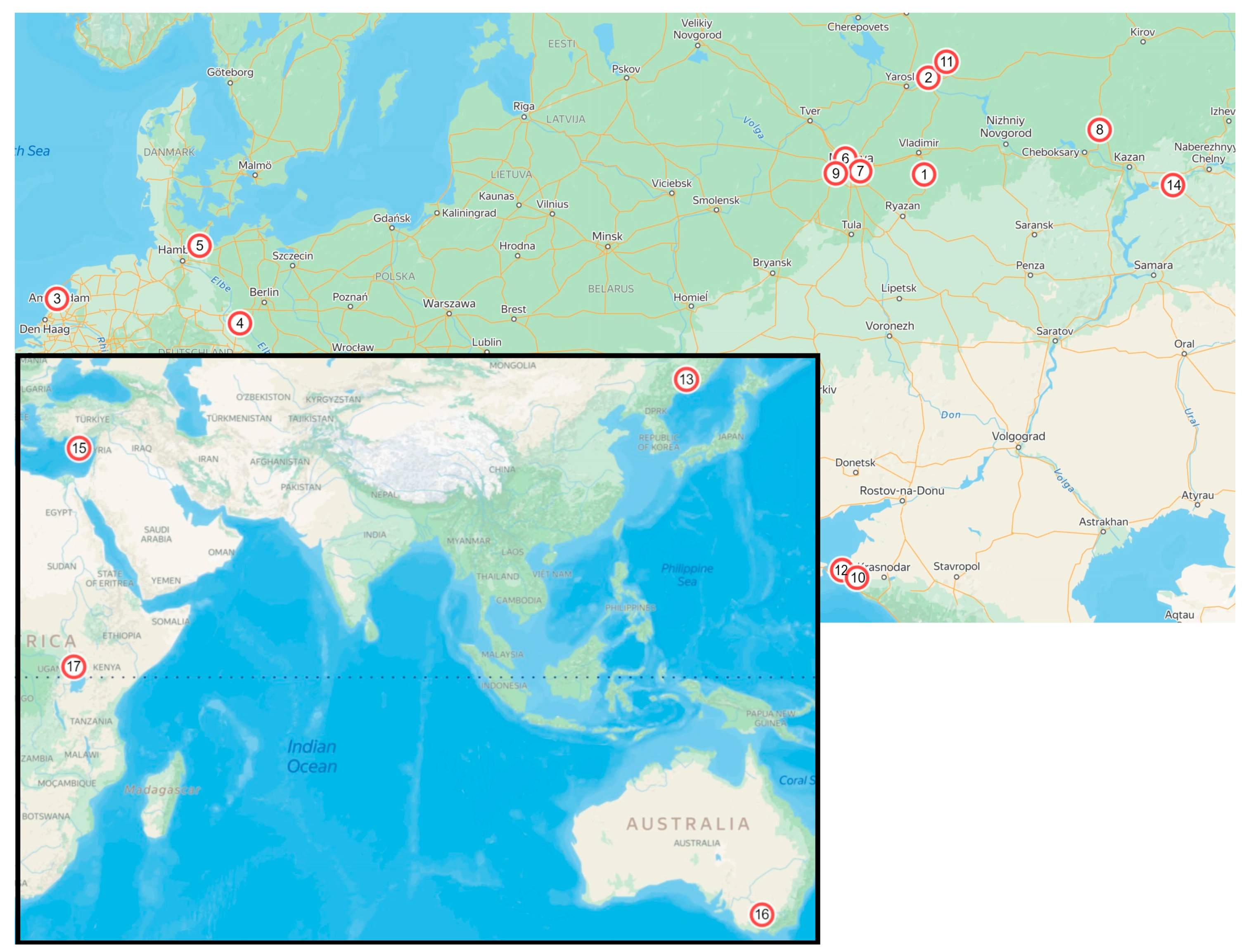
Samples were collected from the fruits of tomato, eggplant, and pepper (Table 1 and Figure 1) as well as from potato tubers, leaves, and stems. Seed potato tubers from the Netherlands, Germany, Australia, Cyprus, and Uganda were taken for comparison. All isolation sources were surface-sterilized with sodium hypochlorite (2% solution) to remove possible contamination, sliced, and put in wet chambers at 24 ± 1 °C. For isolation, small black sclerotia from the tuber peel or diseased tissue were taken using a preparation needle under a binocular microscope (MBS10, Russia), and transferred to culture media (potato-dextrose agar, PDA) amended with antibiotic (benzylpenicillin sodium salt, 100 mg/L).
Table 1.
Details of isolates used in the study.
Figure 1.
Location of collection sites (see also Table 1). Russia: 1—Vladimir Region; 2, 11—Kostroma Region; 6, 7, 9—Moscow Region; 8—the Mari El Republic; 10, 12—Krasnodar Krai; 13—Primorsky Krai; 14—the Republic of Tatarstan; 3—the Netherlands; 4, 5—Germany; 15—the Republic of Cyprus; 16—Australia; 17—Uganda.
2.2. DNA Isolation, PCR, Sequencing, and Phylogenetic Analysis
To extract DNA, the mycelium of fungi was grown on a liquid pea medium (180 g of green pea boiled for 10 min in 1 L of water, then filtered and autoclaved for 30 min at 1 atm). DNA was extracted according to the standard CTAB protocol [27,28]. ITS, act, and gaphd amplifications were performed in a SSI microtube strips in a 25 μL total volume reaction containing 1 μL of a DNA template (50 ng/μL), 2.5 μL of 10× PCR buffer (Applied Biosystems, Waltham, MA, USA), 0.5 μL of 10 mM each deoxyribonucleotide triphosphates (dNTP), 0.4 μL of 100μM each primer (Evrogen Co, Moscow, Russia), 1.5 U of Taq polymerase (5U/μL, Promega, Madison, WI, USA), and Milli-Q water (MQ). For the amplification of gs 2.8 μL of each dNTP was used; the concentrations of the other components remained the same. The following primers were used: ITS1 5’-TCCGTAGGTGAACCTGCGG-’3 and ITS4 5’-TCCTCCGCTTATTGATATGC-3’ for the ITS region [29], GSF1 5’-ATGGCCGAGTACATCTGG-’3 and GSR1 5’-GAACCGTCGAAGTTCCAC-’3 for the gs gene [30], GDF-1 5’-GCCGTCAACGACCCCTTCATTGA-’3 and GDR-1 5’-GGGTGGAGTCGTACTTGAGCATGT-’3 for gaphd [31], and ACT-512F 5’-ATGTGCAAGGCCGGTTTCGC-’3 and ACT-783R 5’-TACGAGTCCTTCTGGCCCAT-’3 for act [32].
The PCR protocol included initial denaturation at 94 °C for 3 min, 35 amplification cycles, and an additional extending step at 72 °C for 3 min. For the primer pair ITS1/ITS4, the amplification cycles were 94 °C for 30 s, 55 °C for 30 s, and 72 °C for 45 s. For the primer pairs GDF-1/GDR-1 and ACT-512F/ACT783R, the amplification cycles were 94 °C for 30 s, 60 °C for 30 s, and 72 °C for 30 s. For the primer pair GSF1/GSR1, the amplification cycles were 94 °C for 30 s, 61 °C for 30 s, and 72 °C for 120 s.
The amplification was performed on a T100 Thermal Cycler (Bio-Rad Laboratories, Inc., Hercules, CA, USA). PCR products were run in 0.7–1.2% agarose gel amended with ethidium bromide; the agarose concentration depended on the PCR fragment length. The gel extraction was performed with a Cleanup Mini Kit (Evrogen Co., Russia). The PCR fragments were sequenced using the BigDye® Terminator v.3.1 Cycle Sequencing Kit (Applied Biosystems, USA) and the Applied Biosystems 3730 × l automated sequencer (Applied Biosystems, USA). Each fragment was sequenced in both directions. Consensus sequences for each locus were assembled using Geneious version 7.1 (created using Biomatters) and MEGA X [33], and aligned with available type species sequences (Table 2). Species identification was based primarily on the gaphd sequence.
Table 2.
Reference strains used in this study.
2.3. Morphological Analysis
The cultures were grown on synthetic nutrient-poor agar medium (SNA) [34] amended with Anthriscus sylvestris double autoclaved stems [16] for 10 days. Microscopic preparations were made in clear lactic acid. The width and length of conidia were measured, with 30 measurements per structure, using a Leica DM 2500 (Leica Microsystems, Germany).
2.4. Pathogenicity Tests
To compare the pathogenic activity, 10 isolates (two from potato, two from pepper, three from tomato, and three from eggplant) were chosen. Healthy cherry tomato fruits and potato tubers (cultivar “Gala”) were washed and surface-sterilised in 0.5% sodium hypochlorite solution for 5 min, rinsed in distilled water, and air-dried. The potato tubers were sliced to imitate wounding. Two types of tomato fruit were used: wounded with sterile tips and unwounded. The experiment was conducted with three repeats for each strain of each kind of inoculation. The wounded fruits were internally inoculated with 100 μL of conidial suspension (concentration 105 spores/mL). The unwounded fruits and potato slices were surface-inoculated with mycelium and conidia, and placed in sterile wet chambers. The control fruits and tubers were surface or internally inoculated with distilled water. Each wet chamber was stored at 10 °C for 21 or 35 days, and the radius of the lesion was measured. To fulfil Koch’s postulate, a small tissue sample was taken from the margin of the disease area with a sterile scalpel and placed in a Petri dish on PDA.
2.5. In Vitro Assessment of Fungicide Sensitivity
Three chemical fungicides: azoxystrobin (Quadris®, Syngenta, Basel, Switzerland), difenoconazole (Score®, Syngenta), and thiabendazole (Tecto®, Syngenta) were chosen to evaluate their efficiencies against Colletotrichum isolates. The fungicides were selected based on their current use in Russia for tuber or in-furrow treatment, and they were obtained from local suppliers. The sensitivity was evaluated in Petri plates with PDA. The fungicides were added at different concentrations to autoclaved PDA medium to produce a concentration series of 0, 0.1, 1, 10, and 100 mg/L for each fungicide (active ingredient). The mycelial plug (5 mm in diameter) of each isolate was punched from the margin of an actively growing colony of a 5-day-old culture and placed in the centre of a 90 mm PDA plate amended with fungicide, as well as on non-amended PDA plates (controls). Three replicates per treatment were produced, and the plates were incubated at 24 ± 1 °C for 4 days. The diameter of the fungal colony on each plate was measured at perpendicular angles. The average of the two measurements was used to calculate the fungicide concentration inhibiting linear colony growth of 50% over control (EC50) [35,36].
3. Results
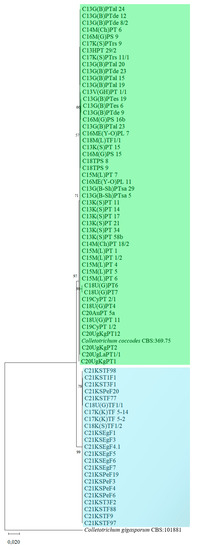
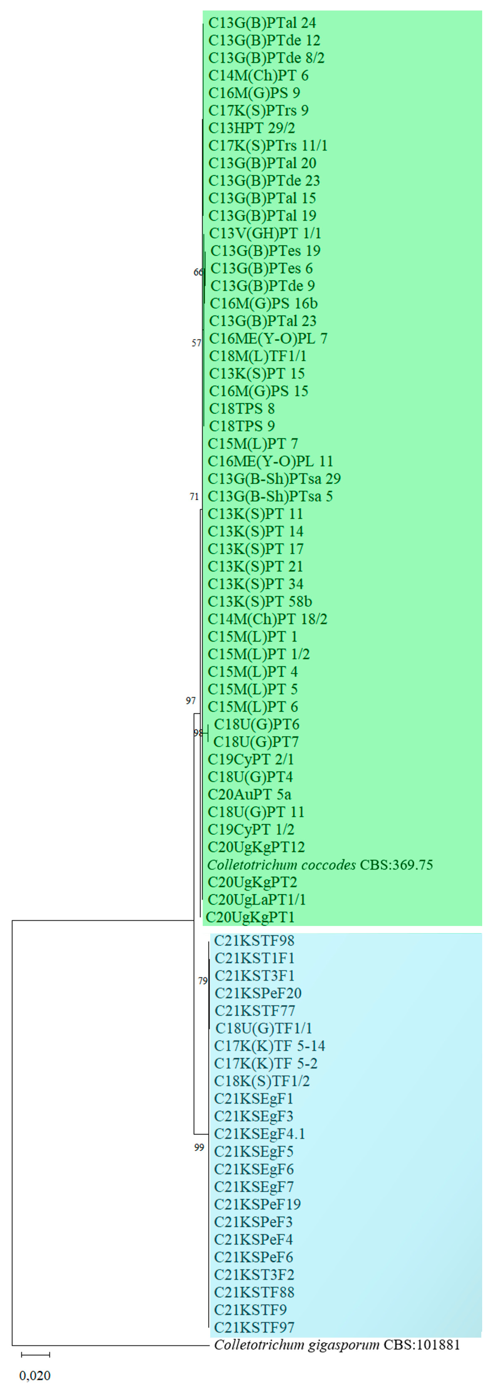
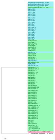
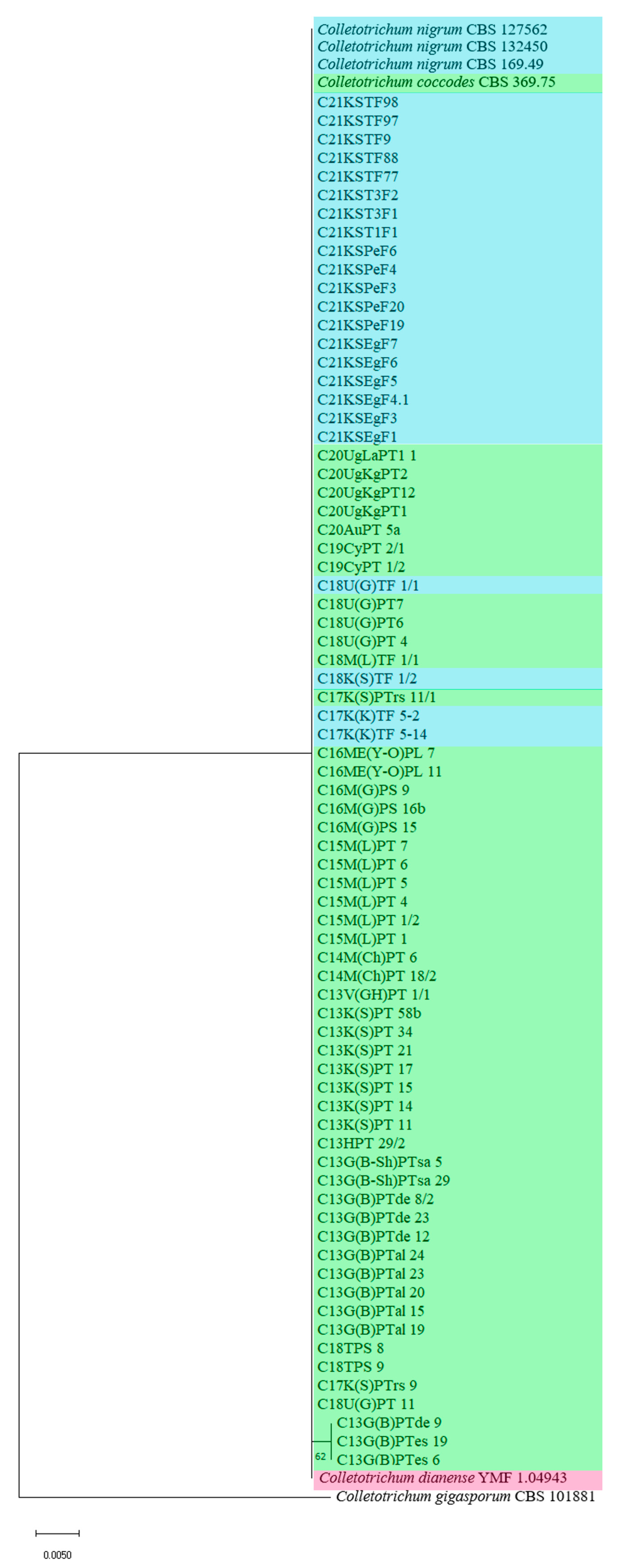
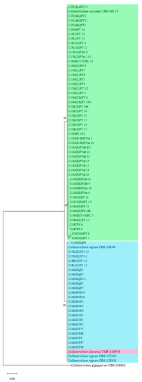
In total, 74 isolates were analysed: 50 from potato (Solanum tuberosum L.), 11 from tomato (Solanum lycopersicum L.), 7 from pepper (Capsicum annuum L.), and 6 from eggplant (Solanum melongena L.). All strains isolated from potato, regardless of the plant organ, were identified as C. coccodes, while those from eggplant and pepper proved to be related to C. nigrum (Figure 2, Figure 3, Figure 4, Figure 5 and Figure 6). Almost all tomato isolates except one strain (C18M(L)TF1/1) were identified as C. nigrum. The aligned concatenated sequence dataset of the original isolates was 1690 bp long (440, 207, 233, and 810 bp for the ITS, act, gaphd, and gs sequences, respectively). It contained 65 variable sites: 2 in the ITS region, 2 in the act intron, 12 in the gaphd intron, and 49 the in gs second intron (including three deletions); among them, 2 in act, 8 in gaphd, and 17 in the gs intron seemed to be specific to either C. coccodes or C. nigrum, and they may be used to differentiate between these species.
Figure 2.
Phylogenetic tree inferred from maximum-likelihood analysis of the concatenated alignment, including the ITS region, and the partial act, gaphd, and gs gene regions. The confidence values are indicated at the branches. Green indicates C. coccodes clade, and blue indicates the C. nigrum clade.
Figure 3.
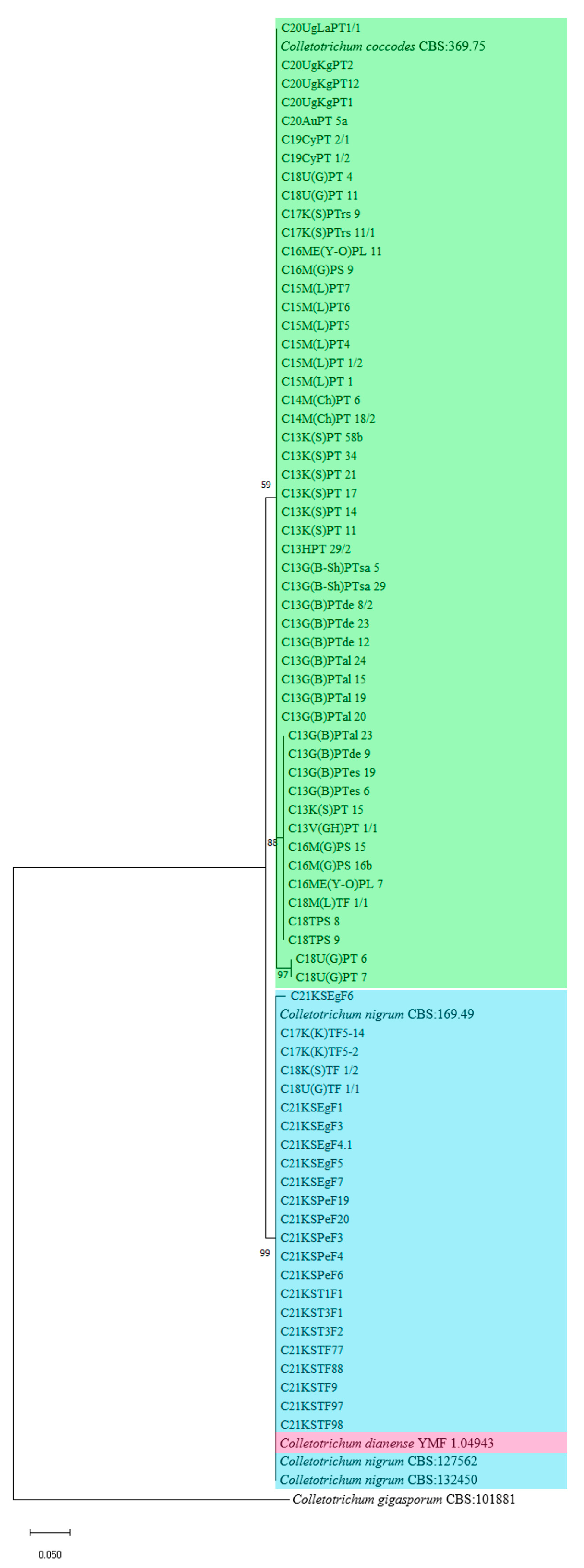
Phylogenetic tree inferred from maximum-likelihood analysis of the ITS1-5, 8S-ITS2 region alignment. Bootstrap 1000 replicates. The confidence values are indicated at the branches. Green indicates C. coccodes isolates; blue indicates C. nigrum isolates, and red marks C. dianense type species.
Figure 4.
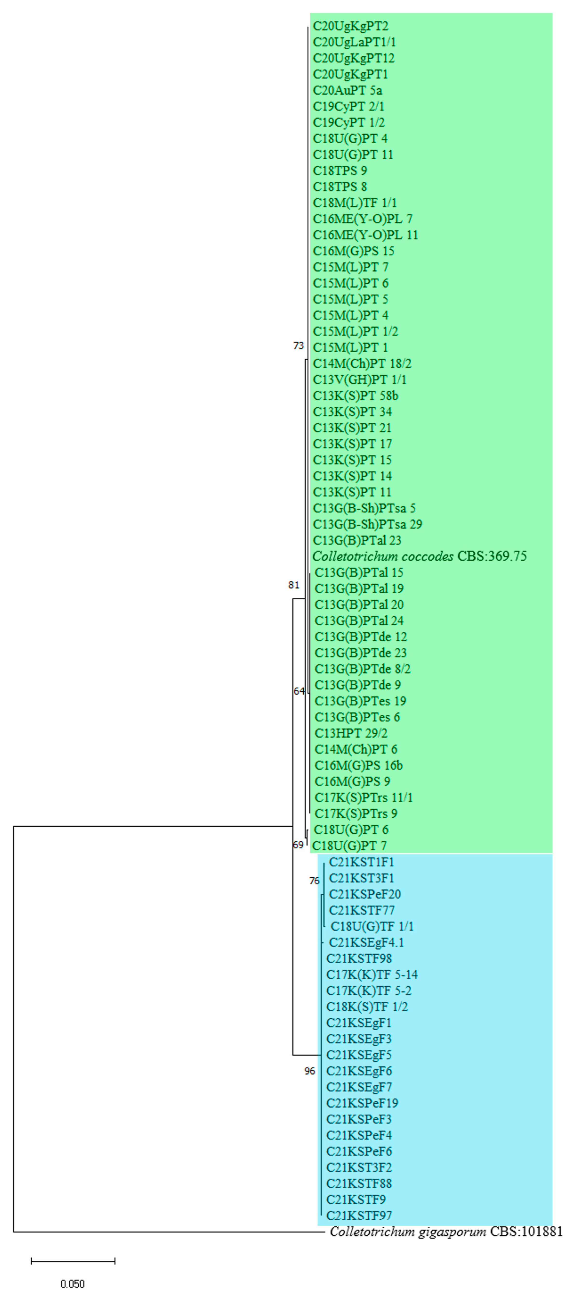
Phylogenetic tree inferred from maximum-likelihood analysis of the actin intron alignment. Bootstrap 1000 replicates. The confidence values are indicated at the branches. Green indicates C. coccodes clade, blue indicates the C. nigrum clade, and red marks C. dianense type species.
Figure 5.
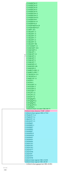
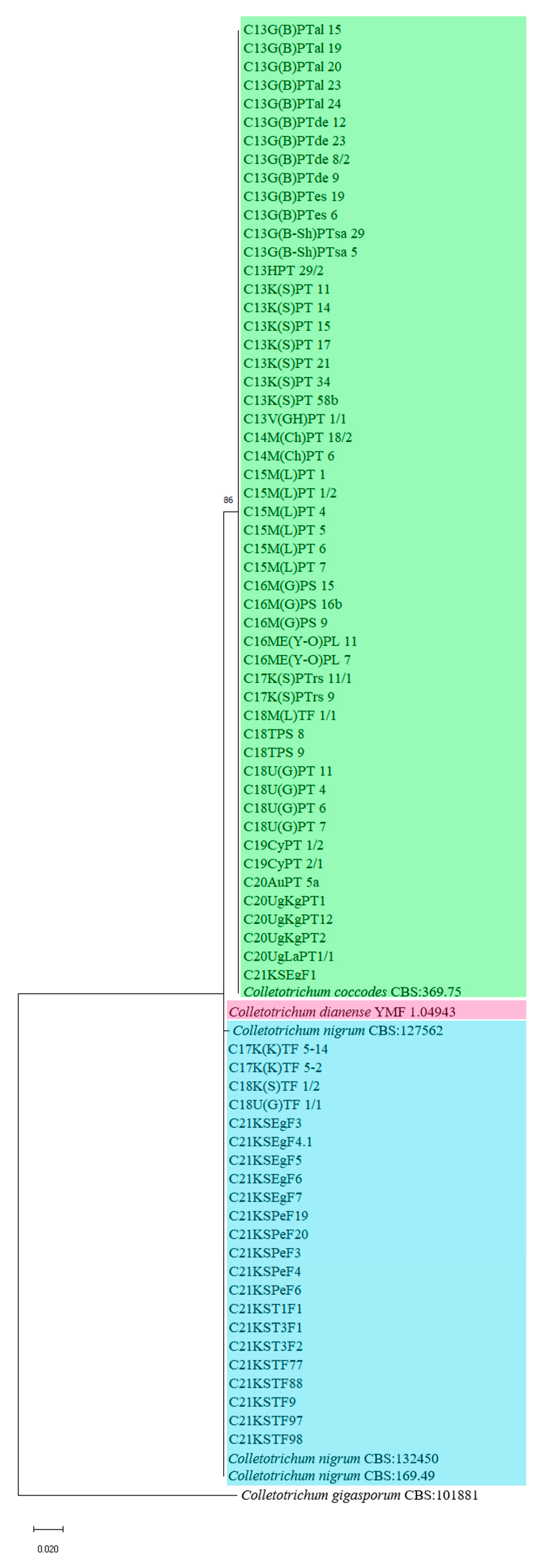
Phylogenetic tree inferred from maximum-likelihood analysis of the glyceraldehyde-3-phosphate dehydrogenase intron alignment. Bootstrap 1000 replicates. The confidence values are indicated at the branches. Green indicates C. coccodes clade, blue indicates the C. nigrum clade, and red marks C. dianense type species.
Figure 6.
Phylogenetic tree inferred from maximum-likelihood analysis of the glutamine synthetase intron alignment. Bootstrap 1000 replicates. The confidence values are indicated at the branches. Green indicates C. coccodes clade; blue indicates C. nigrum clade.
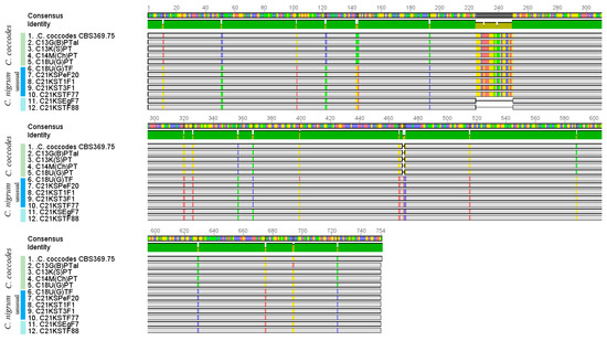
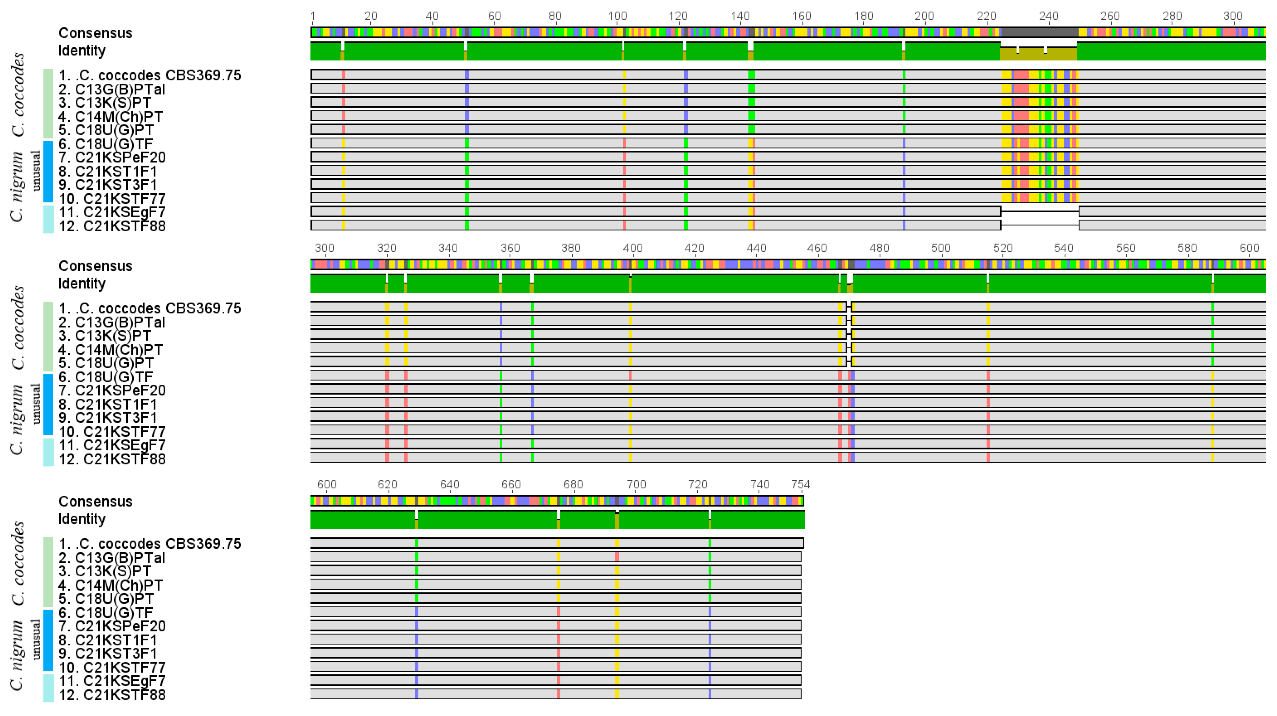
Most C. coccodes, but also several C. nigrum (possible hybrids) isolates, contained a 25 bp insertion in the gs intron (Figure 7). All the nucleotide differences were found in the non-coding regions. All the phylogenetic trees, except one based on the ITS region (Figure 3), showed two well-delimited clades corresponding to two segregate species: C. coccodes and C. nigrum (Figure 2, Figure 4, Figure 5 and Figure 6). The recently described C. dianense was in the same clade as C. nigrum (Figure 4 and Figure 5).
Figure 7.
Comparison of glutamine synthetase second intron sequences of intriguing isolates (C18U(G)TF1/1, C21KST1F1, C21KST3F1, C21KSTF77, and C21KSPeF20) to type strain C. coccodes CBS 369.75. SNPs, including A, T, C, and G, are marked with red, green, blue, and yellow, respectively.
Among the tomato and pepper C. nigrum strains, five intriguing isolates (C18U(G)TF1/1, C21KST1F1, C21KST3F1, C21KSTF77, and C21KSPeF20) were found (Figure 7). Presumably, they shared the features of both species—C. coccodes and C. nigrum. All of the isolates were identified as C. nigrum, based on the gaphd sequence (Table 3). The similarity to C. dianense is discussed further. In total, within 810 bp sequences of the gs gene, we revealed one 25 bp insertion (positions 225–249) and one 1 bp (position 470) insertion typical of C. coccodes CBS 369.75, and 17 single nucleotide polymorphisms (SNP) atypical of C. coccodes (positions 11, 51, 102, 122, 143, 144, 193, 320, 326, 357, 467, 471, 515, 588, 629, 675, and 724).
Table 3.
Comparison of intriguing isolates to type strains *.
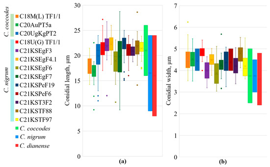
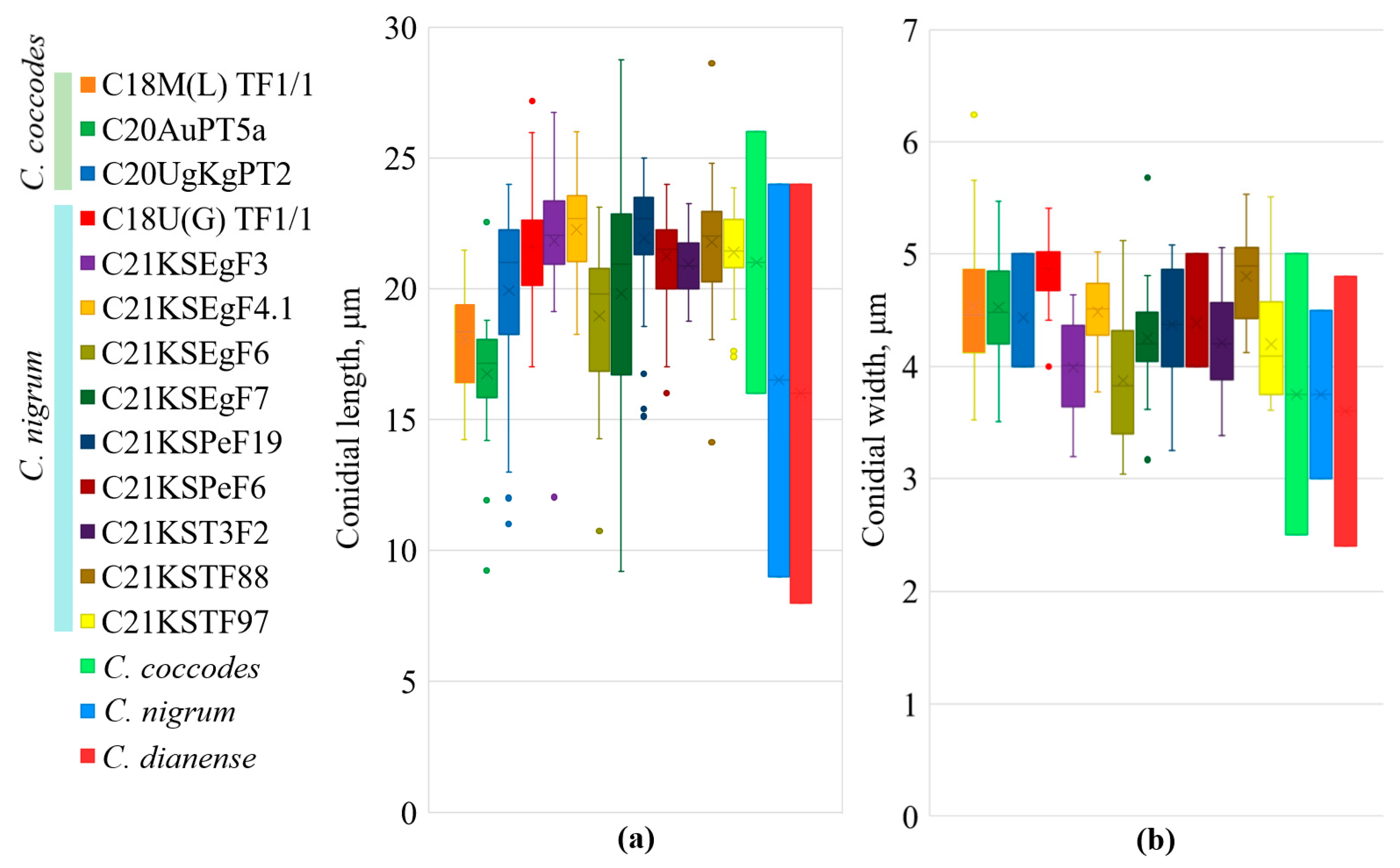
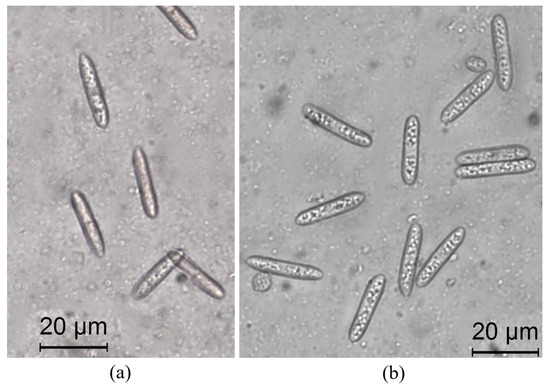
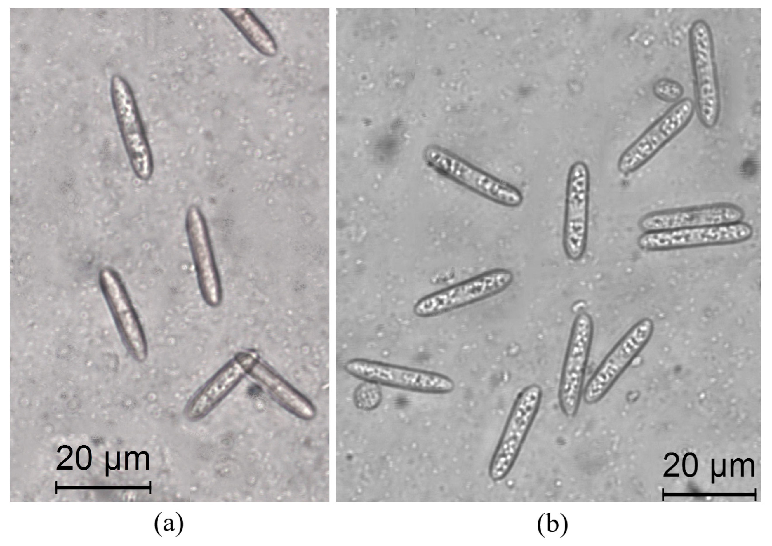
Morphological differences were not found between the sizes of the C. coccodes and C. nigrum conidia (for C. coccodes, the conidial length was 18.23 ± 6.13 μm and the width was 4.49 ± 1.04 μm; for C. nigrum, the conidial length was 21.17 ± 5.10 μm and the width was 4.34 ± 1.03 μm; Figure 8). The obtained conidial measurements overlapped with those of the type strains of C. coccodes, C. nigrum, and C. dianense [16,21,37]. All of the isolates produced aseptate, smooth-walled, hyaline, oval to cylindrical conidia with acute, subacute or obtuse apices, typical of C. coccodes or C. nigrum (Figure 9).
Figure 8.
Comparison of the conidial lengths (a) and widths (b) of Colletotrichum isolates and type strains. Boxes indicate quartiles (first and third), whiskers indicate the minimum and the maximum values, and points outside the boundary are outliers.
Figure 9.
Conidia of C. coccodes isolate C20UgKgPT2 (a) and C. nigrum isolate C21KSPeF6 (b).
All the tested strains of both species were able to cause tomato fruit and potato tuber infection (Table 4). Regardless of the species used to infect the tomatoes, the infected fruit had typical dark lesions of anthracnose with sclerotia, milky-white swellings with conidia occasionally developed. In the case of tomato wound inoculation, the C. coccodes- and C. nigrum-caused disease radii were 5.2–6.2 mm and 3.2–8.3 mm, respectively, after 21 days of infection. On the intact fruit, the disease radius did not exceed 1.2 mm after 21 days for all the isolates of both species, but after 35 days, one anthracnose-causing strain of C. nigrum reached a disease radius of 9.8 mm in width. All the tested strains of C. coccodes and C. nigrum were able to spread on potato slices. The disease radius was 2.5–5.2 mm for C. coccodes and 1.2–8.3 mm for C. nigrum after 21 days. No correlation between the disease severity and the original host was found: the isolates from tomatoes and potatoes could infect plants of both species. Nevertheless, the tomato fruit disease caused by both species was more rapid and extensive under the same temperature conditions compared with the potato tuber disease.
Table 4.
Pathogenicity tests.
No isolate resistant to any examined fungicide was found (Table 5). Thiabendazole EC50 for C. coccodes was 0.65–58.38 mg/L, and that for C. nigrum was 0.58–20.29 mg/L. Six isolates (five C. coccodes from potato tubers and stem, and one C. nigrum from tomato fruit) were less sensitive to the chemical (EC50 > 10 mg/L). No resistance was found for azoxystrobin, EC50 for C. coccodes was 0.05 and 9.07 mg/L, EC50 for C. nigrum was 0.08–8.50 mg/L. Difenoconazole was the most effective chemical; EC50 for all the tested isolates was less than 0.12 mg/L.
Table 5.
Sensitivity to fungicides.
4. Discussion
The efficiencies of the known genetic markers in differentiating Colletotrichum species vary among different species complexes [4]. The ITS region is widely used in routine studies, although the result may be doubtful. For instance, in northern Italy, C. coccodes was reported as an agent of pepper root disease [10]. Undoubtedly, the species can cause root disease; still, ITS-based identification remains insufficient. In Turkey, unusual symptoms of Colletotrichum disease leading to extremely high crop losses were discovered, and the pathogen was identified as C. coccodes [38]. However, the only molecular marker used in the study was the ITS region, so the identification seems uncertain.
Dos Santos Vieira and colleagues [39] propose using gaphd and several other regions to distinguish between Colletotrichum species, while ITS and act are less effective. According to our study, both the act and gaphd genes are suitable, at least for C. coccodes and C. nigrum division (Figure 5 and Figure 6).
The Gs intron also proved useful for delineating Colletotrichum species. This gene sequence is mainly used to distinguish the species within C. gigasporum, C. orbiculare, and C. gloeosporoides species complexes. Thus, up to date, GenBank lacks the gs region sequences of the type material for many species. Several GenBank accession numbers marked as the C. coccodes gs gene (GU935816 and GU935817) presumably belong to C. nigrum, as they differ by approximately 2–3% from C. coccodes CBS164.49 or CBS369.75 (GenBank accession numbers HM171675 and HM171676, respectively) but they are similar to our strains that are identified as C. nigrum, based on the act or gaphd genes. We propose that at least 17 single nucleotide changes underlie the differences between the gs second intron of the two species. The C. nigrum currently presumed occurrence and host range seem to be lower than the real ranges. We assume that several reports of C. coccodes, for example [17], may display C. nigrum disease instead.
According to the pertinent literature, the sexual process is unknown for C. coccodes or C. nigrum. The only way for strains of these species to exchange genetic material is via a parasexual process or through a vegetative compatibility reaction [40]. Based on the gs sequence of five isolates (C18U(G)TF1/1, C21KST1F1, C21KST3F1, C21KSTF77, and C21KSPeF20), we suppose that they might represent hybrids between C. coccodes and C. nigrum. Whereas we detected SNPs in all the isolates identified as C. nigrum based on gaphd, we assume these SNPs to be specific to C. nigrum (Figure 2). At least one of the isolates (C18U(G)TF1/1) was collected from tomato fruit grown near potato plants; therefore, it might have had a possibility of interfering with C. coccodes strains. Notwithstanding these putative hybrid isolates, we assume that the second intron of the gs gene is useful for distinguishing between C. coccodes and C. nigrum, and we propose a more active use of the GSF1—GSR1 primers for identifying the Colletotrichum species.
Both C. coccodes and C. nigrum are currently considered as singleton species. According to Liu et al. [16], all potato-associated isolates belong to C. coccodes. At the same time, both C. nigrum and C. coccodes were able to infect tomato and pepper. The statement is supported by other studies [3,4,5] and by our data. Until now, we found no information regarding C. nigrum in Russia.
Based on ITS region sequencing, only one Colletotrichum species—C. coccodes—was previously reported from potato and tomato leaves in Russia [9,23,24,26]. Belov and colleagues [9] used a specific primer pair (Cc1F1 and Cc2R1) [41] to detect C. coccodes [41]. Both test systems [23,42] were developed based on the ITS region, considered the universal fungi barcode [43]. However, they were of limited use in distinguishing C. coccodes and C. nigrum.
C. dianense, which is very similar to C. nigrum, was recently described [37]. The authors of the study stated that it could be distinguished from C. nigrum by its conidial shape and apex. The ITS region and the act gene of the C. dianense type isolate YMF 1.04943 is 100% identical, and the gaphd gene is 99.66% similar to the C. nigrum type strain CBS 169.49 (one nucleotide difference, position 185). We compared our isolates to both species. Although, in our opinion, the two species are slightly different, we named our isolates from tomato, pepper, and eggplant C. nigrum, as C. nigrum is a well-known and earlier described species.
The cross-virulence of Colletotrichum species was reported a while ago [44]. Yet, there are no literature reports of C. nigrum potato infections, as the species was only found on tomato and pepper [3,16], while the GenBank database contains several C. nigrum isolates (e.g., KU821311) reported from potatoes. Colletotrichum disease is known as post-harvest, and is particularly harmful to climacteric fruits (e.g., tomato) [45]. Even though our study demonstrated the possibility of C. nigrum potato infection, no C. nigrum strains were isolated from potato.
Another Colletotrichum species, C. acutatum s. str., was reported as a tomato and pepper infectious agent [5,46]. The presence of C. acutatum s.l. in potato leaves and mini tubers was revealed in our previous studies (unpublished data) based on the reaction with species-specific primers CaInt2/ITS4 [47]. Although C. acutatum s.l. has not been proven to be a potato disease agent, the possibility of its presence on potato tubers and leaves should be kept in mind.
Liu and colleagues [16,21] mentioned that C. coccodes strains from tomato or other hosts produce larger conidia than C. coccodes from potato, and C. nigrum forms longer conidia than C. coccodes. Our results do not support these statements and show no significant difference between the conidial length or width among the three studied species (Figure 8). Contrary to Zheng and colleagues [37], we suppose that the morphological differentiation within the C. coccodes—C. nigrum—C. dianense clade may not be significant.
All the tested chemicals—azoxystrobin, thiabendazole, and difenoconazole—proved to be effective against Colletotrichum spread. The results were in line with previous studies [48,49,50]: normally, EC50 is less than 1 mg/L for all of the tested chemicals. In addition, azoxystrobin reduced black dot on tubers in field conditions [50]. Resistance to azoxystrobin or thiabendazole was reported in other Colletotrichum species complexes [51,52]. We discovered five strains (C13G(B)PTde9, C13G(B)PTes6, C16M(G)PS16b, C18K(S)TF1/2, and C18U(G)PT11), with thiabendazole EC50 ranging over 20–50 mg/L. Sanders and Korsten [52] classified strains with 66–70% growth on 0.5–2.5 mg/L thiabendazole as resistant, but we named them as less sensitive after Leite [53]. In our previous study, we examined the β-tubulin gene, but no specific mutations in any of the Colletotrichum isolates was found [54], contrary to Colletotrichum musae [53], less sensitive strains, Colletotrichum siamense [55], or Helminthosporium solani highly resistant strains [56]. Because even the highest EC50 values for Colletotrichum spp. are much lower than the concentrations in the working liquid for treatment (e.g., 170–250 mg/L for azoxystrobin, 4800–5600 mg/L for thiabendazole, and 187–625 mg/L for difenoconazole), we conclude that in general, all of the studied chemicals could still be considered as an effective strategy for anthracnose control on Solanaceae in Russia.
5. Conclusions
Here, we present the results of the first extensive molecular and morphological analysis of Colletotrichum species affecting Solanaceae plants in Russia. Two morphologically indistinguishable species, C. coccodes and C. nigrum, were revealed. The act and gaphd gene introns are suggested as the most suitable molecular markers to differentiate between these species. The Gs intron sequences give rise to the hypothesis of a parasexual process between these two species; therefore, further research is required. Eggplant and pepper plants were found to be infected exclusively by C. nigrum; tomato plants were infected by both species. Potato infection was caused only by C. coccodes. However, in vitro, both species showed an ability to infect tomato fruit and potato tubers. Three studied chemicals, azoxystrobin, difenoconazole, and thiabendazole, were effective against the isolates of both species, although several isolates were less sensitive to thiabendazole.
Author Contributions
Conceptualization, S.E.; methodology, E.C., S.E. and M.Y.; software, E.C., S.E. and L.K.; validation, I.K., M.Y., M.K., A.B., E.C. and L.K.; formal analysis, E.C., S.E., M.Y. and M.K.; investigation, I.K., M.K., M.Y., A.B., G.B., A.E., M.P., A.T. and Y.T.; resources, S.E., E.C., A.B., M.Y. and I.K.; data curation, E.C., M.Y. and S.E.; writing—original draft preparation, M.Y.; writing—review and editing, S.E., A.B. and L.K.; visualization, M.Y.; supervision, S.E.; project administration, S.E.; funding acquisition, S.E., L.K. and E.C. All authors have read and agreed to the published version of the manuscript.
Funding
This research was funded by the RUDN University Scientific Grant System (project № 202193-2-000), and by the Russian Foundation for Basic Research (grant № 20-016-00139).
Institutional Review Board Statement
Not applicable.
Informed Consent Statement
Not applicable.
Data Availability Statement
Newly generated ITS, act, gaphd, and gs sequences are deposited in GenBank under the accession numbers specified in Table 1.
Acknowledgments
The authors are grateful to Anastasia Sharapkova and Tatiana Gavrilova (Rosetta Stone MSU) for the English improvement of the article.
Conflicts of Interest
The authors declare no conflict of interest. The funders had no role in the design of the study; in the collection, analyses, or interpretation of data; in the writing of the manuscript; or in the decision to publish the results.
References
- Marin-Felix, Y.; Hernández-Restrepo, M.; Iturrieta-González, I.; García, D.; Gené, J.; Groenewald, J.Z.; Cai, L.; Chen, Q.; Quaedvlieg, W.; Schumacher, R.K.; et al. Genera of phytopathogenic fungi: GOPHY 1. Stud. Mycol. 2017, 86, 99–216. [Google Scholar] [CrossRef] [Scilit] [PubMed]
- Jayawardena, R.S.; Bhunjun, C.S.; Hyde, K.D.; Gentekaki, E.; Itthayakorn, P. Colletotrichum: Lifestyles, biology, morpho-species, species complexes and accepted species. Mycosphere 2021, 12, 519–669. [Google Scholar] [CrossRef] [Scilit]
- Talhinhas, P.; Baroncelli, R. Colletotrichum species and complexes: Geographic distribution, host range and conservation status. Fungal Divers. 2021, 110, 109–198. [Google Scholar] [CrossRef] [Scilit]
- Bhunjun, C.S.; Phukhamsakda, C.; Jayawardena, R.S.; Jeewon, R.; Promputtha, I.; Hyde, K.D. Investigating species boundaries in Colletotrichum. Fungal Divers. 2021, 107, 107–127. [Google Scholar] [CrossRef] [Scilit]
- Liu, F.; Ma, Z.Y.; Hou, L.W.; Diao, Y.Z.; Wu, W.P.; Damm, U.; Cai, L. Updating species diversity of Colletotrichum, with a phylogenomic overview. Stud. Mycol. 2022, 101, 1–86. [Google Scholar] [CrossRef] [Scilit]
- Banya, M.; Garg, S.; Meena, N.L. A review: Chilli anthracnose, its spread and management. J. Pharmacogn. Phytochem. 2020, 9, 1432–1438. [Google Scholar]
- Fiers, M.; Edel-Hermann, V.; Chatot, C.; Le Hingrat, Y.; Alabouvette, C.; Steinberg, C. Potato soil-borne diseases. A review. Agron. Sustain. Dev. 2012, 32, 93–132. [Google Scholar] [CrossRef] [Scilit]
- Johnson, D.A.; Geary, B.; (Lahkim) Tsror, L. Potato Black Dot—The Elusive Pathogen, Disease Development and Management. Am. J. Potato Res. 2018, 95, 340–350. [Google Scholar] [CrossRef] [Scilit]
- Belov, G.L.; Belosokhov, A.F.; Kutuzova, I.A.; Statsyuk, N.V.; Chudinova, E.M.; Alexandrova, A.V.; Kokaeva, L.Y. Colletotrichum coccodes in potato and tomato leaves in Russia. J. Plant Dis. Prot. 2018, 125, 311–317. [Google Scholar] [CrossRef] [Scilit]
- Garibaldi, A.; Gilardi, G.; Baudino, M.; Ortu, G.; Gullino, M.L. Colletotrichum coccodes: A soil-borne pathogen dangerous to a pepper rootstock in Italy. J. Plant Pathol. 2012, 94, 91. [Google Scholar]
- Ali, A.; Bordoh, P.K.; Singh, A.; Siddiqui, Y.; Droby, S. Post-harvest development of anthracnose in pepper (Capsicum spp): Etiology and management strategies. Crop Prot. 2016, 90, 132–141. [Google Scholar] [CrossRef] [Scilit]
- Tyvaert, L.; Everaert, E.; Lippens, L.; Cuijpers, W.J.M.; França, S.C.; Höfte, M. Interaction of Colletotrichum coccodes and Verticillium dahliae in pepper plants. European J. Plant Pathol. 2019, 155, 1303–1317. [Google Scholar] [CrossRef] [Scilit]
- Boyette, C.D.; Hoagland, R.E.; Stetina, K.C. Bioherbicidal enhancement and host range expansion of a mycoherbicidal fungus via formulation approaches. Biocontrol Sci. Technol. 2018, 28, 307–315. [Google Scholar] [CrossRef] [Scilit]
- Manova, V.; Stoyanova, Z.; Rodeva, R.; Boycheva, I.; Korpelainen, H.; Vesterinen, E.; Wirta, H.; Bonchev, G. Morphological, Pathological and Genetic Diversity of the Colletotrichum Species, Pathogenic on Solanaceous Vegetable Crops in Bulgaria. J. Fungi 2022, 8, 1123. [Google Scholar] [CrossRef] [Scilit] [PubMed]
- Shivas, R.G.; Tan, Y.P.; Edwards, J.; Dinh, Q.; Maxwell, A.; Andjic, V.; Liberato, J.R.; Anderson, C.; Beasley, D.R.; Bransgrove, K.; et al. Colletotrichum species in Australia. Australas. Plant Pathol. 2016, 45, 447–464. [Google Scholar] [CrossRef] [Scilit]
- Liu, F.; Cai, L.; Crous, P.W.; Damm, U. Circumscription of the anthracnose pathogens Colletotrichum lindemuthianum and C. nigrum. Mycologia 2013, 105, 844–860. [Google Scholar] [CrossRef] [Scilit]
- Rodriguez-Salamanca, L.M.; Enzenbacher, T.B.; Derie, M.L.; du Toit, L.J.; Feng, C.; Correll, J.C.; Hausbeck, M.K. First report of Colletotrichum coccodes causing leaf and neck anthracnose on onions (Allium cepa) in Michigan and the United States. Plant Dis. 2012, 96, 769. [Google Scholar] [CrossRef] [Scilit]
- Halsted, B.D. A new anthracnose of peppers. Bull. Torrey Bot. Club 1891, 18, 14–15. [Google Scholar] [CrossRef] [Scilit]
- Rivera, Y.; Stommel, J.; Dumm, J.; Ismaiel, A.; Wyenandt, C.A.; Crouch, J.A. First report of Colletotrichum nigrum causing anthracnose disease on tomato fruit in New Jersey. Plant Dis. 2016, 100, 2162. [Google Scholar] [CrossRef] [Scilit]
- Fungal Databases. Available online: http://nt.ars-grin.gov/fungaldatabases (accessed on 10 June 2022).
- Liu, F.; Hyde, K.D.; Cai, L. Neotypification of Colletotrichum coccodes, the causal agent of potato black dot disease and tomato anthracnose. Mycologia 2011, 2, 248–254. [Google Scholar]
- Kotova, V.V.; Kungurtseva, O.V. Anthracnose of agricultural plants. Vestn. Zashchity Rastenii 2014, 11, 3–132. [Google Scholar]
- Nikitin, M.; Deych, K.; Grevtseva, I.; Girsova, N.; Kuznetsova, M.; Pridannikov, M.; Golikov, A. Preserved microarrays for simultaneous detection and identification of six fungal potato pathogens with the use of real-time PCR in matrix format. Biosensors 2018, 8, 129. [Google Scholar] [CrossRef] [Scilit] [PubMed]
- Kokaeva, L.; Chudinova, E.; Berezov, A.; Yarmeeva, M.; Balabko, P.; Belosokhov, A.; Elansky, S. Fungal diversity in tomato (Solanum lycopersicum) leaves and fruits in Russia. J. Central Eur. Agric. 2020, 21, 809–816. [Google Scholar] [CrossRef] [Scilit]
- Kazartsev, I.A.; Gomzhinaa, M.M.; Gasicha, E.L.; Khlopunova, L.B.; Gannibal, P.B. Biodiversity of Colletotrichum spp. on Several Wild and Cultivated Plants. Mikol. Fitopatol. 2021, 56, 127–139. [Google Scholar]
- Poluektova, E.V.; Berestetskiy, A.O.; Kutuzova, I.A.; Kokaeva, L.Y.; Yarmeeva, M.M.; Khusnetdinova, T.I.; Balabko, P.N.; Kurchaev, M.L.; Elansky, S.N. Biological properties and susceptibility to fungicides of Colletotrichum coccodes strains of different geographic origin. Probl. Agrochem. Ecol. 2021, 3–4, 45–54. [Google Scholar]
- Belosokhov, A.F.; Yarmeeva, M.M.; Kokaeva, L.Y.; Chudinova, E.M.; Mislavsky, S.M.; Elansky, S.N. Trichocladium solani sp. nov.—A new pathogen on potato tubers causing yellow rot. J. Fungi 2022, 8, 1160. [Google Scholar] [CrossRef] [Scilit]
- Elansky, S.N.; Chudinova, E.M.; Elansky, A.S.; Kah, M.O.; Sandzhieva, D.A.; Mukabenova, B.A.; Dedov, A.G. Microorganisms in spent water-miscible metalworking fluids as a resource of strains for their disposal. J. Cleaner Prod. 2022, 350, 1–9. [Google Scholar] [CrossRef] [Scilit]
- White, T.J.; Bruns, T.; Lee, S.J.W.T.; Taylor, J. Amplification and direct sequencing of fungal ribosomal RNA genes for phylogenetics. In PCR Protocols: A Guide to Methods and Applications; Academic Press: Cambridge, MA, USA, 1990; Volume 18, pp. 315–322. [Google Scholar]
- Stephenson, S.A.; Green, J.R.; Manners, J.M.; Maclean, D.J. Cloning and characterisation of glutamine synthetase from Colletotrichum gloeosporioides and demonstration of elevated expression during pathogenesis on Stylosanthes guianensis. Curr. Genet. 1997, 31, 447–454. [Google Scholar] [CrossRef] [Scilit]
- Templeton, M.D.; Rikkerink, E.H.; Solon, S.L.; Crowhurst, R.N. Cloning and molecular characterization of the glyceraldehyde-3-phosphate dehydrogenase-encoding gene and cDNA from the plant pathogenic fungus Glomerella cingulata. Gene 1992, 122, 225–230. [Google Scholar] [CrossRef] [Scilit]
- Carbone, I.; Kohn, L.M. A method for designing primer sets for speciation studies in filamentous ascomycetes. Mycologia 1999, 91, 553–556. [Google Scholar] [CrossRef] [Scilit]
- Kumar, S.; Stecher, G.; Li, M.; Knyaz, C.; Tamura, K. MEGA X: Molecular evolutionary genetics analysis across computing platforms. Mol. Biol. Evol. 2018, 35, 1547–1549. [Google Scholar] [CrossRef] [Scilit] [PubMed]
- Nirenberg, H. Untersuchungen über die Morphologische und Biologische Differenzierung in der Fusarium-Sektion Liseola; Mitteilungen aus der Biologischen Bundesanstalt für Land- und Forstwirtschaft, Berlin-Dahlem; Kommissionsverlag Paul Parey: Berlin, Germany, 1976; pp. 1–93. [Google Scholar]
- Horsfall, J.G. Chapter II—Fungicidal Action and Its Measurement. Principles of Fungicidal Action; Chronica Botanica: Waltham, MA, USA, 1956; pp. 15–29. [Google Scholar]
- Yarmeeva, M.M.; Kokaeva, L.Y.; Chudinova, E.M.; Kah, M.O.; Kurchaev, M.L.; Zeyruk, V.N.; Elansky, S.N. Anastomosis groups and sensitivity to fungicides of Rhizoctonia solani strains isolated from potato in Russia. J. Plant Dis. Prot. 2021, 128, 1253–1261. [Google Scholar] [CrossRef] [Scilit]
- Zheng, H.; Yu, Z.; Jiang, X.; Fang, L.; Qiao, M. Endophytic Colletotrichum species from aquatic plants in southwest China. J. Fungi 2022, 8, 87. [Google Scholar] [CrossRef] [Scilit] [PubMed]
- Çakır, E.; Karahan, A.; Kurbetli, İ. Involvement of Colletotrichum coccodes causing atypical symptoms of potato tubers in Turkey. J. Plant Dis. Prot. 2019, 126, 173–176. [Google Scholar] [CrossRef] [Scilit]
- Dos Santos Vieira, W.A.; Bezerra, P.A.; da Silva, A.C.; Veloso, J.S.; Câmara, M.P.S.; Doyle, V.P. Optimal markers for the identification of Colletotrichum species. Mol. Phylogenetics Evol. 2020, 143, 1–19. [Google Scholar]
- Aqeel, A.M.; Pasche, J.S.; Gudmestad, N.C. Variability in morphology and aggressiveness among North American vegetative compatibility groups of Colletotrichum coccodes. Phytopathology 2008, 98, 901–909. [Google Scholar] [CrossRef] [Scilit]
- Tozze Júnior, H.J.; Firmino, A.C.; Fischer, I.H.; Furtado, E.L.; Massola Júnior, N.S. Characterization of Colletotrichum spp. isolates associated with fruit trees in the state of São Paulo. Summa Phytopathol. 2015, 41, 270–280. [Google Scholar] [CrossRef] [Scilit]
- Cullen, D.W.; Lees, A.K.; Toth, I.K.; Duncan, J.M. Detection of Colletotrichum coccodes from soil and potato tubers by conventional and quantitative real-time PCR. Plant Pathol. 2002, 51, 281–292. [Google Scholar] [CrossRef] [Scilit]
- Schoch, C.L.; Seifert, K.A.; Huhndorf, S.; Robert, V.; Spouge, J.L.; Levesque, C.A.; Chen, W.; Bolchacova, E.; Voigt, K.; Crous, P.W.; et al. Nuclear ribosomal internal transcribed spacer (ITS) region as a universal DNA barcode marker for Fungi. Proc. Natl. Acad. Sci. USA 2012, 109, 6241–6246. [Google Scholar] [CrossRef] [Scilit]
- Hadden, J.F.; Black, L.L. Anthracnose of Pepper Caused by Colletotrichum spp.; AVRDC: Tainan, Taiwan, 1989; pp. 189–199. [Google Scholar]
- Ciofini, A.; Negrini, F.; Baroncelli, R.; Baraldi, E. Management of Post-Harvest Anthracnose: Current Approaches and Future Perspectives. Plants 2022, 11, 1856. [Google Scholar] [CrossRef] [Scilit]
- Damm, U.; Cannon, P.F.; Woudenberg, J.H.C.; Crous, P.W. The Colletotrichum acutatum species complex. Stud. Mycol. 2012, 73, 37–113. [Google Scholar] [CrossRef] [Scilit] [PubMed]
- Sreenivasaprasad, S.; Sharada, K.; Brown, A.E.; Mills, P.R. PCR-based detection of Colletotrichum acutatum on strawberry. Plant Pathol. 1996, 45, 650–655. [Google Scholar] [CrossRef] [Scilit]
- Aqeel, A.M. Response of Colletotrichum coccodes to QoI fungicide in vitro. Plant Pathol. J. 2007, 4, 283–290. [Google Scholar] [CrossRef] [Scilit]
- Chapin, L.J.G.; Wang, Y.; Lutton, E.; Gardener, B.B.M. Distribution and fungicide sensitivity of fungal pathogens causing anthracnose-like lesions on tomatoes grown in Ohio. Plant Dis. 2006, 90, 397–403. [Google Scholar] [CrossRef] [Scilit] [PubMed]
- Nitzan, N.; Cummings, T.F.; Johnson, D.A. Effect of seed-tuber generation, soilborne inoculum, and azoxystrobin application on development of potato black dot caused by Colletotrichum coccodes. Plant Dis. 2005, 89, 1181–1185. [Google Scholar] [CrossRef] [Scilit]
- Torres-Calzada, C.; Tapia-Tussell, R.; Higuera-Ciapara, I.; Martin-Mex, R.; Nexticapan-Garcez, A.; Perez-Brito, D. Sensitivity of Colletotrichum truncatum to four fungicides and characterization of thiabendazole-resistant isolates. Plant Dis. 2015, 99, 1590–1595. [Google Scholar] [CrossRef] [Scilit]
- Sanders, G.M.; Korsten, L. Survey of fungicide resistance of Colletotrichum gloeosporioides from different avocado production areas. Afr. Avocado Grow. Assoc. 1999, 22, 35–38. [Google Scholar]
- Tsindeliani, A.A.; Skokov, D.A.; Yarmeeva, M.M.; Elansky, S.N.; Chudinova, E.M. Thiabendazole sensitiveness of strains isolated from Solanaceae plants. In Current Mycology in Russia. Materials of the Fifth Mycological Congress in Russia; Academy of Mycology: Moscow, Russia, 2022; pp. 258–259. (In Russian) [Google Scholar]
- Leite, I.C.; Silva, R.A.; Santos, J.E.; Freitas-Lopes, R.L.; Câmara, M.P.; Michereff, S.J.; Lopes, U.P. Analysis of Colletotrichum musae populations from Brazil reveals the presence of isolates with high competitive ability and reduced sensitivity to postharvest fungicides. Plant Pathol. 2020, 69, 1529–1539. [Google Scholar] [CrossRef] [Scilit]
- Hu, M.J.; Grabke, A.; Dowling, M.E.; Holstein, H.J.; Schnabel, G. Resistance in Colletotrichum siamense from peach and blueberry to thiophanate-methyl and azoxystrobin. Plant Dis. 2015, 99, 806–814. [Google Scholar] [CrossRef] [Scilit]
- Chudinova, E.M.; Kokaeva, L.Y.; Elansky, S.N.; Kutuzova, I.A.; Pertsev, A.S.; Pobendinskaya, M.A. The occurrence of thiabendazole-resistant isolates of Helminthosporium solani on potato seed tubers in Russia. J. Plant Dis. Prot. 2020, 127, 421–423. [Google Scholar] [CrossRef] [Scilit]

Disclaimer/Publisher’s Note: The statements, opinions and data contained in all publications are solely those of the individual author(s) and contributor(s) and not of MDPI and/or the editor(s). MDPI and/or the editor(s) disclaim responsibility for any injury to people or property resulting from any ideas, methods, instructions or products referred to in the content. |
© 2023 by the authors. Licensee MDPI, Basel, Switzerland. This article is an open access article distributed under the terms and conditions of the Creative Commons Attribution (CC BY) license (https://creativecommons.org/licenses/by/4.0/).