


Abstract
Background: The use of inertial measurement units (IMUs) in the Timed Up and Go (TUG) test enables the quantitative assessment of functional performance and mobility. It allows for the determination not only of the total test completion time, but also of the durations of individual phases, as well as the derivation of spatiotemporal gait parameters and turning velocity. The aim of this review article was to compile parameters of the instrumented Timed Up and Go (iTUG) test and to identify the parameters most commonly analyzed in populations of healthy adults, older adults, and patients with neurological disorders. Methods: A systematic literature search was conducted in the PubMed, Scopus, and ScienceDirect databases. The authors included studies in which commercial IMUs were used during the TUG test and quantitative parameters were analyzed. Methodological quality was assessed using the JBI Critical Appraisal Checklist for cross-sectional studies. Results: A total of 36 studies were included in the review. Only those disease entities represented by at least four studies were included in the tabular analysis. The study presents results for a total of 1268 individuals, including 192 healthy adults, 514 older adults, 230 patients with multiple sclerosis (MS), and 332 patients with Parkinson’s disease (PD). The analysis showed that temporal parameters, particularly the total test duration and the durations of individual phases, were the most commonly reported across all populations. Conclusions: Turning-related parameters were analyzed frequently, whereas spatiotemporal parameters were assessed less often. The results indicate a lack of standardization both in the selection of iTUG parameters as well as in the measurement methods and systems used.
Keywords:
instrumented Up and Go; test Up and Go; TUG; iTUG; Parkinson’s disease; multiple sclerosis; elderly 1. Introduction
Recent years have seen a rapid development of digital technologies supporting the assessment of motor function in medicine and rehabilitation [1,2]. The use of digital tools in diagnostics enables a more objective and precise assessment of movement parameters and their changes, supporting the work of physicians, physical therapists, and exercise professionals [3]. The use of motion capture systems, including inertial measurement units (IMUs) [1,4], marker-based systems [5] and markerless systems [6], enables the acquisition of detailed movement parameters during the performance of functional tests used in clinical practice for many years [7].
An example of such a test is the Timed Up and Go (TUG) test, which was developed in 1991 by Podsiadlo and Richardson [8]. The test is considered a basic assessment of mobility. It is widely used both in older adults and in patients with neurodegenerative diseases. In older adults, the TUG test is used as a screening tool to assess fall risk [9,10], whereas in patients with neurological disorders, it may provide information on impairments in balance and motor coordination [11,12]. Furthermore, the test is used not only in diagnostics but also in monitoring treatment outcomes [13,14]. The test begins with the participant seated on a chair. The participant then stands up, walks a distance of three meters, turns around, and returns the same distance to the chair, where they sit down again [8]. In the conventional assessment, physical therapists measure only the total test duration using a stopwatch. This does not allow for the analysis of velocity, movement strategies, or the identification of phases in which difficulties occur.
Digital technologies make it possible for the Timed Up and Go test to be equipped with inertial measurement units (IMUs). When performed using such sensors, the test is referred to in the literature as the instrumented Timed Up and Go (iTUG). Inertial measurement units (IMUs) enable the recording of movement parameters such as linear accelerations, angular velocities, and body segment orientation. Based on the recorded signals, it is possible not only to determine the total test duration, which has traditionally been assessed by a physical therapist, but also to automatically identify individual test phases, such as the sit-to-stand, walking, and turning phases. The analysis of these data enables the derivation of additional kinematic parameters, including movement velocity and acceleration during the performance of individual test phases. Additionally, the walking phase in the TUG test, performed over a distance of approximately 3 m, allows for a basic analysis of gait parameters [15].
The iTUG is increasingly used to assess functional performance and mobility. Various clinical populations are studied [10,16,17,18], and different inertial sensor–based systems are used for data acquisition. Some researchers use custom IMUs or self-developed algorithms to detect phases of the Timed Up and Go test [19,20]. As a result, numerous parameters describing test performance are reported in the literature, including, among others, the durations of individual phases, turning velocities, and kinematic and spatiotemporal parameters. However, a significant issue that remains is the considerable heterogeneity in the selection of parameters, analysis methods, and measurement systems used, which makes it difficult to compare results across studies. The lack of standardization hinders the interpretation of results and limits the implementation of the iTUG in routine clinical practice. Despite the growing number of studies, there is a lack of a comprehensive synthesis of reported parameters and their usefulness in relation to age and disease entity.
The aim of this review article was to compile a set of iTUG test parameters and to identify the parameters most commonly analyzed in studies involving different study populations.
2. Materials and Methods
2.1. Search Strategy
Between January 2026 and February 2026, a systematic search of the ScienceDirect, PubMed, and Scopus databases was conducted to identify studies using the instrumented Timed Up and Go test. The searches were performed on 5 February 2026 (PubMed), 3 February 2026 (Scopus), and 5 February 2026 (ScienceDirect). The search was limited to studies published between 2010 and 2026. This systematic review was conducted in accordance with the PRISMA guidelines [21] and it is included as Supplementary Files.
No additional filters were applied in any database. All searches were restricted to title and abstract fields where database functionality allowed.
The following search strategy was used in the PubMed database:
(“instrumented timed up and go” [Title/Abstract] OR “instrumented TUG” [Title/Abstract] OR iTUG [Title/Abstract] OR “sensor-based TUG” [Title/Abstract] OR “wearable-based TUG” [Title/Abstract] OR (“timed up and go” [Title/Abstract] AND (“inertial sensor” [Title/Abstract] OR IMU [Title/Abstract] OR accelerometer [Title/Abstract] OR gyroscope [Title/Abstract] OR “wearable sensor” [Title/Abstract] OR “body-worn sensor” [Title/Abstract]))).
In Scopus, the search strategy was adapted to the database syntax using TITLE-ABS-KEY fields. As Scopus does not use controlled vocabulary such as MeSH, the search was based on keyword combinations equivalent to those used in PubMed. The following query was applied: (TITLE-ABS-KEY (“instrumented timed up and go” OR “instrumented TUG” OR iTUG OR “sensor-based TUG” OR “wearable-based TUG” OR (“timed up and go” AND (“inertial sensor” OR IMU OR accelerometer OR gyroscope OR “wearable sensor” OR “body-worn sensor”)))). In ScienceDirect, the search was conducted using keyword-based queries applied to titles, abstracts, and keywords, in accordance with the platform’s search functionality. Due to the limited availability of advanced field-specific query options compared to PubMed and Scopus, a simplified search strategy was employed, based on the same combination of keywords as used in Scopus.
Differences in query syntax across databases reflect their distinct indexing structures and search functionalities.
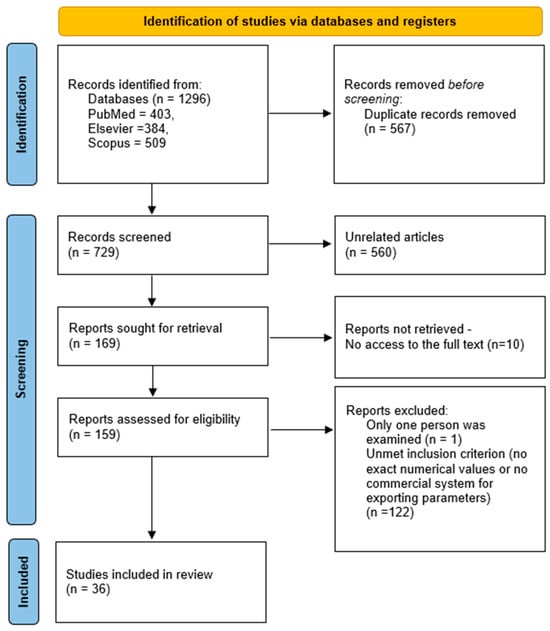
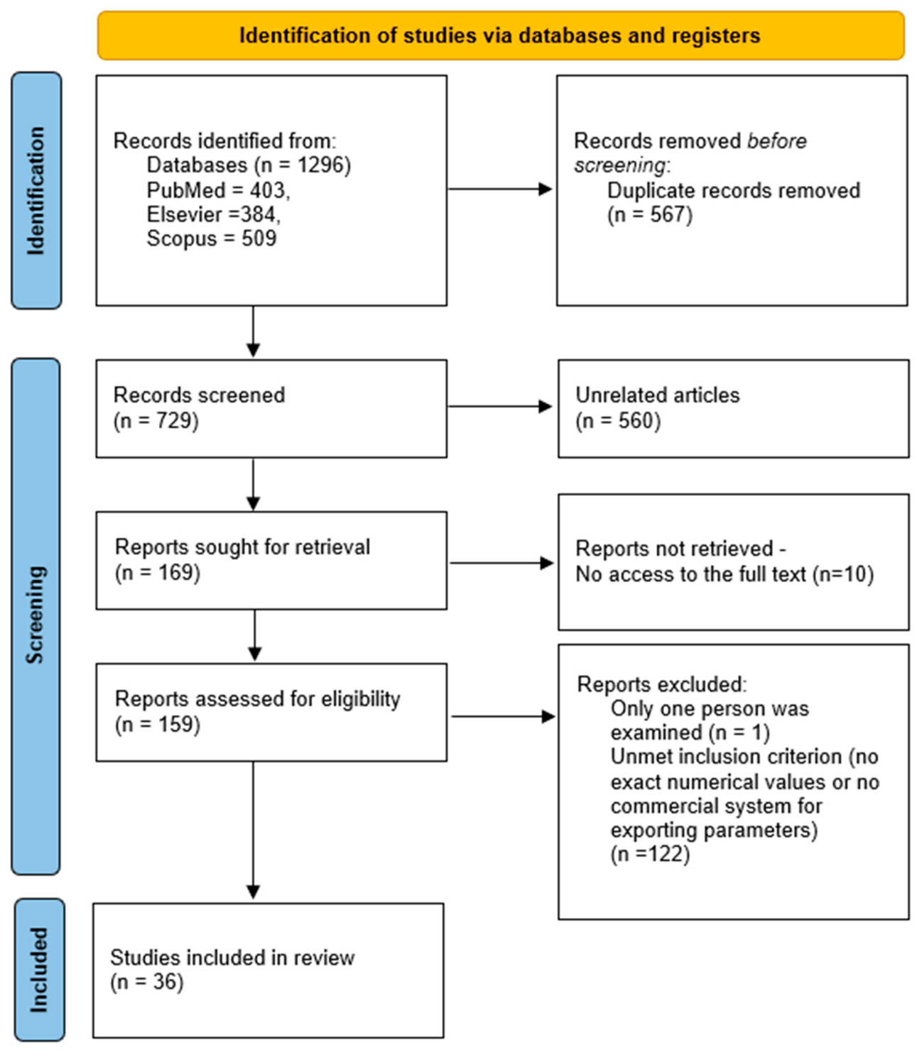
The search results were exported from the databases and imported into Rayyan, where they were merged. Duplicates were initially identified using Rayyan’s built-in algorithm, which relies on bibliographic metadata (including title, authors, publication year, journal, and DOI). Additionally, records with a high degree of similarity (threshold > 0.90) were flagged to identify potential duplicates that may not have been detected automatically. All identified records were then manually verified by the researchers, and the decision to remove a duplicate was made on a case-by-case basis following individual assessment. This approach was intended to minimize the risk of incorrectly excluding relevant publications. In the next stage, the titles and abstracts were screened to identify potentially eligible publications. Screening was conducted independently by two reviewers, including a preliminary assessment of eligibility criteria, such as the availability of numerical data and the use of commercially available systems equipped with IMUs for detecting TUG test phases. Figure 1 presents subsequent steps in the search for articles, along with the number of articles which comply with the assumed criteria.
Figure 1.
PRISMA flow diagram for the systematic review.
2.2. Elegibility Criteria
The studies were selected using the PICOS criteria. The inclusion and exclusion criteria are presented in Table 1. The authors included only cross-sectional studies in which the TUG test was performed using commercially available IMU-based systems enabling the automatic detection of individual test phases and the derivation of their parameters. Additionally, the criterion was restricted to articles reporting parameter values as means and standard deviations, which allowed for comparison and tabular presentation of the results. Two reviewers independently extracted the data, and any discrepancies were identified and resolved through discussion.
Table 1.
Inclusion and exclusion criteria for articles included in the review.
2.3. Quality Assessment
All studies included in this systematic review were cross-sectional in design. Therefore, the Joanna Briggs Institute (JBI) Critical Appraisal Checklist for Analytical Cross-Sectional Studies was selected as the most appropriate tool for methodological quality assessment [22]. The assessment was performed by two independent reviewers. The checklist comprised eight questions and assessed whether the article clearly defined the inclusion criteria, the quality of the population description, the reliability and standardization of measurements, and the analysis of results. Each item of the JBI checklist was rated as “Yes,” “No,” or “Unclear.” For descriptive purposes, studies meeting 7–8 criteria were considered to have a low risk of bias, those meeting 4–6 criteria a moderate risk, and those meeting ≤3 criteria a high risk of bias. Disagreements between reviewers were resolved through discussion and consensus.
2.4. Risk of Bias Across Studies
Risk of bias across studies, including potential publication bias, was assessed qualitatively. Due to substantial clinical and methodological heterogeneity, a formal quantitative assessment (e.g., funnel plot or Egger’s test) was not performed. The assessment considered factors such as selective reporting and the potential overrepresentation of statistically significant findings.
2.5. Data Synthesis/Analysis
Due to the heterogeneity of the analyzed clinical populations and the limited number of studies addressing certain disease entities, a comparative synthesis of reported iTUG parameters was performed only for disease groups represented by at least four independent studies. Conditions represented by a smaller number of publications were described narratively, without conducting a detailed comparative analysis.
To focus on the most frequently analyzed parameters, only parameters reported in at least four different articles were selected for analysis. Additionally, articles that did not report the parameters included in a given table were not included in that table.
Due to substantial heterogeneity in study protocols, sensor configurations, and reported outcome measures, a quantitative meta-analysis was not feasible. Therefore, a qualitative synthesis of the results was performed.
3. Results
Following the literature review, 36 articles meeting the predefined PICOS criteria were included in the analysis. During the selection stage, 123 articles that did not meet the predefined inclusion criteria were excluded. The reasons for exclusion included studies conducted in children populations, the lack of analysis of quantitative iTUG parameters, and the use of nonstandard or noncommercial measurement systems. The detailed study selection process is presented in the PRISMA flow diagram (Figure 1).
3.1. Methodological Quality Results
The methodological quality of the included studies was assessed using the JBI Checklist for Analytical Cross-Sectional Studies. The results of the assessment are presented in Table 2.
Table 2.
Quality assessment scores for the included studies.
Among the 36 included articles, 2 were classified as having a low risk of bias, while 34 were classified as having a moderate risk of bias. None of the included studies were classified as having a high risk of bias.
Due to the criterion of at least four studies per disease entity, which enabled the determination of the range of reported parameter values, 21 articles were included in the subsequent comparative analysis.
In two of the 21 studies, the analysis also included other clinical populations that did not meet the criterion of at least four studies. They were presented in the tables; however, only the results for Parkinson’s disease (PD) were included in the comparative analysis, while the remaining ones were presented in the Other Populations section.
The remaining 15 articles, concerning populations represented by fewer than four studies, were described narratively in the section Other Clinical Populations (<4 studies).
The majority of included studies were classified as having a moderate risk of bias, primarily due to insufficient reporting of confounding factors and limited information on measurement reliability. Only a small number of studies fulfilled all quality criteria, indicating an overall moderate methodological quality of the available evidence.
3.2. Characteristics of Included Studies
The included studies analyzed various patient groups. Table 3 includes only those articles concerning study groups represented in at least four studies, which allowed for the presentation of the range of reported parameter values. Based on the conducted analysis, the most frequently analyzed populations in the selected group of articles were healthy adults [23,24,25,26,27], older adults [9,27,28,29,30,31], patients with multiple sclerosis (MS) [16,32,33,34,35] and patients with Parkinson’s disease (PD) [13,18,36,37,38].
Table 3.
iTUG measurement methodology and study group characteristics (* Study also included patients with fragile X-associated tremor/ataxia syndrome (FXTAS); results for FXTAS are described in Section 3.6), (HC—healthy control group, PD—Parkinson’s disease, MS—multiple sclerosis, F—female, M—male, EDSS—Expanded Disability Status Scale, N/A—this is not a number, no information available).
The walking phase was most commonly performed over a distance of 3 m [9,16,23,26,28,29,30,31,32,33,35,36,37], although distances of 6 m [18], 3.5 m [24] and 7 m [13,25,34,38] were also used.
In many articles, systems such as G-Walk [11,26,27,28,29,30,31,32,33,37], Mobility Lab [9,13,18,24,25,34,35,36,38], Noraxon [16,23] are used to segment the TUG test into periods (phases). Detailed information on the study groups, the systems used, and their placement is presented in Table 3.
3.3. Duration of the Entire Test and TUG Test Phases
In the analyzed articles, the total iTUG duration was most commonly examined. However, when considering the total duration or the duration of the walking phase, the measurement methodology must be taken into account, that is, how the participants were instructed. In most cases, participants moved at a natural, comfortable pace; however, some studies instructed participants to “walk as fast as”—TUG fast [28]. An additional factor that may affect the obtained temporal values is variation in the test protocol, particularly the length of the walking distance used in the TUG test.
In addition to the total test duration, some authors also analyzed the durations of individual iTUG phases, such as the sit-to-stand, walking, and turning. It should be noted, however, that not all authors reported the same test phases, which makes direct comparison of results across studies difficult. The reported values are presented in Table 4.
Table 4.
Duration of iTUG and its subphases (* Study also included patients with fragile X-associated tremor/ataxia syndrome (FXTAS); results for FXTAS are described in Section 3.6, CI-YO—Cognitively impaired young-old, CI-OO—Cognitively impaired old-old, HC-YO—Healthy controls young-old, NAD—iTUG without ADs, CAD—iTUG with a single-tip cane, WAD—iTUG with a four-wheeled walker).
3.4. Gait Parameters
Table 5 presents the reported spatiotemporal parameters obtained during the walking phase of the iTUG. The omitted articles did not analyze the turning phases. The table also presents parameters that were most frequently reported by the authors.
Table 5.
Spatiotemporal parameters obtained during the walking phase of the iTUG (* Study also included patients with fragile X-associated tremor/ataxia syndrome (FXTAS); results for FXTAS are described in Section 3.6, CI-YO—Cognitively impaired young-old, CI-OO—Cognitively impaired old-old, HC-YO—Healthy controls young-old, NAD—iTUG without ADs, CAD—iTUG with a single-tip cane, WAD—iTUG with a four-wheeled walker).
Gait during the iTUG test was analyzed in 9 articles. The most frequently reported parameter was cadence, followed by double support, stance time, swing time, walking speed, stride length, and walking cycle time.
3.5. Turning Parameters
The turning phase is one of the more demanding components of the iTUG test, as it requires simultaneous control, coordination, and a change in direction. Table 6 presents the most frequently reported parameters of the turning phase in the iTUG test. The articles omitted from the table did not analyze the turning phases.
Table 6.
Turning parameters in the iTUG test (* Study also included patients with fragile X-associated tremor/ataxia syndrome (FXTAS); results for FXTAS are described in Section 3, (1) NAD—iTUG without ADs, (2) CAD—iTUG with a single-tip cane, WAD—iTUG with a four-wheeled walker.
Overall, the turning phases were analyzed in 13 articles. The most frequently reported parameters were the maximum velocity of the second turning, followed by the mean velocity of the second turning, the mean and maximum velocity of turning 1, and the mean velocity of turnings 1 and 2.
3.6. Other Clinical Populations (<4 Studies)
Several additional clinical populations were identified in the reviewed studies; however, they were represented by fewer than four publications. Due to the limited number of studies, these conditions were not included in the comparative synthesis of parameter ranges and are instead presented below in a qualitative description.
The aim of Buraschi et al. [49] was to analyze temporal and kinematic parameters in the iTUG. The authors conducted a study on a group of older adults with a mean age of approximately 61 years with low back pain (LBP), along with an age-matched healthy control group. They found significant differences in the following parameters: total TUG duration, sit-to-stand duration, forward walking phase, return walking phase, final turning and stand-to-sit phase, mean velocity of turning 1, and accelerations during sit-to-stand and stand-to-sit phases. The authors concluded that IMUs are a valuable tool for assessing parameters derived from the TUG test and that their use in LBP provides valuable information enabling objective analysis. In turn, the aim of Celletti et al. [14] was to assess therapy for the treatment of the lower spine in older adults. Participants underwent the iTUG test before and after therapy consisting of 10 h of sessions scheduled three times per week, during which they performed various muscle-strengthening exercises. Using this test, they concluded that the training program is a promising new rehabilitative treatment for LBP in improving motor function and reducing functional limitations. The results obtained by the authors indicate significant pre- to post-intervention differences in parameters such as total test duration, sit-to-stand duration, and turning velocity.
There are also studies that investigate differences in iTUG performance among patients after stroke. For example, Spina et al. [41] reported significant differences in iTUG phases and turning velocities between patients after stroke and healthy control participants, suggesting that turning velocity during the Timed Up and Go test may be a sensitive indicator of mobility impairment after stroke. In another study, Tajuddin et al. [45] examined different task conditions (single task, dual motor task, and dual cognitive task) and found that dual-task conditions had a significant effect on gait parameters and increased total test duration in the iTUG, particularly during the dual cognitive task.
The literature includes a study by Winiarski et al. [42] that aimed to determine the long-term effects of COVID-19 on gait and the Timed Up and Go test. The study was conducted in a group of individuals who had been hospitalized due to COVID-19 and for whom more than 8 weeks had elapsed since discharge. The authors reported significant differences in the following parameters: total TUG duration, sit-to-stand duration, walking cycle time, mean speed, mean walking speed, and walking cadence, and observed differences in range of motion compared with individuals who had not been infected with COVID-19.
Assessments using iTUG were conducted by Cimolin et al. [44] in individuals with obesity, with a mean BMI of 41.1 ± 7.9 kg/m2. The authors reported differences in parameters such as total TUG duration, walking phase duration, turning 1 and 2 durations, and turning velocities. The obese group also exhibited increased maximum extension. At the same time, the authors concluded that iTUG is an objective and rapid mobility test and confirmed that it provides valuable information in clinical practice.
Arippa et al. [46] conducted iTUG assessments in individuals who had experienced severe traumatic brain injury. The aim of this study was to quantitatively assess the extent of motor deficits under both static and dynamic conditions. In assessing postural stability, the authors found a significant difference in sway area, and in the TUG test, in total walking time. They concluded that the measurements constitute a reliable tool and that their integration with clinical data is highly important. Prasad et al. [53] conducted similar studies in individuals after traumatic brain injury. The results showed that participants had reduced ankle activity compared with healthy individuals.
Familiari et al. [48] conducted a study to determine whether a shoulder abduction brace after arthroscopic rotator cuff repair affects gait parameters and functional mobility. The authors demonstrated that the brace affects walking speed after surgery, whereas no long-term effect was observed (one week after brace removal).
Monaghan et al. [43] and Mostile et al. [50] conducted iTUG studies in groups with essential tremor (ET). Based on both studies, it can be concluded that the iTUG test may provide valuable information.
Studies have also been conducted in individuals with facioscapulohumeral muscular dystrophy (FSHD). Statland et al. [52] reported that iTUG duration remained unchanged, whereas step length, step velocity, and trunk range of motion changed. Huisinga et al. [39] described motion capture systems as an appealing option for FSHD clinical trials.
Celletti et al. conducted a study in individuals with cervical dystonia [40]. The results were characterized by a significantly longer duration in patients with cervical dystonia compared with the control group, whereas no differences in flexion and extension angular amplitudes were observed. The findings indicate impairment in postural control during walking and postural changes.
In two studies, patients with fragile X–associated tremor/ataxia syndrome (FXTAS) were examined, where one study also analyzed patients with Parkinson’s disease (PD). Due to the limited number of studies, results for FXTAS were not included in the comparative synthesis of parameter ranges. Robertson-Dick et al. [38] conducted a study in groups with FXTAS, Parkinson’s disease (PD), and essential tremor (ET). Based on the results, the authors concluded that patients with FXTAS and ET exhibit different walking profiles than patients with PD. In turn, O’Keefe [47] conducted a study in patients with FXTAS with and without the premutation of the fragile X mental retardation 1 (FMR1) gene. PM carriers with FXTAS were globally impaired across all gait performance domains. Carriers without FXTAS did not differ from controls in any parameters, although double-limb support time was close to significance.
The literature includes a study in which patients with idiopathic normal pressure hydrocephalus (iNPH) and patients with Parkinson’s disease (PD) were examined (results for PD patients are presented in earlier tables) [37]. The authors of the study showed that patients with iNPH-P exhibited specific impairments in motor performance compared with untreated patients with PD, particularly during the transition from the turning phase to the stand-to-sit phase.
4. Discussion
The instrumented Timed Up and Go (iTUG) test is increasingly used as an objective tool for the assessment of mobility, walking, and balance in various clinical populations [16,29,35,36]. The aim of this review was to compile a set of iTUG parameters and to identify the parameters most commonly analyzed in studies involving different study populations. The analysis revealed differences between studies that included not only the measurement systems used, but also variations in TUG protocol methodology and participants at different stages of disease.
The selected articles were assessed using the JBI Checklist for Analytical Cross-Sectional Studies. Only 5.56% of the articles were rated as low risk of bias, whereas 94.44% were classified as moderate risk of bias.
A detailed analysis was conducted for articles concerning clinical populations represented at least four times. In total, the analyzed studies included 1268 participants: 192 healthy adults [23,24,25,26,27], 514 older adults [9,27,28,29,30,31], 230 patients with multiple sclerosis [16,32,33,34,35], and 332 patients with PD [13,18,36,37,38]. The substantial predominance of patients with neurological conditions may indicate growing interest in the use of the iTUG test to assess mobility in this group.
In most of the analyzed articles, iTUG assessments were conducted in accordance with the recommendations of Podsiadlo [8]. However, in some studies, modifications to the protocol were introduced, such as changes in walking distance or instructions regarding movement speed. In four articles, the walking distance was increased from 3 m to 7 m [13,25,34,38], and in one article to 6 m [18], which may be advantageous due to the recording of a greater number of step cycles. This enables a more precise analysis of gait while making direct comparisons between studies more difficult. Importantly, differences in walking distance do not only increase the amount of collected data but may also systematically influence the relative contribution of individual test phases. Longer walking distances may reduce the proportional impact of transitional phases (e.g., sit-to-stand), while amplifying steady-state gait characteristics, which may lead to inconsistencies when comparing studies using different protocols.
Another important methodological difference between studies was the use of different measurement systems, consisting of varying numbers of IMU sensors and their differing placement. The systems used included G-Walk [11,26,27,28,29,30,31,32,33,37], Mobility Lab [9,13,18,24,25,34,35,36,38], and Noraxon [16,23].
It is worth noting that these systems differ substantially in terms of the number of sensors used and the scope of recorded data. For example, the G-Walk system is based on a single sensor placed in the pelvic region, whereas systems such as Noraxon allow the use of up to between 10 and 20 IMU sensors, enabling a more detailed analysis of movement kinematics. These differences may affect the range and accuracy of the derived parameters, thereby complicating direct comparisons between studies. In addition to hardware-related differences, variations in data processing approaches and proprietary algorithms used for signal segmentation and phase detection may further contribute to inconsistencies between studies.
Despite the wide range of analyzed parameters, a lack of consistency in their reporting across studies can be observed. Many parameters were analyzed only in selected populations or in single studies, which substantially limits the possibility of direct comparison. Importantly, the reported iTUG parameters differ in their sensitivity to methodological heterogeneity. Temporal parameters, such as total test duration and phase durations, appear relatively robust across different sensor configurations and test protocols, whereas spatiotemporal and turning-related parameters are more strongly influenced by methodological factors.
Based on the synthesis of the included studies, iTUG parameters can be broadly categorized according to their sensitivity to methodological variability. Temporal parameters demonstrate relatively high robustness, as they are consistently reported and less dependent on sensor configuration or algorithmic processing. In contrast, spatiotemporal gait parameters (e.g., cadence, gait speed) and turning-related parameters (e.g., angular velocity, peak turning velocity) appear to be more susceptible to methodological factors, including sensor placement, sampling frequency, and signal processing techniques.
This distinction is critical for interpreting the literature, as parameters that are highly protocol-dependent may reflect methodological artifacts rather than true differences between populations. Consequently, caution is required when comparing such parameters across studies or when attempting to establish normative ranges.
In many of the analyzed studies, attention was focused on parameters related to the duration of each iTUG phase [9,11,13,16,23,24,27,29,30,31,32,33,34,35,36,37,38]. The most frequently analyzed parameters include total test duration [9,11,16,23,24,27,28,29,30,31,32,33,35], as well as durations of sit-to-stand [9,11,16,23,24,27,28,29,30,31,32,33,35,36,37,38], turning 1 [11,16,23,24,27,28,30,31,32,33,35], and turning 2 [11,16,23,27,28,30,31,32,33,35,38]. In contrast, among spatiotemporal parameters, cadence was the most frequently analyzed parameter [13,16,23,24,25,27,34,36,37]. Spatiotemporal parameters of gait such as step length [25,36,37] or walking cycle time [34,37], were reported less frequently and less consistently across studies. In turn, among turning-related parameters, the most frequently reported parameter was maximum turning velocity during turning 2 [9,18,23,28,33,37,38].
The predominance of temporal parameters may be related to their relatively simple derivation and high interpretability.
Interest in turning-related parameters may result from their high sensitivity in assessing mobility impairments [12,16,54]. The turning phase consists of several components that require coordination, balance, and postural control. For this reason, this phase is considered more susceptible to impairments and is regarded as a reliable indicator of dynamic balance [12]. At the same time, the observed variability in the reporting of turning-related parameters, such as mean velocity, maximum velocity, or their averaged values, further indicates a lack of standardization in iTUG analysis.
From a clinical perspective, not all of the iTUG parameters that are frequently reported provide the same level of diagnostic information. While many parameters are described in the literature, only a subset appears to have consistent clinical relevance across different populations.
Temporal parameters, particularly total test duration and phase durations, appear to be the most clinically robust and practical indicators of overall mobility impairment [11,16,31,35]. Their widespread use, ease of interpretation make them suitable for routine clinical application.
In contrast, turning-related parameters-especially peak or maximum turning velocity-emerge as particularly sensitive indicators of dynamic balance [12]. These parameters appear to be especially valuable in neurological populations, such as patients with Parkinson’s disease [18,38] or multiple sclerosis [16,35], where impairments in turning are often among the earliest and most pronounced functional deficits.
On the other hand, spatiotemporal gait parameters derived from iTUG, such as stride length [36], gait cycle time [34], or even cadence [16,27,34], although potentially informative, appear to have more limited clinical applicability within the iTUG framework. This is primarily due to their lower reporting consistency, higher sensitivity to methodological variability, and the fact that they can be more accurately assessed using dedicated gait analysis protocols. In addition, it should be noted that in the TUG test, gait is typically analyzed over a 3 m distance, which may be too short a distance to assess gait parameters at the preferred walking speed.
In summary, the results of this review indicate the clinical utility of the individual parameters of the iTUG test. The most important components are the overall time indices, followed by the time parameters for individual phases and the indices related to the performance of turns.
The extensive tabular summaries presented in this review provide a comprehensive overview of reported parameter values; however, they should be interpreted with caution. Due to the methodological heterogeneity described above, these tables represent an aggregation of heterogeneous datasets rather than directly comparable measurements.
Therefore, the reported parameter ranges should not be interpreted as normative reference values, but rather as indicative of the variability present in the current literature. Part of the observed variability may reflect methodological differences rather than true clinical variability.
These observations are supported by findings from studies involving various clinical populations. Significant differences in turning duration were observed in patients with multiple sclerosis in studies by Szaflik et al. [16] and Hershkovitz et al. [35]. Similar results were also obtained in patients with PD, where Dewey et al. [18] and Robertson-Dick et al. [38] reported significant differences in turning duration compared with the control group. Similar observations regarding turning parameters were also reported by Mulas et al. [27] in studies involving older adults.
For parameters such as total test duration in patients with multiple sclerosis, Hershkovitz et al. [35] reported significant differences between patients and healthy individuals. However, Szaflik et al. [16] and Pau et al. [32] did not confirm these differences.
A similar inconsistency in results in patients with multiple sclerosis was observed for the sit-to-stand phase, with significant differences reported by Szaflik et al. [16], whereas Pau [32] and Hershkovitz et al. [35] did not observe such differences. For the stand-to-sit phase, none of the authors reported significant differences [16,32,35]. Similarly, for PD, no significant differences were found for the sit-to-stand and stand-to-sit phases. These observations may suggest that turning-related parameters may represent more sensitive and reliable indicators of mobility impairments than traditional total durations or sit-to-stand and stand-to-sit phases, particularly in clinical populations with varying levels of dysfunction.
The frequency of analysis of individual parameters presented in Table 7 enables the identification of the parameters most commonly used in studies involving specific clinical populations. In patients with PD, the most frequently analyzed parameters included total test duration [13,36,37,38], sit-to-stand duration [36,37,38], cadence [13,36,37], double support [13,36,37] and turning 2 peak/max velocity [18,37,38]. In studies involving multiple sclerosis, the most frequently analyzed parameter is total test duration [11,16,32,33,34,35] along with subphase durations, specifically durations of sit-to-stand [11,16,32,33,35], turning 1 [11,16,32,33,35], turning 2 [11,16,32,33,35] and stand-to-sit phases [11,16,32,35]. In older adults, the most frequently analyzed parameters included total test duration [9,27,29,30,31], sit-to-stand duration [9,27,28,29,30,31], turning 1 [27,28,30,31] and turning 2 durations [27,28,30,31] and the duration of stand-to-sit phase [27,28,30,31]. In turn, in studies involving healthy adults, analyses involved cadence [23,24,25], total test time [23,24], sit-to-stand [23,24], turning 1 [23,24] and stand-to-sit [23,24] durations.
Table 7.
List of articles reporting analyzed iTUG parameters by study groups (populations).
4.1. Limitations and Directions of Further Research
A limitation of this literature review is the lack of methodological uniformity, including the use of different equipment and sensor placements, as well as variability in iTUG protocol across studies. The review involved different walking speeds and distances. There is considerable variability in the reported parameters across studies, indicating a lack of standardization in iTUG analysis and limiting the possibility of establishing reference values across different clinical populations.
Moreover, the heterogeneity of measurement protocols may have influenced not only the variability of reported values but also the detectability of between-group differences, which should be taken into account when interpreting the findings of this review. Another limitation of the study was the exclusive use of IMU sensors, without the use of measurement mats or other systems.
A quantitative meta-analysis was not performed due to substantial heterogeneity in study populations, protocols, sensor configurations, and outcome definitions. In addition, many parameters were reported in a limited number of studies or were not consistently defined, which precluded meaningful pooling of results.
Future studies should aim to develop recommendations for a standardized iTUG protocol including a uniform distance and set of analyzed parameters.
In addition, future research should aim to establish consensus guidelines for iTUG assessment, including standardized protocols and reporting, to improve reproducibility and facilitate cross-study comparisons.
Finally, the possibility of publication bias cannot be excluded, as studies reporting statistically significant or clinically relevant findings are more likely to be published, which may have influenced the overall conclusions of this review.
4.2. Clinical Implications and Practical Applications
The compiled set of parameters for different conditions at various stages may assist physicians in interpreting iTUG results and in identifying movement impairments characteristic of specific disease entities.
The use of iTUG enables the identification of subtle movement impairments that may not be detectable in standard clinical assessment. The iTUG parameters can be used not only to assess functional status, but also to monitor disease progression and the effects of therapeutic interventions.
The results indicate a need to standardize the iTUG protocol, particularly with regard to participant instructions, number of trials, walking distance, and the analyzed test phases. Standardization of measurement procedures could improve the comparability of results across studies and enhance their clinical utility.
5. Conclusions
The analysis revealed substantial methodological heterogeneity across studies, including differences in sensor placement, walking distance, and reported parameters, which limits the possibility of direct comparison of results. The observed variability indicates the dynamic development of the iTUG test and its adaptation to new measurement technologies.
Turning-related parameters in the iTUG test demonstrate a greater ability to detect mobility impairments than traditional temporal measures, such as total test duration or the sit-to-stand and stand-to-sit phases. The differences observed in results for populations with multiple sclerosis, PD, and older adults highlight the importance of the turning phase as a reliable component of motor function assessment.
The results indicate that the analysis of iTUG parameters may serve as a valuable tool to support mobility assessment, as well as the monitoring of functional status and progression of movement impairments across different patient populations.
Supplementary Materials
The following supporting information can be downloaded at: https://www.mdpi.com/article/10.3390/jcm15093307/s1, Supplementary Files: PRISMA 2020 Checklist; PRISMA 2020 Abstract Checklist [55].
Author Contributions
Conceptualization, P.S. and K.N.-L.; methodology, P.S. and K.N.-L.; formal analysis, P.S. and K.N.-L.; data curation, P.S. and K.N.-L.; writing—original draft preparation, P.S.; writing—review and editing, K.N.-L.; visualization, P.S.; supervision, K.N.-L.; All authors have read and agreed to the published version of the manuscript.
Funding
The Article Processing Charge was financed under the European Funds for Silesia 2021–2027 Program, co-financed by the Just Transition Fund project entitled “Development of the Silesian biomedical engineering potential in the face of the challenges of the digital and green economy (BioMeDiG)” (project number: FESL.10.25-IZ.01-07G5/23).
Institutional Review Board Statement
Not applicable.
Informed Consent Statement
Not applicable.
Data Availability Statement
The original contributions presented in this study are included in the article. Further inquiries can be directed to the corresponding author.
Conflicts of Interest
The funders had no role in the design of the study; in the collection, analyses, or interpretation of data; in the writing of the manuscript; or in the decision to publish the results.
Abbreviations
The following abbreviations are used in this manuscript:
| IMU | Inertial measurement units |
| TUG | Timed Up and Go |
| iTUG | Instrumented Timed Up and Go |
| MS | Multiple sclerosis |
| PD | Parkinson’s disease |
References
- Adesida, Y.; Papi, E.; McGregor, A.H. Exploring the Role of Wearable Technology in Sport Kinematics and Kinetics: A Systematic Review. Sensors 2019, 19, 1597. [Google Scholar] [CrossRef] [Scilit]
- Boyer, K.A.; Hafer, J.F. Gait Mechanics Contribute to Exercise Induced Pain Flares in Knee Osteoarthritis. BMC Musculoskelet. Disord. 2019, 20, 107. [Google Scholar] [CrossRef] [Scilit]
- Hafer, J.F.; Vitali, R.; Gurchiek, R.; Curtze, C.; Shull, P.; Cain, S.M. Challenges and Advances in the Use of Wearable Sensors for Lower Extremity Biomechanics. J. Biomech. 2023, 157, 111714. [Google Scholar] [CrossRef] [Scilit]
- Santos, G.; Wanderley, M.; Tavares, T.; Rocha, A. A Multi-Sensor Human Gait Dataset Captured through an Optical System and Inertial Measurement Units. Sci. Data 2022, 9, 545. [Google Scholar] [CrossRef] [Scilit]
- Adlou, B.; Wilburn, C.; Weimar, W. Motion Capture Technologies for Athletic Performance Enhancement and Injury Risk Assessment: A Review for Multi-Sport Organizations. Sensors 2025, 25, 4384. [Google Scholar] [CrossRef] [Scilit]
- Mathis, A.; Mamidanna, P.; Cury, K.M.; Abe, T.; Murthy, V.N.; Mathis, M.W.; Bethge, M. DeepLabCut: Markerless Pose Estimation of User-Defined Body Parts with Deep Learning. Nat. Neurosci. 2018, 21, 1281–1289. [Google Scholar] [CrossRef] [Scilit]
- Mancini, M.; Horak, F.B.; Zampieri, C.; Carlson-Kuhta, P.; Nutt, J.G.; Chiari, L. Trunk Accelerometry Reveals Postural Instability in Untreated Parkinson’s Disease. Park. Relat. Disord. 2011, 17, 557–562. [Google Scholar] [CrossRef] [Scilit]
- Podsiadlo, D.; Richardson, S. The Timed “Up & Go”: A Test of Basic Functional Mobility for Frail Elderly Persons. J. Am. Geriatr. Soc. 1991, 39, 142–148. [Google Scholar] [CrossRef] [Scilit]
- Seo, J.; Kim, T.; Lee, J.; Kim, J.; Choi, J.; Tack, G. Fall Prediction of the Elderly with a Logistic Regression Model Based on Instrumented Timed up & Go. J. Mech. Sci. Technol. 2019, 33, 3813–3818. [Google Scholar] [CrossRef] [Scilit]
- Ortega-Bastidas, P.; Gómez, B.; Aqueveque, P.; Luarte-Martínez, S.; Cano-de-la-Cuerda, R. Instrumented Timed Up and Go Test (ITUG)—More Than Assessing Time to Predict Falls: A Systematic Review. Sensors 2023, 23, 3426. [Google Scholar] [CrossRef] [Scilit]
- Pau, M.; Porta, M.; Coghe, G.; Corona, F.; Pilloni, G.; Lorefice, L.; Marrosu, M.G.; Cocco, E. Are Static and Functional Balance Abilities Related in Individuals with Multiple Sclerosis? Mult. Scler. Relat. Disord. 2017, 15, 1–6. [Google Scholar] [CrossRef] [Scilit] [PubMed]
- Picardi, M.; Redaelli, V.; Antoniotti, P.; Pintavalle, G.; Aristidou, E.; Sterpi, I.; Meloni, M.; Corbo, M.; Caronni, A. Turning and Sit-to-Walk Measures from the Instrumented Timed Up and Go Test Return Valid and Responsive Measures of Dynamic Balance in Parkinson’s Disease. Clin. Biomech. 2020, 80, 105177. [Google Scholar] [CrossRef] [Scilit]
- Atterbury, E.M.; Welman, K.E. Balance Training in Individuals with Parkinson’s Disease: Therapist-Supervised vs. Home-Based Exercise Programme. Gait Posture 2017, 55, 138–144. [Google Scholar] [CrossRef] [Scilit]
- Celletti, C.; Mollica, R.; Ferrario, C.; Galli, M.; Camerota, F. Functional Evaluation Using Inertial Measurement of Back School Therapy in Lower Back Pain. Sensors 2020, 20, 531. [Google Scholar] [CrossRef] [Scilit]
- Frenken, T.; Lohmann, O.; Frenken, M.; Steen, E.E.; Hein, A. Performing Gait Analysis within the Timed up & Go Assessment Test: Comparison of ATUG to a Marker-Based Tracking System. Inform. Health Soc. Care 2014, 39, 232–248. [Google Scholar] [CrossRef] [Scilit] [PubMed]
- Szaflik, P.; Kaczmarczyk, A.; Zadoń, H.; Szefler-Derela, J.; Wasiuk-Zowada, D.; Nowakowska-Lipiec, K.; Michnik, R.; Siuda, J. Instrumented Timed Up and Go Test as a Tool to Early Detection of Gait and Functional Mobility Impairments in Multiple Sclerosis. J. Clin. Med. 2026, 15, 679. [Google Scholar] [CrossRef] [Scilit]
- Pino-Ortega, J.; Carvajal-Espinoza, R.; Becerra-Patiño, B.A. Evaluation of Hand Muscle Strength Using Manual Dynamometry: A Reliability and Validity Study of the Activ5 Instrument. Appl. Sci. 2024, 14, 8775. [Google Scholar] [CrossRef] [Scilit]
- Dewey, D.C.; Miocinovic, S.; Bernstein, I.; Khemani, P.; Querry, R.; Chitnis, S.; Dewey, R.B. Automated Gait and Balance Parameters Diagnose and Correlate with Severity in Parkinson Disease. J. Neurol. Sci. 2014, 345, 131. [Google Scholar] [CrossRef] [Scilit]
- Madgwick, S.O.H.; Harrison, A.J.L.; Vaidyanathan, R. Estimation of IMU and MARG Orientation Using a Gradient Descent Algorithm. In Proceedings of the 2011 IEEE International Conference on Rehabilitation Robotics, Rehab Week Zurich, Zurich, Switzerland, 29 June–1 July 2011; ACT Profile Report: State. Graduating Class 2012. Montana; IEEE: New York, NJ, USA, 2011; pp. 1–7. [Google Scholar]
- Silva, C.A.; Casilari, E.; García-Bermúdez, R. UMATUG: A Dataset of Inertial Signals of Older and Young Adults Using a Gerontologic Simulator Collected during Instrumented Timed Up and Go (ITUG) Tests. Data Brief 2024, 55, 110621. [Google Scholar] [CrossRef] [Scilit]
- Liberati, A.; Altman, D.G.; Tetzlaff, J.; Mulrow, C.; Gøtzsche, P.C.; Ioannidis, J.P.A.; Clarke, M.; Devereaux, P.J.; Kleijnen, J.; Moher, D. The PRISMA Statement for Reporting Systematic Reviews and Meta-Analyses of Studies That Evaluate Health Care Interventions: Explanation and Elaboration. J. Clin. Epidemiol. 2009, 62, e1–e34. [Google Scholar] [CrossRef] [Scilit]
- JBI Critical Appraisal Tools|JBI. Available online: https://jbi.global/critical-appraisal-tools (accessed on 27 March 2026).
- Kowal, M.; Winiarski, S.; Morgiel, E.; Madej, M.; Proc, K.; Madziarski, M.; Wedel, N.; Sebastian, A. Functional Mobility Studies in Younger Adults: Instrumented Timed Up and Go (ITUG) Test Using Inertial Devices. J. Clin. Med. 2025, 14, 1944. [Google Scholar] [CrossRef] [Scilit]
- Coulthard, J.T.; Treen, T.T.; Oates, A.R.; Lanovaz, J.L. Evaluation of an Inertial Sensor System for Analysis of Timed-up-and-Go under Dual-Task Demands. Gait Posture 2015, 41, 882–887. [Google Scholar] [CrossRef] [Scilit]
- Abualait, T.S.; Alnajdi, G.K. Effects of Using Assistive Devices on the Components of the Modified Instrumented Timed up and Go Test in Healthy Subjects. Heliyon 2021, 7, e06940. [Google Scholar] [CrossRef] [Scilit]
- Santos, A.C.A.D.; Matheus, D.; Battistella, L.R. Reference Values for Instrumented Timed Up and Go—Outcomes of Vertical Accelerations and Angular Velocities in Healthy Subjects. J. Int. Soc. Phys. Rehabil. Med. 2025, 8, 1–5. [Google Scholar] [CrossRef] [Scilit]
- Mulas, I.; Putzu, V.; Asoni, G.; Viale, D.; Mameli, I.; Pau, M. Clinical Assessment of Gait and Functional Mobility in Italian Healthy and Cognitively Impaired Older Persons Using Wearable Inertial Sensors. Aging Clin. Exp. Res. 2020, 33, 1853–1864. [Google Scholar] [CrossRef] [Scilit]
- Zarzeczny, R.; Nawrat-Szołtysik, A.; Polak, A.; Maliszewski, J.; Kiełtyka, A.; Matyja, B.; Dudek, M.; Zborowska, J.; Wajdman, A. Aging Effect on the Instrumented Timed-Up-and-Go Test Variables in Nursing Home Women Aged 80–93 Years. Biogerontology 2017, 18, 651–663. [Google Scholar] [CrossRef] [Scilit] [PubMed]
- Park, J.; Cheon, S.M.; Lee, M.J.; Ryu, D.W.; Yoo, D. Comparison of the Impact of Various Exercise Modalities on Parkinson’s Disease. J. Mov. Disord. 2025, 18, 222–230. [Google Scholar] [CrossRef] [Scilit] [PubMed]
- Porta, M.; Pilloni, G.; Corona, F.; Fastame, M.C.; Hitchott, P.K.; Penna, M.P.; Pau, M. Relationships between Objectively Assessed Functional Mobility and Handgrip Strength in Healthy Older Adults. Eur. Geriatr. Med. 2018, 9, 201–209. [Google Scholar] [CrossRef] [Scilit]
- Pau, M.; Mulas, I.; Putzu, V.; Asoni, G.; Viale, D.; Mameli, I.; Allali, G. Functional Mobility in Older Women with and without Motoric Cognitive Risk Syndrome: A Quantitative Assessment Using Wearable Inertial Sensors. J. Gerontol. Geriatr. 2022, 70, 1–8. [Google Scholar] [CrossRef] [Scilit]
- Pau, M.; Casu, G.; Porta, M.; Pilloni, G.; Frau, J.; Coghe, G.; Cocco, E. Timed Up and Go in Men and Women with Multiple Sclerosis: Effect of Muscular Strength. J. Bodyw. Mov. Ther. 2020, 24, 124–130. [Google Scholar] [CrossRef] [Scilit] [PubMed]
- Lorefice, L.; Coghe, G.; Fenu, G.; Porta, M.; Pilloni, G.; Frau, J.; Corona, F.; Sechi, V.; Barracciu, M.A.; Marrosu, M.G.; et al. ‘Timed up and Go’ and Brain Atrophy: A Preliminary MRI Study to Assess Functional Mobility Performance in Multiple Sclerosis. J. Neurol. 2017, 264, 2201–2204. [Google Scholar] [CrossRef] [Scilit]
- Craig, J.J.; Bruetsch, A.P.; Lynch, S.G.; Horak, F.B.; Huisinga, J.M. Instrumented Balance and Walking Assessments in Persons with Multiple Sclerosis Show Strong Test-Retest Reliability. J. Neuroeng. Rehabil. 2017, 14, 43. [Google Scholar] [CrossRef] [Scilit]
- Hershkovitz, L.; Malcay, O.; Grinberg, Y.; Berkowitz, S.; Kalron, A. The Contribution of the Instrumented Timed-Up-and-Go Test to Detect Falls and Fear of Falling in People with Multiple Sclerosis. Mult. Scler. Relat. Disord. 2019, 27, 226–231. [Google Scholar] [CrossRef] [Scilit]
- Cakmak, O.O.; Akar, K.; Youssef, H.; Samanci, M.Y.; Ertan, S.; Vural, A. Comparative Assessment of Gait and Balance in Patients with Parkinson’s Disease and Normal Pressure Hydrocephalus. Med. Bull. Sisli Etfal Hosp. 2023, 57, 232. [Google Scholar] [CrossRef] [Scilit]
- Mostile, G.; Contrafatto, F.; Terranova, R.; Terravecchia, C.; Luca, A.; Sinitò, M.; Donzuso, G.; Cicero, C.E.; Sciacca, G.; Nicoletti, A.; et al. Turning and Sitting in Early Parkinsonism: Differences Between Idiopathic Normal Pressure Hydrocephalus Associated with Parkinsonism and Parkinson’s Disease. Mov. Disord. Clin. Pract. 2023, 10, 466–471. [Google Scholar] [CrossRef] [Scilit]
- Robertson-Dick, E.E.; Timm, E.C.; Pal, G.; Ouyang, B.; Liu, Y.; Berry-Kravis, E.; Hall, D.A.; O’Keefe, J.A. Digital Gait Markers to Potentially Distinguish Fragile X-Associated Tremor/Ataxia Syndrome, Parkinson’s Disease, and Essential Tremor. Front. Neurol. 2023, 14, 1308698. [Google Scholar] [CrossRef] [Scilit]
- Huisinga, J.; Bruetsch, A.; Mccalley, A.; Currence, M.; Herbelin, L.; Jawdat, O.; Pasnoor, M.; Dimachkie, M.; Barohn, R.; Statland, J. An Instrumented Timed up and Go in Facioscapulohumeral Muscular Dystrophy. Muscle Nerve 2018, 57, 503–506. [Google Scholar] [CrossRef] [Scilit]
- Celletti, C.; Ferrazzano, G.; Belvisi, D.; Ferrario, C.; Tarabini, M.; Baione, V.; Fabbrini, G.; Conte, A.; Galli, M.; Camerota, F. Instrumental Timed Up and Go Test Discloses Abnormalities in Patients with Cervical Dystonia. Clin. Biomech. 2021, 90, 105493. [Google Scholar] [CrossRef] [Scilit] [PubMed]
- Spina, S.; Facciorusso, S.; D’ascanio, M.C.; Morone, G.; Baricich, A.; Fiore, P.; Santamato, A. Sensor Based Assessment of Turning during Instrumented Timed Up and Go Test for Quantifying Mobility in Chronic Stroke Patients. Eur. J. Phys. Rehabil. Med. 2022, 59, 6–13. [Google Scholar] [CrossRef] [Scilit] [PubMed]
- Kowal, M.; Morgiel, E.; Winiarski, S.; Gieysztor, E.; Madej, M.; Sebastian, A.; Madziarski, M.; Wedel, N.; Proc, K.; Madziarska, K.; et al. Effect of COVID-19 on Musculoskeletal Performance in Gait and the Timed-Up and Go Test. J. Clin. Med. 2023, 12, 4184. [Google Scholar] [CrossRef] [Scilit] [PubMed]
- Monaghan, P.G.; Murrah, W.M.; Walker, H.C.; Neely, K.A.; Roper, J.A. Evaluating Postural Transition Movement Performance in Individuals with Essential Tremor via the Instrumented Timed Up and Go. Sensors 2024, 24, 2216. [Google Scholar] [CrossRef] [Scilit]
- Cimolin, V.; Cau, N.; Malchiodi Albedi, G.; Aspesi, V.; Merenda, V.; Galli, M.; Capodaglio, P. Do Wearable Sensors Add Meaningful Information to the Timed Up and Go Test? A Study on Obese Women. J. Electromyogr. Kinesiol. 2019, 44, 78–85. [Google Scholar] [CrossRef] [Scilit] [PubMed]
- Tajuddin, K.; Justine, M.; Mohd Mustafah, N.; Latif, L.; Manaf, H. Dual-Tasking Effects on Gait and Turning Performance of Stroke Survivors with Diabetic Peripheral Neuropathy. Malays. J. Med. Sci. 2021, 28, 63. [Google Scholar] [CrossRef] [Scilit]
- Arippa, F.; Pau, M.; Marcello, R.; Atzeni, L.; Simone Vullo, S.; Monticone, M. An Integrated Approach to the Assessment of Balance and Functional Mobility in Individuals with History of Severe Traumatic Brain Injury. Heliyon 2024, 10, e30665. [Google Scholar] [CrossRef] [Scilit]
- O’Keefe, J.A.; Robertson-Dick, E.E.; Hall, D.A.; Berry-Kravis, E. Gait and Functional Mobility Deficits in Fragile X-Associated Tremor/Ataxia Syndrome. Cerebellum 2016, 15, 475–482. [Google Scholar] [CrossRef] [Scilit]
- Familiari, F.; Mercurio, M.; Arenas-Miquelez, A.; Barone, A.; Greco, F.; Emerenziani, G.P.; Gasparini, G.; Galasso, O. Shoulder Brace Has No Detrimental Effect on Basic Spatio-Temporal Gait Parameters and Functional Mobility after Arthroscopic Rotator Cuff Repair. Gait Posture 2024, 107, 207–211. [Google Scholar] [CrossRef] [Scilit] [PubMed]
- Buraschi, R.; Pollet, J.; Villafañe, J.H.; Piovanelli, B.; Negrini, S. Temporal and Kinematic Analyses of Timed up and Go Test in Chronic Low Back Pain Patients. Gait Posture 2022, 96, 137–142. [Google Scholar] [CrossRef] [Scilit] [PubMed]
- Mostile, G.; Terranova, R.; Rascunà, C.; Terravecchia, C.; Cicero, C.E.; Giuliano, L.; Davì, M.; Chisari, C.; Luca, A.; Preux, P.M.; et al. Clinical-Instrumental Patterns of Neurodegeneration in Essential Tremor: A Data-Driven Approach. Park. Relat. Disord. 2021, 87, 124–129. [Google Scholar] [CrossRef] [Scilit]
- Brognara, L.; Mafla-España, M.A.; Gil-Molina, I.; Castillo-Verdejo, Y.; Cauli, O. The Effects of 3D Custom Foot Orthotics with Mechanical Plantar Stimulation in Older Individuals with Cognitive Impairment: A Pilot Study. Brain Sci. 2022, 12, 1669. [Google Scholar] [CrossRef] [Scilit]
- Statland, J.M.; Karanevich, A.; Bruetsch, A.; Huisinga, J. A Pilot Study of the Responsiveness of Wireless Motion Analysis in Facioscapulohumeral Muscular Dystrophy. Muscle Nerve 2019, 60, 590–594. [Google Scholar] [CrossRef] [Scilit]
- Tendolkar, P.A.; Pilkar, R.; Karunakaran, K.K. Evaluating Activity Levels in Individuals with TBI Using IMU. In Proceedings of the 2025 47th Annual International Conference of the IEEE Engineering in Medicine and Biology Society, Copenhagen, Denmark, 14–18 July 2025; IEEE: New York, NJ, USA, 2025; Volume 2025, pp. 1–4. [Google Scholar] [CrossRef] [Scilit]
- Salarian, A.; Horak, F.B.; Zampieri, C.; Carlson-Kuhta, P.; Nutt, J.G.; Aminian, K. ITUG, a Sensitive and Reliable Measure of Mobility. IEEE Trans. Neural Syst. Rehabil. Eng. 2010, 18, 303–310. [Google Scholar] [CrossRef] [Scilit] [PubMed]
- Page, M.J.; McKenzie, J.E.; Bossuyt, P.M.; Boutron, I.; Hoffmann, T.C.; Mulrow, C.D.; Shamseer, L.; Tetzlaff, J.M.; Akl, E.A.; Brennan, S.E.; et al. The PRISMA 2020 statement: An updated guideline for reporting systematic reviews. BMJ 2021, 372, n71. [Google Scholar] [CrossRef] [Scilit] [PubMed]

Disclaimer/Publisher’s Note: The statements, opinions and data contained in all publications are solely those of the individual author(s) and contributor(s) and not of MDPI and/or the editor(s). MDPI and/or the editor(s) disclaim responsibility for any injury to people or property resulting from any ideas, methods, instructions or products referred to in the content. |
© 2026 by the authors. Licensee MDPI, Basel, Switzerland. This article is an open access article distributed under the terms and conditions of the Creative Commons Attribution (CC BY) license.