


Abstract
Background/Objectives: Retinal vein occlusion (RVO) is a common retinal vascular disease involving local and systemic inflammation. The pan-immune inflammation value (PIV) integrates multiple circulating blood cells and can serve as an indirect indicator of systemic inflammatory activity. This study evaluated the association between PIV and RVO, comparing its discriminatory performance with other indices. Methods: This retrospective case–control study included 120 participants (60 RVO patients and 60 controls). Complete blood count (CBC) parameters were recorded, and the monocyte-to-lymphocyte ratio (MLR), platelet-to-lymphocyte ratio (PLR), neutrophil-to-lymphocyte ratio (NLR), systemic inflammation response index (SIRI), and PIV were calculated. Receiver operating characteristic (ROC) curve analysis evaluated discrimination between RVO and controls, and binary logistic regression identified factors associated with RVO. Results: Age, gender, and diabetes prevalence were similar between groups, whereas hypertension was significantly more prevalent in the RVO group (p = 0.035). Neutrophil counts, NLR, SIRI, and PIV were significantly higher in patients with RVO after FDR correction (<0.003, <0.003, =0.004, and <0.003, respectively). Areas under the ROC curve for SIRI, PIV, and NLR were 0.664, 0.678, and 0.738, respectively. In univariable logistic regression, elevated PIV was significantly associated with RVO (OR: 3.76, 95% CI: 1.7–8.2, p < 0.001). After adjustment for age, gender, hypertension, diabetes mellitus, and smoking status, continuous PIV was associated with RVO (adjusted OR per 100-unit increase: 1.41; 95% CI: 1.08–1.83; p = 0.010). Conclusions: PIV was associated with RVO after adjustment for clinical covariates, whereas its association was not statistically significant in the model containing other CBC-derived indices.
1. Introduction
Retinal vein occlusion (RVO) is one of the most common retinal vascular diseases [1,2]. The location of the occlusion determines whether RVO is classified as central RVO (CRVO) or branch RVO (BRVO). A multicenter study revealed that RVO affects roughly 17 million adults, with CRVO accounting for 17% and BRVO for 83% [2]. RVO etiology is multifactorial. Common risk factors include diabetes, systemic hypertension, hyperlipidemia, atherosclerotic vascular disease, and vasculitis. The etiology is thought to be based on Virchow’s triad: hypercoagulability, vessel wall damage, and venous stasis [3]. Local and systemic inflammation facilitate the progression of RVO by exacerbating systemic atherosclerosis and hypercoagulability [4]. Moreover, atherosclerosis, one of the main risk factors for RVO, is characterized by low-grade chronic inflammation. Recent studies suggest that inflammation caused by blood immune cell interactions has an important role in RVO development and related ischemia, macular edema, and sub-macular detachment [5,6]. RVO can result in irreversible vision loss due to retinal edema, ischemia, neovascularization, and neovascular glaucoma [1].
Peripheral blood cell counts are readily available, inexpensive laboratory measurements that have been investigated as indicators of systemic inflammation. Lymphocyte (LYM), neutrophil (NEU), monocyte (MONO), and platelet (PLT) counts, together with indices derived from these counts, have been associated with inflammatory processes [7,8]. Previous studies have examined complete blood count (CBC) parameters and CBC-derived inflammatory indices in patients with RVO [9,10,11]. These markers, such as the monocyte-to-lymphocyte ratio (MLR), the platelet-to-lymphocyte ratio (PLR), the neutrophil-to-lymphocyte ratio (NLR), and the systemic inflammation response index (SIRI) calculated from the monocyte count × NLR, were associated with systemic inflammation [12,13].
The pan-immune inflammation value (PIV) is a recently developed composite index calculated from NEU, LYM, PLT, and MONO counts. PIV may reflect aspects of systemic inflammation but does not fully characterize the underlying inflammatory state or directly assess immune cell function or activation. While this index has been documented in a number of systemic inflammatory conditions, it is a novel approach for inflammatory ocular diseases. That index is a valuable tool for the diagnosis and follow-up of inflammatory and rheumatologic diseases, acute coronary syndrome, and various sepsis cases as well as different types of cancers [14,15]. This study is the first report that evaluates PIV in RVO. The primary goal of this study was to investigate the relationship between PIV, a novel inflammatory marker, and RVO. The secondary objective was to explore the associations of PIV with subretinal fluid (SRF), ischemia, and RVO subtype.
2. Materials and Methods
2.1. Study Design and Participants
This single-center retrospective case–control study included 60 patients with RVO and 60 controls evaluated at the ophthalmology clinic of Sivas Cumhuriyet University Health Services Practice and Research Hospital. Patients diagnosed with RVO between February 2024 and November 2025 were identified from hospital records. Demographic characteristics, medical history, ophthalmological findings, and laboratory results were retrospectively extracted from the hospital database. All participants underwent a comprehensive ophthalmological examination, including best-corrected visual acuity assessment using a Snellen chart, intraocular pressure measurement, anterior segment examination, and dilated fundus examination. Snellen visual acuity values were converted to logMAR units for statistical analysis. Patients with RVO were eligible if they had a symptom duration of no more than seven days and an available CBC obtained within this interval. RVO was diagnosed based on characteristic fundus findings, including retinal venous dilation venous tortuosity, and retinal hemorrhages. The diagnosis was supported by fundus fluorescein angiography (FFA) using the CLARUS 700 (Carl Zeiss Meditec, Inc., Dublin, CA, USA) and by spectral-domain optical coherence tomography (SD-OCT) using the RTVue XR (Optovue, Inc., Fremont, CA, USA). FFA findings included delayed arteriovenous transit and hypo-fluorescence caused by blockage from retinal hemorrhages. CRVO causes hyperemia and edema in the optic disc and involves the central retinal venous circulation, whereas BRVO is confined to a branch retinal venous territory. Ischemic RVO was defined by a retinal capillary nonperfusion area of at least 10 disc areas on wide-field FFA. SRF was defined as fluid accumulation between the neurosensory retina and the retinal pigment epithelium on SD-OCT. Controls were selected from individuals attending the same ophthalmology outpatient clinic for routine eye screening during the same study period (February 2024–November 2025) who had no evidence of RVO on ophthalmological examination. Patients with RVO and controls were identified through a retrospective review of hospital records and included based on eligibility criteria and the availability of the required clinical and laboratory data.
2.2. Exclusion Criteria
Aside from diabetes mellitus and controlled hypertension, individuals with systemic disorders such as active infectious diseases, malignancies, cardiovascular diseases, and acute and chronic inflammatory diseases were excluded. Individuals with a history of oral contraceptive, anticoagulant, or nonsteroidal anti-inflammatory drug use were excluded. Similarly, individuals with a history of surgery or ocular diseases potentially associated with inflammation, including diabetic retinopathy, glaucoma, uveitis, and age-related macular degeneration, were also excluded. The same exclusion criteria were applied to patients with RVO and controls.
2.3. Blood Sampling and Calculated Inflammatory Indices
Following a 12-h fast, a sample was collected from the antecubital vein. Within three hours of collection, samples were analyzed using a Sysmex XN 3000 automated hematology analyzer (Sysmex Corporation, Kobe, Japan). In patients with RVO, CBC measurements were obtained within seven days of symptom onset and before the initiation of RVO treatment. For controls, CBC results obtained on the day of the ophthalmological examination or within the following seven days were retrieved from hospital records. The blood cell counts (PLT, LYM, MONO, and NEU) were acquired from the participants’ CBC analyses. We calculated NLR (neutrophil count/lymphocyte count), PLR (platelet count/lymphocyte count), MLR (monocyte count/lymphocyte count), SIRI (monocyte count × neutrophil count/lymphocyte count), and PIV (platelet count × neutrophil count × monocyte count/lymphocyte count). Cell counts were expressed in ×109/L.
2.4. Statistical Analysis
Sample size was calculated using G*Power 3.1 for a two-sided independent-samples t-test, assuming Cohen’s d = 0.70, α = 0.05, a target power of 95%, and equal group sizes. The minimum required sample size was 55 participants per group, corresponding to an achieved power of 95.33%. Data were analyzed using IBM SPSS Statistics software, version 27.0. The Shapiro–Wilk and Kolmogorov–Smirnov tests were used to assess the normality of the data distribution. Descriptive statistics for continuous variables were presented as mean ± standard deviation or median (interquartile range), whereas categorical variables were described using counts and percentages (%). Student’s t-test or Mann–Whitney U test was chosen to compare the continuous variables between the groups. The chi-square test was used to compare the categorical data. The Benjamini–Hochberg procedure was applied separately across the nine hematologic comparisons between patients with RVO and controls and the 27 pairwise hematologic comparisons among CRVO, BRVO, and control groups. Analyses according to RVO subtype, ischemia, and SRF status were considered exploratory and interpreted cautiously given the limited subgroup sizes. Receiver operating characteristic (ROC) analysis was used to evaluate the ability of the inflammatory indices to discriminate patients with RVO from controls. The areas under the curves (AUCs) and 95% confidence intervals were reported. Cut-off values were selected by maximizing the Youden index (sensitivity + specificity − 1). These thresholds were derived and evaluated in the same dataset and were considered exploratory. AUCs were compared using paired nonparametric tests to account for measurements obtained from the same participants. All pairwise comparisons among NLR, PLR, SIRI, and PIV were evaluated using two-sided tests. Univariable binary logistic regression analyses were performed to assess associations with RVO. Two multivariable logistic regression models were evaluated. The first included hypertension and categorical NLR, PLR, and PIV. In this exploratory model, the indices were dichotomized using the Youden-derived thresholds. The additional model included continuous PIV, scaled per 100-unit increase, together with age, gender, hypertension, diabetes mellitus, and smoking status. Adjusted ORs and 95% CIs were reported. Statistical significance was defined as p < 0.05.
The hospital ethics committee approved the application (No.: 26_01_29_30). This study was conducted in accordance with the Helsinki Declaration. Informed consent for participation in the study was obtained from all participants.
3. Results
3.1. Demographic and Clinical Characteristics of the Study Groups
This research comprised 60 RVO patients and 60 controls without RVO. The RVO group had a mean age of 58.10 ± 11.9 years, with 35 males and 25 females, while the control group had a mean age of 55.60 ± 11.7 years, with 31 males and 29 females. Age and gender did not differ statistically significantly (p = 0.259 and p = 0.463, respectively). The prevalence of diabetes mellitus was comparable between the groups (28% vs. 27%, p = 1.000), whereas hypertension was significantly more prevalent in the RVO group (45% vs. 25%, p = 0.035). There were no significant differences between the groups in diastolic blood pressure (DBP), systolic blood pressure (SBP), and HbA1c levels (p = 0.841, p = 0.789, and p = 0.564, respectively). Smoking habits were similarly distributed among RVO patients and controls (37% vs. 30%, p = 0.561). Of the patients with RVO, 68.3% (n = 41) had BRVO and 31.7% (n = 19) had CRVO. Additionally, ischemia was detected in 17 of the RVO patients, and SRF was detected on OCT in 26. Table 1 shows the clinical and demographic characteristics of the RVO and control groups.
Table 1.
The demographic and clinical characteristics of the groups.
3.2. Comparison of the Hematologic Parameters
Table 2 presents the comparisons of platelet, lymphocyte, monocyte, and neutrophil counts and CBC-derived inflammatory indices between patients with RVO and controls. After Benjamini–Hochberg false discovery rate (BH-FDR) correction, no statistically significant differences were observed in platelet, lymphocyte, or monocyte counts or MLR (adjusted p = 0.135, 0.930, 0.930, and 0.930, respectively). In contrast, neutrophil counts, NLR, SIRI, and PIV remained significantly higher in patients with RVO (adjusted p < 0.003, <0.003, =0.004, and <0.003, respectively). PLR was higher in the unadjusted comparison, but this difference did not remain statistically significant after correction (unadjusted p = 0.040; FDR-adjusted p = 0.072). Mann–Whitney U tests revealed no statistically significant differences in NLR, PLR, MLR, SIRI, or PIV according to ischemia status (p = 0.928, 0.787, 0.086, 0.103, and 0.075, respectively). Similarly, comparisons according to SRF status showed no statistically significant differences in these indices (p = 0.241, 0.673, 0.101, 0.155, and 0.137, respectively).
Table 2.
Comparison of the hematologic parameters of patients with RVO and controls.
3.3. Comparison of Hematologic Parameters Among the CRVO, BRVO, and Control Groups
Table 3 shows a comparison of the hematologic parameters and demographic characteristics of the RVO subtypes (BRVO/CRVO) and control groups. The prevalence of hypertension was significantly higher in the BRVO group compared to the control group (p = 0.04). SBP and DBP did not differ significantly between the groups (p = 0.332, p = 0.221, respectively). No statistically significant differences were observed between CRVO and BRVO in the prevalence of ischemia or SRF or in inflammatory index values (Table 3). CRVO was associated with greater visual impairment than BRVO (p < 0.001). Effect sizes for the pairwise comparisons are provided in Supplementary Table S1.
Table 3.
Comparison of demographic, clinical, and hematologic characteristics among the CRVO, BRVO, and control groups.
3.4. Receiver Operating Characteristic (ROC) Curve Analysis and Logistic Regression Analysis Results
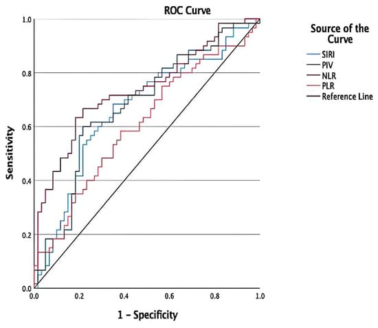
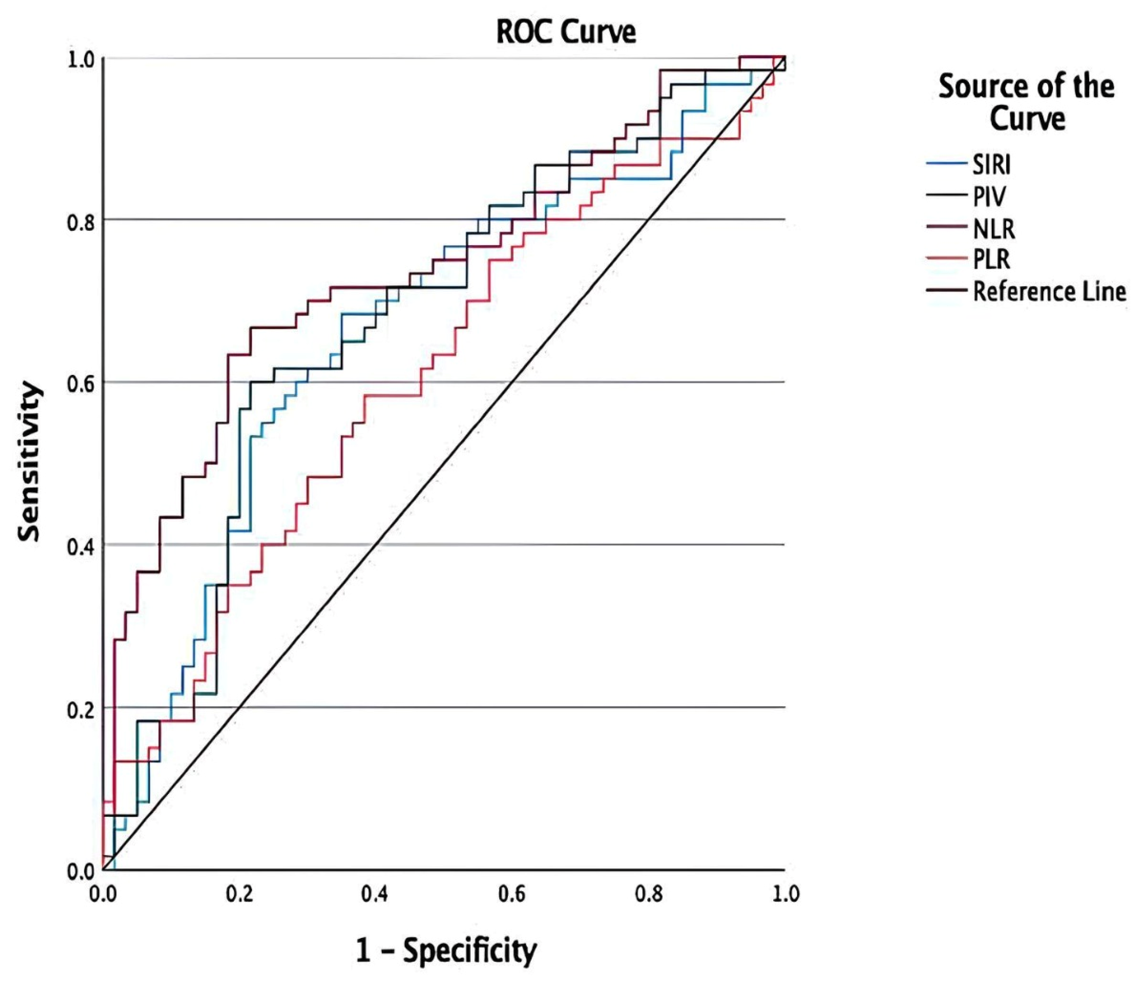
NLR had the highest observed AUC (0.738), followed by PIV (0.678), SIRI (0.664), and PLR (0.608) in ROC curve analysis for discriminating patients with RVO from controls (Figure 1). For PIV, the optimal cut-off value was identified as >292.35, with a sensitivity of 60.0% and a specificity of 78.3%. For NLR, the optimal cut-off value was identified as >2.15; sensitivity and specificity were found to be 66.7% and 78.3%. For SIRI, the optimal cut-off value was identified as >0.91; sensitivity and specificity were found to be 68.3% and 65.0%. For PLR, the optimal cut-off value was identified as >124.97; sensitivity and specificity were found to be 58.3% and 61.7% (Table 4). Paired nonparametric comparisons showed no statistically significant differences in AUC between PIV and NLR (p = 0.172), SIRI (p = 0.765), or PLR (p = 0.164). Likewise, no significant differences were observed between NLR and PLR (p = 0.113), NLR and SIRI (p = 0.152), or PLR and SIRI (p = 0.164).
Figure 1.
Receiver operating characteristic (ROC) curves of NLR, SIRI, PIV, and PLR for discriminating patients with RVO from controls.
Table 4.
Results of the receiver operating characteristic curve analysis.
In univariable logistic regression analyses, hypertension, elevated PLR, NLR, and PIV were significantly associated with RVO. The odds of RVO were 2.46-fold higher in patients with hypertension (95% CI, 1.13–5.33; p = 0.023), 3.37-fold higher in the higher PLR category (95% CI, 1.59–7.15; p = 0.002), 4.10-fold higher in the higher NLR category (95% CI, 1.89–8.93; p < 0.001), and 3.76-fold higher in the higher PIV category (95% CI, 1.71–8.23; p < 0.001). In the multivariable model including all four variables, none remained statistically significant. PIV showed an adjusted OR of 1.96 (95% CI, 0.74–5.23; p = 0.177), whereas PLR demonstrated a borderline association (adjusted OR, 2.36; 95% CI, 0.99–5.61; p = 0.052) (Table 5). In an additional multivariable logistic regression analysis, PIV was entered as a continuous variable, with adjustment for age, gender, hypertension, diabetes mellitus, and smoking status. PIV was significantly associated with higher odds of RVO (adjusted OR per 100-unit increase: 1.41, 95% CI: 1.08–1.83; p = 0.010).
Table 5.
Univariable and Multivariable Logistic Regression Analyses of Factors Associated with Retinal Vein Occlusion.
4. Discussion
Neutrophil counts, NLR, SIRI, and PIV were significantly higher in patients with RVO than in controls after FDR correction, whereas the difference in PLR did not remain statistically significant. No statistically significant differences were observed in platelet, lymphocyte, or monocyte counts or MLR. These findings are broadly consistent with previous reports of associations between RVO and CBC-derived inflammatory indices [9,10,11,12,13,16,17]. The higher prevalence of hypertension in the RVO group is consistent with previous reports [18,19]. No statistically significant between-group differences were observed in diabetes prevalence, SBP, DBP, or HbA1c levels. The comparable distribution of these metabolic and hemodynamic parameters between the groups may have reduced their potential confounding effects on the observed differences in systemic inflammatory indices. Nevertheless, residual confounding, particularly related to the duration and long-term control of hypertension and diabetes, cannot be completely excluded.
Inflammation may contribute to endothelial dysfunction, leukocyte activation, platelet activation, and thrombosis, providing a biologically plausible link between systemic inflammatory indices and RVO [20,21]. CBC-derived markers are attractive because they are readily available and inexpensive. PIV integrates neutrophil, lymphocyte, monocyte, and platelet counts and therefore captures several components of the systemic inflammatory and thrombo-inflammatory response in a single index. Previous studies have reported associations between PIV and several systemic and ocular inflammatory conditions, including diabetic retinopathy and pseudoexfoliation-related disease [22,23,24,25]. To our knowledge, this is the first study to evaluate PIV in patients with RVO.
In the present cohort, PIV was significantly higher in patients with RVO than in controls. In univariable logistic regression, participants in the higher PIV category had approximately 3.76-fold higher odds of having RVO. Importantly, however, the association was not maintained after simultaneous adjustment for hypertension, PLR, and NLR. The loss of statistical significance after multivariable adjustment suggests that the association between PIV and RVO may partly overlap with information captured by other inflammatory indices and vascular risk factors. This is also relevant because PIV is mathematically derived from neutrophil, lymphocyte, monocyte, and platelet counts, while NLR and PLR are themselves components of related inflammatory processes. Consequently, the use of several correlated CBC-derived indices in the same model may reduce the apparent independent contribution of each marker. In our data, the multivariable model including hypertension, PLR, NLR, and PIV did not identify any of these variables as statistically significant at the 0.05 level, although PLR showed a borderline association (adjusted OR, 2.36; 95% CI, 0.99–5.61; p = 0.052). These findings emphasize that univariable associations should not be interpreted as evidence of independent effects. In contrast, continuous PIV was significantly associated with RVO in the additional model adjusted for age, gender, hypertension, diabetes mellitus, and smoking. Because the models differed in both covariate composition and PIV representation, the difference in findings cannot be attributed solely to adjustment for other inflammatory indices.
Elevated neutrophil levels may contribute to the development and progression of atherosclerosis, potentially increasing the risk of RVO [25,26]. Lymphocytes, unlike neutrophils, inhibit cell proliferation and migration. Lymphocytes are indicators of physiological stress. Low lymphocyte levels are linked to cardiovascular disease [27]. Previously, a number of research studies evaluated the utility of NLR and PLR as indicators of RVO risk in clinical practice [5,6,12,13,16]. Previous studies have reported inconsistent findings regarding NLR and PLR in RVO. Both Şahin et al. [11] and Liu et al. [6] reported that patients with RVO had considerably higher NLR and PLR compared to controls. Zhu et al. [13] suggested that elevated NLR and PLR could serve as independent predictors of RVO. In contrast, some other studies found no significant difference in the NLR or PLR [10,28]. In the current study, neutrophil counts and NLR remained significantly higher in patients with RVO after FDR correction, whereas the PLR difference did not. Subgroup analysis revealed no significant differences between BRVO and CRVO. NLR had the highest observed AUC; however, the reported paired comparisons did not demonstrate statistically significant differences between the evaluated indices. These findings do not establish the clinical superiority of any individual marker.
Monocytes migrate to tissues during inflammation. They secrete inflammatory cytokines and phagocytose other cells and toxic molecules. The MLR is a reliable predictor of cardiovascular events and mortality [29], but its significance in RVO pathophysiology has received less attention. In the present study, MLR did not differ significantly between patients with RVO and controls. In contrast to this investigation, Üçer et al. [30] revealed that MLR was significantly higher in RVO patients. Another study found that higher MLR was associated with the response to anti-VEGF therapy in patients with RVO [31]. The discrepancies among these findings may be explained by differences in study populations, disease characteristics, and sample sizes.
SIRI is derived from neutrophil, monocyte, and lymphocyte counts. Previous studies have reported higher SIRI values in patients with RVO than in controls [27,30]. Similarly, SIRI was elevated in the present study, whereas comparisons according to ischemia and SRF status did not reveal statistically significant differences.
Few studies have compared inflammatory indices between RVO subtypes. Consistent with the present findings, Wang et al. [9] and Üçer et al. [30] reported no statistically significant differences between BRVO and CRVO. The absence of statistically significant subgroup differences may reflect limited statistical power or limited biological specificity of these indices for the evaluated RVO phenotypes. Similarly, the nonsignificant PIV association in the model containing other CBC-derived indices may reflect overlapping information or limited additional relevance.
PIV may serve as an indirect indicator of systemic inflammation; however, it does not directly assess immune cell function, activation, or leukocyte–endothelial interactions. Although PIV was elevated in patients with RVO, its modest discriminatory performance does not support its use as a standalone clinical biomarker. NLR had the highest observed AUC among the evaluated indices; however, these findings do not establish sufficient clinical utility for any of these markers. Moreover, their nonspecific nature limits the attribution of elevated values specifically to RVO. To our knowledge, this is the first study to investigate the association between PIV and RVO. Functional and molecular assessments were not available in this retrospective study. Future prospective studies could evaluate candidate measures such as granulocyte alkaline phosphatase activity and low-density lipoprotein receptor-related protein 1 (LRP-1) expression alongside CBC-derived indices. Their relevance and predictive performance in RVO would require specific validation.
This study has several limitations. First, the study had a single-center retrospective case–control design. Hospital-based control selection may have introduced selection bias, as these individuals may differ from the general population in their health characteristics and healthcare-seeking behavior, potentially limiting the generalizability of our findings. Because blood samples were obtained after the RVO event, the elevated indices may reflect a secondary response to vascular occlusion rather than a pre-existing inflammatory state. Therefore, neither temporal directionality nor causality can be established, and these indices cannot be interpreted as predictors of future RVO. Second, the sample size was relatively small, particularly for the CRVO/BRVO and ischemic/non-ischemic subgroup analyses, resulting in wide confidence intervals and limited power to detect moderate associations. Subgroup analyses were exploratory, and their findings require confirmation in adequately powered studies. Third, the data-derived cut-offs were not independently validated and may therefore yield optimistic estimates of sensitivity and specificity. Fourth, some potentially relevant variables, including body mass index, lipid parameters, serum inflammatory proteins, and cytokine profiles, were unavailable or incomplete, leaving the possibility of residual confounding. Likewise, detailed medication data were unavailable; therefore, residual confounding related to medication use could not be excluded. Fifth, although hypertension, diabetes mellitus, SBP, DBP, HbA1c, and smoking status were evaluated, single-visit measurements of blood pressure and glycemic status may not fully capture long-term vascular risk. Finally, the inflammatory indices are derived from routine CBC parameters and may be influenced by unmeasured systemic factors.
Despite these limitations, the study provides preliminary evidence that PIV is elevated in patients with RVO and contributes to the growing literature on CBC-derived inflammatory markers in retinal vascular disease. The present findings support further investigation of PIV, particularly in prospective studies designed to assess incident RVO, disease severity, treatment response, and clinical outcomes. Future multicenter studies should also evaluate whether PIV adds incremental information beyond established vascular risk factors using appropriate multivariable and model-performance analyses.
5. Conclusions
This study demonstrated that PIV, NLR, and SIRI were significantly elevated in RVO patients compared to controls; however, no statistically significant differences in these indices were observed across RVO subgroups or based on the presence of ischemia or SRF. Higher categorical PIV was associated with greater odds of RVO in univariable analysis. Continuous PIV was associated with RVO after adjustment for clinical covariates, whereas categorical PIV was not significantly associated with RVO in the model containing hypertension, NLR, and PLR. These model-dependent findings, together with modest discriminatory performance, do not establish PIV as an RVO-specific or predictive biomarker. Prospective studies are needed to clarify the temporal relationship and potential clinical relevance of these associations.
Supplementary Materials
The following supporting information can be downloaded at: https://www.mdpi.com/article/10.3390/jcm15228805/s1; Table S1: Effect sizes for pairwise comparisons among the CRVO, BRVO, and control groups.
Author Contributions
Conceptualization, M.B. and L.B.; methodology, M.B. and L.B.; software, M.B. and L.B.; validation, M.B. and L.B.; formal analysis, M.B. and L.B.; writing—review and editing, M.B. and L.B.; visualization, M.B. and L.B.; supervision, M.B. and L.B.; funding acquisition, not applicable. All authors have read and agreed to the published version of the manuscript.
Funding
This research received no external funding.
Institutional Review Board Statement
The study was approved by the Sivas Cumhuriyet University Faculty of Medicine Non Interventional Clinical Research Ethics Committee (No: 26_01_29_30; date of approval 29 January 2026) and was conducted in accordance with the principles of the Declaration of Helsinki.
Informed Consent Statement
Informed consent was obtained from all participants involved in the study.
Data Availability Statement
The data presented in this study are available from the corresponding author upon reasonable request. The data are not publicly available due to privacy and ethical restrictions.
Conflicts of Interest
The authors declare no conflicts of interest.
Abbreviations
| RVO | Retinal vein occlusion |
| CRVO | Central retinal vein occlusion |
| BRVO | Branch retinal vein occlusion |
| CBC | Complete blood count |
| LYM | Lymphocytes |
| NEU | Neutrophils |
| MONO | Monocytes |
| PLT | Platelets |
| MLR | Monocyte-to-lymphocyte ratio |
| PLR | Platelet-to-lymphocyte ratio |
| NLR | Neutrophil-to-lymphocyte ratio |
| SIRI | Systemic inflammation response index |
| PIV | Pan-immune inflammation value |
| OR | Odds ratio |
| CI | Confidence interval |
| HT | Hypertension |
| DM | Diabetes mellitus |
| BH-FDR | Benjamini–Hochberg false discovery rate |
| BCVA | Best-corrected visual acuity |
| FFA | Fundus fluorescein angiography |
| SD-OCT | Spectral-domain optical coherence tomography |
| SBP | Systolic blood pressure |
| DBP | Diastolic blood pressure |
| SRF | Subretinal fluid |
| ROC | Receiver operating characteristic |
| AUC | Area under the curve |
| logMAR | Logarithm of the minimum angle of resolution |
References
- Song, P.; Xu, Y.; Zha, M.; Zhang, Y.; Rudan, I. Global Epidemiology of Retinal Vein Occlusion: A Systematic Review and Meta-Analysis of Prevalence, Incidence, and Risk Factors. J. Glob. Health 2019, 9, 010427. [Google Scholar] [CrossRef] [Scilit]
- Rogers, S.; McIntosh, R.L.; Cheung, N.; Lim, L.; Wang, J.J.; Mitchell, P.; Kowalski, J.W.; Nguyen, H.; Wong, T.Y. The Prevalence of Retinal Vein Occlusion: Pooled Data from Population Studies from the United States, Europe, Asia, and Australia. Ophthalmology 2010, 117, 313–319.e1. [Google Scholar] [CrossRef] [Scilit]
- Rehak, M.; Wiedemann, P. Retinal Vein Thrombosis: Pathogenesis and Management. J. Thromb. Haemost. 2010, 8, 1886–1894. [Google Scholar] [CrossRef] [Scilit]
- Kesler, A.; Shalev, V.; Rogowski, O.; Shimron, O.; Shainberg, B.; Sela, B.A.; Shapira, I.; Salomon, O.; Berliner, S. Comparative Analysis of Homocysteine Concentrations in Patients with Retinal Vein Occlusion versus Thrombotic and Atherosclerotic Disorders. Blood Coagul. Fibrinolysis 2008, 19, 259–262. [Google Scholar] [CrossRef] [Scilit]
- Kushwaha, P.; Singh, A.; Shrivastava, P. Inflammatory and Hematologic Biomarker Profiles across Central Branch and Hemi-Retinal Vein Occlusion. Bioinformation 2025, 21, 2781–2784. [Google Scholar] [CrossRef] [Scilit]
- Liu, Z.; Perry, L.A.; Penny-Dimri, J.C.; Raveendran, D.; Hu, M.L.; Arslan, J.; Britten-Jones, A.C.; O’Hare, F.; Ayton, L.N.; Edwards, T.L. The Association of Neutrophil-Lymphocyte Ratio and Platelet-Lymphocyte Ratio with Retinal Vein Occlusion: A Systematic Review and Meta-Analysis. Acta Ophthalmol. 2022, 100, e635–e647. [Google Scholar] [CrossRef] [Scilit]
- Huang, Z.; Fu, Z.; Huang, W.; Huang, K. Prognostic Value of Neutrophil-to-Lymphocyte Ratio in Sepsis: A Meta-Analysis. Am. J. Emerg. Med. 2020, 38, 641–647. [Google Scholar] [CrossRef] [Scilit]
- Corriere, T.; Di Marca, S.; Cataudella, E.; Pulvirenti, A.; Alaimo, S.; Stancanelli, B.; Malatino, L. Neutrophil-to-Lymphocyte Ratio Is a Strong Predictor of Atherosclerotic Carotid Plaques in Older Adults. Nutr. Metab. Cardiovasc. Dis. 2018, 28, 23–27. [Google Scholar] [CrossRef] [Scilit]
- Wang, X.; Wang, L.; Li, X.; Liu, S.; Liu, B. Characteristics of Hematologic Parameters in Young Patients with Retinal Vein Occlusion. Ophthalmic Res. 2023, 66, 1096–1103. [Google Scholar] [CrossRef] [Scilit]
- Turkseven Kumral, E.; Yenerel, N.M.; Ercalik, N.Y.; Imamoglu, S.; Vural, E.T. Neutrophil/Lymphocyte Ratio and Mean Platelet Volume in Branch Retinal Vein Occlusion. Saudi J. Ophthalmol. 2016, 30, 105–108. [Google Scholar] [CrossRef] [Scilit]
- Şahin, M.; Elbey, B.; Şahin, A.; Yüksel, H.; Türkcü, F.M.; Çaça, İ. Neutrophil-to-Lymphocyte Ratio and Platelet-to-Lymphocyte Ratio in Retinal Vein Occlusion. Clin. Exp. Optom. 2020, 103, 490–494. [Google Scholar] [CrossRef] [Scilit]
- Lai, X.J.; Yang, S.Y.; Lei, C.Y.; Xiao, R.H.; Zhang, M.X. Correlations between Inflammatory Biomarkers in Peripheral Blood and Branch Retinal Vein Occlusion. Int. J. Ophthalmol. 2025, 18, 1908–1913. [Google Scholar] [CrossRef] [Scilit]
- Zhu, D.-D.; Liu, X. Neutrophil/Lymphocyte Ratio and Platelet/Lymphocyte Ratio in Branch Retinal Vein Occlusion. J. Ophthalmol. 2019, 2019, 6043612. [Google Scholar] [CrossRef] [Scilit]
- Zhou, C.; Cheng, X.; Zhang, J. Research Advances of Pan-Immune Inflammation Value in Rheumatoid Arthritis. Front. Immunol. 2025, 16, 1693676. [Google Scholar] [CrossRef] [Scilit]
- Yang, X.C.; Liu, H.; Liu, D.C.; Tong, C.; Liang, X.W.; Chen, R.H. Prognostic Value of Pan-Immune-Inflammation Value in Colorectal Cancer Patients: A Systematic Review and Meta-Analysis. Front. Oncol. 2022, 12, 1036890. [Google Scholar] [CrossRef] [Scilit]
- Zuo, W.; Chen, T.; Song, J.; Ai, M. Assessment of Systemic Immune-Inflammation Index Levels in Patients with Retinal Vein Occlusion. Ocul. Immunol. Inflamm. 2023, 31, 491–495. [Google Scholar] [CrossRef] [Scilit]
- Kazantzis, D.; Machairoudia, G.; Kroupis, C.; Theodossiadis, G.; Theodossiadis, P.; Chatziralli, I. Complete Blood Count-Derived Inflammation Indices and Retinal Vein Occlusion: A Case-Control Study. Ophthalmol. Ther. 2022, 11, 1241–1249. [Google Scholar] [CrossRef] [Scilit]
- Wong, T.Y.; Larsen, E.K.M.; Klein, R.; Mitchell, P.; Couper, D.J.; Klein, B.E.K.; Hubbard, L.D.; Siscovick, D.S.; Sharrett, A.R. Cardiovascular Risk Factors for Retinal Vein Occlusion and Arteriolar Emboli: The Atherosclerosis Risk in Communities & Cardiovascular Health Studies. Ophthalmology 2005, 112, 540–547. [Google Scholar] [CrossRef] [Scilit]
- O’Mahoney, P.R.A.; Wong, D.T.; Ray, J.G. Retinal Vein Occlusion and Traditional Risk Factors for Atherosclerosis. Arch. Ophthalmol. 2008, 126, 692–699. [Google Scholar] [CrossRef] [Scilit]
- Afshar-Kharghan, V.; Thiagarajan, P. Leukocyte Adhesion and Thrombosis. Curr. Opin. Hematol. 2006, 13, 34–39. [Google Scholar] [CrossRef] [Scilit]
- Zhu, Y.; Xian, X.; Wang, Z.; Bi, Y.; Chen, Q.; Han, X.; Tang, D.; Chen, R. Research Progress on the Relationship between Atherosclerosis and Inflammation. Biomolecules 2018, 8, 80. [Google Scholar] [CrossRef] [Scilit]
- Tarim, B. The Role of Serum Biomarkers in Determining Systemic Inflammation and Cardiovascular Risk in Pseudoexfoliation Syndrome. Int. Ophthalmol. 2025, 45, 15. [Google Scholar] [CrossRef] [Scilit]
- Candan, O.; Orman, G.; Ünlü, N.; Ozkan, G. Assessment of Pan-Immune-Inflammation Value as a Novel Marker of Proliferative Diabetic Retinopathy Stage. Int. J. Ophthalmol. 2026, 19, 517–525. [Google Scholar] [CrossRef] [Scilit]
- Candan, O.; Orman, G.; Ünlü, N.; Ozkan, G. Diagnostic Efficiency of Pan-Immune-Inflammation Value to Predict Diabetic Macular Edema and Its Relationship with OCT-Based Biomarkers of Inflammation. Int. Ophthalmol. 2025, 45, 365. [Google Scholar] [CrossRef] [Scilit]
- Bulu, A.; Keser, S. The Relationship between Pan-Immune Inflammation Value and Different Stages of Diabetic Retinopathy in Patients with Type 2 Diabetes Mellitus: A Prospective Cross-Sectional Study. BMC Endocr. Disord. 2025, 25, 184. [Google Scholar] [CrossRef] [Scilit]
- Silvestre-Roig, C.; Braster, Q.; Ortega-Gomez, A.; Soehnlein, O. Neutrophils as Regulators of Cardiovascular Inflammation. Nat. Rev. Cardiol. 2020, 17, 327–340. [Google Scholar] [CrossRef] [Scilit]
- Bian, C.; Wu, Y.; Shi, Y.; Xu, G.; Wang, J.; Xiang, M.; Weng, S.; Jiang, J.; Ma, J. Predictive Value of the Relative Lymphocyte Count in Coronary Heart Disease. Heart Vessel. 2010, 25, 469–473. [Google Scholar] [CrossRef] [Scilit]
- Vural, E.; Hazar, L. Is the C Reactive Protein/Albumin Ratio a Good Tool for Retinal Vein Occlusion? BMC Ophthalmol. 2025, 25, 664. [Google Scholar] [CrossRef] [Scilit]
- Hua, Y.; Sun, J.Y.; Lou, Y.X.; Sun, W.; Kong, X.Q. Monocyte-to-Lymphocyte Ratio Predicts Mortality and Cardiovascular Mortality in the General Population. Int. J. Cardiol. 2023, 379, 118–126. [Google Scholar] [CrossRef] [Scilit]
- Üçer, M.B.; Cevher, S. Assessment of Systemic Inflammatory Response Index and Other Inflammatory Indicators in Retinal Vein Occlusion. Medicine 2023, 102, E36512. [Google Scholar] [CrossRef] [Scilit]
- Chatziralli, I.; Kazantzis, D.; Kroupis, C.; Machairoudia, G.; Dimitriou, E.; Theodossiadis, G.; Theodossiadis, P.; Sergentanis, T.N. The Impact of Laboratory Findings and Optical Coherence Tomography Biomarkers on Response to Intravitreal Anti-VEGF Treatment in Patients with Retinal Vein Occlusion. Int. Ophthalmol. 2022, 42, 3449–3457. [Google Scholar] [CrossRef] [Scilit]

Disclaimer/Publisher’s Note: The statements, opinions and data contained in all publications are solely those of the individual author(s) and contributor(s) and not of MDPI and/or the editor(s). MDPI and/or the editor(s) disclaim responsibility for any injury to people or property resulting from any ideas, methods, instructions or products referred to in the content. |
© 2026 by the authors. Licensee MDPI, Basel, Switzerland. This article is an open access article distributed under the terms and conditions of the Creative Commons Attribution (CC BY) license.