


Abstract
Background: Triple-negative breast carcinoma (TNBC) comprises several histological entities with different molecular alterations and clinical behaviour. Recognising these entities is important for accurate classification and for choosing appropriate immunohistochemical and molecular tests. Methods: We conducted a narrative review of English-language literature identified through PubMed/MEDLINE, Scopus, Web of Science Core Collection, citation searching and a Scopus publication list. Journal publications available through 24 August 2026 were eligible. Current reporting and regulatory guidance was obtained from official websites. Findings were synthesised qualitatively, without statistical pooling. Results: The review included 93 journal publications and two official documents. Secretory carcinoma, adenoid cystic carcinoma, tall cell carcinoma with reversed polarity and metaplastic carcinoma require a different diagnostic approach from conventional high-grade invasive breast carcinoma of no special type. Architectural, cytological and stromal findings guide the differential diagnosis and the selection of immunohistochemical stains and molecular assays. Limited sampling, tumour heterogeneity and treatment-related changes can affect tissue selection and interpretation. Conclusions: Pathology reports should specify the histological type and clearly state any remaining diagnostic uncertainty. Morphology should guide diagnostic testing, while validated predictive biomarkers and clinical criteria determine treatment eligibility.
1. Introduction
Triple-negative breast carcinoma (TNBC) is conventionally defined as invasive breast carcinoma with oestrogen receptor (ER) and progesterone receptor (PR) expression in fewer than 1% of tumour-cell nuclei and without human epidermal growth factor receptor 2 (HER2) positivity according to validated immunohistochemical and in situ hybridisation (ISH) algorithms [1,2]. Although essential for clinical classification, receptor status does not establish a histological or molecular diagnosis. ER-low tumours and the HER2-low and HER2-ultralow categories illustrate the continuous nature of receptor expression and the need for defined thresholds in treatment decisions [3,4,5].
An ER-negative/PR-negative/HER2-negative profile can occur in several distinct histological entities. The current World Health Organization (WHO) classification incorporates morphology, immunophenotype, predictive biomarkers, the limitations of small specimens and recurrent molecular alterations [6]. Most TNBCs are high-grade invasive breast carcinomas of no special type (IBC-NST), but the same receptor profile also occurs in metaplastic carcinoma, secretory carcinoma, adenoid cystic carcinoma (AdCC), tall cell carcinoma with reversed polarity (TCCRP), low-grade adenosquamous carcinoma (LGASC), fibromatosis-like metaplastic carcinoma (FLMC), apocrine carcinoma, acinic cell-like carcinoma, mucoepidermoid carcinoma and rare triple-negative lobular carcinomas [6,7,8,9,10,11,12]. Several of these entities differ substantially from conventional high-grade TNBC in their clinical behaviour and molecular drivers. Acinic cell-like and mucoepidermoid carcinomas are considered in this review because, despite their variable receptor expression, they may enter the differential diagnosis of a breast carcinoma initially found to be receptor-negative [6,13,14,15,16,17]. Their classification depends on morphology and context, irrespective of triple-negative status [6,18].
Some of this biological heterogeneity can be recognised on haematoxylin and eosin (H&E) sections. Microcystic architecture with intracellular and extracellular secretory material suggests secretory carcinoma and an associated ETV6::NTRK3 fusion [19,20]; cribriform biphasic architecture suggests AdCC and MYB/MYBL1-pathway activation [21,22,23]; reversed nuclear polarity strongly suggests TCCRP and recurrent IDH-pathway alterations [24,25,26,27,28]. Other morphological features are associated with broader biological phenotypes. Apocrine differentiation may indicate androgen receptor-associated biology [29,30], while high-grade syncytial growth, necrosis, pushing borders and prominent lymphocytes overlap with basal-like and BRCA-associated phenotypes [31,32]. None of these morphological associations alone establishes the corresponding molecular finding.
Triple-negative breast carcinomas show considerable genomic and transcriptional heterogeneity [32,33,34,35,36,37,38]. Common molecular features include homologous recombination deficiency, TP53-associated genomic instability, PI3K/AKT/PTEN and cell-cycle dysregulation, androgen receptor (AR)/luminal androgen receptor (LAR) phenotypes, and basal-like or mesenchymal transcriptional programmes [32,33,34,35,36,37,38]. Immune- and microenvironment-related programmes partly reflect differences in the composition and spatial organisation of the tumour microenvironment (TME) [39]. These features overlap and correspond only partly to conventional histological categories. Molecular findings therefore require interpretation in the context of tumour morphology.
Transcriptomic classifications have advanced the understanding of TNBC biology, but they correspond only partly to routine histological entities and are not required for everyday diagnostic classification [33,34]. A morphology-guided approach recognises the diversity of TNBC and allows ancillary tests to be selected according to the histological differential diagnosis. A recent review described the clinicopathological, molecular, prognostic and therapeutic features of rare TNBC histologies [10]. The present review examines how specific H&E findings can guide immunohistochemistry and indicate when molecular confirmation is needed.
The review addresses the principal components of the diagnostic work-up: confirmation of the triple-negative phenotype, H&E pattern recognition, focused immunohistochemistry, defining or highly characteristic molecular alterations, differential diagnosis and selected tissue-based biomarkers. The tumour microenvironment, post-neoadjuvant pathology, predictive biomarkers and digital or spatial methods are considered where they affect tissue interpretation, sampling or clinically relevant testing. The diagnostic sequence begins with H&E examination and a morphological differential diagnosis, followed by focused immunohistochemistry and, when needed, molecular confirmation to reach an integrated diagnosis.
2. Materials and Methods
2.1. Literature Search Strategy
For this narrative review, PubMed/MEDLINE, Scopus and Web of Science Core Collection were searched, with the database searches last updated on 24 August 2026. English-language journal publications were eligible from database inception through that date. Eligibility was determined by the date of first full publication, including online-first publication, irrespective of the assigned issue year. No study-design or publication-type filters were applied. Older seminal publications were included when relevant to current diagnostic practice. The same publication window and relevance criteria were applied to journal publications identified through all search methods. Guideline, reporting and regulatory information was checked against current official sources.
The three databases were searched using the same free-text terms and Boolean structure, adapted to their syntax and search fields: PubMed title/abstract-related fields [tiab], Scopus TITLE-ABS-KEY and Web of Science Topic fields (TS, including Keywords Plus). Breast or mammary carcinoma/cancer terms were combined with either triple-negative terminology or the names of special histological types and a required block of diagnostic terms. Searches for special types did not require triple-negative terminology, as relevant reports may omit receptor status from the title or abstract. The complete queries are shown below. Reference lists and forward citations of key publications were also screened for relevant reports.
PubMed/MEDLINE
(breast[tiab] OR mammary[tiab]) AND (carcinoma*[tiab] OR cancer*[tiab]) AND (“triple-negative”[tiab] OR “triple negative”[tiab] OR TNBC[tiab] OR “secretory carcinoma”[tiab] OR “secretory breast carcinoma”[tiab] OR “adenoid cystic”[tiab] OR “tall cell”[tiab] OR “reversed polarity”[tiab] OR “reverse polarity”[tiab] OR metaplastic[tiab] OR “fibromatosis-like”[tiab] OR “fibromatosis like”[tiab] OR adenosquamous[tiab] OR apocrine[tiab] OR “acinic cell”[tiab] OR mucoepidermoid[tiab] OR medullary[tiab]) AND (morpholog*[tiab] OR histopatholog*[tiab] OR clinicopatholog*[tiab] OR (histolog*[tiab] AND (classif*[tiab] OR subtype*[tiab] OR “special type*”[tiab] OR “rare type*”[tiab])) OR “differential diagnosis”[tiab] OR ((immunohistochem*[tiab] OR immunophenotyp*[tiab]) AND (diagnos*[tiab] OR classif*[tiab])) OR ETV6[tiab] OR NTRK3[tiab] OR MYB[tiab] OR MYBL1[tiab] OR IDH2[tiab] OR MAML2[tiab] OR NR4A3[tiab] OR TRPS1[tiab] OR GATA3[tiab] OR SOX10[tiab]) AND english[la]
Scopus
TITLE-ABS-KEY((breast OR mammary) AND (carcinoma* OR cancer*) AND (“triple-negative” OR “triple negative” OR TNBC OR “secretory carcinoma” OR “secretory breast carcinoma” OR “adenoid cystic” OR “tall cell” OR “reversed polarity” OR “reverse polarity” OR metaplastic OR “fibromatosis-like” OR “fibromatosis like” OR adenosquamous OR apocrine OR “acinic cell” OR mucoepidermoid OR medullary) AND (morpholog* OR histopatholog* OR clinicopatholog* OR (histolog* AND (classif* OR subtype* OR “special type*” OR “rare type*”)) OR “differential diagnosis” OR ((immunohistochem* OR immunophenotyp*) AND (diagnos* OR classif*)) OR ETV6 OR NTRK3 OR MYB OR MYBL1 OR IDH2 OR MAML2 OR NR4A3 OR TRPS1 OR GATA3 OR SOX10)) AND LANGUAGE(english)
Web of Science Core Collection
TS=((breast OR mammary) AND (carcinoma* OR cancer*) AND (“triple-negative” OR “triple negative” OR TNBC OR “secretory carcinoma” OR “secretory breast carcinoma” OR “adenoid cystic” OR “tall cell” OR “reversed polarity” OR “reverse polarity” OR metaplastic OR “fibromatosis-like” OR “fibromatosis like” OR adenosquamous OR apocrine OR “acinic cell” OR mucoepidermoid OR medullary) AND (morpholog* OR histopatholog* OR clinicopatholog* OR (histolog* AND (classif* OR subtype* OR “special type*” OR “rare type*”)) OR “differential diagnosis” OR ((immunohistochem* OR immunophenotyp*) AND (diagnos* OR classif*)) OR ETV6 OR NTRK3 OR MYB OR MYBL1 OR IDH2 OR MAML2 OR NR4A3 OR TRPS1 OR GATA3 OR SOX10))
English-language limits were applied using english[la] in PubMed, LANGUAGE(english) in Scopus and the Language filter in Web of Science. The database queries did not restrict publication dates, study designs or publication types. Eligibility by publication date was assessed during screening.
A publication list exported from Scopus was screened for relevance to diagnostic interpretation, ancillary testing and pathological reporting. These publications were assessed with the other search results, using the same publication window and relevance criteria. References retained for the review were checked against the statements they supported.
2.2. Literature Selection and Narrative Synthesis
Publications and official documents were included when directly relevant to histological classification, differential diagnosis, ancillary testing, specimen interpretation, tissue selection or pathological reporting. Receptor assessment was reviewed in relation to WHO terminology, ASCO/CAP guidance and current reporting recommendations. Genotype–phenotype associations were examined in clinicopathological series, diagnostic immunohistochemistry studies and molecular investigations. The qualitative appraisal considered study design, cohort size, pathological confirmation, assay validation and consistency across publications. Therapeutic and experimental-biology literature was included only when relevant to the selection or interpretation of tissue-based tests.
The database searches identified 1420 records: 420 from PubMed/MEDLINE, 565 from Scopus and 435 from Web of Science. After 610 duplicates were removed, 810 records underwent title/abstract screening; 642 were excluded because they fell outside the review scope or publication window. Of the 168 reports sought, eight could not be retrieved and 160 underwent full-text assessment. Eighty-six were excluded, leaving 74 journal publications. Citation searching identified 35 documents and official websites yielded two. After 18 duplicates were removed, including documents already identified in the database searches, 19 reports were sought. Three could not be retrieved; the remaining 16 were assessed and retained, comprising 14 journal publications and two official documents. The Scopus publication list contained 98 records, two of which had already been identified through the other searches. The remaining 96 records were assessed for relevance. Ninety-one did not provide sufficiently relevant evidence for the diagnostic or methodological questions addressed, and five journal publications were included.
One principal reason was recorded for each exclusion after full-text assessment: no direct diagnostic relevance to the review (n = 54); non-breast disease or exclusively preclinical evidence without a relevant diagnostic comparison (n = 12); evidence restricted to receptor-positive tumours without a relevant comparison (n = 8); retrieved conference abstracts or short reports with insufficient methodological detail (n = 7); or superseded or redundant guidance (n = 5). Reports that could not be retrieved were counted separately. The review included 93 journal publications from 2006 to 2026 and two official reporting or regulatory documents (95 in total). All are listed in the References, which also include the PRISMA 2020 methodological statement [40]. Literature identification and selection, including reasons for exclusion, are summarised in Figure 1 using a flow diagram adapted from PRISMA 2020 [40].
Figure 1.
Literature identification and selection for the narrative review. The flow diagram was adapted from PRISMA 2020 [40].
The review used a narrative design: no protocol was preregistered, screening was not independently duplicated, and neither a structured risk-of-bias assessment nor formal evidence grading was performed. No new statistical analysis or meta-analysis was undertaken because the publications addressed different histological entities, assays, diagnostic questions and clinical outcomes, with no common endpoint suitable for quantitative synthesis. Numerical findings are attributed to the original publications and were not pooled. Diagnostic testing establishes the tumour type or resolves a differential diagnosis; predictive testing assesses validated biomarkers relevant to treatment eligibility; exploratory testing concerns investigational profiling. The proposed diagnostic approach is based on the literature and has not been externally validated as a clinical decision rule.
2.3. Use of Generative Artificial Intelligence
During the preparation of this manuscript, the authors used ChatGPT, version 5.6 (OpenAI, San Francisco, CA, USA), as an auxiliary writing tool to assist with language editing, improvement of English clarity and readability, and stylistic refinement of selected parts of the text. It was also used to generate initial illustrative images for the figures, which were subsequently edited and finalised by the authors. This tool was not used to generate scientific data, perform analyses, select or interpret references, or draw scientific conclusions. The authors reviewed and edited all AI-assisted outputs, checked the final figures for scientific accuracy, and take full responsibility for the content of this publication.
3. Establishing the Triple-Negative Phenotype
Morphology-guided diagnosis depends on reliable ER, PR and HER2 assessment. Pre-analytical quality is particularly important in pretreatment core needle biopsy (CNB), which may provide the only untreated tissue available before neoadjuvant therapy. Cold ischaemia, fixation, crush artefact, low tumour cellularity and tissue exhaustion can cause misleadingly weak or negative staining. Internal and external controls should therefore be reviewed before a triple-negative result is accepted [1,2].
ER staining in fewer than 1% of invasive tumour-cell nuclei is interpreted as negative, whereas 1–10% staining is classified as ER-low positive [1]. ER-low carcinomas do not formally meet the TNBC definition, although some have TNBC-like biological features. An unusual ER-negative/PR-positive result warrants review of the controls and morphology, with repeat testing when appropriate. Interpretation of negative findings depends on technically adequate staining and correlation with morphology.
HER2 assessment follows the ASCO/CAP IHC–ISH algorithm: IHC 0 and 1+ are negative for overexpression, IHC 2+ is equivocal and requires ISH, and IHC 3+ is positive [2]. Reports should record the actual IHC score and, within the conventional IHC 0 category, distinguish absent membrane staining from faint, incomplete membrane staining in a small proportion of tumour cells [4,41]. The September 2026 CAP template provides separate reporting options for negative 0 with absent membrane staining and negative 0+ with membrane staining [41]. HER2-low denotes IHC 1+ or IHC 2+/ISH-negative; HER2-ultralow denotes IHC 0 with faint, incomplete membrane staining in >0% and ≤10% of tumour cells [4]. Both can occur in receptor-defined TNBC. They describe HER2 expression and do not establish distinct histological entities or independently determine treatment eligibility. Interpretation near these thresholds requires attention to controls and tissue quality, with a second review where needed [4,42].
CNB is generally reliable for determining positive or negative receptor status, but limited sampling may miss intratumoral heterogeneity, and discordant results occur more often near low-expression thresholds [43]. Distinguishing HER2 IHC 0, ultralow and low categories is more susceptible to sampling and interpretive variability [4,42,43]. Repeat testing of an analytically valid pretreatment core with concordant morphology should therefore be selective. It is most useful when the biopsy is scant or poorly fixed, ER or PR expression lies near a decision threshold, HER2 staining is heterogeneous, finer categorisation of low-level HER2 expression may affect management, or the resection contains a morphologically distinct component. Decisions to repeat testing should reflect analytical concerns, morphological discordance and clinical relevance.
Once mammary origin is considered plausible and a technically valid triple-negative receptor profile has been established, the H&E sections are reviewed in this context. The dominant morphology guides the work-up towards conventional high-grade triple-negative IBC-NST or investigation for a special histological type. Subsequent morphological, immunophenotypic or clinicoradiological discordance may prompt reassessment of mammary origin and further testing for tumour lineage. This assessment relates the receptor profile to the histological diagnosis and its biological implications.
4. Morphology-Guided Diagnostic Approach
H&E examination identifies the dominant architecture and cytology and establishes the differential diagnosis. Receptor testing places these findings in the triple-negative context, while ancillary tests are selected to resolve specific diagnostic questions. Special histological types are recognised primarily by morphology rather than receptor status, and occasional cases have receptor profiles outside the conventional TNBC definition. Table 1 details the morphology-guided diagnostic work-up, and Figure 2 summarises the proposed approach.
Table 1.
Morphology-guided differential diagnosis and ancillary testing in TNBC.
Figure 2.
Proposed morphology-guided approach to TNBC diagnosis. H&E examination establishes invasion, dominant morphology and whether mammary origin is plausible. A tailored lineage panel is used when origin is uncertain. The TNBC approach applies once mammary origin is supported and the triple-negative phenotype is technically valid. Conventional high-grade IBC-NST generally undergoes clinically indicated biomarker assessment; distinctive morphological patterns prompt focused diagnostic testing. Ancillary findings are interpreted according to their diagnostic, predictive or exploratory purpose and incorporated into the pathology report. Morphology alone does not establish treatment eligibility. The approach is based on the literature and has not been externally validated. Histological and immunohistochemical vignettes are schematic illustrations and do not represent patient-derived micrographs. Abbreviations: AdCC, adenoid cystic carcinoma; AR, androgen receptor; ER, oestrogen receptor; GATA3, GATA-binding protein 3; H&E, haematoxylin and eosin; HER2, human epidermal growth factor receptor 2; HRD, homologous recombination deficiency; IBC-NST, invasive breast carcinoma of no special type; IDH, isocitrate dehydrogenase; LAR, luminal androgen receptor; LGASC, low-grade adenosquamous carcinoma; MEC, mucoepidermoid carcinoma; MYB, MYB proto-oncogene; MYBL1, MYB proto-oncogene like 1; pan-TRK, pan-tropomyosin receptor kinase; PR, progesterone receptor; SOX10, SRY-box transcription factor 10; sTILs, stromal tumour-infiltrating lymphocytes; TCCRP, tall cell carcinoma with reversed polarity; TNBC, triple-negative breast carcinoma; TRPS1, transcriptional repressor GATA binding 1.
4.1. Conventional High-Grade Triple-Negative IBC-NST: Morphological and Molecular Features
Conventional triple-negative IBC-NST usually presents as a high-grade invasive carcinoma with limited tubule formation, marked nuclear pleomorphism, brisk mitotic activity, solid or syncytial growth, geographic necrosis and variable lymphocytic infiltration [31,32,44]. Pushing or relatively circumscribed borders may also be present [31,32]. These features overlap with the historical morphological description of basal-like carcinoma [31,32]. In a contemporary TNBC series, basal-like morphology was associated with higher grade and proliferation but was not independently associated with outcome, illustrating the limitations of prognostic assessment based on morphology alone [44].
High-grade morphology may suggest basal-like, proliferative or DNA-repair-deficient biology, but it cannot establish these characteristics. TNBC and basal-like carcinoma overlap but are not equivalent categories [32,33,35]. BRCA1-associated tumours frequently show grade 3 morphology, syncytial growth, pushing margins, necrosis and prominent lymphocytes; however, these features also occur in sporadic TNBC [31,32]. Germline BRCA1/2 status cannot be inferred from morphology [31,32,51]. The receptor-defined TNBC category encompasses several partly overlapping molecular abnormalities and cellular phenotypes, including homologous recombination deficiency, TP53-associated genomic instability, PI3K/AKT/PTEN and cell-cycle dysregulation, AR/LAR biology, basal-like and mesenchymal states, and variation in the immune response and tumour microenvironment. These features are summarised in Figure 3.
Figure 3.
Molecular and cellular features contributing to the biological heterogeneity of TNBC. Representative molecular abnormalities, cellular states and phenotypic features are grouped according to recurrent biological processes [32,33,34,35,36,37,38,39]. These processes overlap and do not correspond to mutually exclusive histological types or molecular subtypes used in routine diagnosis. Abbreviations: AKT, protein kinase B; AR, androgen receptor; E2F, E2F transcription-factor family; EGFR, epidermal growth factor receptor; EMT, epithelial–mesenchymal transition; ER, oestrogen receptor; HER2, human epidermal growth factor receptor 2; HRD, homologous recombination deficiency; LAR, luminal androgen receptor; PD-L1, programmed death-ligand 1; PI3K, phosphoinositide 3-kinase; PR, progesterone receptor; PTEN, phosphatase and tensin homologue; TIL, tumour-infiltrating lymphocyte; TME, tumour microenvironment; TNBC, triple-negative breast carcinoma.
Histological assessment should document architecture, grade, necrosis, lymphocytic infiltration, stromal features and any spatially distinct tumour component. Cytokeratin 5/6 (CK5/6), cytokeratin 14 (CK14) and epidermal growth factor receptor (EGFR) can demonstrate basal differentiation [31,32,44], although their routine use solely to classify a conventional TNBC as “basal-like” adds little diagnostic information. Ki-67 can describe proliferative activity but is not an independent treatment-predictive classifier [32,44]. A typical grade 3 triple-negative IBC-NST without a distinctive morphological pattern generally warrants standard clinical biomarker testing rather than an experimental IHC panel intended to assign a molecular subtype.
Homologous recombination deficiency (HRD) provides a further example of the limits of morphological assessment. A “BRCA-like” appearance on H&E may suggest defective DNA repair, but establishing HRD requires a molecular or functional assay [32,45,46]. RAD51 foci assessment is a promising tissue-based measure of homologous recombination competence, although it is not part of routine diagnostic classification of TNBC [45,46]. In conventional high-grade triple-negative IBC-NST, morphology is therefore particularly useful for selecting tissue and recognising unusual components; it does not generally justify broad profiling solely to assign a molecular subtype.
Conventional triple-negative IBC-NST may also be morphologically heterogeneous [32,44]. Syncytial areas with abundant lymphocytes may coexist with desmoplastic, spindle-cell or apocrine-appearing regions. When a distinct component is present, the tissue selected for ancillary testing should include that component, particularly when sampling is limited to CNB or follows systemic therapy. Histological examination thus helps identify the region most appropriate for investigating a specific diagnostic or biological question.
4.2. IBC-NST with Medullary Pattern
Some TNBCs show syncytial growth of high-grade tumour cells, a prominent lymphoplasmacytic infiltrate and pushing borders. These tumours are classified as IBC-NST with medullary pattern rather than as a separate entity designated “medullary carcinoma” [6,9]. The pattern remains diagnostically useful because it identifies tumours with an abundant immune infiltrate. Standardised assessment of stromal tumour-infiltrating lymphocytes (sTILs) provides more useful information than attempting to assign an immunomodulatory molecular subtype from H&E [47,48]. Indications for programmed death-ligand 1 (PD-L1) testing depend on the clinical setting and cannot be determined by lymphocyte abundance alone [57,58,59].
4.3. Apocrine Differentiation and Androgen Receptor Expression
Apocrine morphology comprises large polygonal cells with abundant granular eosinophilic cytoplasm, sharply defined borders, enlarged nuclei and prominent nucleoli. Focal apocrine differentiation should be distinguished from pure apocrine carcinoma, a more restrictive designation. In the literature, “pure apocrine carcinoma” describes tumours with apocrine morphology in at least 90% of tumour cells and an ER-negative/PR-negative/AR-positive immunophenotype [6,29,30]. In a tumour already classified as triple-negative, ER and PR negativity are established, whereas AR immunohistochemistry provides support for the pure apocrine phenotype. GATA3 or gross cystic disease fluid protein 15 (GCDFP-15) may further support mammary or apocrine differentiation [29,30]. Triple-negative carcinomas with apocrine differentiation frequently show AR-related transcriptional features and PI3K/AKT pathway alterations. Recent clinicopathological series also indicate biological differences from conventional basal-like TNBC [30,60].
Apocrine morphology, AR positivity and the transcriptomic LAR subtype overlap but are not synonymous. AR immunohistochemistry can support an apocrine phenotype defined by morphology, but it cannot independently establish a molecular subtype. Cell cohesion should also be assessed in tumours with apocrine or histiocytoid cytology: rare triple-negative lobular carcinomas can show AR-associated biology, and subtle single-file or targetoid infiltration may otherwise be overlooked [11,12].
Triple-negative carcinomas with apocrine differentiation may have lower proliferative activity and different chemotherapy responsiveness from conventional basal-like TNBC [29,30,60]. Neither apocrine morphology nor AR expression alone, however, establishes a predictive biomarker. The diagnosis should describe the histological and immunophenotypic findings, with molecular LAR terminology reserved for tumours evaluated using an appropriate transcriptional classifier.
4.4. Metaplastic Carcinoma: Spindle-Cell, Squamous and Matrix-Producing Patterns
Spindle-cell or overt squamous differentiation, malignant chondroid or osseous matrix, and abrupt transitions between epithelial and mesenchymal-appearing areas suggest metaplastic carcinoma. Metaplastic carcinomas are commonly triple-negative but differ from conventional IBC-NST in morphology, differential diagnosis, molecular alterations and reported response to neoadjuvant treatment [61,62]. Recent response data support documenting histological type, although the small numbers of special-type tumours do not support treatment recommendations based on histology alone [62].
Ancillary testing should address the differential diagnosis suggested by the histology. Broad-spectrum and high-molecular-weight cytokeratins are useful because epithelial marker expression may be focal, particularly in spindle-cell areas [64,65]. p63 and p40 can support squamous or metaplastic differentiation but lack specificity; several diagnostic pitfalls are recognised for p63 in breast pathology [63]. A single negative keratin stain does not exclude metaplastic carcinoma. Conversely, isolated expression of an epithelial-type marker does not outweigh characteristic biphasic morphology in a phyllodes tumour [64,65]. When CNB findings remain indeterminate, deeper sections, additional keratin stains and assessment of the architecture in the excision specimen are often more informative than unfocused molecular testing [64,65]. Section 5.3 discusses the differential diagnosis in relation to architecture and grade. In bland spindle-cell lesions, keratin and/or p63 expression in lesional spindle cells supports FLMC over fibromatosis, scar and nodular fasciitis, which are generally negative for these markers. Staining in FLMC may be very focal and must be distinguished from entrapped benign epithelium [64,65].
4.5. Secretory Carcinoma and ETV6::NTRK3 Fusion
Secretory carcinoma has a well-established association between morphology and its characteristic molecular alteration. Microcystic, tubular, follicular-like or solid architecture with intracellular and extracellular eosinophilic secretory material strongly suggests the diagnosis. S100 is frequently expressed but is not specific. Strong pan-TRK staining in an appropriate distribution provides additional diagnostic support [20].
The defining molecular alteration is the ETV6::NTRK3 fusion, which is present in the great majority of conventional secretory carcinomas [19]. Molecular confirmation is particularly useful when morphology is atypical, pan-TRK staining is equivocal, or demonstrating an NTRK fusion has diagnostic or therapeutic implications [19,20]. In a recent 29-case cohort, the metastatic tumour showed predominantly solid architecture, greater atypia, mitotic activity and necrosis, suggesting that features resembling high-grade transformation may identify rare aggressive cases [19]. Morphological suspicion can therefore be followed by S100 with or without pan-TRK staining and, when indicated, targeted fusion testing.
In small biopsies, microcystic or follicle-like architecture may be misinterpreted as nonspecific low-grade carcinoma, particularly when characteristic secretions are focal. S100 positivity alone does not establish secretory carcinoma because it occurs in several breast and non-breast tumours [20]. Pan-TRK is similarly a screening stain that requires morphological interpretation; positivity does not by itself confirm an NTRK rearrangement. Diagnostic confidence is greatest when architecture, secretory material, immunophenotype and fusion status are concordant.
4.6. Adenoid Cystic Carcinoma: Biphasic Morphology and MYB-Related Alterations
AdCC should be considered in a triple-negative tumour with cribriform, tubular or trabecular architecture, true lumina and pseudolumina containing basement-membrane-like material. Epithelial markers together with p63/p40 or other basal markers can support the biphasic epithelial and basal/myoepithelial phenotype [22,23,67]. MYB immunohistochemistry is particularly useful in difficult cases with solid-basaloid morphology, while molecular testing may identify MYB::NFIB or other MYB/MYBL1-related alterations [21,22,23]. These alterations can support the diagnosis, although the canonical MYB::NFIB fusion is not required, particularly in solid-basaloid AdCC [21,22,23].
The distinction between classic and solid-basaloid AdCC has clinical relevance. Classic cribriform AdCC generally behaves much more indolently than conventional high-grade TNBC. Solid-basaloid AdCC, however, may show higher grade, necrosis, increased mitotic activity, more aggressive behaviour and limited response to neoadjuvant chemotherapy [21,66,67]. Recent molecular series indicate that MYB activation may persist in solid-basaloid tumours despite the relative rarity of canonical MYB::NFIB fusion [21,22,23]. Histological subclassification should therefore be recorded even when molecular findings support the diagnosis.
The differential diagnosis includes cribriform ductal carcinoma in situ (DCIS), collagen spherulosis, basal-like invasive carcinoma and other salivary gland-like neoplasms [22,23,66,67]. Identifying a true biphasic cellular population and pseudolumina containing basement-membrane-like material is more informative than using an extensive panel of basal markers. MYB is particularly helpful when classic architecture is incompletely represented or the tumour is predominantly solid. Absence of the canonical MYB::NFIB fusion does not exclude AdCC, as alternative MYB/MYBL1-related alterations occur.
4.7. Tall Cell Carcinoma with Reversed Polarity and IDH-Related Alterations
TCCRP is a rare entity characterised by solid or solid-papillary architecture and tall or cuboidal cells with nuclei displaced towards the luminal pole. Nuclear grooves, intranuclear pseudoinclusions and resemblance to papillary thyroid carcinoma may be present. The differential diagnosis includes other papillary breast lesions, particularly in core biopsies. Reversed nuclear polarity, interpreted alongside the immunophenotypic and molecular findings, helps distinguish TCCRP from these lesions [24]. Clinical history and thyroid-lineage markers can help exclude metastatic thyroid carcinoma when this is a consideration.
Most TCCRPs harbour recurrent IDH2 R172 hotspot mutations, frequently accompanied by PIK3CA alterations. Mutation-specific IDH2 R172 immunohistochemistry can therefore support the diagnosis [24,25,26]. The commonly used 11C8B1 antibody detects the frequent R172S/R172T substitutions but not every IDH2 R172 variant; a small minority of morphologically typical tumours are also IDH2 wild-type [25]. Rare tumours with otherwise typical morphology and an IDH1 hotspot mutation have also been reported [27]. A TET2 truncating mutation with a concurrent PIK3CA mutation has also been reported in an IDH2-wild-type tumour [28]. Accurate recognition is clinically relevant because TCCRP usually has a low proliferative index and favourable behaviour despite frequently meeting the receptor-based definition of TNBC.
Reversed nuclear polarity is the principal morphological clue. Basally positioned nuclei argue against TCCRP even if an IDH-directed stain is equivocal. Conversely, negative mutation-specific IDH2 staining does not exclude TCCRP with classic morphology, given the variant-dependent coverage of the antibody. Broader molecular testing may be appropriate in such cases [25,26,27,28]. Morphology identifies the suspected entity, while the range of associated alterations informs the choice of a confirmatory assay.
4.8. Bland and Other Unusual Triple-Negative Morphologies
A triple-negative carcinoma with deceptively bland or otherwise unusual morphology warrants consideration of a special histological type before extensive ancillary testing. FLMC consists of bland spindle cells arranged in long infiltrative fascicles, with low mitotic activity and minimal pleomorphism. Broad epithelial markers and p63/p40 may help distinguish it from desmoid-type fibromatosis or scar, although keratin expression can be focal. Nuclear β-catenin can support fibromatosis in the appropriate context [65,68,69]. LGASC typically comprises small infiltrative glands with variable squamous differentiation in a sclerotic stroma, often associated with changes resembling a radial scar or complex sclerosing lesion [70,71]. It may mimic a benign sclerosing lesion on CNB. Assessment of infiltrative architecture and appropriately selected epithelial and myoepithelial markers is more informative than molecular profiling in this setting [70,71].
Acinic cell-like carcinoma is another special type recognised in the current WHO classification [6]. Tumours often have abundant granular eosinophilic cytoplasm and acinar or microglandular architecture. They are not uniformly low-grade, however, and solid high-grade components with marked cytological atypia have been described [72]. The 2026 WHO classification replaced “acinic cell carcinoma” with “acinic cell-like carcinoma” because mammary tumours with this morphology generally lack the characteristic NR4A3 rearrangement and/or NR4A3 protein overexpression of salivary-gland acinic cell carcinoma. Their molecular profile instead resembles conventional TNBC and carcinomas associated with microglandular adenosis [6]. Recent population-level data also argue against assuming a uniformly triple-negative or indolent phenotype. In a 149-case National Cancer Database analysis, ER expression was reported in 57.1%, PR expression in 50.7%, 44.6% of tumours were poorly differentiated or anaplastic, and 27.0% were node-positive [13]. Registry classification without central histological review cannot redefine the entity, but these findings emphasise the need to assess morphology independently of receptor status and presumed low grade. Testing for NR4A3 rearrangement and/or NR4A3 protein expression may be useful when a salivary primary is a plausible differential diagnosis. Mammary acinic cell-like carcinoma remains a diagnosis based on morphology and clinical context, without a requirement for a salivary-type molecular alteration.
Mucoepidermoid carcinoma (MEC) of the breast comprises mucinous, intermediate and squamoid/epidermoid cells arranged in variable solid and cystic patterns [14,15,16,17,18]. Low-grade tumours predominate in published series, but receptor expression varies and a triple-negative phenotype is not required for diagnosis. Small molecular studies have reported discordant findings. Pareja et al. identified CRTC1::MAML2 in the single breast MEC they analysed [14]. In a four-case 2024 series, MAML2 rearrangement was detected in three tumours, with CRTC1::MAML2 confirmed by RNA sequencing in two [15]. A separate 13-case cohort found no MAML2 or EWSR1 rearrangements in the nine cases described in its FISH results [16], whereas both additional breast MECs reported in 2026 showed MAML2 rearrangement [17]. A MAML2 rearrangement or CRTC1::MAML2 fusion therefore provides strong diagnostic support when the morphology is compatible. A negative result does not exclude breast MEC, and the frequency of these rearrangements and fusions in mammary MEC remains uncertain.
Departure from the usual high-grade TNBC morphology should prompt consideration of a special histological type. Bland cytology, distinctive architecture, granular eosinophilic cytoplasm, mixed mucinous-squamoid cells or a characteristic relationship with the stroma warrant focused investigation before classification as triple-negative IBC-NST. A limited panel selected for the specific differential diagnosis may provide more information than a broad “TNBC panel” while conserving tissue.
4.9. Discohesive or Histiocytoid Morphology: Lobular Carcinoma
Rare triple-negative invasive lobular carcinomas may show pleomorphic, apocrine or histiocytoid cytology. Single-file infiltration, targetoid growth, discohesive cells and intracytoplasmic lumina should prompt assessment of E-cadherin and the catenin complex. Aberrant staining patterns can occur, and immunohistochemical findings require interpretation in relation to the morphology [6,11,12]. A triple-negative phenotype does not remove the need to assess cell cohesion.
5. Differential Diagnosis and Ancillary Testing
5.1. Poorly Differentiated Carcinoma: Establishing Breast Origin
Typical primary breast tumours with concordant imaging usually do not require additional confirmation of mammary origin. Such confirmation becomes important in metastatic disease, axillary or distant-site biopsies, poorly differentiated carcinomas with an unusual phenotype, and tumours with atypical morphology or clinicoradiological findings. The absence of an in situ component may increase uncertainty but does not, by itself, indicate metastasis. Mammaglobin and GCDFP-15 have limited sensitivity in TNBC. GATA3 remains useful, although its expression may be reduced in high-grade TNBC. TRPS1 and SOX10 improve sensitivity in selected settings, although neither is entirely specific [49,50]. When lineage is uncertain, the immunophenotypic profile must be considered alongside the clinical and imaging findings [49,50]. Imaging of suspicious regional nodes provides complementary anatomical and N-staging information [75].
An initial panel for a tumour of uncertain breast origin may include TRPS1 + GATA3 ± SOX10, with additional markers selected for the competing diagnosis [49,50]. TRPS1 can be expressed in non-mammary malignancies, while SOX10 also marks melanoma and several salivary and neural crest tumours [50,73,74]. A positive breast-lineage marker therefore needs to be interpreted as part of the overall immunophenotype, with attention to morphology and the clinical setting.
A small panel of sensitive breast-lineage markers can be combined with markers specific for the suspected alternative site [49,50,73]. For example, a high-grade carcinoma in a patient with an adnexal mass warrants comparison of breast and Müllerian markers. In a patient with a lung mass, breast-lineage markers can be assessed alongside thyroid transcription factor-1 (TTF-1) and Napsin A if adenocarcinoma is suspected. This allows testing to address the differential suggested by the morphology and clinical findings.
5.2. Müllerian, Pulmonary, and Melanocytic Mimics
Distinguishing TNBC from high-grade serous Müllerian carcinoma can be difficult in patients with BRCA-associated cancer susceptibility or disseminated disease. PAX8 and WT1 support Müllerian differentiation, whereas TRPS1/GATA3/SOX10 support breast origin, but none should be interpreted without considering recognised pitfalls [50,73]. In a comparative series, TRPS1 was highly sensitive for TNBC but was also expressed in a substantial subset of high-grade serous carcinomas [73]. PAX8 positivity in TNBC was strongly clone-dependent: staining occurred with MRQ50 but not with BC12 in that study [73]. When this differential is relevant, the PAX8 clone should be documented and its result interpreted with the rest of the panel [73]. p53 is usually unhelpful because abnormal staining patterns are common in both tumour types [73]. Morphology, antibody clone, imaging and clinical history remain essential to interpretation.
Poorly differentiated lung carcinoma may also enter the differential. TTF-1 and Napsin A support pulmonary adenocarcinoma, whereas p40/p63 indicate squamous differentiation without establishing the site of origin, since metaplastic squamous breast carcinoma may express the same markers [50,63]. Melanoma is another potential mimic. SOX10 and S100 may be positive in TNBC [49,50,74], and rare melanocyte colonisation can complicate the interpretation of superficial or pigmented breast carcinomas [74,76]. In an amelanotic poorly differentiated tumour, cytokeratins, breast-lineage markers and melanocytic markers should be assessed together [49,50]. Dedifferentiated melanoma may lose conventional melanocytic marker expression, making the clinical history particularly important when the panel is discordant [77].
5.3. Spindle-Cell Lesions
Distinguishing spindle-cell metaplastic carcinoma from malignant phyllodes tumour, primary sarcoma, fibromatosis and reactive spindle-cell lesions is particularly difficult on breast CNB. The initial assessment should establish whether the lesion is biphasic or monophasic and low or high grade [65]. Leaf-like architecture, periductal stromal condensation, benign epithelial elements and stromal overgrowth support phyllodes tumour. A conventional carcinoma component, squamous differentiation, DCIS or convincing epithelial differentiation within malignant spindle cells supports metaplastic carcinoma.
Limited sampling may prevent a definitive diagnosis. A high-grade spindle-cell lesion can be reported as a malignant spindle-cell neoplasm with a favoured differential when the necessary architecture is not represented. A bland keratin-negative lesion should not be classified as fibromatosis until FLMC has been considered and the clinicoradiological findings are concordant. Immunohistochemistry can narrow the differential but cannot compensate for inadequate sampling [64,65].
An isolated immunohistochemical result should not outweigh convincing architecture. Keratin expression in metaplastic carcinoma may be extremely focal [64,65], and focal cytokeratin or p63 expression can also occur in malignant phyllodes tumours [64,65]. Bland FLMC may be difficult to distinguish from fibromatosis on a limited core. If morphology and IHC remain discordant, a descriptive diagnosis with classification deferred to excision may be more appropriate than assignment to a specific entity [64,65].
5.4. Salivary Gland-like Tumours and Site of Origin
Secretory carcinoma and AdCC share recurrent pathway alterations with morphologically similar tumours arising at other anatomical sites. In tumours with compatible morphology, ETV6::NTRK3 supports secretory carcinoma and MYB/MYBL1-pathway alterations support AdCC, but neither finding alone establishes mammary origin [18]. Conversely, the morphological resemblance of mammary acinic cell-like carcinoma to salivary acinic cell carcinoma does not imply shared NR4A3-driven biology [6]. Ancillary markers also need to be interpreted in relation to the site: variable DOG1 immunoreactivity in salivary acinic cell carcinoma does not establish its diagnostic performance in mammary acinic cell-like carcinoma [78]. The molecular findings in breast MEC are less consistent. CRTC1::MAML2 or MAML2 rearrangement has been demonstrated in small breast series and recent cases, including 1/1, 3/4 and 2/2 tested tumours, respectively [14,15,17]. A separate 13-case cohort reported no MAML2 or EWSR1 rearrangements in the nine cases described in its FISH results [16]. Establishing primary breast origin therefore still requires concordant anatomical and clinicopathological findings [18].
5.5. Core Needle Biopsy and Tissue Stewardship
Limited architecture and tissue availability make careful test selection particularly important on CNB [64,65]. H&E examination should establish the plausible differential before a focused ancillary panel is chosen. Repeated broad panels may deplete material needed for ER/PR/HER2 reassessment, PD-L1, ISH, fusion testing or sequencing. Selective deeper levels can reveal an epithelial component, secretory material, cribriform architecture or a relationship with benign ducts, sometimes resolving the diagnosis without further biomarker testing.
Assessment of tissue adequacy should accompany diagnostic planning. If a cytology cell block is used for ancillary testing, its cellularity must also be reviewed [79]. When molecular confirmation is likely to be required, testing should use a representative block. In a heterogeneous tumour, the selected region should contain the morphological component that prompted the test [64,65].
6. Tumour Microenvironment and Morphological Interpretation
sTIL assessment is the best-established standardised morphological measure of the TNBC microenvironment [39,47,48]. The score represents the percentage of stromal area within the invasive tumour occupied by mononuclear inflammatory cells. Necrosis, DCIS, biopsy-related inflammation and tissue outside the invasive tumour are excluded; reporting a continuous percentage is preferred to qualitative categories [47,48].
Recent studies support the prognostic relevance of sTILs and their association with pathological response, including in contemporary chemo-immunotherapy cohorts [52,53]. These findings support careful sTIL assessment but do not establish a stand-alone threshold for withholding systemic treatment or selecting immunotherapy. The report should give the measured stromal percentage separately from any interpretation of its clinical significance.
Spatial organisation may provide information beyond the sTIL percentage. Tertiary lymphoid structures (TLS), immune-cell infiltration of tumour nests and patterns of immune exclusion can help identify representative regions for research assays [54,55,56,80]. In routine reports, these findings should remain descriptive and should not be used as definitive predictors of treatment response or as a reason for additional immune-marker panels. Tissue selection should account for heterogeneity and avoid necrotic or otherwise unrepresentative regions.
7. Post-Neoadjuvant Pathology
Examination of post-neoadjuvant specimens establishes the extent of treatment response and residual disease. Pathological complete response (pCR) is strongly associated with outcome, while Residual Cancer Burden (RCB) quantifies residual disease using tumour-bed dimensions, residual cellularity, in situ proportion, nodal burden and the largest nodal metastasis [81,82]. RCB retains prognostic value in contemporary chemo-immunotherapy settings [83]. Reports should document residual tumour burden and nodal involvement and compare the residual morphology with the pretreatment diagnosis. In a retrospective chemo-immunotherapy cohort, metaplastic carcinoma had lower pCR rates than IBC-NST. The small triple-negative lobular subgroup had an observed pCR rate of 56.2%, compared with 65.4% in IBC-NST, and an exploratory association with less favourable event-free survival [62].
Comparison with the pretreatment specimen can clarify the residual morphology. Treatment may preserve the original pattern or reveal a previously minor spindle, apocrine, discohesive or other distinctive component, prompting selective biomarker reassessment or sampling for molecular testing. Residual sTILs may provide prognostic information beyond residual burden [84], and HER2 IHC categorisation can differ between pretreatment and residual tumour [85]. Selective reassessment may be appropriate when the morphology changes, the pretreatment sample was limited or a new result could affect a clinically relevant treatment option.
8. Biomarkers and Their Implications for Pathological Testing
Diagnostic testing establishes the tumour type or resolves a differential diagnosis, whereas predictive testing determines whether a validated biomarker criterion is met in a specified clinical setting. Morphology can help select diagnostic tests. Treatment eligibility requires the appropriate biomarker result together with the relevant clinical criteria.
PD-L1. The indication for testing depends on the clinical setting, not on lymphocyte abundance. For advanced TNBC indications requiring PD-L1 22C3 pharmDx selection, the relevant threshold is a Combined Positive Score (CPS) ≥10 [57,86]. The report should state the assay, platform and scoring method and give the numerical CPS. Assessment requires sufficient viable tumour, appropriate controls and correct attribution of staining to tumour cells, lymphocytes and macrophages. Different assays and scoring systems should not be assumed to be interchangeable [58,87]. PD-L1 positivity is not required in the established high-risk early-TNBC perioperative setting [59]. A high sTIL percentage therefore does not replace PD-L1 testing or establish immunotherapy eligibility.
BRCA1/2 and HRD. Germline BRCA1/2 pathogenic or likely pathogenic variants have established predictive relevance in specified clinical settings [51,88]. Referral should follow clinical criteria rather than depend on “BRCA-like” morphology, and variants of uncertain significance should not guide management [51]. Germline status requires dedicated genetic testing and cannot be inferred from the tumour phenotype. Genomic HRD scores and functional RAD51 assays assess related but distinct aspects of DNA-repair biology and do not substitute for germline BRCA1/2 status. RAD51 assays remain promising, but evidence is insufficient to establish routine HRD-based treatment selection in TNBC [45,46,89].
HER2 expression. The IHC score, membrane-staining pattern and ISH result should be recorded even when the tumour is HER2-negative under the conventional algorithm [2,4,41]. HER2-low expression may be clinically relevant in advanced hormone receptor-negative disease [90]. The pivotal evidence for treatment selection in HER2-ultralow disease concerns hormone receptor-positive metastatic breast cancer and should not be extrapolated automatically to TNBC [91]. The measured result should be reported clearly, with its therapeutic interpretation determined by the disease setting and validated eligibility criteria.
NTRK and TROP2. Secretory morphology supports targeted testing for ETV6::NTRK3, which may have both diagnostic and predictive significance [19,20]. Pan-TRK IHC can be used for screening but does not, by itself, demonstrate a therapeutically relevant fusion. The current TNBC indication for TROP2-directed therapy does not require tumour TROP2-expression testing, and reflex TROP2 IHC has no established role in the diagnostic panel [86,89].
9. Interpreting the Pathology Report in Clinical Practice
The pathology report should make clear whether a triple-negative tumour is conventional IBC-NST or a special histological type, explain any diagnostic uncertainty and distinguish findings that establish tumour type from treatment-predictive biomarkers [6,9,10]. Table 2 summarises the histology-specific information most useful to medical oncologists.
Table 2.
Histology-specific information in TNBC pathology reports for medical oncologists.
Regardless of histological type, the report should include ER and PR results, the HER2 IHC score and ISH result when applicable, specimen site and treatment context, histological grade and stage-relevant findings [1,2,41]. After neoadjuvant therapy, residual burden and nodal findings should be reported, with comparison to pretreatment morphology where informative [81,82,83,84,85]. An uncertain special type, discrepant receptor result or unusual residual component should prompt discussion with the pathologist before further testing depletes the available tissue. Histological type informs risk assessment and the diagnostic work-up, while treatment planning requires clinical stage and validated biomarkers.
10. Digital and Spatial Pathology
Digital and spatial approaches may help quantify sTILs, tumour–stroma relationships, immune-cell localisation and intratumoral heterogeneity, and help select regions for confirmatory testing [92,93,94,95]. Their potential value in diagnostic practice depends on reliable measurement, sound interpretation and representative tissue selection.
Spatial studies in TNBC, including X-SPATIO, link H&E-defined regions with spatial biomarker expression. Spatial transcriptomic investigations also demonstrate different molecular and immune programmes across morphologically distinct regions [93,94,95]. These observations may guide sampling for research assays, but do not establish a routine alternative to tissue-based biomarker testing.
Computational prediction of biomarkers or treatment response from H&E remains investigational. Clinical implementation requires independent validation across laboratories and representative patient populations, with attention to technical variation and dataset bias [92,96]. Model predictions cannot replace validated receptor, PD-L1 or molecular assays.
11. Limitations and Future Directions
The English-language restriction and purposive selection of literature may have introduced selection bias. The search queries, selection counts and main reasons for exclusion are reported, but no protocol was preregistered, screening was not independently duplicated, and neither a structured risk-of-bias assessment nor formal evidence grading was performed. The proposed diagnostic approach is based on a qualitative synthesis and has not been validated as a diagnostic algorithm. Evidence for several rare histological types comes from small retrospective series or molecular case studies. Assay performance may also vary with antibody clone, platform, specimen type and pre-analytical conditions. Questions remain about the reproducibility and clinical utility of HER2-low/ultralow reporting and H&E-based biomarker prediction. Future studies should assess interobserver agreement for morphological findings that prompt testing, the additional diagnostic yield of focused ancillary panels, and their effects on tissue consumption, turnaround time and cost. Prospective multicentre evaluation and independent validation of computational models are needed before these approaches can support additional clinical decisions.
12. Conclusions
A TNBC pathology report should identify the histological type, document reliable receptor results and explain any unresolved diagnostic uncertainty. Distinctive architecture and cytology should guide the choice of ancillary tests. Conventional high-grade IBC-NST does not require routine molecular subtype assignment.
Findings that confirm tumour type should be distinguished from biomarkers used to select treatment. Detection of an NTRK fusion in secretory carcinoma can support the diagnosis and identify a potential therapeutic target. Treatment selection also depends on the clinical setting and the applicable eligibility criteria.
Diagnostic and predictive tests should be planned together to conserve tissue, particularly in small biopsies. Any specimen limitations that affect interpretation should be clearly stated in the report.
Author Contributions
Conceptualization, V.Z., V.F. and A.I.; methodology, V.Z., M.M. and A.I.; data curation, I.R.; investigation, V.Z., I.R., L.P., T.M.M. and V.F.; resources, P.T., G.R., G.C., F.M.A., M.A.M. and M.B.; writing—original draft preparation, V.Z., I.R., L.P. and T.M.M.; writing—review and editing, all authors; visualisation, G.T., G.F., V.Z. and G.R.; supervision, G.T., G.F., M.M. and A.I. All authors have read and agreed to the published version of the manuscript.
Funding
This research received no external funding.
Institutional Review Board Statement
Not applicable.
Informed Consent Statement
Not applicable.
Data Availability Statement
No new experimental data were generated or analysed. The search strategies, selection counts and principal reasons for exclusion are presented in Section 2. The publications and official documents reviewed are listed in the References.
Acknowledgments
During the preparation of this manuscript, the authors used ChatGPT, version 5.6 (OpenAI, San Francisco, CA, USA), as an auxiliary writing tool to assist with language editing, improvement of English clarity and readability, and stylistic refinement of selected parts of the text. It was also used to generate initial illustrative images for the figures, which were subsequently edited and finalized by the authors. This tool was not used to generate scientific data, perform analyses, select or interpret references, or draw scientific conclusions. The authors reviewed and edited all AI-assisted outputs, checked the final figures for scientific accuracy, and take full responsibility for the content of this publication.
Conflicts of Interest
The authors declare no conflicts of interest.
References
- Allison, K.H.; Hammond, M.E.H.; Dowsett, M.; McKernin, S.E.; Carey, L.A.; Fitzgibbons, P.L.; Hayes, D.F.; Lakhani, S.R.; Chavez-MacGregor, M.; Perlmutter, J.; et al. Estrogen and Progesterone Receptor Testing in Breast Cancer: ASCO/CAP Guideline Update. J. Clin. Oncol. 2020, 38, 1346–1366. [Google Scholar] [CrossRef] [Scilit] [PubMed]
- Wolff, A.C.; Somerfield, M.R.; Dowsett, M.; Hammond, M.E.H.; Hayes, D.F.; McShane, L.M.; Saphner, T.J.; Spears, P.A.; Allison, K.H. Human Epidermal Growth Factor Receptor 2 Testing in Breast Cancer: ASCO-College of American Pathologists Guideline Update. J. Clin. Oncol. 2023, 41, 3867–3872. [Google Scholar] [CrossRef] [Scilit] [PubMed]
- Rakha, E.A. Triple-negative breast cancer: A threshold-defined diagnostic category within a biological continuum. J. Clin. Pathol. 2026, 79, 505–512. [Google Scholar] [CrossRef] [Scilit] [PubMed]
- Rakha, E.A.; Tan, P.H.; Van Bockstal, M.R.; Allison, K.H.; Brogi, E.; Callagy, G.; Cserni, G.; Jaffer, S.; Foschini, M.P.; Gobbi, H.; et al. International Expert Consensus Recommendations for HER2 Reporting in Breast Cancer: Focus on HER2-Low and Ultralow Categories. Mod. Pathol. 2026, 39, 100925. [Google Scholar] [CrossRef] [Scilit] [PubMed]
- Franchina, M.; Pizzimenti, C.; Fiorentino, V.; Martini, M.; Ricciardi, G.R.R.; Silvestris, N.; Ieni, A.; Tuccari, G. Low and Ultra-Low HER2 in Human Breast Cancer: An Effort to Define New Neoplastic Subtypes. Int. J. Mol. Sci. 2023, 24, 12795. [Google Scholar] [CrossRef] [Scilit] [PubMed]
- Quinn, C.; Tan, P.H.; Allison, K.H.; Brogi, E.; Lakhani, S.R.; Schnitt, S.J.; Fox, S.B.; Jaffer, S.; Sahin, A.; Salgado, R.; et al. World Health Organization classification of tumours of the breast 6th edition 2026. Histopathology 2026, 89, 199–218. [Google Scholar] [CrossRef] [Scilit] [PubMed]
- Yang, E.; Wen, H.Y. Special Subtypes of Triple Negative Breast Carcinoma. Surg. Pathol. Clin. 2025, 18, 665–673. [Google Scholar] [CrossRef] [Scilit] [PubMed]
- Jones, V.M.; Bachert, S.E. When three negatives equal a positive: Molecular updates of triple-negative breast carcinomas with favorable prognoses. Semin. Diagn. Pathol. 2026, 43, 151028. [Google Scholar] [CrossRef] [Scilit] [PubMed]
- Thomas, A.; Reis-Filho, J.S.; Geyer, C.E., Jr.; Wen, H.Y. Rare subtypes of triple negative breast cancer: Current understanding and future directions. npj Breast Cancer 2023, 9, 55. [Google Scholar] [CrossRef] [Scilit] [PubMed]
- Gouveia, M.C.; Bonadio, R.C.; Barroso-Sousa, R.; Miranda, D.L.P.; Pessoa, B.M.S.; Rathi, K.K.; Kahila, M.; Krishnamurti, U.; Lustberg, M.; Kahn, A. Beyond a single disease: Review of rare histological subtypes of triple-negative breast cancer with implications for prognosis and treatment. npj Breast Cancer 2026. Online ahead of print. [Google Scholar] [CrossRef] [Scilit]
- Conforti, F.; Pala, L.; Pagan, E.; Guerini Rocco, E.; Bagnardi, V.; Montagna, E.; Peruzzotti, G.; De Pas, T.; Fumagalli, C.; Pileggi, S.; et al. Biological and clinical features of triple negative Invasive Lobular Carcinomas of the breast. Clinical outcome and actionable molecular alterations. Breast 2021, 59, 94–101. [Google Scholar] [CrossRef] [Scilit] [PubMed]
- Bergeron, A.; MacGrogan, G.; Bertaut, A.; Ladoire, S.; Arveux, P.; Desmoulins, I.; Bonnefoi, H.; Loustalot, C.; Auriol, S.; Beltjens, F.; et al. Triple-negative breast lobular carcinoma: A luminal androgen receptor carcinoma with specific ESRRA mutations. Mod. Pathol. 2021, 34, 1282–1296. [Google Scholar] [CrossRef] [Scilit] [PubMed]
- Foo, C.Y.; Sehrawat, O.; Puri, A.; Haque, W.; Teh, B.S. Clinicopathologic features, treatment patterns and survival outcomes of acinic cell carcinoma of the breast: A national cancer database analysis. Breast Cancer Res. Treat. 2026, 218, 13. [Google Scholar] [CrossRef] [Scilit] [PubMed]
- Pareja, F.; Da Cruz Paula, A.; Gularte-Mérida, R.; Vahdatinia, M.; Li, A.; Geyer, F.C.; da Silva, E.M.; Nanjangud, G.; Wen, H.Y.; Varga, Z.; et al. Pleomorphic adenomas and mucoepidermoid carcinomas of the breast are underpinned by fusion genes. npj Breast Cancer 2020, 6, 20. [Google Scholar] [CrossRef] [Scilit] [PubMed]
- Ge, Y.; Lin, X.; He, J.; Chen, W.; Lin, D.; Zheng, Y.; Yang, L.; Xu, F.; Li, Z. Genomics and tumor microenvironment of breast mucoepidermoid carcinoma based on whole-exome and RNA sequencing. Diagn. Pathol. 2024, 19, 15. [Google Scholar] [CrossRef] [Scilit] [PubMed]
- Venetis, K.; Sajjadi, E.; Ivanova, M.; Andaloro, S.; Pessina, S.; Zanetti, C.; Ranghiero, A.; Citelli, G.; Rossi, C.; Lucioni, M.; et al. The molecular landscape of breast mucoepidermoid carcinoma. Cancer Med. 2023, 12, 10725–10737. [Google Scholar] [CrossRef] [Scilit] [PubMed]
- Ding, X.; Sun, H. Breast Mucoepidermoid Carcinoma: A Report of Two Cases. Ann. Clin. Lab. Sci. 2026, 56, 344–349. [Google Scholar] [PubMed]
- Richardson, E.T.; Jo, V.Y.; Schnitt, S.J. Salivary Gland-like Tumors of the Breast. Arch. Pathol. Lab. Med. 2023, 147, 1014–1024. [Google Scholar] [CrossRef] [Scilit] [PubMed]
- Ren, H.; Tong, W.; Ma, J.; Chen, X.; Huang, H.; Wei, N.; Liu, Y.; Yang, M.; Zhang, L.; Li, H. Secretory breast carcinoma: Morphologic and molecular heterogeneity with indicators of aggressive potential in a cohort of 29 cases. J. Pathol. Clin. Res. 2025, 11, e70060. [Google Scholar] [CrossRef] [Scilit] [PubMed]
- Harrison, B.T.; Fowler, E.; Krings, G.; Chen, Y.Y.; Bean, G.R.; Vincent-Salomon, A.; Fuhrmann, L.; Barnick, S.E.; Chen, B.; Hosfield, E.M.; et al. Pan-TRK Immunohistochemistry: A Useful Diagnostic Adjunct For Secretory Carcinoma of the Breast. Am. J. Surg. Pathol. 2019, 43, 1693–1700. [Google Scholar] [CrossRef] [Scilit] [PubMed]
- Schulz-Costello, K.; Fan, F.; Schmolze, D.; Arias-Stella, J.A., III; Taylor, L.; Tseng, J.; Afkhami, M.; Rand, J.G.; Jones, V.; Farmah, P.; et al. Solid basaloid adenoid cystic carcinoma of the breast: A high-grade triple negative breast carcinoma which rarely responds to neoadjuvant chemotherapy. Hum. Pathol. 2025, 157, 105760. [Google Scholar] [CrossRef] [Scilit] [PubMed]
- Wu, Y.; Luo, J.; Zeng, X.; Yu, W.; Zhang, Z.; Wei, B. Adenoid cystic carcinoma of the breast: Molecular profiling updates and multidimensional detection of MYB. Pathol. Res. Pract. 2025, 272, 156091. [Google Scholar] [CrossRef] [Scilit] [PubMed]
- Batra, H.; Bose, P.S.C.; Ding, Y.; Dai, A.; Chen, H.; Albarracin, C.T.; Sun, H.; Sahin, A.A.; Yang, F.; Wistuba, I.I.; et al. MYB expression by immunohistochemistry is highly specific and sensitive for detection of solid variant of adenoid cystic carcinoma of the breast among all triple-negative breast cancers. Histopathology 2024, 85, 503–509. [Google Scholar] [CrossRef] [Scilit] [PubMed]
- Lei, Z.; Wang, Y.X.; Wang, Z.Y.; Yang, C.G.; Pan, G.Q. Case report: Tall cell carcinoma with reversed polarity of the breast: An additional case and review of the literature. Front. Oncol. 2024, 14, 1302196. [Google Scholar] [CrossRef] [Scilit] [PubMed]
- Sasaki, E.; Iwakoshi, A.; Satake, T.; Nakajima, K.; Kobayashi, M.; Ando, Y.; Koshikawa, T.; Masago, K.; Honma, N.; Nishimura, R.; et al. The Diagnostic Utility of IDH2 R172 Immunohistochemistry in Tall Cell Carcinoma with Reversed Polarity of the Breast. Appl. Immunohistochem. Mol. Morphol. 2022, 30, 654–661. [Google Scholar] [CrossRef] [Scilit] [PubMed]
- Lu, Y.; Kong, X.; Zhang, W.; Wang, X.; Pei, S.; Fang, Y.; Gao, J.; Wang, J. Tall Cell Carcinoma with Reversed Polarity of the breast: Clinicopathological insights, molecular profile, and future therapeutic directions. Breast 2025, 82, 104501. [Google Scholar] [CrossRef] [Scilit] [PubMed]
- Krings, G.; Hashem, S.; Apushkin, M.A.; Ang-Rabanes, T.L.; Hoda, R.S. Tall cell carcinoma with reversed polarity of the breast harbouring IDH1 hotspot mutation: Morphologic, immunohistochemical and genetic characterization of two cases. Histopathology 2026, 88, 1212–1220. [Google Scholar] [CrossRef] [Scilit] [PubMed]
- Chiang, S.; Weigelt, B.; Wen, H.C.; Pareja, F.; Raghavendra, A.; Martelotto, L.G.; Burke, K.A.; Basili, T.; Li, A.; Geyer, F.C.; et al. IDH2 Mutations Define a Unique Subtype of Breast Cancer with Altered Nuclear Polarity. Cancer Res. 2016, 76, 7118–7129. [Google Scholar] [CrossRef] [Scilit] [PubMed]
- Saridakis, A.; Berger, E.R.; Harigopal, M.; Park, T.; Horowitz, N.; Le Blanc, J.; Zanieski, G.; Chagpar, A.; Greenup, R.; Golshan, M.; et al. Apocrine Breast Cancer: Unique Features of a Predominantly Triple-Negative Breast Cancer. Ann. Surg. Oncol. 2021, 28, 5610–5616. [Google Scholar] [CrossRef] [Scilit] [PubMed]
- Schwartz, C.J.; Ruiz, J.; Bean, G.R.; Sirohi, D.; Joseph, N.M.; Hosfield, E.M.; Jacobs, T.W.; Mukhtar, R.A.; Chen, Y.Y.; Krings, G. Triple-Negative Apocrine Carcinomas: Toward a Unified Group with Shared Molecular Features and Clinical Behavior. Mod. Pathol. 2023, 36, 100125. [Google Scholar] [CrossRef] [Scilit] [PubMed]
- Fulford, L.G.; Easton, D.F.; Reis-Filho, J.S.; Sofronis, A.; Gillett, C.E.; Lakhani, S.R.; Hanby, A. Specific morphological features predictive for the basal phenotype in grade 3 invasive ductal carcinoma of breast. Histopathology 2006, 49, 22–34. [Google Scholar] [CrossRef] [Scilit] [PubMed]
- Derakhshan, F.; Reis-Filho, J.S. Pathogenesis of Triple-Negative Breast Cancer. Annu. Rev. Pathol. 2022, 17, 181–204. [Google Scholar] [CrossRef] [Scilit] [PubMed]
- Lehmann, B.D.; Bauer, J.A.; Chen, X.; Sanders, M.E.; Chakravarthy, A.B.; Shyr, Y.; Pietenpol, J.A. Identification of human triple-negative breast cancer subtypes and preclinical models for selection of targeted therapies. J. Clin. Investig. 2011, 121, 2750–2767. [Google Scholar] [CrossRef] [Scilit] [PubMed]
- Lehmann, B.D.; Jovanović, B.; Chen, X.; Estrada, M.V.; Johnson, K.N.; Shyr, Y.; Moses, H.L.; Sanders, M.E.; Pietenpol, J.A. Refinement of Triple-Negative Breast Cancer Molecular Subtypes: Implications for Neoadjuvant Chemotherapy Selection. PLoS ONE 2016, 11, e0157368. [Google Scholar] [CrossRef] [Scilit] [PubMed]
- Cancer Genome Atlas Network. Comprehensive molecular portraits of human breast tumours. Nature 2012, 490, 61–70. [Google Scholar] [CrossRef] [Scilit] [PubMed]
- Burstein, M.D.; Tsimelzon, A.; Poage, G.M.; Covington, K.R.; Contreras, A.; Fuqua, S.A.W.; Savage, M.I.; Osborne, C.K.; Hilsenbeck, S.G.; Chang, J.C.; et al. Comprehensive genomic analysis identifies novel subtypes and targets of triple-negative breast cancer. Clin. Cancer Res. 2015, 21, 1688–1698. [Google Scholar] [CrossRef] [Scilit] [PubMed]
- Bareche, Y.; Venet, D.; Ignatiadis, M.; Aftimos, P.; Piccart, M.; Rothé, F.; Sotiriou, C. Unravelling triple-negative breast cancer molecular heterogeneity using an integrative multiomic analysis. Ann. Oncol. 2018, 29, 895–902. [Google Scholar] [CrossRef] [Scilit] [PubMed]
- Jiang, Y.Z.; Ma, D.; Suo, C.; Shi, J.; Xue, M.; Hu, X.; Xiao, Y.; Yu, K.D.; Liu, Y.R.; Yu, Y.; et al. Genomic and Transcriptomic Landscape of Triple-Negative Breast Cancers: Subtypes and Treatment Strategies. Cancer Cell 2019, 35, 428–440.e5. [Google Scholar] [CrossRef] [Scilit] [PubMed]
- Martorana, T.M.; Ricciardi, G.; Tralongo, P.; Fiorentino, V.; Pizzimenti, C.; Franchina, M.; Marino, M.A.; Santarpia, M.; Tuccari, G.; Ieni, A.; et al. Targeting the tumor microenvironment in triple-negative breast cancer: Biological insights and therapeutic opportunities. Pathol. Res. Pract. 2025, 275, 156226. [Google Scholar] [CrossRef] [Scilit] [PubMed]
- Page, M.J.; McKenzie, J.E.; Bossuyt, P.M.; Boutron, I.; Hoffmann, T.C.; Mulrow, C.D.; Shamseer, L.; Tetzlaff, J.M.; Akl, E.A.; Brennan, S.E.; et al. The PRISMA 2020 statement: An updated guideline for reporting systematic reviews. BMJ 2021, 372, n71. [Google Scholar] [CrossRef] [Scilit] [PubMed]
- College of American Pathologists. Template for Reporting Results of Biomarker Testing of Specimens from Patients with Carcinoma of the Breast. Version 1.6.2.0. September 2026. Available online: https://www.cap.org/wp-content/uploads/2026/09/Breast.Bmk_1.6.2.0.REL_CAPCP.pdf (accessed on 10 October 2026).
- Fernandez, A.I.; Liu, M.; Bellizzi, A.; Brock, J.; Fadare, O.; Hanley, K.; Harigopal, M.; Jorns, J.M.; Kuba, M.G.; Ly, A.; et al. Examination of Low ERBB2 Protein Expression in Breast Cancer Tissue. JAMA Oncol. 2022, 8, 607–610. [Google Scholar] [CrossRef] [Scilit] [PubMed]
- Xu, M.; Yan, Y.; Chen, Y.; Chen, X.; Gong, K.; Fu, F. Expression and Subtype Discordance Between Core Needle Biopsy and Surgical Specimen in Breast Cancer. J. Surg. Res. 2025, 307, 42–52. [Google Scholar] [CrossRef] [Scilit] [PubMed]
- Čeprnja, T.; Tomić, S.; Perić Balja, M.; Marušić, Z.; Blažićević, V.; Spagnoli, G.C.; Juretić, A.; Čapkun, V.; Vuger, A.T.; Pogorelić, Z.; et al. Prognostic Value of “Basal-like” Morphology, Tumor-Infiltrating Lymphocytes and Multi-MAGE-A Expression in Triple-Negative Breast Cancer. Int. J. Mol. Sci. 2024, 25, 4513. [Google Scholar] [CrossRef] [Scilit] [PubMed]
- Llop-Guevara, A.; Loibl, S.; Villacampa, G.; Vladimirova, V.; Schneeweiss, A.; Karn, T.; Zahm, D.M.; Herencia-Ropero, A.; Jank, P.; van Mackelenbergh, M.; et al. Association of RAD51 with homologous recombination deficiency (HRD) and clinical outcomes in untreated triple-negative breast cancer (TNBC): Analysis of the GeparSixto randomized clinical trial. Ann. Oncol. 2021, 32, 1590–1596. [Google Scholar] [CrossRef] [Scilit] [PubMed]
- Llop-Guevara, A.; Pellegrino, B.; Pimentel, I.; Villacampa, G.; Solinas, C.; Torres-Esquius, S.; Campanini, N.; Simonetti, S.; Tommasi, C.; Serra, O.; et al. RAD51-based homologous recombination deficiency is associated with treatment response and survival in early breast cancer. npj Breast Cancer 2026, 12, 58. [Google Scholar] [CrossRef] [Scilit] [PubMed]
- Hendry, S.; Salgado, R.; Gevaert, T.; Russell, P.A.; John, T.; Thapa, B.; Christie, M.; van de Vijver, K.; Estrada, M.V.; Gonzalez-Ericsson, P.I.; et al. Assessing Tumor-infiltrating Lymphocytes in Solid Tumors: A Practical Review for Pathologists and Proposal for a Standardized Method from the International Immunooncology Biomarkers Working Group: Part 1: Assessing the Host Immune Response, TILs in Invasive Breast Carcinoma and Ductal Carcinoma In Situ, Metastatic Tumor Deposits and Areas for Further Research. Adv. Anat. Pathol. 2017, 24, 235–251. [Google Scholar] [CrossRef] [Scilit] [PubMed]
- Kos, Z.; Roblin, E.; Kim, R.S.; Michiels, S.; Gallas, B.D.; Chen, W.; van de Vijver, K.K.; Goel, S.; Adams, S.; Demaria, S.; et al. Pitfalls in assessing stromal tumor infiltrating lymphocytes (sTILs) in breast cancer. npj Breast Cancer 2020, 6, 17. [Google Scholar] [CrossRef] [Scilit] [PubMed]
- Yoon, E.C.; Wang, G.; Parkinson, B.; Huo, L.; Peng, Y.; Wang, J.; Salisbury, T.; Wu, Y.; Chen, H.; Albarracin, C.T.; et al. TRPS1, GATA3, and SOX10 expression in triple-negative breast carcinoma. Hum. Pathol. 2022, 125, 97–107. [Google Scholar] [CrossRef] [Scilit] [PubMed]
- Cimino-Mathews, A. Novel uses of immunohistochemistry in breast pathology: Interpretation and pitfalls. Mod. Pathol. 2021, 34, 62–77. [Google Scholar] [CrossRef] [Scilit] [PubMed]
- Bedrosian, I.; Somerfield, M.R.; Achatz, M.I.; Boughey, J.C.; Curigliano, G.; Friedman, S.; Kohlmann, W.K.; Kurian, A.W.; Laronga, C.; Lynce, F.; et al. Germline Testing in Patients with Breast Cancer: ASCO-Society of Surgical Oncology Guideline. J. Clin. Oncol. 2024, 42, 584–604. [Google Scholar] [CrossRef] [Scilit] [PubMed]
- Wang, Y.; Drubay, D.; Jonas, S.F.; Dixon-Douglas, J.; Acs, B.; Adams, S.; André, F.; Badve, S.; Bataillon, G.; Carter, J.M.; et al. Including tumor-infiltrating lymphocytes into the PREDICT prognostic model for triple-negative breast cancer survival. Ann. Oncol. 2026, 37, 1093–1105. [Google Scholar] [CrossRef] [Scilit] [PubMed]
- Colombo Bonadio, R.; Cavalcanti Balint, F.; Waisberg, F.; Silva de Souza, Z.; de Jesus Barros Gomes, N.; Dal Ponte Ferreira, R.; dos Anjos, C.H.; de Melo Gagliato, D.; Lopes de Brito, M.; Assad-Suzuki, D.; et al. Role of tumor-infiltrating lymphocytes in early-stage triple-negative breast cancer treated with KEYNOTE-522 in real-world setting. npj Breast Cancer 2026. Online ahead of print. [Google Scholar] [CrossRef] [Scilit]
- Boissière-Michot, F.; Chateau, M.C.; Thézenas, S.; Lafont, V.; Crapez, E.; Sharma, P.; Bobrie, A.; Roger, P.; Guiu, S.; Jacot, W. Prognostic value of tertiary lymphoid structures in triple-negative breast cancer: Integrated analysis with the tumor microenvironment and clinicopathological features. Front. Immunol. 2024, 15, 1507371. [Google Scholar] [CrossRef] [Scilit] [PubMed]
- Carter, J.M.; Chumsri, S.; Hinerfeld, D.A.; Ma, Y.; Wang, X.; Zahrieh, D.; Hillman, D.W.; Tenner, K.S.; Kachergus, J.M.; Brauer, H.A.; et al. Distinct spatial immune microlandscapes are independently associated with outcomes in triple-negative breast cancer. Nat. Commun. 2023, 14, 2215. [Google Scholar] [CrossRef] [Scilit] [PubMed]
- Gruosso, T.; Gigoux, M.; Manem, V.S.K.; Bertos, N.; Zuo, D.; Perlitch, I.; Saleh, S.M.I.; Zhao, H.; Souleimanova, M.; Johnson, R.M.; et al. Spatially distinct tumor immune microenvironments stratify triple-negative breast cancers. J. Clin. Investig. 2019, 129, 1785–1800. [Google Scholar] [CrossRef] [Scilit] [PubMed]
- Cortes, J.; Rugo, H.S.; Cescon, D.W.; Im, S.A.; Yusof, M.M.; Gallardo, C.; Lipatov, O.; Barrios, C.H.; Perez-Garcia, J.; Iwata, H.; et al. Pembrolizumab plus Chemotherapy in Advanced Triple-Negative Breast Cancer. N. Engl. J. Med. 2022, 387, 217–226. [Google Scholar] [CrossRef] [Scilit] [PubMed]
- Fusco, N.; Ivanova, M.; Frascarelli, C.; Criscitiello, C.; Cerbelli, B.; Pignataro, M.G.; Pernazza, A.; Sajjadi, E.; Venetis, K.; Cursano, G.; et al. Advancing the PD-L1 CPS test in metastatic TNBC: Insights from pathologists and findings from a nationwide survey. Crit. Rev. Oncol. Hematol. 2023, 190, 104103. [Google Scholar] [CrossRef] [Scilit] [PubMed]
- Schmid, P.; Cortes, J.; Dent, R.; McArthur, H.; Pusztai, L.; Kümmel, S.; Denkert, C.; Park, Y.H.; Hui, R.; Harbeck, N.; et al. Overall Survival with Pembrolizumab in Early-Stage Triple-Negative Breast Cancer. N. Engl. J. Med. 2024, 391, 1981–1991. [Google Scholar] [CrossRef] [Scilit] [PubMed]
- Basoglu, T.; Ozkerim, U.; Oksuz, S.; Kinikoglu, O.; Yildirim, S.; Kokten, S.; Surmeli, H.; Isik, D.; Sever, O.N.; Ay Ersoy, S.; et al. Triple-Negative Apocrine Carcinoma: Largest Cohort Highlights Unique Biology and Survival Advantage. J. Clin. Med. 2025, 14, 7103. [Google Scholar] [CrossRef] [Scilit] [PubMed]
- Yam, C.; Abuhadra, N.; Sun, R.; Adrada, B.E.; Ding, Q.Q.; White, J.B.; Ravenberg, E.E.; Clayborn, A.R.; Valero, V.; Tripathy, D.; et al. Molecular Characterization and Prospective Evaluation of Pathologic Response and Outcomes with Neoadjuvant Therapy in Metaplastic Triple-Negative Breast Cancer. Clin. Cancer Res. 2022, 28, 2878–2889. [Google Scholar] [CrossRef] [Scilit] [PubMed]
- Barroso-Sousa, R.; Gouveia, M.C.; Varela, M.; Nunes, N.C.C.; Testa, L.; Tavares, M.C.; Balint, F.C.; Souza, Z.S.; Anjos, C.H.; Gagliato, D.M.; et al. Differential response and survival across rare histologic subtypes of triple-negative breast cancer following neoadjuvant chemo-immunotherapy. npj Breast Cancer 2026. Online ahead of print. [Google Scholar] [CrossRef] [Scilit]
- Alkhayyat, R.; Abbas, A.; Quinn, C.M.; Rakha, E.A. Tumour 63 protein (p63) in breast pathology: Biology, immunohistochemistry, diagnostic applications, and pitfalls. Histopathology 2024, 84, 723–741. [Google Scholar] [CrossRef] [Scilit] [PubMed]
- Quintana, L.M.; Collins, L.C. Diagnostic Pitfalls in Breast Cancer Pathology with an Emphasis on Core Needle Biopsy Specimens. Arch. Pathol. Lab. Med. 2023, 147, 1025–1038. [Google Scholar] [CrossRef] [Scilit] [PubMed]
- Ni, Y.; Tse, G.M. Spindle Cell Lesions of the Breast: A Diagnostic Algorithm. Arch. Pathol. Lab. Med. 2023, 147, 30–37. [Google Scholar] [CrossRef] [Scilit] [PubMed]
- Cserni, G.; Quinn, C.M.; Foschini, M.P.; Bianchi, S.; Callagy, G.; Chmielik, E.; Decker, T.; Fend, F.; Kovács, A.; van Diest, P.J.; et al. Triple-Negative Breast Cancer Histological Subtypes with a Favourable Prognosis. Cancers 2021, 13, 5694. [Google Scholar] [CrossRef] [Scilit] [PubMed]
- Cima, L.; Kaya, H.; Marchiò, C.; Nishimura, R.; Wen, H.Y.; Fabbri, V.P.; Foschini, M.P. Triple-negative breast carcinomas of low malignant potential: Review on diagnostic criteria and differential diagnoses. Virchows Arch. 2022, 480, 109–126. [Google Scholar] [CrossRef] [Scilit] [PubMed]
- Naveed, H.; Idress, R.; Ehsan, M.; Din, N.U.; Kayani, N. Fibromatosis-Like Metaplastic Carcinoma of Breast: A Clinicopathological Study of 6 Patients. Int. J. Surg. Pathol. 2026. Online ahead of print. [Google Scholar] [CrossRef] [Scilit] [PubMed]
- Rathi, A.; Sahay, A.; Patil, A.; Pai, T.; Panjwani, P.; Desai, S.B.; Shet, T.M. Understanding Low-Grade Fibromatosis-Like Metaplastic Breast Carcinoma, a Tertiary Care Center Experience: Case Series and Review of Literature. Int. J. Surg. Pathol. 2026, 34, 678–689. [Google Scholar] [CrossRef] [Scilit] [PubMed]
- Lewis, G.; Fong, N.; Gjeorgjievski, S.G.; Li, X.B.; Li, Z.; Wei, S.; Sturgis, C.D.; Wang, C.; Komforti, M.; Zhang, H.; et al. Low-grade adenosquamous carcinoma of the breast: A clinical, morphological and immunohistochemical analysis of 25 patients. Histopathology 2023, 83, 252–263. [Google Scholar] [CrossRef] [Scilit] [PubMed]
- Tamminen, A.; Boström, P. Low-Grade Adenosquamous Carcinoma of the Breast: A Single-Center Retrospective Study and a Systematic Literature Review. Cancers 2024, 16, 4246. [Google Scholar] [CrossRef] [Scilit] [PubMed]
- Ge, Y.; Wei, X.; Liu, J.N.; Sun, P.L.; Gao, H. Elucidating the nature of acinic cell carcinoma of the breast with high-grade morphology: Evidence from case report. Diagn. Pathol. 2024, 19, 100. [Google Scholar] [CrossRef] [Scilit] [PubMed]
- Jain, P.V.; Molina, M.; Moh, M.; Bishop, E.; Rader, J.S.; Jorns, J.M. Immunohistochemistry in the Differential Diagnosis of Triple Negative Breast Carcinoma and High-grade Serous Carcinoma: Old and New Markers. Appl. Immunohistochem. Mol. Morphol. 2024, 32, 456–461. [Google Scholar] [CrossRef] [Scilit] [PubMed]
- Newman, J.; Brahmbhatt, M.; Stoff, B.K.; Martinez, A.P. S-100 protein and SOX10-positive breast carcinoma mimicking metastatic melanoma. J. Cutan. Pathol. 2020, 47, 1187–1191. [Google Scholar] [CrossRef] [Scilit] [PubMed]
- Di Paola, V.; Mazzotta, G.; Pignatelli, V.; Bufi, E.; D’Angelo, A.; Conti, M.; Panico, C.; Fiorentino, V.; Pierconti, F.; Kilburn-Toppin, F.; et al. Beyond N Staging in Breast Cancer: Importance of MRI and Ultrasound-based Imaging. Cancers 2022, 14, 4270. [Google Scholar] [CrossRef] [Scilit] [PubMed]
- Santoro, A.; Angelico, G.; Fiorentino, V.; Zhang, Q.; Spadola, S.; Carlino, A.; Sanchez, A.M.; Franceschini, G.; Zannoni, G.F.; Mulè, A. Melanocyte Colonization and Pigmentation of Breast Carcinoma: Description of Two Pathological Cases and Review of Literature. Diagnostics 2021, 11, 709. [Google Scholar] [CrossRef] [Scilit] [PubMed]
- Pepe, L.; Fiorentino, V.; Ieni, A.; Santarpia, M.; Fazio, A.; Vaccaro, M.; Borgia, F.; Ferrara, G.; Lentini, M. Metastatic melanoma with heterologous bone after neoadjuvant immunotherapy: Diagnostic insights. Pathologica 2026, 118, 142–144. [Google Scholar] [CrossRef] [Scilit] [PubMed]
- Fiorentino, V.; Straccia, P.; Tralongo, P.; Musarra, T.; Pierconti, F.; Martini, M.; Fadda, G.; Rossi, E.D.; Larocca, L.M. DOG1 as an Immunohistochemical Marker of Acinic Cell Carcinoma: A Systematic Review and Meta-Analysis. Int. J. Mol. Sci. 2022, 23, 9711. [Google Scholar] [CrossRef] [Scilit] [PubMed]
- Kirbis, I.S.; Kholova, I.; Huhtala, H.; Flezar, M.S.; Roque, R.R.; Mäkinen, H.-H.; Litmanen, M.; Vrdoljak-Mozetič, D.; Harabajsa, S.; Hewer, E.; et al. Cellularity of Routinely Prepared Cell Blocks: Insights from an International Study. Cytopathology 2026, 37, 214–221. [Google Scholar] [CrossRef] [Scilit] [PubMed]
- Lu, X.; Gou, Z.; Chen, H.; Li, L.; Chen, F.; Bao, C.; Bu, H.; Zhang, Z. Extracellular matrix cancer-associated fibroblasts promote stromal fibrosis and immune exclusion in triple-negative breast cancer. J. Pathol. 2025, 265, 385–399. [Google Scholar] [CrossRef] [Scilit] [PubMed]
- Cortazar, P.; Zhang, L.; Untch, M.; Mehta, K.; Costantino, J.P.; Wolmark, N.; Bonnefoi, H.; Cameron, D.; Gianni, L.; Valagussa, P.; et al. Pathological complete response and long-term clinical benefit in breast cancer: The CTNeoBC pooled analysis. Lancet 2014, 384, 164–172, Erratum in Lancet 2019, 393, 986. https://doi.org/10.1016/S0140-6736(18)32772-7. [Google Scholar] [CrossRef] [Scilit] [PubMed]
- Yau, C.; Osdoit, M.; van der Noordaa, M.; Shad, S.; Wei, J.; de Croze, D.; Hamy, A.S.; Laé, M.; Reyal, F.; Sonke, G.S.; et al. Residual cancer burden after neoadjuvant chemotherapy and long-term survival outcomes in breast cancer: A multicentre pooled analysis of 5161 patients. Lancet Oncol. 2022, 23, 149–160. [Google Scholar] [CrossRef] [Scilit] [PubMed]
- Pusztai, L.; Denkert, C.; O’Shaughnessy, J.; Cortes, J.; Dent, R.; McArthur, H.; Kümmel, S.; Bergh, J.; Park, Y.H.; Hui, R.; et al. Event-free survival by residual cancer burden with pembrolizumab in early-stage TNBC: Exploratory analysis from KEYNOTE-522. Ann. Oncol. 2024, 35, 429–436. [Google Scholar] [CrossRef] [Scilit] [PubMed]
- Luen, S.J.; Salgado, R.; Dieci, M.V.; Vingiani, A.; Curigliano, G.; Gould, R.E.; Castaneda, C.; D’Alfonso, T.; Sanchez, J.; Cheng, E.; et al. Prognostic implications of residual disease tumor-infiltrating lymphocytes and residual cancer burden in triple-negative breast cancer patients after neoadjuvant chemotherapy. Ann. Oncol. 2019, 30, 236–242. [Google Scholar] [CrossRef] [Scilit] [PubMed]
- Raghavendra, A.S.; Zakon, D.B.; Jin, Q.; Strahan, A.; Grimm, M.; Hughes, M.E.; Cherian, M.; Vincuilla, J.; Parker, T.; Tarantino, P.; et al. Clinical outcomes of early-stage triple-negative breast cancer after neoadjuvant chemotherapy according to HER2-low status. ESMO Open 2024, 9, 103973. [Google Scholar] [CrossRef] [Scilit] [PubMed]
- U.S. Food and Drug Administration. FDA Approves Sacituzumab Govitecan-Hziy as Monotherapy and in Combination with Pembrolizumab for First-Line Treatment of Triple-Negative Breast Cancer. 24 June 2026. Available online: https://www.fda.gov/drugs/resources-information-approved-drugs/fda-approves-sacituzumab-govitecan-hziy-monotherapy-and-combination-pembrolizumab-first-line (accessed on 10 October 2026).
- Fiorentino, V.; Pizzimenti, C.; Franchina, M.; Pepe, L.; Russotto, F.; Tralongo, P.; Micali, M.G.; Militi, G.B.; Lentini, M. Programmed Cell Death Ligand 1 Immunohistochemical Expression and Cutaneous Melanoma: A Controversial Relationship. Int. J. Mol. Sci. 2024, 25, 676. [Google Scholar] [CrossRef] [Scilit] [PubMed]
- Geyer, C.E., Jr.; Garber, J.E.; Gelber, R.D.; Yothers, G.; Taboada, M.; Ross, L.; Rastogi, P.; Cui, K.; Arahmani, A.; Aktan, G.; et al. Overall survival in the OlympiA phase III trial of adjuvant olaparib in patients with germline pathogenic variants in BRCA1/2 and high-risk, early breast cancer. Ann. Oncol. 2022, 33, 1250–1268. [Google Scholar] [CrossRef] [Scilit] [PubMed]
- Henry, N.L.; Somerfield, M.R.; Dayao, Z.; Elias, A.; Kalinsky, K.; McShane, L.M.; Moy, B.; Park, B.H.; Shanahan, K.M.; Sharma, P.; et al. Biomarkers for Systemic Therapy in Metastatic Breast Cancer: ASCO Guideline Update. J. Clin. Oncol. 2022, 40, 3205–3221. [Google Scholar] [CrossRef] [Scilit] [PubMed]
- Modi, S.; Jacot, W.; Yamashita, T.; Sohn, J.; Vidal, M.; Tokunaga, E.; Tsurutani, J.; Ueno, N.T.; Prat, A.; Chae, Y.S.; et al. Trastuzumab Deruxtecan in Previously Treated HER2-Low Advanced Breast Cancer. N. Engl. J. Med. 2022, 387, 9–20. [Google Scholar] [CrossRef] [Scilit] [PubMed]
- Bardia, A.; Hu, X.; Dent, R.; Yonemori, K.; Barrios, C.H.; O’Shaughnessy, J.A.; Wildiers, H.; Pierga, J.Y.; Zhang, Q.; Saura, C.; et al. Trastuzumab Deruxtecan after Endocrine Therapy in Metastatic Breast Cancer. N. Engl. J. Med. 2024, 391, 2110–2122. [Google Scholar] [CrossRef] [Scilit] [PubMed]
- Hanna, M.G.; Rashidi, H.; Pantanowitz, L. Practical applications of computational pathology: Bridging technology and clinical practice. Pathology 2026. Online ahead of print. [Google Scholar] [CrossRef] [Scilit] [PubMed]
- Rao, V.R.; Sadanandappa, M.K.; Black, C.C.; Palisoul, S.M.; Workman, A.A.; MacKenzie, T.A.; Liu, X.; Chamberlin, M.D.; Vaickus, L.J.; Zanazzi, G.J.; et al. An Explanatory Deep Learning Pipeline for the Prediction and Visualization of Spatially Resolved Biomarker Expression in Triple-Negative Breast Cancer. Am. J. Pathol. 2026. Online ahead of print. [Google Scholar] [CrossRef] [Scilit] [PubMed]
- Donati, B.; Reggiani, F.; Torricelli, F.; Santandrea, G.; Rossi, T.; Bisagni, A.; Gasparini, E.; Neri, A.; Cortesi, L.; Ferrari, G.; et al. Spatial Distribution of Immune Cells Drives Resistance to Neoadjuvant Chemotherapy in Triple-Negative Breast Cancer. Cancer Immunol. Res. 2024, 12, 120–134. [Google Scholar] [CrossRef] [Scilit] [PubMed]
- Wang, X.; Venet, D.; Lifrange, F.; Larsimont, D.; Rediti, M.; Stenbeck, L.; Dupont, F.; Rouas, G.; Garcia, A.J.; Craciun, L.; et al. Spatial transcriptomics reveals substantial heterogeneity in triple-negative breast cancer with potential clinical implications. Nat. Commun. 2024, 15, 10232. [Google Scholar] [CrossRef] [Scilit] [PubMed]
- Carlini, A.; Foca, F.; Sirico, M.; Farolfi, A.; Casadei, C.; Gianni, C.; Palleschi, M.; Gentili, N.; Mariotti, M.; Di Menna, G.; et al. Artificial intelligence for predicting pathological complete response to neoadjuvant therapy in triple-negative breast cancer: A systematic review and meta-analysis. Breast 2026, 89, 104861. [Google Scholar] [CrossRef] [Scilit] [PubMed]
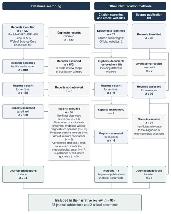
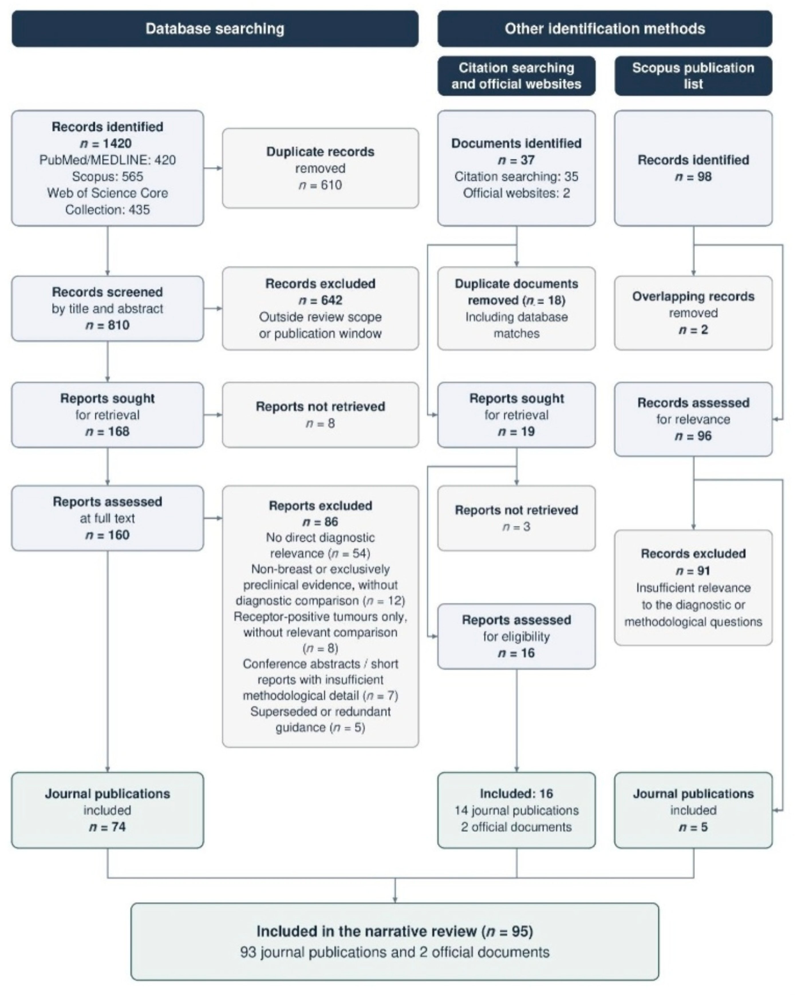
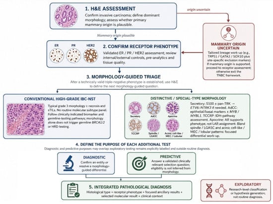
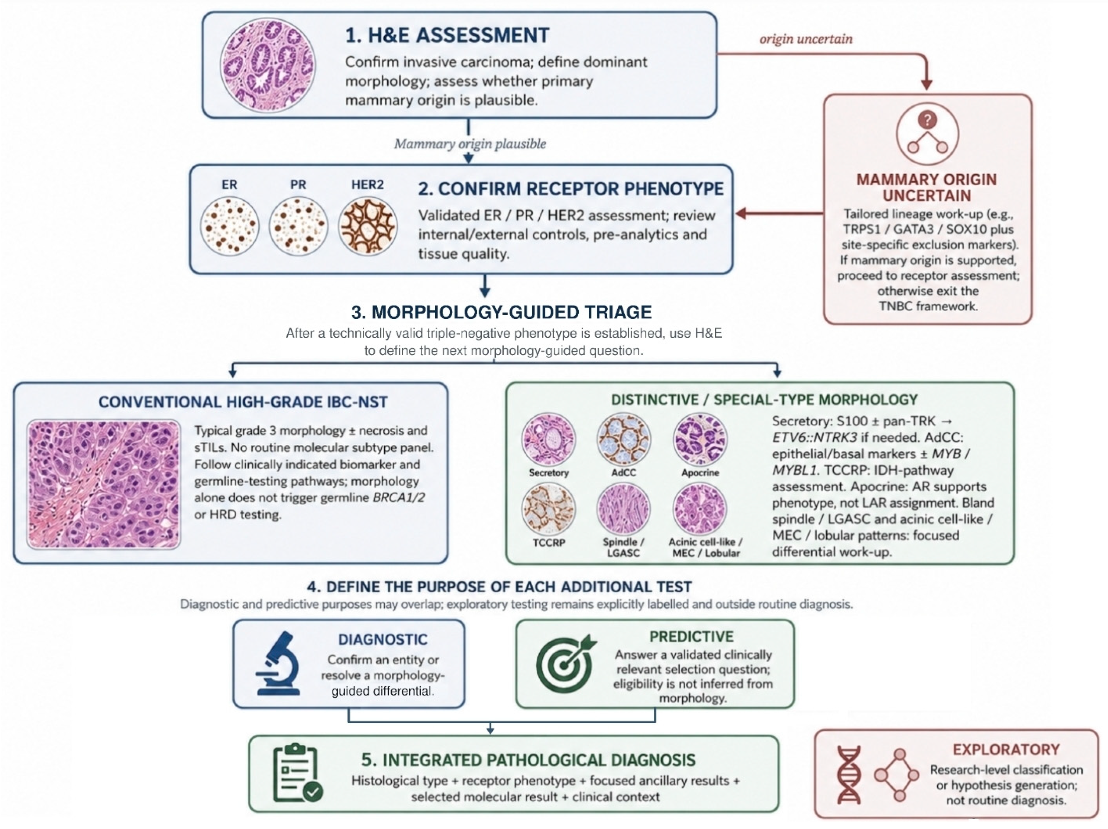
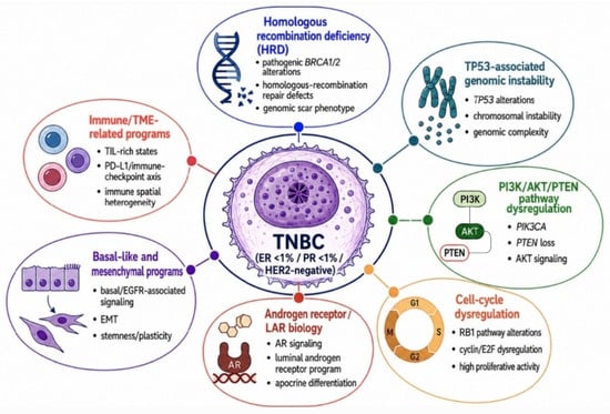
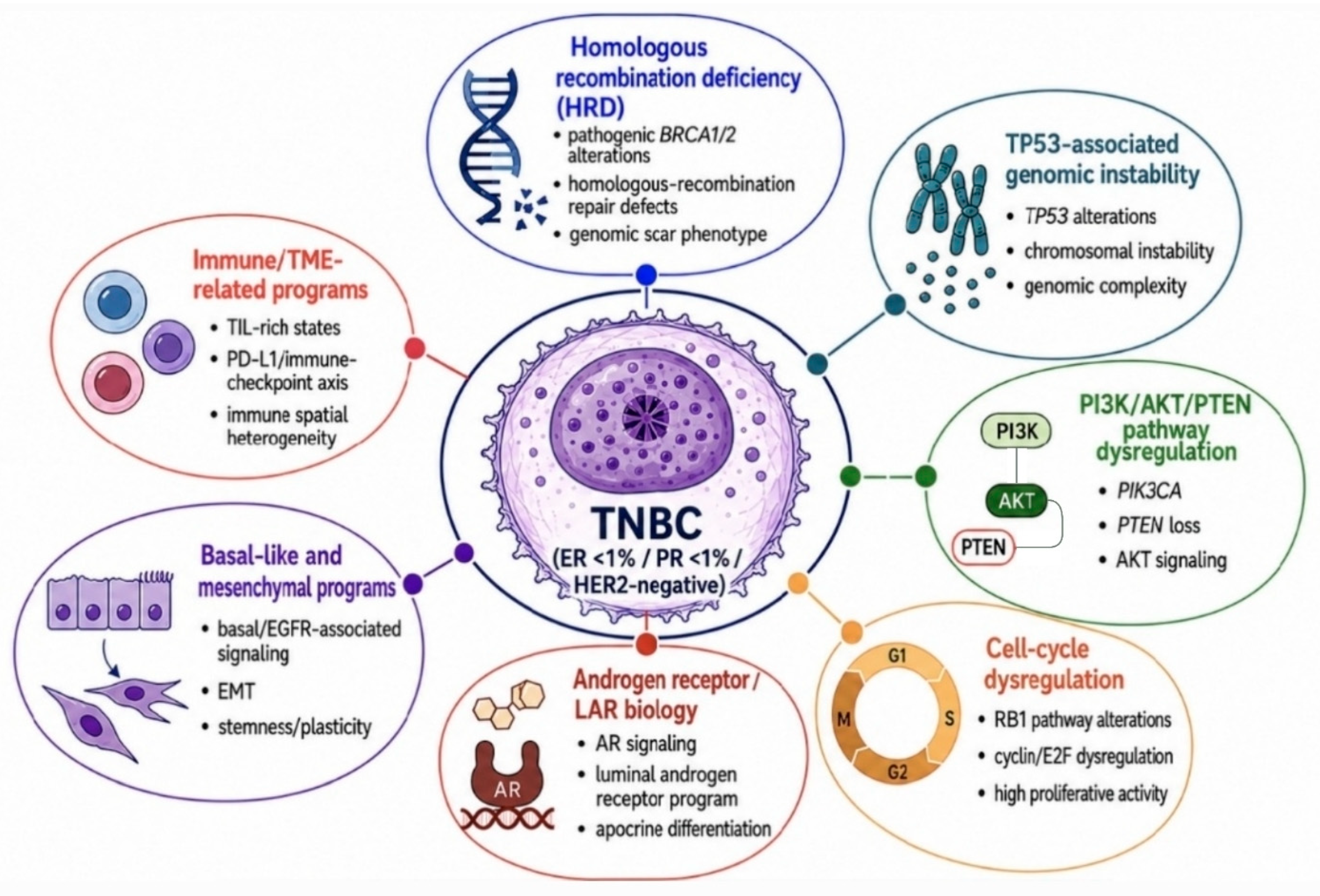

Disclaimer/Publisher’s Note: The statements, opinions and data contained in all publications are solely those of the individual author(s) and contributor(s) and not of MDPI and/or the editor(s). MDPI and/or the editor(s) disclaim responsibility for any injury to people or property resulting from any ideas, methods, instructions or products referred to in the content. |
© 2026 by the authors. Licensee MDPI, Basel, Switzerland. This article is an open access article distributed under the terms and conditions of the Creative Commons Attribution (CC BY) license.