


Abstract
Background: To evaluate longitudinal radiographic outcomes, exploratory radiographic response, and planned versus unplanned revision burden after magnetically controlled growing rod treatment in patients with early-onset scoliosis. Methods: We retrospectively reviewed 55 patients with early-onset scoliosis treated with magnetically controlled growing rods at a single center. Complete year-3 radiographic measurements were available for 40 patients; therefore, year-3 estimates were conditional on imaging availability. Cobb angle, T1–S1 length, and apical vertebral translation (AVT) were assessed preoperatively and at postoperative years 1, 2, and 3. Longitudinal changes were evaluated using generalized estimating equations. Exploratory radiographic responder endpoints included ≥20° Cobb correction, ≥30% Cobb correction, and a ≥30 mm preoperative-to-year-3 T1–S1 length difference. Freedom from unplanned revision and composite endpoints were also evaluated. The radiographic thresholds were pragmatic and were not validated measures of clinically important improvement. Results: The cohort included 33 female and 22 male patients, with a mean age at surgery of 8.10 ± 1.86 years. Neuromuscular and congenital scoliosis accounted for 41.8% and 40.0% of cases, respectively, and dual-rod constructs were used in 83.6%. At year 3, mean Cobb angle was 40.15° ± 19.03°, mean radiographic T1–S1 length was 383.02 ± 54.42 mm, and mean AVT was 32.80 ± 23.56 mm among 40 patients with complete measurements. GEE models showed significant reductions in Cobb angle and AVT and increases in T1–S1 length at each postoperative year (all p < 0.001). At year 3, ≥20° Cobb correction, ≥30% Cobb correction, and ≥30 mm preoperative-to-year-3 T1–S1 length difference were achieved in 50.0%, 55.0%, and 67.5% of patients, respectively; 35.0% achieved both radiographic thresholds and 25.0% achieved both thresholds with no recorded unplanned revision. Patient-level fields recorded any complication in 20.0%, unplanned revision in 16.4%, internal revision classification in 43.6%, and definitive fusion in 16.4%; these fields were not treated as mutually exclusive. Conclusions: MCGR treatment was associated with longitudinal radiographic improvement among patients with available measurements. The preoperative-to-year-3 T1–S1 difference includes the effect of index surgery and should not be interpreted as biological spinal growth alone. These findings suggest that MCGR success should be assessed not only by curve correction, but also by measured spinal-length gain and revision burden.
1. Introduction
Early-onset scoliosis (EOS) comprises a heterogeneous spectrum of spinal deformities diagnosed before 10 years of age and may adversely affect thoracic growth, pulmonary development, and global spinal alignment when progressive curves remain untreated [1]. The objective of growth-friendly surgery is to control coronal deformity and preserve spinal and thoracic growth until definitive fusion [1,2]. Traditional growing rods are a standard distraction-based strategy; however, repeated open lengthenings are associated with cumulative anesthetic exposure, morbidity, implant complications, and substantial treatment burden [2,3].
Magnetically controlled growing rods (MCGRs) reduce this burden by enabling noninvasive outpatient distraction while maintaining growth-friendly deformity control [3,4]. Early clinical series and comparative studies have shown that MCGR treatment can achieve meaningful coronal curve correction and spinal height gain in selected patients with EOS, while reducing the need for scheduled open lengthening procedures compared with traditional growing rods [3,4,5]. These advantages have established MCGR as an important surgical option in progressive EOS.
Nevertheless, the clinical effectiveness of MCGR treatment cannot be judged solely by the magnitude of initial radiographic correction. Durability of correction, maintenance of spinal height gain, and cumulative revision burden are equally important determinants of treatment success. Implant-related complications, including distraction failure, rod fracture, fixation failure, anchor pull-out, infection, and loss of correction, remain clinically relevant limitations [6,7,8]. Unplanned reoperations due to failed distraction, proximal foundation failure, rod breakage, or infection may offset part of the intended benefit of avoiding repeated planned open lengthenings [7]. Systematic review data have similarly demonstrated substantial complication and unplanned revision rates after MCGR treatment, supporting the need to evaluate outcomes beyond early coronal correction alone [8].
Longitudinal assessment is particularly important because MCGR construct performance may evolve over time. Progressive reduction in achieved distraction, often discussed as “diminishing returns,” may limit spinal height gain and contribute to treatment failure during longer follow-up [9]. In addition, scoliosis etiology, baseline curve magnitude, rod configuration, and previous growth-friendly surgery may influence both radiographic response and mechanical complication risk [6,8,10]. Therefore, studies integrating longitudinal radiographic outcomes with patient-level response measures and planned versus unplanned revision patterns are needed to better define the real-world effectiveness of MCGR treatment in EOS.
The present study aimed to describe longitudinal radiographic outcomes and revision burden after MCGR treatment for EOS in a single-center cohort. We evaluated serial Cobb angle, T1–S1 length, and apical vertebral translation measurements, together with year-3 radiographic changes and exploratory responder endpoints. Complications, revision procedures, and definitive fusion were summarized using separately recorded patient-level variables. We also described outcomes by scoliosis etiology and year-3 radiographic availability and explored the association between baseline Cobb angle and time to unplanned revision.
2. Materials and Methods
2.1. Study Design and Ethical Approval
This single-center retrospective cohort study evaluated longitudinal radiographic outcomes, exploratory radiographic response, and revision burden after magnetically controlled growing rod (MCGR/MAGEC) (MAGnetic Expansion Control; NuVasive, Audubon, PA, USA) treatment for early-onset scoliosis (EOS). Patients who underwent MCGR/MAGEC instrumentation between January 2020 and December 2024 were screened for eligibility. The Institutional Ethics Committee of Ankara Bilkent City Hospital approved this study (TABED 1-26-2713; 17 June 2026). Informed consent was waived due to the retrospective design.
2.2. Patient Population
Patients with EOS who underwent MCGR/MAGEC treatment at our institution and had available clinical and/or radiographic follow-up data were included. EOS was defined as scoliosis diagnosed before 10 years of age. A total of 55 patients constituted the final cohort.
Demographic, etiological, surgical, and clinical variables were retrospectively collected from medical records, operative reports, outpatient notes, and radiographic archives. Variables included age at index MCGR surgery, sex, scoliosis etiology, surgery date, follow-up duration, primary versus revision/conversion MCGR implantation, previous external revision or conversion history, rod configuration, number of rods, and previous growth-friendly surgery when available. Etiology was categorized as neuromuscular, congenital, idiopathic, or other/unclear, and rod configuration as single-rod or dual-rod instrumentation. Nutritional status variables, including BMI-for-age z-score and weight-for-age z-score, were not systematically available in the source records and were not analyzed.
The decision to perform MCGR implantation, select rod configuration and anchor strategy, and schedule subsequent procedures was based on patient characteristics, curve pattern, remaining growth potential, previous surgical history, and surgeon judgment. Postoperative distractions were performed during outpatient follow-up according to institutional practice and individualized decision-making. Because detailed distraction-session data were not consistently available, the analysis focused on serial radiographic outcomes and revision burden rather than achieved distraction per session. The institutional lengthening protocol consisted of outpatient distraction according to the treating surgeon’s schedule and individualized clinical/radiographic assessment. Detailed session-level distraction dates, achieved millimeters per session, and standardized frequency were not consistently available. Planned rod or system exchange was defined as a procedure recorded as part of the expected growth-friendly pathway; the specific operative indication for each exchange was not consistently documented and was therefore not inferred retrospectively.
2.3. Inclusion and Exclusion Criteria
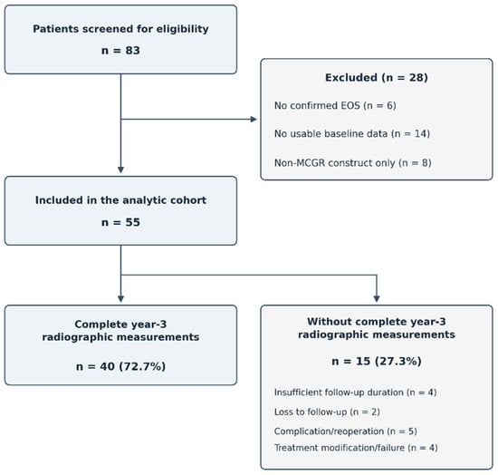
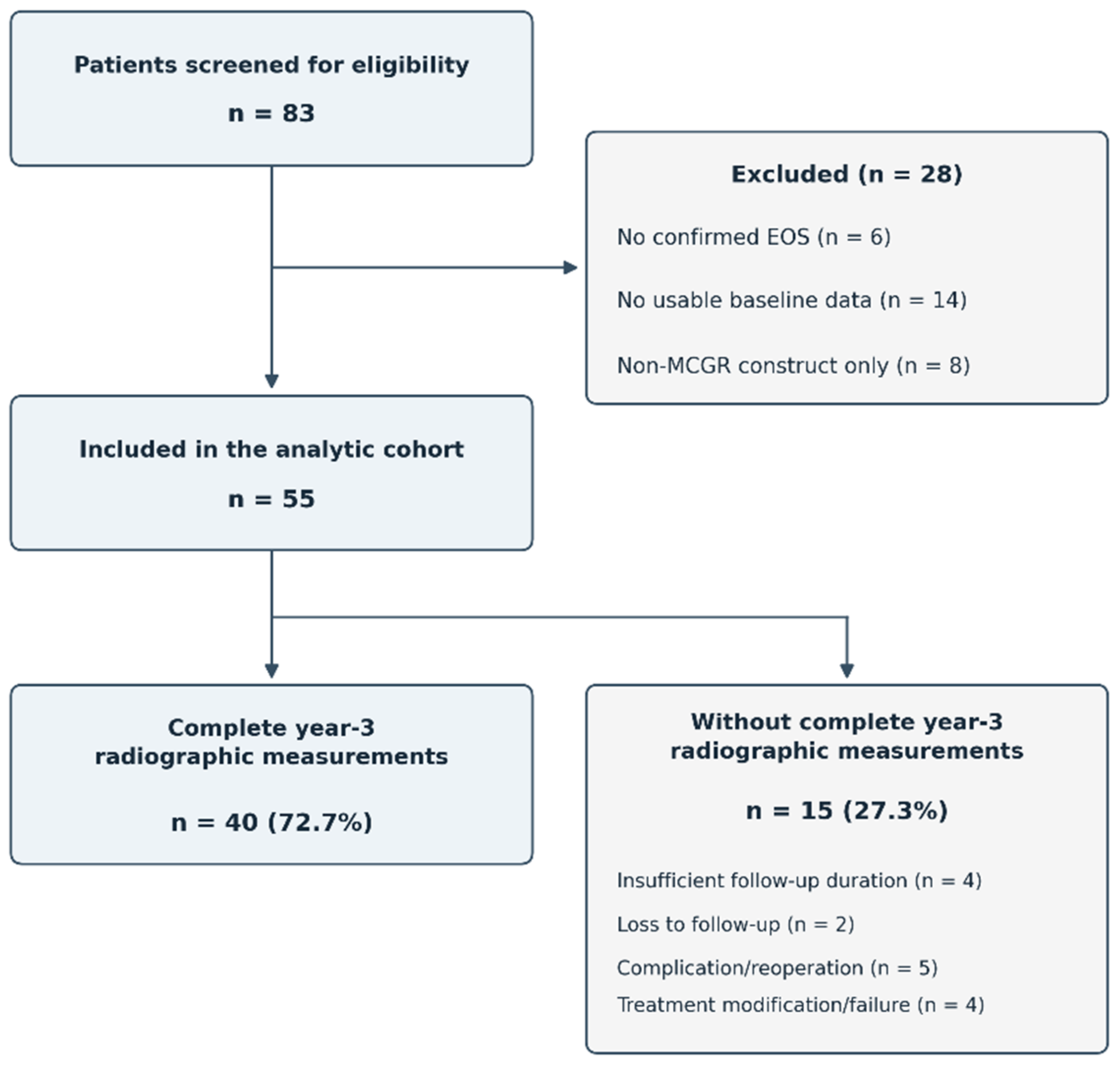
Patients were eligible if they had (1) confirmed EOS, (2) MCGR/MAGEC treatment at our institution, (3) available surgical and/or follow-up records, and (4) at least one evaluable preoperative radiographic parameter, including Cobb angle, T1–S1 length, or apical vertebral translation (AVT). The institutional screening log identified 83 patients who were assessed for eligibility for the present study. Of these, 28 were excluded before formation of the analytic cohort: six because EOS could not be confirmed, 14 because usable baseline data were unavailable, and eight because a non-MCGR construct was used exclusively. The final analytic cohort therefore comprised 55 patients (Figure 1).
Figure 1.
Patient selection and study cohort flowchart.
2.4. Radiographic Assessment
Radiographic assessment was performed using routinely obtained standing whole-spine scoliosis radiographs and institutional radiographic records. The main parameters were Cobb angle, T1–S1 length, and AVT, recorded preoperatively and at postoperative years 1, 2, and 3 when available. Patients with partially available repeated measurements were retained in longitudinal analyses. Complete year-3 radiographic measurements were available for 40/55 patients (72.7%). Baseline characteristics, follow-up duration, and recorded outcomes were descriptively compared between patients with and without year-3 radiographs. The primary recorded reasons for unavailable year-3 radiographs were identified during the updated review of follow-up records and summarized as mutually exclusive categories. These reasons could not be reliably cross-tabulated by scoliosis etiology in the available extraction. Comparisons between patients with and without year-3 radiographs were descriptive; no inferential testing was performed. The comparison was interpreted cautiously because the unavailable group was small and the age field showed inconsistencies with date-derived age.
Cobb angle quantified coronal angular deformity, T1–S1 length served a radiographic measure of spinal height, and AVT assessed coronal translational alignment. All measurements were performed using Surgimap software (version 2.3.2.1) by a spine surgeon experienced in spinal deformity assessment.
2.5. Outcomes
The primary outcomes were longitudinal changes in Cobb angle and T1–S1 length from baseline to postoperative years 1, 2, and 3. Secondary outcomes included longitudinal changes in AVT, absolute and percentage Cobb correction at year 3, preoperative-to-year-3 and first-postoperative-to-year-3 T1–S1 length differences, and preoperative-to-year-3 AVT improvement. Exploratory radiographic responder endpoints were defined as ≥20° absolute Cobb correction, ≥30% Cobb correction, and a ≥30 mm preoperative-to-year-3 T1–S1 length difference. These pragmatic thresholds were used to describe the proportions of patients achieving specified radiographic changes and should not be interpreted as validated minimal clinically important differences. A dual radiographic response was defined as achievement of both ≥30% Cobb correction and a ≥30 mm preoperative-to-year-3 T1–S1 length difference. An additional composite endpoint combined this dual radiographic response with the absence of a recorded unplanned revision during follow-up. All year-3 responder analyses were restricted to patients with complete year-3 radiographic measurements.
2.6. Revision and Complication Assessment
Revision status was evaluated at two levels. Previous external revision or conversion history was recorded for patients who had undergone prior scoliosis or growth-friendly surgery elsewhere or underwent MCGR implantation as a revision/conversion procedure; these were considered baseline surgical-history variables and were not counted as post-index revision outcomes. All additional surgical procedures performed at our institution after index MCGR implantation were recorded as post-index internal revisions and classified as planned or unplanned.
Planned post-index revisions were procedures considered part of the expected growth-friendly treatment pathway, including planned rod exchange, system exchange, or final definitive surgery. Definitive fusion was considered an expected endpoint of growth-friendly treatment rather than a complication-related treatment failure. Because rod exchange, system exchange, definitive fusion, internal revision classification, external revision classification, and unplanned revision were recorded in separate fields that could overlap, they were not combined into a single planned-versus-unplanned procedure total. Unplanned revisions were procedures performed for unexpected clinical or radiographic problems, including infection, wound complications, implant failure, screw pull-out, rod fracture, system removal, loss of correction, or other unforeseen complications. Complications were recorded from medical records, operative notes, and follow-up documentation. Planned and unplanned revisions were reported separately to distinguish expected staged management from unexpected revision-related treatment failure.
2.7. Statistical Analysis
Continuous variables were summarized as mean ± standard deviation (SD) and, where appropriate, median and interquartile range (IQR). Categorical variables were summarized as counts and percentages. The number of available observations was reported for each outcome and time point because radiographic data availability varied during follow-up. Age recorded in months was converted to years by dividing by 12. Normality of within-patient differences was assessed using the Shapiro–Wilk test. Paired changes in Cobb angle, T1–S1 length, and apical vertebral translation (AVT) from preoperative measurements were evaluated at each postoperative assessment using the Wilcoxon signed-rank test. p values were adjusted using the Holm method across the four postoperative comparisons within each outcome. Longitudinal changes in Cobb angle, T1–S1 length, and AVT were assessed using separate generalized estimating equation (GEE) models. Time was entered as a categorical predictor, with the preoperative assessment as the reference category. Models used a Gaussian distribution, an identity link, an exchangeable working correlation structure, and robust sandwich standard errors. All available repeated measurements were included. Estimates are reported as mean differences with 95% confidence intervals (CIs). Because measurements were not available for every patient at every time point, the estimates may be affected if missingness was informative. Year-3 radiographic outcomes were calculated for patients with available year-3 measurements. Responder endpoints were summarized as counts and percentages; the thresholds were exploratory. Outcomes stratified by scoliosis etiology and by year-3 radiograph availability were summarized descriptively, without inferential subgroup comparisons. Unplanned revision-free survival was estimated using the Kaplan–Meier method and compared between baseline Cobb-angle groups (<60° and ≥60°) using the log-rank test. The ≥60° cutoff was exploratory. Separate univariable Cox proportional hazards models were used to estimate the hazard ratio (HR) for Cobb angle ≥ 60° versus <60° and the HR per 10° increase in baseline Cobb angle. Cox estimates were considered exploratory because few unplanned revision events were recorded. All tests were two-sided, and p < 0.05 was considered statistically significant. Holm-adjusted p values were used for the paired comparisons as described above. Statistical analyses were performed using R version 4.3.3 (R Foundation for Statistical Computing, Vienna, Austria). Figures were generated using Python version 3.12.14 and Matplotlib version 3.10.8.
3. Results
3.1. Patient Cohort and Follow-Up Availability
The cohort included 55 patients. Mean age at surgery was 8.10 ± 1.86 years; 33 patients (60.0%) were female and 22 (40.0%) were male. Neuromuscular scoliosis was present in 23 patients (41.8%), congenital scoliosis in 22 (40.0%), idiopathic scoliosis in 7 (12.7%), and other/unclear etiologies in 3 (5.5%). Dual-rod constructs were used in 46 patients (83.6%) and single-rod constructs in 9 (16.4%) (Table 1). Complete year-3 radiographic measurements were available for 40/55 patients (72.7%). The year-3 summaries and all derived year-3 response endpoints were therefore conditional on complete year-3 imaging.
Table 1.
Baseline characteristics and radiological follow-up availability.
Patient flow was as follows: 83 patients were initially identified; 28 were excluded before analysis (no confirmed EOS, n = 6; no usable baseline data, n = 14; non-MCGR construct, n = 8); and 55 patients were included in the final cohort. Among the 55 included patients, 40 (72.7%) had complete year-3 radiographic measurements and 15 (27.3%) did not. The primary recorded reasons for unavailable year-3 imaging were insufficient follow-up duration (n = 4), loss to follow-up (n = 2), complication/reoperation (n = 5), and treatment modification/failure (n = 4). Each patient was assigned to one primary reason category. Insufficient follow-up duration was distinguished from loss to follow-up, and absence of year-3 imaging was not itself considered evidence of treatment failure.
3.2. Longitudinal Radiographic Outcomes
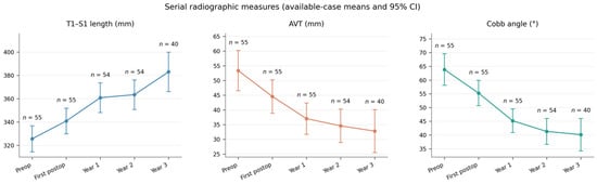
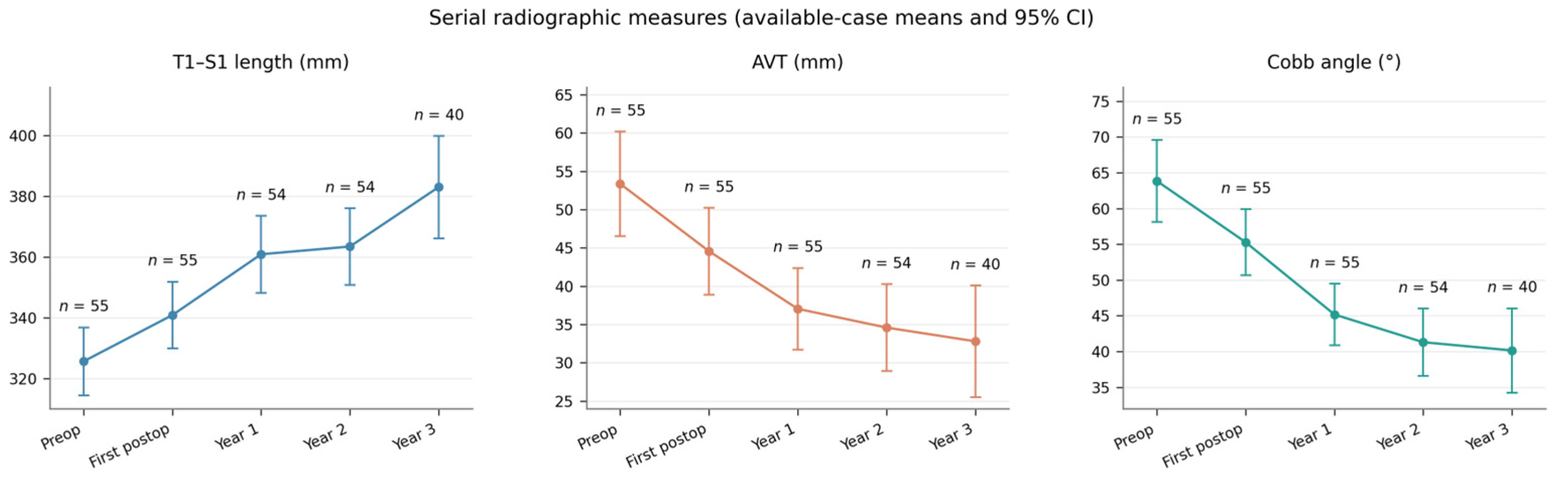
Radiographic measurements were available for all 55 patients at baseline and at the first postoperative assessment. At year 1, Cobb angle and AVT were available for 55 patients and T1–S1 length for 54; at year 2, 54 patients had Cobb angle, T1–S1 length, and AVT data; and at year 3, 40 patients had complete measurements for all three outcomes (Table 2, Figure 2).
Table 2.
Serial radiographic summaries.
Figure 2.
Serial radiographic measurements from the preoperative assessment to postoperative year 3. Points represent available-case means, and error bars indicate 95% confidence intervals for these means. The number of patients contributing to each estimate is shown at each time point. These descriptive summaries correspond to Table 2 and do not represent within-patient paired changes or GEE-estimated changes. Lower Cobb angle and apical vertebral translation (AVT) indicate improvement, whereas higher T1–S1 length indicates greater measured spinal length.
Available-case mean T1–S1 length increased from 325.60 mm preoperatively to 340.84 mm at the first postoperative assessment and reached 383.02 mm at year 3. These descriptive means were calculated from the patients with measurements available at each time point; differences between these means should therefore not be interpreted as within-patient length changes. Cobb angle and AVT decreased, with most of the observed mean reduction occurring by year 1 and smaller decreases thereafter. These are available-case descriptive patterns; the year-3 summaries are conditional on measurements being available.
3.3. Paired Changes and GEE Models
Within-patient paired analyses demonstrated significant changes from preoperative values at the first postoperative assessment and at years 1, 2, and 3 for all three radiographic outcomes (all Holm-adjusted p < 0.001; Supplementary Table S1). At year 3, the median paired change was −20.00° for Cobb angle, +58.00 mm for T1–S1 length, and −19.50 mm for AVT among 40 paired observations. Longitudinal changes were evaluated using separate GEE models including all available repeated measurements from the 55 patients. Time was entered as a categorical predictor, the preoperative measurement was the reference, an exchangeable working correlation structure was used, and robust sandwich standard errors were calculated. Compared with preoperative values, Cobb angle and AVT decreased and T1–S1 length increased at each postoperative year (all p < 0.001; Table 3). Estimated year-3 changes were −23.70° for Cobb angle, +53.06 mm for T1–S1 length, and −21.11 mm for AVT.
Table 3.
GEE estimates of radiographic changes from preoperative measurements.
3.4. Year-3-Derived Outcomes and Exploratory Response Endpoints
Among the 40 patients with complete year-3 radiographic measurements, mean Cobb correction was 22.93° ± 20.70°, mean percentage Cobb correction was 33.60% ± 28.33%, mean preoperative-to-year-3 T1–S1 length difference was 53.75 ± 40.01 mm, mean first-postoperative-to-year-3 T1–S1 length difference was 36.53 ± 37.73 mm, and mean AVT improvement was 20.33 ± 22.36 mm (Table 4). The preoperative-to-year-3 T1–S1 difference includes the index surgical change and subsequent measured change; it should not be interpreted as biological spinal growth alone.
Table 4.
Year-3 radiographic outcomes and exploratory response endpoints.
The exploratory thresholds were achieved by 20/40 patients (50.0%) for ≥20° Cobb correction, 22/40 (55.0%) for ≥30% Cobb correction, and 27/40 (67.5%) for ≥30 mm preoperative-to-year-3 T1–S1 length difference. Both radiographic thresholds were achieved by 14/40 patients (35.0%), and both thresholds plus no recorded unplanned revision were achieved by 10/40 (25.0%).
3.5. Complications, Revision Fields, and Definitive Fusion
Eleven patients (20.0%) had a recorded complication. Infection-related complications were most frequent (8/11; 14.5% of the cohort), followed by screw pull-out (2/55; 3.6%) and rod fracture (1/55; 1.8%). Complication management included debridement in 6/11 patients, antibiotics in 2/11, screw revision in 2/11, and rod exchange in 1/11. Nine patients (16.4%) were flagged in the patient-level unplanned-revision field. Recorded indications were superficial infection/wound complications in 6/9, screw pull-out in 2/9, and rod fracture in 1/9. Internal revision classification was recorded for 24 patients (43.6%), external revision classification for 7 (12.7%), and no internal/external revision classification for 24 (43.6%). Rod exchange was recorded in 16 patients (29.1%), system exchange in 6 (10.9%), and definitive fusion in 9 (16.4%) (Table 5). These fields were recorded separately and may overlap; they were therefore not combined into a single planned-versus-unplanned procedure total.
Table 5.
Patient-level complications and revision records.
3.6. Exploratory Outcomes by Etiology
Year-3 radiographs were available for 18/23 neuromuscular patients (78.3%), 16/22 congenital patients (72.7%), 6/7 idiopathic patients (85.7%), and 0/3 patients with other/unclear etiology. Any complication was recorded in 4/23 neuromuscular patients (17.4%), 4/22 congenital patients (18.2%), 3/7 idiopathic patients (42.9%), and 0/3 other/unclear patients. Unplanned revision was recorded in 3/23 neuromuscular patients (13.0%), 3/22 congenital patients (13.6%), 3/7 idiopathic patients (42.9%), and 0/3 other/unclear patients (Table 6). These subgroup summaries were descriptive; no inferential comparisons were performed because of small group sizes.
Table 6.
Exploratory outcomes by scoliosis etiology.
3.7. Comparison by Year-3 Radiographic Availability
Patients with year-3 radiographs (n = 40) and those without year-3 radiographs (n = 15) had broadly comparable baseline radiographic means. Mean age was 8.30 ± 1.86 versus 7.56 ± 1.81 years, mean preoperative Cobb angle was 63.08° ± 22.67° versus 65.93° ± 19.69°, mean preoperative T1–S1 length was 329.28 ± 43.17 versus 315.80 ± 40.01 mm, and mean preoperative AVT was 53.13 ± 26.79 versus 54.00 ± 23.99 mm. Recorded complication proportions were identical (20.0% in each group). The year-3-available group had longer recorded follow-up (45.38 ± 13.11 vs. 27.07 ± 4.03 months) (Table 7).
Table 7.
Characteristics and recorded outcomes by year-3 radiographic availability.
3.8. Exploratory Unplanned Revision-Free Survival Analysis
In the analysis, nine patients were classified as having an unplanned revision. Estimated unplanned revision-free survival for the overall cohort was 89.1%, 87.2%, and 82.6% at 1, 2, and 3 years, respectively. At 3 years, survival was 95.7% in the preoperative Cobb < 60° group and 70.8% in the Cobb ≥ 60° group; the log-rank p value was 0.017. In separate exploratory univariable Cox models, preoperative Cobb ≥ 60° was associated with a higher hazard of unplanned revision than Cobb < 60° (HR 8.30, 95% CI 1.04–66.44; p = 0.046). For each 10° increase in preoperative Cobb angle, the estimated HR was 1.28 (95% CI 0.96–1.69; p = 0.090) (Table 8). These estimates are hypothesis-generating because few events occurred and confidence intervals were wide.
Table 8.
Exploratory univariable Cox proportional hazards analyses of unplanned revision.
4. Discussion
In this single-center longitudinal cohort of 55 patients with early-onset scoliosis treated with magnetically controlled growing rods, MCGR treatment was associated with longitudinal radiographic improvement among patients with available 3-year measurements. The principal findings were a mean year-3 Cobb angle of 40.15°, a mean radiographic T1–S1 length of 383.02 mm, and a mean AVT of 32.80 mm among the 40 patients with complete year-3 measurements. Model-based longitudinal analyses confirmed significant improvements in Cobb angle, T1–S1 length, and AVT at each postoperative time point compared with baseline. These findings support MCGR as a growth-friendly technique capable of providing coronal deformity correction, radiographic spinal-length increase, and improved coronal alignment in a clinically heterogeneous EOS population. However, these radiographic findings should be interpreted together with the cumulative surgical burden and incomplete follow-up. Patient-level records showed internal revision classification in 43.6%, unplanned revision in 16.4%, and definitive fusion in 16.4%; these fields were recorded separately and were not mutually exclusive. Definitive fusion represents an expected endpoint of growth-friendly treatment and should not automatically be interpreted as treatment failure. The cohort derivation identified 83 patients initially and excluded 28 before the final 55-patient analytic cohort. Complete year-3 radiographic measurements were available for 40 patients, whereas 15 did not have complete year-3 imaging. Baseline radiographic means and recorded complication proportions were broadly similar between patients with and without year-3 imaging, although the unavailable group had shorter recorded follow-up. This descriptive comparison reduces, but does not eliminate, concern about attrition or survivorship bias. Because no inferential comparison was performed and the unavailable group was small, year-3 findings and all responder rates remain conditional on continued availability of adequate radiographic follow-up.
Our radiographic improvements are consistent with previous MCGR studies [3,4,5,11,12,13,14,15]. In the present dataset, the mean preoperative-to-year-3 Cobb correction was 22.93°, the mean percentage Cobb correction was 33.60%, and the mean preoperative-to-year-3 T1–S1 length difference was 53.75 mm, paralleling prior reports of coronal deformity reduction and spinal height gain [5,10,11,12,13,14,15]. The observed T1–S1 length difference is clinically relevant because the aim of growth-friendly surgery is not only angular correction, but also preservation of longitudinal spinal and thoracic growth until skeletal maturity or definitive fusion becomes appropriate [1,2,12]. Nevertheless, the measured preoperative-to-year-3 difference includes the index surgical correction and should not be interpreted as biological spinal growth alone.
A key strength is the longitudinal evaluation of radiographic outcomes, as MCGR construct performance may change over time. Gardner et al. questioned whether the “law of diminishing returns,” originally described in traditional growing rods, also applies to MCGR lengthening [9]. Longer-term MCGR studies have emphasized that radiographic gains and implant-related events should be assessed across the entire treatment course rather than at a single endpoint [16]. Sankar et al. demonstrated progressively smaller T1–S1 gains with repeated lengthenings of dual growing rods [17]. In our cohort, the largest improvements in Cobb angle and AVT occurred early, whereas measured T1–S1 length continued to increase through year 3. The preoperative-to-year-3 T1–S1 length difference was 53.75 mm, whereas the first-postoperative-to-year-3 difference was 36.53 mm. This distinction is important: the former includes the index surgical correction, while neither measure can be interpreted as biological spinal growth alone without a complete distraction and imaging history. Accordingly, the present study reports measured radiographic length differences and T1–S1 length gain rather than pure biological growth.
Responder analyses were exploratory. Fifty percent achieved ≥20° Cobb correction, 55.0% achieved ≥30% correction, and 67.5% achieved a ≥30 mm preoperative-to-year-3 T1–S1 length difference. Both radiographic thresholds were achieved by 35.0%, and both thresholds plus no recorded unplanned revision by 25.0%. These proportions are descriptive and should not be interpreted as validated clinical success rates. The ≥20° Cobb, ≥30% Cobb, and ≥30 mm T1–S1 thresholds were pragmatic exploratory thresholds rather than validated minimal clinically important differences. Similarly, the 60° preoperative Cobb cutoff was used as a pragmatic descriptive stratification threshold, not as a validated or outcome-optimized cutoff.
Revision burden remains central when interpreting MCGR efficacy. The theoretical advantage of MCGR over traditional growing rods is reduction in scheduled open lengthening procedures [3,4,12]. However, MCGR constructs remain vulnerable to mechanical and biological complications, including rod fracture, distraction failure, anchor or foundation failure, infection, implant wear, and loss of correction [6,7,8,18,19,20,21,22,23,24]. In the present cohort, 83.6% of patients were free of a recorded unplanned revision, whereas 16.4% had an unplanned-revision flag. However, post-index internal revision classification was recorded in 43.6%, indicating that MCGR treatment remains surgically demanding even when unexpected reoperations are less frequent. This should not be interpreted as construct failure alone, because the revision fields were separate and most recorded procedure-type fields reflected the staged nature of growth-friendly treatment. In addition, 12.7% of patients had previous external revision or conversion history, representing baseline surgical complexity rather than a post-index outcome. Therefore, previous/conversion surgery, planned post-index revision, definitive fusion, and unplanned post-index revision should be reported separately to distinguish baseline complexity, expected treatment-pathway procedures, expected definitive treatment, and unexpected revision-related clinical events.
Our 20.0% complication rate was lower than several published series [6,8,10,13,19,20,21,25], possibly reflecting variability in definitions, techniques, and case mix. Moreover, biological and implant-related concerns such as wear debris, metallosis, and serum metal ion changes have emerged as important long-term issues in MCGR treatment [22,23,24]. These outcomes were not available in the present cohort but remain relevant for family counseling and long-term surveillance. The patient-level complication data showed 11 recorded complications: eight infection-related events, two screw pull-outs, and one rod fracture. Management included debridement, antibiotics, screw revision, and rod exchange. The timing of individual complications was not reliably preserved in the structured retrospective extraction and is therefore not interpreted in detail. Nine patients had an unplanned-revision flag, 24 had internal revision classification, and nine had definitive-fusion fields. These variables were not mutually exclusive and were recorded in separate data fields. Definitive fusion represents an expected endpoint of growth-friendly treatment and should not be equated with complication-related treatment failure.
The survival analysis suggested a possible association between greater baseline Cobb severity and unplanned revision, but this finding is exploratory and accompanied by wide confidence intervals. It should be considered hypothesis-generating rather than a validated clinical prediction rule Given the small number of unplanned-revision events, the Kaplan–Meier estimates and Cox associations should be interpreted cautiously, and the findings remain exploratory and hypothesis-generating. The baseline Cobb stratification should not be described as a clinical risk signal or prediction rule. Outcomes by scoliosis etiology were summarized descriptively. The small and heterogeneous etiological subgroups preclude firm conclusions regarding differences between etiologies. Year-3 radiographs were available in 18/23 neuromuscular patients, 16/22 congenital patients, 6/7 idiopathic patients, and 0/3 patients with other/unclear etiology. Any complication and unplanned revisions were more frequent numerically in the idiopathic subgroup, but these observations are descriptive and should not be interpreted as evidence of etiology-specific risk.
Limitations
The study remains limited by its retrospective single-center design and incomplete year-3 imaging. Although the primary reasons for unavailable year-3 imaging were identified, their distribution across etiological subgroups could not be reliably established from the available extraction. Comparisons between patients with and without year-3 radiographs were descriptive, the unavailable group was small, and the age field contained inconsistencies. Some missing assessments were attributed to complications, reoperations, or treatment modification/failure, raising concern that imaging availability may have been related to the treatment course. Therefore, descriptive similarities in baseline characteristics do not exclude attrition or survivorship bias, and year-3 outcomes should be interpreted as conditional on radiographic availability. T1–S1 changes were measured radiographic length differences. Immediate surgical correction, subsequent distraction, and growth-related change could not be completely separated, and the preoperative-to-year-3 difference should not be interpreted as biological spinal growth alone. Responder cutoffs were pragmatic and exploratory rather than validated minimal clinically important differences. Only nine unplanned revisions were recorded, limiting the stability of survival and Cox estimates despite the use of robust descriptive interpretation. Separate revision, procedure-type, unplanned-revision, and definitive-fusion fields could overlap and could not be collapsed into a mutually exclusive procedure pathway. Patient-reported outcomes, pulmonary function, health-related quality of life, distraction compliance, implant wear, metallosis, and serum metal ion levels were unavailable [22,23,24]. Measurements were performed by a single surgeon using Surgimap, and interobserver reliability was not assessed. Baseline and longitudinal nutritional status, including BMI-for-age and weight-for-age z-scores, was not available. Therefore, the association between nutritional status and complications, revision surgery, follow-up completion, or change across etiological subgroups could not be assessed. The median follow-up by year-3 availability, exact timing of individual complications, and patient-level indications for planned exchanges could not be reconstructed reliably from the available retrospective extraction. These limitations constrain detailed time-to-event and procedure-pathway interpretation.
5. Conclusions
In this retrospective EOS cohort, MCGR treatment was associated with improved Cobb angle, measured radiographic T1–S1 length, and AVT through three years among patients with available measurements. The preoperative-to-year-3 T1–S1 difference included the effect of index surgery and should not be interpreted as biological spinal growth alone. Year-3 response estimates were conditional on imaging availability, and revision/fusion fields were not mutually exclusive. Given the incomplete follow-up and small number of unplanned revision events, these findings should be interpreted as descriptive and hypothesis-generating rather than definitive evidence of treatment efficacy or risk.
Supplementary Materials
The following supporting information can be downloaded at: https://www.mdpi.com/article/10.3390/jcm15208049/s1, Table S1: Paired changes from preoperative measurements.
Author Contributions
Conceptualization, Y.U.; methodology, Y.U. and K.C.; formal analysis, Y.U.; investigation, Y.U. and R.K.; data curation, E.E.; writing—original draft preparation, Y.U.; writing—review and editing, Y.U. and E.B.; visualization, Y.U.; supervision, E.B. All authors have read and agreed to the published version of the manuscript.
Funding
This research received no external funding.
Institutional Review Board Statement
The study was conducted in accordance with the Declaration of Helsinki and approved by the Institutional Ethics Committee of Ankara Bilkent City Hospital approved this study (TABED 1-26-2713; 17 June 2026).
Informed Consent Statement
Patient consent was waived owing to the retrospective design of the study and the use of anonymized data.
Data Availability Statement
The data supporting the findings of this study are available from the corresponding author upon reasonable request, subject to institutional and ethical restrictions.
Conflicts of Interest
The authors declare no conflicts of interest.
Abbreviations
| AVT | Apical vertebral translation |
| CI | Confidence interval |
| EOS | Early-onset scoliosis |
| GEE | Generalized estimating equations |
| HR | Hazard ratio |
| MAGEC | Magnetic Expansion Control |
| MCGR | Magnetically controlled growing rod |
| OR | Odds ratio |
| Q1–Q3 | First and third quartiles |
| SD | Standard deviation |
| SE | Standard error |
| T1–S1 | First thoracic vertebra to first sacral vertebra length |
References
- Akbarnia, B.A. Management themes in early onset scoliosis. J. Bone Jt. Surg. Am. 2007, 89, 42–54. [Google Scholar] [CrossRef] [Scilit] [PubMed]
- Helenius, I.J. Standard and magnetically controlled growing rods for the treatment of early onset scoliosis. Ann. Transl. Med. 2020, 8, 26. [Google Scholar] [CrossRef] [Scilit] [PubMed]
- Akbarnia, B.A.; Cheung, K.; Noordeen, H.; Elsebaie, H.; Yazici, M.; Dannawi, Z.; Kabirian, N. Next generation of growth-sparing techniques: Preliminary clinical results of a magnetically controlled growing rod in 14 patients with early-onset scoliosis. Spine 2013, 38, 665–670. [Google Scholar] [CrossRef] [Scilit] [PubMed]
- Akbarnia, B.A.; Pawelek, J.B.; Cheung, K.M.C.; Demirkiran, G.; Elsebaie, H.; Emans, J.B.; Johnston, C.E.; Mundis, G.M.; Noordeen, H.; Skaggs, D.L.; et al. Traditional growing rods versus magnetically controlled growing rods for the surgical treatment of early-onset scoliosis: A case-matched 2-year study. Spine Deform. 2014, 2, 493–497. [Google Scholar] [CrossRef] [Scilit] [PubMed]
- Lebon, J.; Batailler, C.; Wargny, M.; Choufani, E.; Violas, P.; Fron, D.; Kieffer, J.; Accadbled, F.; De Gauzy, J.S. Magnetically controlled growing rod in early onset scoliosis: A 30-case multicenter study. Eur. Spine J. 2017, 26, 1567–1576. [Google Scholar] [CrossRef] [Scilit] [PubMed]
- Choi, E.; Yaszay, B.; Mundis, G.; Hosseini, P.; Pawelek, J.; Alanay, A.; Berk, H.; Cheung, K.; Demirkiran, G.; Ferguson, J.; et al. Implant complications after magnetically controlled growing rods for early onset scoliosis: A multicenter retrospective review. J. Pediatr. Orthop. 2017, 37, e588–e592. [Google Scholar] [CrossRef] [Scilit] [PubMed]
- Kwan, K.Y.H.; Alanay, A.; Yazici, M.; Demirkiran, G.; Helenius, I.; Nnadi, C.; Ferguson, J.; Akbarnia, B.A.; Cheung, J.P.Y.; Cheung, K.M.C. Unplanned reoperations in magnetically controlled growing rod surgery for early onset scoliosis with a minimum of two-year follow-up. Spine 2017, 42, E1410–E1414. [Google Scholar] [CrossRef] [Scilit] [PubMed]
- Thakar, C.; Kieser, D.C.; Mardare, M.; Haleem, S.; Fairbank, J.; Nnadi, C. Systematic review of the complications associated with magnetically controlled growing rods for the treatment of early onset scoliosis. Eur. Spine J. 2018, 27, 2062–2071. [Google Scholar] [CrossRef] [Scilit] [PubMed]
- Gardner, A.; Beaven, A.; Marks, D.; Spilsbury, J.; Mehta, J.; Newton Ede, M. Does the law of diminishing returns apply to the lengthening of the MCGR rod in early onset scoliosis with reference to growth velocity? J. Spine Surg. 2017, 3, 525–530. [Google Scholar] [CrossRef] [Scilit] [PubMed]
- Abdelaal, A.; Munigangaiah, S.; Trivedi, J.; Davidson, N. Magnetically controlled growing rods in the treatment of early onset scoliosis: A single centre experience of 44 patients with mean follow-up of 4.1 years. Bone Jt. Open. 2020, 1, 405–414. [Google Scholar] [CrossRef] [PubMed]
- Tang, N.; Zhao, H.; Shen, J.-X.; Zhang, J.-G.; Li, S.-G. Magnetically controlled growing rod for early-onset scoliosis: Systematic review and meta-analysis. World Neurosurg. 2019, 125, e593–e601. [Google Scholar] [CrossRef] [Scilit] [PubMed]
- Tsirikos, A.I.; Roberts, S.B. Magnetic controlled growth rods in the treatment of scoliosis: Safety, efficacy and patient selection. Med. Devices 2020, 13, 75–85. [Google Scholar] [CrossRef] [Scilit] [PubMed]
- Obid, P.; Yiu, K.; Cheung, K.; Kwan, K.; Ruf, M.; Cheung, J.P.Y. Magnetically controlled growing rods in early onset scoliosis: Radiological results, outcome, and complications in a series of 22 patients. Arch. Orthop. Trauma. Surg. 2021, 141, 1163–1174. [Google Scholar] [CrossRef] [Scilit] [PubMed]
- Bednar, E.D.; Bergin, B.; Kishta, W. Comparison of magnetically controlled growing rods with other distraction-based surgical technologies for early-onset scoliosis: A systematic review and meta-analysis. JBJS Rev. 2021, 9, e20.00062. [Google Scholar] [CrossRef] [Scilit] [PubMed]
- Migliorini, F.; Chiu, W.O.; Scrofani, R.; Chiu, W.K.; Baroncini, A.; Iaconetta, G.; Maffulli, N. Magnetically controlled growing rods in the management of early onset scoliosis: A systematic review. J. Orthop. Surg. Res. 2022, 17, 309. [Google Scholar] [CrossRef] [Scilit] [PubMed]
- Cheung, J.P.Y.; Yiu, K.; Kwan, K.Y.H.; Cheung, K.M.C. Mean 6-year follow-up of magnetically controlled growing rod patients with early onset scoliosis: A glimpse of what happens to graduates. Neurosurgery 2019, 84, 1112–1123. [Google Scholar] [CrossRef] [Scilit] [PubMed]
- Sankar, W.N.; Skaggs, D.L.; Yazici, M.; Johnston, C.E.; Shah, S.A.; Javidan, P.; Kadakia, R.V.; Day, T.F.; Akbarnia, B.A. Lengthening of dual growing rods and the law of diminishing returns. Spine 2011, 36, 806–809. [Google Scholar] [CrossRef] [Scilit] [PubMed]
- Kim, G.; El Sammak, S.; Michalopoulos, G.D.; Mualem, W.; Pinter, Z.W.; Freedman, B.A.; Bydon, M. Comparison of surgical interventions for the treatment of early-onset scoliosis: A systematic review and meta-analysis. J. Neurosurg. Pediatr. 2022, 31, 342–357. [Google Scholar] [CrossRef] [Scilit] [PubMed]
- Ellinger, F.; Tropp, H.; Gerdhem, P.; Hallgren, H.B.; Ivars, K. Magnetically controlled growing rod treatment for early-onset scoliosis: Analysis of 52 consecutive cases demonstrates improvement of coronal deformity. J. Spine Surg. 2023, 9, 259–268. [Google Scholar] [CrossRef] [Scilit] [PubMed]
- Grabala, P.; Gupta, M.C.; Pereira, D.E.; Latalski, M.; Danielewicz, A.; Glowka, P.; Grabala, M. Radiological outcomes of magnetically controlled growing rods for the treatment of children with various etiologies of early-onset scoliosis—A multicenter study. J. Clin. Med. 2024, 13, 1529. [Google Scholar] [CrossRef] [Scilit] [PubMed]
- Glowka, P.; Grabala, P.; Gupta, M.C.; Pereira, D.E.; Latalski, M.; Danielewicz, A.; Grabala, M.; Tomaszewski, M.; Kotwicki, T. Complications and health-related quality of life in children with various etiologies of early-onset scoliosis treated with magnetically controlled growing rods—A multicenter study. J. Clin. Med. 2024, 13, 4068. [Google Scholar] [CrossRef] [Scilit] [PubMed]
- Zhang, T.; Sze, K.Y.; Peng, Z.W.; Cheung, K.M.C.; Lui, Y.F.; Wong, Y.W.; Kwan, K.Y.H.; Cheung, J.P.Y. Systematic investigation of metallosis associated with magnetically controlled growing rod implantation for early-onset scoliosis. Bone Jt. J. 2020, 102-B, 1375–1383. [Google Scholar] [CrossRef] [Scilit] [PubMed]
- Lüders, K.A.; Braunschweig, L.; Zioła-Frankowska, A.; Stojek, A.; Jakkielska, D.; Wichmann, A.; Dihazi, G.H.; Streit, F.; Güsewell, S.E.; Trüe, T.C.; et al. Titanium wear from magnetically controlled growing rods (MCGRs) for the treatment of spinal deformities in children. Sci. Rep. 2022, 12, 10811. [Google Scholar] [CrossRef] [Scilit] [PubMed]
- Cheung, J.P.Y.; Cheung, K.M.C. Current status of the magnetically controlled growing rod in treatment of early-onset scoliosis: What we know after a decade of experience. J. Orthop. Surg. 2019, 27, 2309499019886945. [Google Scholar] [CrossRef] [Scilit] [PubMed]
- Bess, S.; Akbarnia, B.A.; Thompson, G.H.; Sponseller, P.D.; Shah, S.A.; El Sebaie, H.; Boachie-Adjei, O.; Karlin, L.I.; Canale, S.; Poe-Kochert, C.; et al. Complications of growing-rod treatment for early-onset scoliosis: Analysis of one hundred and forty patients. J. Bone Jt. Surg. Am. 2010, 92, 2533–2543. [Google Scholar] [CrossRef] [Scilit] [PubMed]

Disclaimer/Publisher’s Note: The statements, opinions and data contained in all publications are solely those of the individual author(s) and contributor(s) and not of MDPI and/or the editor(s). MDPI and/or the editor(s) disclaim responsibility for any injury to people or property resulting from any ideas, methods, instructions or products referred to in the content. |
© 2026 by the authors. Licensee MDPI, Basel, Switzerland. This article is an open access article distributed under the terms and conditions of the Creative Commons Attribution (CC BY) license.