


Abstract
Background/Objectives: Early risk stratification in acute myocardial infarction (AMI) requires accessible and cost-effective tools. Complete blood count-derived inflammatory indices may provide readily available markers of immunothrombotic activity. The objective of this study was to evaluate the association between inflammatory indices derived from the complete blood count at admission and in-hospital mortality among patients with acute myocardial infarction treated at a high-complexity cardiovascular center. Methods: We conducted a retrospective cohort analysis of prospectively collected data from adults with type 1 AMI treated at a high-complexity cardiovascular center in Colombia between 2021 and 2025. Complete blood count parameters at admission were used to calculate hematological inflammatory indices. The primary outcome was all-cause in-hospital mortality. Associations were evaluated using logistic regression models. Discrimination was assessed using ROC curves, and restricted cubic splines and sensitivity analyses were used to evaluate nonlinearity and robustness. Results: Among 3768 patients, 143 (3.8%) died during hospitalization. Non-survivors had higher NLR, SIRI, SII, and AISI values (all p < 0.001). After adjustment, SIRI (OR 1.94, 95% CI 1.62–2.32), NLR (OR 1.89, 95% CI 1.57–2.27), NPR (OR 1.86, 95% CI 1.56–2.22), and AISI (OR 1.83, 95% CI 1.53–2.19) showed the numerically largest adjusted associations with mortality. Discriminatory performance was modest, with the numerically highest AUROC observed for NPR (0.723), followed by SIRI (0.716) and NLR (0.706). Conclusions: Inflammatory indices were associated with in-hospital mortality in type 1 AMI, but their discriminatory ability was modest. These indices may serve as complementary markers of inflammatory risk.
1. Introduction
Acute myocardial infarction (AMI) remains a major cause of morbidity and mortality and poses a substantial challenge in cardiovascular emergency care [1]. Although high-sensitivity cardiac troponin and established risk scores such as GRACE (Global Registry of Acute Coronary Events) and TIMI (Thrombolysis in Myocardial Infarction) support diagnosis and early risk stratification, their performance depends on the availability and completeness of several clinical and laboratory variables [2,3]. Therefore, accessible and inexpensive biomarkers that may complement current risk assessment remain clinically relevant [4].
Inflammation plays a central role in the pathophysiology and progression of AMI. Myocardial ischemia and necrosis activate the innate immune response through the release of damage-associated molecular patterns [5]. Neutrophil activation contributes to endothelial injury, microvascular obstruction, platelet aggregation, and thrombosis, whereas stress-related lymphopenia and altered monocyte activation may impair the transition from inflammation to tissue repair [3,6]. Metabolic disturbances, including stress hyperglycemia, may further amplify myocardial injury and adverse ventricular remodeling [3,7]. For instance, stress hyperglycemia ratio is associated with a higher incidence of type 4a myocardial infarction in patients with non-ST-segment elevation myocardial infarction (NSTEMI) undergoing percutaneous coronary intervention [8].
These changes can be partially captured through complete blood count-derived inflammatory indices that integrate neutrophils, lymphocytes, monocytes, and platelets. Measures such as the neutrophil-to-lymphocyte ratio (NLR), monocyte-to-lymphocyte ratio (MLR), platelet-to-lymphocyte ratio (PLR), systemic immune-inflammation index (SII), aggregate index of systemic inflammation (AISI), and systemic inflammation response index (SIRI) are inexpensive, reproducible, and widely available [9,10,11]. However, their mathematical overlap and shared cellular components may result in substantial correlation, and their comparative clinical relevance remains uncertain.
Previous studies have associated individual hematological inflammatory indices with adverse outcomes after AMI, but results have varied according to the population, index, follow-up period, and analytical approach [4,9,10,11]. Moreover, much of the available evidence has focused on single indices or selected populations, such as STEMI, patients undergoing percutaneous coronary intervention, or patients with specific comorbidities, limiting direct comparison across markers. Few studies have simultaneously evaluated conventional complete blood count parameters and multiple composite inflammatory indices within the same AMI cohort while accounting for clinical severity, intercorrelation between indices, potential nonlinear associations, and their standalone discriminatory performance [12,13]. Evidence from Latin American cohorts evaluating hematological parameters in AMI remains limited, and additional data from cardiovascular referral centers are needed to determine whether these associations are consistent across different patient profiles and health-system contexts [14]. Therefore, this study aimed to evaluate the association between complete blood count parameters at admission and derived hematological inflammatory indices and all-cause in-hospital mortality among patients with AMI treated at a high-complexity cardiovascular referral center.
2. Methods
2.1. Study Design and Setting
We conducted a retrospective cohort analysis of prospectively collected data from consecutive adults with type 1 AMI treated between January 2021 and December 2025 at a high-complexity cardiovascular referral center in Colombia. The study was conducted within the Acute Myocardial Infarction Center of Excellence (AMICE), a multidisciplinary program established in 2021 to standardize the diagnosis, treatment, and continuity of care of patients with AMI. The program integrates early clinical assessment, risk stratification, reperfusion when indicated, guideline-directed medical therapy, discharge planning, patient education, cardiac rehabilitation, and longitudinal follow-up.
2.2. Study Population
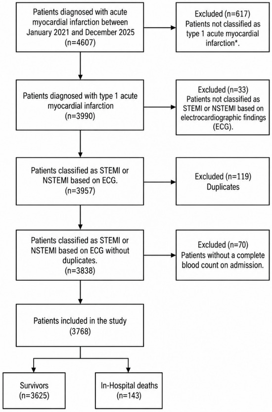
Patients aged 18 years or older with a confirmed diagnosis of type 1 AMI, including ST-segment elevation myocardial infarction (STEMI) and NSTEMI, were eligible (Figure 1). AMI was diagnosed according to contemporary international criteria, requiring a rise and/or fall in cardiac troponin concentrations with at least one value above the 99th percentile upper reference limit, evidence of acute myocardial ischemia, and at least one of the following: symptoms of myocardial ischemia, new ischemic ECG changes, development of pathologic Q waves, imaging evidence of new loss of viable myocardium or new regional wall motion abnormality in a pattern consistent with an ischemic etiology, or identification of a coronary thrombus by angiography or autopsy [15,16]. For patients with more than one AMI hospitalization during the study period, only the first eligible admission was considered the index hospitalization. Subsequent admissions were excluded.
Figure 1.
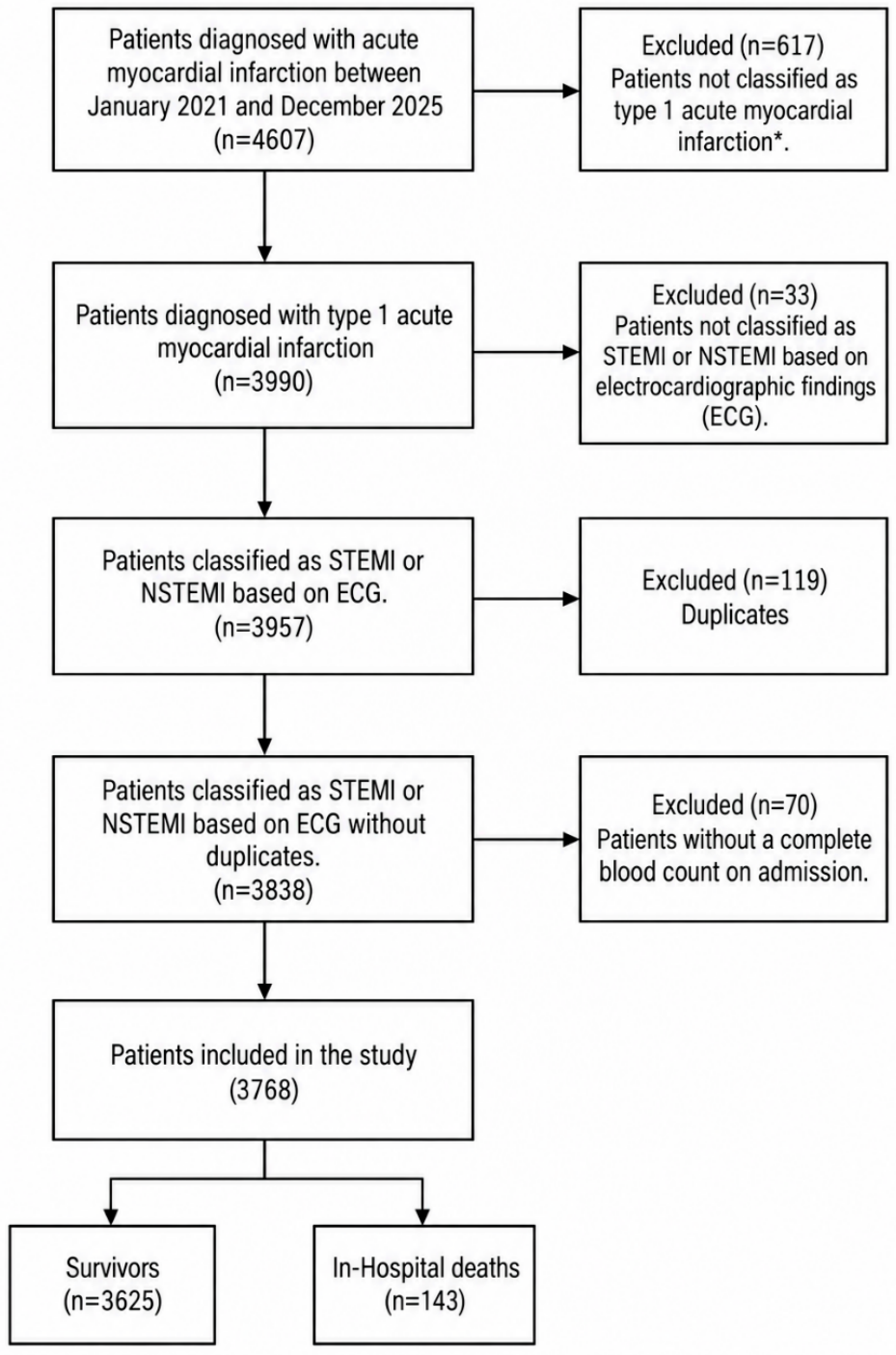
Study flowchart of included participants. * Patients not classified as type 1 myocardial infarction included patients with type 2 myocardial infarction, non-ischemic injury, or medical records with insufficient information. ECG: electrocardiogram; NSTEMI: non-ST-segment elevation myocardial infarction; STEMI: ST-segment elevation myocardial infarction.
Patients were excluded if they had type 2 myocardial infarction, myocardial injury without evidence of acute atherothrombotic coronary disease, insufficient information to confirm AMI, or no complete blood count available during the index hospitalization. In routine practice, the final diagnosis and differentiation between type 1 AMI, type 2 MI, or non-ischemic myocardial injury were established by the treating cardiology team at discharge following the institutional AMICE protocol. For critically ill or high-risk NSTEMI patients, coronary angiography was the primary tool used to confirm acute atherothrombosis (plaque rupture or occlusion). In patients managed conservatively, type 2 MI or myocardial injury was adjudicated when a documented supply–demand mismatch (e.g., severe sepsis, tachyarrhythmia, severe hemorrhage) or non-ischemic etiology was deemed the primary clinical driver. Consequently, a diagnosis of type 1 AMI in these patients required typical ischemic symptoms or dynamic electrocardiographic changes in the absence of such severe secondary triggers. For analyses of individual inflammatory indices, patients were additionally excluded when any cell count required for its calculation was missing or mathematically undefined.
2.3. Data Collection
Clinical information was recorded in a structured REDCap (Research Electronic Data Capture), version 17.3.0, registry [17,18] integrated with the institutional electronic health record system. Trained research personnel reviewed the records, validated clinical information, and resolved inconsistencies using standardized procedures.
Sociodemographic variables included age, sex, educational level, marital status, and area of residence. Clinical variables included smoking status, obesity, hypertension, diabetes mellitus, dyslipidemia, chronic kidney disease, previous coronary artery disease, heart failure, cerebrovascular disease, and chronic obstructive pulmonary disease (COPD).
Variables related to the index event included AMI subtype, Killip class, GRACE and TIMI risk scores, left ventricular ejection fraction, reperfusion strategy, percutaneous coronary intervention, coronary artery bypass grafting, and hospital length of stay.
Laboratory variables included total leukocyte count, red blood cell count, hemoglobin, hematocrit, mean corpuscular volume, mean corpuscular hemoglobin, mean corpuscular hemoglobin concentration, red cell distribution width, platelet count, mean platelet volume, and absolute neutrophil, lymphocyte, monocyte, eosinophil, and basophil counts. Serum creatinine and cardiac troponin were also collected when available.
2.4. Hematological Measurements
The first complete blood count obtained during the index hospitalization was used. For all included patients, the complete blood count was obtained within the first 24 h of hospital admission and before coronary revascularization. Complete blood counts were performed as part of routine clinical care using a Sysmex XN-1000 automated hematology analyzer (Sysmex Corporation, Kobe, Japan). Total and differential leukocyte counts and platelet measurements were extracted from the electronic health record. Before analysis, measurement units were standardized and values were assessed for plausibility, timing, and internal consistency.
2.5. Hematological Inflammatory Indices
Hematological inflammatory indices were calculated from the absolute blood cell counts obtained from the admission complete blood count (Table 1).
Table 1.
Hematological inflammatory index definitions.
2.6. Outcome
The primary outcome was all-cause in-hospital mortality, defined as death from any cause during the index AMI hospitalization. Patients discharged alive were classified as survivors. Vital status was obtained from the AMICE registry and verified against the institutional electronic health record and discharge documentation.
2.7. Statistical Analysis
Analyses were conducted using complete cases for the variables included in each model. Consequently, the analytic sample varied slightly across indices and model specifications. The number and percentage of missing observations were summarized for all variables considered in the multivariable analyses and are presented in Supplementary Table S1. No imputation of missing data was performed.
The extent of missing information was described for the principal variables. Continuous variables were assessed for distributional characteristics; normally distributed variables are presented as means with their respective standard deviations, whereas non-normally distributed variables are summarized as medians with interquartile ranges. Categorical variables are reported as frequencies and percentages. Baseline clinical, sociodemographic, and hematological characteristics were compared according to in-hospital survival status using the independent-samples Mann–Whitney U test for continuous variables, as appropriate, and χ2 test for categorical variables. Correlations among hematological inflammatory indices were examined using Spearman’s rank correlation coefficients. Correlation estimates were displayed in a heat map to describe the degree of overlap among indices.
The association between each complete blood count parameter or hematological inflammatory index and all-cause in-hospital mortality was evaluated using binary logistic regression. Conventional hematological parameters were standardized and modeled per one-standard-deviation increase. The inflammatory indices were naturally log-transformed and subsequently standardized. Their odds ratios therefore represent the change in the odds of in-hospital mortality associated with a one-standard-deviation increase in the log-transformed index.
Crude and multivariable-adjusted odds ratios with 95% confidence intervals were estimated. Each hematological parameter and inflammatory index was evaluated in a separate regression model because several indices share mathematical components and exhibited substantial intercorrelation. The prespecified adjusted model included age, sex, acute myocardial infarction subtype, serum creatinine, log-transformed cardiac troponin, and diabetes mellitus. The primary adjustment set was prespecified to capture demographic characteristics, infarction phenotype, renal function, myocardial injury, and relevant metabolic comorbidity. Acute clinical severity was further evaluated through complementary models incorporating Killip class, GRACE score, and TIMI score, while sensitivity analyses included additional adjustment for PCI and CABG.
The assumption of linearity between each continuous inflammatory index and the log odds of mortality was evaluated using restricted cubic splines applied to the log-transformed index. Four knots were used whenever supported by the observed distribution; three knots were used for indices with insufficient variability for a four-knot specification. Nonlinearity was assessed by jointly testing the spline terms representing deviation from linearity. For indices showing evidence of nonlinearity, adjusted spline plots were generated to characterize the shape of the association. Odds ratios and 95% confidence intervals were estimated using the median value of each log-transformed index as the reference. To reduce instability caused by sparse observations at the distributional extremes, spline curves were restricted to the 5th–95th percentiles. When no evidence of nonlinearity was identified, the association was summarized using the standardized linear term. For indices showing significant nonlinearity, the linear odds ratio was interpreted as an average association across the observed range, and spline findings were considered complementary evidence regarding the shape of the relationship.
The discriminatory ability of each inflammatory index considered individually was evaluated using ROC curves and the area under the curve with 95% confidence intervals. Several sensitivity analyses were conducted. First, the primary models were additionally adjusted for percutaneous coronary intervention and coronary artery bypass grafting. Second, associations were re-estimated using each index on its original scale. Third, the models were repeated using the natural log-transformed but non-standardized index. Robustness was assessed by examining the consistency of the direction and approximate magnitude of associations and the overlap of confidence intervals across model specifications. Analyses were performed using complete cases for the variables included in each model; consequently, sample sizes could vary slightly across analyses. All tests were two-sided, and a p value < 0.05 was considered statistically significant. We conducted analyses using Stata version 16 (StataCorp LLC, College Station, TX, USA). Detailed results of the sensitivity analyses are presented in Supplementary Table S1.
3. Results
A total of 3768 patients were included in the study. One hundred and forty-three participants (3.8%) experienced in-hospital death (Figure 1). Compared with survivors, patients who died were older (median age, 71.0 vs. 66.0 years; p < 0.001) and more frequently presented with STEMI (59.4% vs. 43.8%; p < 0.001). No significant differences were observed in terms of sex, marital status, or area of residence. Educational level differed between groups, with a higher proportion of patients without formal education among those who died (17.5% vs. 7.7%; p < 0.001). Among prior medical conditions, chronic kidney disease (p < 0.001) and peripheral vascular disease (p = 0.010) were more frequent in patients who died, whereas dyslipidemia was less common (p = 0.012). Other comorbidities did not differ significantly between groups. Length of hospital stay was also longer among patients who died (median, 6.0 vs. 4.0 days; p = 0.025). According to the Killip score, clinical severity (Killip III-IV) was greater among those who died in the hospital compared to survivors (p < 0.001). Nonetheless, the proportion of missing values was significant (25.4%). In addition, patients who died had statistically significant higher serum creatinine levels, higher cardiac troponin concentrations, and higher GRACE and TIMI scores than survivors (p < 0.001) (Table 2).
Table 2.
Baseline Sociodemographic and Clinical Characteristics According to In-Hospital Mortality.
A total of 4607 patients admitted with suspected acute coronary syndrome between January 2021 and December 2025 were initially screened. After the clinical adjudication process, 617 patients were excluded because they did not meet the criteria for type 1 AMI (this group included patients with type 2 myocardial infarction, non-ischemic myocardial injury, or insufficient clinical information). This left a cohort of 3990 patients with confirmed type 1 acute myocardial infarction. From this group, we further excluded 33 patients without electrocardiographic classification as STEMI or NSTEMI, 119 duplicate records, and 70 patients without a complete blood count at admission, resulting in 3768 patients included in the final analysis. Of these, 3625 survived to hospital discharge and 143 died during hospitalization (Figure 1).
Regarding complete blood count parameters at admission, patients who died had significantly higher white blood cell counts (median, 12.90 vs. 9.85 × 10³/µL; p < 0.001), absolute neutrophil counts (10.20 vs. 6.91 × 10³/µL; p < 0.001), and absolute monocyte counts (0.79 vs. 0.68 × 10³/µL; p < 0.001) compared with survivors. Conversely, red blood cell counts (4.50 vs. 4.76 × 10³/µL; p < 0.001), hemoglobin levels (13.70 vs. 14.20 g/dL; p = 0.007), and absolute lymphocyte counts (1.42 vs. 1.80 × 10³/µL; p < 0.001) were significantly lower in the non-survivor group. No significant differences were observed between the groups in terms of platelet count (p = 0.319), mean platelet volume (p = 0.453), mean corpuscular hemoglobin (p = 0.129), or absolute basophil count (p = 0.678). Furthermore, hematological inflammatory indices were markedly elevated among patients who died during hospitalization. Compared with survivors, non-survivors presented with significantly higher median values for the neutrophil-to-lymphocyte ratio (7.02 vs. 3.78; p < 0.001), systemic immune-inflammation index (1625.15 vs. 929.99; p < 0.001), aggregate index of systemic inflammation (1384.73 vs. 620.65; p < 0.001), and systemic inflammation response index (6.10 vs. 2.50; p < 0.001), among other positive indices. In contrast, the monocyte-to-neutrophil ratio was significantly lower in patients who died (0.08 vs. 0.11; p < 0.001) (Table 3).
Table 3.
Baseline Complete Blood Count Parameters and Hematological Inflammatory Indices According to In-Hospital Mortality.
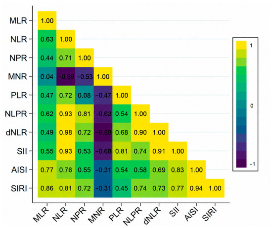
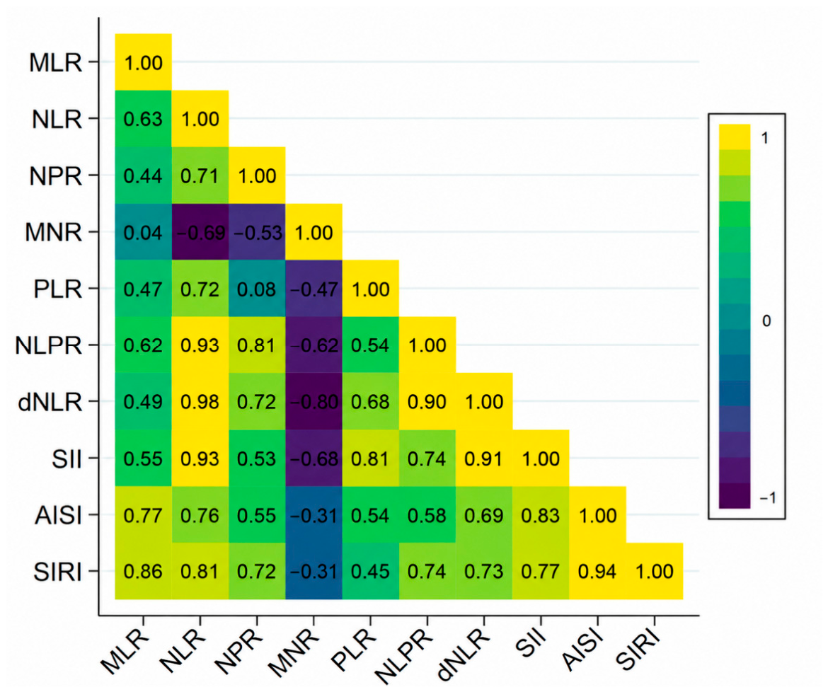
Hematological inflammatory indices showed substantial intercorrelation, reflecting their shared cellular components. The strongest positive correlations were observed between NLR and dNLR (ρ = 0.98), AISI and SIRI (ρ = 0.94), NLR and NLPR (ρ = 0.93), NLR and SII (ρ = 0.93), and dNLR and SII (ρ = 0.91). MNR showed inverse correlations with several neutrophil-based indices, particularly dNLR (ρ = −0.80), NLR (ρ = −0.69), SII (ρ = −0.68), and NLPR (ρ = −0.62) (Figure 2).
Figure 2.
Spearman Correlation Matrix of Hematological Inflammatory Indices.
The heat map displays pairwise Spearman rank correlation coefficients among complete blood count-derived inflammatory indices. Positive correlations are represented by progressively lighter yellow-green shades, whereas negative correlations are shown in darker blue-purple shades. Values range from −1 to 1, with coefficients closer to either extreme indicating stronger correlations. AISI: aggregate index of systemic inflammation; dNLR: derived neutrophil-to-lymphocyte ratio; MLR: monocyte-to-lymphocyte ratio; MNR: monocyte-to-neutrophil ratio; NLR: neutrophil-to-lymphocyte ratio; NLPR: neutrophil-to-lymphocyte-to-platelet ratio; NPR: neutrophil-to-platelet ratio; PLR: platelet-to-lymphocyte ratio; SII: systemic immune-inflammation index; SIRI: systemic inflammation response index.
In multivariable analyses, higher white blood cell count (adjusted OR 1.58, 95% CI 1.38–1.81), absolute neutrophil count (OR 1.67, 95% CI 1.45–1.92), absolute monocyte count (OR 1.25, 95% CI 1.09–1.43), red cell distribution width (OR 1.33, 95% CI 1.12–1.57), and mean corpuscular volume (OR 1.21, 95% CI 1.02–1.45) were associated with greater odds of in-hospital mortality. In contrast, higher red blood cell count (OR 0.75, 95% CI 0.62–0.89), hemoglobin (OR 0.79, 95% CI 0.66–0.95), and hematocrit (OR 0.79, 95% CI 0.66–0.95) were associated with lower odds. Absolute lymphocyte count showed an inverse association that did not reach conventional statistical significance after adjustment (OR 0.78, 95% CI 0.61–1.00; p = 0.052). All hematological inflammatory indices were significantly associated with in-hospital mortality after adjustment, although the direction and magnitude of association varied across indices (Table 4).
Table 4.
Crude and Adjusted Associations of Complete Blood Count Parameters and Hematological Inflammatory Indices With In-Hospital Mortality.
Missingness was greatest for TIMI score (42.9%), Killip class (25.4%), and GRACE score (24.3%). Cardiac troponin and serum creatinine were unavailable in 8.5% and 0.7% of patients, respectively. The primary adjusted models included 3435–3436 patients, depending on the hematological parameter or inflammatory index evaluated. The one-patient variation across some models resulted from an unavailable or mathematically undefined inflammatory index value. A detailed description of missing data is provided in Supplementary Table S2.
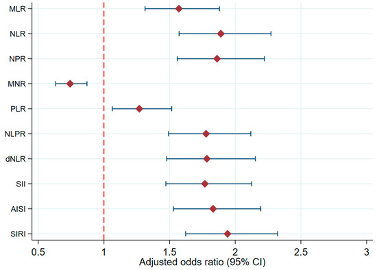
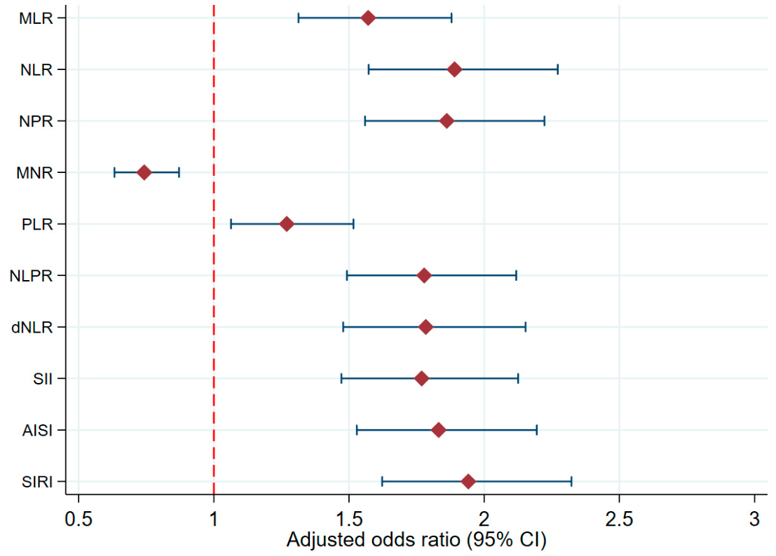
Among the hematological inflammatory indices, SIRI showed the numerically largest positive association with in-hospital mortality (adjusted OR 1.94, 95% CI 1.62–2.32), followed by NLR (OR 1.89, 95% CI 1.57–2.27), NPR (OR 1.86, 95% CI 1.56–2.22), AISI (OR 1.83, 95% CI 1.53–2.19), NLPR (OR 1.78, 95% CI 1.49–2.12), dNLR (OR 1.78, 95% CI 1.48–2.15), and SII (OR 1.77, 95% CI 1.47–2.13). MLR (OR 1.57, 95% CI 1.31–1.88) and PLR (OR 1.27, 95% CI 1.06–1.52) also showed positive associations, whereas MNR was inversely associated with mortality (OR 0.74, 95% CI 0.63–0.87) (Figure 3).
Figure 3.
Adjusted associations between hematological inflammatory indices and in-hospital mortality. Diamonds represent adjusted odds ratios and horizontal lines indicate 95% confidence intervals. The red dashed vertical line indicates the null value of association (adjusted odds ratio = 1).
Across the three multivariable models, NLR, NPR, NLPR, dNLR, SII, AISI, and SIRI remained significantly associated with higher odds of in-hospital mortality. NPR showed the numerically largest association in the GRACE and TIMI models (OR 1.76, 95% CI 1.37–2.25; and OR 1.78, 95% CI 1.27–2.50, respectively), whereas SIRI showed the numerically largest association in the clinical model (OR 1.54, 95% CI 1.21–1.97). MLR remained significant in the clinical and TIMI models but was borderline in the GRACE model. MNR was inversely associated with mortality only in the GRACE model, while PLR was not significantly associated with mortality in any model (Table 5).
Table 5.
Adjusted Associations Between Hematological Inflammatory Indices and In-Hospital Mortality Across Clinical, GRACE, and TIMI Models.
Sensitivity analyses were generally consistent with the primary models. Associations remained in the same direction after additional adjustment for PCI and CABG and when indices were modeled using alternative scales. NPR and NLPR showed imprecise estimates when modeled on their original scales, likely reflecting scale-related instability; however, their associations were more stable after log transformation.
Restricted cubic spline analyses showed no evidence of nonlinearity for NLR, NLPR, or dNLR. In contrast, significant nonlinear associations were observed for MLR, NPR, MNR, PLR, AISI, and SIRI, while SII showed borderline evidence of nonlinearity. The adjusted spline curves showed J-shaped patterns for MLR, NPR, PLR, AISI, and SIRI, characterized by lower estimated odds near the lower-to-intermediate portions of their distributions followed by progressively increasing odds at higher values. In contrast, MNR exhibited an inverse nonlinear pattern, with higher estimated odds at low values, decreasing odds across the intermediate range, and a modest increase toward the upper end of its distribution. These associations are presented in Supplementary Figure S1. These findings suggest that, for several indices, the association with in-hospital mortality may not be fully captured by a single linear term.
In discrimination analyses, the individual indices demonstrated modest standalone performance. NPR showed the numerically highest AUROC (0.723), followed by SIRI (0.716), NLR (0.706), and NLPR (0.705). These results indicate that although inflammatory indices were associated with mortality, their ability to discriminate between survivors and non-survivors when used individually was limited. Sensitivity analyses were generally consistent with the primary models. Associations remained in the same direction after additional adjustment for PCI and CABG and when indices were modeled using alternative scales. NPR and NLPR showed imprecise estimates when modeled on their original scales, likely reflecting scale-related instability; however, their associations were more stable after log transformation. Detailed results are presented in Supplementary Table S1.
4. Discussion
In this large cohort of patients with type 1 acute myocardial infarction, inflammatory markers derived from the admission complete blood count were consistently associated with all-cause in-hospital mortality. Patients who died had higher leukocyte, neutrophil, and monocyte counts, lower lymphocyte counts, and higher values of most composite inflammatory indices. After multivariable adjustment, SIRI, NLR, NPR, AISI, NLPR, dNLR, and SII showed the largest positive associations, whereas MNR was inversely associated with mortality. However, their standalone discriminatory performance was modest, indicating that these indices should be considered complementary risk markers rather than substitutes for validated clinical scores.
The associations of leukocytosis and neutrophilia with mortality are consistent with previous evidence in acute coronary syndromes. In the TACTICS–TIMI 18 substudy, higher baseline leukocyte counts were associated with more extensive coronary disease, impaired myocardial perfusion, and increased mortality [13]. Similar findings have been reported in STEMI, where neutrophilia has been related to greater infarct size, microvascular obstruction, heart failure, and death. Our study adds evidence to a contemporary population including both STEMI and NSTEMI and shows that these associations persist after accounting for major clinical risk factors.
These findings are biologically plausible because neutrophils play a central role in ischemic and reperfusion injury. Myocardial necrosis promotes neutrophil mobilization and recruitment to injured tissue, where activated cells release proteolytic enzymes, reactive oxygen species, cytokines, and neutrophil extracellular traps. Although this response contributes to debris clearance and tissue repair, excessive activation may aggravate endothelial dysfunction, thrombosis, microvascular obstruction, and myocardial injury. Conversely, lymphopenia may reflect stress-induced neuroendocrine activation, immune dysregulation, comorbidity, or hemodynamic severity. Because lymphocytes form the denominator of several indices, their reduction amplifies NLR, MLR, PLR, NLPR, SII, AISI, and SIRI.
NLR was one of the most consistent markers across the different model specifications. This agrees with the findings of Tamhane et al. in 2833 patients with acute coronary syndromes [19], subsequent studies in STEMI [20], and meta-analytic evidence showing that elevated NLR is associated with mortality and major adverse cardiovascular events after myocardial infarction [21]. Its prognostic relevance may arise from simultaneously capturing neutrophil-mediated innate activation and relative lymphocyte depletion. Nevertheless, NLR is nonspecific and can be influenced by infection, corticosteroid use, smoking, malignancy, chronic inflammation, physiological stress, and the timing of blood collection. Moreover, recent evidence in STEMI patients undergoing primary PCI has shown that the Advanced Lung Cancer Inflammation Index (ALI), a composite inflammatory–nutritional marker, has greater prognostic value than the NLR. These findings suggest that readily available hematological indices may enhance their predictive performance when combined with other easily accessible clinical variables, such as nutritional status, in the setting of acute cardiovascular care [22].
SIRI presented the numerically largest adjusted association in the primary analysis and remained significant across the clinical, GRACE, and TIMI models. By combining neutrophils, monocytes, and lymphocytes, SIRI may reflect innate immune activation, monocyte recruitment, and relative adaptive immune suppression. Previous studies have also associated elevated SIRI with adverse outcomes in acute coronary syndrome and STEMI [23,24]. SII and AISI were similarly associated with mortality, in agreement with reports linking these indices to adverse cardiovascular events and survival after PCI and AMI [24,25,26].
However, the more complex indices did not clearly outperform simpler markers. NLR was almost perfectly correlated with dNLR and strongly correlated with NLPR and SII, while AISI and SIRI were also highly correlated. These indices should therefore be understood as alternative mathematical representations of a common inflammatory phenotype dominated by neutrophilia, lymphopenia, and, to a lesser extent, monocytosis and platelet variation. This overlap supports evaluating them in separate models and limits claims of biological independence, or that any single index represents a distinct biological pathway or is definitively superior.
NPR produced the numerically highest AUROC and remained associated with mortality across the adjusted analyses. Nevertheless, NPR has been less extensively studied than NLR, PLR, SII, or SIRI, and its estimates on the original scale were imprecise. Because platelet count alone was not associated with mortality, the NPR result may have been driven mainly by neutrophil elevation rather than an independent platelet-related mechanism. In contrast, PLR showed weaker and less consistent results: although significant in the primary model, it lost significance after adjustment for clinical severity and established risk scores. Previous meta-analyses have associated PLR with adverse outcomes after acute coronary syndromes [27,28], but their findings have been heterogeneous, possibly because of differences in populations, cutoff values, treatment strategies, follow-up, and covariate adjustment.
MNR was inversely associated with mortality in the primary model but was not consistently significant in the more comprehensive models. This inverse association is mathematically compatible with the increased neutrophil burden among patients who died and should not be interpreted as evidence that monocytes are protective. Monocytes participate in both inflammatory injury and myocardial repair, and their prognostic role may depend on the timing and relative kinetics of leukocyte mobilization.
Among conventional hematological parameters, higher RDW remained independently associated with mortality, in agreement with previous meta-analytic evidence in acute coronary syndrome [29]. RDW may integrate inflammation, oxidative stress, impaired erythropoiesis, renal dysfunction, nutritional deficiencies, and chronic disease burden. Lower red blood cell count, hemoglobin, and hematocrit were also associated with mortality, possibly reflecting reduced oxygen-carrying capacity or greater underlying comorbidity. However, these variables are strongly interrelated and should not be interpreted as independent biological effects.
Despite statistically significant associations, the AUROC values of the individual indices were modest. Thus, their distributions overlapped substantially between survivors and patients who died, limiting their use as standalone classifiers. Their main potential advantage is that they are inexpensive, rapidly available, and routinely measured at admission. However, the present analysis does not establish that they improve prediction beyond GRACE, TIMI, or other validated models. Such a conclusion would require direct comparison of models with and without each index, together with assessment of calibration, internal validation, and clinical net benefit.
The nonlinear relationships observed for MLR, NPR, MNR, PLR, AISI, and SIRI also indicate that a single linear odds ratio or fixed cutoff may oversimplify their associations with mortality. This may partly explain the heterogeneous thresholds reported in previous studies. Continuous modeling using logarithmic transformations and flexible functions may provide a more reliable representation than data-derived cutoffs, which frequently have limited transportability.
This study has several strengths, including its large contemporary cohort, the inclusion of both STEMI and NSTEMI, and the simultaneous evaluation of conventional blood count parameters and multiple inflammatory indices. The analyses also addressed intercorrelation, nonlinear associations, discrimination, and robustness across alternative adjustment models. Nevertheless, several limitations should be acknowledged. First, the retrospective analysis of registry data precludes causal inference and remains susceptible to residual confounding. Information on concurrent infections, chronic inflammatory or hematological diseases, active malignancy, and corticosteroid exposure was not systematically captured. These non-cardiac determinants of blood count abnormalities may therefore have contributed to unmeasured confounding. Future prospective studies should systematically document these conditions to better distinguish the inflammatory response associated with myocardial ischemia from other systemic triggers.
Although complementary models incorporated Killip class, GRACE score, and TIMI score, and sensitivity analyses additionally adjusted for PCI and CABG, acute clinical severity may not have been fully captured. LVEF was not included in the multivariable models, and direct indicators of hemodynamic instability, including cardiogenic shock, vasopressor requirement, mechanical circulatory support, and sustained hypotension, were not modeled separately. Moreover, adjustment for PCI and CABG did not encompass all aspects of reperfusion management, such as fibrinolysis, absence of reperfusion, and treatment timing. Residual confounding related to ventricular dysfunction, hemodynamic compromise, infarct severity, and treatment selection therefore cannot be excluded.
All complete blood counts were obtained within the first 24 h of admission and before coronary revascularization; however, the exact timing relative to symptom onset was not uniformly available. This is relevant because inflammatory indices may change dynamically during the acute course of myocardial infarction. In addition, the complete-case approach may have introduced selection bias, particularly in models incorporating Killip class, GRACE score, and TIMI score, for which substantial proportions of observations were missing. The single-center design and the potential influence of local care pathways may also limit external generalizability, and the findings should therefore be considered primarily hypothesis-generating. Furthermore, evaluating multiple correlated indices increases the possibility of chance findings. Finally, discrimination was assessed for each index individually, without formally evaluating incremental predictive value beyond established risk scores or standard clinical models through changes in C-statistics, calibration, reclassification measures, or decision-curve analysis. Accordingly, these findings should not be interpreted as evidence that the inflammatory indices improve clinical risk prediction.
5. Conclusions
Admission complete blood count-derived inflammatory indices were associated with all-cause in-hospital mortality in patients with type 1 AMI. Neutrophil-dominant indices, particularly SIRI, NLR, NPR, AISI, NLPR, dNLR, and SII, showed the most consistent associations, whereas PLR and MNR were less robust after adjustment for clinical severity. Their high intercorrelation suggests that these indices reflect a shared inflammatory phenotype. Although they were associated with in-hospital mortality, their standalone discriminatory ability was modest, and the present study did not demonstrate incremental predictive value beyond established clinical assessment. Accordingly, these indices should be interpreted as exploratory markers of inflammatory risk rather than as validated tools for improving clinical risk prediction. Rather than supporting immediate broad clinical implementation, our findings serve primarily as a robust local validation of previously described indices within a Latin American healthcare context. External validation across diverse populations is needed to determine whether these selected indices provide meaningful prognostic information beyond established tools like the GRACE and TIMI scores.
Supplementary Materials
The following supporting information can be downloaded at https://www.mdpi.com/article/10.3390/jcm15197416/s1, Table S1: Sensitivity analysis of the association between hematological inflammatory indices and in-hospital mortality. Table S2: Missing data for variables considered in the multivariable analyses. Figure S1. Restricted cubic spline associations between selected inflammatory indices and in-hospital mortality.
Author Contributions
Conceptualization, A.H.-O., E.F.M.-H., M.L.-A. and J.F.S.; methodology, A.H.-O., E.F.M.-H., M.L.-A. and M.S.P.-M.; formal analysis, A.H.-O., E.F.M.-H. and M.L.-A.; data curation, A.H.-O., M.S.P.-M., A.F.M.-S., J.L.S. and B.M.P.; writing—original draft preparation, A.H.-O., M.S.P.-M. and M.L.-A.; writing—review & editing, A.H.-O., M.S.P.-M., A.F.M.-S., J.L.S., B.M.P., E.F.M.-H., M.L.-A., J.F.S. and A.M.C.-G.; supervision, E.F.M.-H., M.L.-A. and J.F.S. All authors have read and agreed to the published version of the manuscript.
Funding
This research received no external funding.
Institutional Review Board Statement
The study was conducted in accordance with the Declaration of Helsinki and was approved by the Institutional Research Ethics Committee of Fundación Cardiovascular de Colombia (approval number Act No. 650; CEI-2024-07461-47; 9 March 2026).
Informed Consent Statement
Patient consent was waived due to the retrospective nature of the study and the use of a de-identified research database.
Data Availability Statement
The datasets presented in this article are not readily available because of institutional privacy restrictions regarding patient data. Requests to access the datasets should be directed to the corresponding author.
Acknowledgments
During the preparation of this manuscript, the authors used ChatGPT (GPT-5.5; OpenAI) for the purposes of language refinement and to improve manuscript readability. The authors have reviewed and edited the output and take full responsibility for the content of this publication.
Conflicts of Interest
The authors declare no conflicts of interest.
References
- Byrne, R.; Coughlan, J.J.; Rossello, X.; Ibanez, B.; Members of the Task Force for the 2023 ESC Guidelines for the Management of Acute Coronary Syndromes; Barbato, E.; Berry, C.; Chieffo, A.; Claeys, M.J.; Dan, G.-A.; et al. 2023 ESC Guidelines for the management of acute coronary syndromes. Eur. Heart J. 2023, 44, 3720–3826. [Google Scholar]
- Tadesse, S.; Tegene, E.; Yilma, D.; Yemane, T.; Gudina, E.K.; Mossie, A. Predictive role of hematological indices in patients with acute coronary syndrome in Ethiopia: Intrahospital outcomes. Heliyon 2024, 10, e36790. [Google Scholar] [CrossRef] [Scilit] [PubMed]
- Zhou, T.; Yang, K.; Yang, Y.; Liu, S.M. Complete Blood Count-Derived Inflammation Indices to Predict 3-Year All-Cause Mortality in Patients with Diabetes and Acute Myocardial Infarction in Critical Care: Retrospective Cohort Study With Single-Center External Validation. JMIR Med. Inform. 2026, 14, e83328. [Google Scholar] [CrossRef] [Scilit] [PubMed]
- Zhang, W.-C.; Shuai, W.-L.; Huang, X.-Q.; Dai, J.-Q.; Huo, J.-H.; Shen, M.; Chen, J.-J.; Yang, Z.-M.; Xia, X.-X. Eosinophil count trajectories are associated with the prognosis of acute myocardial infarction patients: Insights from ICU data analysis. PLoS ONE 2026, 21, e0349827. [Google Scholar] [CrossRef] [Scilit] [PubMed]
- Lin, G.; Dai, C.; Chen, W. The AHEAD-NLR score: A novel inflammatory-comorbidity integrated tool for predicting long-term mortality in patients with heart failure after acute myocardial infarction. Sci. Rep. 2026, 16, 12436. [Google Scholar] [CrossRef] [Scilit] [PubMed]
- Döring, Y.; Soehnlein, O.; Weber, C. Neutrophil extracellular traps in atherosclerosis and atherothrombosis. Circ. Res. 2017, 120, 736–743. [Google Scholar] [CrossRef] [Scilit] [PubMed]
- van der Laan, A.M.; Hirsch, A.; Robbers, L.F.; Nijveldt, R.; Lommerse, I.; Delewi, R.; van der Vleuten, P.A.; Biemond, B.J.; Zwaginga, J.J.; van der Giessen, W.J.; et al. A proinflammatory monocyte response is associated with myocardial injury and impaired functional outcome in patients with ST-segment elevation myocardial infarction: Monocytes and myocardial infarction. Am. Heart J. 2012, 163, 57–65.e2. [Google Scholar] [CrossRef] [Scilit] [PubMed]
- Armillotta, M.; Bergamaschi, L.; Angeli, F.; Belmonte, M.; Alvarez, M.C.; Sansonetti, A.; Fedele, D.; Amicone, S.; Canton, L.; Bertolini, D.; et al. Impact of glucometabolic status on type 4a myocardial infarction in patients with non-ST-segment elevation myocardial infarction: The role of stress hyperglycemia ratio. Cardiovasc. Diabetol. 2025, 24, 394–405. [Google Scholar] [CrossRef] [Scilit] [PubMed]
- Huang, W.-M.; Cheng, H.-M.; Huang, C.-J.; Guo, C.-Y.; Lu, D.-Y.; Lee, C.-W.; Hsu, P.-F.; Yu, W.-C.; Chen, C.-H.; Sung, S.-H. Hemographic indices are associated with mortality in acute heart failure. Sci. Rep. 2017, 7, 17828. [Google Scholar] [CrossRef] [Scilit] [PubMed]
- Tao, Z.; Yin, J.; Li, M.; Li, G.; Liu, K.; Wang, Z. Predictive Value of IBI for In-Hospital Death in Elderly Patients with Non-ST-Segment Elevation Myocardial Infarction. Med. Sci. Monit. 2025, 31, e950592. [Google Scholar] [CrossRef] [Scilit] [PubMed]
- Kraus, M.; Schmitz, T.; Raake, P.; Linseisen, J.; Meisinger, C. Evaluation of the platelet-to-white-cell ratio (PWR) as predictor of long-term all-cause mortality in patients with acute myocardial infarction. Sci. Rep. 2026, 16, 17860. [Google Scholar] [CrossRef] [Scilit] [PubMed]
- Karadeniz, F.Ö.; Karadeniz, Y.; Altuntaş, E. Systemic immune-inflammation index, and neutrophilto-lymphocyte and platelet-to-lymphocyte ratios can predict clinical outcomes in patients with acute coronary syndrome. Cardiovasc. J. Afr. 2024, 35, 82–88. [Google Scholar] [CrossRef] [Scilit] [PubMed]
- Sabatine, M.S.; Morrow, D.A.; Cannon, C.P.; Murphy, S.A.; Demopoulos, L.A.; Di Battiste, P.M.; McCabe, C.H.; Braunwald, E.; Gibson, C.M. Relationship between baseline white blood cell count and degree of coronary artery disease and mortality in patients with acute coronary syndromes: A TACTICS-TIMI 18 (Treat Angina with Aggrastat and determine Cost of Therapy with an Invasive or Conservative Strategy- Thrombolysis in Myocardial Infarction 18 trial)substudy. J. Am. Coll. Cardiol. 2002, 40, 1761–1768. [Google Scholar] [PubMed]
- Monteiro Júnior, J.G.D.M.; Torres, D.D.O.C.; da Silva, M.C.F.C.; Martins, C.M.D.H.; da Silva, I.K.; do Nascimento, M.E.M.; Dos Santos, A.C.O.; Montarroyos, U.R.; Filho, D.C.S. Prognostic value of hematological parameters in patients with acute myocardial infarction: Intrahospital outcomes. PLoS ONE 2018, 13, e0194897. [Google Scholar] [CrossRef] [Scilit] [PubMed]
- Thygesen, K.; Alpert, J.S.; Jaffe, A.S.; Chaitman, B.R.; Bax, J.J.; Morrow, D.A.; White, H.D.; Executive Group on behalf of the Joint European Society of Cardiology (ESC)/American College of Cardiology (ACC)/American Heart Association (AHA)/World Heart Federation (WHF) Task Force for the Universal Definition of Myocardial Infarction. Fourth Universal Definition of Myocardial Infarction (2018). Circulation 2018, 138, e618–e651. [Google Scholar] [PubMed]
- Rao, S.V.; O’dOnoghue, M.L.; Ruel, M.; Rab, T.; Tamis-Holland, J.E.; Alexander, J.H.; Baber, U.; Baker, H.; Cohen, M.G.; Cruz-Ruiz, M.; et al. 2025 ACC/AHA/ACEP/NAEMSP/SCAI Guideline for the Management of Patients with Acute Coronary Syndromes: A Report of the American College of Cardiology/American Heart Association Joint Committee on Clinical Practice Guidelines. Circulation 2025, 151, e771–e862, Correction in Circulation 2025, 151, 1098. [Google Scholar] [CrossRef] [Scilit] [PubMed]
- Harris, P.A.; Taylor, R.; Thielke, R.; Payne, J.; Gonzalez, N.; Conde, J.G. Research electronic data capture (REDCap)--a metadata-driven methodology and workflow process for providing translational research informatics support. J. Biomed. Inform. 2009, 42, 377–381. [Google Scholar] [CrossRef] [Scilit] [PubMed]
- Harris, P.A.; Taylor, R.; Minor, B.L.; Elliott, V.; Fernandez, M.; O’Neal, L.; McLeod, L.; Delacqua, G.; Delacqua, F.; Kirby, J.; et al. The REDCap consortium: Building an international community of software platform partners. J. Biomed. Inform. 2019, 95, 103208. [Google Scholar] [CrossRef] [Scilit] [PubMed]
- Tamhane, U.U.; Aneja, S.; Montgomery, D.; Rogers, E.-K.; Eagle, K.A.; Gurm, H.S. Association between admission neutrophil to lymphocyte ratio and outcomes in patients with acute coronary syndrome. Am. J. Cardiol. 2008, 102, 653–657. [Google Scholar] [CrossRef] [Scilit] [PubMed]
- Gazi, E.; Bayram, B.; Gazi, S.; Temiz, A.; Kirilmaz, B.; Altun, B.; Barutcu, A. Prognostic Value of the Neutrophil-Lymphocyte Ratio in Patients With ST-Elevated Acute Myocardial Infarction. Clin. Appl. Thromb. Hemost. 2015, 21, 155–159. [Google Scholar] [CrossRef] [Scilit] [PubMed]
- Banahene, N.O.; Sinha, T.; Shaikh, S.; Zin, A.K.; Khreis, K.; Chaudhari, S.S.; Wei, C.R.; Palleti, S.K. Effect of Elevated Neutrophil-to-Lymphocyte Ratio on Adverse Outcomes in Patients with Myocardial Infarction: A Systematic Review and Meta-Analysis. Cureus 2024, 16, e61647. [Google Scholar] [CrossRef] [Scilit] [PubMed]
- Trimarchi, G.; Pizzino, F.; Lilli, A.; De Caterina, A.R.; Esposito, A.; Dalmiani, S.; Mazzone, A.; Di Bella, G.; Berti, S.; Paradossi, U. Advanced Lung Cancer Inflammation Index as Predictor of All-Cause Mortality in ST-Elevation Myocardial Infarction Patients Undergoing Primary Percutaneous Coronary Intervention. J. Clin. Med. 2024, 13, 6059–6071. [Google Scholar] [CrossRef] [Scilit] [PubMed]
- Han, K.; Shi, D.; Yang, L.; Wang, Z.; Li, Y.; Gao, F.; Liu, Y.; Ma, X.; Zhou, Y. Prognostic value of systemic inflammatory response index in patients with acute coronary syndrome undergoing percutaneous coronary intervention. Ann. Med. 2022, 54, 1667–1677. [Google Scholar] [CrossRef] [Scilit] [PubMed]
- Marchi, F.; Pylypiv, N.; Parlanti, A.; Storti, S.; Gaggini, M.; Paradossi, U.; Berti, S.; Vassalle, C. Systemic Immune-Inflammation Index and Systemic Inflammatory Response Index as Predictors of Mortality in ST-Elevation Myocardial Infarction. J. Clin. Med. 2024, 13, 1256. [Google Scholar] [CrossRef] [Scilit] [PubMed]
- Zhang, C.; Li, M.; Liu, L.; Deng, L.; Yulei, X.; Zhong, Y.; Liao, B.; Yu, L.; Feng, J. Systemic immune-inflammation index as a novel predictor of major adverse cardiovascular events in patients undergoing percutaneous coronary intervention: A meta-analysis of cohort studies. BMC Cardiovasc. Disord. 2024, 24, 189. [Google Scholar] [CrossRef] [Scilit] [PubMed]
- Luo, J.; Qin, X.; Zhang, X.; Zhang, Y.; Yuan, F.; Shi, W.; Liu, B.; Wei, Y.; NOAFCAMI-SH Registry Investigators. Prognostic implications of systemic immune-inflammation index in myocardial infarction patients with and without diabetes: Insights from the NOAFCAMI-SH registry. Cardiovasc. Diabetol. 2024, 23, 41. [Google Scholar] [CrossRef] [Scilit] [PubMed]
- Willim, H.A.; Harianto, J.C.; Cipta, H. Platelet-to-Lymphocyte Ratio at Admission as a Predictor of In-Hospital and Long-Term Outcomes in Patients With ST-Segment Elevation Myocardial Infarction Undergoing Primary Percutaneous Coronary Intervention: A Systematic Review and Meta-Analysis. Cardiol. Res. 2021, 12, 109–116. [Google Scholar] [CrossRef] [Scilit] [PubMed]
- Li, H.; Zhou, Y.; Ma, Y.; Han, S.; Zhou, L. The prognostic value of the platelet-to-lymphocyte ratio in acute coronary syndrome: A systematic review and meta-analysis. Kardiol. Pol. 2017, 75, 666–673. [Google Scholar] [CrossRef] [Scilit] [PubMed]
- Abrahan, L.L., 4th; Ramos, J.D.A.; Cunanan, E.L.; Tiongson, M.D.A.; Punzalan, F.E.R. Red Cell Distribution Width and Mortality in Patients with Acute Coronary Syndrome: A Meta-Analysis on Prognosis. Cardiol. Res. 2018, 9, 144–152. [Google Scholar] [CrossRef] [Scilit] [PubMed]

Disclaimer/Publisher’s Note: The statements, opinions and data contained in all publications are solely those of the individual author(s) and contributor(s) and not of MDPI and/or the editor(s). MDPI and/or the editor(s) disclaim responsibility for any injury to people or property resulting from any ideas, methods, instructions or products referred to in the content. |
© 2026 by the authors. Licensee MDPI, Basel, Switzerland. This article is an open access article distributed under the terms and conditions of the Creative Commons Attribution (CC BY) license.