


Abstract
Background: Urinary cytology is routinely used as an adjunct to cystoscopy in the diagnosis and surveillance of urothelial carcinoma. Although it has high specificity for high-grade tumors, its sensitivity for low-grade papillary lesions is limited. Activating mutations in the fibroblast growth factor receptor 3 (FGFR3) gene are frequently encountered in non-muscle-invasive bladder cancer and may provide a complementary molecular method for detecting tumors that are not identified by conventional cytology. Objective: To compare conventional urinary cytology with urinary detection of the FGFR3 hotspot mutations S249C and R248C in patients undergoing evaluation for urothelial carcinoma. Materials and Methods: This single-center prospective pilot study included adult patients with clinically and radiologically confirmed bladder tumors evaluated between February 2022 and December 2023. Voided urine specimens were collected before cystoscopy or urinary tract instrumentation. Urine cytology was performed after centrifugation, conventional smear preparation, alcohol fixation, and Papanicolaou staining, with interpretation according to The Paris System for Reporting Urinary Cytology. DNA extracted from paired urine specimens was analyzed for the FGFR3 S249C and R248C hotspot mutations using TaqMan mutation-detection assays and real-time polymerase chain reaction. Histopathological examination of transurethral resection, bladder biopsy, or radical surgical specimens served as the reference standard. Results: Twenty-six patients were included, with a mean age of 69.2 ± 11.5 years; 24 patients (92.3%) were male. At least one FGFR3 mutation was detected in 17 patients (65.4%), whereas nine patients (34.6%) had no detectable mutation. The S249C mutation was identified in three patients, while the R248C mutation was detected in nine patients, 5 patients were positive for both mutations.
1. Introduction
Bladder cancer (BC) is among the ten most frequently diagnosed malignancies worldwide and remains a major cause of morbidity, mortality, and healthcare expenditure. More than 600,000 new cases and 220,000 deaths occur annually, with increasing incidence because of population aging and continued exposure to carcinogens such as tobacco smoke and occupational aromatic amines [1]. More than 90% of bladder tumors are urothelial carcinomas; approximately 70–75% present as non-muscle-invasive bladder cancer (NMIBC), including Ta, T1, and carcinoma in situ (CIS), while the remainder are muscle-invasive at diagnosis [2,3,4]. Although NMIBC generally has favorable cancer-specific survival, 50–70% of patients experience recurrence and approximately 10–20% progress to muscle-invasive disease [5,6]. Repeated endoscopic procedures and prolonged surveillance consequently make bladder cancer one of the most expensive malignancies to manage per patient [7].
Guidelines from the European Association of Urology (EAU), American Urological Association/Society of Urologic Oncology (AUA/SUO), and National Comprehensive Cancer Network (NCCN) identify cystoscopy with histopathological confirmation following transurethral resection as the diagnostic standard [2,3,4]. Cystoscopy permits direct lesion visualization and tissue acquisition, while NMIBC surveillance remains based mainly on periodic cystoscopy because of the high recurrence risk [2,3]. Urinary biomarkers may provide complementary information in selected situations, but current molecular assays cannot replace cystoscopy across the full spectrum of urothelial carcinoma [2,3,4]. Urinary cytology has been used routinely for decades and remains recommended as an adjunct, particularly for suspected high-grade urothelial carcinoma, CIS, unexplained positive urinary findings despite negative cystoscopy, and surveillance of intermediate- or high-risk NMIBC [2,3,4]. Its principal value lies in detecting high-grade disease and providing cellular information that complements endoscopic findings [2,3,4].
Urine cytology is based on microscopic examination of exfoliated urothelial cells obtained from voided urine, bladder washings, or instrumented specimens. Malignant cells may show nuclear enlargement, hyperchromasia, coarse chromatin, irregular contours, increased nuclear-to-cytoplasmic ratio, and prominent nucleoli [8]. High-grade tumors generally exfoliate abundant atypical cells, whereas low-grade papillary tumors often retain near-normal architecture and release few abnormal cells; performance therefore depends on tumor biology, specimen quality, and cytopathological expertise [9,10]. Cytology has very high specificity, usually above 90% and often approaching 95–100% in experienced laboratories [11,12,13]. Overall sensitivity is lower, commonly 35–55%, and varies markedly by grade [11,14]. Sensitivity for high-grade carcinoma and CIS often reaches 70–90% or more [9,11,15], whereas low-grade tumors are frequently detected in fewer than 30% of cases because their cytological atypia is limited [11,14].
These limitations prompted the development of The Paris System for Reporting Urinary Cytology (TPS), which focuses primarily on identifying high-grade urothelial carcinoma rather than attempting reliable diagnosis of all urothelial neoplasms [16,17,18]. Its standardized categories—Negative for High-Grade Urothelial Carcinoma, Atypical Urothelial Cells, Suspicious for High-Grade Urothelial Carcinoma, and High-Grade Urothelial Carcinoma—improve communication and reduce interobserver variability [16,17,18,19]. Validation studies indicate that TPS enhances reproducibility without sacrificing specificity [17,18,19,20]. Nevertheless, cytological interpretation remains influenced by cellularity, fixation, preparation, and observer experience. Infection, instrumentation, intravesical Bacillus Calmette–Guérin therapy, urolithiasis, radiation, and inflammation may produce reactive atypia [8,16], while hypocellular specimens or poorly exfoliating tumors can cause false-negative results. Cytology therefore has strong positive predictive value but modest negative predictive value, especially for low-grade disease [11,12,13,14,15]. It is relatively inexpensive, generally costing US$30–100 per specimen [21,22], although repeated testing, confirmatory cystoscopy, and dependence on specialist interpretation limit its economic and diagnostic impact [17,18].
The poor sensitivity of cytology for low-grade tumors has encouraged the development of molecular urinary biomarkers that detect tumor-associated alterations independently of cell morphology. Among these, activating fibroblast growth factor receptor 3 (FGFR3) mutations are especially relevant because they arise early and are common in low-grade papillary NMIBC. FGFR3 encodes a transmembrane receptor tyrosine kinase involved in proliferation, differentiation, migration, apoptosis, and tissue homeostasis through pathways including RAS–MAPK, PI3K–AKT, PLCγ, and STAT [5,23,24]. Activating point mutations cause ligand-independent receptor dimerization and constitutive signaling, promoting urothelial tumorigenesis [24,25,26].
More than ten activating FGFR3 mutations have been described, but R248C, S249C, G372C, and Y375C account for most cases [24,27]. S249C is the most frequent mutation, followed by R248C, and together they represent more than half of FGFR3-mutated urothelial tumors [24]. Mutation frequencies may approach 70–80% in low-grade Ta NMIBC but are substantially lower in high-grade and muscle-invasive disease [23,25,28]. This distribution complements cytology: cytology performs best in high-grade tumors, whereas FGFR3 alterations predominate in low-grade lesions that often escape morphological detection. FGFR3-mutated low-grade tumors generally show lower proliferative and metastatic potential and a more indolent course than tumors driven by TP53 or RB1 alterations [29]. Although recurrence remains frequent, progression is comparatively uncommon, supporting FGFR3 activation as a distinct pathway of urothelial carcinogenesis and a potential surveillance marker [24,29].
Urinary FGFR3 assays detect tumor-derived DNA released into urine and may remain informative when intact atypical cells are scarce. Available platforms include allele-specific, real-time, and multiplex polymerase chain reaction, droplet digital polymerase chain reaction, and next-generation sequencing [30,31,32]. Modern assays can detect low mutant-allele fractions within abundant wild-type DNA [31]. Across studies, specificity has generally been high, although the performance of individual assays varies according to mutation prevalence, disease characteristics, specimen type, and analytical method [30,33]. Zuiverloon and colleagues demonstrated the feasibility of FGFR3 mutation analysis in voided urine during surveillance of patients with low-grade non-muscle-invasive bladder cancer [33], while other studies have similarly supported the potential role of urinary molecular testing in recurrence surveillance [34,35].
The present study therefore compared conventional urinary cytology with targeted urinary FGFR3 hotspot analysis for S249C and R248C in paired urine specimens from patients with histopathologically confirmed urothelial carcinoma. Paired test patterns and discordance were assessed descriptively and examined in relation to clinicopathological characteristics. The study was designed as a pilot, hypothesis-generating analysis of whether these two urine-based approaches provide non-identical and potentially complementary information rather than as a formal diagnostic-accuracy study.
2. Materials and Methods
2.1. Study Design and Patients
We conducted a single-center observational pilot study with prospective data collection between February 2022 and December 2023 at the Clinical Institute of Urology and Renal Transplantation, Cluj-Napoca, Romania. Eligible patients were adults with diagnosed via cystoscopy or imaging techniques with bladder tumors. Upon enrollment in the pilot study, patients did not have pathological reports regarding tumor stage or grade. Exclusion criteria were age < 18 years, pregnancy, active urinary tract infection, another confirmed or suspected malignancy, previous radical cystectomy, or previous intravesical Bacillus Calmette–Guérin or intravesical chemotherapy. The final analysis included 26 patients with clinical and radiological confirmation of bladder tumors in whom urothelial carcinoma was confirmed. Histopathology was used to characterize the tumors; because all patients in the analyzed cohort had urothelial carcinoma, it could not provide a disease-negative comparison group for formal diagnostic-accuracy calculations.
2.2. Urine Collection and Cytology
Freshly voided urine was collected before TurB/bladder biopsy. When possible, first morning urine was avoided to reduce cellular degeneration associated with prolonged intravesical retention. The whole urine sample was divided into aliquots for cytological and molecular analyses. Cytology specimens were transported promptly to the Department of Pathology and processed within approximately 2–4 h. Whole urine samples were centrifuged at 600× g for 10 min; the supernatant was discarded and the cellular pellet was used for conventional smear preparation. Slides were fixed in an alcohol-based fixative and stained using the Papanicolaou method; May–Grünwald–Giemsa staining was used when required for morphological assessment.
Cytological specimens were interpreted according to TPS, Second Edition, using the categories Negative for High-Grade Urothelial Carcinoma (NHGUC), Atypical Urothelial Cells (AUC), Suspicious for High-Grade Urothelial Carcinoma (SHGUC), High-Grade Urothelial Carcinoma (HGUC), Low-Grade Urothelial Neoplasm, or Other Malignancies. For the descriptive paired analysis, SHGUC and HGUC were categorized as positive cytology; all remaining results are referred to as non-positive rather than as true-negative results. Cytological assessment was performed without knowledge of the urinary FGFR3 result.
2.3. Urine-Derived DNA Preparation and FGFR3 Mutation Testing
DNA extraction from midstream urine was performed with the QIAamp DNA Mini Kit (Manufacturer: Qiagen, City: Hilden, Country: Germany), according to the manufacturer’s instructions. DNA obtained from the urine aliquot was assessed using a Pearl® NanoPhotometer (Manufacturer: Implen GmbH, City: Munich, Country: Germany) by ultraviolet absorbance and the A260/A280 ratio. Samples with an A260/A280 ratio < 1.7 underwent additional purification with the MasterPure™ Complete DNA and RNA Purification Kit (Manufacturer: Biosearch Technologies, City: Hoddesdon, Country: UK). Purification included protein precipitation with MPC reagent, centrifugation at ≥10,000× g, isopropanol precipitation, two washes with 75% ethanol, and resuspension of the nucleic-acid pellet in 35 µL nuclease-free water.
Targeted mutation detection was performed using TaqMan® Mutation Detection Assays with castPCR™ technology (Manufacturer: Thermo Fisher Scientific, City: Waltham, State: MA, Country: USA). The FGFR3 gene-reference assay was FGFR3_rf (Assay ID Hs00001015_rf). The R248C hotspot was tested with FGFR3_714_mu (Assay ID Hs00000811_mu), and the S249C hotspot with FGFR3_715_mu (Assay ID Hs00000812_mu). DNA was adjusted to approximately 10 ng/µL. Each 20 µL reaction contained 10 µL TaqMan Genotyping Master Mix (2×), 2 µL genomic DNA (20 ng), 2 µL of the corresponding TaqMan assay, and 6 µL nuclease-free water. A nuclease-free-water no-template control was included in each analytical run. Positive mutation controls were not used because they were not available during the study.
Amplification was performed on a QuantStudio™ 5 Real-Time PCR System (Manufacturer: Thermo Fisher Scientific, City: Waltham, State: MA, Country: USA) in standard run mode. Thermal cycling consisted of 95 °C for 10 min; five cycles of 92 °C for 15 s and 58 °C for 1 min; and 40 cycles of 92 °C for 15 s and 60 °C for 1 min, with fluorescence acquisition in the FAM channel. Real-time PCR data were analyzed with a manual fluorescence threshold of 0.2 and automatic baseline correction. For each mutation target, the mutant assay was interpreted relative to the paired FGFR3 reference assay. ΔCt was calculated as Ct (mutant assay) − Ct (reference assay), and mutation calling was based on whether the resulting ΔCt met the assay-specific detection cutoff used for that assay. The assay information available for the study did not specify a separate numerical analytical limit of detection; therefore, no independent limit-of-detection value is claimed. Reference-assay amplification curves were reviewed to confirm the presence of adequate amplifiable DNA before interpreting the mutation assay.
Each mutant and reference assay was initially performed as a single technical replicate per sample. Samples generating an invalid, inconclusive, or technically uninterpretable result were retested with the same assay from the original DNA extract according to the manufacturer’s instructions, and the repeat result was used for final interpretation. Molecular assessment was performed blinded to both urinary cytology and histopathological findings, and molecular results were recorded before integration with the clinicopathological dataset.
2.4. Histopathological Evaluation
Tissue obtained by transurethral resection, bladder biopsy, or radical surgery was evaluated by genitourinary pathologists according to the 2022 World Health Organization Classification of Urinary and Male Genital Tumours. Pathological stage was assigned using the TNM classification and tumor grade was classified as low-grade or high-grade urothelial carcinoma where applicable.
2.5. Statistical Analysis
Given the pilot nature of the study and the sample size of 26 patients, the analysis is descriptive. Categorical variables are presented as counts and percentages and age as mean ± standard deviation. Paired cytology and FGFR3 results are shown in cross-tabulated form. No inferential hypothesis testing was performed. Sensitivity, specificity, positive predictive value, negative predictive value, and overall diagnostic accuracy were not calculated because the analyzed cohort did not include a disease-negative control group.
3. Results
3.1. Cohort and Tumor Characteristics
Twenty-six patients were included. Mean age was 69.2 ± 11.5 years and 24/26 patients (92.3%) were male. Histopathological stage was Ta in 16 patients (61.5%), T1 in 3 (11.5%), and T2 in 7 (26.9%). The principal clinicopathological and urinary test findings are summarized in Table 1.
Table 1.
Principal characteristics and urinary test results of the pilot cohort (n = 26).
3.2. Paired Urinary Cytology and FGFR3 Results
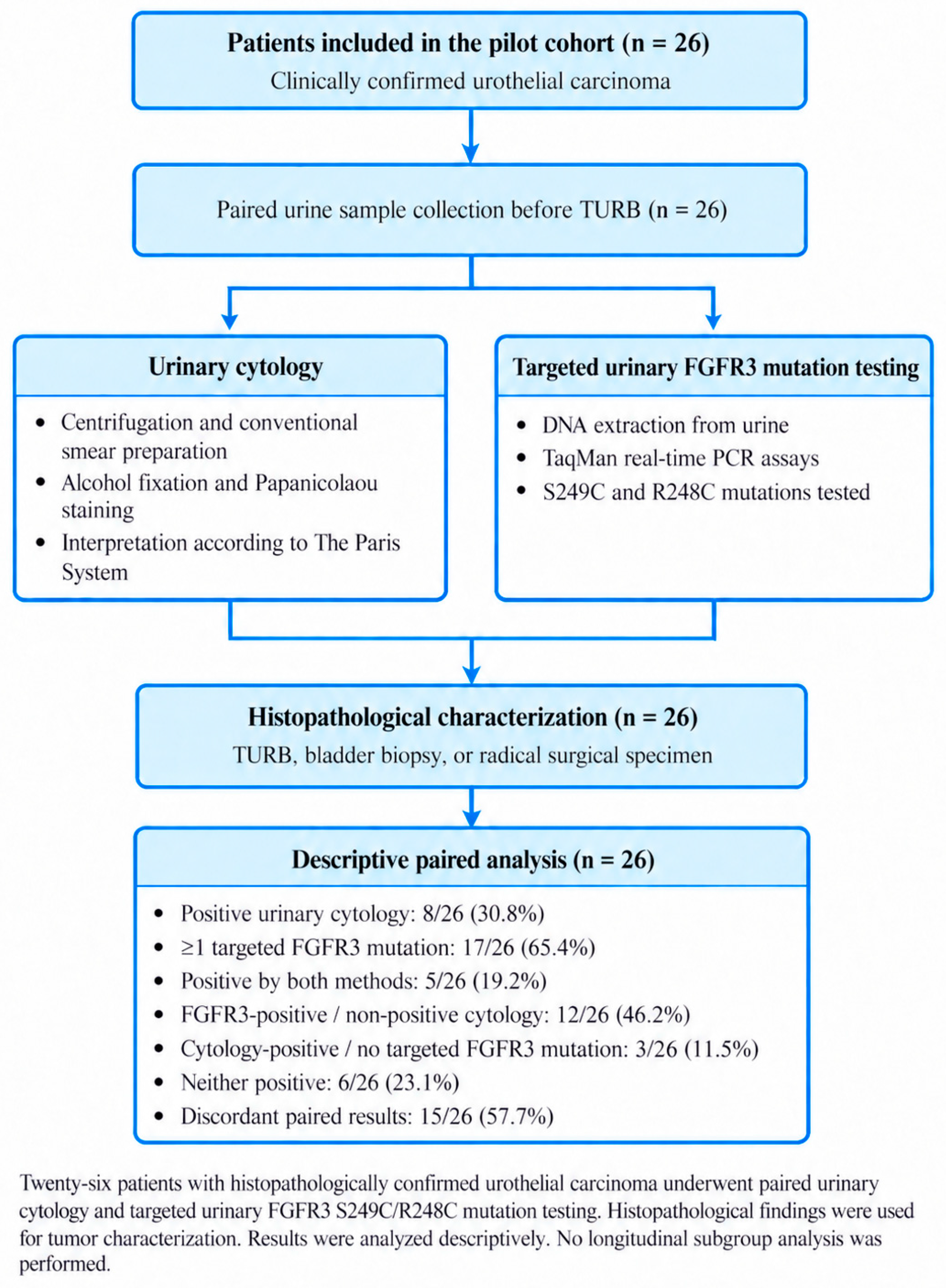
At least one targeted FGFR3 mutation was detected in 17/26 patients (65.4%), whereas cytology was positive in 8/26 patients (30.8%). Five patients were positive by both approaches. Twelve patients had one or both targeted FGFR3 mutations despite non-positive cytology, while three had positive cytology without detectable S249C or R248C. Six patients had neither positive cytology nor either targeted mutation. Thus, the two urine-based approaches were discordant in 15/26 patients (57.7%) (Table 2). This discordance is reported descriptively and is not interpreted as evidence of superiority of either method.
Table 2.
Paired urinary cytology and targeted urinary FGFR3 mutation results.
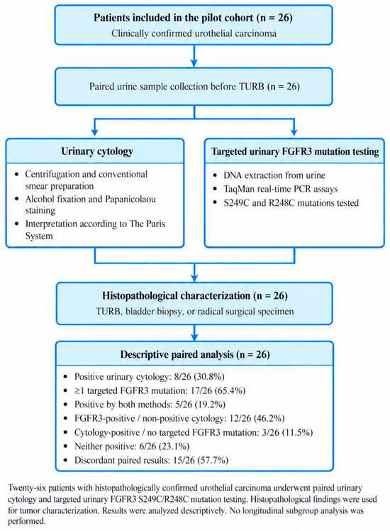
Within the Ta subgroup, 11/16 patients (68.8%) had at least one targeted FGFR3 mutation and 4/16 (25.0%) had positive cytology. Among the combined T1/T2 subgroup, 6/10 patients (60.0%) had at least one targeted mutation and 4/10 (40.0%) had positive cytology. These subgroup counts are descriptive only and were not subjected to significance testing Figure 1.
Figure 1.
Patient flow and paired urinary testing in the pilot cohort.
4. Discussion
This pilot study was focused on a single question: how conventional urinary cytology and targeted urinary detection of FGFR3 S249C/R248C behave when assessed in the same small cohort. The main finding is the frequency of discordant paired results. Although 17/26 patients had at least one targeted FGFR3 mutation, only 8/26 had positive cytology; 12 patients were FGFR3-positive despite non-positive cytology, while 3 showed the opposite pattern. These observations do not demonstrate that FGFR3 testing is more sensitive than cytology because the study lacks disease-negative controls, comprehensive tumor-tissue genotyping, and adequate sample size for a formal diagnostic-accuracy analysis. Rather, they support the narrower hypothesis that morphology-based cytology and mutation-specific urinary testing can provide non-identical information.
The biological rationale for such discordance is plausible. FGFR3 mutations are particularly frequent in papillary NMIBC, and S249C and R248C are among the recurrent activating hotspots described in urothelial carcinoma [23,24,25,26,28]. By contrast, urinary cytology is primarily optimized for recognition of high-grade morphological atypia and performs substantially less well in many low-grade papillary tumors [8,9,11,12,13,14,15]. Rieger-Christ et al. demonstrated that FGFR3 mutations detected in urine sediment DNA could complement cytology in bladder tumor detection [27]. Subsequent studies further demonstrated the feasibility of urinary FGFR3 mutation testing in NMIBC surveillance [29,30]. The findings of the present pilot study are compatible with this concept but do not allow quantification of incremental diagnostic benefit. The study by Bannier et al. provides an important contemporary context for FGFR3 testing [36]. Using 1222 cases, the investigators developed and externally validated a deep-learning model that pre-screened FGFR3 mutational status from routine H&E-stained tumor slides. The model achieved > 93% sensitivity in advanced and metastatic validation cohorts while potentially reducing molecular testing by approximately 40%. Notably, S249C and R248C were among the recurrent hotspots represented in their dataset, and S249C was reported as the easiest hotspot for the model to detect. Their approach is fundamentally different from ours: Bannier et al. inferred mutation probability from tumor histomorphology and used tissue molecular status as ground truth, whereas the present study directly interrogated two predefined mutations in urine-derived DNA and did not perform paired tumor-tissue FGFR3 genotyping [36]. The comparison therefore highlights both the promise of alternative FGFR3 pre-screening strategies and the stronger validation framework required before clinical implementation.
The 2026 ASCO Genitourinary Cancers Symposium report by Basin et al. further illustrates the importance of biospecimen type and tumor-derived DNA fraction in molecular FGFR3 testing [37]. In clinically advanced urothelial bladder cancer, the investigators compared large tissue-biopsy and plasma liquid-biopsy datasets and found that detection of FGFR3 alterations in circulating tumor DNA increased with plasma tumor fraction; when tumor fraction was <1%, FGFR3-positive disease could be missed. These results concern plasma circulating tumor DNA, not urine, and should not be extrapolated directly to urinary assays. They nevertheless reinforce a relevant principle for the present work: the analytical yield of a non-tissue molecular assay depends on the biological source and quantity of tumor-derived DNA, and negative results cannot be interpreted independently of these factors.
The clinical relevance of urinary FGFR3 testing in this setting is therefore complementary rather than substitutive. Cystoscopy and histopathology remain essential for diagnosis and risk stratification, and cytology remains clinically valuable for high-grade disease [1,2,3,4,5,6,7]. A targeted urinary assay that interrogates only S249C and R248C cannot detect FGFR3-wild-type tumors or tumors carrying other FGFR3 alterations. Conversely, mutation detection in urine does not by itself establish the presence, location, grade, or stage of a tumor. The potential value of combining cytology with molecular urine testing must therefore be demonstrated prospectively using predefined clinical endpoints rather than inferred from mutation prevalence alone.
Several limitations are central to interpretation of this pilot study. First, the cohort comprised only 26 patients from a single center, making inferential statistical testing inappropriate. Second, all analyzed patients had histopathological confirmed urothelial carcinoma; without a disease-negative control group, sensitivity, specificity, predictive values, and diagnostic accuracy cannot be estimated. Third, only S249C and R248C were tested, so a negative urinary result does not indicate FGFR3 wild-type status. Fourth, tumor tissue was not genotyped for FGFR3 in parallel; therefore, concordance between urinary mutations and the molecular status of the corresponding tumor cannot be determined. Fifth, the study did not quantify mutant allele fraction or evaluate broader genomic panels.
The principal strength is the prospective paired assessment of cytology and targeted FGFR3 testing from the same urine collection in a clinically characterized cohort. This design permits a direct description of within-patient discordance without implying that either test is a diagnostic reference standard. The findings can therefore serve as pilot data for a larger study that includes disease-negative controls, comprehensive tissue and urine FGFR3 profiling, prespecified cytology categories, and clinically meaningful endpoints such as detection of recurrence or high-grade disease.
5. Conclusions
In this 26-patient pilot study, conventional urinary cytology and targeted urinary detection of FGFR3 S249C/R248C frequently yielded discordant paired results. This pattern is compatible with the two methods interrogating different biological features of urothelial carcinoma, but it does not establish diagnostic superiority, sensitivity, or clinical utility. Targeted urinary FGFR3 testing should not replace cystoscopy, histopathology, or cytology based on these data. Larger prospective studies should include disease-negative controls, paired tumor-tissue genotyping, broader FGFR3 and genomic panels, and predefined clinical endpoints to determine whether urinary molecular testing adds clinically meaningful information to conventional evaluation.
Author Contributions
Conceptualization, B.-P.T. and A.B.; methodology, B.-P.T., I.G., A.-D.C., M.L.V.M. and A.F. writing—original draft preparation, B.-P.T.; writing—review and editing, B.-P.T., A.B. and M.L.V.M. All authors have read and agreed to the published version of the manuscript.
Funding
This paper was funded by a PCD grant awarded by the University of Medicine and Pharmacy “Iuliu Hatieganu” Cluj Napoca No. 1032/69/13.01.2021. The APC was funded by the main author Bogdan-Petru Tichil.
Institutional Review Board Statement
Investigations were conducted in accordance with the principles outlined in the Declaration of Helsinki (1975, revised in 2013). Local Ethics Commission/IRB approval was obtained (Approval code: Nr. AVZ7/Approval date: 26 January 2021) from The Ethics Committee of “Iuliu Hatieganu” University of Medicine and Pharmacy Cluj-Napoca, Romania.
Informed Consent Statement
Informed consent was obtained from all subjects involved in the study.
Data Availability Statement
The data presented in this study are available on request from the corresponding author due to privacy, legal and ethical restrictions.
Acknowledgments
The authors have reviewed and edited the output and take full responsibility for the content of this publication.
Conflicts of Interest
The authors declare no conflicts of interest. The funders had no role in the design of the study; in the collection, analyses, or interpretation of data; in the writing of the manuscript; or in the decision to publish the results.
Abbreviations
The following abbreviations are used in this manuscript:
| AKT | Protein kinase B |
| AS-PCR | Allele-specific polymerase chain reaction |
| AUC | Atypical urothelial cells |
| AUA | American Urological Association |
| BC | Bladder cancer |
| BCG | Bacillus Calmette–Guérin |
| BMI | Body mass index |
| CIS | Carcinoma in situ |
| CT | Computed tomography |
| ddPCR | Droplet digital polymerase chain reaction |
| DNA | Deoxyribonucleic acid |
| EAU | European Association of Urology |
| FAM | 6-Carboxyfluorescein |
| FGFR | Fibroblast growth factor receptor |
| FGFR3 | Fibroblast growth factor receptor 3 |
| HGUC | High-grade urothelial carcinoma |
| LGUN | Low-grade urothelial neoplasm |
| MAPK | Mitogen-activated protein kinase |
| MIBC | Muscle-invasive bladder cancer |
| MPC | Protein precipitation reagent used in the MasterPure purification protocol |
| NCCN | National Comprehensive Cancer Network |
| NGS | Next-generation sequencing |
| NHGUC | Negative for high-grade urothelial carcinoma |
| NMIBC | Non-muscle-invasive bladder cancer |
| NTC | No-template control |
| PCR | Polymerase chain reaction |
| PI3K | Phosphoinositide 3-kinase |
| PLCγ | Phospholipase C gamma |
| PPV | Positive predictive value |
| NPV | Negative predictive value |
| RB1 | Retinoblastoma 1 gene |
| RNA | Ribonucleic acid |
| SHGUC | Suspicious for high-grade urothelial carcinoma |
| STAT | Signal transducer and activator of transcription |
| SUO | Society of Urologic Oncology |
| TERT | Telomerase reverse transcriptase |
| TNM | Tumor–Node–Metastasis classification |
| TP53 | Tumor protein p53 gene |
| TPS | The Paris System for Reporting Urinary Cytology |
| UC | Urothelial carcinoma |
References
- Yang, Z.; Song, F.; Zhong, J. Urinary biomarkers in bladder cancer: FDA-approved tests and emerging tools for diagnosis and surveillance. Cancers 2025, 17, 3425. [Google Scholar] [CrossRef] [Scilit] [PubMed]
- Gontero, P.; Mariappan, P.; Pradere, B.; Rai, B.P.; Liedberg, F.; Compérat, E.; Teoh, J.Y.; Moschini, M.; Soria, F.; van Rhijn, B.W.G.; et al. EAU Guidelines on Non-Muscle-Invasive Bladder Cancer (TaT1 and CIS)—A Summary of the 2026 Guidelines Update. Eur. Urol. 2026. advance online publication. [Google Scholar] [CrossRef] [Scilit] [PubMed]
- Holzbeierlein, J.M.; Bixler, B.R.; Buckley, D.I.; Chang, S.S.; Holmes, R.; James, A.C.; Kirkby, E.; McKiernan, J.M.; Schuckman, A.K. Diagnosis and treatment of non-muscle invasive bladder cancer: AUA/SUO guideline: 2024 amendment. J. Urol. 2024, 211, 533–538. [Google Scholar] [CrossRef] [Scilit] [PubMed]
- National Comprehensive Cancer Network. NCCN Clinical Practice Guidelines in Oncology: Bladder Cancer; Version 2.2026; National Comprehensive Cancer Network: Plymouth Meeting, PA, USA, 2026. [Google Scholar]
- Matuszczak, M.; Salagierski, M. Diagnostic and prognostic potential of biomarkers CYFRA 21.1, ERCC1, p53, FGFR3 and TATI in bladder cancers. Int. J. Mol. Sci. 2020, 21, 3360. [Google Scholar] [CrossRef] [Scilit] [PubMed]
- Batista, R.; Vinagre, J.; Prazeres, H.; Sampaio, C.; Peralta, P.; Conceição, P.; Sismeiro, A.; Leão, R.; Gomes, A.; Furriel, F.; et al. Validation of a novel, sensitive, and specific urine-based test for recurrence surveillance of patients with non-muscle-invasive bladder cancer in a comprehensive multicenter study. Front. Genet. 2019, 10, 1237. [Google Scholar] [CrossRef] [Scilit] [PubMed]
- Hentschel, A.E.; van der Toom, E.E.; Vis, A.N.; Ket, J.C.F.; Bosschieter, J.; Heymans, M.W.; van Moorselaar, R.J.; Steenbergen, R.D.M.; A Nieuwenhuijzen, J. A systematic review on mutation markers for bladder cancer diagnosis in urine. BJU Int. 2021, 127, 12–27. [Google Scholar] [CrossRef] [Scilit] [PubMed]
- Sullivan, P.S.; Chan, J.B.; Levin, M.R.; Rao, J. Urine cytology and adjunct markers for detection and surveillance of bladder cancer. Am. J. Transl. Res. 2010, 2, 412–440. [Google Scholar] [PubMed]
- Strojan Flezar, M. Urine and bladder washing cytology for detection of urothelial carcinoma: Standard test with new possibilities. Radiol. Oncol. 2010, 44, 207–214. [Google Scholar] [CrossRef] [Scilit] [PubMed]
- Bisharah, S.; Raap, M.; Abbas, M. Urine cytological diagnostics: Possibilities and limitations—A 25-year review and overview at Hannover Medical School. Clin. Pract. 2025, 15, 234. [Google Scholar] [CrossRef] [Scilit] [PubMed]
- Chou, R.; Gore, J.L.; Buckley, D.; Fu, R.; Gustafson, K.; Griffin, J.C.; Grusing, S.; Selph, S. Urinary biomarkers for diagnosis of bladder cancer: A systematic review and meta-analysis. Ann. Intern. Med. 2015, 163, 922–931. [Google Scholar] [CrossRef] [Scilit] [PubMed]
- Nikas, I.P.; Seide, S.; Proctor, T.; Kleinaki, Z.; Kleinaki, M.; Reynolds, J.P. The Paris System for Reporting Urinary Cytology: A meta-analysis. J. Pers. Med. 2022, 12, 170. [Google Scholar] [CrossRef] [Scilit] [PubMed]
- Kars, M.; Çetin, M.; Toper, M.H.; Filinte, D.; Çam, K. Contemporary role of urine cytology in bladder cancer. Bull. Urooncol. 2024, 23, 73–77. [Google Scholar] [CrossRef] [Scilit]
- Rakhshan, A.; Arvin, E.; Alahyari, S.; Kazeminegad, B.; Mollasharifi, T.; Bagheri, A.; Aliakbari, F.; Hosseini, S.J.; Soleimani, M.; Ahadi, M.; et al. Accuracy of Paris 2016 System for non-invasive diagnosis bladder malignancy. Iran. J. Pathol. 2022, 17, 328–334. [Google Scholar] [CrossRef] [Scilit] [PubMed]
- Phruttinarakorn, B.; Plumworasawat, S.; Kayankarnnavee, J.; Lualon, J.; Pongtippan, A. Application of the Paris Reporting System for urine cytology: The three-year experience of a single tertiary care institute in Thailand. Acta Cytol. 2022, 66, 134–141. [Google Scholar] [CrossRef] [Scilit] [PubMed]
- Wojcik, E.M. The Paris System for reporting urinary cytology: What worked and what still needs to be improved. Histopathology 2026, 88, 131–136. [Google Scholar] [CrossRef] [Scilit] [PubMed]
- Long, T.; Layfield, L.J.; Esebua, M.; Frazier, S.R.; Giorgadze, D.T.; Schmidt, R.L. Interobserver reproducibility of The Paris System for Reporting Urinary Cytology. CytoJournal 2017, 14, 17. [Google Scholar] [CrossRef] [Scilit] [PubMed]
- Yamasaki, M.; Taoka, R.; Katakura, K.; Matsunaga, T.; Kani, N.; Honda, T.; Harada, S.; Tohi, Y.; Matsuoka, Y.; Kato, T.; et al. The Paris System for reporting urinary cytology improves the negative predictive value of high-grade urothelial carcinoma. BMC Urol. 2022, 22, 51. [Google Scholar] [CrossRef] [Scilit] [PubMed]
- Chen, F.; Lin, X. The Paris System for Reporting Urinary Cytology: An updated review. J. Clin. Transl. Pathol. 2023, 3, 59–74. [Google Scholar] [CrossRef] [Scilit]
- Kalogeraki, A.M.; Tamiolakis, D.I.; Mamoulakis, C. Urine cytology and axillary testing for interpretation and follow-up of urinary tumors. Acta Clin. Croat. 2022, 61, 505–510. [Google Scholar] [CrossRef] [Scilit] [PubMed]
- Mowatt, G.; Zhu, S.; Kilonzo, M.; Boachie, C.; Fraser, C.; Griffiths, T.R.L.; N’Dow, J.; Nabi, G.; Cook, J.; Vale, L. Systematic review of the clinical effectiveness and cost-effectiveness of photodynamic diagnosis and urine biomarkers and cytology for the detection and follow-up of bladder cancer. Health Technol. Assess. 2010, 14, 1–331. [Google Scholar] [CrossRef] [Scilit] [PubMed]
- Lee, H.H.; Kim, S.H. Review of non-invasive urinary biomarkers in bladder cancer. Transl. Cancer Res. 2020, 9, 6554–6564. [Google Scholar] [CrossRef] [Scilit] [PubMed]
- Wei, H.; Wan, W.; Zhan, H.; Wang, J.; Chen, J. The role of FGFR3 in the diagnosis and treatment of bladder cancer: A review. Cancer Plus 2021, 3, 28–34. [Google Scholar] [CrossRef] [Scilit]
- Kompier, L.C.; Lurkin, I.; van der Aa, M.N.M.; van Rhijn, B.W.G.; van der Kwast, T.H.; Zwarthoff, E.C. FGFR3, HRAS, KRAS, NRAS and PIK3CA mutations in bladder cancer and their potential as biomarkers for surveillance and therapy. PLoS ONE 2010, 5, e13821. [Google Scholar] [CrossRef] [Scilit] [PubMed]
- García-Perdomo, H.A.; Usubillaga-Velasquez, J.P.; Zapata-Copete, J.A.; Reis, L.O. Mutations in CDKN2A and the FGFR3 genes on bladder cancer diagnosis: A systematic review and meta-analysis. World J. Urol. 2019, 37, 2001–2007. [Google Scholar] [CrossRef] [Scilit] [PubMed]
- van Oers, J.M.M.; Lurkin, I.; van Exsel, A.J.M.; Nijsen, Y.; van Rhijn, B.W.G.; van der Aa, M.N.M.; Zwarthoff, E.C. A simple and fast method for the simultaneous detection of nine fibroblast growth factor receptor 3 mutations in bladder cancer and voided urine. Clin. Cancer Res. 2005, 11, 7743–7748. [Google Scholar] [CrossRef] [Scilit] [PubMed]
- Rieger-Christ, K.M.; Mourtzinos, A.; Lee, P.J.; Zagha, R.M.; Cain, J.; Silverman, M.; Libertino, J.A.; Summerhayes, I.C. Identification of fibroblast growth factor receptor 3 mutations in urine sediment DNA samples complements cytology in bladder tumor detection. Cancer 2003, 98, 737–744. [Google Scholar] [CrossRef] [Scilit] [PubMed]
- Yu, S.H.; Kim, S.S.; Kim, S.; Lee, H.; Kang, T.W. FGFR3 mutations in urothelial carcinoma: A single-center study using next-generation sequencing. J. Clin. Med. 2024, 13, 1305. [Google Scholar] [CrossRef] [Scilit] [PubMed]
- Roperch, J.P.; Hennion, C. A novel ultra-sensitive method for the detection of FGFR3 mutations in urine of bladder cancer patients—Design of the Urodiag PCR kit for surveillance of NMIBC. BMC Med. Genet. 2020, 21, 112. [Google Scholar] [CrossRef] [Scilit] [PubMed]
- Zuiverloon, T.C.M.; van der Aa, M.N.M.; van der Kwast, T.H.; Steyerberg, E.W.; Lingsma, H.F.; Bangma, C.H.; Zwarthoff, E.C. Fibroblast growth factor receptor 3 mutation analysis on voided urine for surveillance of patients with low-grade non-muscle-invasive bladder cancer. Clin. Cancer Res. 2010, 16, 3011–3018. [Google Scholar] [CrossRef] [Scilit] [PubMed]
- Hayashi, Y.; Fujita, K.; Matsuzaki, K.; Matsushita, M.; Kawamura, N.; Koh, Y.; Nakano, K.; Wang, C.; Ishizuya, Y.; Yamamoto, Y.; et al. Diagnostic potential of TERT promoter and FGFR3 mutations in urinary cell-free DNA in upper tract urothelial carcinoma. Cancer Sci. 2019, 110, 1771–1779. [Google Scholar] [CrossRef] [Scilit] [PubMed]
- de Jong, F.C.; Iflé, I.G.; van der Made, A.C.; Kooper, D.; de Jong, J.J.; Franckena, M.; Zuiverloon, T.C.M.; van Criekinge, W.; Incrocci, L.; Zwarthoff, E.C.; et al. A genomic urine assay for surveillance of patients with bladder cancer treated with radiotherapy. Eur. Urol. Open Sci. 2024, 62, 131–139. [Google Scholar] [CrossRef] [Scilit] [PubMed]
- Humayun-Zakaria, N.; Ward, D.G.; Arnold, R.; Bryan, R.T. Trends in urine biomarker discovery for urothelial bladder cancer: DNA, RNA, or protein? Transl. Androl. Urol. 2021, 10, 2787–2808. [Google Scholar] [CrossRef] [Scilit] [PubMed]
- Oyaert, M.; Van Praet, C.; Delrue, C.; Speeckaert, M. Novel urinary biomarkers for the detection of bladder cancer. Cancers 2025, 17, 1283. [Google Scholar] [CrossRef] [Scilit] [PubMed]
- Wan, X.; Wang, D.; Zhang, X.; Xu, M.; Huang, Y.; Qin, W.; Chen, S. Unleashing the power of urine-based biomarkers in diagnosis, prognosis and monitoring of bladder cancer. Int. J. Oncol. 2025, 66, 18. [Google Scholar] [CrossRef] [Scilit] [PubMed]
- Bannier, P.A.; Saillard, C.; Mann, P.; Touzot, M.; Maussion, C.; Matek, C.; Klümper, N.; Breyer, J.; Wirtz, R.; Sikic, D.; et al. AI allows pre-screening of FGFR3 mutational status using routine histology slides of muscle-invasive bladder cancer. Nat. Commun. 2024, 15, 10914. [Google Scholar] [CrossRef] [Scilit] [PubMed]
- Basin, M.; Pavlick, D.C.; Daneshmand, S.; Grivas, P.; Agarwal, N.; Gupta, S.; Cheng, L.; Kamat, A.M.; Spiess, P.E.; Li, R.; et al. Detecting FGFR3 genomic alterations in liquid biopsies from patients with clinically advanced urothelial bladder cancer. J. Clin. Oncol. 2026, 44, 830. [Google Scholar] [CrossRef] [Scilit]

Disclaimer/Publisher’s Note: The statements, opinions and data contained in all publications are solely those of the individual author(s) and contributor(s) and not of MDPI and/or the editor(s). MDPI and/or the editor(s) disclaim responsibility for any injury to people or property resulting from any ideas, methods, instructions or products referred to in the content. |
© 2026 by the authors. Licensee MDPI, Basel, Switzerland. This article is an open access article distributed under the terms and conditions of the Creative Commons Attribution (CC BY) license.