


Abstract
Background: The association of sex hormone-binding globulin (SHBG) with heart failure (HF) remains a topic of ongoing debate, particularly in the light of type 2 diabetes mellitus (T2DM). We aimed to assess the association of SHBG with clinical and echocardiographic parameters of HF in men according to the presence of T2DM. Methods: Data on baseline characteristics, cardiovascular risk factors and medications, laboratory findings including serum SHBG and total testosterone concentrations, and echocardiographic parameters were prospectively collected for 215 male patients consecutively hospitalized for an acute episode of HF. Results: Patients with T2DM were older (p = 0.013), had a greater body mass index (p = 0.009) and NYHA class (p = 0.001), and were more likely to have hypertension (p < 0.001) or hyperlipidemia (p = 0.032). A moderate correlation among SHBG and total testosterone with the left ventricular ejection fraction (LVEF) was observed only in T2DM patients (r = 0.456) but not among non-T2DM patients (r = 0.194). A multivariate analysis revealed the independent association of increased SHBG levels with lower LVEF values among T2DM patients (ß = −0.542, p < 0.0001), whereas in the same group higher total testosterone was an independent predictor of higher LVEF (ß = 0.531, p < 0.0001) and lower LVDD (ß = −0.442, p = 0.0002) levels. Conclusions: In men with HF and T2DM, in contrast to testosterone, SHBG may have an independent adverse impact on the LVEF, which may account for 12.5% of the variance in LVEF levels. The possible subcellular mechanisms of SHBG in men with diabetic myocardial disorder should be additionally explored.
1. Introduction
In addition to other clinical symptoms and signs, heart failure (HF) is a clinical syndrome commonly associated with a multiple hormonal imbalance [1]. These imbalances include decreased testosterone [2,3], disturbed thyroid hormone levels [4], insulin resistance, inappropriate antidiuretic hormone secretion [1], and the downregulation of growth hormone [1,5] and insulin-like growth factor-1 [1,6], as well as increased parathyroid hormone and cortisol levels [7].
Sex hormone-binding globulin (SHBG) is a glycoprotein that binds most of the circulating testosterone. Within the context of the hormone imbalance in HF and cardiovascular outcomes, previous studies have reported conflicting results on SHBG, including a lower risk of HF with a low SHBG level [8], a non-linear association with cardiovascular mortality [9], or no association with HF [10]. Recently, increased SHBG has been suggested as a predictor of HF hospitalizations only in dysglycemic men and not in dysglycemic women [11]. Considering cardiac function, only a few studies [12,13] have investigated the association of SHBG with left ventricular (LV) ejection fraction (LVEF), providing limited information on this issue. To the best of our knowledge, there are no data on the association of SHBG with LV diastolic dysfunction (LVDD).
To stress the importance of the independent contribution of type 2 diabetes mellitus (T2DM) to the progression of HF, the term “diabetic myocardial disorder” has been proposed for systolic and/or diastolic myocardial dysfunction in the presence of T2DM [14]. Factors such as a more progressed renal impairment and lower testosterone levels with T2DM may be independent contributors to a worse prognosis of HF in those patients [15]. However, little is known about the possible mechanisms that could mediate the linkages among T2DM, HF, and SHBG. By separating the effect of testosterone, we aimed to assess the independent association of SHBG with clinical and echocardiographic parameters of HF in men according to the presence of T2DM.
2. Materials and Methods
2.1. Study Patients
Our investigation of the association of serum hormone levels with clinical parameters of HF included men hospitalized due to an acute episode of HF; the findings on thyroid hormones have been reported elsewhere [16]. The present cross-sectional study enrolled 215 male patients, and the inclusion criteria were: (1) clinical presentation typical for HF; (2) LVEF < 50% and/or LVDD established by transthoracic echocardiography; (3) unchanged medical therapy during at least one month. The exclusion criteria were: (1) acute or chronic systemic illness that could affect the hormonal metabolism (i.e., a primary endocrine disorder, liver disease or liver cirrhosis, autoimmune or malignant disease, infection, terminal phase of renal failure, or body mass index (BMI) < 18.5 kg/m2); (2) any hormonal treatment or drug intake at the time of the study or in the past (corticosteroids, synthetic thyroid hormones, antithyroid drugs, dopamine, dobutamine); (3) cardiac surgery, acute coronary syndrome, or coronary revascularization within six months before the study; (4) C-reactive protein levels above 15 mg/d. This study complied with the Declaration of Helsinki. The study design and protocol were approved by the Ethics Committee of the University Hospital Center Split (2181-147-10-01/01-M.J.). Each participant provided written informed consent.
2.2. Data Collection
The presence of T2DM was defined as previously known diabetes patients on stable therapy for at least 3 months or newly detected T2DM based on an oral glucose tolerance test. For all patients, blood samples for a biochemical analysis and blood count were taken on admission during the initial evaluation in the emergency department. Serum concentrations of SHBG (nmol/L), total testosterone (nmol/L), total triiodothyronine (nmol/L), and plasma N-terminal pro-B type natriuretic peptide (NT-proBNP, pmol/L) were measured using a chemiluminescence immunoassay (Roche Elecsys 2010/Elecsys 1010, Roche Diagnostics GmbH, Mannheim, Germany). Blood samples for the hormones of interest were taken during the first three days of hospitalization between 08:00 and 09:00 to account for circadian variations in circulating hormone levels. For all hormone assays, the intra- and inter-assay variation coefficients were <9% and <13%, respectively.
To measure general adiposity, we calculated the BMI as the body weight in kilograms divided by the squared height in meters. The glomerular filtration rate (GFR, in mL/min/1.73 m2) was estimated using the simplified Modification of Diet in Renal Disease formula [17]. Given the minor clinical importance of C-reactive protein levels under 10 mg/L [18], and to account for a low-grade systemic inflammation that accompanies HF [19], the cut-off value for inclusion in the study was set to a maximum of 15 mmol/L, corresponding to a maximum increase of 50% in the C-reactive protein level [20].
2.3. Echocardiographic Examination
All subjects underwent a standard transthoracic echocardiographic examination at rest during the first 2 days of hospitalization. The examinations were performed by certified cardiologists using a Vivid 9E device (GE Medical System, Milwaukee WI, USA). All echocardiographic parameters were obtained in accordance with the standard guidelines of the American Society of Echocardiography and the European Association of Cardiovascular Imaging [21]. The LVEF was assessed using the biplane Simpson’s method, whereas the severity of LVDD (3 basic grades) was classified by assessing the early filling (E) and atrial (A) filling peak velocities, E/A ratio, E-wave deceleration time, isovolumic relaxation time, septal and lateral diastolic é and á peak annular tissue velocities, é/á ratio, and LV filling index E/é ratio. The mitral valve inflow for assessment of LVDD was measured on a pulsed-wave Doppler device in the four-chamber view and the sample volume was positioned within one centimeter of the septal and lateral insertion of the mitral valve leaflets [21].
2.4. Statistical Analyses
The normally distributed continuous variables are presented as means with standard deviations, and those with a skewed distribution as medians with interquartile ranges. The intergroup differences between those with and without T2DM were tested using the Student’s t-test, Mann–Whitney U test, or χ2 test where appropriate. The multivariate analysis was deployed by using hierarchical linear regression. To assess the predictive associations of variables of interest for the clinical parameters of HF, the data were entered in 4 blocks (models). In model 1, adjustments were made for age, BMI, and GFR because of their important role in the pathophysiology, presentation, and prognosis of HF, and also because of their independent associations with circulating SHBG levels [22]. Model 2 contained relevant clinical variables and risk factors, model 3 contained serum total testosterone and total triiodothyronine values, and in model 4 SHBG values were added. The change in R2 was evaluated for each block and individual contributions of independent variables were calculated by squaring the semi-partial correlation. The predictive values of the independent variables in multiple regression models are expressed using the standardized partial regression coefficient ß and the corresponding p value.
All statistical analyses were conducted using IBM SPSS Statistics for Windows, version 26 (IBM Corp., Armonk, NY, USA). A 2-sided α of <0.05 was considered statistically significant.
3. Results
The clinical characteristics, laboratory and echocardiographic findings, and medication taken by the study patients according to presence of diabetes are presented in Table 1 and Table 2. The diabetic patients were older, had a greater BMI and NYHA class (Table 1), and were more likely to have hypertension or hyperlipidemia and to take furosemide, calcium antagonists, angiotensin II-receptor blockers, aspirin, or statins (Table 2). All study patients had LVDD with an average grade of 2.5 ± 0.6 (Table 1).
Table 1.
Baseline characteristics and prehospital medication in the study population and in patients according to the presence of T2DM.
Table 2.
Laboratory findings for the study population and for patients according to the presence of T2DM.
Considering the clinical HF parameters, in the univariable analysis, the SHBG levels inversely correlated with NYHA and NT-proBNP values in the subgroup of patients with T2DM, whereas no significant correlations were observed for echocardiographic parameters of cardiac function or duration of HF (Table 3). In the subgroup of patients without T2DM, no significant correlation was observed between SHBG and any of the clinical or echocardiographic parameters of HF.
Table 3.
Correlation of serum SHBG and total testosterone levels with echocardiographic, laboratory, and clinical parameters of HF in the study population and in patients according to the presence of T2DM.
Total testosterone positively correlated with the LVEF in both subgroups according to T2DM. Furthermore, in patients with T2DM, testosterone inversely correlated with the LVDD, NT-proBNP, and NYHA class, whereas there was no significant association with the duration of HF. In patients without T2DM, testosterone inversely correlated with NT-proBNP, showed no significant correlation with LVDD or the NYHA class, and showed a borderline significance with the duration of HF (Table 3).
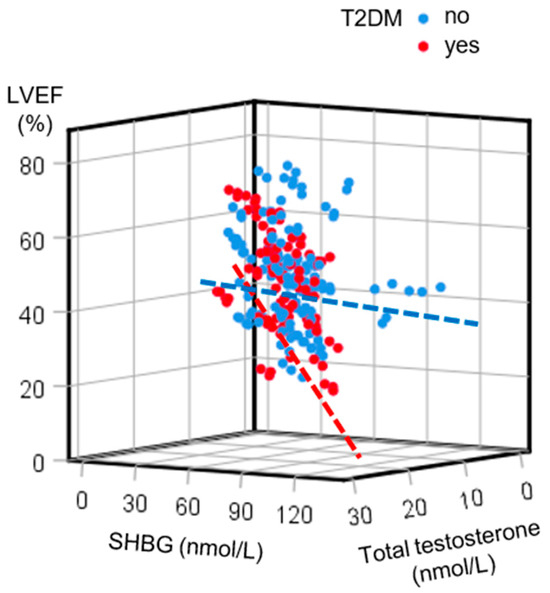
When plotting a combined relationship among sex hormones and the LVEF, a moderate inverse correlation among SHBG and total testosterone with the LVEF was observed in T2DM patients, whereas a very weak correlation was observed in those without T2DM (Figure 1).
Figure 1.
A 3D scatterplot depicting the correlations among sex hormone-binding globulin (SHBG), total testosterone, and left ventricular ejection fraction (LVEF) levels in patients with heart failure according to type 2 diabetes mellitus (T2DM) status. In contrast to a very weak correlation in patients without T2DM (blue dots and dashed line; r = 0.194, linear regression equation LVEF = 42.367 − 0.075 × SHBG + 0.305 × total testosterone), a moderate inverse correlation was observed in patients with T2DM (red dots and dashed line; r = 0.456, linear regression equation LVEF = 45.376 − 0.434 × SHBG + 0.535 × total testosterone).
In the multivariable analysis, among the T2DM patients, the adjustments for clinical variables including triiodothyronine and testosterone revealed that SHBG was the most important predictor, contributing 12.5% of the unique variance in LVEF levels in the final model (Table 4). In the same subgroup, SHBG explained 4.6% of the unique variance in the progression of LVDD (Supplemental Table S1). In the final models for the NYHA class (Supplemental Table S2) and duration of HF (Supplemental Table S3), the SHBG levels showed small, non-significant changes of 2.5% and 0.1% from the previous models, respectively.
Table 4.
A hierarchical regression analysis for LVEF in men with HF and T2DM.
In patients without T2DM, the SHBG levels showed no significant improvements from the previous models for the echocardiographic and clinical parameters of HF. In the final models, SHBG explained 2.4% of the variance in LVEF values (Supplemental Table S4), 6.9% of the variance in LVDD values (Supplemental Table S5), 1.4% of the variance in NYHA Class values (Supplemental Table S6), and 1.1% of the variance in the duration of HF (Supplemental Table S7).
In patients with T2DM, total testosterone was an independent predictor of higher LVEF and lower LVDD or NYHA values, and showed no predictive association for the duration of HF (Supplemental Table S8). For all of these analyses, there was no significant interaction between testosterone and SHBG (Supplemental Table S8). In patients without T2DM, of the echocardiographic or clinical parameters of HF, testosterone showed a predictive association only with the duration of HF (Supplemental Table S9). In these analyses, a significant interaction between testosterone and SHBG was observed for LVDD, and the interaction for LVEF showed a borderline statistical significance (Supplemental Table S9).
4. Discussion
The principal finding of the present study is the adverse effect of increased SHBG with lower LVEF, independent of the serum total testosterone levels, only in male HF patients with coexisting T2DM. In these patients, SHBG may explain 12.5% of the unique variance in LVEF.
Sex hormones have a substantial role in body composition, metabolism, inflammatory processes, and cardiovascular function. In contrast to previous thinking, recent evidence suggests that SHBG is not just a passive protein carrier that simply binds sex hormones, thereby regulating their circulating levels, but that it is a multifunctional protein and mediator involved in a number of physiological and pathophysiological processes, conditions, and diseases [23,24]. SHBG may partly be an inhibitor of sex steroids [12], whereby low levels increase and high levels decrease anabolic activity, favouring catabolic processes [25]. While free testosterone can easily enter the cell via passive diffusion, endocytosis of testosterone bound to SHBG is another means of cellular uptake and partly contributes to the intracellular testosterone activity [26]. The intracellular effects of SHBG are mediated through cell surface signalling, cellular delivery, and the direct activation of specific plasma receptors responsible for sex hormones’ biological actions [27,28]. However, the complex interplay among SHBG, testosterone, and their intracellular mechanisms affecting cardiac function is poorly understood.
The interactions among SHBG, testosterone, and T2DM represent an important facet of this multidimensional problem. A large systematic review with a meta-analysis of observational studies has suggested a link between lower levels of SHBG and a higher risk of T2DM [29]. A somewhat weaker causal interplay of SHBG with insulin resistance and T2DM has been observed in Mendelian randomization studies [29,30]. At the same time, mechanisms linking androgens to the development of T2DM have been well-established; higher total testosterone is associated with a lower T2DM risk in men, whereas in women, conversely, it is associated with an increased T2DM risk [29]. The mediating effect of SHBG in the association between sex and glucose homeostasis, independent of confounders such as age, obesity, and testosterone level, has also been recently reported [31]. Finally, it has been suggested that SHBG may play a more significant role in T2DM than androgens [32].
Data about the association of SHBG with cardiac function in HF are scarce. In men with HF and LVEF values <40%, higher SHBG levels have been associated with an increased 3-year cardiovascular mortality [12]. In men aged 40 to 69 years, independent of total testosterone and other covariates, those with lower SHBG levels had a lower risk for developing HF during a median 9-year follow-up period [8]. In a study using cardiac magnetic resonance to estimate the LV mass, LV mass index, cardiac output, and LVEF, total testosterone showed no significant association with cardiac mass or function after adjustment for clinically relevant variables [13]. In contrast, the associations with SHBG have been weak, explaining less than 1% of the variance in cardiac variables [13]. Compared to that study, our multivariate adjustment included a greater number of clinically relevant variables, and revealed that in the presence of T2DM, a high SHBG level was a predictor of low LVEF (explaining 12.5% of the variance in LVEF) and was associated with a trend toward more progressed LVDD. To the best of our knowledge, this is the first study suggesting that T2DM may play a role in the effects of SHBG on cardiomyocytes of men with HF.
Regardless of the presence of ischemic heart disease or hypertension, the presence of T2DM is associated with a greater likelihood of both LV systolic and LVDD, whereas the echocardiographic abnormalities often include lower LVEF levels, reduced global longitudinal strain, an abnormal E/e’ ratio, LV hypertrophy, and left atrial enlargement [14]. The term diabetic myocardial disorder has been proposed for systolic and/or diastolic myocardial dysfunction in the presence of T2DM. However, T2DM is rarely exclusively responsible for this condition and commonly operates in addition to comorbidities such as arterial hypertension, chronic kidney disease, obesity, or coronary artery disease [14]. While the clinical and echocardiographic features of diabetic myocardial disorder have been defined, the intracellular and metabolic mechanisms that may underlay this condition are less known.
Metabolically, the myocardium is a very active tissue that chiefly uses fatty acids to maintain oxidative phosphorylation and energy production [33]. Changes in cardiac energy metabolism contribute to the pathophysiology, progression, and severity of HF. The coexistence of T2DM independently and substantially influences these processes. In contrast to HF associated with hypertension or ischemia, where myocardial fatty acid oxidation decreases, in HF associated with T2DM fatty acid oxidation increases, making the failing heart even less efficient [34]. Enhanced glucose uptake into the myocardium in the setting of increased fatty acid uptake further contributes to the development of diabetic myocardial disorder. Hyperglycemia and insulin resistance enhance lipid accumulation in the heart, causing lipotoxicity, which is an important mediator of impaired mitochondrial dynamism and cardiomyocyte cytotoxicity [35,36]. These processes promote myocardial collagen deposition, extracellular matrix expansion, and fibrosis, eventually leading to HF [37,38].
A disruption of homeostasis caused by lipotoxicity, mitochondrial dysfunction, and other cardiotoxic processes that accompany T2DM disturb energy production and induce cardiomyocyte death and cardiac fibrosis. Alterations in the metabolic pathways may be associated with transcriptional changes in enzymes responsible for some of these pathways [34]. Whether SHBG, in the presence of T2DM, may influence post-translational epigenetic changes and the expression of genes associated with energy metabolism that may affect the function of cardiomyocytes and LVEF remains to be elucidated. This possibility may be supported by the observation that in men with dysglycemia, increased SHBG levels are associated with a greater risk of HF hospitalizations [11]. Nevertheless, our study suggests a need for future research on the expression of SHBG and its intracellular mechanisms in both healthy myocardial tissue and in HF.
Our study is in agreement with several previous observations that total testosterone may independently affect cardiac function. However, our study also suggests that this effect is confined to patients with T2DM. Chronic inflammation with enhanced production of growth factors, cytokines, and inflammatory mediators that accompany T2DM has been associated with damage caused by disturbed glucose homeostasis [30]. Glucose dysregulation induces metabolic damage and oxidative stress, and through epigenetic mechanisms leads to transcriptional changes [39,40]. In a mouse model, it has been suggested that a reduction in miR-146a, a non-coding RNA, caused by glucose dysregulation in T2DM may be one of the most important mediators of the proinflammatory state responsible for structural and functional cardiac changes, particularly cardiac fibrosis [41]. By opposing such processes, testosterone could be more important for cardioprotection in T2DM patients, with beneficial effects on both the LVEF and LVDD, as suggested by our results. Specifically, the favourable effects of testosterone on the LVEF and LVDD could be mediated through antiproliferative, anticollagen, and antifibrotic properties [2,42,43].
Two earlier meta-analyses of randomized controlled trials of testosterone supplementation within a physiological range in men with chronic HF have shown an improved functional capacity and quality of life expressed through exercise capacity and muscle strength [44,45]. The improvement has not been associated with significant changes in LVEF [44,45]. Nevertheless, it could be explained by effects such as peripheral and coronary vasodilation, an increase in baroreceptor sensitivity, beneficial changes in muscle structure and function, an improved ventilatory response, and an increase in hemoglobin levels and oxygen delivery [2,45]. However, a 2020 updated meta-analysis suggested that significantly improved exercise tolerance is present only with an endpoint of total testosterone of at least 25 nmol/L but not with an endpoint of TT < 25 nmol/L [46]. In terms of major adverse cardiac events, supplementation therapy in men with hypogonadism and a pre-existing or a high risk of cardiovascular disease seems safe [47,48]. Given that male hypogonadism is an important prognostic indicator in HF, this group of patients may be an optimal target population for this therapeutic approach. However, at the moment, there is no consensus about the definition of testosterone deficiency in patients with HF [49,50].
A recent 2024 individual participant data meta-analysis has suggested that lower SHBG levels in men are associated with lower all-cause and cardiovascular mortality, whereas low testosterone is associated with increased all-cause mortality [9]. Therefore, lower SHBG levels coupled with higher testosterone could be a favorable sex hormone profile for cardiovascular and health in general. Our study provides evidence that within this framework, T2DM may play an important independent role by influencing the effects of sex hormones on cardiac function. As described above, men with HF and testosterone deficiency may be a target population for testosterone supplementation. None of the clinical trials investigating testosterone supplementation have investigated the concomitant changes in SHBG levels. Therefore, we can only speculate about the effect of testosterone supplementation on SHBG levels, as well as possible net effects of such hormonal changes on cardiac function. With all that in mind, the most important implication of our study is that in future research, sex hormones in patients with HF should be separately analyzed in those with and those without T2DM.
Strengths and Limitations
The main strength of our study was the meticulous exclusion criteria designed to avoid potential influence on hormone metabolism. Additionally, we adjusted the associations of interest for a greater number of specific clinical confounders when compared to the previous clinical studies on this topic. To reduce the problem of a close correlation between testosterone and SHBG, and to estimate their separate effects, we standardized the individual values of these hormones and assessed their interactions. The statistical model allowed us to estimate the magnitude of the effects and possible impacts. The main limitation of our study was its observational nature, which prevented us from establishing causal relationships. We did not collect data on glycemic control in patients with T2DM nor on the presence of subclinical coronary artery disease or its extension. Next, we included only male patients with HF, and it is questionable whether our results can be generalized to the female HF population. Finally, we determined serum levels of the investigated hormones, whereas their levels within cardiac tissue remained unknown.
5. Conclusions
We found that in men with HF and T2DM, in contrast to testosterone, SHBG may have an independent adverse impact on the LVEF, which may account for 12.5% of the variance in LVEF levels. Our study provides the basis for avenues of future investigation of the potential pathophysiological pathways linking SHBG, testosterone, T2DM, cardiac function, and HF.
Supplementary Materials
The following supporting information can be downloaded at https://www.mdpi.com/article/10.3390/jcm14072132/s1: Table S1. A hierarchical regression analysis for LVDD in men with HF and T2DM. Table S2. A hierarchical regression analysis for NYHA in men with HF and T2DM. Table S3. A hierarchical regression analysis for the duration of HF in men with T2DM. Table S4. A hierarchical regression analysis for LVEF in men with HF without T2DM. Table S5. A hierarchical regression analysis for LVDD in men with HF but without T2DM. Table S6. A hierarchical regression analysis for NYHA in men with HF without T2DM. Table S7. A hierarchical regression analysis for the duration of HF in men without T2DM. Table S8. Predictive associations of baseline characteristics, risk factors, and circulating SHBG and total testosterone levels for echocardiographic and clinical parameters of HF in men with T2DM. Table S9. Predictive associations of baseline characteristics, risk factors, and circulating SHBG and total testosterone levels for echocardiographic and clinical parameters of HF in men without T2DM.
Author Contributions
Conceptualization, V.Č.; methodology, V.Č.; software, T.M.; validation, V.Č. and I.V.; formal analysis, V.Č., Ž.B. and I.V.; investigation, V.Č., Ž.B. and I.V.; data curation, V.Č., Ž.B. and I.V.; writing—original draft preparation, V.Č.; writing—review and editing, V.Č. and R.V.; visualization, V.Č. and T.M. All authors have read and agreed to the published version of the manuscript.
Funding
This research received no external funding.
Institutional Review Board Statement
The study was conducted in accordance with the Declaration of Helsinki, and approved by the Ethics Committee of the University Hospital Centre Split (2181-147-10-01/01-M.J.) on 30 March 2010.
Informed Consent Statement
Informed consent was obtained from all subjects involved in the study.
Data Availability Statement
The data sets analyzed in the current study are available from the corresponding author on reasonable request.
Acknowledgments
The authors thank Mary Louise Stover for her helpful comments and for editing the manuscript.
Conflicts of Interest
The authors declare no conflicts of interest. The funders had no role in the design of the study; in the collection, analyses, or interpretation of data; in the writing of the manuscript; or in the decision to publish the results.
Abbreviations
The following abbreviations are used in this manuscript:
| HF | Heart failure |
| SHBG | Sex hormone-binding globulin |
| T2DM | Type 2 diabetes mellitus |
| GFR | Glomerular filtration rate |
| LVEF | Left ventricular ejection fraction |
| LVDD | Left ventricular diastolic dysfunction |
| NT-proBNP | N-terminal pro-B type natriuretic peptide |
| E | Early filling |
| A | Atrial filling |
References
- Saccà, L. Heart failure as a multiple hormonal deficiency syndrome. Circ. Heart Fail. 2009, 2, 151–156. [Google Scholar] [CrossRef] [Scilit] [PubMed]
- Bušić, Ž.; Čulić, V. Central and peripheral testosterone effects in men with heart failure: An approach for cardiovascular research. World J. Cardiol. 2015, 7, 504–510. [Google Scholar] [CrossRef] [Scilit] [PubMed]
- Schäfer, S.; Aydin, M.A.; Appelbaum, S.; Kuulasmaa, K.; Palosaari, T.; Ojeda, F.; Blankenberg, S.; Jousilahti, P.; Salomaa, V.; Karakas, M. Low testosterone concentrations and prediction of future heart failure in men and in women: Evidence from the large FINRISK97 study. ESC Heart Fail. 2021, 8, 2485–2491. [Google Scholar] [CrossRef] [Scilit] [PubMed]
- Razvi, S.; Jabbar, A.; Pingitore, A.; Danzi, S.; Biondi, B.; Klein, I.; Peeters, R.; Zaman, A.; Iervasi, G. Thyroid hormones and cardiovascular function and diseases. J. Am. Coll. Cardiol. 2018, 71, 1781–1796. [Google Scholar] [CrossRef] [Scilit]
- Arcopinto, M.; Salzano, A.; Giallauria, F.; Bossone, E.; Isgaard, J.; Marra, A.M.; Bobbio, E.; Vriz, O.; Åberg, D.N.; Masarone, D.; et al. Growth hormone deficiency is associated with worse cardiac function, physical performance, and outcome in chronic heart failure: Insights from the T.O.S.CA. GHD study. PLoS ONE 2017, 12, e0170058. [Google Scholar] [CrossRef] [Scilit]
- De Giorgi, A.; Marra, A.M.; Iacoviello, M.; Triggiani, V.; Rengo, G.; Cacciatore, F.; Maiello, C.; Limongelli, G.; Masarone, D.; Perticone, F.; et al. Insulin-like growth factor-1 (IGF-1) as predictor of cardiovascular mortality in heart failure patients: Data from the T.O.S.CA. registry. Intern. Emerg. Med. 2022, 17, 1651–1660. [Google Scholar] [CrossRef] [Scilit]
- Nägele, M.P.; Barthelmes, J.; Kreysing, L.; Haider, T.; Nebunu, D.; Ruschitzka, F.; Sudano, I.; Flammer, A.J. Endocrine hormone imbalance in heart failure with reduced ejection fraction: A cross-sectional study. Health Sci. Rep. 2022, 5, e880. [Google Scholar] [CrossRef] [Scilit]
- Yeap, B.B.; Marriott, R.J.; Antonio, L.; Raj, S.; Dwivedi, G.; Reid, C.M.; Anawalt, B.D.; Bhasin, S.; Dobs, A.S.; Handelsman, D.J.; et al. Associations of serum testosterone and sex hormone–binding globulin with incident cardiovascular events in middle-aged to older men. Ann. Intern. Med. 2022, 175, 159–170. [Google Scholar] [CrossRef] [Scilit]
- Yeap, B.B.; Marriott, R.J.; Dwivedi, G.; Adams, R.J.; Antonio, L.; Ballantyne, C.M.; Bauer, D.C.; Bhasin, S.; Biggs, M.L.; Cawthon, P.M.; et al. Associations of testosterone and related hormones with all-cause and cardiovascular mortality and incident cardiovascular disease in men: Individual participant data meta-analyses. Ann. Intern. Med. 2024, 177, 768–781. [Google Scholar] [CrossRef] [Scilit]
- Zhao, D.; Guallar, E.; Ballantyne, C.M.; Post, W.S.; Ouyang, P.; Vaidya, D.; Jia, X.; Ying, W.; Subramanya, V.; Ndumele, C.E.; et al. Sex hormones and incident heart failure in men and postmenopausal women: The Atherosclerosis Risk in Communities study. J. Clin. Endocrinol. Metab. 2020, 105, e3798–e3807. [Google Scholar] [CrossRef] [Scilit]
- Wang, A.; Hess, S.; Lee, S.F.; Gerstein, H.C. Sex hormone-binding globulin and heart failure hospitalizations in patients with dysglycemia: Experiences from the outcome reduction with an Initial Glargine Intervention trial. Diabetes Res. Clin. Pract. 2023, 206, 111010. [Google Scholar] [CrossRef] [Scilit] [PubMed]
- Pascual-Figal, D.A.; Tornel, P.L.; Nicolas, F.; Sanchez-Mas, J.; Martinez, M.D.; Gracia, M.R.; Garrido, I.P.; Ruipérez, J.A.; Valdés, M. Sex hormone-binding globulin: A new marker of disease severity and prognosis in men with chronic heart failure. Rev. Esp. Cardiol. 2009, 62, 1381–1387. [Google Scholar] [CrossRef] [PubMed]
- Pencina, K.M.; Travison, T.G.; Bhasin, S.; Li, Z.; Nigam, N.; Manning, W.J.; Vasan, R.S.; Hoffmann, U.; O’Donnell, C.J.; Basaria, S. Endogenous circulating testosterone and sex hormone-binding globulin levels and measures of myocardial structure and function: The Framingham Heart Study. Andrology 2019, 7, 307–314. [Google Scholar] [CrossRef] [Scilit]
- Seferović, P.M.; Paulus, W.J.; Rosano, G.; Polovina, M.; Petrie, M.C.; Jhund, P.S.; Tschöpe, C.; Sattar, N.; Piepoli, M.; Papp, Z.; et al. Diabetic myocardial disorder. A clinical consensus statement of the Heart Failure Association of the ESC and the ESC Working Group on Myocardial & Pericardial Diseases. Eur. J. Heart Fail. 2024, 26, 1893–1903. [Google Scholar] [CrossRef] [Scilit]
- Čulić, V.; Bušić, Ž. Severity of acute heart failure in men according to diabetes mellitus: The role of testosterone and renal dysfunction. Int. J. Cardiol. 2013, 168, 5039–5041. [Google Scholar] [CrossRef] [Scilit]
- Turić, I.; Velat, I.; Bušić, Ž.; Čulić, V. Circulating thyroid hormones and clinical parameters of heart failure in men. Sci. Rep. 2023, 13, 20319. [Google Scholar] [CrossRef] [Scilit]
- Levey, A.S.; Coresh, J.; Greene, T.; Stevens, L.A.; Zhang, Y.L.; Hendriksen, S.; Kusek, J.W.; Van Lente, F.; Chronic Kidney Disease Epidemiology Collaboration. Using standardized serum creatinine values in the modification of diet in renal disease study equation for estimating glomerular filtration rate. Ann. Intern. Med. 2006, 145, 247–254. [Google Scholar] [CrossRef] [Scilit]
- Black, S.; Kushner, I.; Samols, D. C-reactive protein. J. Biol. Chem. 2004, 279, 48487–48490. [Google Scholar] [CrossRef] [Scilit] [PubMed]
- Anker, S.D.; von Haehling, S. Inflammatory mediators in chronic heart failure: An overview. Heart 2004, 90, 464–470. [Google Scholar] [CrossRef] [Scilit]
- Čulić, V.; Bušić, Ž.; Bušić, M. Circulating sex hormones, alcohol consumption and echocardiographic parameters of cardiac function in men with heart failure. Int. J. Cardiol. 2016, 224, 245–251. [Google Scholar] [CrossRef] [Scilit]
- Nagueh, S.F.; Smiseth, O.A.; Appleton, C.P.; Byrd, B.F., 3rd; Dokainish, H.; Edvardsen, T.; Flachskampf, F.A.; Gillebert, T.C.; Klein, A.L.; Lancellotti, P.; et al. Recommendations for the evaluation of left ventricular diastolic function by echocardiography: An update from the American Society of Echocardiography and the European Association of Cardiovascular Imaging. Eur. Heart J. Cardiovasc. Imaging 2016, 17, 1321–1360. [Google Scholar] [CrossRef] [PubMed]
- Marriott, R.J.; Murray, K.; Adams, R.J.; Antonio, L.; Ballantyne, C.M.; Bauer, D.C.; Bhasin, S.; Biggs, M.L.; Cawthon, P.M.; Couper, D.J.; et al. Factors associated with circulating sex hormones in men: Individual participant data meta-analyses. Ann. Intern. Med. 2023, 176, 1221–1234. [Google Scholar] [CrossRef] [Scilit] [PubMed]
- Arathimos, R.; Millard, L.A.; Bell, J.A.; Relton, C.L.; Suderman, M. Impact of sex hormone-binding globulin on the human phenome. Hum. Mol. Genet. 2020, 29, 1824–1832. [Google Scholar] [CrossRef] [Scilit]
- Kautzky-Willer, A.; Harreiter, J.; Pacini, G. Sex and gender differ ences in risk, pathophysiology and complications of type 2 diabe tes mellitus. Endocr. Rev. 2016, 37, 278–316. [Google Scholar] [CrossRef] [Scilit] [PubMed]
- Ding, E.L.; Song, Y.; Manson, J.E.; Hunter, D.J.; Lee, C.C.; Rifai, N.; Buring, J.E.; Gaziano, J.M.; Liu, S. Sex hormone-binding globulin and risk of type 2 diabetes in women and men. N. Engl. J. Med. 2009, 361, 1152–1163. [Google Scholar] [CrossRef] [Scilit]
- Hammes, A.; Andreassen, T.K.; Spoelgen, R.; Raila, J.; Hubner, N.; Schulz, H.; Metzger, J.; Schweigert, F.J.; Luppa, P.B.; Nykjaer, A.; et al. Role of endocytosis in cellular uptake of sex steroids. Cell 2005, 122, 751–762. [Google Scholar] [CrossRef] [Scilit]
- Perry, J.R.; Weedon, M.N.; Langenberg, C.; Jackson, A.U.; Lyssenko, V.; Sparsø, T.; Thorleifsson, G.; Grallert, H.; Ferrucci, L.; Maggio, M.; et al. Genetic evidence that raised sex hormone binding globulin (SHBG) levels reduce the risk of type 2 diabetes. Hum. Mol. Genet. 2010, 19, 535–544. [Google Scholar] [CrossRef] [Scilit]
- Fortunati, N.; Catalano, M.G.; Boccuzzi, G.; Frairia, R. Sex hormone-binding globulin (SHBG), estradiol and breast cancer. Mol. Cell. Endocrinol. 2010, 316, 86–92. [Google Scholar] [CrossRef] [Scilit]
- Ding, E.L.; Song, Y.; Malik, V.S.; Liu, S. Sex differences of endogenous sex hormones and risk of type 2 diabetes: A systematic review and meta-analysis. JAMA 2006, 295, 1288–1299. [Google Scholar] [CrossRef] [Scilit]
- Wang, Q.; Kangas, A.J.; Soininen, P.; Tiainen, M.; Tynkkynen, T.; Puukka, K.; Ruokonen, A.; Viikari, J.; Kähönen, M.; Lehtimäki, T.; et al. Sex hormone-binding globulin associations with circulating lipids and metabolites and the risk for type 2 diabetes: Observational and causal effect esti mates. Int. J. Epidemiol. 2015, 44, 623–637. [Google Scholar] [CrossRef] [Scilit]
- Raeisi-Dehkordi, H.; Amiri, M.; Rathmann, W.; Zeller, T.; Adamski, J.; Bano, A.; van der Schouw, Y.T.; Thorand, B.; Muka, T.; Nano, J. Sex hormone-binding globulin may explain sex differences for glucose homeostasis and incidence of type 2 diabetes: The KORA study. Eur. J. Epidemiol. 2024, 39, 915–924. [Google Scholar] [CrossRef] [Scilit] [PubMed]
- Raeisi-Dehkordi, H.; Thorand, B.; Beigrezaei, S.; Peters, A.; Rathman, W.; Adamski, J.; Chatelan, A.; van der Schouw, Y.T.; Franco, O.H.; Muka, T.; et al. The mediatory role of androgens on sex differences in glucose homeostasis and incidence of type 2 diabetes: The KORA study. Cardiovasc. Diabetol. 2024, 23, 411. [Google Scholar] [CrossRef] [Scilit]
- Kadkhodayan, A.; Coggan, A.R.; Peterson, L.R. A “PET” area of interest: Myocardial metabolism in human systolic heart failure. Heart Fail. Rev. 2013, 18, 567–574. [Google Scholar] [CrossRef] [Scilit] [PubMed]
- Lopaschuk, G.D.; Karwi, Q.G.; Tian, R.; Wende, A.R.; Abel, E.D. Cardiac energy metabolism in heart failure. Circ. Res. 2021, 128, 1487–1513. [Google Scholar] [CrossRef] [Scilit]
- Glass, C.K.; Olefsky, J.M. Inflammation and lipid signaling in the etiology of insulin resistance. Cell Metabol. 2012, 15, 635–645. [Google Scholar] [CrossRef] [Scilit]
- Abourjaili, G.; Shtaynberg, N.; Wetz, R.; Costantino, T.; Abela, G.S. Current concepts in triglyceride metabolism, pathophysiology, and treatment. Metabolism 2010, 59, 1210–1220. [Google Scholar] [CrossRef] [Scilit] [PubMed]
- Falcăo-Pires, I.; Hamdani, N.; Borbély, A.; Gavina, C.; Schalkwijk, C.G.; van der Velden, J.; van Heerebeek, L.; Stienen, G.J.; Niessen, H.W.; Leite-Moreira, A.F.; et al. Diabetes mellitus worsens diastolic left ventricular dysfunction in aortic stenosis through altered myocardial structure and cardiomyocyte stiffness. Circulation 2011, 124, 1151–1159. [Google Scholar] [CrossRef] [Scilit]
- Shimizu, I.; Minamino, T.; Toko, H.; Okada, S.; Ikeda, H.; Yasuda, N.; Tateno, K.; Moriya, J.; Yokoyama, M.; Nojima, A.; et al. Excessive cardiac insulin signaling exacerbates systolic dysfunction induced by pressure overload in rodents. J. Clin. Investig. 2010, 120, 1506–1514. [Google Scholar] [CrossRef] [Scilit]
- Khan, Z.A.; Farhangkhoee, H.; Chakrabarti, S. Towards newer molecular targets for chronic diabetic complications. Curr. Vasc. Pharmacol. 2006, 4, 45–57. [Google Scholar] [CrossRef] [Scilit]
- Prattichizzo, F.; Giuliani, A.; Ceka, A.; Rippo, M.R.; Bonfigli, A.R.; Testa, R.; Procopio, A.D.; Olivieri, F. Epigenetic mechanisms of endothelial dysfunction in type 2 diabetes. Clin. Epigenetics 2015, 7, 56. [Google Scholar] [CrossRef] [Scilit]
- Feng, B.; Chen, S.; Gordon, A.D.; Chakrabarti, S. miR-146a mediates inflammatory changes and fibrosis in the heart in diabetes. J. Mol. Cell Cardiol. 2017, 105, 70–76. [Google Scholar] [CrossRef] [Scilit] [PubMed]
- Chehab, O.; Shabani, M.; Varadarajan, V.; Wu, C.O.; Watson, K.E.; Yeboah, J.; Post, W.S.; Ambale-Venkatesh, B.; Bluemke, D.A.; Michos, E.; et al. Endogenous sex hormone levels and myocardial fibrosis in men and postmenopausal women. JACC Adv. 2023, 2, 100320. [Google Scholar] [CrossRef] [Scilit]
- Čulić, V. Androgens in cardiac fibrosis and other cardiovascular mechanisms. Int. J. Cardiol. 2015, 179, 190–192. [Google Scholar] [CrossRef] [Scilit] [PubMed]
- Wang, W.; Jiang, T.; Li, C.; Chen, J.; Cao, K.; Qi, L.W.; Li, P.; Zhu, W.; Zhu, B.; Chen, Y. Will testosterone replacement therapy become a new treatment of chronic heart failure? A review based on 8 clinical trials. J. Thorac. Dis. 2016, 8, E269–E277. [Google Scholar] [CrossRef] [Scilit]
- Toma, M.; McAlister, F.A.; Coglianese, E.E.; Vidi, V.; Vasaiwala, S.; Bakal, J.A.; Armstrong, P.W.; Ezekowitz, J.A. Testosterone supplementation in heart failure: A meta-analysis. Circ. Heart Fail. 2012, 5, 315–321. [Google Scholar] [CrossRef] [Scilit]
- Tao, J.; Liu, X.; Bai, W. Testosterone supplementation in patients with chronic heart failure: A meta-analysis of randomized controlled trials. Front. Endocrinol. 2020, 11, 110. [Google Scholar] [CrossRef] [Scilit]
- Corona, G.; Rastrelli, G.; Sparano, C.; Carinci, V.; Casella, G.; Vignozzi, L.; Sforza, A.; Maggi, M. Cardiovascular safety of testosterone replacement therapy in men: An updated systematic review and meta-analysis. Expert. Opin. Drug Saf. 2024, 23, 565–579. [Google Scholar] [CrossRef] [Scilit] [PubMed]
- Lincoff, A.M.; Bhasin, S.; Flevaris, P.; Mitchell, L.M.; Basaria, S.; Boden, W.E.; Cunningham, G.R.; Granger, C.B.; Khera, M.; Thompson, I.M., Jr.; et al. Cardiovascular safety of testosterone-replacement therapy. N. Engl. J. Med. 2023, 389, 107–117. [Google Scholar] [CrossRef] [Scilit]
- McBride, J.A.; Carson, C.C.; Coward, R.M. Diagnosis and management of testosterone deficiency. Asian J. Androl. 2015, 17, 177–186. [Google Scholar] [CrossRef] [Scilit]
- Paduch, D.A.; Brannigan, R.E.; Fuchs, E.F.; Kim, E.D.; Marmar, J.L.; Sandlow, J.I. The laboratory diagnosis of testosterone deficiency. Urology 2014, 83, 980–988. [Google Scholar] [CrossRef] [Scilit]

Disclaimer/Publisher’s Note: The statements, opinions and data contained in all publications are solely those of the individual author(s) and contributor(s) and not of MDPI and/or the editor(s). MDPI and/or the editor(s) disclaim responsibility for any injury to people or property resulting from any ideas, methods, instructions or products referred to in the content. |
© 2025 by the authors. Licensee MDPI, Basel, Switzerland. This article is an open access article distributed under the terms and conditions of the Creative Commons Attribution (CC BY) license (https://creativecommons.org/licenses/by/4.0/).