


Abstract
Classical trigeminal neuralgia (TN) is a chronic pain disorder characterized by severe, unilateral facial pain, often resulting from vasculonervous conflict. A less common cause of TN is vertebrobasilar dolichoectasia (VBD). Microvascular decompression (MVD) is the preferred surgical intervention for TN, but in case of VBD, the surgical procedure is more complex due to the aberrant vascular anatomy. This study aims to review the evidence on MVD for VBD-induced TN, and analyze operative challenges, efficacy, and postoperative outcomes. An illustrative case is also presented. We report on the case of a 62-year-old male with a 7-year history of right-sided TN affecting the maxillary (V2) and mandibular (V3) territories. The patient underwent MVD using the interposition technique, where Teflon sponges were placed between the basilar artery and the nerve. Postoperatively, the patient experienced complete pain relief without neurological deficits. At 12 months follow-up, the patient remained pain-free and off medications. We performed an extensive literature review using PubMed, Scopus, and Web of Sciences, highlighting the most relevant studies and findings on the topic. The literature review showed that, while MVD is effective in providing long-term pain relief in VBD-induced TN, the choice between interposition and transposition techniques remains debated. Interposition is easier to perform but may inadvertently increase nerve compression in some cases, whereas transposition offers more definitive decompression but carries higher technical risks. Our case and the available literature highlight the importance of individualized treatment planning in achieving optimal outcomes for patients with VBD-induced TN. Further research is needed to refine surgical techniques and minimize complications in this subset of TN patients.
1. Introduction
Classical Trigeminal Neuralgia (TN) due to vasculonervous conflict is a common chronic pain disorder characterized by recurrent episodes of electric shock-like or stabbing pain affecting the dermatomal distribution of trigeminal nerve [1,2,3]. This condition is almost always unilateral [4]; it manifests more commonly on the left side and within the territories of the maxillary (V2) and mandibular (V3) branches of the trigeminal nerve [5,6]. The incidence of TN is higher in women and increases with age [7]. The most implicated blood vessels responsible for compression of the trigeminal root are the superior cerebellar artery, in about 75% of cases, the anterior inferior cerebellar artery, in 10%, or a vein, in 7% [3,8]. Less commonly, TN may be secondary to vertebrobasilar dolichoectasia (VBD). VBD, as originally described by Smoker et al., is a rare cerebrovascular disease characterized by ectatic, elongated, and tortuous vertebrobasilar arteries (VBA) [9]. The abnormal arteries may sometimes compress, directly or indirectly—thorough displacements of adjacent vessels [7]—the root of the trigeminal nerve, resulting in TN [10]. Although still largely unknown, researchers have associated VBD pathogenesis to a combination of congenital factors (defect of the arterial elastic layer) and acquired vascular factors such as hypertension and atherosclerosis [7,11,12,13].
Accurate diagnosis of trigeminal nerve compression is a critical step before considering surgical treatment. This is typically achieved using brain MRI, including 3D-CISS (T2-weighted), 3D-TOF, or contrast-enhanced angio-MRI (T1-weighted). The 3D-FIESTA sequence provides high-resolution images of the cranial nerves and vascular structures in contrast with cerebrospinal fluid (CSF), while the other sequences show vascular structures in hypersignal, allowing for clear differentiation with cranial nerves. Additionally, a recent study [14] has demonstrated the utility of arterial spin labeling (ASL) MRI, a dedicated sequence capable of accurately detecting complex vascular malformations.
MVD is currently proposed as the first surgical choice for the treatment of TN [6,7,15,16]. Several variations of decompression procedures for prominent offending arteries have been proposed over the past two decades. In VBD cases, given the unusual anatomy and the low elasticity of the atherosclerotic vertebrobasilar artery, surgery is highly challenging and requires considerable experience and dexterity.
Our work aims to review the available evidence on MVD for trigeminal neuralgia secondary to VBD, including a case from our institution, and analyze surgical strategies, efficacy, and rate of postoperative complications.
2. Case Presentation
A 62-year-old male patient presented to our department with a seven-year history of paroxysmal, lancinating right-sided trigeminal neuralgia in maxillary (V2) and mandibular (V3) territories. The pain was described as sharp and electrical-like and was triggered by talking and chewing. Over time, the pain significantly worsened in terms of frequency and intensity of episodes, particularly in the four months before admission. The pain was not satisfactorily controlled by oral opioids, tricyclic, or dual antidepressants (Tapentadol 100 mg/day, Carbamazepine 400 mg/day) and worsened every time the treatment was scaled down (Grade V pain according to the Barrow Neurological Institute grading system, Table 1 [2]).
Table 1.
Barrow Neurological Institute (BNI) pain intensity score.
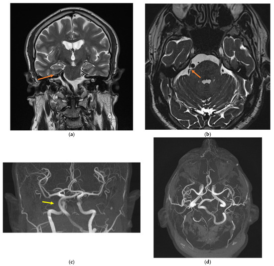
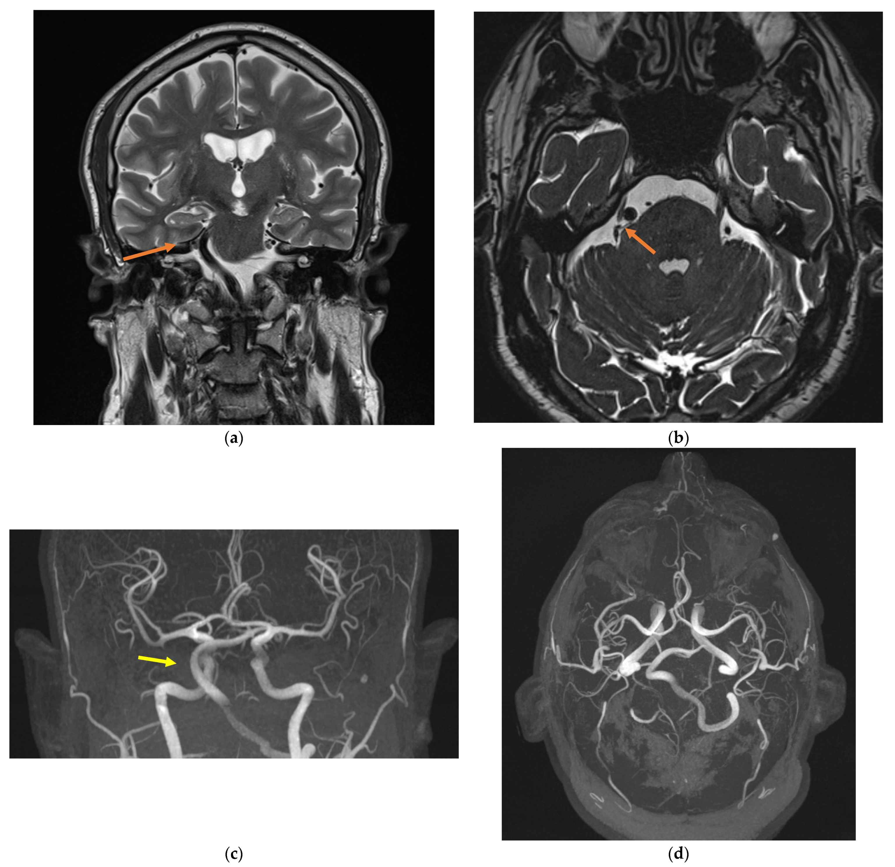
The patient’s past medical history included bilateral high-frequency hearing loss, bronchial asthma, essential hypertension, and myocardial infarction treated with percutaneous coronary intervention and double antiplatelet therapy. On examination, the motor and sensory functions of the trigeminal nerve and corneal reflex were intact; no neurological deficit was found. Routine blood tests (metabolic panel and complete blood count) were normal. Brain magnetic resonance imaging (MRI) with gadolinium contrast revealed the presence of an abnormality of the right vertebral artery (VA) (no visualization of V4 segment), with ectasia of the left VA, and a dolichoectatic basilar artery (BA) with a tortuous course causing, at the right cerebellopontine angle (CPA), a mechanical compression on the trigeminal nerve (Figure 1). The compression of the TN was grade II according to Sindou et al. classification [17] (Table 2).
Figure 1.
Magnetic resonance imaging showing the neurovascular conflict (a,b; orange arrow) and the vertebrobasilar dolichoectasia (c,d; yellow arrow). (a): coronal plane, T2-weighted sequence; (b): axial plane, 3D-CISS sequence; (c): coronal plane, MRI angiogram; (d): sagittal plan, MRI angiogram.
Table 2.
Grade of compression of the Trigeminal Nerve.
Considering the clinical and radiological findings, in addition to low tolerance and ineffectiveness of medical therapy, microvascular decompression surgery was proposed.
A right retrosigmoid approach was performed under general anesthesia, with the patient placed in a left park bench position. After reaching the trigeminal cistern, we opened the dura and drained the cerebrospinal fluid (CSF). Following the release of CSF, the cerebellum was retracted medially, exposing the CPA and trigeminal nerve. Arachnoid dissection revealed the dolichoectatic BA compressing the right anterolateral region of the brainstem and the trigeminal nerve root. Microvascular decompression of the dolichoectatic BA was obtained using the interposition technique: two layers of Teflon sponge, held with fibrin glue, were placed between the artery and the nerve. Particular care was taken to avoid injury of the artery during the vascular microdissection, given the potentially fragile vessel walls in VBD. The Dandy vein was preserved.
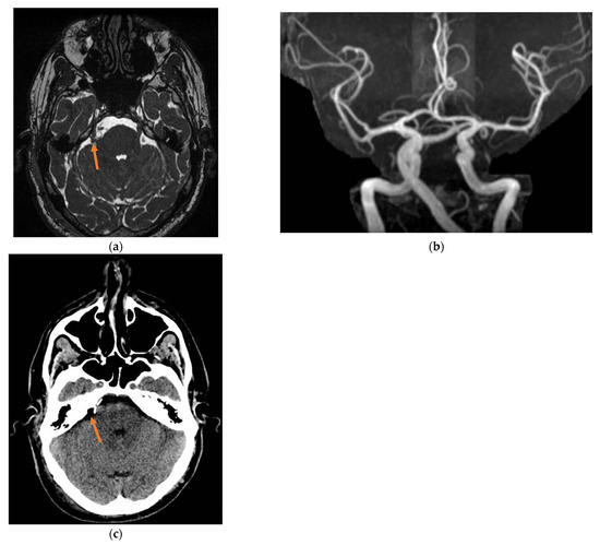
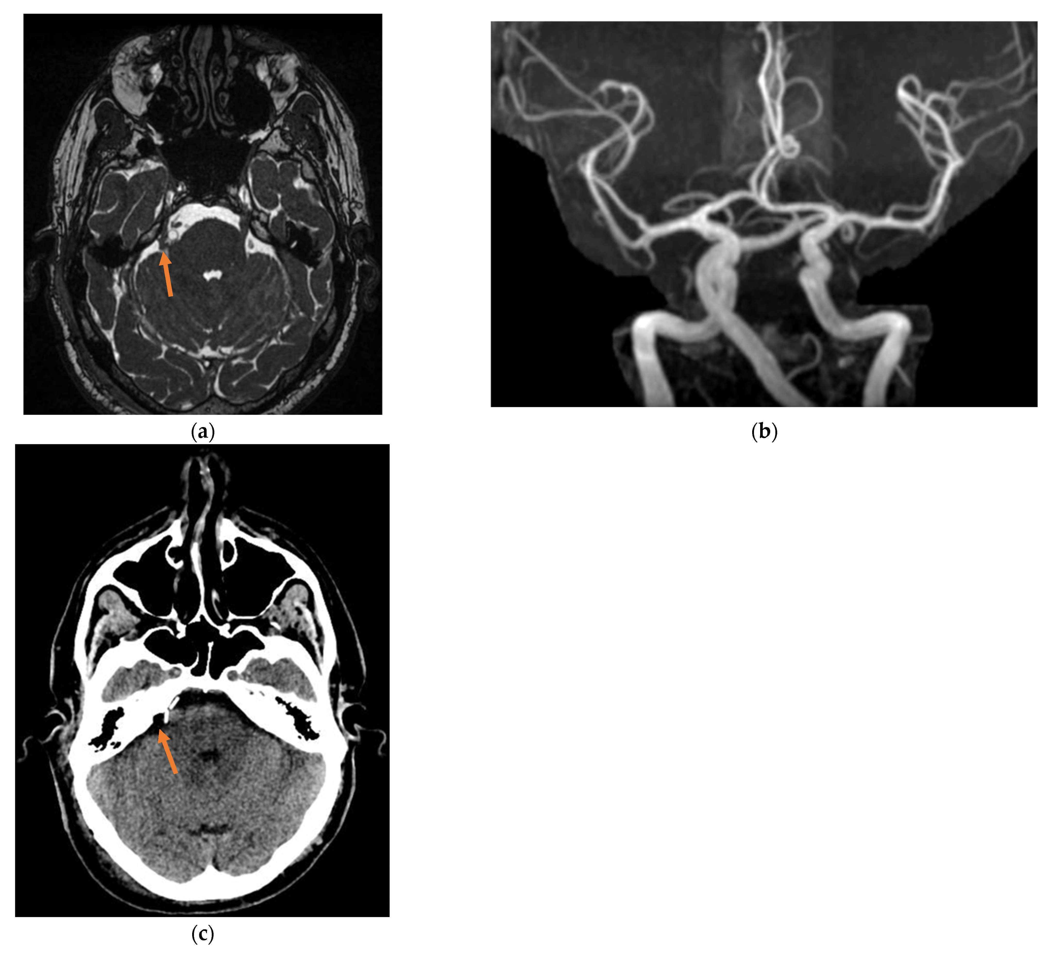
Pain completely resolved after surgery and no neurological deficits were reported. Postoperative brain CT scan and MRI (Figure 2) showed satisfactory surgical outcomes and excluded bleeding complications. The patient was discharged on day 6 after surgery. The patient reported satisfactory pain control (BNI grade 1) at 1-month follow-up. Four weeks after surgery, medications were completely and definitively stopped. No pain recurrence (BNI grade I) and/or sensory abnormalities were found at 3/6/12 months follow-up, and the patient is still on a medication-free status.
Figure 2.
Post-surgery imaging showing Teflon sponges placed between the dolichoectatic basilar artery and the right trigeminal root (orange arrow). (a): MRI, axial plane, (b): MRI Angiogram, (c): CT scan, axial plane.
3. Discussion
Vertebrobasilar dolichoectasia (VBD; from the Greek dolicho, “elongated,” and ectasia, “dilated”) is an uncommon cause of trigeminal neuralgia (TN). Previous reports on this topic are limited. We conducted an extensive search of the literature and collected the most relevant studies on VBA in TN. A summary of the main findings is reported in Table 3.
VBD refers to a vascular abnormality characterized by expansion, elongation, and tortuosity of the vertebrobasilar system. Diagnostic criteria, first proposed by Ubogu et al. [18], include basilar artery (BA) or vertebral artery (VA) diameter > 4.5 mm, deviation of any portion >10 mm from the shortest expected course, BA length > 29.5 mm or intracranial VA length > 23.5 mm; also, for the BA, when bifurcation is above the suprasellar cistern or if any portion lies adjacent to the margin of the clivus or dorsum sellae, elongation is considered to be present [19].
3.1. Pathophysiology, Clinical Characteristics, and Medical Treatment
Although sometimes asymptomatic, VBD can be associated with posterior circulation transient ischemic attacks and strokes [18,20,21,22], subarachnoid bleeding [23], brainstem compression [24], hemifacial spasm [25,26], and trigeminal neuralgia [27]. According to previous studies, VBD-induced TN accounts for 2% to 7.7% of all cases of TN [3,13,28,29,30]. Although the etiopathogenesis of VBD is still largely unknown, most of the evidence points towards a multifactorial process combining congenital vascular wall abnormalities with acquired factors related to atherosclerosis [31,32]. Compared to other patients affected by TN, patients with VBD-related TN are more likely to be older, male, and with a history of diabetes, hyperlipidemia, hypertension, and myocardial infarction [10,13,16,33,34,35].
The elongation and tortuosity of the VBA system can dislocate adjacent vessels, which consequently reach the trigeminal nerve and contribute to compression. Among these, the anterior inferior cerebellar artery (AICA) is the most common additional vessel found in TN cases secondary to VBD [6,10]. The involvement of multiple vessels necessitates a thorough surgical approach, as all compressive forces must be addressed to relieve the trigeminal nerve effectively. In TN secondary to VBD, pain occurs more commonly, but not exclusively, as our case shows, on the left side, and left predominance may be attributed to the asymmetry in the caliber of the two vertebral arteries that commonly occurs or a stronger transmission of pulse pressure to the left VA from a subclavian artery that has its origin from the aortic arch rather than the brachiocephalic trunk. [10,27,33,36]. Most commonly, patients complain of pain in the V2 and/or V3 dermatome. As with other forms of classical TN, medical therapy—primarily with carbamazepine [37] or oxcarbazepine—remains the first-line treatment in VBD-associated TN. When pharmacological therapy proves ineffective or is not well tolerated, surgical options such as trigeminal rhizotomy, percutaneous thermocoagulation, mechanical balloon compression, microvascular decompression (MVD), and gamma knife radiosurgery can be considered [13,35].
3.2. Surgical Management
Microvascular decompression (MVD) has demonstrated the highest efficacy among surgical treatments for VBD-related TN [38,39,40,41,42,43], as it is nondestructive and directly addresses the vascular etiology. Historically, the retrosigmoid approach has been the standard method to reach the trigeminal nerve at the root entry zone; however, in recent years, the endoscopic transorbital approach (ETOA) has emerged as a viable, minimally invasive, surgical option, particularly for treating Schwannomas of the trigeminal nerve and tumors affecting the middle cranial fossa [44,45]. MVD, first described by Jannetta et al. in 1967 [40], aims at isolating the trigeminal nerve from the vessel responsible for compression by placing a piece of material between the vessel and the nerve. When the artery involved is a dolichoectatic VBA, the surgery is notably more challenging; dolichoectatic arteries present with atherosclerosis, abnormal course, increased diameter, low elasticity, and limited mobility. These factors significantly amplify the complexity of the surgery, particularly during vessel displacement, posing potential risks such as plaque dislodgement, injury to brainstem perforators, and vasospasm [36]. Several studies have demonstrated the superiority of MVD over alternative surgical procedures in ensuring long-term pain relief with a low complication rate [3,38,46,47]. However, reports on the efficacy and safety of MVD specifically in cases involving VBD are scarce. Di Carlo et al. [27] conducted a random-effects meta-analysis, including 167 patients with TN secondary to VBD, and found that 97% of cases experienced immediate postoperative pain relief, with a slight decrease to 92.9%, at the last follow-up (55 months). Early post-operative complications included facial numbness (21% of cases), diplopia (5%), hearing impairment (5%), and cerebellar ataxia (4%). By the last follow-up, hearing impairment remained stable (5%), while rates of facial numbness, diplopia, and cerebellar ataxia decreased to 13%, 0%, and 3%, respectively. In a cohort study by Zheng et al. [7], no significant differences were found in the safety and efficacy of MVD between the VBD group and the conventional TN group (p > 0.05). However, some authors have reported higher failure or recurrence rates in patients with elongated, ectatic VBA treated with MVD [33,48,49].
3.3. Interposition vs. Transposition
Various techniques have been applied during MVD to separate the offending dolichoectatic VBA from the compressed trigeminal nerve. These can be broadly classified into the interposition method and transposition method [7,13,35]. The former, which is the traditional method and is widely used in patients with neurovascular compression syndrome due to small vessels such as the SCA and AICA, involves the insertion of an implant between the offending vessel and the nerve [3,10,16,30,36,50]; the latter method involves repositioning the VBA using biomedical glue [51], Teflon slings or pads [28,32,52], and aneurysm clips [53]. Both methods have advantages and disadvantages.
The conventional interposition technique is widely accepted as it is easy to perform, can be effectively achieved through a relatively small craniotomy, and requires minimal manipulation of the VBA complex. However, some authors argue that, in cases of VBD, this technique may worsen the symptoms as the offending VBA follows a prominent upward loop squeezing the CN V against the cerebellar tentorium; therefore, inserting material between the artery and the nerve may ultimately increase the compression effect [6,7,52]. Despite these concerns, several case series have reported successful long-term outcomes with the interposition in VBD cases [3,10,16,28,30,36,50].
The transposition technique has been proposed as an effective and safe alternative to interposition [13]. It relieves the pressure on the nerves by repositioning the VBA—thereby eliminating the possibility of indirect compression—using materials such as biomedical glue, Teflon, and aneurysm clips. This approach is not free of risks. When trying to mobilize the VBA, the following factors must be considered: (i) the VBA complex is stiff, tortuous, and ectatic, making it difficult to dislocate; (ii) overstretching and injury of the perforator branches originating from VBA can result in brainstem infarction; (iii) atherosclerotic plaques have the potential to break during mobilization, which can serve as a source of emboli and may cause ischemic stroke or cerebral hemorrhage [51]. Chai et al. [13] found that the 5-year rate of pain relief (BNI I) for the transposition technique was 91.1%; this was compared to 58.7% for the interposition technique, with no significant difference in terms of postoperative complication rate. Inoue et al. [6] decompressed the trigeminal nerve by displacing the VBA and found a long-term pain relief rate of 96%. Concerning the adequate and secure distancing of the VBA from CN V needed to ensure effective decompression, different theories have been proposed. According to Amagasaki et al. [52], the surgeon must mobilize the VBA as caudally as possible; this is because the trigeminal nerve is often shifted rostrally by the VBA, and after repositioning the vessel, CN V often droops and may contact the VBA again. However, according to other authors, such extensive mobilization dramatically increases surgical times and may result in kinking and stenosis of the parent artery, overstretching and injury to the perforating arteries, and brain stem infarction [53]. Also, it does not significantly impact long-term pain relief, as shown in the study by Inoue et al. [6]: they compared the “separation group”—where complete separation of the VBA from the trigeminal nerve was achieved—and the “contact group”—where slight vascular contact remained—and found no difference in terms of pain relief, indicating that once the tense trigeminal nerve is loosened, further attempts to mobilize the VBA for complete nerve separation are not necessary. Several surgeons prefer to mobilize the VBA using Teflon felts placed piece by piece between the vessel and surrounding structures [6,7,32,54]. However, large volumes of Teflon increase the risk of inflammation, granuloma formation, and adhesions, ultimately resulting in TN recurrence. In the case series by Wang et al. [54], including patients treated with VBA repositioning using Teflon felts, one patient developed recurrent post-operative pain that required a second surgery to remove the redundant Teflon and adhesions. Techniques that involve fixing the transposed VBA to the skull base dura (at the clivus or petrous region) have been developed to secure the repositioning. Liu et al. [51] successfully treated 22 cases of VBD-induced TN by mobilizing the VBA with Teflon pads and fixing it to the petrous dura using biomedical glue. A similar technique was used in previous smaller case series, with comparable outcomes reported [32,35]. Although relatively simple to perform, glue fixation suffers several limitations. Above all, the adhesive is not strong enough to guarantee a permanent fixation. An effective solution to overcome this problem is to coagulate the portion of the dura where the artery will be fixed to induce a scar to promote fibrous adhesion [35,51,53]; pieces of Surgicel may also be used [32]. Additionally, the use of glue poses the potential risk of inducing vasculitis and cranial nerve damage from glue overflow or dripping. Also, recurrence due to the adhesion of neurovascular structures by the glue has been reported in the literature [26,55].
A safer alternative to glue fixation is the use of aneurysm clips, as described by Lin et al. in 2012. [53] They reported a series of three cases of VBD-induced TN successfully treated by transposing the offending vessel using an unabsorbable dural tape sling and fixing it with an aneurysm clip passed through a petrous dural bridge. Besides ensuring effective and durable fixation, this technique has the major advantage of allowing an easy modulation of the strength of traction (the more of the sling the clip takes, the stronger the traction) and the direction of transposition, thus avoiding kinking of the parent artery, overstretching of the perforating branches, and damaging the adjacent cranial nerves. However, when considering the fixation technique, it is worth noting that placing vascular clips introduces radiological artifacts that may affect later imaging evaluation.
Although MVD, whether through the interposition or transposition method, has proven highly successful in cases of VBD, significant postoperative complications can arise. Notably, diplopia and hearing loss are relatively common, due to mechanical stimulation of the fourth, sixth, or eighth cranial nerves during decompression, especially in cases of extensive manipulation of the VBA complex [13,35].
Our study has some limitations. The included articles were primarily small, retrospective, single-institution case series, and many reported incomplete data. Follow-up ranged from 1 year up to 15 years, which may not provide a homogeneous long-term perspective. Future research should prioritize larger, prospective studies with standardized reporting to enable better comparison of surgical techniques and strategies.
Table 3.
Summary of the studies included in the literature review.
4. Conclusions
Despite potential risks, MVD offers the best chance for long-term pain relief in patients suffering from VBD-induced TN. Future research should aim to further refine these surgical techniques and reduce the risk of postoperative complications, ultimately improving outcomes for this subset of patients.
Author Contributions
Conceptualization, M.V., F.S. and F.Z.; methodology, F.Z. and F.S.; software, F.Z.; validation, M.V., F.S. and F.Z.; formal analysis, F.Z.; investigation, F.Z.; resources, F.Z.; data curation, F.S. and F.Z.; writing—original draft preparation, F.S. and F.Z.; writing—review and editing, F.S. and M.V.; visualization, F.Z. and M.V.; supervision, M.V.; project administration, M.V. All authors have read and agreed to the published version of the manuscript.
Funding
This research received no external funding.
Institutional Review Board Statement
The study was conducted in accordance with the Declaration of Helsinki. Due to the nature of the study, no approval by the institutional review board was necessary.
Informed Consent Statement
Informed consent was obtained from all subjects involved in the study. Written informed consent has been obtained from the patient to publish this paper.
Data Availability Statement
All the data generated in the present study have been included in this paper after anonymization.
Conflicts of Interest
The authors declare no conflicts of interest.
References
- Jones, M.R.; Urits, I.; Ehrhardt, K.P.; Cefalu, J.N.; Kendrick, J.B.; Park, D.J.; Cornett, E.M.; Kaye, A.D.; Viswanath, O. A Comprehensive Review of Trigeminal Neuralgia. Curr. Pain Headache Rep. 2019, 23, 74. [Google Scholar] [CrossRef] [Scilit]
- Leland Rogers, C.; Shetter, A.G.; Fiedler, J.A.; Smith, K.A.; Han, P.P.; Speiser, B.L.; Joseph, S. PII S0360-3016(00)00513-7 Gamma knife radiosurgery for trigeminal neuralgia: The initial experience of the barrow neurological institute. Int. J. Radiat. Oncol. Biol. Phys. 2000, 47, 1013–1019. [Google Scholar] [CrossRef] [Scilit]
- Apra, C.; Lefaucheur, J.-P.; Le Guerinel, C.; Caroline, A.; Jean-Pascal, L.; Guérinel Caroline, L. Microvascular Decompression Is an Effective Therapy for Trigeminal Neuralgia Due to Dolichoectatic Basilar Artery Compression: Case Reports and Literature Review. Neurosurg. Rev. 2017, 40, 577–582. [Google Scholar] [CrossRef] [Scilit]
- Headache Classification Committee of the International Headache Society (IHS). The International Classification of Headache Disorders, 3rd Edition. Cephalalgia 2018, 38, 1–211. [Google Scholar] [CrossRef] [Scilit]
- Cruccu, G.; Di Stefano, G.; Truini, A. Trigeminal Neuralgia. N. Engl. J. Med. 2020, 383, 754–762. [Google Scholar] [CrossRef] [Scilit]
- Inoue, T.; Shitara, S.; Goto, Y.; Prasetya, M.; Fukushima, T. Microvascular Decompression for Trigeminal Neuralgia Attributable to the Vertebrobasilar Artery: Decompression Technique and Significance of Separation from the Nerve Root. Acta Neurochir. 2021, 163, 1037–1043. [Google Scholar] [CrossRef] [Scilit]
- Zheng, W.; Wang, L.; Wang, H.; Zhou, H.; Du, Q. Trigeminal Neuralgia Caused by Vertebrobasilar Dolichoectasia: Efficacy of Stepwise Decompression Technique. Acta Neurochir. 2023, 165, 3019–3026. [Google Scholar] [CrossRef] [Scilit]
- Bašić Kes, V. Accommodation to Diagnosis of Trigeminal Neuralgia. Acta Clin. Croat. 2017, 56, 157–161. [Google Scholar] [CrossRef] [Scilit]
- Smoker’, W.R.K.; Corbett2, J.J.; Gentry’, L.R.; Keyes’, W.D.; Price, M.J.; Mckuske, S. High-Resolution Computed Tomography of the Basilar Artery: 2. Vertebrobasilar Dolichoectasia: Clinical-Pathologic Correlation and Review. Am. J. Neuroradiol. 1986, 7, 61–72. [Google Scholar]
- Yu, F.; Yin, J.; Lu, P.-G.; Zhao, Z.-Y.; Zhang, Y.-Q.; Men, X.-Z. Microvascular Decompression by Interposition Method for Treatment of Trigeminal Neuralgia Due to Vertebrobasilar Dolichoectasia: A Retrospective Single-Center Study. Neurosurg. Rev. 2022, 45, 2709–2715. [Google Scholar] [CrossRef] [Scilit]
- Ikeda, K.; Nakamura, Y.; Hirayama, T.; Sekine, T.; Nagata, R.; Kano, O.; Kawabe, K.; Kiyozuka, T.; Tamura, M.; Iwasaki, Y. Cardiovascular Risk and Neuroradiological Profiles in Asymptomatic Vertebrobasilar Dolichoectasia. Cerebrovasc. Dis. 2010, 30, 23–28. [Google Scholar] [CrossRef] [Scilit]
- Gavazzi, C.; Borsini, W.; Guerrini, L.; Della Nave, R.; Rocca, M.A.; Tessa, C.; Buchner, S.; Belli, G.; Filippi, M.; Villari, N.; et al. Subcortical Damage and Cortical Functional Changes in Men and Women with Fabry Disease: A Multifaceted MR Study. Radiology 2006, 241, 492–500. [Google Scholar] [CrossRef] [Scilit]
- Chai, S.; Xu, H.; Wang, Q.; Li, J.; Wang, J.; Wang, Y.; Pool, H.; Lin, M.; Xiong, N. Microvascular Decompression for Trigeminal Neuralgia Caused by Vertebrobasilar Dolichoectasia: Interposition Technique versus Transposition Technique. Acta Neurochir. 2020, 162, 2811–2821. [Google Scholar] [CrossRef] [Scilit]
- De Simone, M.; Fontanella, M.M.; Choucha, A.; Schaller, K.; Machi, P.; Lanzino, G.; Bijlenga, P.; Kurz, F.T.; Lövblad, K.O.; De Maria, L. Current and Future Applications of Arterial Spin Labeling MRI in Cerebral Arteriovenous Malformations. Biomedicines 2024, 12, 753. [Google Scholar] [CrossRef] [Scilit]
- Honey, C.M.; Kaufmann, A.M. Trigeminal Neuralgia Due to Vertebrobasilar Artery Compression. World Neurosurg. 2018, 118, e155–e160. [Google Scholar] [CrossRef] [Scilit]
- El-Ghandour, N.M.F. Microvascular Decompression in the Treatment of Trigeminal Neuralgia Caused by Vertebrobasilar Ectasia. Neurosurgery 2010, 67, 330–337. [Google Scholar] [CrossRef] [Scilit]
- Sindou, M.; Amrani, F.; Mertens, P. Microsurgical Vascular Decompression in Trigeminal Neuralgia. Comparison of 2 Technical Modalities and Physiopathologic Deductions. A Study of 120 Cases. Neurochirurgie 1990, 36, 16–25, discussion 25–26. [Google Scholar]
- Ubogu, E.E.; Zaidat, O.O. Vertebrobasilar Dolichoectasia Diagnosed by Magnetic Resonance Angiography and Risk of Stroke and Death: A Cohort Study. J. Neurol. Neurosurg. Psychiatry 2004, 75, 22–26. [Google Scholar]
- Stone, J.L.; Lichtor, T.; Crowell, R.M. Microvascular Sling Decompression for Trigeminal Neuralgia Secondary to Ectatic Vertebrobasilar Compression. J. Neurosurg. 1993, 79, 943–945. [Google Scholar] [CrossRef] [Scilit]
- Passero, S.G.; Rossi, S. Natural History of Vertebrobasilar Dolichoectasia. Neurology 2008, 70, 66–72. [Google Scholar] [CrossRef] [Scilit]
- Yuh, S.-J.; Alkherayf, F.; Lesiuk, H. Dolichoectasia of the Vertebral Basilar and Internal Carotid Arteries: A Case Report and Literature Review. Surg. Neurol. Int. 2013, 4, 153. [Google Scholar] [CrossRef] [Scilit] [PubMed]
- Kwon, H.M.; Lee, Y.S. Dolichoectasia of the Intracranial Arteries. Curr. Treat. Options Cardiovasc. Med. 2011, 13, 261–267. [Google Scholar] [CrossRef] [Scilit] [PubMed]
- Rabb, C.H.; Barnwell, S.L. Catastrophic Subarachnoid Hemorrhage Resulting from Ruptured Vertebrobasilar Dolichoectasia: Case Report. Neurosurgery 1998, 42, 379–382. [Google Scholar] [CrossRef] [Scilit] [PubMed]
- Tomasello, F.; Alafaci, C.; Salpietro, F.M.; Longo, M. Bulbar Compression by an Ectatic Vertebral Artery: A Novel Neurovascular Construct Relieved by Microsurgical Decompression. Neurosurgery 2005, 56, 117–124. [Google Scholar] [CrossRef] [Scilit] [PubMed]
- Revuelta-Gutiérrez, R.; Velasco-Torres, H.S.; Vales Hidalgo, L.O.; Martínez-Anda, J.J. Tic Convulsivo Doloroso: Serie de Casos y Revisión de La Literatura. Cirugía Cir. 2016, 84, 493–498. [Google Scholar] [CrossRef] [Scilit]
- Kurokawa, Y.; Maeda, Y.; Toyooka, T.; Inaba, K.I. Microvascular Decompression for Hemifacial Spasm Caused by the Vertebral Artery: A Simple and Effective Transposition Method Using Surgical Glue. Surg. Neurol. 2004, 61, 398–403. [Google Scholar] [CrossRef] [Scilit]
- Di Carlo, D.T.; Benedetto, N.; Marani, W.; Montemurro, N.; Perrini, P. Microvascular Decompression for Trigeminal Neuralgia Due to Vertebrobasilar Artery Compression: A Systematic Review and Meta-Analysis. Neurosurg. Rev. 2022, 45, 285–294. [Google Scholar] [CrossRef] [Scilit]
- Yang, X.-S.; Li, S.-T.; Zhong, J.; Zhu, J.; Du, Q.; Zhou, Q.-M.; Jiao, W.; Guan, H.-X. Microvascular Decompression on Patients with Trigeminal Neuralgia Caused by Ectatic Vertebrobasilar Artery Complex: Technique Notes. Acta Neurochir. 2012, 154, 793–797. [Google Scholar] [CrossRef] [Scilit]
- Park, K.-J.; Kondziolka, D.; Kano, H.; Berkowitz, O.; Ahmed, S.F.; Liu, X.; Niranjan, A.; Flickinger, J.C.; Lunsford, L.D. Outcomes of Gamma Knife Surgery for Trigeminal Neuralgia Secondary to Vertebrobasilar Ectasia. J. Neurosurg. 2012, 116, 73–81. [Google Scholar] [CrossRef] [Scilit]
- Ma, X.; Sun, X.; Yao, J.; Ni, S.; Gong, J.; Wang, J.; Li, X. Clinical Analysis of Trigeminal Neuralgia Caused by Vertebrobasilar Dolichoectasia. Neurosurg. Rev. 2013, 36, 573–578. [Google Scholar] [CrossRef] [Scilit]
- Campos, W.K.; Guasti, A.A.; da Silva, B.F.; Guasti, J.A. Trigeminal Neuralgia Due to Vertebrobasilar Dolichoectasia. Case Rep. Neurol. Med. 2012, 2012, 367304. [Google Scholar] [CrossRef] [Scilit] [PubMed]
- Yamahata, H.; Tokimura, H.; Hanaya, R.; Tajitsu, K.; Hirabaru, M.; Yamagami, M.; Arita, K. Microvascular Decompression for Trigeminal Neuralgia Due to Compression by the Vertebral Artery: Report of 3 Cases. Minim. Invasive Neurosurg. 2011, 54, 79–82. [Google Scholar] [CrossRef] [Scilit] [PubMed]
- Linskey, M.E.; Jho, H.D.; Jannetta, P.J. Microvascular Decompression for Trigeminal Neuralgia Caused by Vertebrobasilar Compression. J. Neurosurg. 1994, 81, 1–9. [Google Scholar] [CrossRef] [Scilit] [PubMed]
- Wang, Y.; Cheng, W.; Lian, Y. Vertebrobasilar Dolichoectasia and Basilar Artery Dissection Presenting with Trigeminal Neuralgia: A Case Report. Front. Neurol. 2019, 10, 491. [Google Scholar] [CrossRef] [Scilit]
- Vanaclocha, V.; Herrera, J.M.; Martínez-Gómez, D.; Rivera-Paz, M.; Calabuig-Bayo, C.; Vanaclocha, L. Is There a Safe and Effective Way to Treat Trigeminal Neuralgia Associated with Vertebrobasilar Dolichoectasia? Presentation of 8 Cases and Literature Review. World Neurosurg. 2016, 96, 516–529. [Google Scholar] [CrossRef] [Scilit]
- Shulev, Y.A.; Gordienko, K.S.; Trashin, A.V.; Pechiborshch, D.A. Microvascular Decompression in Trigeminal Neuralgia Following Vertebrobasilar Dolichoectasia. Zhurnal Vopr. Nejrokhirurgii Im. N. N. Burdenko 2020, 84, 50–63. [Google Scholar] [CrossRef] [Scilit]
- Gambeta, E.; Chichorro, J.G.; Zamponi, G.W. Trigeminal Neuralgia: An Overview from Pathophysiology to Pharmacological Treatments. Mol. Pain 2020, 16, 1744806920901890. [Google Scholar] [CrossRef] [Scilit]
- Kraemer, J.L.; Filho, A.d.A.P.; de David, G.; Faria, M.d.B. Vertebrobasilar Dolichoectasia as a Cause of Trigeminal Neuralgia: The Role of Microvascular Decompression. Case Report. Arq. Neuropsiquiatr. 2006, 64, 128–131. [Google Scholar] [CrossRef] [Scilit]
- Ashkan, K.; Marsh, H. Microvascular Decompression for Trigeminal Neuralgia in the Elderly: A Review of the Safety and Efficacy. Neurosurgery 2004, 55, 840–850. [Google Scholar] [CrossRef] [Scilit]
- Jannetta, P.J. Arterial Compression of the Trigeminal Nerve at the Pons in Patients with Trigeminal Neuralgia. J. Neurosurg. 2007, 107, 216–237. [Google Scholar] [CrossRef] [Scilit]
- Jannetta, P.J. Microsurgical Management of Trigeminal Neuralgia. Arch. Neurol. 1985, 42, 800. [Google Scholar] [CrossRef] [Scilit] [PubMed]
- Oesman, C.; Mooij, J.J.A. Long-Term Follow-up of Microvascular Decompression for Trigeminal Neuralgia. Skull Base 2011, 21, 313–321. [Google Scholar] [CrossRef] [Scilit] [PubMed]
- Tatli, M.; Satici, O.; Kanpolat, Y.; Sindou, M. Various Surgical Modalities for Trigeminal Neuralgia: Literature Study of Respective Long-Term Outcomes. Acta Neurochir. 2008, 150, 243–255. [Google Scholar] [CrossRef] [Scilit] [PubMed]
- De Simone, M.; Zoia, C.; Choucha, A.; Kong, D.S.; De Maria, L. The Transorbital Approach: A Comprehensive Review of Targets, Surgical Techniques, and Multiportal Variants. J. Clin. Med. 2024, 13, 2712. [Google Scholar] [CrossRef] [Scilit] [PubMed]
- De Simone, M.; Choucha, A.; Dannhoff, G.; Kong, D.S.; Zoia, C.; Iaconetta, G. Treating Trigeminal Schwannoma through a Transorbital Approach: A Systematic Review. J. Clin. Med. 2024, 13, 3701. [Google Scholar] [CrossRef] [Scilit]
- Shi, J.; Qian, Y.; Han, W.; Dong, B.; Mao, Y.; Cao, J.; Guan, W.; Zhou, Q. Risk Factors for Outcomes After Microvascular Decompression for Trigeminal Neuralgia. World Neurosurg. 2020, 136, e559–e566. [Google Scholar] [CrossRef] [Scilit]
- Wei, Y.; Pu, C.; Li, N.; Cai, Y.; Shang, H.; Zhao, W. Long-Term Therapeutic Effect of Microvascular Decompression for Trigeminal Neuralgia: Kaplan-Meier Analysis in a Consecutive Series of 425 Patients. Turk. Neurosurg. 2016, 28, 88–93. [Google Scholar] [CrossRef] [Scilit]
- Suzuki, S.; Tsuchita, H.; Kurokawa, Y.; Kitami, K.; Sohma, T.; Takeda, T.; Suzuki, S. New Method of MVD Using a Vascular Tape for Neurovascular Compression Involving the Vertebrobasilar Artery. Report of Two Cases. Neurol. Med.-Chir. 1990, 30, 1020–1023. [Google Scholar] [CrossRef] [Scilit]
- Yoshimoto, Y.; Tsutsumi, Y. Pain encircling method of trigeminal nerve decompression for neuralgia caused by tortuous vertebrobasilar artery: Technical note. Surg. Neurol. 1995, 43, 151–153. [Google Scholar] [CrossRef] [Scilit]
- Sun, S.; Jiang, W.; Wang, J.; Gao, P.; Zhang, X.; Jiao, L.; Liu, W.; Shu, K.; Lei, T. Clinical Analysis and Surgical Treatment of Trigeminal Neuralgia Caused by Vertebrobasilar Dolichoectasia: A Retrospective Study. Int. J. Surg. 2017, 41, 183–189. [Google Scholar] [CrossRef] [Scilit]
- Liu, J.; Chen, Z.; Feng, T.; Jiang, B.; Yuan, Y.; Yu, Y. Biomedical Glue Sling Technique in Microvascular Decompression for Trigeminal Neuralgia Caused by Atherosclerotic Vertebrobasilar Artery: A Description of Operative Technique and Clinical Outcomes. World Neurosurg. 2019, 128, e74–e80. [Google Scholar] [CrossRef] [Scilit] [PubMed]
- Amagasaki, K.; Takusagawa, Y.; Tatebayashi, K.; Nakaguchi, H. Macrovascular Decompression with the Transposition Method Using Teflon Sling for Trigeminal Neuralgia Caused by the Vertebrobasilar Artery. World Neurosurg. 2022, 167, e1395–e1401. [Google Scholar] [CrossRef] [Scilit] [PubMed]
- Lin, C.F.; Chen, H.H.; Hernesniemi, J.; Lee, C.C.; Liao, C.H.; Chen, S.C.; Chen, M.H.; Shih, Y.H.; Hsu, S.P.C. An Easy Adjustable Method of Ectatic Vertebrobasilar Artery Transposition for Microvascular Decompression. Clin. Neurol. Neurosurg. 2012, 114, 951–956. [Google Scholar] [CrossRef] [Scilit] [PubMed]
- Wang, X.; Wang, H.; Chen, S.; Liang, H.; Wang, H.; Xu, M.; Xu, L. The Long-Term Clinical Outcomes of Microvascular Decompression for Treatment of Trigeminal Neuralgia Compressed by the Vertebra-Basilar Artery: A Case Series Review. BMC Neurol. 2019, 19, 217. [Google Scholar] [CrossRef] [Scilit]
- Ohta, M.; Komatsu, F.; Abe, H.; Sakamoto, S.; Tsugu, H.; Oshiro, S.; Fukushima, T. Complication Caused by Use of Fibrin Glue in Vessel. Transposition for Trigeminal Neuralgia-Case Report. Neurol. Med. Chir. 2008, 48, 30–32. [Google Scholar] [CrossRef] [Scilit]

Disclaimer/Publisher’s Note: The statements, opinions and data contained in all publications are solely those of the individual author(s) and contributor(s) and not of MDPI and/or the editor(s). MDPI and/or the editor(s) disclaim responsibility for any injury to people or property resulting from any ideas, methods, instructions or products referred to in the content. |
© 2024 by the authors. Licensee MDPI, Basel, Switzerland. This article is an open access article distributed under the terms and conditions of the Creative Commons Attribution (CC BY) license (https://creativecommons.org/licenses/by/4.0/).