


The technique of thoracic ultrasound is living through a progressive rise in clinical routine.
After initial skepticism due to the impossibility of studying lung parenchyma with the available ultrasound machines, the increasing evidence and the growth of ultrasound technology have permitted the development of ultrasound use in every application in respiratory care.
In pleural diseases, ultrasound has become the gold standard in the management of pleural effusions, as a diagnostic tool and as a guide for interventional procedures [1].
The study of diaphragmatic features and motions, after the evaluation of normal features [2], is being characterized for use in muscle dysfunctions in neuromuscular diseases [3] and in intensive care settings, being a tool for predicting outcomes of critical patients [4].
The ultrasound assessment of lung diseases is continuously growing; today, lung ultrasound is a fundamental tool to diagnose and follow-up pneumonia [5] and has a promising role in the early detection of interstitial lung diseases [6].
However, the technique has limits, given the impossibility of ultrasound waves to cross the ventilated (even partially) peripheral air spaces.
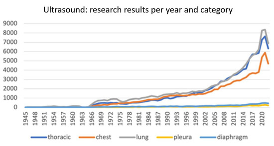
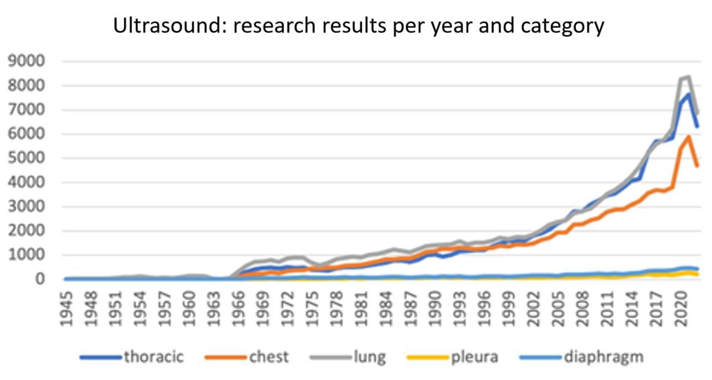
From a search on Pubmed for the terms “thoracic ultrasound”, “chest ultrasound”, “lung ultrasound”, “pleura ultrasound”, and “diaphragm ultrasound” we obtained the number of papers written yearly since 1945. This number is progressively increasing, reaching a huge peak of more than 21,000 total papers in 2021 and 2022, thanks in part to COVID-19-related research (Figure 1).
Figure 1.
Research results on Pubmed for specific types of ultrasound.
This literature jungle has evolved from case reports and descriptions of artifacts and signs for lung study protocols and clinical trials, up to physical models to explain the origin of different vertical artifacts [7] and studies on the possible role of artificial intelligence [8]. In the pandemic era, the role of lung ultrasound was crucial, particularly in acute diagnostic settings and in critical care management [9,10].
So, what do we need today?
Much has been written about lungs and pleura, but the study of the whole respiratory muscular system and the global mechanics of the chest wall have yet to be fully explained and described.
Moreover, all the known notions must be put in order, to finally assess the correct terminology and establish an appropriate and universal methodology. Thoracic ultrasound escapes from the pure “diagnostic imaging” because its features do not permit a precise diagnosis when only considering the obtained images. In fact, it becomes wholly the responsibility of an operator, who needs “good eyes” to correctly interpret the image, “good hands” to adequately guide the probe and create a good quality image, and finally a “good mind”, to understand the clinical situation of the patient and integrate clinical data with the procedure, to obtain a final, accurate diagnosis.
That is art.
Funding
This research received no external funding.
Conflicts of Interest
The author declares no conflict of interest.
References
- Zanforlin, A.; Giannuzzi, R.; Nardini, S.; Testa, A.; Soldati, G.; Copetti, R.; Marchetti, G.; Valente, S.; Inchingolo, R.; Smargiassi, A. The role of chest ultrasonography in the management of respiratory diseases: Document I. Multidiscip. Respir. Med. 2013, 8, 54. [Google Scholar] [CrossRef] [Scilit] [PubMed]
- Boussuges, A.; Gole, Y.; Blanc, P. Diaphragmatic Motion Studied by M-Mode Ultrasonography: Methods, reproducibility, and normal values. Chest 2009, 135, 391–400. [Google Scholar] [CrossRef] [Scilit] [PubMed]
- Fayssoil, A.; Behin, A.; Ogna, A.; Mompoint, D.; Amthor, H.; Clair, B.; Laforet, P.; Mansart, A.; Prigent, H.; Orlikowski, D.; et al. Diaphragm: Pathophysiology and Ultrasound Imaging in Neuromuscular Disorders. J. Neuromuscul. Dis. 2018, 5, 1–10. [Google Scholar] [CrossRef] [Scilit] [PubMed]
- Llamas-Álvarez, A.M.; Tenza-Lozano, E.M.; Latour-Pérez, J. Diaphragm and Lung Ultrasound to Predict Weaning Outcome: Systematic Review and Meta-Analysis. Chest 2017, 152, 1140–1150. [Google Scholar] [CrossRef] [Scilit] [PubMed]
- Chavez, M.A.; Shams, N.; Ellington, L.E.; Naithani, N.; Gilman, R.H.; Steinhoff, M.C.; Santosham, M.; Black, R.E.; Price, C.; Gross, M.; et al. Lung ultrasound for the diagnosis of pneumonia in adults: A systematic review and meta-analysis. Respir. Res. 2014, 15, 50. [Google Scholar] [CrossRef] [Scilit] [PubMed]
- Pitsidianakis, G.; Vassalou, E.E.; Vasarmidi, E.; Bolaki, M.; Klontzas, M.E.; Xirouchaki, N.; Georgopoulos, D.; Karantanas, A.H.; Tzanakis, N.; Antoniou, K.M. Performance of Lung Ultrasound for Monitoring Interstitial Lung Disease. J. Ultrasound Med. 2022, 41, 1077–1084. [Google Scholar] [CrossRef] [Scilit] [PubMed]
- Demi, M.; Soldati, G.; Ramalli, A. Lung Ultrasound Artifacts Interpreted as Pathology Footprints. Diagnostics 2023, 13, 1139. [Google Scholar] [CrossRef] [Scilit] [PubMed]
- Fatima, N.; Mento, F.; Zanforlin, A.; Smargiassi, A.; Torri, E.; Perrone, T.; Demi, L. Human-to-AI Interrater Agreement for Lung Ultrasound Scoring in COVID-19 Patients. J. Ultrasound Med. 2023, 42, 843–851. [Google Scholar] [CrossRef] [Scilit] [PubMed]
- Zanforlin, A.; Tursi, F.; Accademia di Ecografia Toracica Group. How Is COVID-19 Changing Lung Ultrasound? A Survey by the Thoracic Ultrasound Academy. J. Ultrasound Med. 2021, 40, 417–418. [Google Scholar] [CrossRef] [Scilit] [PubMed]
- Zanforlin, A.; Strapazzon, G.; Falk, M.; Gallina, V.; Viteritti, A.; Valzolgher, L.; La Guardia, M.; Ferro, F.; Pagani, L.; Vezzali, N. Lung Ultrasound in the Emergency Department for Early Identification of COVID-19 Pneumonia. Respiration 2021, 100, 145–153. [Google Scholar] [CrossRef] [Scilit] [PubMed]
Disclaimer/Publisher’s Note: The statements, opinions and data contained in all publications are solely those of the individual author(s) and contributor(s) and not of MDPI and/or the editor(s). MDPI and/or the editor(s) disclaim responsibility for any injury to people or property resulting from any ideas, methods, instructions or products referred to in the content. |
© 2023 by the author. Licensee MDPI, Basel, Switzerland. This article is an open access article distributed under the terms and conditions of the Creative Commons Attribution (CC BY) license (https://creativecommons.org/licenses/by/4.0/).