Skip Content
You are currently on the new version of our website. Access the old version .

Journal of Clinical Medicine

Journal of Clinical Medicine is an international, peer-reviewed, open access journal of clinical medicine, published semimonthly online by MDPI.
Indexed in PubMed | Quartile Ranking JCR - Q1 (Medicine, General and Internal)

All Articles (46,887)

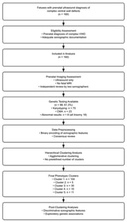
Background/Objectives: To identify distinct sonographic phenotypes of complex malformations of the fetal ventral wall. Methods: We performed a retrospective analysis of ultrasound reports from 160 fetuses diagnosed with complex ventral wall defects at a single tertiary referral center between 1997 and 2021. Agglomerative hierarchical clustering was applied to identify distinct sonographic phenotypes based on the level of the ventral wall defect and associated anomalies. Results: Ventral wall defects involved the abdominal wall in 150 cases, the thoracic wall in 42 cases, and the pelvic wall in 28 cases, either in isolation or in combination. Open neural tube defects were present in 58 fetuses (36.3%), spinal defects in 110 fetuses (68.8%), and limb anomalies in 45 fetuses (28.1%). Additional anomalies were identified in 38 fetuses (23.8%), including cardiac anomalies in 18 cases (11.3%). Amniotic bands were observed in seven cases (4.4%). Using agglomerative hierarchical clustering, five groups of fetuses with differing numbers of observations were identified (cluster 1, n = 104; cluster 2, n = 5; cluster 3, n = 30; cluster 4, n = 10; cluster 5, n = 11). The silhouette score of the clustering model was 0.3285. The most discriminative features for each cluster, expressed as feature importance values, were as follows: kyphoscoliosis for cluster 1 (0.924), pelvic wall defect for cluster 2 (0.852), ectopia cordis for cluster 3 (0.662), limb anomalies for cluster 4 (0.767), and spina bifida for cluster 5 (0.691). Conclusions: Complex malformations of the fetal ventral wall are associated with a wide spectrum of additional anomalies. Hierarchical clustering identified five distinct sonographic phenotypes of complex ventral wall defects, highlighting the heterogeneity of these conditions.

8 February 2026

Study flow diagram.

Background: Prolonged mechanical ventilation and tracheostomy in patients with COVID-19 is associated with longer hospital stays. Guidance on which patients are at risk for tracheostomy due to the progression of COVID-19 is limited. Objectives: This study aimed to identify risk factors associated with the need for tracheostomy in patients intubated for COVID-19 between 1 March and 31 December 2020. Methods: The methodology for this study involved a single-center retrospective analysis of 120 patients who were intubated due to COVID-19 infection between 1 March 2020 and 31 December 2020. A comparison of variables was performed using the Wilcoxon test, Chi-squared test, and Fisher’s exact test alongside univariate analysis. Results: Several risk factors were found to be significantly associated with the need for tracheostomy, including age, P/F ratio, creatinine level, and history of arrhythmia. Conclusions: Initial exploration indicates the presence of certain factors that can help us understand future need for tracheostomy earlier in the patient’s clinical course. Further analysis should be performed with a larger sample size to validate these findings and increase the generalizability of the present study.

8 February 2026

Flow diagram of patients tested for COVID-19 between 1 March and 31 December 2020 (n = 2788). Of these, 416 tested positive, with 263 requiring intubation. Among intubated patients, 146 were intubated due to COVID-19, of whom 120 had complete data for analysis.

Background: Pulmonary embolism (PE) is a significant cause of cardiovascular mortality, and emergency department (ED) management requires early risk assessment to guide monitoring and disposition. Because key decisions are often needed while diagnostic evaluation is ongoing, the simplified Pulmonary Embolism Severity Index (sPESI) may provide limited discrimination for in-hospital outcomes. We evaluated whether explainable machine-learning (ML) models integrating routine ED variables with validated risk scores can predict in-hospital mortality in adults evaluated for suspected acute PE. Methods: A retrospective single-center cohort study was performed, including 220 consecutive adults evaluated for suspected acute PE in the ED between January 2021 and March 2025, comprising both PE-confirmed and PE-excluded cases. Predictors included demographics, vital signs, arterial blood gas indices, available imaging/echocardiographic findings, and Wells, Revised Geneva, and sPESI scores. Seven ML algorithms were trained and internally evaluated using the area under the receiver operating characteristic curve (AUC) and complementary metrics. Model interpretability was assessed using SHAP (SHAPley Additive exPlanations), and a sensitivity analysis was conducted in the PE-confirmed subgroup. Results: Tree-based ensemble models demonstrated higher discrimination for in-hospital all-cause mortality than simpler classifiers. SHAP analyses consistently highlighted sPESI, oxygenation/arterial blood gas indices, and malignancy as key contributors to mortality risk. Findings were similar in the PE-confirmed sensitivity analysis. Conclusions: Explainable ML models combining established risk scores with routinely collected ED variables may complement risk stratification along the suspected-PE pathway. External multicenter validation and prospective impact studies are warranted before clinical implementation.

8 February 2026

Patient selection and dataset allocation.

Semi-Automated Lung Segmentation Based on Region-Growing Methods in Interstitial Lung Disease

  • Mădălin-Cristian Moraru,
  • Cristiana-Iulia Dumitrescu and
  • Daniela Dumitrescu
  • + 10 authors

Background: One of the main tools for investigating pulmonary disorders is computed tomography. Starting with a CT, analyses can be qualitative (e.g., direct interpretation of 2D slices, virtual bronchoscopy) or quantitative (e.g., fibrosis score). Qualitative analyses can be performed without segmentation, but quantitative analyses require lung segmentation. Methods: We present the concepts for a class of lung segmentation methods that use region-growing algorithms, the implementation and testing details, and the results obtained in our software platform. Accurate segmentation of lung regions from medical images is a crucial step in computer-aided diagnosis (CAD) systems for pulmonary diseases such as chronic obstructive pulmonary disease (COPD), pneumonia, and lung cancer. Manual segmentation is time-consuming and subjective, while fully automated methods may fail under challenging imaging conditions. Results: This article presents a semi-automated lung segmentation approach, based on region-growing methods, that balances automation with user control. Conclusions: The proposed technique effectively delineates lung boundaries in computed tomography (CT), minimizing computational complexity and manual effort.

8 February 2026

Theoretical lungs model—unsegmented (two separated regions with similar HU intensities, with one of them being a tracheobronchial tree with “red” structures-HU outside the lung parenchyma interval-protruding outside the lung).

News & Conferences

Issues

Open for Submission

Editor's Choice

Get Alerted

Add your email address to receive forthcoming issues of this journal.

XFacebookLinkedIn
J. Clin. Med. - ISSN 2077-0383