


Abstract
Fusion peptides within the coronavirus (CoV) Spike glycoprotein interact with host membranes and contribute to the structural rearrangements required for membrane fusion. Here, we investigated the upstream fusion peptide (FPu) from severe acute respiratory syndrome (SARS)-CoV and the conserved internal fusion peptide (IFP) of SARS-CoV/SARS-CoV-2 in zwitterionic and anionic lysolipid micelles using steady-state and time-resolved fluorescence, circular dichroism (CD), electron spin resonance (ESR), and molecular dynamics (MD) simulations. Both peptides strongly associated with the micellar phase, with IFP displaying higher partition coefficients and a clearer preference for negatively charged micelles. FPu exhibited pronounced environment-sensitive fluorescence changes, whereas CD revealed greater α-helical propensity for IFP and higher conformational heterogeneity for FPu. ESR showed that both peptides restricted spin-probe rotational dynamics, with broader and stronger effects in anionic micelles, particularly under acidic pH. MD simulations further indicated dynamic micelle-bound topologies in which central peptide regions were preferentially lipid-associated and less solvent-exposed, while terminal regions remained more flexible. Together, the results demonstrate that peptide affinity, conformation, bound-state topology, and perturbation of micellar dynamics are related but non-equivalent properties, highlighting the importance of membrane composition and environmental conditions in modulating coronavirus fusion-peptide interactions.
1. Introduction
Severe acute respiratory syndrome coronavirus 2 (SARS-CoV-2) is a positive-sense single-stranded ribonucleic acid (RNA) virus belonging to the Betacoronavirus genus [1,2,3] and the etiological agent of COVID-19, which has caused millions of deaths worldwide since its emergence in 2019 [4,5,6]. Like other enveloped viruses, SARS-CoV-2 enters host cells through membrane fusion, whereby the viral envelope merges with a host cell membrane [7,8]. This process is mediated by the Spike (S) glycoprotein, which is anchored in the viral envelope [9,10,11]. The S protein comprises two functional subunits: S1, which primarily binds the host cell receptor angiotensin-converting enzyme 2 (ACE2), and S2, which drives membrane fusion and anchors the protein to the viral membrane [6,12,13]. Proteolytic processing of Spike is critical for its activation. In SARS-CoV-2, the polybasic S1/S2 boundary is efficiently cleaved by furin-like proprotein convertases during viral maturation, whereas subsequent cleavage at the S2′ site in the target cell can be mediated by transmembrane serine protease 2 (TMPRSS2) at the cell surface or by endosomal cathepsins, particularly cathepsin L [8,14]. These proteolytic events, together with ACE2 engagement, promote large-scale conformational rearrangements within S2 that expose membrane-active regions and initiate membrane fusion [8]. Notably, the presence of a polybasic furin cleavage site at the S1/S2 boundary distinguishes SARS-CoV-2 from SARS-CoV, whose Spike lacks this pre-cleavage site and relies more extensively on proteolytic activation in the target cell [8,15,16].
Within the S2 subunit, distinct membranotropic regions known as fusion peptides (FPs) are critical for mediating viral–host membrane fusion [17]. These short, predominantly hydrophobic segments interact with lipid membranes and can modify physicochemical properties such as lipid ordering, hydration, and curvature, thereby contributing to the membrane remodeling required for fusion [18]. In SARS-CoV, three main membranotropic regions have been proposed as fusion peptide domains (Figure 1a) [19]: the fusion peptide upstream of the S2’ cleavage site (FPu) [20], the S2′-adjacent downstream fusion peptide (FP), often regarded as the canonical or “bona fide” fusion peptide [21], and the internal fusion peptide (IFP), located closer to HR1 [22]. FP and IFP are identical in sequence between SARS-CoV and SARS-CoV-2, whereas FPu differs by four amino acid substitutions, two of which involve conservative changes (Figure 1b,c and Supplementary Figure S1) [23].
Figure 1.
Domain organization of the SARS-CoV and SARS-CoV-2 Spike glycoprotein and d sequence conservation of fusion peptides. (a) Schematic representation of the Spike glycoprotein showing the major functional domains of the S1 and S2 subunits, the S1/S2 and S2′ proteolytic cleavage sites, and the locations of the upstream (FPu), downstream (FP), and internal (IFP) fusion peptide regions. (b) Sequence alignment of FPu and IFP from SARS-CoV (Urbani strain) and SARS-CoV-2 (Wuhan-Hu-1 strain). FPu is highly conserved between the two viruses, whereas the IFP sequence is identical. Asterisks (*) indicate identical residues, colons (:) strongly conservative substitutions. (c) Sequence logos derived from alignments of FPu and IFP sequences from 47 lineage B betacoronavirus Spike glycoproteins (see Supplementary Figure S1), illustrating the high conservation of both regions across this viral lineage. NTD, N-terminal domain; RBD, receptor-binding domain; CTD1 and CTD2, C-terminal domains 1 and 2 of the S1 subunit; FPu, upstream fusion peptide; FP, downstream fusion peptide; IFP, internal fusion peptide; HR1 and HR2, heptad-repeat regions 1 and 2; TM, transmembrane domain; CT, cytoplasmic tail.
FPu was initially identified as a 19-residue membrane-active segment of the SARS-CoV Spike that strongly partitions into lipid vesicles, acquires ordered secondary structure upon membrane binding, and promotes both lipid mixing and membrane leakage [20]. Subsequent studies demonstrated that its membrane affinity, conformation, and insertion depend strongly on lipid composition, with preferential interaction with negatively charged phospholipids and substantial perturbation of the surrounding lipid environment [24,25]. In detergent micelles, solution nuclear magnetic resonance further showed that FPu can adopt a predominantly α-helical conformation with a characteristic distortion around its central glycine residues and can become embedded within the micellar environment [26]. The IFP, corresponding to a highly conserved region immediately upstream of HR1, was independently identified as another strongly membrane-active segment [22,27]. Like FPu, IFP binds model membranes preferentially when negatively charged phospholipids are present and alters lipid-chain dynamics, while its secondary structure is highly dependent on the membrane environment [27]. In dodecylphosphocholine micelles, IFP adopts a comparatively straight amphipathic α-helix and is substantially associated with the micellar interior [26]. Together, these studies established FPu and IFP as structurally plastic and strongly membranotropic regions whose membrane-bound conformations and topologies depend on the physicochemical properties of the lipid environment.
More recent comparative studies extended this analysis to a longer internal fusion peptide domain encompassing residues 851–888, referred to as the complete or extended IFP (IFPext). When FPu, FP, and IFPext were examined under equivalent membrane conditions, IFPext displayed the highest membranotropic and fusogenic activities, followed by FPu, whereas FP was substantially less effective in promoting membrane fusion, permeabilization, and lipid ordering [28]. IFPext also showed a pronounced propensity to form α-helical coiled-coil assemblies upon membrane interaction and contains several conserved GxxxG-like motifs, features that may facilitate peptide oligomerization and interactions with other membrane-active regions of Spike [28,29]. Consistent with this view, cryo-electron microscopy of membrane-embedded postfusion SARS-CoV-2 Spike revealed that the broader region encompassing the IFP participates directly in a membrane-associated oligomeric structure and interacts with the transmembrane domain [19]. Together with previous observations of interactions between shorter IFP-containing sequences and membrane-proximal regions of Spike [30], these findings have strengthened the proposal that the internal fusion peptide region plays a central role in coronavirus membrane fusion [28,31].
Given these findings, understanding the molecular mechanisms by which FPu and IFP interact with lipid assemblies is essential to elucidating their contributions to viral fusion. Despite these advances, a comparative understanding of how FPu and IFP respond to membrane charge and pH in highly curved lipid assemblies, and how membrane affinity relates to peptide conformation, localization, and lipid-dynamics perturbation, remains incomplete. While most previous studies have used lipid bilayers as model membranes, membrane fusion proceeds through highly curved and dynamically reorganized intermediates, including stalk and hemifusion structures, whose physicochemical properties planar or conventional vesicular bilayers do not fully reproduce [32]. In this regard, lysolipid micelles provide a useful complementary membrane-mimetic system. The use of phosphocholine (PC)- and phosphoglycerol (PG)-containing lysolipids preserves headgroup chemistries closely related to those used in previous bilayer and monolayer studies of these peptides [23,24,25,27,28], while allowing comparison between zwitterionic and anionic micellar environments. Their high curvature and dynamic molecular packing capture selected physicochemical features of strongly curved membrane environments without reproducing the complex curvature field or topology of stalk and hemifusion intermediates. Importantly, the quantitative partitioning of FPu and IFP into zwitterionic and anionic micelles, its dependence on pH, and its relationship with peptide structure, side-chain dynamics, radial localization, and micellar perturbation have not been systematically established. Lysolipid micelles therefore provide an experimentally accessible environment to examine these relationships [33] and are highly amenable to complementary spectroscopic and computational approaches [34].
In this study, we focused on the interactions of FPu from SARS-CoV and the conserved IFP sequence of SARS-CoV/SARS-CoV-2 with anionic and zwitterionic micellar membrane mimetics. We employed a combination of steady-state and time-resolved fluorescence spectroscopy, circular dichroism, electron spin resonance, and molecular dynamics simulations to explore complementary structural and dynamic aspects of peptide–membrane interactions. Specifically, we examined peptide partitioning and the local environment of intrinsic fluorophores, membrane-induced conformational changes, peptide-dependent alterations in micellar dynamics, and the organization and localization of the micelle-bound states. By comparing these properties under different membrane charge and pH conditions, we sought to determine how the physicochemical environment modulates FPu and IFP behavior and to clarify the relationships between membrane affinity, peptide conformation, bound-state topology, and lipid assembly perturbation.
2. Materials and Methods
2.1. Materials
The FPu (MWKTPTLKYFGGFNFSQIL) and IFP (GAALQIPFAMQMAYRF) peptides were either commercially obtained from GenScript (Piscataway Township, NJ, USA) or synthesized by standard Fmoc solid-phase peptide synthesis on a Rink-Amide resin, as previously described [23,35]. Because FPu and IFP are internal regions of the Spike S2 subunit, both peptides were N-terminally acetylated and C-terminally amidated to neutralize artificial terminal charges introduced upon peptide isolation and better approximate their native electrostatic context. Table 1 summarizes the physicochemical properties of FPu and IFP. FPu contained a Y2W substitution introduced to enhance its intrinsic fluorescence signal; this conservative substitution has previously been shown not to affect its fusogenic activity [20]. The lysophospholipids 1-palmitoyl-2-hydroxy-sn-glycero-3-phosphocholine (16:0 lyso-PC; LPC) and 1-palmitoyl-2-hydroxy-sn-glycero-3-phospho-(1’-rac-glycerol) (16:0 lyso-PG; LPG) were purchased from Avanti Polar Lipids, Inc. (Alabaster, AL, USA). The spin labels 5-doxyl stearic acid (5-SASL), 12-doxyl stearic acid (12-SASL), 16-doxyl stearic acid (16-SASL), and 16-doxyl-stearic acid methyl ester (16-MESL), as well as dimethyl sulfoxide (DMSO), 2-(N-morpholino)ethanesulfonic acid (MES), 4-(2-hydroxyethyl)piperazine-1-ethanesulfonic acid (HEPES), 2,2,2-trifluoroethanol (TFE), and sodium chloride (NaCl) were purchased from Sigma-Aldrich (St. Louis, MO, USA). All reagents were used without further purification.
Table 1.
Physicochemical characteristics of the fusion peptides used in this study. Aromatic amino acid residues are shown in green, and positively charged residues are highlighted in red. FPu corresponds to the Y2W variant used in the fluorescence experiments. Both peptides were N-terminally acetylated (Ac–) and C-terminally amidated (–NH2). Residue positions correspond to the SARS-CoV Spike protein. pI, theoretical isoelectric point; MW, molecular weight; , molar extinction coefficient at 280 nm.
2.2. Methods
2.2.1. Sample Preparation
Peptide stock solutions were prepared in DMSO, and their concentrations were determined spectrophotometrically at 280 nm using a NanoDrop One microvolume spectrophotometer (Thermo Fisher Scientific, Wilmington, DE, USA) and the corresponding molar extinction coefficients provided in Table 1. Concentrated LPC and LPG stock solutions were prepared gravimetrically by weighing the appropriate amount of lysolipid and directly hydrating it in the buffer used for each experiment. For time-resolved fluorescence and CD measurements, lysolipid stock solutions were prepared in 10 mM phosphate–borate–acetate (PBA) buffer at pH 5.0 or 7.4. For steady-state fluorescence and ESR experiments, lysolipid solutions were prepared in 10 mM MES containing 150 mM NaCl at pH 5.0 or in 10 mM HEPES containing 150 mM NaCl at pH 7.4. The samples were vigorously vortexed until complete dispersion of the lysolipid and subsequently centrifuged at 15,000 rpm for 10 min before use. Working samples were prepared by diluting the concentrated stock solutions to the desired lysolipid concentration. Above their corresponding critical micelle concentrations (CMCs), LPC and LPG spontaneously form micellar aggregates. At 25 °C, the reported CMC of 16:0 LPC is approximately 7 µM, whereas that of 16:0 LPG is strongly dependent on ionic strength, decreasing from approximately 600 µM under low-ionic-strength conditions to approximately 18 µM under saline conditions [36]. These values are provided as reference values because CMCs depend on the exact buffer composition and ionic strength.
For ESR experiments, the spin labels 5-SASL, 12-SASL, 16-SASL, or 16-MESL were incorporated at 1 mol% relative to the total lysolipid concentration. Appropriate aliquots of spin-label stock solutions prepared in ethanol or chloroform were transferred to glass tubes, dried under a stream of , and kept under vacuum for at least 2 h to remove residual organic solvent. The resulting spin-label films were hydrated with the corresponding lysolipid solution and buffer, followed by vigorous mixing and bath sonication (SPlabor SSBu 10 L) for 10 min. Samples were then equilibrated at room temperature for 2 h before ESR measurements.
2.2.2. Steady-State and Time-Resolved Fluorescence Measurements
All fluorescence measurements were performed at 25 °C using rectangular quartz cuvettes with a 1-cm optical path length. Steady-state fluorescence spectra were recorded on a Shimadzu RF-5301PC spectrofluorometer. FPu was excited at 280 nm to monitor its intrinsic tryptophan fluorescence, whereas IFP was excited at 275 nm to monitor tyrosine fluorescence. Emission spectra were collected from 290 to 450 nm for FPu and from 290 to 380 nm for IFP. The excitation and emission slit widths were set to 1.5 and 10 nm, respectively, for FPu, and to 1.5 and 15 nm for IFP. The peptide concentration was maintained at 10 μM in a final sample volume of 1 mL. LPC or LPG was progressively added to the peptide samples to obtain total lysolipid concentrations ranging from 12.5 μM to 5 mM at pH 5.0 or 7.4, with continuous stirring. Within this concentration range, the lowest point tested (12.5 μM) was above the reported LPC CMC (~7 μM) and close to, but slightly below, the reported LPG CMC (~18 μM), whereas all subsequent concentrations exceeded the corresponding CMCs.
Partitioning of FPu and IFP into the lysolipid micelles was evaluated by monitoring the fluorescence intensity at the emission maximum as a function of the total lysolipid concentration. Spectra were corrected for dilution and light-scattering contributions. Apparent membrane–water partition coefficients () were obtained by fitting the experimental data according to
where is the observed relative fluorescence intensity, and are the minimum and maximum relative fluorescence intensities, respectively, [L] and [W] correspond to the molar concentrations of lysolipid and water (55.3 M), respectively. The expression was adapted from reference [20] to account for the concentration dependence observed in the present micellar systems.
Time-resolved fluorescence measurements were performed using time-correlated single-photon counting (TCSPC), as previously described [37]. Fluorescence decays were acquired with an Edinburgh FL900 spectrometer using pulsed excitation at the appropriate wavelength. The full width at half maximum of the instrument response function (IRF) was approximately 100 ps, with a temporal resolution of 12 ps per channel. Experimental fluorescence decays were analyzed by accounting for the IRF using the software provided by Edinburgh Instruments (Livingston, UK) and fitted to a multiexponential decay function:
where represents the contribution to the intensity decay of the ith component with lifetime . The quality of the fits was assessed from the reduced values and the distribution of the residuals. The mean fluorescence lifetime, , was calculated from the intensity-weighted lifetime components according to
Excitation and emission wavelengths were set, respectively, to 290 and 350 nm for FPu, and to 277 and 308 nm for IFP. Steady-state and time-resolved fluorescence measurements were performed in duplicate using two independently prepared samples for each experimental condition.
2.2.3. Circular Dichroism (CD) Experiments
Far-UV CD spectra were recorded from 190 to 250 nm on a JASCO J-815 spectropolarimeter (JASCO Corporation, Hachioji, Japan) using a 1-mm-path-length quartz cuvette and a scan rate of 100 . Measurements were performed for FPu and IFP in the presence of LPC or LPG at pH 5.0 and 7.4. Spectra were also acquired in aqueous solution containing 75% (v/v) TFE as a helix-promoting environment. The peptide and lysolipid concentrations were 50 μM and 5 mM, respectively. Corresponding peptide-free control spectra were recorded under identical conditions and subtracted from the peptide-containing spectra. The resulting data were converted to mean residue molar ellipticity and analyzed using the BeStSel web server to estimate the relative contributions of the different secondary-structure elements [38]. CD measurements were performed in duplicate using two independently prepared samples for each experimental condition.
2.2.4. Electron Spin Resonance (ESR) and Spectral Analysis
Samples containing lysolipid and peptide at a 100:1 lipid-to-peptide molar ratio were prepared with 1 mol% spin label relative to the total lysolipid concentration. The samples were transferred into glass capillaries and subsequently sealed. ESR spectra were recorded at 25 °C using an X-band (9.5 GHz) Bruker ELEXSYS E500 spectrometer (Bruker BioSpin, Ettlingen, Germany). The magnetic-field modulation amplitude was 0.4 G for 16-SASL and 16-MESL and 1.0 G for 5-SASL and 12-SASL, with a microwave power of 20 mW.
Peptide-induced changes in the local organization and rotational mobility of the spin-labeled lysolipids were evaluated using empirical parameters directly extracted from the ESR spectra. Specifically, the ratios and were calculated from the peak-to-peak amplitudes of the low-field (), central (), and high-field () resonance lines, respectively. These amplitude ratios are sensitive to changes in the spectral line shape arising from variations in nitroxide rotational mobility and motional restriction and have been widely used to characterize lipid organization in spin-labeled membrane systems [39,40]. Under otherwise identical experimental conditions, decreases in and are generally associated with increased restriction of spin-label motion, whereas increases indicate greater rotational mobility. Because these empirical parameters may reflect combined contributions from molecular ordering and dynamics, peptide-induced effects were evaluated comparatively relative to the corresponding peptide-free micelles. ESR measurements were performed using two independent sample preparations for each experimental condition, prepared from separate peptide and lysolipid stock solutions. Reported values correspond to the mean of the two independent measurements, and variability is expressed as the standard deviation.
2.2.5. Molecular Dynamics (MD) Simulations
Peptide–micelle systems were constructed using CHARMM-GUI [41]. Initial peptide coordinates were obtained from the experimentally determined solution nuclear magnetic resonance (NMR) structures of SARS-CoV FPu and IFP in n-dodecylphosphocholine (DPC) micelles (PDB entries: 2RUM for FPu, and 2RUO for IFP) [26]. The 2RUM structure contains the Y2W substitution used experimentally in the present study. The first conformer of the deposited NMR ensemble for each peptide was embedded in DPC or 1-myristoyl-2-hydroxy-sn-glycero-3-phospho-(1’-rac-glycerol) (LMPG) micelles and solvated with TIP3P water in cubic simulation boxes under periodic boundary conditions. Each system contained 100 detergent molecules. The FPu–DPC, FPu–LMPG, IFP–DPC, and IFP–LMPG systems contained 13,922, 16,082, 10,496, and 12,221 water molecules, respectively. The DPC systems were neutralized with two Cl− ions for FPu and one Cl− ion for IFP, whereas the LMPG systems were neutralized with 98 K+ ions for FPu and 99 K+ ions for IFP. No additional bulk salt was added. Energy minimization followed by NVT and NPT equilibration was performed while positional restraints on peptide and micelle atoms were progressively released. Production simulations were carried out with NAMD [42] using the CHARMM36 force field [43] for 200 ns per trajectory in the NPT ensemble at 303.15 K and 1 atm. Temperature was controlled using Langevin dynamics with a damping coefficient of 1 ps−1, and pressure was maintained using the Nosé–Hoover Langevin piston [44]. Long-range electrostatic interactions were treated using the particle-mesh Ewald method [45], with a 12 Å cutoff for short-range nonbonded interactions and a switching function applied from 10 to 12 Å. The initial production-cell dimensions are shown in Table 2, and the representative initial configurations within the periodic simulation cells are shown in Supplementary Figure S3. A 2-fs integration timestep was used, and coordinates were saved every 1 ps. Two independent replicates were performed for each FPu/DPC, FPu/LMPG, IFP/DPC, and IFP/LMPG system, yielding a total production simulation time of 1.6 μs.
Table 2.
Average cubic box side size for each system, FPu or IFP with either DPC or LMPG, during the production run.
Atomic coordinates were retrieved from the simulations using in-house Python (v3.12.2) scripts. Periodic boundary conditions were accounted for before calculating peptide and micelle positions. We defined the micelle center of mass (COM) as the mean of all lipid heavy atoms and computed the instantaneous micelle radius and its standard deviation from distances of the lipid phosphorus atoms to the micelle center. Distances of the COM of each peptide residue to the center of the micelle were computed from main chain atoms. To account for differences in micelle size, residue positions were also expressed as the normalized radial coordinate (r/Rmicelle), where r is the residue-to-micelle-center distance and Rmicelle is the instantaneous micelle radius. Both quantities were calculated frame-by-frame before time averaging. Each replicate was analyzed independently, and the resulting residue profiles were subsequently combined with equal weight. Interactions between peptide and lipid or water were considered whenever any heavy atom of each group was closer than 4 Å. Secondary structures were calculated with the DSSP (Dictionary of Secondary Structure of Proteins) program [46,47]. All Python scripts for plotting are available on GitHub (https://github.com/InsilicoGenebankProteomics/fp_ifp_simulation_analyses/ (accessed on 25 October 2026)).
3. Results
3.1. Peptide–Micelle Fluorescence Binding Experiments
The interaction of fusion peptides with membrane-mimetic environments was investigated by steady-state intrinsic fluorescence spectroscopy through titration of micelles into peptide solutions. Figure 2 illustrates the fluorescence emission spectra of FPu and IFP in aqueous buffer and upon the addition of increasing concentrations of LPC and LPG micelles at pH 5.0 and 7.4. The fluorescence intensities generally increased with rising micelle concentration, suggesting transfer of the aromatic residues to more hydrophobic environments, consistent with peptide insertion into the micelles. Interestingly, FPu displayed an initial decrease in fluorescence intensity at low micelle concentrations, followed by an increase at higher concentrations (Figure 2a–d). This biphasic behavior, more evident with LPG micelles, likely reflects sequential events: initial interaction with surfactant monomers and subsequent binding to well-formed micelles. This effect may be related to FPu-induced modulation of the critical micelle concentration or altered local aggregation dynamics. Additionally, FPu, which contains a Trp residue introduced by a Y2W substitution, exhibited an emission maximum at 352 nm (λmax) in aqueous solution, which “blue-shifted” upon micelle binding, suggesting relocation of the Trp side chain to a less polar environment. The extent of the shift varied with micelle type and pH and was most pronounced in LPG micelles at both pH conditions (Figure 3a). In contrast, IFP—lacking tryptophan—displayed negligible changes in emission maximum, maintaining λmax around 308 nm regardless of micelle presence.
Figure 2.
FP-micelles binding probed by steady-state fluorescence spectroscopy at pHs 5.0 and 7.4. Titration of 10 µM FPu (a–d) or IFP (e–h) with micelles of LPC at pH 5.0 (a,e), LPC at pH 7.4 (b,f), LPG at pH 5.0 (c,g), and LPG at pH 7.4 (d,h). In panels (a–d), dashed lines indicate the positions of the FPu fluorescence emission maxima in the absence and presence of micelles, and arrows indicate the direction of the micelle-induced shift in the emission maximum. Measurements were conducted at 25 °C. Buffers: 10 mM MES, 150 mM NaCl, pH 5.0, and 10 mM HEPES, 150 mM NaCl, pH 7.4.
Figure 3.
Micelle-induced spectral shift and association behavior of fusion peptides. (a) λmax shift in FPu as a function of LPC and LPG micelle concentrations at pH 5.0 and 7.4. (b,c) Fluorescence titration curves of (b) FPu and (c) IFP under the same conditions. For FPu, the binding curves were constructed by monitoring the fluorescence intensity at fixed wavelengths corresponding to the maximum blue shift observed in the emission spectra (panel (a)): 341 nm for LPC at pH 5.0, 339 nm for LPC at pH 7.4, 336 nm for LPG at pH 5.0, and 334 nm for LPG at pH 7.4.
To further characterize peptide–micelle interactions, the experimental F/F0 versus micelle concentration curves were fitted using a binding model that accounts for the maximum and minimum fluorescence intensity variations (see Section 2.2.2). The initial peptide fluorescence intensity in aqueous solution was defined as F0, and used to normalize all fluorescence data. Due to the biphasic behavior observed for FPu during micelle titration, the minimum fluorescence intensity was lower than F0 and varied with micelle composition and pH. In contrast, IFP showed no initial fluorescence decrease, and therefore in all conditions. Nonlinear least squares fits of the experimental data to Equation (1) are shown in Figure 3b,c, and the corresponding partition coefficients are summarized in Table 3.
Table 3.
Partition coefficients (Kp) of FPu and IFP in LPC and LPG micelles determined from steady-state fluorescence measurements at 25 °C. Kp values were obtained by nonlinear regression of fluorescence titration data averaged from two independent experiments using Equation (1) and the reported ± values correspond to the fitting error of Kp.
IFP displayed substantially higher affinity for micelles than FPu under all experimental conditions, indicating a stronger overall affinity for micellar environments. FPu exhibited similar partition coefficients in LPG micelles at both pH values and in LPC at pH 5.0, whereas a slightly lower affinity was observed for LPC micelles at pH 7.4. In addition, the larger blue shift observed in LPG micelles indicates that the FPu Trp residue experiences a less polar local environment, consistent with reduced exposure to the aqueous phase. Similarly, IFP also exhibited preferential binding to LPG micelles, particularly at neutral pH, while lower partition coefficients were observed in LPC micelles. Overall, the fluorescence binding data indicate that both peptides preferentially associate with negatively charged micelles, with IFP exhibiting consistently stronger micelle association than FPu.
To further investigate peptide–micelle interactions, time-resolved fluorescence decay measurements of the tryptophan and tyrosine residues were performed. Figure 4 shows the fluorescence decay curves of the FPu tryptophan and IFP tyrosine residues in aqueous solution and in the presence of LPC and LPG micelles at pH 5.0 and 7.4, as well as in 75% TFE/water solution (or 3:1 v/v). Table 4 and Table 5 summarize the fluorescence lifetimes, normalized amplitudes, population distributions, average lifetimes, and reduced χ2 values from the decay-curve analyses for FPu and IFP, respectively.
Figure 4.
Time-resolved fluorescence decay profiles of SARS-CoV fusion peptides. Fluorescence decay curves of 50 µM FPu (a) and IFP (b) in aqueous solution, in the presence of 5 mM LPC and LPG micelles in 10 mM PBA at pH 5.0 and 7.4, and in 75% TFE/water solution. Decay traces were acquired over a 50 ns time window at 25 °C.
Table 4.
Fluorescence lifetime components (τi), normalized amplitudes (αi), average lifetimes (⟨τ⟩), and reduced χ2 values obtained from the analysis of the FPu Trp fluorescence decay curves in aqueous solution, LPC and LPG micelles, and 75% TFE/water solution at 25 °C.
Table 5.
Fluorescence lifetime components (τi), normalized amplitudes (αi), average lifetimes (⟨τ⟩), and reduced χ2 values obtained from the analysis of the IFP Tyr fluorescence decay curves in aqueous solution, LPC and LPG micelles, and 75% TFE/water solution at 25 °C.
Qualitatively, both peptides exhibited markedly different decay profiles in micellar and TFE environments compared with those observed in aqueous buffer (Figure 4). In general, the average fluorescence lifetimes increased in the presence of micelles, indicating reduced solvent exposure of the aromatic residues upon peptide binding to membrane-mimetic environments. For both peptides, average lifetimes were longer in LPC than in LPG micelles. FPu displayed a moderate pH dependence, with slightly longer lifetimes at pH 5.0 than at pH 7.4 in LPC micelles and a reversed pattern in LPG micelles, whereas IFP exhibited essentially pH-independent behavior. Interestingly, opposite trends were observed in 75% TFE solution: the average lifetime of the FPu Trp residue decreased relative to buffer, while the lifetime of the IFP Tyr residue increased significantly.
Additionally, the decay curves of the peptide fluorophores exhibit multi-exponential behavior (Table 4 and Table 5). The deconvoluted curves revealed two components for FPu tryptophan—with emission monitored at 350 nm, ensuring no direct contribution from the tyrosine residue in the peptide sequence—and three components for IFP, which contains a single tyrosine residue. The multi-exponential decay is most likely due to the presence of rotational isomers (rotamers) in the side chain, which can adopt different conformations in solution or upon interaction with membranes, switching slowly on the nanosecond timescale [48].
For FPu, the lifetime of the first component (~0.9 to ~1.6 ns) was consistently shorter than that of the second component (~2.1 to ~4.8 ns). The peptide in LPC micelles exhibited the longest lifetimes across all conditions, with an average lifetime of 3.996 ns at pH 5.0 and 3.841 ns at pH 7.4. The shortest decay was observed in 75% TFE solution, with an average lifetime of 1.612 ns, whereas in buffer, <τ> values were about 2.0 ns. For IFP, the average lifetimes were approximately 1.3 ns in solution, 2.3 ns in 75% TFE, 3.0 ns in LPC micelles, and 2.5 ns in LPG micelles. Unlike FPu, IFP showed the shortest decay in buffer solution rather than in TFE. These quantitative decay-lifetime values correlated directly with the qualitative data, highlighting the pH-dependent behavior of FPu and the pH-independent behavior of IFP.
3.2. CD Measurements and Secondary Structure Analysis
The secondary structure propensity of FPu and IFP was investigated by CD spectroscopy in the presence of LPC and LPG micelles at pH 5.0 and 7.4, as well as in 75% TFE/water solution. The CD spectra of FPu in micellar environments displayed two characteristic negative bands: a broad band between 220 and 223 nm and a slightly less intense minimum between 205 and 207 nm (Figure 5a). Subtle variations in band position and intensity were observed depending on micelle composition and pH. In addition, a shoulder-like feature near 216 nm was consistently detected in the spectra obtained in micelles. In 75% TFE solution, FPu exhibited a pronounced negative band centered at 206 nm together with weaker contributions around 216 and 221 nm. Overall, these spectral profiles suggest that FPu adopts conformationally heterogeneous structures with mixed α-helical and β-like characteristics rather than a fully canonical α-helix. Such structural heterogeneity is consistent with the relatively modest helical content obtained from spectral deconvolution and may reflect the coexistence of distinct peptide conformations in equilibrium [49].
Figure 5.
Secondary structure of fusion peptides in membrane-mimetic environments. CD spectra of 50 µM of (a) FPu and (b) IFP in the presence of 5 mM LPC and LPG micelles in 10 mM PBA buffer at pH 5.0 and 7.4, and in 75% TFE/water solution. The experiments were carried out at 25 °C.
In contrast, the CD spectra of IFP displayed two well-defined negative bands centered at approximately 207–208 nm and 220 nm under all conditions investigated (Figure 5b), which are characteristic of predominantly α-helical conformations. Interestingly, the spectrum obtained in LPC micelles at pH 7.4 exhibited a more intense minimum at 207 nm than at 220 nm, suggesting subtle alterations in helix packing, orientation, or conformational dynamics in this particular environment. Compared with FPu, the spectral features of IFP were considerably more homogeneous and strongly indicative of stable helical conformations in membrane-mimetic systems.
The estimated secondary structure contents obtained from spectral deconvolution using the BeStSel are summarized in Table 6. Given the short length of the peptides and the membrane-mimetic environment, the deconvolution-derived secondary-structure fractions were interpreted cautiously and primarily used to support qualitative comparisons among conditions. FPu exhibited a marked increase in α-helical content in micellar environments (~18–27%) relative to 75% TFE (~11%), indicating that the micellar environments promoted greater helix formation under the conditions tested. In addition, FPu displayed slightly higher contributions of ordered secondary structures (α-helices and β-sheets) under acidic conditions compared with neutral pH, particularly in LPC micelles. The peptide also exhibited significant contributions from β-like structures under all conditions, supporting the interpretation of conformational heterogeneity derived from the spectral profiles. IFP, in turn, displayed substantially higher α-helical content than FPu under all experimental conditions, ranging from ~36% to ~46% in micelles and ~33% in TFE solution. LPC micelles at pH 7.4 promoted the highest helical content observed for IFP, whereas LPG micelles yielded slightly lower helicity values. In contrast to FPu, β-structure contributions in IFP were minimal or absent, particularly in LPG micelles, further supporting the predominance of α-helical conformations for this peptide. Overall, the CD data indicate that IFP possesses a markedly greater intrinsic tendency to adopt ordered α-helical structures in membrane-mimetic environments than FPu, in agreement with its stronger micelle affinity and longer fluorescence lifetimes observed in the fluorescence experiments.
Table 6.
Secondary structure content (%) of FPu and IFP in LPC and LPG micelles at pH 5.0 and 7.4 and in 75% TFE/water solution, estimated by deconvolution of the CD spectra using BeStSel. Measurements were performed in duplicate using independently prepared samples, and one representative spectrum from each condition was selected for deconvolution because only minor differences were observed between replicate spectra. NRMSD: Normalized root-mean square deviation between the experimental and fitted spectra.
3.3. ESR Analysis of Spin-Labeled Lipids Embedded in Micelles
To investigate how FPu and IFP affect the local structural dynamics of lysolipid micelles, we employed X-band continuous-wave ESR spectroscopy using the fatty-acid spin probes 5-SASL, 12-SASL, and 16-SASL, which contain the nitroxide moiety at different positions along the hydrocarbon chain, together with the methyl-ester derivative 16-MESL (Supplementary Figure S2). Spin-label ESR is particularly sensitive to local changes in lipid packing and rotational dynamics, because alterations in the motion of the nitroxide modulate the anisotropic hyperfine and Zeeman interactions and, consequently, the ESR line shape [23,50]. Different positions of the doxyl group therefore provide complementary information on distinct regions along the surfactant hydrocarbon chains. However, in micellar assemblies these probes should be regarded as reporters of different local microenvironments rather than as markers of strictly defined radial depths, since the effective position of the nitroxide within the micelle depends on probe structure and the local organization and hydration of the aggregate [51].
Representative ESR spectra of the spin-labeled micelles in the absence and presence of FPu or IFP are shown in Figure 6. Although peptide addition produced relatively subtle changes upon visual inspection, alterations in the relative amplitudes and line shapes of the resonance signals can report changes in the rotational dynamics of the spin probes. We therefore quantified the peptide-induced spectral changes using empirical parameters derived directly from the experimental ESR line shapes, an approach extensively employed to monitor the mobility and packing of spin-labeled lipid assemblies [40,52].
Figure 6.
Representative ESR spectra of spin-labeled lysolipid micelles in the absence and presence of fusion peptides. X-band CW-ESR spectra of 5-SASL, 12-SASL, 16-SASL, and 16-MESL incorporated into LPG micelles in the absence (black) and presence (red) of (a) FPu or (b) IFP at pH 5.0 and 22 °C. The lysolipid-to-peptide molar ratio was 100:1.
Specifically, the ratios between the peak-to-peak amplitudes of the low-field () and central () lines, , and between the high-field () and central lines, , were determined. These empirical line-height ratios are sensitive to nitroxide rotational dynamics and approach unity as the motion becomes faster and more isotropic, with generally displaying greater sensitivity to changes in the local packing of highly mobile spin labels [40]. Accordingly, decreases in these ratios indicate increased restriction of spin-label motion, whereas increases indicate enhanced local mobility. Because these empirical parameters contain contributions from both molecular ordering and rotational rate, they are interpreted here primarily as comparative reporters of changes in local spin-label mobility rather than as direct measures of lipid order.
In LPC micelles, the effects of FPu and IFP depended strongly on both the position of the spin probe along the hydrocarbon chain and pH (Figure 7a,c). The largest decreases in were generally detected with 5-SASL, particularly at pH 5.0, where FPu and IFP reduced this parameter by approximately 7% and 6%, respectively. At pH 7.4, FPu still produced a pronounced decrease in the 5-SASL parameter (~5%), whereas the effect of IFP was minimal. Conversely, the response of 12-SASL was more pronounced at pH 7.4 for both peptides. Changes detected with 16-SASL using were comparatively small, indicating that this empirical parameter was less sensitive to peptide-induced alterations in the chain-segment microenvironment sampled by this probe. Analysis of , however, revealed that peptide-induced changes in LPC micelles were not confined to the microenvironment sampled by probes positioned closer to the beginning of the hydrocarbon chain. FPu decreased the 16-SASL ratio by approximately 6% at pH 5.0 and 12% at pH 7.4, whereas the corresponding decreases induced by IFP were approximately 2% and 9%, respectively (Figure 7c). Thus, both peptides restricted the rotational dynamics sensed by 16-SASL, particularly at neutral pH. In contrast, 16-MESL displayed smaller changes and a slight increase in mobility at pH 7.4. The different responses of 16-SASL and 16-MESL, despite the same nominal position of their nitroxide groups along the hydrocarbon chain, further emphasize that probe structure, anchoring, and local microenvironment influence the ESR response and caution against assigning these changes to strictly defined radial depths within the micelle.
Figure 7.
Fusion peptide-induced changes in empirical ESR mobility parameters of LPC and LPG micelles. Percentage changes in the (a,b) and (c,d) line-height ratios upon addition of FPu or IFP to LPC (a,c) and LPG (b,d) micelles at pH 5.0 and 7.4. , , and correspond to the peak-to-peak amplitudes of the low-, central-, and high-field resonance lines, respectively. Negative values indicate decreased spin-label mobility, whereas positive values indicate increased mobility relative to peptide-free micelles. Data are presented as mean ± SD from two independent sample preparations. The lysolipid-to-peptide molar ratio was 100:1. Buffers were 10 mM MES, 150 mM NaCl, pH 5.0, and 10 mM HEPES, 150 mM NaCl, pH 7.4.
A different and generally more pronounced pattern was observed in negatively charged LPG micelles (Figure 7b,d). Both peptides caused decreases in the empirical mobility parameters for several spin probes, indicating restriction of hydrocarbon-chain dynamics across multiple local microenvironments. At pH 5.0, FPu reduced by approximately 7% and 6% for 5-SASL and 12-SASL, respectively, whereas IFP produced decreases of approximately 5% and 7%. Smaller but still negative changes were detected for probes positioned farther along the hydrocarbon chain. These peptide-induced restrictions were consistently attenuated at pH 7.4, indicating that perturbation of LPG micelles by both peptides is favored under acidic conditions. The pH dependence was particularly evident from the parameter of the C16 probes. At pH 5.0, FPu decreased by approximately 10% for 16-SASL and 18% for 16-MESL, whereas IFP produced decreases of approximately 8% and 15%, respectively. At pH 7.4, these effects were substantially attenuated. Taken together, these results indicate that FPu and IFP restrict the rotational dynamics of spin probes reporting on different hydrocarbon-chain microenvironments in LPG micelles, with the strongest perturbations occurring under acidic conditions.
Overall, the empirical ESR analysis demonstrates that peptide-induced changes in micellar dynamics depend on surfactant headgroup charge, pH, and the hydrocarbon-chain region sampled by the spin probe. LPG micelles were generally more strongly perturbed than LPC micelles, particularly at pH 5.0. Notably, the magnitude of the ESR changes did not parallel the partition coefficients obtained from fluorescence spectroscopy. Although IFP displayed higher values than FPu under all conditions, FPu produced comparable or even greater restrictions of spin-probe mobility at several positions. Thus, peptide affinity for the micellar phase and the ability to modify its local structural dynamics appear to represent distinct properties: stronger partitioning into the micelle does not necessarily translate into a greater perturbation of hydrocarbon-chain dynamics.
3.4. MD Simulations of Peptide–Micelle Interactions
Molecular dynamics simulations were performed to characterize the bound-state organization and conformational dynamics of FPu and IFP in zwitterionic DPC and anionic LMPG micelles. Initial peptide conformations were taken from their experimentally determined NMR structures in DPC micelles, in which both peptides display substantial α-helical structure [26]. Therefore, the simulations were used to evaluate the persistence and rearrangement of these membrane-bound conformations rather than spontaneous peptide folding or micelle insertion.
Residue-resolved normalized radial profiles showed that both FPu and IFP remained associated with DPC and LMPG micelles throughout the simulations, while their relative localization depended on the micellar environment (Figure 8). After normalization by the instantaneous micelle radius, the replicate-averaged residue profiles of both peptides remained shifted toward smaller r/Rmicelle values in DPC than in LMPG, indicating tendency toward more central localization relative to micelle size in zwitterionic micelles. In addition, the central regions of FPu and IFP tended to remain closer to the micelle center, whereas the terminal segments occupied more peripheral positions. This pattern was particularly evident for FPu in DPC, where several residues in the central portion of the peptide displayed substantially lower r/Rmicelle values than those near the termini. In LMPG, both peptides displayed larger normalized radial positions, consistent with a more peripheral average localization. However, the DPC systems showed greater replicate-to-replicate variability then the LMPG systems, indicating that the magnitude of the difference between the two micellar environments depended on the configurations sampled by the individual trajectories.
Figure 8.
Normalized radial position of fusion peptide residues within the micelles during MD simulations. The plots show the residue-wise normalized radial positions, expressed as r/Rmicelle, for FPu (left) and IFP (right) in DPC micelles (top panels) and LMPG micelles (bottom panels). Here, r corresponds to the distance between the backbone center of mass of each residue and the micelle center of mass, and Rmicelle is the instantaneous micelle radius. Each replicate was analyzed independently, and the resulting residue profiles were combined with equal weight. Solid lines represent the replicate-averaged profiles, whereas shaded regions represent the mean of the within-trajectory temporal standard deviations calculated independently for each replicate. The dashed green line represents the mean normalized peptide backbone center of mass position, and the dashed orange line at r/Rmicelle = 1 represents the micelle radius.
The peptide–environment contact analysis was consistent with these radial profiles (Figure 9). When the two independent simulations were averaged for each peptide–micelle system, residues in the central regions of FPu and IFP generally exhibited fewer contacts with water and a larger contribution from micellar lipids, whereas the terminal regions showed greater solvent exposure. Together, the radial-distance and contact analyses indicate that the peptides adopt heterogeneous micelle-bound conformations in which central hydrophobic regions preferentially interact with the micellar interior while the termini remain more exposed to the aqueous phase.
Figure 9.
Residue-resolved peptide contacts with water and micellar lipids during MD simulations. Average numbers of contacts formed by individual FPu and IFP residues with water (blue) and lipid molecules (orange) during 200-ns simulations in DPC and LMPG micelles. Two independent replicates are shown for each peptide–micelle system. Contacts were averaged over the trajectory for each residue. A contact was defined when any peptide and water or lipid heavy atoms were within 4 Å.
The time evolution of peptide and micelle radial positions further showed that these average profiles arise from dynamic, replicate-dependent configurations rather than from a single well-defined insertion geometry (Supplementary Figure S4). The radial dimensions of the LMPG micelles remained relatively constant at approximately 27 Å in both replicates, whereas DPC micelles generally displayed smaller dimensions of ~24–25 Å. Inspection of the representative configurations showed transient changes in micelle shape and anisotropy, but no apparent separation into distinct detergent aggregates (Supplementary Figure S5). The peptide radial positions remained strongly replicate-dependent. In DPC, one FPu trajectory sampled comparatively central positions for most of the simulation, whereas the second showed a gradual displacement toward more peripheral positions. In LMPG, FPu sampled more peripheral and variable radial positions, with the trajectories eventually fluctuating within broader but comparatively stable ranges. IFP showed a similar replicate dependence in DPC, with one trajectory sampling more peripheral states and the other remaining closer to the micelle center, whereas in LMPG both trajectories predominantly sampled more peripheral positions. Differences between the radial positions of the N- and C-terminal regions throughout the trajectories further indicate continuous peptide reorientation within the micellar assemblies.
Analysis of secondary structure using DSSP showed substantial α-helical persistence for both fusion peptides (Supplementary Figure S6). FPu remained predominantly α-helical throughout most trajectories, although transient local disruptions were observed, particularly at terminal and central segments in individual replicates. IFP displayed a persistent central α-helical region, approximately encompassing residues 6–15, whereas its N-terminal residues were substantially more conformationally dynamic and sampled turns, bends, and other non-helical states. Thus, the simulations indicate that micelle-bound IFP is characterized by a relatively stable central helix flanked by more dynamic terminal regions.
Overall, the simulations reveal that FPu and IFP remain associated with both phosphocholine and phosphatidylglycerol micellar environments while sampling distinct radial positions and orientations. DPC showed lower normalized radial positions on average, consistent with a more central localization relative to micelle size, whereas LMPG favored more peripheral configurations. Importantly, these differences should not be interpreted as direct measures of micelle affinity: the simulations characterize the topology and dynamics of already bound peptides, whereas the fluorescence-derived partition coefficients report the thermodynamic preference for the micellar phase. Together, the two approaches therefore provide complementary information on peptide–micelle association and the structural organization of the micelle-associated configurations.
4. Discussion
Membrane fusion is an essential step in the entry of enveloped viruses and requires coordinated protein conformational changes and membrane-remodeling events that overcome the energetic barriers separating the viral and host lipid bilayers [32]. In coronaviruses, proteolytic activation and receptor engagement trigger large-scale rearrangements of the Spike S2 subunit, exposing membrane-active regions that interact with the target membrane [8,18]. Here, we investigated two such regions: the upstream fusion peptide (FPu) from SARS-CoV and the internal fusion peptide (IFP), whose sequence is identical in SARS-CoV and SARS-CoV-2. Using complementary fluorescence, CD, ESR, and molecular dynamics approaches, we show that both peptides associate with lysolipid micelles but differ in their affinity, conformational response, localization within the micellar environment, and effects on local surfactant-chain dynamics. These results also show that different aspects of peptide–membrane interaction are not necessarily coupled: stronger micelle partitioning does not invariably correspond to deeper localization or greater perturbation of micellar dynamics.
The fluorescence-derived partition coefficients demonstrated a stronger association of IFP with micelles than FPu. IFP exhibited values ranging from approximately to , whereas FPu values ranged from approximately to . Both peptides showed a preference for the negatively charged LPG environment, although this effect was more evident for IFP. These values fall within the range previously reported for SARS-CoV fusion-peptide sequences interacting with lipid vesicles. FPu partition coefficients of approximately have been reported for POPC- and POPC/PI-containing vesicles [20,24], whereas values approaching were observed in negatively charged PG membranes [24]. Similarly, IFP displays stronger partitioning into negatively charged than zwitterionic bilayers [27]. Quantitative comparisons between micelles and lipid vesicles should, however, be made cautiously because depends on lipid composition, aggregate architecture, experimental conditions, and the partition model employed. The present data therefore support a general preference for anionic lipid environments without implying quantitative equivalence between micellar and bilayer partition coefficients.
Electrostatic interactions likely contribute to this preference because both peptides carry a net positive charge under the conditions investigated, favoring association with negatively charged LPG. Hydrophobic partitioning and the local organization of the peptide within the aggregate are also important, as indicated by intrinsic fluorescence. FPu exhibited a substantial blue shift in its Trp emission upon micelle association, particularly in LPG, indicating reduced exposure of the indole group to the aqueous phase and transfer to a less polar environment. For IFP, changes in Tyr fluorescence intensity and lifetime upon micelle association indicated alterations in the local environment and quenching of the fluorophore. Because Tyr fluorescence is considerably less sensitive than Trp to changes in environmental polarity, its emission maximum was not used as a reporter of peptide localization.
Interestingly, stronger micelle affinity did not correlate directly with fluorescence lifetime. For both peptides, average lifetimes were longer in LPC than in LPG despite preferential partitioning toward negatively charged micelles. These observables report different properties: the partition coefficient describes the distribution of the peptide between aqueous and micellar phases, whereas fluorescence lifetime and intensity depend on the local fluorophore environment and the balance between radiative and non-radiative decay pathways [53]. Similarly, a Trp blue shift reports decreased local solvent polarity or accessibility but does not imply a proportional increase in fluorescence intensity or quantum yield. The larger FPu blue shift in LPG together with shorter fluorescence lifetimes is therefore consistent with a distinct micelle-bound microenvironment. The biphasic intensity behavior observed during the initial FPu titrations may additionally reflect interactions with surfactant monomers or premicellar aggregates and/or peptide-induced changes in micellization.
Time-resolved fluorescence further indicated heterogeneity in the local environments of the aromatic residues. FPu fluorescence was adequately described by two lifetime components, whereas IFP required three components under most conditions. Multi-exponential Trp fluorescence is commonly associated with distinct rotameric and local conformational states with different quenching efficiencies [48,53]. Interestingly, Guillén et al. reported three lifetime components for FPu in both aqueous and lipid environments [24], whereas addition of a third component did not significantly improve the fits obtained here. Despite this difference, both studies showed longer FPu average lifetimes in PC-containing environments than in negatively charged PG- or PS-containing membranes, supporting a lipid-dependent modulation of the Trp microenvironment [24]. For IFP, an ultrashort component was required mainly to reproduce the earliest portion of the decay. Because this component represents only a small fraction of the population and approaches the temporal resolution relevant to the early decay, a specific molecular assignment cannot be established from the present data. The two longer Tyr lifetimes are more consistent with heterogeneous local conformations and quenching environments [54,55,56].
The CD results show that micelle association is accompanied by peptide-specific structural rearrangements. FPu displayed broad spectral features consistent with a heterogeneous conformational ensemble containing α-helical and extended/β-like contributions. Spectral deconvolution indicated that its α-helical content increased from approximately 11% in 75% TFE to 18–27% in micelles, indicating stabilization of helical conformations by the lipid environment. Previous studies likewise showed considerable structural variability for this region: FPu can adopt β-rich or heterogeneous conformations in bilayers [20,24], whereas an H7-tagged construct becomes predominantly helical in LPC and LPG micelles [28]. Thus, FPu structure is sensitive to peptide construct, lipid organization, charge, and aggregation state.
IFP displayed a greater α-helical propensity, with BeStSel analysis yielding approximately 36–46% helical structure in micelles compared with ~33% in TFE. This agrees with previous observations that lipid-mimetic environments promote IFP helix formation [27,28]. Of particular relevance, Mahajan and Bhattacharjya determined the structures of the same SARS-CoV FPu and IFP sequences used here in DPC micelles by solution NMR [26]. FPu formed an α-helix with a pronounced distortion around its central Gly residues, whereas IFP adopted a comparatively straight amphipathic α-helix. Paramagnetic NMR also demonstrated association of both peptides with the micellar interior [26]. These experimentally determined structures provided the initial conformations for our MD simulations and support the greater conformational heterogeneity of FPu and the stronger helical propensity of IFP observed by CD.
The dependence of FPu structure on peptide context is further illustrated by NMR analysis of a longer 64-residue SARS-CoV S2 fragment encompassing this region [57]. In this construct, the sequence corresponding to FPu adopted predominantly loop-like conformations with short helical segments, while paramagnetic relaxation experiments using 16-DSA showed that the central portion of the larger fusion-peptide region penetrated into the hydrophobic region of DPC micelles. Thus, both peptide length and the surrounding sequence can influence the secondary structure and topology adopted by FPu in membrane-mimetic environments.
The MD simulations extend these observations by providing residue-level information on the organization of the micelle-bound peptides. Because the simulations were initiated from experimentally determined DPC-bound NMR structures that were already predominantly α-helical, the trajectories primarily probe the persistence, local rearrangement, and topology of these conformations on the simulated timescale. Both FPu and IFP remained associated with DPC and LMPG throughout the trajectories but sampled different radial positions and orientations. After normalization by micelle radius, the replicate-average profiles still tended toward deeper localization in DPC than LMPG, although the magnitude of this difference varied between independent trajectories, particularly in DPC. This variability indicates the presence of multiple accessible micelle-bound states. Contact analysis showed that central peptide regions generally had fewer contacts with water and greater interactions with micellar lipids, whereas terminal regions were more solvent-exposed. A comparable organization has been reported for the HIV-1 gp41 fusion domain in micelles, for which MD and NMR studies identified a structured hydrophobic region associated with the micellar interior together with more dynamic and solvent-exposed peptide segments [58,59].
DPC and LMPG were used in the simulations to reproduce the principal headgroup chemistries represented experimentally by LPC and LPG, respectively, despite differences in hydrocarbon-chain length. Simulated radial positions therefore cannot be mapped quantitatively onto the experimental LPC/LPG systems or onto lipid bilayers. This distinction is relevant when comparing our results with studies of coronavirus fusion peptides in bilayer models. Simulations of SARS-CoV-2-derived fusion-peptide sequences have reported shallower or more heterogeneous membrane-associated states and conformational ensembles containing random-coil and β-like structures, with marked dependence on lipid composition [60,61]. Together with the NMR data [26,57], these observations indicate that fusion-peptide structure and topology depend on peptide length, lipid composition, membrane geometry, and packing. Because the simulations were constructed as charge-normalized systems without added bulk salt, their ionic environment do not directly reproduce the 150 mM NaCl condition used in the steady-state fluorescence and ESR experiments. However, this condition is consistent with the experimental structural context used to initialize the simulations, since the NMR structures of FPu and IFP in DPC micelles were determined without added NaCl [26]. The simulations should therefore be interpreted primarily as providing structural and topological information on already micelle-associated peptides under these conditions, rather than as direct counterparts of the higher-ionic-strength partitioning and ESR experiments. In addition, the relatively small initial distance between the IFP–LMPG micelle and its nearest periodic image means that finite-size effects cannot be completely excluded for this system.
Structural studies of intact Spike provide additional context for the behavior of the IFP. In prefusion and earlier postfusion structures, portions of the membrane-interacting S2 regions were unresolved or conformationally variable [62,63,64,65]. More recently, cryo-EM analysis of membrane-embedded postfusion SARS-CoV-2 Spike resolved a broader membrane-interacting region encompassing IFP, which forms a hairpin-like structure that penetrates the lipid bilayer and interacts with the transmembrane region [19]. This provides structural evidence for direct membrane engagement by the IFP-containing region. The isolated 16-residue IFP investigated here represents only part of this larger element, but its tendency to adopt an α-helical structure and associate with the hydrophobic region of micelles is consistent with the membrane-associated organization observed in the intact postfusion Spike.
ESR provides a complementary view by reporting how peptide association modifies the local dynamics of the micellar amphiphiles. Both FPu and IFP decreased spin-probe mobility, although the magnitude and distribution of the effects depended on micelle composition and pH. In LPC, the changes were generally modest and probe-dependent. In LPG, the effects were broader and larger, particularly at pH 5.0, with reduced mobility detected at several labeled positions. Thus, negatively charged micelles favor peptide association and also exhibit greater peptide-induced changes in local hydrocarbon-chain dynamics. The pH dependence of the ESR response was more pronounced than that of the partition coefficients, indicating that acidification affects the structural consequences of peptide association with LPG more strongly than the overall amount of peptide partitioned into the micellar phase.
These results are consistent with our previous studies of SARS-CoV fusion peptides in lipid bilayers. FPu and IFP altered ordering, mobility, hydration, and curvature of negatively charged membranes more strongly than zwitterionic membranes [23]. An extended IFP construct and FPu likewise produced greater perturbations in DMPC/DMPG bilayers than in pure DMPC [28]. These observations support the present finding that negative lipid charge enhances the coupling between SARS-CoV fusion peptides and lipid dynamics. Studies of other viral fusion peptides also illustrate that the magnitude of these changes depends on the peptide and micellar system. Chang, Cheng, and Chien observed decreased mobility of doxyl-stearate probes upon association of the HIV-1 gp41 fusion peptide with sodium dodecyl sulfate (SDS) micelles [66], whereas Esposito et al. reported only limited perturbation of 5- and 16-doxylstearic-acid probes by a feline immunodeficiency virus (FIV) gp36-derived peptide in DPC and SDS micelles [67].
Integration of the fluorescence, CD, MD, and ESR results shows that micelle affinity, peptide localization, conformation, and perturbation of surfactant dynamics are related but distinct properties. This behavior is consistent with the broader concept of membranotropism, in which membrane association encompasses a continuum of coupled states, including partitioning, membrane-induced folding, insertion, and membrane remodeling, that need not vary in parallel and are jointly determined by molecular architecture and membrane physicochemistry [68]. When integrating the different experimental approaches, the distinct ionic-strength conditions should also be considered. Steady-state fluorescence and ESR measurements were performed in the presence of 150 mM NaCl, whereas time-resolved fluorescence and CD experiments were conducted without added salt. Ionic strength can modulate electrostatic peptide–lipid interactions and their consequences for membrane organization, as previously observed for SARS-CoV fusion peptides [23,28]. Accordingly, comparisons across techniques are interpreted qualitatively and differences in their responses should not be attributed solely to lipid charge or pH. IFP displayed higher partition coefficients than FPu under all experimental conditions, particularly in LPG, but FPu produced comparable or larger restrictions of spin-probe mobility at several positions. MD also indicated lower normalized radial positions in DPC for both peptides, consistent with a more central average localization relative to micelle size, despite their experimentally stronger association with LPG. A peptide can therefore associate strongly with the lipid–water interface without occupying the deepest radial position, and deeper localization does not necessarily produce the largest change in surfactant dynamics. A related observation was reported by Morris, Gao, and Wong for the HIV-1 gp41 fusion peptide: the fusogenic wild-type peptide and the poorly fusogenic V2E mutant displayed similar partition coefficients in DPC-containing micelles but differed substantially in their insertion topology [69]. Thus, membrane affinity alone does not define the structural consequences of peptide association.
These distinctions among affinity, localization, and lipid perturbation should also be considered in the context of the membrane model itself. The present results should therefore be interpreted within the physicochemical boundaries of the micellar model. Micelles and phospholipid bilayers differ in curvature, lateral packing, hydrophobic dimensions, interfacial hydration, and compositional complexity, and these differences can influence peptide conformation, orientation, and localization. Accordingly, the radial localization and conformational behavior observed here cannot be quantitatively extrapolated to lipid bilayers. This distinction is particularly relevant because studies in bilayer systems have shown that ionic conditions, lipid composition, and cholesterol can substantially modulate the association, structure, oligomerization, and fusogenic activity of SARS-CoV/SARS-CoV-2 fusion peptides [70,71,72,73]. The micellar systems used here should therefore be viewed as complementary models that isolate the effects of peptide sequence, headgroup charge, pH, and lipid organization on peptide association and micellar perturbation.
Within this context, micellar systems can provide valuable insights into early peptide–lipid interactions and into molecular properties that may contribute to membrane remodeling during fusion. In particular, micellar models allow the identification of intrinsic peptide preferences for specific lipid headgroups, membrane environments, and conformational arrangements that may influence subsequent membrane-remodeling events required for fusion. In this context, the preferential partitioning of SARS-CoV/SARS-CoV-2 fusion peptides into negatively charged lipid environments may be associated with the enhanced fusogenic activity reported for these peptides in negatively charged lipid vesicles [74]. Furthermore, the importance of specific lipid headgroups in membrane fusion has been demonstrated for the intact SARS-CoV-2 Spike protein, whose fusion peptide-containing S2 subunit preferentially interacts with phosphatidylserine in the target membrane, promoting Spike-mediated fusion [75]. Membrane fusion involves substantial lipid rearrangements and changes in membrane curvature, including the formation of a hemifusion stalk, whose local geometry is spatially heterogeneous and includes regions of negative curvature in the contacting leaflets, followed by fusion-pore formation involving regions of positive curvature. In this context, the highly curved and dynamic environment of micelles may facilitate the association and structural rearrangements of isolated fusion peptides by reducing some of the energetic constraints imposed by lipid bilayers. Under physiological conditions, however, fusion peptides act as part of the larger Spike fusion machinery, whose extensive conformational rearrangements provide the driving force to overcome the energetic barriers associated with membrane fusion, while peptide–lipid interactions and peptide self-assembly may further contribute to the membrane remodeling required for this process [76,77]. Thus, although micelles cannot reproduce the successive stages of membrane fusion, the complex geometry of fusion stalks, or the complete activity of fusion peptides within the intact Spike protein, they provide a useful platform for characterizing their intrinsic interaction preferences, conformational tendencies, and ability to perturb lipid organization, which may contribute to their roles in membrane remodeling during viral entry.
Taken together, the present results show that FPu and IFP use both hydrophobic and electrostatic interactions to associate with highly curved lipid assemblies but display distinct membrane-associated behaviors. IFP partitions more strongly into the micellar phase and shows a greater tendency to maintain an α-helical conformation. FPu displays greater conformational heterogeneity while still sampling buried micellar positions and modifying local surfactant dynamics. Negative surface charge enhances peptide association and, more clearly, the perturbation of micellar dynamics, whereas acidic conditions have a larger effect on the consequences of peptide binding than on partitioning itself. These differences emphasize that the membrane-associated behavior of coronavirus fusion peptides is determined by the combined effects of peptide sequence and conformation, lipid charge, and membrane organization.
5. Conclusions
FPu and IFP display distinct but complementary modes of interaction with membrane-mimetic micelles. IFP exhibits stronger micelle partitioning and a greater propensity to retain α-helical structure, whereas FPu shows greater conformational heterogeneity while still inducing substantial perturbations of micellar dynamics. The combined fluorescence, CD, ESR, and MD results further indicate that peptide affinity, local fluorophore environment, bound-state topology, and perturbation of surfactant dynamics are related but non-equivalent properties. In particular, the preferential interaction with negatively charged micelles and the pH-dependent changes detected by ESR highlight the importance of membrane composition and environmental conditions in modulating fusion-peptide behavior. Overall, these findings provide a more integrated molecular picture of how distinct Spike fusion-peptide regions associate with and reorganize highly curved lipid assemblies relevant to coronavirus membrane fusion.
Supplementary Materials
The following supporting information can be downloaded at https://www.mdpi.com/article/10.3390/membranes16110361/s1. Figure S1: Schematic representation of the Betacoronavirus Spike glycoprotein and localization of fusion peptides. Multiple sequence alignment of FPu and IFP SARS-CoV fusion peptides. Sequence alignment of 47 lineage B betacoronavirus Spike glycoproteins was performed using Clustal Omega (v.1) from the EMBL’s European Bioinformatics Institute (available at https://www.ebi.ac.uk/jdispatcher/msa/clustalo). Sequence conservation is indicated by symbols: asterisks (*) denote fully conserved residues; colons (:) indicate strongly similar properties; and periods (.) denote weakly similar properties. Amino acid sequences of the Spike proteins used in the analyses were obtained from NCBI GenBank. The corresponding accession numbers are as follows: Bat-CoV-273/2005 (ABG47060.1), Bat-CoV-Cp/Yunnan2011 (AGC74176.1), Bat-CoV-RaTG13 (QHR63300.2), Bat-SL-CoVZC45 (AVP78031.1), Bat-SL-HKU3–1 (AAY88866.1), Bat-SL-RS672/2006 (ACU31032.1), Bat-SL-Rf1/2004 (ABD75323.1), Bat-SL-Rs4231 (ATO98157.1), Bat-SL-Rs4255 (ATO98193.1), Bat-SL-Rs4874 (ATO98205.1), Bat-SL-WIV1 (AGZ48828.1), BtRf-BetaCoV/HeB2013 (AIA62290.1), BtRs-BetaCoV/YN2018C (QDF43830.1), BtRs-BetaCoV/YN2018D (QDF43835.1), Civet-SARS-CoV-007/2004 (AAU04646.1), RhinoCoV/LYRa11 (AHX37558.1), RhinoCoV/LYRa3 (AHX37569.1), SARS-CoV-2/Brazil/SP02 (QIG55994.1), SARS-CoV-2/Finland (QHU79173.2), SARS-CoV-2/Hu/DP/Kng/19-031 (BCB97891.1), SARS-CoV-2/Italy/INMI1 (QIA98554.1), SARS-CoV-2/Spain/Valencia003 (QIM47467.2), SARS-CoV-2/USA/IL1 (QHO62877.1), SARS-CoV-2/Vietnam-01S (QIK50438.1), SARS-CoV-2/Wuhan-Hu-1 (QHD43416.1), SARS-CoV-A013 (AAV97985.1), SARS-CoV-AS (AAQ94060.1), SARS-CoV-B039 (AAV49730.1), SARS-CoV-BJ162 (AAY60780.1), SARS-CoV-BJ302 (AAR07630.1), SARS-CoV-C013 (AAV97995.1), SARS-CoV-CDC#200301157 (AAU81608.1), SARS-CoV-CS21 (ABF68958.1), SARS-CoV-CUHK-AG01 (AAP94737.1), SARS-CoV-CUHKtc14NP (ABD72984.1), SARS-CoV-ES191 (ABF68956.1), SARS-CoV-FRA (AAP50485.1), SARS-CoV-GD01 (AAP51227.1), SARS-CoV-GZ02 (AAS00003.1), SARS-CoV-HHS-2004 (AAT74874.1), SARS-CoV-HKU39849 (AFK10254.1), SARS-CoV-PC4-137 (AAV49720.1), SARS-CoV-ShanghaiQXC1 (AAR86788.1), SARS-CoV-Taiwan-TC1 (AAQ01597.1), SARS-CoV-Tor2 (AAP41037.1), SARS-CoV-Urbani (AAP13441.1), SARS-CoV-ZJ01 (AAP73417.1). Figure S2. Chemical structure representation of the spin-labeled lipids used in this work. Adapted from Vishnyakova et al. [RS1]. Figure S3. Initial configurations of the peptide–micelle systems within the periodic simulation cells. Starting configurations for the production trajectories are shown for FPu–DPC (top left), IFP–DPC (top right), FPu–LMPG (bottom left), and IFP–LMPG (bottom right). Simulation-cell boundaries are shown as wireframes. Peptides are shown as cartoons, detergent molecules as sticks, and neutralizing counterions as spheres; water molecules are omitted for clarity. The cubic box lengths were approximately 77.8, 71.7, 82.0, and 75.8 Å for FPu–DPC, IFP–DPC, FPu–LMPG, and IFP–LMPG, respectively. Figure S4. Time evolution of peptide and micelle radial positions during the 200-ns MD simulations. Rows correspond, from top to bottom, to FPu–DPC, FPu–LMPG, IFP–DPC, and IFP–LMPG systems; left and right panels show Replicates 1 and 2, respectively. Curves represent the micelle radius and the radial positions of the peptide center of mass and N- and C-terminal regions relative to the micelle center. Figure S5. Representative configurations of the peptide–micelle systems during the MD simulations. Snapshots are shown for FPu–DPC, FPu–LMPG, IFP–DPC, and IFP–LMPG from top to bottom, with the two rows for each system corresponding to the two independent replicates. Columns show representative configurations at 0, 100, 150, and 200 ns. Peptides are shown as cartoons and detergent molecules as sticks; water molecules and ions are omitted for clarity. The snapshots illustrate the dynamic changes in peptide orientation and micelle morphology observed during the simulations. Although transient changes in micelle shape and anisotropy occur, no persistent separation into two distinct detergent aggregates was observed. Figure S6. Secondary structure profiles of fusion peptides FPu and IFP obtained using the DSSP algorithm. The plots show the secondary structure conformations of FPu and IFP during their interaction with DPC and LMPG micelle models. Two independent molecular dynamics replicates were performed for each system. The color code indicates the following structures: random coil (white), β-sheet (red), β-bridge (black), bend (green), turn (yellow), α-helix (blue), π-helix (purple), and 310-helix (gray).
Author Contributions
Conceptualization, L.G.M.B. and A.J.C.-F.; methodology, M.E.S.A., G.V.T.B., E.F.V. and L.G.M.B.; software, M.E.S.A., V.H.R.N., A.S.C. and L.G.M.B.; validation, M.E.S.A., V.H.R.N., A.S.C. and L.G.M.B.; formal analysis, M.E.S.A., V.H.R.N., A.S.C. and L.G.M.B.; investigation, M.E.S.A., V.H.R.N., A.S.C., G.V.T.B. and L.G.M.B.; resources, E.F.V., A.E.Z., A.J.C.-F. and L.G.M.B.; data curation, M.E.S.A., A.S.C. and L.G.M.B.; writing—original draft preparation, M.E.S.A., G.V.T.B. and L.G.M.B.; writing—review and editing, A.E.Z., E.F.V., A.J.C.-F. and L.G.M.B.; supervision, A.J.C.-F. and L.G.M.B.; project administration, A.J.C.-F. and L.G.M.B.; funding acquisition, A.E.Z., A.J.C.-F. and L.G.M.B. All authors have read and agreed to the published version of the manuscript.
Funding
This research was funded in part by the Coordination for the Improvement of Higher Education Personnel—CAPES (Finance Code 001). AEZ thanks the Rio de Janeiro Research Foundation—FAPERJ (grants numbers 200.177/2023, 210.094/2022, and 211.860/2021). The authors gratefully acknowledge the financial support of the National Institute of Science and Technology in Innovative Research in Health Sciences—from Nanotechnology to Artificial Intelligence (INCT PICS) sponsored by Brazil’s National Council for Scientific and Technological Development—CNPq (grant no. 408417/2024-2), CAPES (grant no. 88887.197686/2025-00), and São Paulo Research Foundation—FAPESP (grant no. 2025/26818-7). AJCF also thanks FAPESP (grant no. 2023/04532-9) and CNPq (grant no. 309476/2025-9). L.G.M.B. acknowledges a Research Productivity Fellowship from CNPq (grant no. 312339/2025-9) and a Young Scientist of Our State Fellowship (JCNE) from FAPERJ (grant no. 204.310/2025). EFV thanks FAPESP (grant no. 2021/06706-9). ASC was supported by the Deutsche Forschungsgemeinschaf (DFG, grant no. SO2132/1-1). VHRN was supported by the Ministry of Science, Energy, Climate Protection, and Environment of the State Saxony-Anhalt. The authors also acknowledge the financial support of Universidade Estadual do Norte Fluminense Darcy Ribeiro (PAPIC UENF/PROPPG Call 05/2024).
Institutional Review Board Statement
Not applicable.
Informed Consent Statement
Not applicable.
Data Availability Statement
The raw data supporting the conclusions of this article will be made available by the authors on request.
Acknowledgments
During the preparation of this manuscript/study, the authors used OpenAI ChatGPT-5.5 for the purposes of improving language clarity, readability, and overall textual flow. The authors have reviewed and edited the output and take full responsibility for the content of this publication.
Conflicts of Interest
The authors declare no conflicts of interest.
Abbreviations
The following abbreviations are used in this manuscript:
| 5-SASL | 5-doxyl stearic acid spin label |
| 12-SASL | 12-doxyl stearic acid spin label |
| 16-DSA | 16-doxyl stearic acid |
| 16-MESL | 16-doxyl-stearic acid methyl ester spin label |
| 16-SASL | 16-doxyl stearic acid spin label |
| ACE2 | Angiotensin-converting enzyme 2 |
| CD | Circular dichroism |
| CHARMM36 | CHARMM36 force field |
| COVID-19 | Coronavirus disease 2019 |
| CT | Cytoplasmic tail |
| CTD1 | C-terminal domain 1 |
| CTD2 | C-terminal domain 2 |
| CW-ESR | Continuous-wave electron spin resonance |
| DMPC | 1,2-dimyristoyl-sn-glycero-3-phosphocholine |
| DMPG | 1,2-dimyristoyl-sn-glycero-3-phospho-(1′-rac-glycerol) |
| DMSO | Dimethyl sulfoxide |
| DPC | Dodecylphosphocholine |
| DSSP | Define Secondary Structure of Proteins Program |
| EMBL | European Molecular Biology Laboratory |
| ESR | Electron spin resonance |
| FIV | Feline immunodeficiency virus |
| Fmoc | 9-fluorenylmethoxycarbonyl |
| FP | Fusion peptide |
| FPs | Fusion peptides |
| FPu | Upstream fusion peptide |
| Gly | Glycine |
| HEPES | 4-(2-hydroxyethyl)piperazine-1-ethanesulfonic acid |
| HIV-1 | Human immunodeficiency virus type 1 |
| HR1 | Heptad-repeat region 1 |
| HR2 | heptad-repeat region 2 |
| IFP | Internal fusion peptide |
| IFPext | Complete/extended internal fusion peptide |
| IRF | Instrument response function |
| Kp | Partition coefficient |
| LMPG | 1-myristoyl-2-hydroxy-sn-glycero-3-phospho-(1′-rac-glycerol (14:0 lyso PG) |
| LPC | 1-palmitoyl-2-hydroxy-sn-glycero-3-phosphocholine (16:0 lyso-PC) |
| LPG | 1-palmitoyl-2-hydroxy-sn-glycero-3-phospho-(1′-rac-glycerol) (16:0 lyso-PG) |
| MD | Molecular dynamic |
| MES | 2-(N-morpholino)ethanesulfonic acid |
| MW | Molecular weight |
| NaCl | Sodium chloride |
| NAMD | Nanoscale Molecular Dynamics Program |
| NCBI | National Center for Biotechnology Information |
| NMR | Nuclear magnetic resonance |
| NPT | Constant particle number, pressure, and temperature ensemble |
| NRMSD | Normalized root-mean-square deviation |
| NTD | N-terminal domain |
| NVT | Constant particle number, volume, and temperature ensemble |
| PBA | Phosphate–borate–acetate buffer |
| PC | Phosphatidylcholine |
| PDB | Protein Data Bank |
| PG | Phosphatidylglycerol |
| PI | Phosphatidylinositol |
| pI | Theoretical isoelectric point |
| POPC | 1-palmitoyl-2-oleoyl-sn-glycero-3-phosphocholine |
| PS | Phosphatidylserine |
| RBD | Receptor-binding domain |
| RNA | Ribonucleic acid |
| S | Spike glycoprotein |
| S1 | Spike subunit 1 |
| S1/S2 | S1/S2 proteolytic cleavage boundary/site |
| S2 | Spike subunit 2 |
| SARS-CoV | Severe acute respiratory syndrome coronavirus |
| SARS-CoV-2 | Severe acute respiratory syndrome coronavirus 2 |
| SDS | Sodium dodecyl sulfate |
| TCSPC | Time-correlated single-photon counting |
| TFE | 2,2,2-trifluoroethanol |
| TIP3P | Transferable interatomic potential 3-point water model |
| TM | Transmembrane domain |
| TMPRSS2 | Transmembrane serine protease 2 |
| Trp | Tryptophan |
| Tyr | Tyrosine |
| Y2W | Tyr-to-Trp substitution at residue 2 |
References
- Gorbalenya, A.E.; Baker, S.C.; Baric, R.S.; de Groot, R.J.; Drosten, C.; Gulyaeva, A.A.; Haagmans, B.L.; Lauber, C.; Leontovich, A.M.; Neuman, B.W.; et al. The Species Severe Acute Respiratory Syndrome-Related Coronavirus: Classifying 2019-NCoV and Naming It SARS-CoV-2. Nat. Microbiol. 2020, 5, 536–544. [Google Scholar] [CrossRef] [Scilit] [PubMed]
- Holmes, E.C. The Emergence and Evolution of SARS-CoV-2. Annu. Rev. Virol. 2024, 11, 21–42. [Google Scholar] [CrossRef] [Scilit] [PubMed]
- Steiner, S.; Kratzel, A.; Barut, G.T.; Lang, R.M.; Aguiar Moreira, E.; Thomann, L.; Kelly, J.N.; Thiel, V. SARS-CoV-2 Biology and Host Interactions. Nat. Rev. Microbiol. 2024, 22, 206–225. [Google Scholar] [CrossRef] [Scilit] [PubMed]
- Zhu, N.; Zhang, D.; Wang, W.; Li, X.; Yang, B.; Song, J.; Zhao, X.; Huang, B.; Shi, W.; Lu, R.; et al. A Novel Coronavirus from Patients with Pneumonia in China, 2019. N. Engl. J. Med. 2020, 382, 727–733. [Google Scholar] [CrossRef] [Scilit] [PubMed]
- Zhou, P.; Yang, X.L.; Wang, X.G.; Hu, B.; Zhang, L.; Zhang, W.; Si, H.R.; Zhu, Y.; Li, B.; Huang, C.L.; et al. A Pneumonia Outbreak Associated with a New Coronavirus of Probable Bat Origin. Nature 2020, 579, 270–273. [Google Scholar] [CrossRef] [Scilit] [PubMed]
- Wu, F.; Zhao, S.; Yu, B.; Chen, Y.M.; Wang, W.; Song, Z.G.; Hu, Y.; Tao, Z.W.; Tian, J.H.; Pei, Y.Y.; et al. A New Coronavirus Associated with Human Respiratory Disease in China. Nature 2000, 579, 265–269. [Google Scholar] [CrossRef] [Scilit] [PubMed]
- Hofmann, H.; Pöhlmann, S. Cellular Entry of the SARS Coronavirus. Trends Microbiol. 2004, 12, 466–472. [Google Scholar] [CrossRef] [Scilit] [PubMed]
- Jackson, C.B.; Farzan, M.; Chen, B.; Choe, H. Mechanisms of SARS-CoV-2 Entry into Cells. Nat. Rev. Mol. Cell Biol. 2021, 23, 3–20. [Google Scholar] [CrossRef] [Scilit] [PubMed]
- Du, L.; He, Y.; Zhou, Y.; Liu, S.; Zheng, B.J.; Jiang, S. The Spike Protein of SARS-CoV—A Target for Vaccine and Therapeutic Development. Nat. Rev. Microbiol. 2009, 7, 226–236. [Google Scholar] [CrossRef] [Scilit] [PubMed]
- Li, X.; Yuan, H.; Li, X.; Wang, H. Spike Protein Mediated Membrane Fusion during SARS-CoV-2 Infection. J. Med. Virol. 2023, 95, e28212. [Google Scholar] [CrossRef] [Scilit] [PubMed]
- Zhang, J.; Xiao, T.; Cai, Y.; Chen, B. Structure of SARS-CoV-2 Spike Protein. Curr. Opin. Virol. 2021, 50, 173–182. [Google Scholar] [CrossRef] [Scilit] [PubMed]
- Gui, M.; Song, W.; Zhou, H.; Xu, J.; Chen, S.; Xiang, Y.; Wang, X. Cryo-Electron Microscopy Structures of the SARS-CoV Spike Glycoprotein Reveal a Prerequisite Conformational State for Receptor Binding. Cell Res. 2016, 27, 119–129. [Google Scholar] [CrossRef] [Scilit] [PubMed]
- Arya, R.; Kumari, S.; Pandey, B.; Mistry, H.; Bihani, S.C.; Das, A.; Prashar, V.; Gupta, G.D.; Panicker, L.; Kumar, M. Structural Insights into SARS-CoV-2 Proteins. J. Mol. Biol. 2021, 433, 166725. [Google Scholar] [CrossRef] [Scilit] [PubMed]
- Bestle, D.; Heindl, M.R.; Limburg, H.; van Lam van, T.; Pilgram, O.; Moulton, H.; Stein, D.A.; Hardes, K.; Eickmann, M.; Dolnik, O.; et al. TMPRSS2 and Furin Are Both Essential for Proteolytic Activation of SARS-CoV-2 in Human Airway Cells. Life Sci. Alliance 2020, 3, e202000786. [Google Scholar] [CrossRef] [Scilit] [PubMed]
- Follis, K.E.; York, J.; Nunberg, J.H. Furin Cleavage of the SARS Coronavirus Spike Glycoprotein Enhances Cell–Cell Fusion but Does Not Affect Virion Entry. Virology 2006, 350, 358–369. [Google Scholar] [CrossRef] [Scilit] [PubMed]
- Hörnich, B.F.; Großkopf, A.K.; Schlagowski, S.; Tenbusch, M.; Kleine-Weber, H.; Neipel, F.; Stahl-Hennig, C.; Hahn, A.S. SARS-CoV-2 and SARS-CoV Spike-Mediated Cell-Cell Fusion Differ in Their Requirements for Receptor Expression and Proteolytic Activation. J. Virol. 2021, 95, 10-1128. [Google Scholar] [CrossRef] [Scilit] [PubMed]
- Epand, R.M. Fusion Peptides and the Mechanism of Viral Fusion. Biochim. Biophys. Acta (BBA)-Biomembr. 2003, 1614, 116–121. [Google Scholar] [CrossRef] [Scilit] [PubMed]
- Mishra, S.; Panda, M.S.; Chakraborty, H. Fusion Peptide Induced Modification of Membrane Organization and Dynamics: Implications in Developing Fusion Inhibitors. Chem. Phys. Impact 2023, 7, 100287. [Google Scholar] [CrossRef] [Scilit]
- Shi, W.; Cai, Y.; Zhu, H.; Peng, H.; Voyer, J.; Rits-Volloch, S.; Cao, H.; Mayer, M.L.; Song, K.; Xu, C.; et al. Cryo-EM Structure of SARS-CoV-2 Postfusion Spike in Membrane. Nature 2023, 619, 403–409. [Google Scholar] [CrossRef] [Scilit] [PubMed]
- Sainz, B.; Rausch, J.M.; Gallaher, W.R.; Garry, R.F.; Wimley, W.C. Identification and Characterization of the Putative Fusion Peptide of the Severe Acute Respiratory Syndrome-Associated Coronavirus Spike Protein. J. Virol. 2005, 79, 7195–7206. [Google Scholar] [CrossRef] [Scilit] [PubMed]
- Madu, I.G.; Roth, S.L.; Belouzard, S.; Whittaker, G.R. Characterization of a Highly Conserved Domain within the Severe Acute Respiratory Syndrome Coronavirus Spike Protein S2 Domain with Characteristics of a Viral Fusion Peptide. J. Virol. 2009, 83, 7411–7421. [Google Scholar] [CrossRef] [Scilit] [PubMed]
- Guillén, J.; Pérez-Berná, A.J.; Moreno, M.R.; Villalaín, J. Identification of the Membrane-Active Regions of the Severe Acute Respiratory Syndrome Coronavirus Spike Membrane Glycoprotein Using a 16/18-Mer Peptide Scan: Implications for the Viral Fusion Mechanism. J. Virol. 2005, 79, 1743–1752. [Google Scholar] [CrossRef] [Scilit] [PubMed]
- Basso, L.G.M.; Vicente, E.F.; Crusca, E.; Cilli, E.M.; Costa-Filho, A.J. SARS-CoV Fusion Peptides Induce Membrane Surface Ordering and Curvature. Sci. Rep. 2016, 6, 37131. [Google Scholar] [CrossRef] [Scilit] [PubMed]
- Guillén, J.; De Almeida, R.F.M.; Prieto, M.; Villalaín, J. Structural and Dynamic Characterization of the Interaction of the Putative Fusion Peptide of the S2 SARS-CoV Virus Protein with Lipid Membranes. J. Phys. Chem. B 2008, 112, 6997–7007. [Google Scholar] [CrossRef] [Scilit] [PubMed]
- Guillén, J.; Kinnunen, P.K.J.; Villalaín, J. Membrane Insertion of the Three Main Membranotropic Sequences from SARS-CoV S2 Glycoprotein. Biochim. Biophys. Acta (BBA)-Biomembr. 2008, 1778, 2765–2774. [Google Scholar] [CrossRef] [Scilit] [PubMed]
- Mahajan, M.; Bhattacharjya, S. NMR Structures and Localization of the Potential Fusion Peptides and the Pre-Transmembrane Region of SARS-CoV: Implications in Membrane Fusion. Biochim. Biophys. Acta (BBA)-Biomembr. 2015, 1848, 721–730. [Google Scholar] [CrossRef] [Scilit] [PubMed]
- Guillén, J.; Pérez-Berná, A.J.; Moreno, M.R.; Villalaín, J. A Second SARS-CoV S2 Glycoprotein Internal Membrane-Active Peptide. Biophysical Characterization and Membrane Interaction. Biochemistry 2008, 47, 8214–8224. [Google Scholar] [CrossRef] [Scilit] [PubMed]
- Basso, L.G.M.; Zeraik, A.E.; Felizatti, A.P.; Costa-Filho, A.J. Membranotropic and Biological Activities of the Membrane Fusion Peptides from SARS-CoV Spike Glycoprotein: The Importance of the Complete Internal Fusion Peptide Domain. Biochim. Biophys. Acta (BBA)-Biomembr. 2021, 1863, 183697. [Google Scholar] [CrossRef] [Scilit] [PubMed]
- Lall, S.; Vijayasarathy, M.; Joshi, N.V.; Balaram, P. Resurrection of the Helical Hairpin Hypothesis for Understanding Coronavirus Fusion. J. Membr. Biol. 2025, 258, 361–373. [Google Scholar] [CrossRef] [Scilit] [PubMed]
- Liao, Y.; Zhang, S.M.; Neo, T.L.; Tam, J.P. Tryptophan-Dependent Membrane Interaction and Heteromerization with the Internal Fusion Peptide by the Membrane Proximal External Region of SARS-CoV Spike Protein. Biochemistry 2015, 54, 1819–1830. [Google Scholar] [CrossRef] [Scilit] [PubMed]
- Ou, X.; Zheng, W.; Shan, Y.; Mu, Z.; Dominguez, S.R.; Holmes, K.V.; Qian, Z. Identification of the Fusion Peptide-Containing Region in Betacoronavirus Spike Glycoproteins. J. Virol. 2016, 90, 5586–5600. [Google Scholar] [CrossRef] [Scilit] [PubMed]
- Harrison, S.C. Viral Membrane Fusion. Virology 2015, 479–480, 498–507. [Google Scholar] [CrossRef] [Scilit] [PubMed]
- Galdiero, S.; Falanga, A.; Cantisani, M.; Vitiello, M.; Morelli, G.; Galdiero, M. Peptide-Lipid Interactions: Experiments and Applications. Int. J. Mol. Sci. 2013, 14, 18758–18789. [Google Scholar] [CrossRef] [Scilit] [PubMed]
- Garavito, R.M.; Ferguson-Miller, S. Detergents as Tools in Membrane Biochemistry. J. Biol. Chem. 2001, 276, 32403–32406. [Google Scholar] [CrossRef] [Scilit] [PubMed]
- Merrifield, R.B.; Vizioli, L.D.; Boman, H.G. Synthesis of the Antibacterial Peptide Cecropin A(1-33). Biochemistry 1982, 21, 5020–5031. [Google Scholar] [CrossRef] [Scilit] [PubMed]
- Marsh, D. Handbook of Lipid Bilayers, 2nd ed.; CRC Press: Göttingen, Germany, 2013; ISBN 9781420088328. [Google Scholar]
- Romani, A.P.; Marquezin, C.A.; Soares, A.E.E.; Ito, A.S. Study of the Interaction between Apis Mellifera Venom and Micro-Heterogeneous Systems. J. Fluoresc. 2006, 16, 423–430. [Google Scholar] [CrossRef] [Scilit] [PubMed]
- Micsonai, A.; Moussong, É.; Wien, F.; Boros, E.; Vadászi, H.; Murvai, N.; Lee, Y.H.; Molnár, T.; Réfrégiers, M.; Goto, Y.; et al. BeStSel: Webserver for Secondary Structure and Fold Prediction for Protein CD Spectroscopy. Nucleic Acids Res. 2022, 50, W90–W98. [Google Scholar] [CrossRef] [Scilit] [PubMed]
- Schreier, S.; Polnaszek, C.F.; Smith, I.C.P. Spin Labels in Membranes Problems in Practice. Biochim. Biophys. Acta (BBA)-Rev. Biomembr. 1978, 515, 395–436. [Google Scholar] [CrossRef] [Scilit] [PubMed]
- Rozenfeld, J.H.K.; Duarte, E.L.; Oliveira, T.R.; Lamy, M.T. Structural Insights on Biologically Relevant Cationic Membranes by ESR Spectroscopy. Biophys. Rev. 2017, 9, 633–647. [Google Scholar] [CrossRef] [Scilit] [PubMed]
- Jo, S.; Kim, T.; Iyer, V.G.; Im, W. CHARMM-GUI: A Web-Based Graphical User Interface for CHARMM. J. Comput. Chem. 2008, 29, 1859–1865. [Google Scholar] [CrossRef] [Scilit] [PubMed]
- Phillips, J.C.; Hardy, D.J.; Maia, J.D.C.; Stone, J.E.; Ribeiro, J.V.; Bernardi, R.C.; Buch, R.; Fiorin, G.; Hénin, J.; Jiang, W.; et al. Scalable Molecular Dynamics on CPU and GPU Architectures with NAMD. J. Chem. Phys. 2020, 153, 044130. [Google Scholar] [CrossRef] [Scilit] [PubMed]
- Huang, J.; Mackerell, A.D. CHARMM36 All-Atom Additive Protein Force Field: Validation Based on Comparison to NMR Data. J. Comput. Chem. 2013, 34, 2135–2145. [Google Scholar] [CrossRef] [Scilit] [PubMed]
- Feller, S.E.; Zhang, Y.; Pastor, R.W.; Brooks, B.R. Constant Pressure Molecular Dynamics Simulation: The Langevin Piston Method. J. Chem. Phys. 1995, 103, 4613–4621. [Google Scholar] [CrossRef] [Scilit]
- Essmann, U.; Perera, L.; Berkowitz, M.L.; Darden, T.; Lee, H.; Pedersen, L.G. A Smooth Particle Mesh Ewald Method. J. Chem. Phys. 1995, 103, 8577–8593. [Google Scholar] [CrossRef] [Scilit]
- Joosten, R.P.; Te Beek, T.A.H.; Krieger, E.; Hekkelman, M.L.; Hooft, R.W.W.; Schneider, R.; Sander, C.; Vriend, G. A Series of PDB Related Databases for Everyday Needs. Nucleic Acids Res. 2011, 39, D411–D419. [Google Scholar] [CrossRef] [Scilit] [PubMed]
- Kabsch, W.; Sander, C. Dictionary of Protein Secondary Structure: Pattern Recognition of Hydrogen-bonded and Geometrical Features. Biopolymers 1983, 22, 2577–2637. [Google Scholar] [CrossRef] [Scilit] [PubMed]
- Clayton, A.H.A.; Sawyer, W.H. Tryptophan Rotamer Distributions in Amphipathic Peptides at a Lipid Surface. Biophys. J. 1999, 76, 3235–3242. [Google Scholar] [CrossRef] [Scilit] [PubMed]
- Greenfield, N.J. Using Circular Dichroism Spectra to Estimate Protein Secondary Structure. Nat. Protoc. 2007, 1, 2876–2890. [Google Scholar] [CrossRef] [Scilit] [PubMed]
- Basso, L.G.M.; Mendes, L.F.S.; Costa-Filho, A.J. The Two Sides of a Lipid-Protein Story. Biophys. Rev. 2016, 8, 179–191. [Google Scholar] [CrossRef] [Scilit] [PubMed]
- Lebedeva, N.; Bales, B.L. Location of Spectroscopic Probes in Self-Aggregating Assemblies. I. The Case for 5-Doxylstearic Acid Methyl Ester Serving as a Benchmark Spectroscopic Probe to Study Micelles. J. Phys. Chem. B 2006, 110, 9791–9799. [Google Scholar] [CrossRef] [Scilit] [PubMed]
- Benatti, C.R.; Epand, R.M.; Lamy, M.T. Low Cholesterol Solubility in DODAB Liposomes. Chem. Phys. Lipids 2007, 145, 27–36. [Google Scholar] [CrossRef] [Scilit] [PubMed]
- Lakowicz, J.R. Principles of Fluorescence Spectroscopy, 3rd ed.; Lakowicz, J.R., Ed.; Springer: New York, NY, USA, 1999; Volume 2. [Google Scholar]
- Rousslang, K.W.; Reid, P.J.; Holloway, D.M.; Haynes, D.R.; Dragavon, J.; Ross, J.B.A. Time-Resolved Phosphorescence of Tyrosine, Tyrosine Analogs, and Tyrosyl Residues in Oxytocin and Small Peptides. J. Protein Chem. 2002, 21, 547–555. [Google Scholar] [CrossRef] [Scilit] [PubMed]
- Turner, R.J.; Moore, G.J. Long-Lived Fluorescence Lifetime from Tyrosine in a Peptide Derived from S-100b. Biochim. Biophys. Acta (BBA)-General. Subj. 1992, 1117, 265–270. [Google Scholar] [CrossRef] [Scilit] [PubMed]
- Laws, W.R.; Alexander Ross, J.B.; Wyssbrod, H.R.; Beechem, J.M.; Brand, L.; Sutherland, J.C. Time-Resolved Fluorescence and 1H NMR Studies of Tyrosine and Tyrosine Analogues: Correlation of NMR-Determined Rotamer Populations and Fluorescence Kinetics. Biochemistry 1986, 25, 599–607. [Google Scholar] [CrossRef] [Scilit] [PubMed]
- Mahajan, M.; Chatterjee, D.; Bhuvaneswari, K.; Pillay, S.; Bhattacharjya, S. NMR Structure and Localization of a Large Fragment of the SARS-CoV Fusion Protein: Implications in Viral Cell Fusion. Biochim. Biophys. Acta (BBA)-Biomembr. 2018, 1860, 407–415. [Google Scholar] [CrossRef] [Scilit] [PubMed]
- Langham, A.; Kaznessis, Y. Simulation of the N-Terminus of HIV-1 Glycoprotein 41000 Fusion Peptide in Micelles. J. Pept. Sci. 2005, 11, 215–224. [Google Scholar] [CrossRef] [Scilit] [PubMed]
- Jaroniec, C.P.; Kaufman, J.D.; Stahl, S.J.; Viard, M.; Blumenthal, R.; Wingfield, P.T.; Bax, A. Structure and Dynamics of Micelle-Associated Human Immunodeficiency Virus gp41 Fusion Domain. Biochemistry 2005, 44, 16167–16180. [Google Scholar] [CrossRef] [Scilit] [PubMed]
- Henao, M.C.; Ocasion, C.; Puentes, P.R.; González-Melo, C.; Quezada, V.; Cifuentes, J.; Yepes, A.; Burgos, J.C.; Cruz, J.C.; Reyes, L.H. Translocating Peptides of Biomedical Interest Obtained from the Spike (S) Glycoprotein of the SARS-CoV-2. Membranes 2022, 12, 600. [Google Scholar] [CrossRef] [Scilit] [PubMed]
- Hao, Y.; Wu, R.; Wang, F.; Zhang, L.; Wang, Z.; Song, X.; Liu, L. Functional Peptides from SARS-CoV-2 Binding with Cell Membrane: From Molecular Dynamics Simulations to Cell Demonstration. Cells 2022, 11, 1738. [Google Scholar] [CrossRef] [Scilit] [PubMed]
- Kirchdoerfer, R.N.; Wang, N.; Pallesen, J.; Wrapp, D.; Turner, H.L.; Cottrell, C.A.; Corbett, K.S.; Graham, B.S.; McLellan, J.S.; Ward, A.B. Stabilized Coronavirus Spikes Are Resistant to Conformational Changes Induced by Receptor Recognition or Proteolysis. Sci. Rep. 2018, 8, 15701. [Google Scholar] [CrossRef] [Scilit] [PubMed]
- Fan, X.; Cao, D.; Kong, L.; Zhang, X. Cryo-EM Analysis of the Post-Fusion Structure of the SARS-CoV Spike Glycoprotein. Nat. Commun. 2020, 11, 3618. [Google Scholar] [CrossRef] [Scilit] [PubMed]
- Walls, A.C.; Park, Y.J.; Tortorici, M.A.; Wall, A.; McGuire, A.T.; Veesler, D. Structure, Function, and Antigenicity of the SARS-CoV-2 Spike Glycoprotein. Cell 2020, 181, 281–292.e6. [Google Scholar] [CrossRef] [Scilit] [PubMed]
- Wrapp, D.; Wang, N.; Corbett, K.S.; Goldsmith, J.A.; Hsieh, C.-L.; Abiona, O.; Graham, B.S.; McLellan, J.S. Cryo-EM Structure of the 2019-NCoV Spike in the Prefusion Conformation. Science (1979) 2020, 367, 1260–1263. [Google Scholar] [CrossRef] [Scilit] [PubMed]
- Chang, D.K.; Cheng, S.F.; Chien, W.J. The Amino-Terminal Fusion Domain Peptide of Human Immunodeficiency Virus Type 1 Gp41 Inserts into the Sodium Dodecyl Sulfate Micelle Primarily as a Helix with a Conserved Glycine at the Micelle-Water Interface. J. Virol. 1997, 71, 6593–6602. [Google Scholar] [CrossRef] [Scilit] [PubMed]
- Esposito, C.; D’Errico, G.; Armenante, M.R.; Giannecchini, S.; Bendinelli, M.; Rovero, P.; D’Ursi, A.M. Physicochemical Characterization of a Peptide Deriving from the Glycoprotein Gp36 of the Feline Immunodeficiency Virus and Its Lipoylated Analogue in Micellar Systems. Biochim. Biophys. Acta (BBA)-Biomembr. 2006, 1758, 1653–1661. [Google Scholar] [CrossRef] [Scilit] [PubMed]
- Siqueira, N.L.; Sumari, Y.; Figueiredo, T.R.; Aurich, M.E.S.; Machado, A.T.; de Sousa, L.Y.M.; Magalhães, F.S.d.S.; Zeraik, A.E.; Basso, L.G.M. Biophysical Determinants of Membranotropism: How Proteins and Peptides Recognize, Insert into and Remodel Lipid Bilayers. Biophys. Rev. 2026. [Google Scholar] [CrossRef] [Scilit]
- Morris, K.F.; Gao, X.; Wong, T.C. The Interactions of the HIV Gp41 Fusion Peptides with Zwitterionic Membrane Mimics Determined by NMR Spectroscopy. Biochim. Biophys. Acta (BBA)-Biomembr. 2004, 1667, 67–81. [Google Scholar] [CrossRef] [Scilit] [PubMed]
- Shen, H.; Chen, L.; Yang, H. The Critical Role of Aromatic Residues in the Binding of the SARS-CoV-2 Fusion Peptide to Phospholipid Bilayer Membranes. Phys. Chem. Chem. Phys. 2024, 26, 26342–26354. [Google Scholar] [CrossRef] [Scilit] [PubMed]
- Shen, H.; Wu, Q.; Chen, L. Binding of SARS-CoV-2 Fusion Peptides to Hybrid Phospholipid Bilayers: Exploring the Influence of Ether-Linked Phospholipids. Phys. Chem. Chem. Phys. 2025, 27, 16962–16971. [Google Scholar] [CrossRef] [Scilit] [PubMed]
- Pattnaik, G.P.; Bhattacharjya, S.; Chakraborty, H. Enhanced Cholesterol-Dependent Hemifusion by Internal Fusion Peptide 1 of SARS Coronavirus-2 Compared to Its N-Terminal Counterpart. Biochemistry 2021, 60, 559–562. [Google Scholar] [CrossRef] [Scilit] [PubMed]
- Meher, G.; Bhattacharjya, S.; Chakraborty, H. Membrane Cholesterol Regulates the Oligomerization and Fusogenicity of SARS-CoV Fusion Peptide: Implications in Viral Entry. Phys. Chem. Chem. Phys. 2023, 25, 7815–7824. [Google Scholar] [CrossRef] [Scilit] [PubMed]
- Mishra, S.; Mahapatra, P.; Chakraborty, H. Negatively Charged Lipids Provide a Preferred Membrane Ecosystem for Higher Fusogenicity of SARS-CoV-2 Internal Fusion Peptide. Langmuir 2026, 42, 22214–22224. [Google Scholar] [CrossRef] [Scilit] [PubMed]
- Singh, P.; Pahari, P.; Mukherjee, S.; Karmakar, S.; Hoffmann, M.; Mandal, T.; Das, D.K. SARS-CoV-2 Spike Fusion Peptide Trans Interaction with Phosphatidylserine Lipid Triggers Membrane Fusion for Viral Entry. mBio 2024, 15, e0107724. [Google Scholar] [CrossRef] [Scilit] [PubMed]
- White, J.M.; Ward, A.E.; Odongo, L.; Tamm, L.K. Viral Membrane Fusion: A Dance Between Proteins and Lipids. Annu. Rev. Virol. 2023, 10, 139–161. [Google Scholar] [CrossRef] [Scilit] [PubMed]
- Pawar, N.; Santamaria, A.; Romano, B.; Batchu, K.C.; Laux, V.; Guzman, E.; Zaccai, N.R.; Alvarez-Fernandez, A.; Maestro, A. Insights into the Self-Assembly and Interaction of Sars-Cov-2 Fusion Peptides with Biomimetic Plasma Membranes. Nat. Commun. 2026, 17, 915. [Google Scholar] [CrossRef] [Scilit] [PubMed]
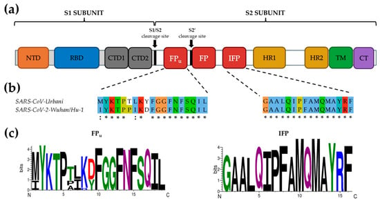
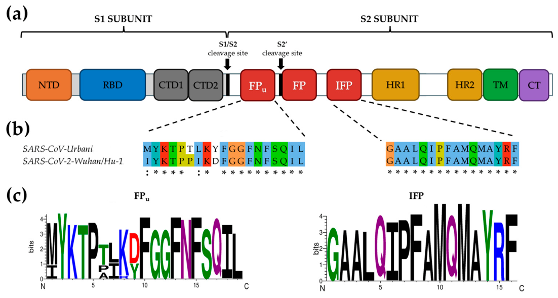
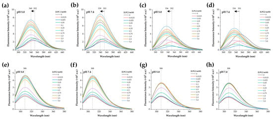
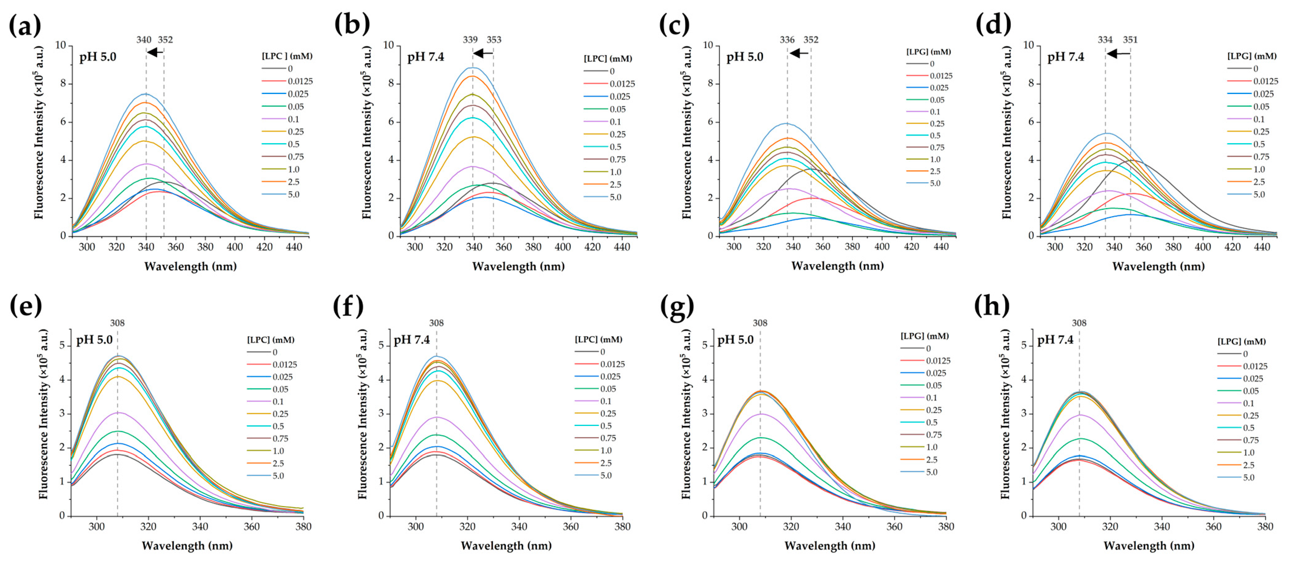
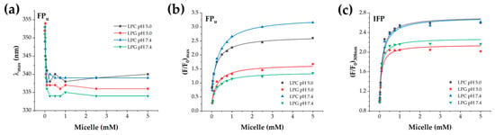
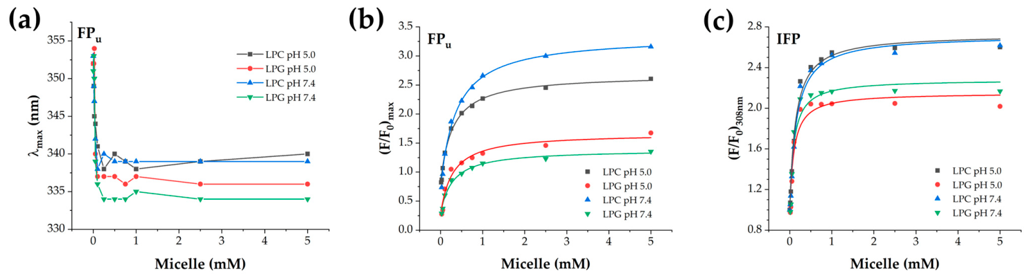
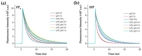
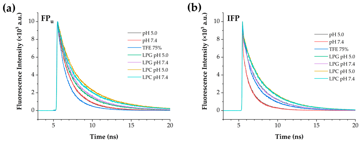
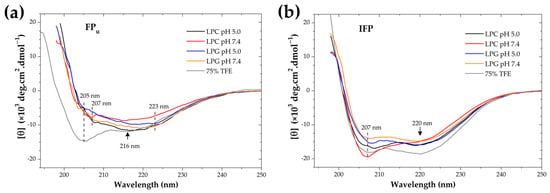
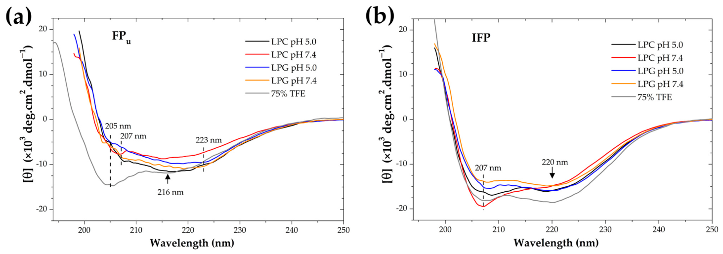
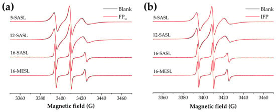
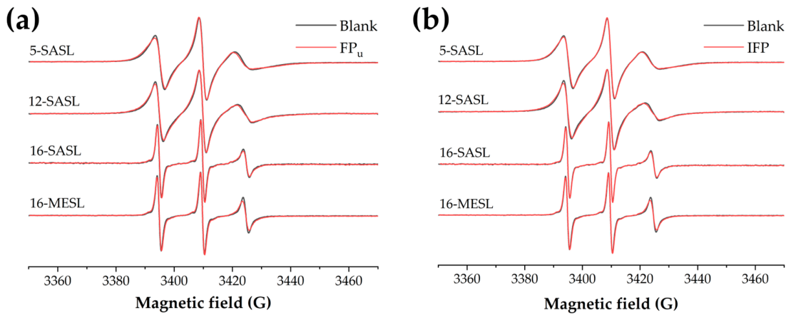
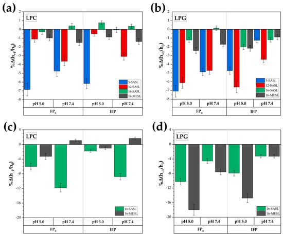
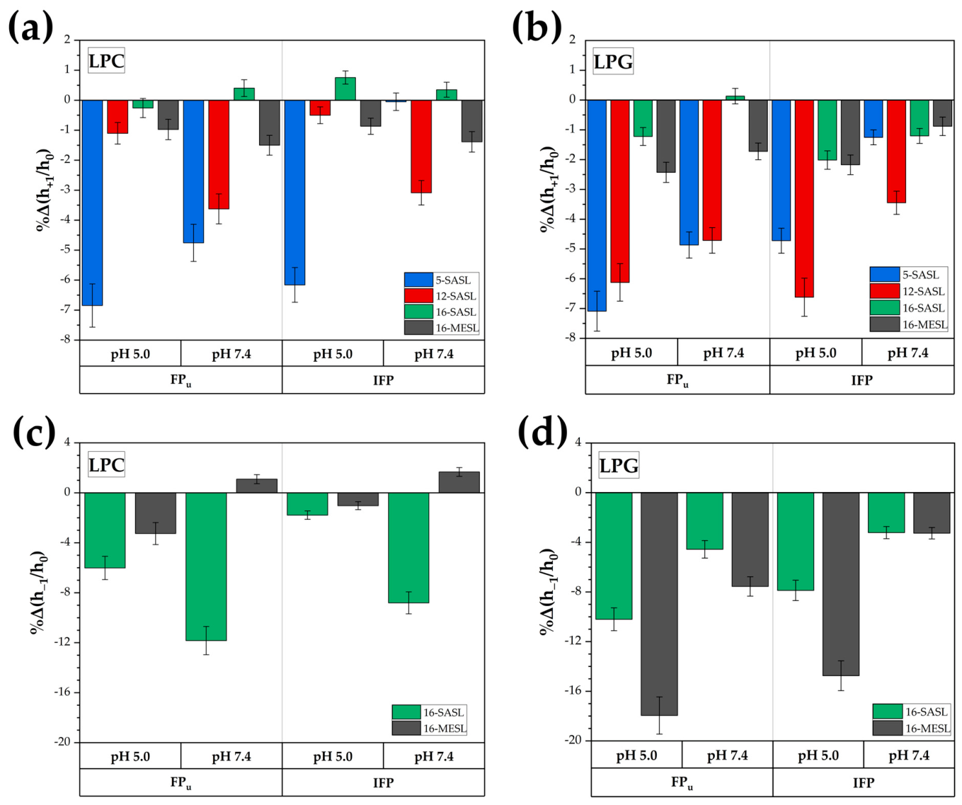
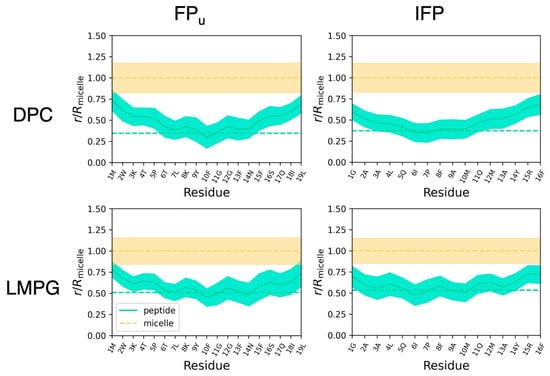
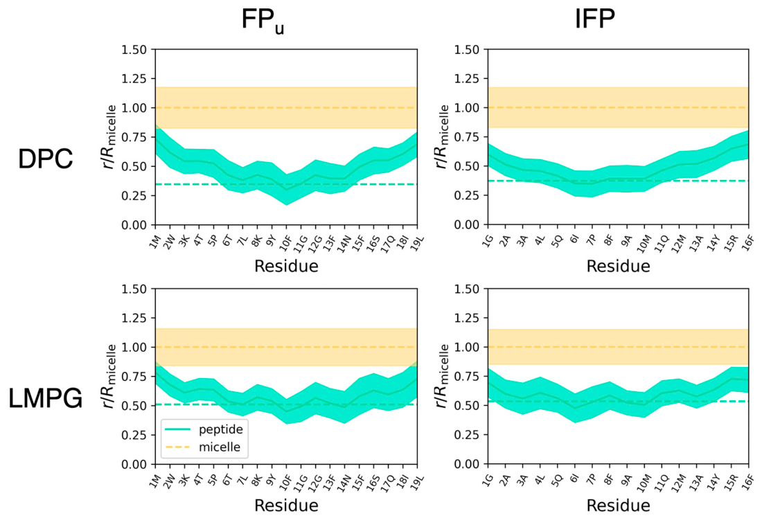
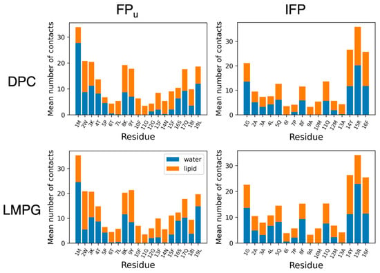
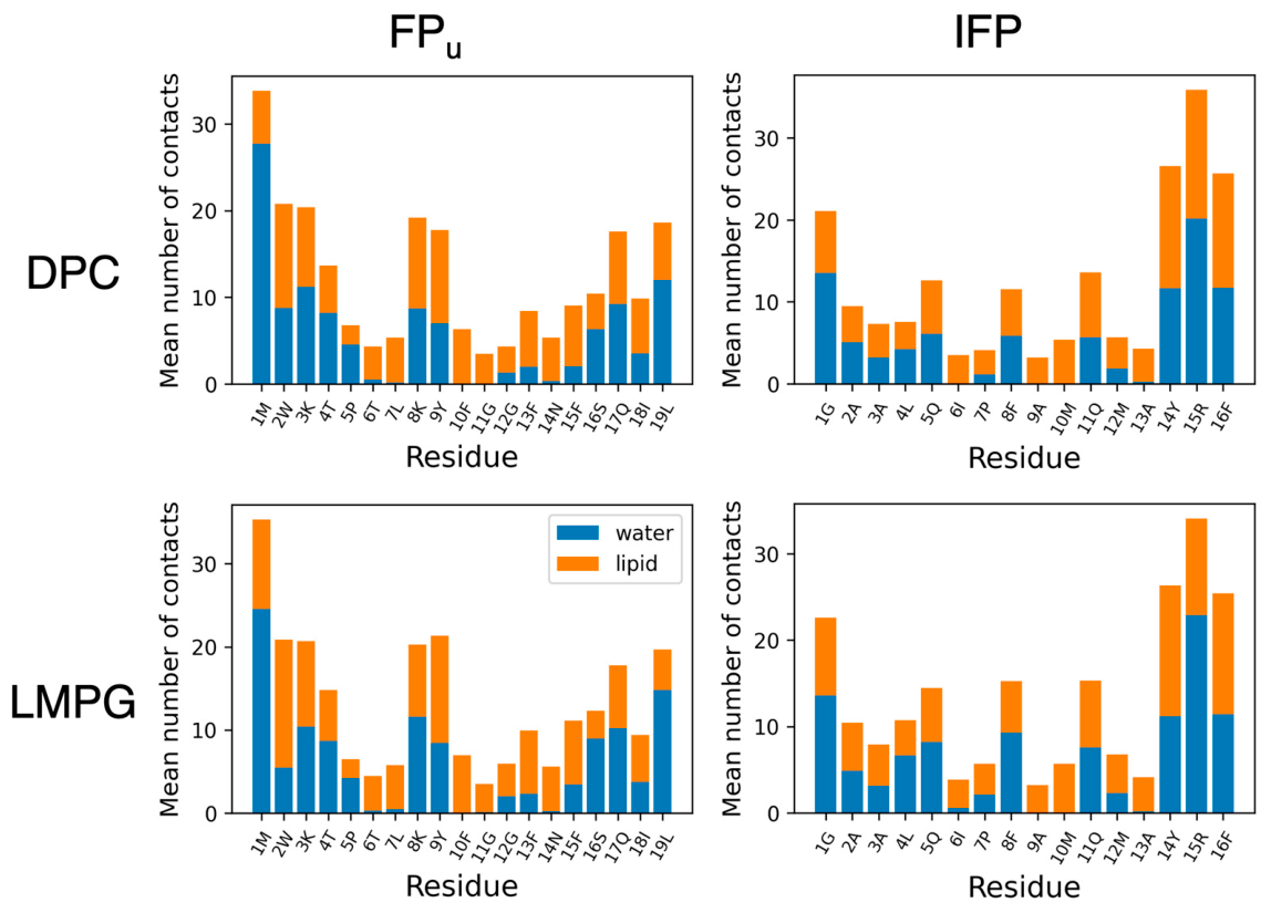

Disclaimer/Publisher’s Note: The statements, opinions and data contained in all publications are solely those of the individual author(s) and contributor(s) and not of MDPI and/or the editor(s). MDPI and/or the editor(s) disclaim responsibility for any injury to people or property resulting from any ideas, methods, instructions or products referred to in the content. |
© 2026 by the authors. Licensee MDPI, Basel, Switzerland. This article is an open access article distributed under the terms and conditions of the Creative Commons Attribution (CC BY) license.