


Abstract
Diabetic cardiomyopathy (DCM) is a highly specific cardiovascular complication of diabetes that occurs independently of hypertension and coronary atherosclerotic heart disease, and is primarily characterized by diffuse myocardial fibrosis and impaired diastolic and systolic function. In recent years, mitochondria-associated membranes (MAMs), as dynamic membrane contact points connecting the endoplasmic reticulum and mitochondria, have attracted much attention in maintaining calcium homeostasis in myocardial cells, regulating energy metabolism and cell survival fate. This article systematically reviews the structural composition of MAMs and their core roles in apoptosis, mitophagy, inflammatory response and calcium homeostasis regulation. It focuses on the molecular mechanisms by which FUNDC1/IP3R2, Bmal1/Bcl-2/IP3R and PPARα/Mfn2 MAM-related signaling pathways mediate calcium overload in DCM, and reviews the research progress of drugs such as dapagliflozin, ferulic acid, quercetin, and ginsenoside Rb1 in improving DCM by targeting the MAM–calcium homeostasis axis. Finally, this review discusses the key scientific issues and future translational research directions in this field, aiming to provide references for mechanism research and new drug development in DCM.
1. Introduction
Diabetic cardiomyopathy (DCM) is one of the most common and severe cardiovascular complications of diabetes. Its typical pathological features include cardiomyocyte hypertrophy and hyperplasia, extracellular matrix deposition, microvascular basement membrane thickening, and interstitial fibrosis. These changes ultimately progress to heart failure, significantly increasing mortality in diabetic patients [1,2]. The pathogenesis of DCM is complex and involves the interplay of multiple factors, including abnormal cardiomyocyte metabolism, calcium homeostasis imbalance, mitochondrial dysfunction, myocardial inflammation and apoptosis, and endoplasmic reticulum stress [3,4].
Mitochondria-associated membranes (MAMs) represent an important discovery in cell biology in recent years. As dynamic and contact structures between mitochondria and the endoplasmic reticulum, MAMs are composed of various proteins and serve not only as a channel for material and information exchange between these two organelles, but also as a functional platform integrating multiple cellular signals [5,6]. Extensive research has established that MAMs serve as central hubs governing critical cellular processes, including mitophagy, endoplasmic reticulum stress, inflammatory responses, and calcium homeostasis. Consequently, structural and functional aberrations in MAMs have been closely linked to the pathogenesis of diverse diseases, such as diabetes, cardiovascular disorders, neurodegenerative conditions, and tumors [7,8]. Disruption of MAMs is regarded as one of the early indicators of mitochondrial dysfunction and insulin resistance [9]. Under the pathological context of DCM, the high-glucose environment can alter the spacing and composition of MAMs, leading to abnormal intracellular signal transduction, thereby impairing the maintenance of calcium homeostasis in cardiomyocytes and inducing mitochondrial dysfunction and aberrant apoptosis. It is worth noting that although the crucial role of MAMs in DCM has gradually been recognized, the strategies for treating DCM by targeting MAMs and restoring calcium homeostasis remain in the exploratory stage. Elucidating the specific mechanisms by which MAMs mediate calcium homeostasis dysregulation in DCM is of great significance for the development of novel therapeutic strategies.
2. The Structure and Composition of MAMs
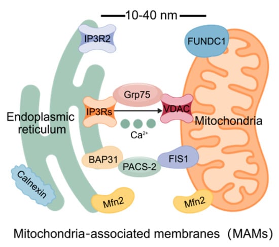
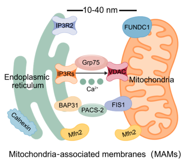
The endoplasmic reticulum and mitochondria are closely interwoven organelles in eukaryotic cells, playing crucial roles in maintaining cellular homeostasis. The endoplasmic reticulum is essential for the transport, storage, synthesis, transfer, modification, and processing of Ca2+ [10,11]. Mitochondria, as the energy factories of the cell, provide a stable energy supply for cellular functions and survival through oxidative phosphorylation reactions [12,13,14]. Although their functional divisions are different, they achieve close functional synergy through the physical coupling structure of MAMs. Fluorescence and electron microscopic observations show that MAMs are dynamically juxtaposed contact points between the endoplasmic reticulum membrane and the outer mitochondrial membrane, with a distance maintained within a narrow range of 10 to 40 nm. MAMs are not merely simple physical connections; they are functional interfaces composed of various proteins [15,16]. In terms of molecular composition, MAMs are enriched in two types of functional proteins: anchoring proteins such as mitochondrial fusion protein 2 (Mfn2) and acidic cluster sorting protein 2 (PACS-2), which are responsible for maintaining the physical coupling between the endoplasmic reticulum and the mitochondria; Ca2+ transport-related proteins such as inositol 1,4,5-triphosphate receptors (IP3Rs), voltage-dependent anion channel (VDAC), glucose-regulated protein 75 (Grp75), FUN14 domain protein 1 (FUNDC1), and calcium-binding protein (Calnexin) which form the core channels for Ca2+ release from the endoplasmic reticulum to the mitochondria (Figure 1). These MAM-associated proteins work together to maintain the physical proximity between the ER and mitochondria at MAM contact sites, while preserving the distinct identities of the two organelles. This configuration enables MAMs to serve both structural support and signal integration functions.
Figure 1.
The structure and composition of MAMs [17].
3. MAMs Regulate Cellular Physiological Functions
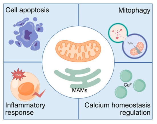
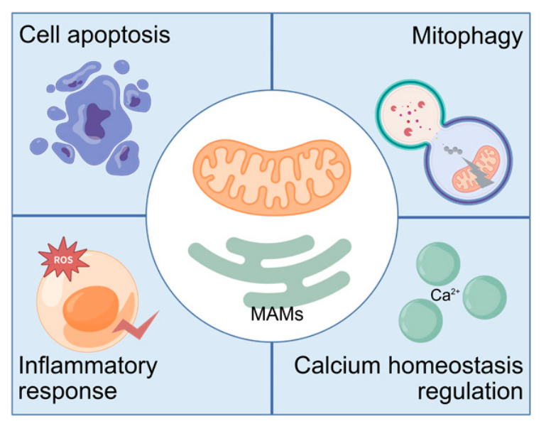
MAMs serve as the functional interface between mitochondria and the endoplasmic reticulum, participating in and regulating various physiological functions of cells. They play a pivotal role in regulating cell apoptosis, mitophagy, reactive oxygen species (ROS) production, inflammatory responses and calcium signal transduction (Figure 2) [18,19]. Dysfunction of MAMs is associated with cancer, neurodegenerative diseases, diabetes, and cardiovascular diseases [20,21,22,23]. Therefore, a comprehensive understanding of the biological functions of MAMs is significant for elucidating the pathogenesis, progression, and phenotypic manifestations of these conditions.
Figure 2.
MAMs regulate cellular physiological functions [17].
3.1. Cell Apoptosis
Cell apoptosis is a programmed cell death mechanism that maintains tissue homeostasis. Its initiation and execution are strictly regulated by internal and external signals, and MAMs are key nodes in apoptotic signal transduction, capable of mediating the apoptotic process [24,25,26,27]. Research has shown that dysregulation of MAMs can lead to poly (ADP-ribose) polymerase (PARP) 1-dependent cell death in retinal progenitor cells of rats [28]; in fluoride-induced mouse spermatocyte injury models and di (2-ethylhexyl) phthalate-induced mouse testicular interstitial cell injury models, structural damage to MAMs and abnormal increases in their quantity can cause mitochondrial Ca2+ overload, thereby triggering significant oxidative stress responses and apoptosis [29,30]; and in aged hen models, stabilizing the structural integrity of ovarian MAMs and normal expression of their anchoring proteins can inhibit granulosa cell apoptosis [31]. The incorrect localization of endoplasmic reticulum protein Calnexin and reduced levels of the Ca2+-binding protein calreticulin in MAMs can impair Ca2+ transfer and biochemical processes in mouse embryonic fibroblasts, and even lead to apoptosis [32]. In addition, in the cardiovascular system, MAMs play a particularly crucial role in regulating cell apoptosis. Disrupting the contact sites of MAMs can damage mitochondrial formation and mitophagy, thereby enhancing the apoptosis of vascular smooth muscle cells [33]. The above evidence indicates that the integrity of MAMs and their regulation of Ca2+ transport between the endoplasmic reticulum and mitochondria play a crucial role in cell apoptosis.
3.2. Mitophagy
Mitophagy is a selective autophagy process aimed at maintaining mitochondrial quality control by eliminating damaged or dysfunctional mitochondria. Recent studies have revealed that MAMs are not only platforms for autophagosome formation but also key sites for initiating mitophagy. It is noteworthy that MAMs exert bidirectional regulation of autophagy: both promoting and inhibiting the process. This phenomenon is closely related to the complex protein composition and multi-dimensional functions of MAMs [34]. In starvation-induced autophagy of renal epithelial HEK293 cells, autophagosomes originate from MAMs. During this process, the autophagy-related proteins PINK1 and BECN1 relocalize to MAMs, where they promote the formation of autophagic precursors, thereby initiating mitophagy [35,36]. However, reductions in MAM number and structural defects can lead to mitochondrial bioenergetic deficiency and decreased Ca2+ uptake, impaired mitochondrial quality control, and altered autophagy flux in primary mouse brain neurons, thereby impairing autophagosome maturation and disrupting the autophagy process [37]. In conclusion, the impact of MAMs on autophagy depends on both the cell type and the nature of the stress.
3.3. Inflammatory Response
The inflammatory response is an important defense mechanism of the body against damage and infection, but excessive or persistent inflammatory signals are core driving factors of tissue damage and various diseases [38]. The role of MAMs in inflammatory regulation has attracted much attention in recent years [39]. Imbalance or dysfunction of MAMs, as well as reductions in MAM length, can activate the NLRP3 inflammasome, leading to increased expression of inflammatory factors such as NLRP3, caspase-1, IL-1β, IL-6, and TNF-α, along with significant increases in ROS and Ca2+ release, and ATP loss [40,41,42,43]. However, similar to autophagy, MAMs also exhibit a bidirectional effect in inflammatory regulation. Under hypoxic conditions, knockdown of the MAM constituent proteins PACS-2 and IP3R1 inhibits the increase in mitochondrial Ca2+ in hypoxia-induced human umbilical vein endothelial cells (HUVECs) and human pulmonary artery endothelial cells (HPAECs), alleviates mitochondrial damage, leads to increased mitochondrial membrane potential and ATP production, reduces ROS, and downregulates the expression of inflammatory molecules such as IL-6 and IL-1β, while enhancing the activity of the eNOS-NO pathway [44]. Therefore, the dual ability of MAMs to promote and inhibit inflammation may be regulated by their structural composition and the cellular survival environment.
3.4. Calcium Homeostasis Regulation
Among the numerous physiological functions mediated by MAMs, calcium homeostasis regulation is regarded as one of the most core and fundamental functions [45,46]. Calcium, as a second messenger in cells, participates in regulating muscle contraction, gene expression, energy metabolism, and cell apoptosis, and serves a crucial function in mitochondria [47,48]. Mitochondrial calcium overload or dyshomeostasis not only impairs mitochondrial function but also disrupts overall cellular homeostasis [49]. MAMs are the channels through which Ca2+ enters mitochondria from the endoplasmic reticulum, and the structural integrity of MAMs is vital for regulating cellular Ca2+ level [50,51,52]. Under pathological conditions, increased MAM formation is typically accompanied by an increase in the contact length between the endoplasmic reticulum and mitochondria, which in turn triggers mitochondrial Ca2+ overload and ultimately leads to mitochondrial dysfunction [53]. In cardiomyocytes and human umbilical vein endothelial cells, the abnormal activation of MAM-related signaling pathways IP3R1-GRP75-VDAC1, IP3R2/FUNDC1, and cGMP/PKG can release Ca2+ from the endoplasmic reticulum into mitochondria and the cytoplasm, resulting in mitochondrial damage and oxidative stress. Inhibiting IP3R-mediated MAM calcium transport can alleviate calcium overload and has a protective effect on mitochondria [49,54]. Thus, MAMs occupy a central position in Ca2+ homeostasis regulation. When the structure or function of MAMs is impaired, it may disrupt the balance of Ca2+ transport, and this phenomenon is particularly prominent in the context of DCM.
4. Abnormal Calcium Signaling Mediated by MAMs in DCM
Abnormal Ca2+ signaling mediated by MAMs is a core factor driving the progression of DCM. Current evidence reveals two seemingly contradictory but potentially corresponding abnormal patterns at different pathological stages. On one hand, the excessive formation of MAMs is an important factor leading to calcium overload and mitochondrial dysfunction. In a high-glucose environment, the length of MAM contact sites and their ratio to mitochondrial perimeter significantly increase, accompanied by a transformation of mitochondria into shorter, spherical shapes. This process leads to decreased Ca2+ levels in the endoplasmic reticulum and an abnormal increase in the level of Ca2+ in mitochondria, thereby inducing ROS generation, autophagy inhibition, and cardiomyocyte apoptosis [55,56]. Accordingly, inhibiting excessive MAM formation is regarded as a potential therapeutic strategy for DCM. On the other hand, in the early stage of DCM, the structure of MAMs undergoes densification, and their thickness tends to be incompatible with efficient calcium transport, resulting in reduced Ca2+ transfer from the endoplasmic reticulum to mitochondria mediated by the IP3R1-Grp75-VDAC1 complex, thereby serving as an early trigger for mitochondrial dysfunction [57]. These two pathways diverge but converge towards exacerbating mitochondrial damage and energy metabolism disorders in DCM.
5. Treatment of Diabetic Cardiomyopathy with the MAM–Calcium Homeostasis Axis
Diabetic cardiomyopathy is a disease involving multiple factors, such as abnormal cardiomyocyte metabolism, calcium homeostasis imbalance, mitochondrial dysfunction, and endoplasmic reticulum stress [58]. With the continuous increase in the global number of diabetic patients, DCM has become one of the main causes of cardiovascular disease risk in these patients [59]. The pathological progression of DCM is related to defective intracellular calcium homeostasis [60], and MAMs participate in the abnormal activation of multiple signaling pathways by mediating calcium homeostasis imbalance, thereby affecting myocardial injury. Exploring these pathways to treat the mitochondrial Ca2+ dysfunction induced by MAMs may help restore normal cardiac function and prevent the deterioration of DCM [57].
5.1. FUNDC1/IP3R2
FUNDC1 is a receptor protein located on the outer mitochondrial membrane. Initially, it was found to be involved in regulating mitophagy induced by hypoxia. Recent studies have expanded the functional repertoire of FUNDC1, revealing its critical role in MAM formation and Ca2+ transfer between the endoplasmic reticulum and mitochondria [61,62,63]. IP3R2 is one of the IP3 receptor subtypes on the endoplasmic reticulum membrane and is highly expressed in cardiomyocytes. It is a key channel for Ca2+ release from the endoplasmic reticulum mediated by MAMs [64,65]. Under normal physiological conditions, the binding of FUNDC1 to IP3R2 remains at a basal level. However, in the pathological context of DCM, high-glucose stimulation significantly enhances the binding between FUNDC1 and IP3R2, which competitively inhibits the ubiquitination and proteasome-mediated degradation of IP3R2. Under high-glucose conditions, the integrity of MAMs is altered, and upregulation of the MAM-associated proteins IP3R2 and FUNDC1 leads to intracellular Ca2+ overload, cellular injury, and even apoptosis. Moreover, disordered Ca2+ transfer via MAMs significantly promotes the progression of DCM [57,66]. In a mouse model of DCM induced by a high-fat and high-fructose diet combined with streptozotocin, the expression levels of FUNDC1 and IP3R2 in the myocardium significantly increased, accompanied by an increase in MAM contact sites and a reduction in inter-organelle distance, as well as a marked rise in the proportion of the endoplasmic reticulum adjacent to mitochondria and Ca2+ content in these regions. Conversely, by specifically knocking out the FUNDC1 gene in the myocardium, the abundance of IP3R2 was reduced, the contact between mitochondria and the endoplasmic reticulum was decreased, the formation of MAMs was eliminated, increases in mitochondrial Ca2+, mitochondrial rupture, and cell apoptosis were prevented, and, at the same time, the mitochondrial functional capacity and cardiac function were improved [60]. These findings collectively establish the FUNDC1/IP3R2 axis as a critical regulator in the pathogenesis of DCM.
5.2. Bmal1/Bcl-2/IP3Rs
Brain and muscle ARNT-like protein 1 (Bmal1) is a core circadian transcription factor that regulates the periodic expression of approximately 10% of the genome under clock control. In the heart, the rhythmic expression of Bmal1 exerts significant regulatory effects on the metabolic rhythm, calcium signal oscillation, and contractile function of cardiomyocytes [67,68]. The rhythmic expression of Bmal1 significantly weakens the coupling between mitochondria and the endoplasmic reticulum [69]. B-cell lymphoma 2 (Bcl-2) controls intrinsic apoptosis and dynamics of mitochondria. Bcl-2 not only inhibits apoptosis at the mitochondrial level but can also directly target IP3Rs and inhibit their channel activity, thereby blocking the Ca2+-dependent apoptotic signal and reducing the release of Ca2+ from the endoplasmic reticulum [70,71]. Recent studies have revealed that overexpression of Bmal1 can directly upregulate Bcl-2 transcription, which increases Bcl-2 protein levels and potentiates the inhibitory effect of Bcl-2 on IP3R activity. This leads to an increased spatial distance between the endoplasmic reticulum and mitochondria, consequently reducing Ca2+ transfer from MAMs to mitochondria. The mitochondrial Ca2+ overload mediated by Bcl-2/IP3Rs is effectively inhibited, indicating that upregulation of Bmal1 can alleviate the progression of DCM through Bcl-2/IP3R-mediated mitochondrial Ca2+ overload [72]. Maintaining the dynamic balance of the circadian rhythm represents a promising direction for future treatment of DCM.
5.3. PPARα/Mfn2
Mfn2 is an important member of the mitochondrial fusion protein family. It has long been believed to mainly participate in the fusion process of the outer mitochondrial membrane. However, recent studies have suggested another essential function of Mfn2. It is one of the key anchor proteins for maintaining the structure of MAMs, and is also located on the outer mitochondrial membrane and endoplasmic reticulum membrane, and expressed on the surface of mitochondria and the endoplasmic reticulum [15,18,73,74] It is worth noting that the regulation of MAM formation by Mfn2 exhibits complex conditional dependence. In the human embryonic kidney immortalized cell line HEK293, knockout of the Mfn2 gene leads to an increase in the coupling between the endoplasmic reticulum and mitochondria, the length of their contact, and the ratio of contact length to total mitochondrial perimeter, thereby increasing the formation of MAMs [75]. In DCM, the protein level of Mfn2 significantly increases in cardiomyocytes treated with high glucose. Overexpression of Mfn2 can promote contact between mitochondria and the endoplasmic reticulum, thereby increasing the Ca2+ level and ROS production in mitochondria, inducing mitochondrial oxidative stress and dysfunction, and leading to endoplasmic reticulum stress in cardiomyocytes [76]. These observations suggest that both Mfn2 deficiency and overexpression ultimately result in MAM dysfunction. The former may result from the loss of functional proteins at the contact sites, leading to an increase in non-specific contacts, while the latter may be caused by excessive physical coupling at the contact sites. Peroxisome proliferator-activated receptor alpha (PPARα) is a significant nuclear receptor regulating fatty acid oxidation and energy metabolism, and is also a direct transcriptional regulatory factor of Mfn2. Decreased expression of PPARα is able to induce downregulation of Mfn2 expression; PPARα directly binds to the promoter of Mfn2 to positively regulate its expression and restore Mfn2 to its physiological level, rather than completely deleting or overexpressing it, thereby promoting mitochondrial fusion, restoring mitochondrial membrane potential, inhibiting mitochondrial oxidative stress and improving the mitochondrial function of cardiomyocytes, decreasing the Ca2+ level in mitochondria, and maintaining intracellular calcium homeostasis and the stable structure of MAMs. This indicates that downregulation of PPARα/Mfn2 may be a key strategy for maintaining the dynamic balance of MAMs and improving DCM [77].
6. The Therapeutic Effect of Drugs on Diabetic Cardiomyopathy
Based on the above mechanistic studies, regulating the MAM–calcium homeostasis axis has become an important direction for the development of therapeutic drugs for DCM. Although no drugs that can directly target the MAM–calcium homeostasis axis have yet been identified, existing research findings show that both chemical drugs and active monomers of traditional Chinese medicine can restore calcium homeostasis in cardiomyocytes by regulating the expression or function of MAM-related proteins, thereby improving the pathological process of DCM and providing important insights for the further development of novel DCM therapeutic drugs (Table 1).
Table 1.
The therapeutic effect of drugs on DCM.
6.1. Chemical Drugs
Sodium–glucose cotransporter 2 inhibitors (SGLT2Is) have been a major breakthrough in the field of diabetes treatment in recent years. Drugs such as dapagliflozin and empagliflozin, which belong to this class of medications, have demonstrated cardiorenal protective effects beyond their hypoglycemic effects in cardiovascular trials. Although their classical target is SGLT2 in the proximal convoluted tubules of the kidneys, increasing evidence indicates that SGLT2Is can directly exert protective effects on the heart [78,79,80,81]. Research has found that dapagliflozin can reduce the generation of oxygen free radicals, lower the activity of membrane channels related to calcium transport, regulate MAMs, decrease cardiomyocyte fibrosis and inflammation, and improve contractile function to treat DCM [82].
6.2. Active Monomers of Traditional Chinese Medicine
Ferulic acid is a type of monophenolic plant chemical substance widely present in Chinese medicinal herbs such as Angelica sinensis, Ligusticum chuanxiong, and Myristica fragrans. It exhibits a range of biological activities, including antioxidant, anti-inflammatory, and anti-apoptotic properties. In H9c2 cardiomyocytes cultured under high-glucose conditions, ferulic acid treatment can significantly downregulate MAM-related proteins such as FUNDC1, while ameliorating oxidative stress caused by Ca2+ overload in the DCM microenvironment, maintaining the integrity of MAMs, and alleviating pathological changes such as cardiomyocyte damage [83]. Quercetin is a flavonoid compound found in various plants including apples, berries, tea, and Tetrapleura tetraptera. Numerous studies have revealed its unique role in maintaining cardiac calcium homeostasis. Specifically, quercetin and its secondary metabolites have been shown to reduce cardiomyocyte sensitivity to Ca2+ by activating the sarcoplasmic/endoplasmic reticulum calcium ATPase (SERCA), thereby preserving cardiac calcium homeostasis and MAM integrity, and effectively mitigating the progression of DCM [84]. Ginsenoside Rb1, one of the principal active constituents of Panax ginseng, has been utilized in the treatment of diabetes and obesity, as well as in reducing inflammation and cardiovascular risk [85]. In DCM research, ginsenoside Rb1 alleviates aberrant Ca2+ distribution and transport dysregulation within MAMs by restoring key Ca2+-handling proteins SERCA2a and RyR2, effectively improving diabetes-induced cardiac dysfunction and cardiomyocyte calcium signaling disturbances [86,87,88]. Although these three natural products have different chemical structures, they can all treat DCM by regulating the MAM–calcium homeostasis axis, providing a theoretical basis for subsequent drug screening and combination strategies based on MAMs for the treatment of DCM.
7. Summary and Outlook
As a central functional platform connecting mitochondria and the endoplasmic reticulum, MAMs play an irreplaceable role in maintaining calcium homeostasis, regulating energy metabolism, achieving the autophagy–apoptosis balance, and integrating inflammatory signals in cardiomyocytes [89]. In the pathological context of diabetic cardiomyopathy, the high-glucose and high-lipid environment disrupts the structural integrity and functional regulation of MAMs, leading to dysregulation of multiple signaling pathways, and subsequently causing mitochondrial calcium overload, oxidative stress, and cardiomyocyte apoptosis, ultimately driving the progression of DCM [90]. In the current research context, strategies targeting the MAM–calcium homeostasis axis show significant potential for treating myocardial damage in DCM. Based on this, MAMs are gradually being recognized as a key target for DCM treatment. However, several key issues in this field still need to be addressed urgently. Firstly, the identified MAM constituent proteins exceed 1000, but the specific functions of most of these proteins in DCM remain unclear [91,92]. In the future, combined with single-cell transcriptomics and spatial proteomics technologies, the dynamic changes in MAM protein groups in cardiomyocytes at different stages of DCM should be systematically mapped to identify key molecules for early diagnosis and targeted intervention [93,94,95,96]. Secondly, in the regulation of calcium homeostasis, excessive or insufficient calcium influx is harmful, and it is necessary to restore the calcium transport function of MAMs while preventing mitochondrial calcium overload. In summary, the MAM–calcium homeostasis axis provides a new perspective and idea for treating DCM. From the perspective of intercellular interaction, examining the pathogenesis of cardiovascular diseases not only helps to reveal the fundamental pathological links of DCM, but also opens a path for developing drugs with novel mechanisms for treating DCM, and is expected to truly bring MAMs, as potential targets, to clinical application [97,98].
Author Contributions
J.S. was responsible for conducting literature research and writing the article. J.S. was responsible for the content of each section and chapter, and also drew charts and tables. X.L. assisted in drawing the charts and participated in the revision of the paper. P.L. provided in-depth opinions on the structure and composition of MAMs as well as the future outlook section. All authors have read and agreed to the published version of the manuscript.
Funding
This research received no external funding.
Institutional Review Board Statement
Not applicable.
Data Availability Statement
No new data were created or analyzed in this study. Data sharing is not applicable to this article.
Conflicts of Interest
The authors declare no conflicts of interest.
Abbreviations
| DCM | Diabetic cardiomyopathy |
| MAMs | Mitochondria-associated membranes |
| Mfn2 | Mitochondrial fusion protein 2 |
| PACS-2 | Acidic cluster sorting protein 2 |
| IP3Rs | Inositol 1, 4, 5—triphosphate receptors |
| VDAC | Voltage-dependent anion channel |
| Grp75 | Glucose-regulated protein 75 |
| FUNDC1 | FUN14 domain protein 1 |
| Calnexin | Calcium-binding protein |
| ROS | Reactive oxygen species |
| PARP | Poly (ADP-ribose) polymerase |
| PINK1 | PTEN-induced putative kinase 1 |
| BECN1 | Beclin 1 |
| NLRP3 | NACHT, LRR and PYD domains-containing protein 3 |
| IL-1β | Interleukin-1β |
| IL-6 | Interleukin-6 |
| TNF-α | Tumor Necrosis Factor-α |
| ATP | Adenosine Triphosphate |
| cGMP | Cyclic Guanosine Monophosphate |
| PKG | cGMP-Dependent Protein Kinase |
| Bmal1 | Brain and muscle ARNT-like protein 1 |
| Bcl-2 | B-cell lymphoma 2 |
| PPARα | Peroxisome proliferator-activated receptor α |
| SGLT2Is | Sodium–glucose cotransporter 2 inhibitors |
| SERCA | Sarcoplasmic reticulum/endoplasmic reticulum calcium ATPase |
| RyR2 | Ryanodine Receptor 2 |
References
- Saeedi, P.; Petersohn, I.; Salpea, P.; Malanda, B.; Karuranga, S.; Unwin, N.; Colagiuri, S.; Guariguata, L.; Motala, A.A.; Ogurtsova, K.; et al. Global and regional diabetes prevalence estimates for 2019 and projections for 2030 and 2045: Results from the international diabetes federation diabetes atlas, 9th edition. Diabetes Res. Clin. Pract. 2019, 157, 107843. [Google Scholar] [CrossRef] [Scilit]
- Zhan, J.; Jin, K.; Xie, R.; Fan, J.; Tang, Y.; Chen, C.; Li, H.; Wang, D.W. Ago2 protects against diabetic cardiomyopathy by activating mitochondrial gene translation. Circulation 2024, 149, 1102–1120. [Google Scholar] [CrossRef] [Scilit]
- Yu, F.; Zong, B.; Tian, Z.; Jia, D.; Wang, R. Exerkines: Potential regulators of diabetic cardiomyopathy. Ageing Res. Rev. 2025, 110, 102816. [Google Scholar] [CrossRef] [Scilit]
- Wang, H.; Wang, J.; Cui, H.; Fan, C.; Xue, Y.; Liu, H.; Li, H.; Li, J.; Li, H.; Sun, Y.; et al. Inhibition of fatty acid uptake by tgr5 prevents diabetic cardiomyopathy. Nat. Metab. 2024, 6, 1161–1177. [Google Scholar] [CrossRef] [Scilit]
- Chen, Y.; Xu, Y.M.; Zhu, L.L.; Shan, Z.Y.; Yao, Z.G.; Zhao, H. Mfn2-perk axis regulates er stress in parotid glands of aged mice via mams. J. Dent. Res. 2026, 105, 1643–1654. [Google Scholar] [CrossRef] [Scilit]
- Li, X.; Wang, Y.; Wu, F.; Huang, X.; Ni, Z.; Xu, G.; Wang, W.; Tie, J.; Huang, Y.; Sun, Y.; et al. The mcu-mecom axis orchestrates glioblastoma progression by remodeling mitochondrial dynamics and quality control via mams. Int. J. Biol. Sci. 2026, 22, 5054–5082. [Google Scholar] [CrossRef] [Scilit]
- Wang, A.; Yu, Z.; Guo, R.; Tang, M.; Liang, L.; Tu, H.; Duan, X.; Zhou, Y.; Li, W.; Tang, Z.; et al. Cgrp mitigates myocardial ischemia-reperfusion injury by attenuating ferroptosis through the inhibition of mams and mitochondrial division. Int. Immunopharmacol. 2026, 182, 116839. [Google Scholar] [CrossRef] [Scilit]
- Liu, H.; Jiang, Y.; Cong, L.; Zhang, X.; Zhou, Y.; Pan, X.; Liu, S.; Wang, R.; Cao, X. Intranasal insulin administration affecting perioperative neurocognitive dysfunction by regulating calcium transport protein complex ip3r/grp75/vdac1 on mams. Free Radic. Biol. Med. 2025, 228, 240–250. [Google Scholar] [CrossRef] [Scilit]
- Tao, X.; Zhang, Y.; Pang, J.; Liao, Z.; Chen, G.; Zhong, X.; Cao, X.; Lin, Y.; Guan, D.; Tian, Y.; et al. Er stress sensor perk drives ferroptosis by disrupting mams-mediated mitochondrial homeostasis and promoting mtros accumulation in acute myocardial infarction. Pharmacol. Res. 2025, 222, 108029. [Google Scholar] [CrossRef] [Scilit]
- Gulati, A.; Ahn, D.; Suades, A.; Hult, Y.; Wolf, G.; Iwata, S.; Superti-Furga, G.; Nomura, N.; Drew, D. Stepwise atp translocation into the endoplasmic reticulum by human slc35b1. Nature 2025, 643, 855–864. [Google Scholar] [CrossRef] [Scilit]
- Jang, W.; Puchkov, D.; Samso, P.; Liang, Y.; Nadler-Holly, M.; Sigrist, S.J.; Kintscher, U.; Liu, F.; Mamchaoui, K.; Mouly, V.; et al. Endosomal lipid signaling reshapes the endoplasmic reticulum to control mitochondrial function. Science 2022, 378, 1188. [Google Scholar] [CrossRef] [Scilit]
- Wang, J.; Wu, H.; Gao, J.; Zhang, T. Mitochondrion in pulmonary disease: Small organelle but big function. Small Methods 2026, 10, e2500674. [Google Scholar] [CrossRef] [Scilit]
- Choi, Y.; No, M.; Heo, J.; Cho, E.; Park, D.; Kang, J.; Kim, C.; Seo, D.Y.; Han, J.; Kwak, H. Resveratrol attenuates aging-induced mitochondrial dysfunction and mitochondria-mediated apoptosis in the rat heart. Nutr. Res. Pract. 2025, 19, 186–199. [Google Scholar] [CrossRef] [Scilit]
- Jiang, L.; Zhou, S.; Zhang, X.; Li, C.; Ji, S.; Mao, H.; Jiang, X. Mitochondrion-specific dendritic lipopeptide liposomes for targeted sub-cellular delivery (vol 12, 2390, 2021). Nat. Commun. 2025, 16, 5405. [Google Scholar]
- Lin, Z.; Wang, J.; Li, J.; Zheng, Y.; Zheng, B.; Li, Q.; Huang, X. Baicalin alleviates oxidative stress in df-1 cell line via the mfn2/mams/Ca2+ pathway. Poult. Sci. 2025, 104, 105818. [Google Scholar] [CrossRef] [Scilit]
- Huang, S.; Zeng, Z.; Chu, Y.; Zhang, S.; Zhou, J.; Hu, Z.; Yang, Y.; Zhou, C.; Cheng, W.; Yang, S.; et al. Mitigation of lipopolysaccharide-induced intestinal injury in rats by chimonanthus nitens oliv. Essential oil via suppression of mitochondrial fusion protein mitofusin 2 (mfn2)-mediated mitochondrial-associated endoplasmic reticulum membranes (mams) formation. J. Ethnopharmacol. 2025, 337, 118856. [Google Scholar] [CrossRef] [Scilit]
- Jiang, S.; Li, H.; Zhang, L.; Mu, W.; Zhang, Y.; Chen, T.; Wu, J.; Tang, H.; Zheng, S.; Liu, Y.; et al. Generic diagramming platform (gdp): A comprehensive database of high-quality biomedical graphics. Nucleic Acids Res. 2024, 53, D1670–D1676. [Google Scholar] [CrossRef] [Scilit]
- Wang, S.; Sang, X.; Li, S.; Yang, W.; Wang, S.; Chen, H.; Lu, C. Increased Ca2+ transport across the mitochondria-associated membranes by mfn2 inhibiting endoplasmic reticulum stress in ischemia/reperfusion kidney injury. Sci. Rep. 2023, 13, 17257. [Google Scholar] [CrossRef] [Scilit]
- Dai, L.; Wang, M.; Wang, B.; Liang, H.; Guan, Y.; Du, Z.; Wang, H. Yiqi huoxue recipe ameliorates diabetic nephropathy by mediating vapb-ptpip51 complex to activate autophagy and regulate mam contact. Front. Nutr. 2025, 12, 1634555. [Google Scholar] [CrossRef] [Scilit]
- Pi, S.; Liu, W.; Zhao, J.; Chen, H.; Luo, X.; Wu, S.; Song, J.; Chen, X.; Li, X.; Luo, W.; et al. Serca2 dysfunction drives vascular calcification via coupling with tspo-mcu at mitochondria-associated endoplasmic reticulum membranes. Pharmacol. Res. 2026, 227, 108177. [Google Scholar] [CrossRef] [Scilit]
- Xie, W.; Chen, M.; Loor, J.J.; Song, X.; Ma, N.; Zhou, S.; Zhang, H.; Chang, G.; Shen, X. Ampk-endoplasmic reticulum stress axis contributes to lipopolysaccharide-caused mitochondrial dysfunction by regulating mitochondria-associated membrane function in bovine hepatocytes. J. Dairy Sci. 2023, 106, 5146–5164. [Google Scholar] [CrossRef] [Scilit]
- Xue, N.; Liu, Y.; Lin, Z.; Huang, W.; Zhang, F.; Zheng, R.; Si, X.; Gu, L.; Fan, Y.; Pu, J.; et al. Parkin deficiency impairs er-mitochondria associations and calcium homeostasis via ip3r-grp75-vdac1 complex. Int. J. Biol. Sci. 2026, 22, 731–749. [Google Scholar] [CrossRef] [Scilit]
- Lin, S.; Cao, L.; Zhou, Y.; Liu, S.; Yan, Y.; Zhang, Q.; Yuan, Q.; Gao, Y.; Du, G.; Hu, J.; et al. Er stress promoted mams formation and er-mito Ca2+ transfer: Implication for fluoride-induced hippocampal neuronal injury and cognitive deficits in mice. Chem. Biol. Interact. 2026, 437, 112252. [Google Scholar] [CrossRef] [Scilit]
- Cao, Y.; Chen, Z.; Hu, J.; Feng, J.; Zhu, Z.; Fan, Y.; Lin, Q.; Ding, G. Mfn2 regulates high glucose-induced mams dysfunction and apoptosis in podocytes via perk pathway. Front. Cell Dev. Biol. 2021, 9, 769213. [Google Scholar] [CrossRef] [Scilit]
- Cao, Z.; Liu, R.; Li, K.; Sun, J.; Wen, X.; Tang, F.; Liu, L.; Peng, F.; Peng, C.; Ao, H. Mechanism of shenzhu components in alleviating chemotherapy-induced diarrhea via mitochondria-associated endoplasmic reticulum membranes (mams) regulation. J. Ethnopharmacol. 2026, 358, 120958. [Google Scholar] [CrossRef] [Scilit]
- Legiot, A.; Cere, C.; Dupoiron, T.; Kaabouni, M.; Camougrand, N.; Manon, S. Mitochondria-associated membranes (mams) are involved in bax mitochondria! Localization and cytochrome c release. Microb. Cell 2019, 6, 257–266. [Google Scholar] [CrossRef] [Scilit]
- Li, H.; Dong, A.; Liu, C.; He, P.; Ma, Y.; Chen, S.; Dong, S.; Zhang, S.; Zhang, M.; Zhang, M. Schisandra chinensis mixture attenuates diabetic kidney disease via VDAC1/GRP75/IP3R-mediated mams stabilization and apoptosis-autophagy regulation. Phytomedicine 2025, 147, 157164. [Google Scholar] [CrossRef] [Scilit]
- Yang, Y.; Wu, J.; Lu, W.; Dai, Y.; Zhang, Y.; Sun, X. Mitochondria-associated endoplasmic reticulum membranes dysfunction contributes to parp-1-dependent cell death under oxidative stress in retinal precursor cells. J. Biochem. Mol. Toxicol. 2023, 37, e23303. [Google Scholar] [CrossRef] [Scilit]
- Guo, X.; Wang, L.; Xuan, J.; Chen, T.; Du, Y.; Qiao, H.; Zhang, S.; Sun, Z.; Wang, J.; Niu, R. Fluoride induces spermatocyte apoptosis by ip3r1/mcu-mediated mitochondrial calcium overload through mams. J. Hazard. Mater. 2025, 489, 137514. [Google Scholar] [CrossRef] [Scilit]
- Wang, J.; Wei, Y.; Wu, Y.; Zhao, T.; Kang, L.; Han, L.; Chen, J.; Long, C.; Wei, G.; Shen, L.; et al. Di -(2-ethylhexyl) phthalate induces prepubertal testicular injury through mam-related mitochondrial calcium overload in leydig and sertoli cell apoptosis. Toxicology 2024, 509, 153956. [Google Scholar] [CrossRef] [Scilit]
- Si, C.; Chen, Y.; Liu, L.; Liu, S.; Zhang, Z.; Guo, L.; Zhao, M.; Chen, Y.; Shi, Y.; Shi, W.; et al. Dietary semen cuscuta improves laying performance by stabilizing mitochondria-associated membranes (mams) and inhibiting granulosa cell apoptosis in laying hens. Poult. Sci. 2026, 105, 106749. [Google Scholar] [CrossRef] [Scilit]
- Carpio, M.A.; Means, R.E.; Brill, A.L.; Sainz, A.; Ehrlich, B.E.; Katz, S.G. Bok controls apoptosis by Ca2+ transfer through er-mitochondrial contact sites. Cell Rep. 2021, 34, 108827. [Google Scholar] [CrossRef] [Scilit]
- Moulis, M.; Grousset, E.; Faccini, J.; Richetin, K.; Thomas, G.; Vindis, C. The multifunctional sorting protein pacs-2 controls mitophagosome formation in human vascular smooth muscle cells through mitochondria-er contact sites. Cells 2019, 8, 638. [Google Scholar] [CrossRef] [Scilit]
- Wu, W.; Li, W.; Chen, H.; Jiang, L.; Zhu, R.; Feng, D. Fundc1 is a novel mitochondrial-associated-membrane (mam) protein required for hypoxia-induced mitochondrial fission and mitophagy. Autophagy 2016, 12, 1675–1676. [Google Scholar] [CrossRef] [Scilit]
- Gelmetti, V.; De Rosa, P.; Torosantucci, L.; Marini, E.S.; Romagnoli, A.; Di Rienzo, M.; Arena, G.; Vignone, D.; Fimia, G.M.; Valente, E.M. Pink1 and becn1 relocalize at mitochondria-associated membranes during mitophagy and promote er-mitochondria tethering and autophagosome formation. Autophagy 2017, 13, 654–669. [Google Scholar] [CrossRef] [Scilit]
- Gu, J.; Zhang, T.; Guo, J.; Chen, K.; Li, H.; Wang, J. Pink1 activation and translocation to mitochondria-associated membranes mediates mitophagy and protects against hepatic ischemia/reperfusion injury. Shock 2020, 54, 783–793. [Google Scholar] [CrossRef] [Scilit]
- Patergnani, S.; Bataillard, M.S.; Danese, A.; Alves, S.; Cazevieille, C.; Valéro, R.; Tranebjærg, L.; Maurice, T.; Pinton, P.; Delprat, B.; et al. The wolfram-like variant wfs1(e864k) destabilizes mam and compromises autophagy and mitophagy in human and mice. Autophagy 2024, 20, 2055–2066. [Google Scholar] [CrossRef] [Scilit]
- Ali, S.; Akhtar, M.S.; Amin, A.; Khalil, A.A.K.; Zaman, W. Pharmaco-therapeutic modulation of the glo1-nrf2-rage functional network in chronic inflammation: Polyphenols in the context of current anti-inflammatory therapy. Inflammopharmacology 2026, 34, 3873–3886. [Google Scholar] [CrossRef] [Scilit]
- Zhou, Q.; Wang, Y.; Lu, Z.; He, C.; Li, L.; You, M.; Wang, L.; Cao, T.; Zhao, Y.; Li, Q.; et al. Cx43 acts as a mitochondrial calcium regulator that promotes obesity by inducing the polarization of macrophages in adipose tissue. Cell. Signal. 2023, 105, 110606. [Google Scholar] [CrossRef] [Scilit]
- Liu, H.; Li, X.; Wang, Z.; Li, L.; Li, Y.; Yan, H.; Yuan, Y. Elaidic acid leads to mitochondrial dysfunction via mitochondria-associated membranes triggers disruption of mitochondrial calcium fluxes. Food Sci. Hum. Wellness 2024, 13, 287–298. [Google Scholar] [CrossRef] [Scilit]
- Chang, H.; Yang, F.; Bai, H.; Lu, Z.; Xing, C.; Dai, X.; Wan, W.; Liao, S.; Cao, H. Molybdenum and/or cadmium induce nlrp3 inflammasome production by causing mitochondria-associated endoplasmic reticulum membrane dysfunction in sheep hepatocytes. Chem. Biol. Interact. 2023, 382, 110617. [Google Scholar] [CrossRef] [Scilit]
- Jiang, T.; Peng, D.; Shi, W.; Guo, J.; Huo, S.; Men, L.; Zhang, C.; Li, S.; Lv, J.; Lin, L. Il-6/stat3 signaling promotes cardiac dysfunction by upregulating fundc1-dependent mitochondria-associated endoplasmic reticulum membranes formation in sepsis mice. Front. Cardiovasc. Med. 2022, 8, 790612. [Google Scholar] [CrossRef] [Scilit]
- Nawaz, S.; Kulyar, M.F.; Mo, Q.; Zhang, Z.; Quan, C.; Iqbal, M.; Imad, E.F.; Li, J. Thiram-induced er stress promotes mitochondrial calcium signaling and nlrp3 inflammasome activation in a tissue specific manner. Ecotoxicol. Environ. Saf. 2025, 293, 118026. [Google Scholar] [CrossRef] [Scilit]
- Yang, Y.; Li, M.; Xu, G.; Zhang, E.; Chen, J.; Sun, B.; Chen, D.; Gao, Y. Targeting mitochondria-associated membranes as a potential therapy against endothelial injury induced by hypoxia. J. Cell. Biochem. 2019, 120, 18967–18978. [Google Scholar] [CrossRef] [Scilit]
- Li, Y.; Du, T.; Zhang, Y.; Lu, Y.; Li, X.; Xie, W.; Guo, S. Cardioprotective effect of yiqi huoxue decoction on post-myocardial infarction injury mediated by Ca2+ flux through mams. Front. Cardiovasc. Med. 2025, 12, 1596757. [Google Scholar] [CrossRef] [Scilit]
- Zhang, J.; Jiang, J.; Li, H.; Deng, J.; Dong, W.; Deng, H. Mam-mediated mitochondrial Ca2+ overload and endoplasmic reticulum stress aggravates synaptic plasticity impairment in diabetic mice. Brain Sci. 2025, 15, 1157. [Google Scholar] [CrossRef] [Scilit]
- Cherubini, M.; Lopez-Molina, L.; Gines, S. Mitochondrial fission in huntington’s disease mouse striatum disrupts er-mitochondria contacts leading to disturbances in Ca2+ efflux and reactive oxygen species (ROS) homeostasis. Neurobiol. Dis. 2020, 136, 104741. [Google Scholar] [CrossRef] [Scilit]
- Ahuja, K.; Motiani, R.K. Calcium acts as a critical determinant of mitochondria-nuclear networking driven retrograde signaling. Cell Calcium 2025, 127, 103017. [Google Scholar] [CrossRef] [Scilit]
- Guo, J.; Ma, T.; Wang, B.; Xing, B.; Huang, L.; Li, X.; Zheng, H.; He, Y.; Xi, J. Zn2+ protects h9c2 cardiomyocytes by alleviating mams-associated apoptosis and calcium signaling dysregulation. Cell. Signal. 2025, 127, 111629. [Google Scholar] [CrossRef] [Scilit]
- Mohan, S.; Nair, A.; Poornima, M.S.; Raghu, K.G. Vanillic acid mitigates hyperinsulinemia induced er stress mediated altered calcium homeostasis, mams distortion and surplus lipogenesis in hepg2 cells. Chem. Biol. Interact. 2023, 375, 110365. [Google Scholar] [CrossRef] [Scilit]
- Hayashi, T.; Su, T. Sigma-1 receptor chaperones at the er-mitochondrion interface regulate Ca2+ signaling and cell survival. Cell 2007, 131, 596–610. [Google Scholar] [CrossRef] [Scilit]
- Marchi, S.; Bittremieux, M.; Missiroli, S.; Morganti, C.; Patergnani, S.; Sbano, L.; Rimessi, A.; Kerkhofs, M.; Parys, J.B.; Bultynck, G.; et al. Endoplasmic reticulum-mitochondria communication through Ca2+ signaling: The importance of mitochondria-associated membranes (mams). Organelle Contact Sites Mol. Mech. Dis. 2017, 997, 49–67. [Google Scholar] [CrossRef] [Scilit]
- Li, Y.; Jing, M.; Wang, W.; Lin, W.; Zhang, Y.; Nie, T.; Liu, P.; Lu, W.; Chen, Y.; Fielding Hejtmancik, J.; et al. Dapl1 restrains rpe panoptosis in experimental amd by inhibiting grp75-mediated mitochondria-associated endoplasmic reticulum membranes. Proc. Natl. Acad. Sci. USA 2026, 123, e1783041173. [Google Scholar] [CrossRef] [Scilit]
- Du, T.; Zhang, Y.; Wang, H.; Xie, W.; Liang, C.; Guo, J.; Zheng, C.; Guo, S. Yiqi huoxue formula promotes post-myocardial infarction angiogenesis by modulating mitochondria-associated membranes (mams) and calcium homeostasis via the cgmp/pkg signaling pathway. J. Ethnopharmacol. 2026, 370, 121936. [Google Scholar] [CrossRef] [Scilit]
- Wei, Y.; Ji, Y.; Meng, J.; Yu, L.; Tang, Y.; Fang, W. Acid sphingomyelinase promotes diabetic cardiomyopathy via disruption of mitochondrial calcium homeostasis. Cardiovasc. Diabetol. 2025, 24, 272. [Google Scholar] [CrossRef] [Scilit]
- Wang, B.; Dai, L.; Liang, H.; He, J.; Zhou, J.; Guan, Y.; Wang, H. Mitochondrial ultrastructural pathology in diabetic cardiomyopathy: Integrated analysis via scanning electron microscopy and 3d visualization imaging. Cardiovasc. Diabetol. 2025, 24, 331. [Google Scholar] [CrossRef] [Scilit]
- Dia, M.; Gomez, L.; Thibault, H.; Tessier, N.; Leon, C.; Chouabe, C.; Ducreux, S.; Gallo-Bona, N.; Tubbs, E.; Bendridi, N.; et al. Reduced reticulum-mitochondria Ca2+ transfer is an early and reversible trigger of mitochondrial dysfunctions in diabetic cardiomyopathy. Basic Res. Cardiol. 2020, 115, 74. [Google Scholar] [CrossRef] [Scilit]
- Norambuena-Soto, I.; Zhang, P.; Wang, Y.; Fu, L.; Jimenez-Gallegos, D.; Jiang, Y.; Yuan, V.; Li, H.; Chiong, M.; Zapata-Torres, G.; et al. Angiotensin-(1-9) attenuates diabetic cardiomyopathy by improving insulin resistance. Br. J. Pharmacol. 2026, 183, 3032–3048. [Google Scholar] [CrossRef] [Scilit]
- Chen, X.; Guo, W.; Jing, Z.; Zhang, T.; Wu, Z.; Cui, H.; Zheng, W.; Chen, Y.; Chen, H. The mitochondrial calcium uniporter induces apoptosis in cardiomyocytes cultured with high-glucose medium by affecting mitochondrial function. Res. Sq. 2020. [Google Scholar] [CrossRef] [Scilit]
- Wu, S.; Lu, Q.; Ding, Y.; Wu, Y.; Qiu, Y.; Wang, P.; Mao, X.; Huang, K.; Xie, Z.; Zou, M. Hyperglycemia-driven inhibition of amp-activated protein kinase α2 induces diabetic cardiomyopathy by promoting mitochondria-associated endoplasmic reticulum membranes in vivo. Circulation 2019, 139, 1913–1936. [Google Scholar] [CrossRef] [Scilit]
- Ma, J.; Yuan, M.; Guo, S.; Gao, T.; Li, C.; Liu, L. Fundc1-dependent mitochondrial-er membranes mediate mitochondrial dysfunction and melanocyte damage under oxidative stress. Free Radic. Biol. Med. 2025, 240, 108–123. [Google Scholar] [CrossRef] [Scilit]
- He, H.; Huang, W.; Xiong, L.; Ma, C.; Wang, Y.; Sun, P.; Shi, D.; Li, L.; Yan, H.; Wu, Y. Fundc1-mediated mitophagy regulates photodamage independently of the pink1/parkin-dependent pathway. Free Radic. Biol. Med. 2024, 225, 630–640. [Google Scholar] [CrossRef] [Scilit]
- Wei, X.; Wang, Y.; Lao, Y.; Weng, J.; Deng, R.; Li, S.; Lu, J.; Yang, S.; Liu, X. Effects of honokiol protects against chronic kidney disease via bnip3/nix and fundc1-mediated mitophagy and ampk pathways. Mol. Biol. Rep. 2023, 50, 6557–6568. [Google Scholar] [CrossRef] [Scilit]
- Xu, L.; Xu, Y.; Jiang, Y.; Jiang, J.; Chen, S.; Sun, D.; Li, S.; Wei, F.; Zhu, H. Ip3r2 regulates apoptosis by Ca2+ transfer through mitochondria-er contacts in hypoxic photoreceptor injury. Exp. Eye Res. 2024, 245, 109965. [Google Scholar] [CrossRef] [Scilit]
- Atakpa-Adaji, P.; Ivanova, A.; Kujawa, K.; Taylor, C.W. Krap regulates mitochondrial Ca2+ uptake by licensing ip3 receptor activity and stabilizing er-mitochondrial junctions. J. Cell Sci. 2024, 137, jcs261728. [Google Scholar] [CrossRef] [Scilit]
- Salin Raj, P.; Nair, A.; Preetha Rani, M.R.; Rajankutty, K.; Ranjith, S.; Raghu, K.G. Ferulic acid attenuates high glucose-induced mam alterations via pacs2/ip3r2/fundc1/vdac1 pathway activating proapoptotic proteins and ameliorates cardiomyopathy in diabetic rats. Int. J. Cardiol. 2023, 372, 101–109. [Google Scholar] [CrossRef] [Scilit]
- Weng, Y.; Xiong, J.; Zhao, Q.; Tan, Z. Hif-1α and bmal1 in bone regeneration: Crosstalk between hypoxia response and circadian rhythm. Bone Res. 2026, 14, 25. [Google Scholar] [CrossRef] [Scilit]
- Shen, Y.; Zhao, Y.; Sun, X.; Ji, G.; Xu, D.; Wang, Z. Circadian genes clock and bmal1 in cancer: Mechanistic insights and therapeutic strategies. J. Zhejiang Univ. Sci. B 2025, 26, 935–948. [Google Scholar] [CrossRef] [Scilit]
- Jouffe, C.; Weger, B.D.; Martin, E.; Atger, F.; Weger, M.; Gobet, C.; Ramnath, D.; Charpagne, A.; Morin-Rivron, D.; Powell, E.E.; et al. Disruption of the circadian clock component bmal1 elicits an endocrine adaption impacting on insulin sensitivity and liver disease. Proc. Natl. Acad. Sci. USA 2022, 119, e2094884177. [Google Scholar] [CrossRef] [Scilit]
- Dulloo, I.; Atakpa-Adaji, P.; Yeh, Y.; Levet, C.; Muliyil, S.; Lu, F.; Taylor, C.W.; Freeman, M. Irhom pseudoproteases regulate er stress-induced cell death through IP3 receptors and bcl-2. Nat. Commun. 2022, 13, 1257. [Google Scholar] [CrossRef] [Scilit]
- Rosa, N.; Ivanova, H.; Wagner, L.E.N.; Kale, J.; La Rovere, R.; Welkenhuyzen, K.; Louros, N.; Karamanou, S.; Shabardina, V.; Lemmens, I.; et al. Bcl-xl acts as an inhibitor of IP3R channels, thereby antagonizing Ca2+-driven apoptosis. Cell Death Differ. 2022, 29, 788–805. [Google Scholar] [CrossRef] [Scilit]
- Zhang, N.; Yu, H.; Liu, T.; Zhou, Z.; Feng, B.; Wang, Y.; Qian, Z.; Hou, X.; Zou, J. Bmal1 downregulation leads to diabetic cardiomyopathy by promoting bcl2/ip3r-mediated mitochondrial Ca2+ overload. Redox Biol. 2023, 64, 102788. [Google Scholar] [CrossRef] [Scilit]
- Lv, Y.; Chen, L.; Li, L.; Liao, Z.; Fang, Z.; Cheng, L.; Peng, F. The structure and function of mitofusin 2 and its role in cardiovascular disease through mediating mitochondria-associated endoplasmic reticulum membranes. Front. Cardiovasc. Med. 2025, 12, 1535401. [Google Scholar] [CrossRef] [Scilit]
- Narendradev, N.D.; Ravindran, R.; Jain, P.; Chaudhary, S.; Velikkakath, A.K.G.; Sudharman, A.; Janardhanan, A.; Nag, T.C.; Yadav, S.C.; Srinivasula, S.M. Endosomal rffl ubiquitin ligase regulates mitochondrial morphology by targeting mitofusin 2. J. Cell Sci. 2025, 138, jcs263830. [Google Scholar] [CrossRef] [Scilit]
- Leal, N.S.; Schreiner, B.; Pinho, C.M.; Filadi, R.; Wiehager, B.; Karlström, H.; Pizzo, P.; Ankarcrona, M. Mitofusin-2 knockdown increases er-mitochondria contact and decreases amyloid β-peptide production. J. Cell. Mol. Med. 2016, 20, 1686–1695. [Google Scholar] [CrossRef] [Scilit]
- Zhao, T.; Chen, C.; Zhao, W.; Han, J.; Omar Jan, M.; Lou, H.; Chen, Z.; Liu, X.; Jing, S. Sap30bp aggravates mitochondrial-related ferroptosis in diabetic cardiomyopathy by regulating mfn2-acsl4 axis. Eur. J. Pharmacol. 2025, 1005, 178046. [Google Scholar] [CrossRef] [Scilit]
- Hu, L.; Ding, M.; Tang, D.; Gao, E.; Li, C.; Wang, K.; Qi, B.; Qiu, J.; Zhao, H.; Chang, P.; et al. Targeting mitochondrial dynamics by regulating mfn2 for therapeutic intervention in diabetic cardiomyopathy. Theranostics 2019, 9, 3687–3706. [Google Scholar] [CrossRef] [Scilit]
- Zhang, A.; Yu, J.; Ke, J.; Yin, P.; Zhang, J.; Wang, C.; Tang, Z.; Gu, J. Sglt2 inhibitors, glp-1 receptor agonists, and their combination in diabetic cardiomyopathy: A prospective cohort study. Pol. Heart J. (Kardiol. Pol.) 2025, 83, 814–822. [Google Scholar]
- Saha, S.; Fang, X.; Green, C.D.; Das, A. Mtorc1 and sglt2 inhibitors-a therapeutic perspective for diabetic cardiomyopathy. Int. J. Mol. Sci. 2023, 24, 15078. [Google Scholar] [CrossRef] [Scilit]
- Severino, A.; Reyes-Gaido, O.E.; Nguyen, P.; Elkarim, A.; Luczak, E.D.; Mesubi, O.O. Sglt2 inhibitors protect against diabetic cardiomyopathy and atrial fibrillation through a camkii independent mechanism. bioRxiv 2024. [Google Scholar] [CrossRef] [Scilit]
- Li, N.; Zhou, H. Sglt2 inhibitors: A novel player in the treatment and prevention of diabetic cardiomyopathy. Drug Des. Dev. Ther. 2020, 14, 4775–4788. [Google Scholar] [CrossRef] [Scilit]
- Arow, M.; Waldman, M.; Yadin, D.; Nudelman, V.; Shainberg, A.; Abraham, N.G.; Freimark, D.; Kornowski, R.; Aravot, D.; Hochhauser, E.; et al. Sodium-glucose cotransporter 2 inhibitor dapagliflozin attenuates diabetic cardiomyopathy. Cardiovasc. Diabetol. 2020, 19, 7. [Google Scholar] [CrossRef] [Scilit]
- Salin Raj, P.; Swapna, S.U.S.; Raghu, K.G. High glucose induced calcium overload via impairment of serca/pln pathway and mitochondrial dysfunction leads to oxidative stress in h9c2 cells and amelioration with ferulic acid. Fundam. Clin. Pharmacol. 2019, 33, 412–425. [Google Scholar] [CrossRef] [Scilit]
- Hamaguchi, S.; Abe, K.; Komatsu, M.; Kainuma, J.; Namekata, I.; Tanaka, H. Positive lusitropic effect of quercetin on isolated ventricular myocardia from normal and streptozotocin-induced diabetic mice. Biol. Pharm. Bull. 2021, 44, 1894–1897. [Google Scholar] [CrossRef] [Scilit]
- Yu, H.; Zhen, J.; Yang, Y.; Gu, J.; Wu, S.; Liu, Q. Ginsenoside rg1 ameliorates diabetic cardiomyopathy by inhibiting endoplasmic reticulum stress-induced apoptosis in a streptozotocin-induced diabetes rat model. J. Cell. Mol. Med. 2016, 20, 623–631. [Google Scholar] [CrossRef] [Scilit]
- Qin, L.; Wang, J.; Zhao, R.; Zhang, X.; Mei, Y. Ginsenoside-rb1 improved diabetic cardiomyopathy through regulating calcium signaling by alleviating protein o-glcnacylation. J. Agric. Food Chem. 2019, 67, 14074–14085. [Google Scholar] [CrossRef] [Scilit]
- Ji, L.; Han, H.; Shan, X.; Zhao, P.; Chen, H.; Zhang, C.; Xu, M.; Lu, R.; Guo, W. Ginsenoside rb1 ameliorates lipotoxicity-induced myocardial injury in diabetes mellitus by regulating mfn2. Eur. J. Pharmacol. 2024, 974, 176609. [Google Scholar] [CrossRef] [Scilit]
- Zhi, F.; Pu, X.; Wei, W.; Liu, L.; Liu, C.; Chen, Y.; Chang, X.; Xu, H. Modulating mitochondrial dynamics ameliorates left ventricular dysfunction by suppressing diverse cell death pathways after diabetic cardiomyopathy. Int. J. Med. Sci. 2024, 21, 2324–2333. [Google Scholar] [CrossRef] [Scilit]
- Zhuang, Z.; Huang, S.; Zhang, X.; Han, X.; Hua, M.; Liang, Z.; Lou, N.; Lv, L.; Zheng, F.; Zhang, L.; et al. Lipin1 ameliorates cognitive ability of diabetic encephalopathy via regulating Ca2+ transfer through mitochondria-associated endoplasmic reticulum membranes. Int. Immunopharmacol. 2025, 150, 114266. [Google Scholar] [CrossRef] [Scilit]
- Xian, T.; Liu, Y.; Ye, Y.; Peng, B.; Huang, J.; Liang, L.; Zhang, J.; Wu, H.; Lin, Z. Human salivary histatin 1 regulating ip3r1/grp75/vdac1 mediated mitochondrial-associated endoplasmic reticulum membranes (mams) inhibits cell senescence for diabetic wound repair. Free Radic. Biol. Med. 2024, 225, 164–180. [Google Scholar] [CrossRef] [Scilit]
- Sun, Y.; Wang, T.; Wan, C. Proteomic study of mitochondria-associated endoplasmic reticulum in Saccharomyces cerevisiae under different nutritional conditions. J. Chin. Mass Spectrom. Soc. 2024, 45, 832–842. [Google Scholar]
- Li, Y.; Wang, H.; Cao, J.; Zhang, W.; Wang, H.; Xu, C.; Li, K.; Liu, Y.; Wang, J.; Ha, H.; et al. Proteomic analysis of mitochondria associated membranes in renal ischemic reperfusion injury. J. Transl. Med. 2024, 22, 261. [Google Scholar] [CrossRef] [Scilit]
- Chen, D.; Sindone, A.; Huang, M.L.H.; Peter, K.; Jenkins, A.J. Diabetic cardiomyopathy: Insights into pathophysiology, diagnosis and clinical management. J. Mol. Cell. Cardiol. 2025, 206, 55–69. [Google Scholar] [CrossRef] [Scilit]
- Li, J.; Richmond, B.; Cluntun, A.A.; Bia, R.; Walsh, M.A.; Shaw, K.; Symons, J.D.; Franklin, S.; Rutter, J.; Funai, K.; et al. Cardiac gene therapy treats diabetic cardiomyopathy and lowers blood glucose. JCI Insight 2023, 8, e166713. [Google Scholar] [CrossRef] [Scilit]
- Deng, G.; Ren, J.; Li, R.; Li, M.; Jin, X.; Li, J.; Liu, J.; Gao, Y.; Zhang, J.; Wang, X.; et al. Systematic investigation of the underlying mechanisms of glp-1 receptor agonists to prevent myocardial infarction in patients with type 2 diabetes mellitus using network pharmacology. Front. Pharmacol. 2023, 14, 1125753. [Google Scholar] [CrossRef] [Scilit]
- Zhang, H.; Han, S.; Su, Z.; Zheng, Z.; Ru, D.; Xu, B.; Jiang, X.; Zhao, F. Single-cell transcriptomic analysis and machine learning identify atad3a as a key gene that stabilizes mitochondrial-endoplasmic reticulum membranes, promoting bladder cancer progression. J. Transl. Med. 2026, 24, 427. [Google Scholar] [CrossRef] [Scilit]
- Shi, J.; Sun, X.; You, J.; Ji, X.; Xie, J.; Li, Y.; Gu, K.; Wei, X. Targeting mitochondria-associated endoplasmic reticulum membranes (mams): A novel therapeutic strategy for diabetic complications. Diabetes Metab. Syndr. Obes. 2026, 19, 599670. [Google Scholar] [CrossRef] [Scilit]
- Ding, Y.; Liu, N.; Zhang, D.; Guo, L.; Shang, Q.; Liu, Y.; Ren, G.; Ma, X. Mitochondria-associated endoplasmic reticulum membranes as a therapeutic target for cardiovascular diseases. Front. Pharmacol. 2024, 15, 1398381. [Google Scholar] [CrossRef] [Scilit]

Disclaimer/Publisher’s Note: The statements, opinions and data contained in all publications are solely those of the individual author(s) and contributor(s) and not of MDPI and/or the editor(s). MDPI and/or the editor(s) disclaim responsibility for any injury to people or property resulting from any ideas, methods, instructions or products referred to in the content. |
© 2026 by the authors. Licensee MDPI, Basel, Switzerland. This article is an open access article distributed under the terms and conditions of the Creative Commons Attribution (CC BY) license.