


Abstract
Human viral entry receptor mouse models exist to overcome a fundamental experimental barrier: many clinically important viruses bind their human entry factors far more efficiently than the corresponding murine orthologs, leaving conventional mice unable to support authentic infection, physiological tissue tropism, or meaningful countermeasure evaluation. This review is organized around the receptor-humanization concept rather than around a single coronavirus model. Engineering strategies compared here include random transgenesis, endogenous-locus knock-in, minimal receptor-interface humanization, conditional and inducible expression, and transient vector-mediated delivery. Receptor systems covered span human angiotensin-converting enzyme 2 (hACE2)-dependent sarbecoviruses, human dipeptidyl peptidase 4 (hDPP4)-dependent Middle East respiratory syndrome coronavirus (MERS-CoV), human cluster of differentiation 4/human C-C chemokine receptor type 5 (hCD4/hCCR5)-dependentt human immunodeficiency virus type 1 (HIV-1), adenovirus receptor models, human intercellular adhesion molecule 1 (hICAM-1) rhinovirus systems, hepatitis C virus (HCV), hepatitis B virus (HBV), and hepatitis D virus (HDV) entry-factor models, measles receptor models, poliovirus receptor/CD155 (PVR/CD155) models, human scavenger receptor class B member 2 (hSCARB2) enterovirus systems, and human transferrin receptor 1 (hTfR1) arenavirus models. We then discuss how these platforms support antibody evaluation, Fc-effector analysis, vaccine protection, variant benchmarking, and safety assessment. These models yield the most reliable data when the experimental question is explicitly entry-dependent and when receptor expression level, anatomical distribution, pathology window, and immune context have all been independently validated. They are least informative when receptor expression is non-physiological, when disease readouts are driven by promoter artifacts, or when post-entry species barriers remain the dominant bottleneck. A validation-centered framework is therefore proposed to guide the selection of each model for the specific antiviral antibody or vaccine question it can legitimately answer.
1. Introduction: From Species Barriers to Receptor-Humanized Infection Models
Mouse models humanized for viral entry receptors represent one class within the broader family of genetically humanized infection platforms. Their greatest value arises when receptor engagement itself constitutes the dominant species barrier. The central issue is not simply that mice are experimentally convenient, but that many human viruses recognize their cognate human receptors far more efficiently than the corresponding murine orthologs. Previous reviews have therefore emphasized that receptor-humanized mice are best used as hypothesis-driven platforms for restoring viral entry competence rather than as generic replicas of human disease [1,2,3,4,5]. This principle applies across otherwise unrelated virus families, including human angiotensin-converting enzyme 2 (hACE2) and human dipeptidyl peptidase 4 (hDPP4) for coronaviruses, human intercellular adhesion molecule 1 (hICAM-1) for major-group rhinoviruses, human sodium taurocholate cotransporting polypeptide (hNTCP) for hepatitis B and D viruses, poliovirus receptor/CD155 (PVR/CD155) for poliovirus, human scavenger receptor class B member 2 (hSCARB2) for enterovirus A71 (EV-A71), human transferrin receptor 1 (hTfR1) for New World arenaviruses, and human cluster of differentiation 4 (hCD4), human C-C chemokine receptor type 5 (hCCR5), or liver-specific entry factors for viruses that encounter additional post-entry restrictions [1,3,4,5].
Receptor-humanized mice are most informative when the experimental question is explicitly receptor dependent. For example, when the objective is to determine whether a neutralizing antibody blocks a defined human receptor-dependent entry event or whether vaccine-induced immunity protects against a virus whose host range is governed by that receptor, these models provide a rigorous in vivo bridge between cell-culture assays and large-animal or clinical studies. By contrast, receptor humanization alone is generally insufficient for investigating chronic infection, adaptive immune selection, antibody maturation, human leukocyte antigen (HLA)-restricted T-cell immunodominance, or human fragment crystallizable (Fc) receptor biology. Murine immune signaling, Fc receptor networks, stromal architecture, cytokine programs, and tissue-repair pathways remain distinct from their human counterparts; therefore, model selection must account for immune and tissue context in addition to viral entry [6,7].
In this narrative review, we synthesize recent advances in the development, validation, and application of mouse models humanized for viral entry receptors. We examine how distinct engineering strategies, including random transgenesis, endogenous-locus knock-in, minimal receptor-interface humanization, conditional or inducible expression, and transient vector-mediated delivery, shape viral susceptibility, tissue tropism, disease phenotypes, and the interpretation of antiviral studies. This review has three main objectives: to compare model designs across representative viral receptor systems; to summarize their applications in evaluating antiviral antibodies, vaccine efficacy, Fc-dependent functions, variant breadth, and safety; and to define the biological and technical limitations that constrain their translational relevance. Although hACE2- and hDPP4-based coronavirus models receive particular emphasis because they represent the most extensively developed evidence base, representative models for other clinically important viruses are also discussed to provide a broader cross-viral perspective.
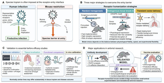
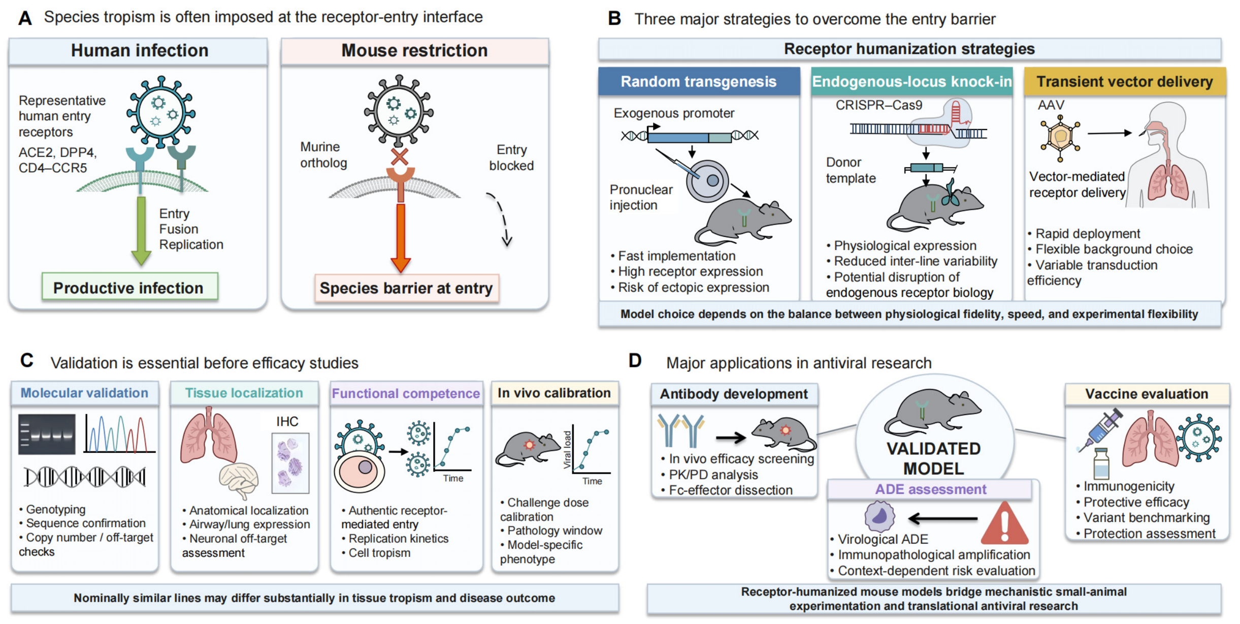
The engineering strategy chosen at the outset constrains every downstream interpretation. Random transgenesis remains fast and experimentally accessible, but copy number, insertion site, and promoter choice can create expression patterns that are useful for robust challenge yet misleading for tissue tropism. Endogenous-locus knock-in preserves more physiological regulation but may yield a narrower disease window. Conditional, inducible, and precision-editing strategies add temporal or cell-type control, and prime editing illustrates how small receptor-interface changes may eventually allow entry competence without replacing an entire gene [8,9,10,11,12,13]. This review therefore emphasizes a validation-centered framework: first define the receptor-dependent question, then choose the model architecture, and only then interpret antibody, vaccine, or pathogenesis readouts within the limits of that architecture. As shown in Figure 1, this decision path links model-engineering choices to the downstream antibody, vaccine, and variant-benchmarking questions that each platform can legitimately support.
Figure 1.
Engineering, validation, and applications of receptor-humanized mouse models. (A) Many clinically important human viruses fail to infect standard laboratory mice because the corresponding murine receptor orthologs do not support efficient viral attachment or entry. In permissive human cells, receptor engagement enables attachment, entry, and productive infection, whereas inefficient engagement of the murine ortholog creates a species barrier at the entry stage. (B) This entry barrier can be overcome through three major receptor-humanization strategies: random transgenesis, clustered regularly interspaced short palindromic repeats/CRISPR-associated protein 9 (CRISPR/Cas9)-mediated endogenous-locus knock-in, and transient adeno-associated virus (AAV)-mediated receptor delivery. These approaches differ in implementation speed, physiological fidelity, experimental flexibility, and susceptibility to artifacts such as ectopic expression or variable transduction efficiency. (C) Rigorous validation is required before efficacy studies, including molecular confirmation of the engineered allele, anatomical characterization of receptor expression, immunohistochemistry (IHC) where appropriate, functional demonstration of authentic receptor-dependent entry, and in vivo calibration of the challenge dose and pathology window. (D) Once established and validated, receptor-humanized mouse models support multiple applications in antiviral research, including in vivo antibody screening, pharmacokinetic/pharmacodynamic (PK/PD) analysis, antibody-dependent enhancement (ADE) assessment, and vaccine evaluation based on immunogenicity, protective efficacy, immune readouts, and variant benchmarking.
2. Building Receptor-Humanized Models: Design Choices That Shape Interpretation
The first practical decision is whether susceptibility should be maximized or physiological fidelity should be prioritized. Random transgenesis can rapidly produce a line in which a human receptor is expressed at high levels, often under an epithelial or broadly active promoter. This can be valuable when the goal is to establish a reproducible disease window for screening many antibody or vaccine candidates. The limitation is that the construct, the integration site, and the founder line become part of the phenotype. Two animals may carry the same receptor sequence but differ substantially in receptor abundance, tissue distribution, and disease severity because random integration places the transgene under different chromatin and regulatory influences. Classic transgenic technology therefore remains powerful, but it should be interpreted as a line-specific experimental system rather than as a direct physiological substitute for the human receptor locus [8,9,10].
Targeted knock-in and minimal interface humanization address a different problem. By placing the human receptor sequence, or selected human-like residues, into a defined genomic context, these approaches reduce founder-line variability and align expression more closely with endogenous regulatory logic. They are particularly attractive when tissue tropism, cell-type restriction, or chronic tissue injury is part of the question. The trade-off is that physiological receptor distribution may produce a milder phenotype, requiring more careful endpoint selection and larger cohorts [8]. Conditional and inducible systems introduce a further layer of experimental precision: tetracycline-responsive designs allow receptor expression to be tuned over time, while Cre-loxP strategies confine it to selected cell types or developmental windows [11,12]. Prime editing and related precision-editing approaches are conceptually important because receptor usage is often determined by a small number of interface residues; in such cases, precise nucleotide changes may create entry competence while leaving the remainder of the receptor and its regulatory architecture intact [13].
Validation is a core experimental result, not a technical appendix. At minimum, a receptor-humanized model must be characterized at the allele, RNA, protein, anatomical, and functional levels. Molecular confirmation verifies that the intended receptor sequence is present and that no obvious rearrangement has occurred; tissue-level validation asks whether the receptor is found in the relevant cell types; and functional validation asks whether infection is actually receptor dependent. This is especially important for sarbecoviruses because severe acute respiratory syndrome coronavirus (SARS-CoV) and severe acute respiratory syndrome coronavirus 2 (SARS-CoV-2) both use ACE2 but differ in receptor-binding details, protease usage, and experimental disease phenotype. Structural and entry studies therefore support the principle that SARS-CoV data cannot automatically substitute for SARS-CoV-2 data, or vice versa, when the claim concerns a pathogen-specific entry event [14,15,16,17].
A useful model-selection rule is to align the model with the narrowest claim the experiment needs to support. When the intended claim is entry blockade, high and reproducible receptor-dependent susceptibility may be acceptable even if expression is not fully physiological. When the claim is tissue tropism, disease mechanism, or safety, promoter and locus become central. When the claim concerns vaccine immunogenicity or antibody effector function, receptor humanization may need to be paired with immune-system or Fc-receptor humanization. This is why a single ranking of receptor-humanized models is misleading: the better model is the one whose engineered feature matches the biological claim and whose remaining limitations have been measured rather than assumed [6,7,8,9,10,11,12,13].
The same validation logic also explains why apparently similar hACE2 models can answer different questions. Keratin-18 promoter-driven hACE2 (K18-hACE2) mice, other transgenic hACE2 lines, endogenous hACE2 knock-in mice, adeno-associated virus (AAV)-mediated hACE2 delivery systems, and mouse-adapted SARS-CoV-2 all solve the susceptibility problem in different ways. A strong transgenic model may be excellent for rapid countermeasure ranking but may also carry promoter-driven tissue distribution that complicates interpretation. A knock-in model may better preserve receptor regulation but can produce a subtler disease window. AAV-hACE2 delivery can be deployed quickly in multiple mouse backgrounds, whereas mouse-adapted viruses bypass receptor humanization altogether and therefore answer a different question. For this reason, model reports should specify receptor locus or promoter, receptor distribution, genetic background, viral strain, challenge window, and the endpoint being used to judge protection or disease [18,19,20,21,22,23,24,25,26,27,28].
6. Vaccine Evaluation: Protection, Immune Readouts, and Variant Breadth
For vaccines, receptor-humanized mice test whether immunization prevents authentic receptor-dependent infection and disease. MERS-CoV vaccine candidates have been evaluated in hDPP4 transgenic models, and SARS-CoV-2 mRNA, adenoviral, and intranasal vaccine studies have used hACE2 or related challenge systems to quantify protection in the respiratory tract [92,93,94,95]. These models are most valuable when the vaccine claim depends on preventing infection by a virus whose host range is shaped by a human receptor. They are less informative when the primary endpoint is long-term human immune memory, HLA-restricted T-cell specificity, or mucosal architecture that is not reproduced in the mouse.
Humoral readouts should be paired with tissue and immunological endpoints. Antibody titer alone cannot distinguish durable germinal-center-derived protection from short-lived plasmablast responses, nor can it identify whether protection reflects sterilizing entry blockade, accelerated clearance, or reduced pathology. LNP-based vaccines can induce strong T follicular helper and humoral responses, and SARS-CoV-2 mRNA vaccination can generate persistent germinal-center activity in humans [96,97]. Receptor-humanized challenge models can connect such immune signatures to viral-load and pathology endpoints, but the interpretation remains bounded by murine lymphoid architecture, murine Fc receptors, and the absence of human HLA unless additional humanization is incorporated.
Vaccine safety and interpretation also depend on the type of receptor-humanized model used. A high-susceptibility model may be ideal for detecting incomplete protection because residual infection is easy to measure, but it may overrepresent pathology driven by promoter-defined receptor expression. A more physiological knock-in model may better represent tissue entry but require more sensitive virological or histological endpoints. Neither choice is universally superior. The critical point is to match the model to the vaccine question: prevention of infection, reduction of viral burden, reduction of tissue damage, comparison across variants, or mechanistic analysis of immune correlates [92,93,94,95,96,97,98,99,100,101,102].
T-cell and variant-breadth analyses reinforce the same interpretive boundary. SARS-CoV-specific memory CD8 T cells can protect against lethal challenge in mice, and SARS-CoV-2 epitope mapping in humans has defined immunodominance and immunoprevalence patterns in depth [98,99]. Mouse MHC presentation and human HLA restriction are not interchangeable, however, so T-cell conclusions from receptor-humanized models require either careful framing of their limits or incorporation of HLA/immune-system humanization. Variant benchmarking is similarly powerful but context dependent. Omicron immune escape, vaccine-induced neutralization loss across variants, and mosaic RBD nanoparticle protection against diverse sarbecoviruses show why in vivo breadth testing is needed [100,101,102]. Receptor-humanized models are one important layer of that workflow when the benchmark depends on human receptor usage, but variant studies should use the receptor and immune context most relevant to the intended clinical claim.
7. Translational Boundaries: Validation Standards and Model Limits
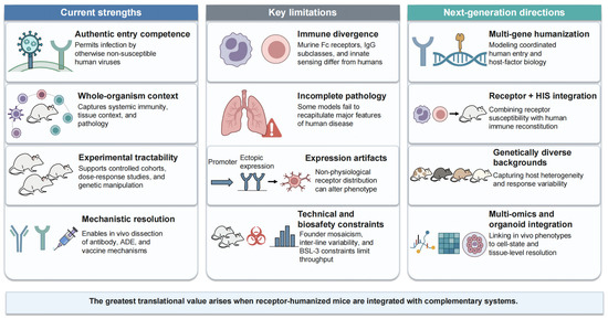
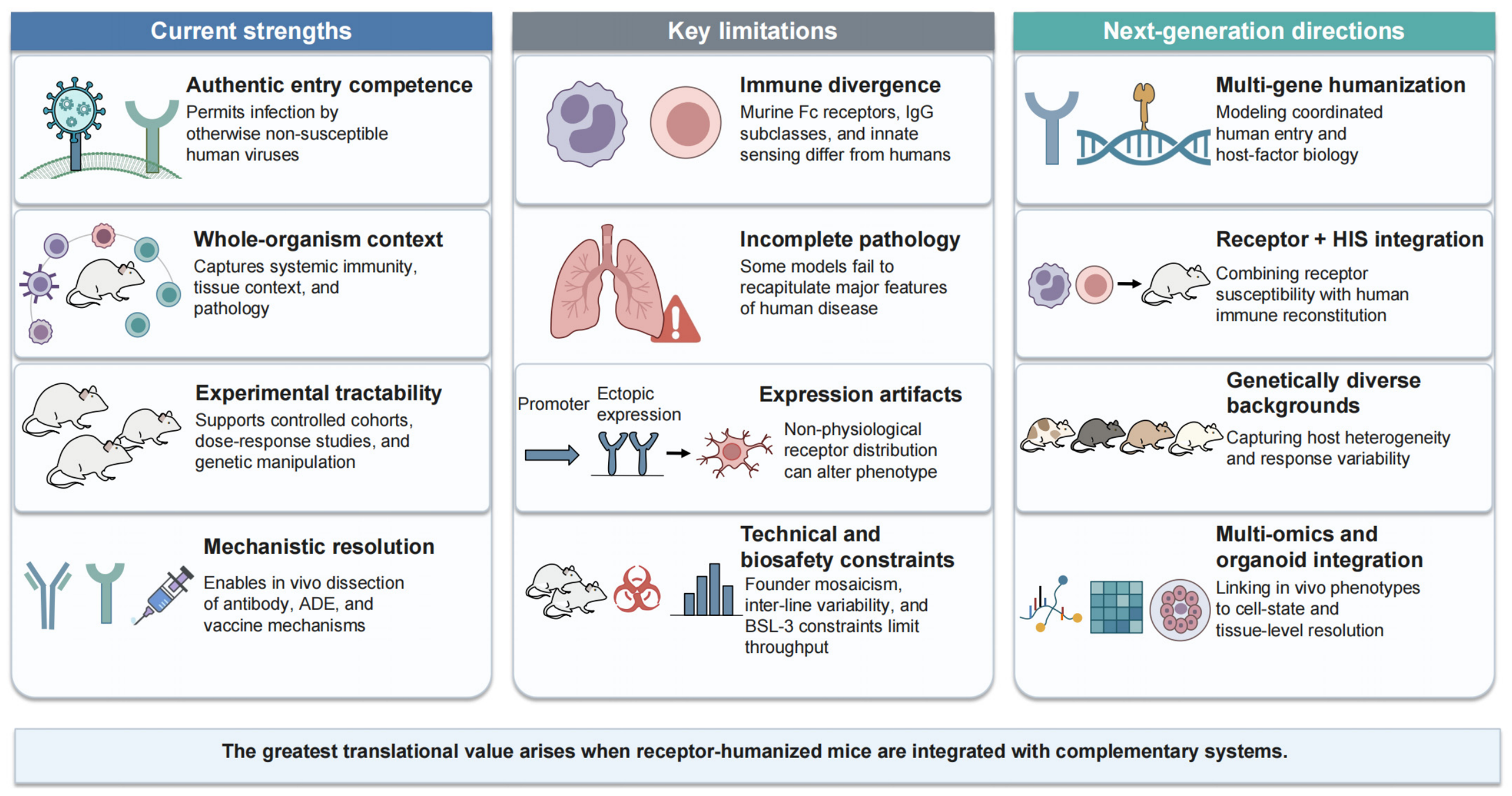
The most fundamental limitation of receptor-humanized models is precisely what makes them useful: they address the entry step specifically, but entry competence does not automatically reproduce human disease. Human liver chimeric mice demonstrate that organ-humanized platforms may be required for hepatitis virus replication and treatment studies, while human immune-system models are often necessary when vaccine responses, antibody maturation, Fc-effector mechanisms, or immune escape are the primary endpoints [103,104,105,106]. A receptor-humanized mouse should therefore be selected for the question it can answer, not for the disease label attached to the virus.
Receptor expression level, anatomical distribution, and genetic background are the main axes along which interpretation can drift. Overexpression under strong ubiquitous promoters can generate non-physiological tropism, artifactual lethality, or tissue damage that has no counterpart in the natural disease process. Endogenous knock-in configurations typically yield more physiological expression patterns, but often at the cost of a narrower intervention window. Immune background matters as well: mouse and human innate and adaptive responses differ, and host genetic background can reshape pathogenesis even when the virus and receptor are unchanged [6,7,107]. Model descriptions should therefore include receptor locus or promoter, copy number when known, cell-type distribution, age, sex, background strain, viral strain, and the validated endpoint window.
Future model development will likely move from single-receptor substitution toward layered humanization. For hepatitis viruses, liver-humanized mice already show how organ context can complement receptor engineering; for antibody and vaccine studies, immune-system humanization may be needed to evaluate human B-cell maturation, HLA-restricted T-cell responses, and human Fc-effector pathways [103,104,105,106]. At the same time, the field should avoid assuming that more humanization is always better. Each added human component can introduce its own artifacts, engraftment variability, or immunodeficient background. The most useful future systems will therefore be modular: receptor-humanized when the question is entry, organ-humanized when the question is tissue replication, and immune-humanized when the question is adaptive or effector immunity [3,6,103,104,105,106].
A practical validation checklist follows from these limitations. Investigators should ask whether the human receptor is expressed at biologically relevant sites; whether infection is receptor dependent; whether tissue tropism resembles the intended human disease feature; whether viral replication and pathology provide a measurable intervention window; whether antibody or vaccine readouts require human Fc receptors, HLA, or human immune cells; and whether complementary organoid, human cell, immune-humanized, liver-humanized, or non-human primate data are needed for confirmation [1,2,3,4,5,6,7,103,104,105,106,107]. Framed this way, receptor-humanized mice remain powerful but bounded tools: they are neither simple susceptibility switches nor complete human disease replicas.
8. Conclusions
Human viral entry receptor mouse models are best understood as a diverse, receptor-defined experimental toolkit extending far beyond hACE2/SARS-CoV-2 systems. hACE2 and hDPP4 models remain central for coronavirus countermeasures, but CD4/CCR5, hCAR, hCD46, hICAM-1, hCD81/hOCLN, hNTCP, hSLAM/CD150, PVR/CD155, hSCARB2, and hTfR1 models demonstrate that the same receptor-humanization principle applies across many medically relevant viral infections [14,15,16,17,18,19,20,21,22,23,24,25,26,27,28,29,30,31,32,33,34,35,36,37,38,39,40,41,42,43,44,45,46,47,48,49,50,51,52,53,54,55,56,57,58,59,60,61,62,63,64,65,66,67,68,69,70,71,72,73,74,75,76,77,78,79,80]. Several limitations of this review and the underlying mouse models should be acknowledged. First, although the scope of this review was deliberately extended beyond coronaviruses, both the available evidence and the depth of discussion remain weighted toward hACE2/SARS-CoV-2 and hDPP4/MERS-CoV models, which are more extensively developed than most non-coronavirus systems. The cross-virus comparisons are therefore qualitative and should not be interpreted as an exhaustive inventory or quantitative ranking. Second, as a narrative review, this article relies on targeted literature selection and does not include a systematic risk-of-bias or certainty-of-evidence assessment; selection and publication bias cannot be excluded. Third, receptor-humanized mice reproduce a defined entry barrier rather than the complete human disease state. Murine immune signaling, Fc receptor and IgG subclass biology, HLA-restricted antigen presentation, stromal and tissue-repair programs, and organ physiology remain incompletely modeled. In addition, promoter choice, transgene copy number, ectopic expression, founder effects, age, sex, genetic background, challenge dose, viral adaptation, and endpoint selection can materially influence apparent efficacy. These limitations should be considered when extrapolating antibody- or vaccine-protection data to humans.
The practical lesson from the available models is that engineering strategy determines interpretation. Random transgenic systems can provide rapid and robust susceptibility, but may introduce promoter- and copy-number-driven artifacts. Endogenous knock-in or minimal receptor-interface editing can better preserve physiological expression, but may produce a narrower disease window and may not remove post-entry species restrictions. Conditional, inducible, and transient expression strategies add flexibility, yet they also require careful controls for timing, tissue distribution, and expression level [8,9,10,11,12,13,18,19,20,21,22,23,24,25,26,27,28,31,32,33,34,35,36,37]. No platform is intrinsically superior to another; each is scientifically defensible only when its construction, receptor localization, infection phenotype, and endpoint window are all aligned with the specific experimental question being asked.
For antiviral antibody and vaccine development, the strongest use of receptor-humanized mice is not to claim comprehensive human disease mimicry, but to test specific entry-dependent hypotheses under controlled in vivo conditions. These models can connect in vitro neutralization to reductions in tissue viral load and pathology, Fc-effector function, and challenge protection, and they are particularly useful when candidate antibodies or vaccines target a human receptor-dependent entry pathway [81,82,83,84,85,86,87,88,89,90,91,92,93,94,95,96,97,98,99,100,101,102,103,104,105,106]. Their translational value therefore depends on restraint as much as breadth: investigators should state exactly which receptor-dependent question the model can answer and identify which human immune or organ-level features require complementary systems.
Future progress will likely come from layered humanization rather than from simply adding more receptor-transgenic lines. Combining receptor-humanized mice with human immune-system reconstitution, organ-humanized platforms, HLA or Fc-receptor humanization, organoids, primary human cell systems, and, when necessary, non-human primate studies will allow entry competence to be interpreted within a more clinically relevant immune and tissue context [1,2,3,4,5,6,7,103,104,105,106,107]. Equally indispensable is transparent reporting: manuscripts should specify the receptor allele design, promoter or locus, copy number where known, tissue distribution, animal background, challenge virus, endpoint window, and—critically—the explicit rationale for selecting that particular model over available alternatives. Used in this disciplined way, human viral entry receptor mouse models can serve as a rigorous bridge between reductionist cell-culture assays and later-stage animal or clinical studies, while avoiding the overinterpretation that has sometimes limited confidence in this field.
Author Contributions
Conceptualization, N.Z., R.I., D.R. and A.H.; methodology, N.Z., X.Z. and R.I.; investigation, N.Z., X.Z. and R.I.; resources, D.R. and A.H.; writing—original draft preparation, N.Z. and X.Z.; writing—review and editing, N.Z., X.Z., R.I., D.R. and A.H.; supervision, X.Z. and A.H.; funding acquisition, D.R. and A.H. All authors have read and agreed to the published version of the manuscript.
Funding
This research was funded by the Fundamental Research Funds for the Central Universities, grant number 2026300391.
Data Availability Statement
No new data were created or analyzed in this study. Data sharing is not applicable to this article.
Conflicts of Interest
The authors declare no conflicts of interest.
References
- Masemann, D.; Ludwig, S.; Planz, O. Advances in transgenic mouse models to study infections by human pathogenic viruses. Int. J. Mol. Sci. 2020, 21, 9289. [Google Scholar] [CrossRef] [Scilit] [PubMed]
- Muñoz-Fontela, C.; Dowling, W.E.; Funnell, S.G.P.; Gsell, P.S.; Riveros-Balta, A.X.; Albrecht, R.A.; Andersen, H.; Baric, R.S.; Carroll, M.W.; Cavaleri, M.; et al. Animal models for COVID-19. Nature 2020, 586, 509–515. [Google Scholar] [CrossRef] [Scilit] [PubMed]
- Wahl, A.; Garcia, J.V. Humanized mouse systems to study viral infection: A new era in immunology research. Annu. Rev. Immunol. 2025, 43, 143–167. [Google Scholar] [CrossRef] [Scilit] [PubMed]
- Douam, F.; Ploss, A. The use of humanized mice for studies of viral pathogenesis and immunity. Curr. Opin. Virol. 2018, 29, 62–71. [Google Scholar] [CrossRef] [Scilit] [PubMed]
- Lai, F.; Chen, Q. Humanized mouse models for the study of infection and pathogenesis. Viruses 2018, 10, 643. [Google Scholar] [CrossRef] [Scilit] [PubMed]
- Shultz, L.D.; Brehm, M.A.; Garcia-Martinez, J.V.; Greiner, D.L. Humanized mice for immune system investigation: Progress, promise and challenges. Nat. Rev. Immunol. 2012, 12, 786–798. [Google Scholar] [CrossRef] [Scilit] [PubMed]
- Mestas, J.; Hughes, C.C.W. Of mice and not men: Differences between mouse and human immunology. J. Immunol. 2004, 172, 2731–2738. [Google Scholar] [CrossRef] [Scilit] [PubMed]
- Gurumurthy, C.B.; Lloyd, K.C.K. Generating mouse models for biomedical research: Technological advances. Dis. Model. Mech. 2019, 12, dmm029462. [Google Scholar] [CrossRef] [Scilit] [PubMed]
- Gordon, J.W.; Ruddle, F.H. Integration and stable germ line transmission of genes injected into mouse pronuclei. Science 1981, 214, 1244–1246. [Google Scholar] [CrossRef] [Scilit] [PubMed]
- Palmiter, R.D.; Brinster, R.L. Germ-line transformation of mice. Annu. Rev. Genet. 1986, 20, 465–499. [Google Scholar] [CrossRef] [Scilit] [PubMed]
- Gossen, M.; Bujard, H. Tight control of gene expression in mammalian cells by tetracycline-responsive promoters. Proc. Natl. Acad. Sci. USA 1992, 89, 5547–5551. [Google Scholar] [CrossRef] [Scilit] [PubMed]
- Feil, R.; Brocard, J.; Mascrez, B.; LeMeur, M.; Metzger, D.; Chambon, P. Ligand-activated site-specific recombination in mice. Proc. Natl. Acad. Sci. USA 1996, 93, 10887–10890. [Google Scholar] [CrossRef] [Scilit] [PubMed]
- Anzalone, A.V.; Randolph, P.B.; Davis, J.R.; Sousa, A.A.; Koblan, L.W.; Levy, J.M.; Chen, P.J.; Wilson, C.; Newby, G.A.; Raguram, A.; et al. Search-and-replace genome editing without double-strand breaks or donor DNA. Nature 2019, 576, 149–157. [Google Scholar] [CrossRef] [Scilit] [PubMed]
- Li, F.; Li, W.; Farzan, M.; Harrison, S.C. Structure of SARS coronavirus spike receptor-binding domain complexed with receptor. Science 2005, 309, 1864–1868. [Google Scholar] [CrossRef] [Scilit] [PubMed]
- Hoffmann, M.; Kleine-Weber, H.; Schroeder, S.; Krüger, N.; Herrler, T.; Erichsen, S.; Schiergens, T.S.; Herrler, G.; Wu, N.-H.; Nitsche, A.; et al. SARS-CoV-2 cell entry depends on ACE2 and TMPRSS2 and is blocked by a clinically proven protease inhibitor. Cell 2020, 181, 271–280.e8. [Google Scholar] [CrossRef] [Scilit] [PubMed]
- Lan, J.; Ge, J.; Yu, J.; Shan, S.; Zhou, H.; Fan, S.; Zhang, Q.; Shi, X.; Wang, Q.; Zhang, L.; et al. Structure of the SARS-CoV-2 spike receptor-binding domain bound to the ACE2 receptor. Nature 2020, 581, 215–220. [Google Scholar] [CrossRef] [Scilit] [PubMed]
- Shang, J.; Ye, G.; Shi, K.; Wan, Y.; Luo, C.; Aihara, H.; Geng, Q.; Auerbach, A.; Li, F. Structural basis of receptor recognition by SARS-CoV-2. Nature 2020, 581, 221–224. [Google Scholar] [CrossRef] [Scilit] [PubMed]
- McCray, P.B., Jr.; Pewe, L.; Wohlford-Lenane, C.; Hickey, M.; Manzel, L.; Shi, L.; Netland, J.; Jia, H.P.; Halabi, C.; Sigmund, C.D.; et al. Lethal infection of K18-hACE2 mice infected with severe acute respiratory syndrome coronavirus. J. Virol. 2007, 81, 813–821. [Google Scholar] [CrossRef] [Scilit] [PubMed]
- Bao, L.; Deng, W.; Huang, B.; Gao, H.; Liu, J.; Ren, L.; Wei, Q.; Yu, P.; Xu, Y.; Qi, F.; et al. The pathogenicity of SARS-CoV-2 in hACE2 transgenic mice. Nature 2020, 583, 830–833. [Google Scholar] [CrossRef] [Scilit] [PubMed]
- Jiang, R.-D.; Liu, M.-Q.; Chen, Y.; Shan, C.; Zhou, Y.-W.; Shen, X.-R.; Li, Q.; Zhang, L.; Zhu, Y.; Si, H.-R.; et al. Pathogenesis of SARS-CoV-2 in transgenic mice expressing human angiotensin-converting enzyme 2. Cell 2020, 182, 50–58.e8. [Google Scholar] [CrossRef] [Scilit] [PubMed]
- Winkler, E.S.; Bailey, A.L.; Kafai, N.M.; Nair, S.; McCune, B.T.; Yu, J.; Fox, J.M.; Chen, R.E.; Earnest, J.T.; Keeler, S.P.; et al. SARS-CoV-2 infection of human ACE2-transgenic mice causes severe lung inflammation and impaired function. Nat. Immunol. 2020, 21, 1327–1335. [Google Scholar] [CrossRef] [Scilit] [PubMed]
- Oladunni, F.S.; Park, J.-G.; Pino, P.A.; Gonzalez, O.; Akhter, A.; Allué-Guardia, A.; Olmo-Fontánez, A.; Gautam, S.; Garcia-Vilanova, A.; Ye, C.; et al. Lethality of SARS-CoV-2 infection in K18 human angiotensin-converting enzyme 2 transgenic mice. Nat. Commun. 2020, 11, 6122. [Google Scholar] [CrossRef] [Scilit] [PubMed]
- Sun, S.-H.; Chen, Q.; Gu, H.-J.; Yang, G.; Wang, Y.-X.; Huang, X.-Y.; Liu, S.-S.; Zhang, N.-N.; Li, X.-F.; Xiong, R.; et al. A mouse model of SARS-CoV-2 infection and pathogenesis. Cell Host Microbe 2020, 28, 124–133.e4. [Google Scholar] [CrossRef] [Scilit] [PubMed]
- Zhou, X.; Sun, W.; Zhang, Y.; Gu, H.; Wang, R.; Xie, P.; Zhu, Y.; Qiu, M.; Ding, X.; Wang, H.; et al. A novel hACE2 knock-in mouse model recapitulates pulmonary and intestinal SARS-CoV-2 infection. Front. Microbiol. 2023, 14, 1175188. [Google Scholar] [CrossRef] [Scilit] [PubMed]
- Song, I.W.; Washington, M.; Leynes, C.; Hsu, J.; Rayavara, K.; Bae, Y.; Haelterman, N.; Chen, Y.; Jiang, M.M.; Drelich, A.; et al. Generation of a humanized mAce2 and a conditional hACE2 mouse models permissive to SARS-CoV-2 infection. Mamm. Genome 2024, 35, 113–121. [Google Scholar] [CrossRef] [Scilit] [PubMed]
- Liu, G.; Zhang, M.; Wu, B.; Zhang, C.; Wang, Y.; Han, X.; Wang, R.; Li, L.; Wei, Y.; Sun, Y.; et al. A highly susceptible hACE2-transgenic mouse model for SARS-CoV-2 research. Front. Microbiol. 2024, 15, 1348405. [Google Scholar] [CrossRef] [Scilit] [PubMed]
- Israelow, B.; Song, E.; Mao, T.; Lu, P.; Meir, A.; Liu, F.; Alfajaro, M.M.; Wei, J.; Dong, H.; Homer, R.J.; et al. Mouse model of SARS-CoV-2 reveals inflammatory role of type I interferon signaling. J. Exp. Med. 2020, 217, e20201241. [Google Scholar] [CrossRef] [Scilit] [PubMed]
- Dinnon, K.H., III; Leist, S.R.; Schäfer, A.; Edwards, C.E.; Martinez, D.R.; Montgomery, S.A.; West, A.; Yount, B.L., Jr.; Hou, Y.J.; Adams, L.E.; et al. A mouse-adapted model of SARS-CoV-2 to test COVID-19 countermeasures. Nature 2020, 586, 560–566. [Google Scholar] [CrossRef] [Scilit] [PubMed]
- Raj, V.S.; Mou, H.; Smits, S.L.; Dekkers, D.H.W.; Müller, M.A.; Dijkman, R.; Muth, D.; Demmers, J.A.A.; Zaki, A.; Fouchier, R.A.M.; et al. Dipeptidyl peptidase 4 is a functional receptor for the emerging human coronavirus-EMC. Nature 2013, 495, 251–254. [Google Scholar] [CrossRef] [Scilit] [PubMed]
- Cockrell, A.S.; Peck, K.M.; Yount, B.L.; Agnihothram, S.S.; Scobey, T.; Curnes, N.R.; Baric, R.S.; Heise, M.T. Mouse dipeptidyl peptidase 4 is not a functional receptor for Middle East respiratory syndrome coronavirus infection. J. Virol. 2014, 88, 5195–5199. [Google Scholar] [CrossRef] [Scilit] [PubMed]
- Zhao, J.; Li, K.; Wohlford-Lenane, C.; Agnihothram, S.S.; Fett, C.; Zhao, J.; Gale, M.J., Jr.; Baric, R.S.; Enjuanes, L.; Gallagher, T.; et al. Rapid generation of a mouse model for Middle East respiratory syndrome. Proc. Natl. Acad. Sci. USA 2014, 111, 4970–4975. [Google Scholar] [CrossRef] [Scilit] [PubMed]
- Agrawal, A.S.; Garron, T.; Tao, X.; Peng, B.-H.; Wakamiya, M.; Chan, T.-S.; Couch, R.B.; Tseng, C.-T.K. Generation of a transgenic mouse model of Middle East respiratory syndrome coronavirus infection and disease. J. Virol. 2015, 89, 3659–3670. [Google Scholar] [CrossRef] [Scilit] [PubMed]
- Zhao, G.; Jiang, Y.; Qiu, H.; Gao, T.; Zeng, Y.; Guo, Y.; Yu, H.; Li, J.; Kou, Z.; Du, L.; et al. Multi-organ damage in human dipeptidyl peptidase 4 transgenic mice infected with Middle East respiratory syndrome-coronavirus. PLoS ONE 2015, 10, e0145561. [Google Scholar] [CrossRef] [Scilit] [PubMed]
- Li, K.; Wohlford-Lenane, C.; Perlman, S.; Zhao, J.; Jewell, A.K.; Reznikov, L.R.; Gibson-Corley, K.N.; Meyerholz, D.K.; McCray, P.B., Jr. Middle East respiratory syndrome coronavirus causes multiple organ damage and lethal disease in mice transgenic for human dipeptidyl peptidase 4. J. Infect. Dis. 2016, 213, 712–722. [Google Scholar] [PubMed]
- Cockrell, A.S.; Yount, B.L.; Scobey, T.; Jensen, K.; Douglas, M.; Beall, A.; Tang, X.C.; Marasco, W.A.; Heise, M.T.; Baric, R.S. A mouse model for MERS coronavirus-induced acute respiratory distress syndrome. Nat. Microbiol. 2016, 2, 16226. [Google Scholar] [CrossRef] [Scilit] [PubMed]
- Li, K.; Wohlford-Lenane, C.L.; Channappanavar, R.; Park, J.-E.; Earnest, J.T.; Bair, T.B.; Bates, A.M.; Brogden, K.A.; Flaherty, H.A.; Gallagher, T.; et al. Mouse-adapted MERS coronavirus causes lethal lung disease in human DPP4 knockin mice. Proc. Natl. Acad. Sci. USA 2017, 114, E3119–E3128. [Google Scholar] [CrossRef] [Scilit] [PubMed]
- Fan, C.; Wu, X.; Liu, Q.; Li, Q.; Liu, S.; Lu, J.; Yang, Y.; Cao, Y.; Huang, W.; Liang, C.; et al. A human DPP4-knockin mouse’s susceptibility to infection by authentic and pseudotyped MERS-CoV. Viruses 2018, 10, 448. [Google Scholar] [CrossRef] [Scilit] [PubMed]
- Browning, J.; Horner, J.W.; Pettoello-Mantovani, M.; Raker, C.; Yurasov, S.; DePinho, R.A.; Goldstein, H. Mice transgenic for human CD4 and CCR5 are susceptible to HIV infection. Proc. Natl. Acad. Sci. USA 1997, 94, 14637–14641. [Google Scholar] [CrossRef] [Scilit] [PubMed]
- Seay, K.; Qi, X.; Zheng, J.H.; Zhang, C.; Chen, K.; Dutta, M.; Deneroff, K.; Ochsenbauer, C.; Kappes, J.C.; Littman, D.R.; et al. Mice transgenic for CD4-specific human CD4, CCR5 and cyclin T1 expression: A new model for investigating HIV-1 transmission and treatment efficacy. PLoS ONE 2013, 8, e63537. [Google Scholar] [CrossRef] [Scilit] [PubMed]
- Garcia, J.V. Humanized mice for HIV and AIDS research. Curr. Opin. Virol. 2016, 19, 56–64. [Google Scholar] [CrossRef] [Scilit]
- Gillgrass, A.; Wessels, J.M.; Yang, J.X.; Kaushic, C. Advances in humanized mouse models to improve understanding of HIV-1 pathogenesis and immune responses. Front. Immunol. 2021, 11, 617516. [Google Scholar] [CrossRef] [Scilit] [PubMed]
- Wan, Y.Y.; Leon, R.P.; Marks, R.; Cham, C.M.; Schaack, J.; Gajewski, T.F.; DeGregori, J. Transgenic expression of the coxsackie/adenovirus receptor enables adenoviral-mediated gene delivery in naïve T cells. Proc. Natl. Acad. Sci. USA 2000, 97, 13784–13789. [Google Scholar] [CrossRef] [Scilit] [PubMed]
- Zhang, Y.; Bergelson, J.M. Adenovirus receptors. J. Virol. 2005, 79, 12125–12131. [Google Scholar] [CrossRef] [Scilit] [PubMed]
- Verhaagh, S.; de Jong, E.; Goudsmit, J.; Lecollinet, S.; Gillissen, G.; de Swart, R.L.; Rimmelzwaan, G.F.; Osterhaus, A.D.M.E. Human CD46-transgenic mice in studies involving replication-incompetent adenoviral type 35 vectors. J. Gen. Virol. 2006, 87, 255–265. [Google Scholar] [CrossRef] [Scilit] [PubMed]
- Bartlett, N.W.; Walton, R.P.; Edwards, M.R.; Aniscenko, J.; Caramori, G.; Zhu, J.; Glanville, N.; Choy, K.J.; Jourdan, P.; Burnet, J.; et al. Mouse models of rhinovirus-induced disease and exacerbation of allergic airway inflammation. Nat. Med. 2008, 14, 199–204. [Google Scholar] [CrossRef] [Scilit] [PubMed]
- Traub, S.; Nikonova, A.; Carruthers, A.; Dunmore, R.; Vousden, K.A.; Gogsadze, L.; Hao, W.; Zhu, Q.; Bernard, K.; Zhu, J.; et al. An anti-human ICAM-1 antibody inhibits rhinovirus-induced exacerbations of lung inflammation. PLoS Pathog. 2013, 9, e1003520. [Google Scholar] [CrossRef] [Scilit] [PubMed]
- Ploss, A.; Evans, M.J.; Gaysinskaya, V.A.; Panis, M.; You, H.; de Jong, Y.P.; Rice, C.M. Human occludin is a hepatitis C virus entry factor required for infection of mouse cells. Nature 2009, 457, 882–886. [Google Scholar] [CrossRef] [Scilit] [PubMed]
- Dorner, M.; Horwitz, J.A.; Robbins, J.B.; Barry, W.T.; Feng, Q.; Mu, K.; Jones, C.T.; Schoggins, J.W.; Catanese, M.T.; Burton, D.R.; et al. A genetically humanized mouse model for hepatitis C virus infection. Nature 2011, 474, 208–211. [Google Scholar] [CrossRef] [Scilit] [PubMed]
- Dorner, M.; Horwitz, J.A.; Donovan, B.M.; Labitt, R.N.; Budell, W.C.; Friling, T.; Vogt, A.; Catanese, M.T.; Satoh, T.; Kawai, T.; et al. Completion of the entire hepatitis C virus life cycle in genetically humanized mice. Nature 2013, 501, 237–241. [Google Scholar] [CrossRef] [Scilit] [PubMed]
- Dorner, M.; Rice, C.M.; Ploss, A. Study of hepatitis C virus entry in genetically humanized mice. Methods 2013, 59, 249–257. [Google Scholar] [CrossRef] [Scilit] [PubMed]
- Ding, Q.; von Schaewen, M.; Hrebikova, G.; Heller, B.; Sandmann, L.; Plaas, M.; Ploss, A. Mice expressing minimally humanized CD81 and occludin genes support hepatitis C virus uptake in vivo. J. Virol. 2017, 91, e01799-16. [Google Scholar] [CrossRef] [Scilit] [PubMed]
- Giang, E.; Dorner, M.; Prentoe, J.C.; Dreux, M.; Evans, M.J.; Bukh, J.; Rice, C.M.; Ploss, A.; Burton, D.R.; Law, M. Human broadly neutralizing antibodies to the envelope glycoprotein complex of hepatitis C virus. Proc. Natl. Acad. Sci. USA 2012, 109, 6205–6210. [Google Scholar] [CrossRef] [Scilit] [PubMed]
- de Jong, Y.P.; Dorner, M.; Mommersteeg, M.C.; Xiao, J.W.; Balazs, A.B.; Robbins, J.B.; Winer, B.Y.; Gerges, S.; Vega, K.; Labitt, R.N.; et al. Broadly neutralizing antibodies abrogate established hepatitis C virus infection. Sci. Transl. Med. 2014, 6, 254ra129. [Google Scholar] [CrossRef] [Scilit] [PubMed]
- Yan, H.; Zhong, G.; Xu, G.; He, W.; Jing, Z.; Gao, Z.; Huang, Y.; Qi, Y.; Peng, B.; Wang, H.; et al. Sodium taurocholate cotransporting polypeptide is a functional receptor for human hepatitis B and D virus. eLife 2012, 1, e00049. [Google Scholar] [CrossRef] [Scilit] [PubMed]
- He, W.; Ren, B.; Mao, F.; Jing, Z.; Li, Y.; Liu, Y.; Peng, B.; Yan, H.; Qi, Y.; Sun, Y.; et al. Hepatitis D virus infection of mice expressing human sodium taurocholate co-transporting polypeptide. PLoS Pathog. 2015, 11, e1004840. [Google Scholar] [CrossRef] [Scilit] [PubMed]
- He, W.; Cao, Z.; Mao, F.; Ren, B.; Li, Y.; Li, D.; Li, H.; Peng, B.; Yan, H.; Qi, Y.; et al. Modification of three amino acids in sodium taurocholate cotransporting polypeptide renders mice susceptible to infection with hepatitis D virus in vivo. J. Virol. 2016, 90, 8866–8874. [Google Scholar] [CrossRef] [Scilit] [PubMed]
- Lempp, F.A.; Wiedtke, E.; Qu, B.; Roques, P.; Chemin, I.; Vondran, F.W.R.; Le Grand, R.; Grimm, D.; Urban, S. Sodium taurocholate cotransporting polypeptide is the limiting host factor of hepatitis B virus infection in macaque and pig hepatocytes. Hepatology 2017, 66, 703–716. [Google Scholar] [CrossRef] [Scilit] [PubMed]
- Giersch, K.; Hermanussen, L.; Volz, T.; Kah, J.; Allweiss, L.; Casey, J.; Sureau, C.; Dandri, M.; Lütgehetmann, M. Murine hepatocytes do not support persistence of hepatitis D virus mono-infection in vivo. Liver Int. 2021, 41, 410–419. [Google Scholar] [PubMed]
- Wettengel, J.M.; Burwitz, B.J. Innovative HBV animal models based on the entry receptor NTCP. Viruses 2020, 12, 828. [Google Scholar] [CrossRef] [Scilit] [PubMed]
- Horvat, B.; Rivailler, P.; Varior-Krishnan, G.; Cardoso, A.; Gerlier, D.; Rabourdin-Combe, C. Transgenic mice expressing human measles virus receptor CD46 provide cells exhibiting different permissivities to measles virus infection. J. Virol. 1996, 70, 6673–6681. [Google Scholar] [CrossRef] [Scilit] [PubMed]
- Rall, G.F.; Manchester, M.; Daniels, L.R.; Callahan, E.M.; Belman, A.R.; Oldstone, M.B.A. A transgenic mouse model for measles virus infection of the brain. Proc. Natl. Acad. Sci. USA 1997, 94, 4659–4663. [Google Scholar] [CrossRef] [Scilit] [PubMed]
- Oldstone, M.B.A.; Lewicki, H.; Thomas, D.; Tishon, A.; Dales, S.; Patterson, J.; Manchester, M.; Homann, D.; Naniche, D.; Holz, A.; et al. Measles virus infection in a transgenic model: Virus-induced immunosuppression and central nervous system disease. Cell 1999, 98, 629–640. [Google Scholar] [CrossRef] [Scilit] [PubMed]
- Sellin, C.I.; Davoust, N.; Guillaume, V.; Baas, D.; Buckland, R.; Wild, T.F.; Horvat, B. High pathogenicity of wild-type measles virus infection in CD150 (SLAM) transgenic mice. J. Virol. 2006, 80, 6420–6429. [Google Scholar] [CrossRef] [Scilit] [PubMed]
- Ohno, S.; Ono, N.; Takeda, M.; Takeuchi, K.; Yanagi, Y. Measles virus infection of SLAM (CD150) knockin mice reproduces tropism and immunosuppression. J. Virol. 2007, 81, 1650–1659. [Google Scholar] [CrossRef] [Scilit] [PubMed]
- Ren, R.; Costantini, F.; Gorgacz, E.J.; Lee, J.J.; Racaniello, V.R. Transgenic mice expressing a human poliovirus receptor: A new model for poliomyelitis. Cell 1990, 63, 353–362. [Google Scholar] [CrossRef] [Scilit] [PubMed]
- Koike, S.; Taya, C.; Kurata, T.; Abe, S.; Ise, I.; Yonekawa, H.; Nomoto, A. Transgenic mice susceptible to poliovirus. Proc. Natl. Acad. Sci. USA 1991, 88, 951–955. [Google Scholar] [CrossRef] [Scilit] [PubMed]
- Horie, H.; Koike, S.; Kurata, T.; Sato-Yoshida, Y.; Ise, I.; Ota, Y.; Abe, S.; Hioki, K.; Kato, H.; Taya, C.; et al. Transgenic mice carrying the human poliovirus receptor: New animal model for study of poliovirus neurovirulence. J. Virol. 1994, 68, 681–688. [Google Scholar] [CrossRef] [Scilit] [PubMed]
- Abe, S.; Ota, Y.; Koike, S.; Kurata, T.; Horie, H.; Nomura, T.; Hashizume, S.; Nomoto, A. Neurovirulence test for oral live poliovaccines using poliovirus-sensitive transgenic mice. Virology 1995, 206, 1075–1083. [Google Scholar] [CrossRef] [Scilit] [PubMed]
- Ohka, S.; Igarashi, H.; Nagata, N.; Sakai, M.; Koike, S.; Nochi, T.; Kiyono, H.; Nomoto, A. Establishment of a poliovirus oral infection system in human poliovirus receptor-expressing transgenic mice that are deficient in alpha/beta interferon receptor. J. Virol. 2007, 81, 7902–7912. [Google Scholar] [CrossRef] [Scilit] [PubMed]
- Yamayoshi, S.; Yamashita, Y.; Li, J.; Hanagata, N.; Minowa, T.; Takemura, T.; Koike, S. Scavenger receptor B2 is a cellular receptor for enterovirus 71. Nat. Med. 2009, 15, 798–801. [Google Scholar] [CrossRef] [Scilit] [PubMed]
- Lin, Y.-W.; Yu, S.-L.; Shao, H.-Y.; Lin, H.-Y.; Liu, C.-C.; Hsiao, K.-N.; Chitra, E.; Tsou, Y.-L.; Chang, H.-W.; Sia, C.; et al. Human SCARB2 transgenic mice as an infectious animal model for enterovirus 71. PLoS ONE 2013, 8, e57591. [Google Scholar] [CrossRef] [Scilit] [PubMed]
- Liou, A.-T.; Wu, S.-Y.; Liao, C.-C.; Chang, Y.-S.; Chang, C.-S.; Shih, C. A new animal model containing human SCARB2 and lacking stat-1 is highly susceptible to EV71. Sci. Rep. 2016, 6, 31151. [Google Scholar] [CrossRef] [Scilit] [PubMed]
- Yang, C.-H.; Liang, C.-T.; Jiang, S.-T.; Chen, K.-H.; Yang, C.-C.; Cheng, M.-L.; Ho, H.-Y.; Jong, Y.-J.; Lin, T.-Y.; Chiu, C.-H.; et al. A novel murine model expressing a chimeric mSCARB2/hSCARB2 receptor is highly susceptible to oral infection with clinical isolates of enterovirus 71. J. Virol. 2019, 93, e00183-19. [Google Scholar] [CrossRef] [Scilit] [PubMed]
- Wu, C.-Y.; Lin, Y.-W.; Kuo, C.-H.; Liu, W.-H.; Tai, H.-F.; Pan, C.-H.; Chen, Y.-T.; Hsiao, P.-W.; Chan, C.-H.; Chang, C.-C.; et al. Inactivated enterovirus 71 vaccine produced by 200-L scale serum-free microcarrier bioreactor system provides cross-protective efficacy in human SCARB2 transgenic mouse. PLoS ONE 2015, 10, e0136420. [Google Scholar] [CrossRef] [Scilit] [PubMed]
- Radoshitzky, S.R.; Abraham, J.; Spiropoulou, C.F.; Kuhn, J.H.; Nguyen, D.; Li, W.; Nagel, J.; Schmidt, P.J.; Nunberg, J.H.; Andrews, N.C.; et al. Transferrin receptor 1 is a cellular receptor for New World haemorrhagic fever arenaviruses. Nature 2007, 446, 92–96. [Google Scholar] [CrossRef] [Scilit] [PubMed]
- Radoshitzky, S.R.; Kuhn, J.H.; Spiropoulou, C.F.; Albariño, C.G.; Nguyen, D.P.; Salazar-Bravo, J.; Dorfman, T.; Lee, A.S.; Wang, E.; Ross, S.R.; et al. Receptor determinants of zoonotic transmission of New World hemorrhagic fever arenaviruses. Proc. Natl. Acad. Sci. USA 2008, 105, 2664–2669. [Google Scholar] [CrossRef] [Scilit] [PubMed]
- Abraham, J.; Kwong, J.A.; Albariño, C.G.; Lu, J.G.; Radoshitzky, S.R.; Salazar-Bravo, J.; Farzan, M.; Spiropoulou, C.F.; Choe, H. Host-species transferrin receptor 1 orthologs are cellular receptors for nonpathogenic New World clade B arenaviruses. PLoS Pathog. 2009, 5, e1000358. [Google Scholar] [CrossRef] [Scilit] [PubMed]
- Hickerson, B.T.; Sefing, E.J.; Bailey, K.W.; Van Wettere, A.J.; Penichet, M.L.; Gowen, B.B. Type I interferon underlies severe disease associated with Junín virus infection in mice. eLife 2020, 9, e55352. [Google Scholar] [CrossRef] [Scilit] [PubMed]
- Hickerson, B.T.; Daniels-Wells, T.R.; Payes, C.; Clark, L.E.; Candelaria, P.V.; Bailey, K.W.; Sefing, E.J.; Zink, S.; Ziegenbein, J.; Abraham, J.; et al. Host receptor-targeted therapeutic approach to counter pathogenic New World mammarenavirus infections. Nat. Commun. 2022, 13, 558. [Google Scholar] [CrossRef] [Scilit] [PubMed]
- Iyer, K.; Zeltina, A.; Canard, B.; Gutsche, I. Entry inhibitors as arenavirus antivirals. Front. Microbiol. 2024, 15, 1382953. [Google Scholar] [CrossRef] [Scilit] [PubMed]
- Hansen, J.; Baum, A.; Pascal, K.E.; Russo, V.; Giordano, S.; Wloga, E.; Fulton, B.O.; Yan, Y.; Koon, K.; Patel, K.; et al. Studies in humanized mice and convalescent humans yield a SARS-CoV-2 antibody cocktail. Science 2020, 369, 1010–1014. [Google Scholar] [CrossRef] [Scilit] [PubMed]
- Baum, A.; Fulton, B.O.; Wloga, E.; Copin, R.; Pascal, K.E.; Russo, V.; Giordano, S.; Lanza, K.; Negron, N.; Ni, M.; et al. Antibody cocktail to SARS-CoV-2 spike protein prevents rapid mutational escape seen with individual antibodies. Science 2020, 369, 1014–1018. [Google Scholar] [CrossRef] [Scilit] [PubMed]
- Barnes, C.O.; Jette, C.A.; Abernathy, M.E.; Dam, K.-M.A.; Esswein, S.R.; Gristick, H.B.; Malyutin, A.G.; Sharaf, N.G.; Huey-Tubman, K.E.; Lee, Y.E.; et al. SARS-CoV-2 neutralizing antibody structures inform therapeutic strategies. Nature 2020, 588, 682–687. [Google Scholar] [CrossRef] [Scilit] [PubMed]
- Pinto, D.; Park, Y.-J.; Beltramello, M.; Walls, A.C.; Tortorici, M.A.; Bianchi, S.; Jaconi, S.; Culap, K.; Zatta, F.; De Marco, A.; et al. Cross-neutralization of SARS-CoV-2 by a human monoclonal SARS-CoV antibody. Nature 2020, 583, 290–295. [Google Scholar] [CrossRef] [Scilit] [PubMed]
- Corti, D.; Zhao, J.; Pedotti, M.; Simonelli, L.; Agnihothram, S.; Fett, C.; Fernandez-Rodriguez, B.; Foglierini, M.; Agatic, G.; Vanzetta, F.; et al. Prophylactic and postexposure efficacy of a potent human monoclonal antibody against MERS coronavirus. Proc. Natl. Acad. Sci. USA 2015, 112, 10473–10478. [Google Scholar] [CrossRef] [Scilit] [PubMed]
- Pascal, K.E.; Coleman, C.M.; Mujica, A.O.; Kamat, V.; Badithe, A.; Fairhurst, J.; Hunt, C.; Strein, J.; Berrebi, A.; Sisk, J.M.; et al. Pre- and postexposure efficacy of fully human antibodies against Spike protein in a novel humanized mouse model of MERS-CoV infection. Proc. Natl. Acad. Sci. USA 2015, 112, 8738–8743. [Google Scholar] [CrossRef] [Scilit] [PubMed]
- Winkler, E.S.; Gilchuk, P.; Yu, J.; Bailey, A.L.; Chen, R.E.; Chong, Z.; Zost, S.J.; Jang, H.; Huang, Y.; Allen, J.D.; et al. Human neutralizing antibodies against SARS-CoV-2 require intact Fc effector functions for optimal therapeutic protection. Cell 2021, 184, 1804–1820.e16. [Google Scholar] [CrossRef] [Scilit] [PubMed]
- Bournazos, S.; Klein, F.; Pietzsch, J.; Seaman, M.S.; Nussenzweig, M.C.; Ravetch, J.V. Broadly neutralizing anti-HIV-1 antibodies require Fc effector functions for in vivo activity. Cell 2014, 158, 1243–1253. [Google Scholar] [CrossRef] [Scilit] [PubMed]
- Balsitis, S.J.; Williams, K.L.; Lachica, R.; Flores, D.; Kyle, J.L.; Mehlhop, E.; Johnson, S.; Diamond, M.S.; Beatty, P.R.; Harris, E. Lethal antibody enhancement of dengue disease in mice is prevented by Fc modification. PLoS Pathog. 2010, 6, e1000790. [Google Scholar] [CrossRef] [Scilit] [PubMed]
- Liu, L.; Wei, Q.; Lin, Q.; Fang, J.; Wang, H.; Kwok, H.; Tang, H.; Nishiura, K.; Peng, J.; Tan, Z.; et al. Anti-spike IgG causes severe acute lung injury by skewing macrophage responses during acute SARS-CoV infection. JCI Insight 2019, 4, e123158. [Google Scholar] [CrossRef] [Scilit] [PubMed]
- Nimmerjahn, F.; Ravetch, J.V. Fcγ receptors as regulators of immune responses. Nat. Rev. Immunol. 2008, 8, 34–47. [Google Scholar] [CrossRef] [Scilit] [PubMed]
- Wang, L.; Shi, W.; Joyce, M.G.; Modjarrad, K.; Zhang, Y.; Leung, K.; Lees, C.R.; Zhou, T.; Yassine, H.M.; Kanekiyo, M.; et al. Evaluation of candidate vaccine approaches for MERS-CoV. Nat. Commun. 2015, 6, 7712. [Google Scholar] [CrossRef] [Scilit] [PubMed]
- Hashem, A.M.; Algaissi, A.; Agrawal, A.S.; Al-Amri, S.S.; Alhabbab, R.Y.; Sohrab, S.S.; Almasoud, A.S.; Alharbi, N.K.; Peng, B.H.; Russell, M.; et al. A highly immunogenic, protective, and safe adenovirus-based vaccine expressing Middle East respiratory syndrome coronavirus S1-CD40L fusion protein in a transgenic human dipeptidyl peptidase 4 mouse model. J. Infect. Dis. 2019, 220, 1558–1567. [Google Scholar] [CrossRef] [Scilit] [PubMed]
- Corbett, K.S.; Edwards, D.K.; Leist, S.R.; Abiona, O.M.; Boyoglu-Barnum, S.; Gillespie, R.A.; Himansu, S.; Schäfer, A.; Ziwawo, C.T.; DiPiazza, A.T.; et al. SARS-CoV-2 mRNA vaccine design enabled by prototype pathogen preparedness. Nature 2020, 586, 567–571. [Google Scholar] [CrossRef] [Scilit] [PubMed]
- Hassan, A.O.; Kafai, N.M.; Dmitriev, I.P.; Fox, J.M.; Smith, B.K.; Harvey, I.B.; Chen, R.E.; Winkler, E.S.; Wessel, A.W.; Case, J.B.; et al. A single-dose intranasal ChAd vaccine protects upper and lower respiratory tracts against SARS-CoV-2. Cell 2020, 183, 169–184.e13. [Google Scholar] [CrossRef] [Scilit] [PubMed]
- Alameh, M.G.; Tombácz, I.; Bettini, E.; Lederer, K.; Sittplangkoon, C.; Wilmore, J.R.; Gaudette, B.T.; Soliman, O.Y.; Pine, M.; Hicks, P.; et al. Lipid nanoparticles enhance the efficacy of mRNA and protein subunit vaccines by inducing robust T follicular helper cell and humoral responses. Immunity 2021, 54, 2877–2892.e7. [Google Scholar] [CrossRef] [Scilit] [PubMed]
- Turner, J.S.; O’Halloran, J.A.; Kalaidina, E.; Kim, W.; Schmitz, A.J.; Zhou, J.Q.; Lei, T.; Thapa, M.; Chen, R.E.; Case, J.B.; et al. SARS-CoV-2 mRNA vaccines induce persistent human germinal centre responses. Nature 2021, 596, 109–113. [Google Scholar] [CrossRef] [Scilit] [PubMed]
- Channappanavar, R.; Fett, C.; Zhao, J.; Meyerholz, D.K.; Perlman, S. Virus-specific memory CD8 T cells provide substantial protection from lethal severe acute respiratory syndrome coronavirus infection. J. Virol. 2014, 88, 11034–11044. [Google Scholar] [CrossRef] [Scilit] [PubMed]
- Tarke, A.; Sidney, J.; Kidd, C.K.; Dan, J.M.; Ramirez, S.I.; Yu, E.D.; Mateus, J.; da Silva Antunes, R.; Moore, E.; Rubiro, P.; et al. Comprehensive analysis of T cell immunodominance and immunoprevalence of SARS-CoV-2 epitopes in COVID-19 cases. Cell Rep. Med. 2021, 2, 100204. [Google Scholar] [CrossRef] [Scilit] [PubMed]
- Cele, S.; Jackson, L.; Khoury, D.S.; Khan, K.; Moyo-Gwete, T.; Tegally, H.; San, J.E.; Cromer, D.; Scheepers, C.; Amoako, D.G.; et al. Omicron extensively but incompletely escapes Pfizer BNT162b2 neutralization. Nature 2022, 602, 654–656. [Google Scholar] [PubMed]
- Garcia-Beltran, W.F.; Lam, E.C.; Denis, K.S.; Nitido, A.D.; Garcia, Z.H.; Hauser, B.M.; Feldman, J.; Pavlovic, M.N.; Gregory, D.J.; Poznansky, M.C.; et al. Multiple SARS-CoV-2 variants escape neutralization by vaccine-induced humoral immunity. Cell 2021, 184, 2372–2383.e9. [Google Scholar] [CrossRef] [Scilit] [PubMed]
- Cohen, A.A.; van Doremalen, N.; Greaney, A.J.; Andersen, H.; Sharma, A.; Starr, T.N.; Keeffe, J.R.; Fan, C.; Schulz, J.E.; Gnanapragasam, P.N.P.; et al. Mosaic RBD nanoparticles protect against challenge by diverse sarbecoviruses in animal models. Science 2022, 377, eabq0839. [Google Scholar] [CrossRef] [Scilit] [PubMed]
- Bissig, K.-D.; Le, T.T.; Woods, N.-B.; Verma, I.M. Repopulation of adult and neonatal mice with human hepatocytes: A chimeric animal model. Proc. Natl. Acad. Sci. USA 2007, 104, 20507–20511. [Google Scholar] [CrossRef] [Scilit] [PubMed]
- Mercer, D.F.; Schiller, D.E.; Elliott, J.F.; Douglas, D.N.; Hao, C.; Rinfret, A.; Addison, W.R.; Fischer, K.P.; Churchill, T.A.; Lakey, J.R.; et al. Hepatitis C virus replication in mice with chimeric human livers. Nat. Med. 2001, 7, 927–933. [Google Scholar] [CrossRef] [Scilit] [PubMed]
- Bissig, K.-D.; Wieland, S.F.; Tran, P.; Isogawa, M.; Le, T.T.; Chisari, F.V.; Verma, I.M. Human liver chimeric mice provide a model for hepatitis B and C virus infection and treatment. J. Clin. Investig. 2010, 120, 924–930. [Google Scholar] [CrossRef] [Scilit] [PubMed]
- Douam, F.; Ziegler, C.G.K.; Hrebikova, G.; Fant, B.; Leach, R.; Parsons, L.; Wang, W.; Gaska, J.M.; Winer, B.Y.; Heller, B.; et al. Selective expansion of myeloid and natural killer cells in humanized mice yields human-like vaccine responses. Nat. Commun. 2018, 9, 5031. [Google Scholar] [CrossRef] [Scilit] [PubMed]
- Ferris, M.T.; Aylor, D.L.; Bottomly, D.; Whitmore, A.C.; Aicher, L.D.; Bell, T.A.; Bradel-Tretheway, B.; Bryan, J.T.; Buus, R.J.; Gralinski, L.E.; et al. Modeling host genetic regulation of influenza pathogenesis in the Collaborative Cross. PLoS Pathog. 2013, 9, e1003196. [Google Scholar] [CrossRef] [Scilit] [PubMed]

Disclaimer/Publisher’s Note: The statements, opinions and data contained in all publications are solely those of the individual author(s) and contributor(s) and not of MDPI and/or the editor(s). MDPI and/or the editor(s) disclaim responsibility for any injury to people or property resulting from any ideas, methods, instructions or products referred to in the content. |
© 2026 by the authors. Licensee MDPI, Basel, Switzerland. This article is an open access article distributed under the terms and conditions of the Creative Commons Attribution (CC BY) license.