


Abstract
Background: Infectious bursal disease (IBD) remains a major challenge for poultry production, with very virulent IBD virus (vvIBDV) strains causing severe bursal damage, immunosuppression, and performance losses. This study evaluated the protective efficacy of a commercially available in ovo immune-complex vaccine against heterologous vvIBDV challenge in broiler chickens. Methods: Seventy-two 18-day-old embryonated eggs received either the vaccine or phosphate-buffered saline (PBS) in ovo. At 24 days of age, 10 birds per group were euthanized to assess vaccine-associated alterations. The remaining birds were challenged with 105 EID50 of the vvIBDV strain VG-248 or received PBS, and were necropsied at 6 days post-challenge. Vaccine efficacy was assessed based on clinical signs, body-weight gain, relative bursal (BF) and spleen weights, macroscopic bursal lesions, and microscopic histopathological scores. Inflammatory, edematous, necrotic, and regenerative alterations were evaluated independently of lymphoid depletion. Results: Control challenged birds developed moderate-to-severe macroscopic bursal lesions, increased relative bursal and spleen weights and reduced body-weight gain. Clinical signs were observed in 5/10 control challenged birds but in none of the vaccinated birds (p = 0.014). Body-weight gain was significantly lower in control challenged birds than in vaccinated challenged birds (339.85 ± 59.35 vs. 541.38 ± 67.10 g; p = 4.36 × 10⁻⁶), while the BF ratio was significantly higher (4.26 ± 2.95 vs. 0.95 ± 0.30; p = 1.92 × 10⁻⁶). Histologically, control challenged birds showed marked inflammatory infiltration, edema, and lymphoid necrosis, whereas vaccinated challenged birds showed mainly lymphoid depletion and plical atrophy, with minimal inflammation, no edema, low necrosis, and early lymphoid repopulation. Conclusions: In ovo vaccination protected against the clinical, performance-related, and acute pathological consequences of heterologous vvIBDV challenge.
1. Introduction
Infectious bursal disease (IBD), also known as Gumboro disease, is caused by a double-stranded RNA virus belonging to the genus Avibirnavirus, family Birnaviridae [1]. The virus (IBDV) mainly targets immature B lymphocytes of the bursa of Fabricius (BF), a primary lymphoid organ in avian species, and subsequently causes B-cell depletion in bursal follicles [2]. Since the first discovery of classical IBDV strains in the USA in 1957, the virus has spread throughout the world, representing a major constraint for all poultry producers and a leading cause of economic losses [2,3]. Progressive changes in the genetic and antigenic landscape of IBDV over time have led to the occurrence of antigenic variants during the 1980s in the USA, as well as to the emergence of strains with increased virulence, including the first detection of very virulent strains (vvIBDV) in Europe during the late 1980s [4]. Therefore, the development of a new generation of vaccines based on less attenuated strains was required, providing improved efficacy against vvIBDV but at the expense of increased damage to the BF [5].
Live IBD vaccines, including immune-complex vaccines administered in ovo, were replicate in the BF and may induce lymphoid depletion and plical atrophy as part of the expected post-vaccination replication [6]. Nevertheless, substantial lymphoid depletion does not necessarily imply the alteration of bursal functionality or failure of vaccine-induced protection [7]. In this context, functional damage of the bursa has been reported to involve specific lesions affecting all levels of follicular structures, beyond simple lymphoid depletion [8]. These severe lesions have been reported as not being caused by vaccine strains, while they consistently occur with high evidence and severity in very virulent strains infections.
Under field conditions, IBDV, and especially vvIBDV strains, can cause clear clinical disease to broiler birds [2,3]. However, under controlled experimental conditions, the inoculation of vvIBDV strains to broiler birds often fails to induce clinical signs or mortality [9,10]. Despite this variability, characteristic pathological effects of the infection at BF level are evident. During the acute phase of infection, non-protected chickens inoculated with vvIBDV typically develop swollen bursae, a thin layer of gelatinous bursal edema covering the serous surface, and splenomegaly [3,10]. Consequently, lymphoid depletion, when interpreted alone, cannot reliably distinguish expected vaccine-associated alterations from acute lesions resulting from insufficient protection against challenge. In this context, the European Pharmacopoeia (Ph. Eur.) provides a standardized histopathological score largely based on lymphoid depletion [11]. Although other pathological features, including inflammation, edema, necrosis, and structural alterations, are considered, they are not independently scored according to their severity. Therefore, an approach assessing these lesion components separately may provide additional information to distinguish expected vaccine-associated bursal changes from acute bursal damage, particularly following vvIBDV challenge.
Histopathological evaluation of the BF has been widely used to assess the impact of live IBD vaccination. Previous studies have predominantly described vaccine-associated bursal alterations, followed by lesion regression and histological regeneration over time [12]. However, studies specifically addressing vaccine efficacy through a detailed histological differentiation between expected post-vaccination alterations and acute vvIBDV-induced bursal dysfunction remain limited.
Therefore, the objective of this study was to evaluate the protective efficacy of a commercially available in ovo immune-complex IBD vaccine against challenge with a heterologous vvIBDV strain in broiler chickens. It was hypnotized that the independent evaluation of inflammatory, edematous, necrotic, and regenerative alterations, alongside lymphoid depletion and other parameters relevant to the assessment of vaccine efficacy and safety, would improve the more comprehensive interpretation of vaccine protection and help in differentiating expected vaccine-associated bursal changes from acute pathological lesions induced by vvIBDV challenge.
2. Materials and Methods
2.1. Birds and Husbandry
Eighteen-day-old embryonated eggs from broiler chickens of a commercial farm were used in this study. Eggs were incubated until hatching, and newly hatched birds were transferred to isolation rooms. Birds were allocated on the floor with wood shavings and water, and feed was given ad libitum. Room temperature was maintained between 22 and 28 °C, and relative humidity was between 40 and 80%.
2.2. Experimental Design
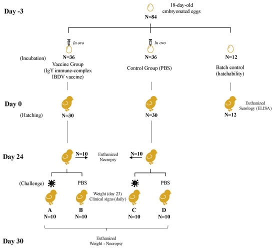
The study was conducted as a randomized controlled experimental trial combining in ovo vaccination and subsequent challenge with a vvIBDV strain (Figure 1). At the beginning of the study (day-3), a total of 84 embryonated eggs at 18 days of incubation were randomly allocated into three groups: two experimental groups of 36 eggs each and one hatchability control group of 12 eggs. Eggs from the experimental groups received either the vaccine or 0.05 mL of phosphate-buffered saline solution (PBS, prepared in-house), via in ovo into the amnion. Eggs from the hatchability control group (batch control) did not receive any treatment.
Figure 1.
Experimental design and group allocation of the vvIBDV challenge model. PBS, phosphate-buffered saline solution.
All eggs were transferred to the hatcher (MOD.65-N PDS; Masalles S.L., Sant Cugat del Vallès, Spain), separated by groups, and maintained at 37.5 °C. Upon hatching (day 0), birds from the batch control were euthanized and used for baseline serological evaluation. Maternal antibodies against IBDV were measured using a commercial ELISA kit (CIVTEST® AVI IBD, product code 80059, Hipra S.A., Amer, Spain). All birds in the batch control group were seropositive for IBDV at hatch, with a mean ELISA titer of 4471.39 ± 1563.93. Thirty birds from each experimental in ovo group were selected and allocated by treatment group to different rooms.
On day 24, ten birds from each treatment group were euthanized and necropsied to assess vaccine-associated alterations. The remaining birds (N = 20 per treatment group) were identified and randomly subdivided into four experimental subgroups (A–D). Birds of groups A (vaccinated and challenged) and C (control and challenged) were transferred to a room in isolation and infected with a vvIBDV strain, whereas birds in groups B (vaccinated and non-challenged) and D (control and non-challenged) received PBS.
On day 30, corresponding to 6 days post-challenge, all remaining birds were euthanized and necropsied. Necropsy and histopathological assessments were performed by personnel blinded to treatment and challenge group allocation.
All procedures were approved by the Ethical Committee at Hipra Scientific S.L.U and by the competent authority (Department of Agriculture, Generalitat de Catalunya: code 11601 and date of approval 15 March 2022), fulfilling European regulations on animal welfare (2010/63/EU). Qualified personnel evaluated animals daily to ensure the animals’ welfare and apply corrective measures if required.
2.3. Vaccine
A commercially available IBD immune-complex vaccine containing a live attenuated IBDV, strain 1052, and IgY extracted from eggs, was used (GUMBOHATCH®, manufactured by Hipra, S.A., Amer, Spain). The vaccine was administered by the in ovo route in a preparation of 0.05 mL of the reconstituted vaccine into each chicken egg at 18 days of embryonation, according to the manufacturer’s instructions.
2.4. Challenge
The virus strain used to perform the challenge was a vvIBDV heterologous to the strain included in the vaccine. The challenge strain (VG-248) was isolated in Girona, Spain, by Hipra, S.A. The genome RNA of this strain was isolated and cDNA was digested by the TaqI, SspI and StyI enzymes. The sequences obtained place it closely related to other European very virulent strains, such as UK661, CS89, 74-89A, Brown and Tula 94 [13]. It was prepared to administer 105 EID50/animal at a dose of 0.2 mL/animal via oculo-nasal (half dose in one eye and half in one nostril).
2.5. Procedure at Necropsy (Macroscopic Evaluation)
On day-24 and day-30 necropsies, birds were weighed, and the BF and spleens were extracted and weighed. Relative organ weights were expressed as BF:body-weight (BF:BW) and spleen:body-weight (spleen:BW) ratios. Bursas were sectioned from the site with the greatest diameters, and a macroscopic evaluation was performed on a scale of 0–3 (0 = no lesion, 1 = slight or subtle, 2 = moderate, 3 = severe alteration), assessing the presence of external edema, congestion, petechiae, hemorrhage, exudate, fibrin deposition, and loss of normal bursal structure. After macroscopic evaluation, bursa samples were fixed in 10% formalin in individual containers.
2.6. Histopathology of Bursas of Fabricius
For the histopathological evaluation, representative sections of each BF were collected and processed using standard histological procedures. Tissue samples were included in individually labeled plastic tissue-tek capsules, dehydrated by means of serial alcohol baths, cleared in xylene, and embedded in paraffin wax using an automatic tissue processor system. Tissue blocks were mounted, and sections produced with a microtome were five µm. Sections were automatically deparaffined and stained with hematoxylin and eosin. Each sample of BF was evaluated independently regarding different pathological alterations, including lymphoid depletion, heterophil infiltration, mononuclear infiltration, multifocal hemorrhage, plical edema, edema of the muscular wall, serosal edema, lumen exudate, lymphoid necrosis, cystic degeneration, plical atrophy and early lymphoid repopulation, based on previously described criteria [8,14]. The degree of bursal damage was scored using the scale proposed in the Ph. Eur. [11]. One hundred follicles were randomly selected and individually examined for each sample. Additionally, a semi-quantitative score (ranging from 0 to 5) was assigned to each pathological alteration, 0 being the absence of lesions (normal architecture) and 5 a severe alteration (complete loss of bursal architecture, fibroplasia). Lymphoid depletion was graded considering 0 (absence of follicular atrophy) to 5 (100% of the follicles showing atrophy). Signs of regeneration of the tissue (early lymphoid repopulation) were also evaluated as previously described [6].
2.7. Clinical Signs and Body-Weight Gain Monitoring
From day 24 to day 30 of the study, birds from the four experimental subgroups (A to D) were monitored daily for clinical signs including depression, and nervous signs (trembling, torticollis, paralysis, and circular motion), as previously described [6]. Each clinical sign was graded on a scale from 0 (normality or absence), 1 (mild), 2 (moderate) to 3 (severe expression of the sign). Fecal alterations (loss of consistency, liquid or aqueous), ruffled or fluffed feathers (neck or body), body condition (slow growth or severe emaciation), abnormal behavior (stereotypical or abnormal pecking), mortality, euthanasia, and any other noteworthy findings were also evaluated.
Birds were weighed the day before the challenge (day 23) and again at necropsy (day 30). Body-weight gain between days 23 and 30 of the study was then calculated.
2.8. Statistical Analysis
The experimental unit was the individual chicken. Sample size was determined with reference to Ph. Eur. [11], using 10 birds per group to minimize animal use while maintaining a group size consistent with in vivo IBD vaccine assessments.
All variables were described and compared between groups; statistical significance was set at p < 0.05. Normality and homogeneity of variances were assessed before parametric analyses. Non-parametric tests were used when the assumptions for parametric analysis were not met. Relative weights (BF:BW and spleen:BW ratios) were analyzed using the Kruskal–Wallis test (or Mann–Whitney test for two-by-two comparisons). Body-weight gain was analyzed by one-way ANOVA with the Tukey post hoc test when appropriate. Histopathological scores were analyzed using the Kruskal–Wallis test followed, when appropriate, by pairwise Wilcoxon rank sum test comparisons (or Mann–Whitney test for two-by-two comparisons). Appearances of clinical signs were evaluated using a survival analysis based on Kaplan–Meier estimates, with group comparisons performed using the log-rank test. All statistical analyses were performed using R version 4.1.0 with RStudio version 1.4.1717.
3. Results
3.1. Protection Against Macroscopic Lesions
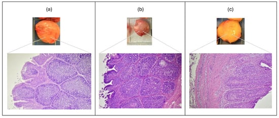
On day 24, two out of ten bursas of the vaccinated group presented mild congestion or exudate. On day 30, 2/10, 1/10 and 1/10 of groups A, B and D, respectively, presented a normal appearance showing only mild congestion (see Figure 2b). In contrast, all bursas of group C presented moderate to severe alterations, including petechiae, hemorrhages, fibrin and an evident inflammation and a thin layer of gelatinous bursal edema covering the serous surface (see Figure 2c).
Figure 2.
Bursas of Fabricius taken at necropsy (upper part) and its histological examination (lower part). (a) Bursa with normal appearance (Group D: non-challenged control); (b) bursa with normal appearance, presenting a certain degree of lymphoid depletion (Group A: vaccinated +vvIBDV); and (c) swollen bursa with gelatinous bursal edema presenting a severe acute lesional stage including massiveinflammation (heterophil and mononuclear infiltrates plus edema) and lymphoid necrosis (Group C: non-vaccinated control +vvIBDV). Magnification: 25×. (Jaume Altimira, Histovet).
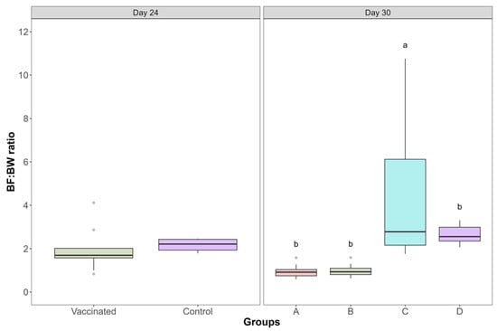
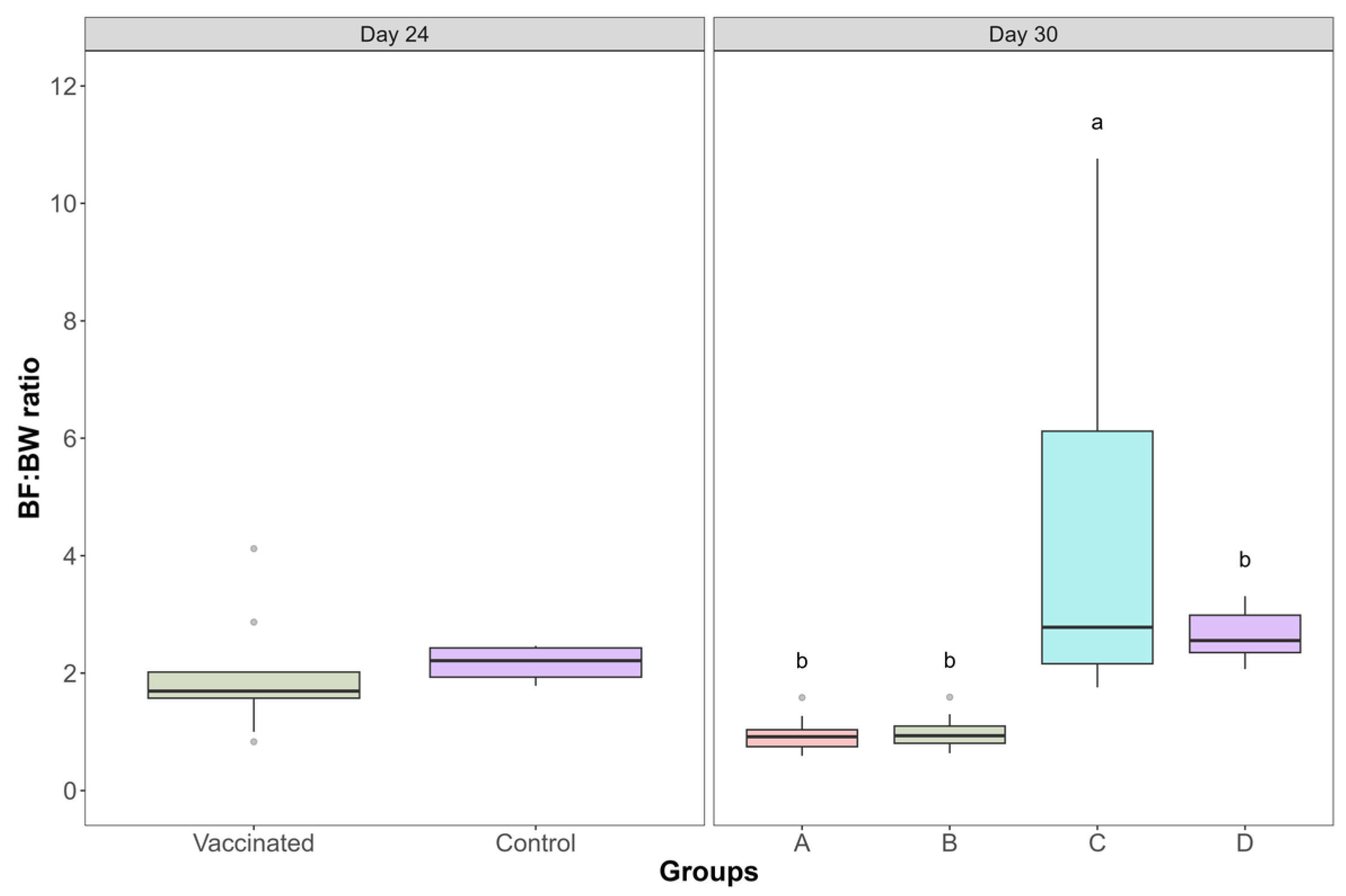
The relative BF:BW ratios evaluated on day 24 (pre-challenge) and on day 30 (6 days post-challenge) are shown in Figure 3. On day 24, BF:BW ratios were comparable between vaccinated and control groups (1.95 ± 1.00 and 24 2.16 ± 0.26, respectively), with no statistically significant differences detected between groups (Mann–Whitney test, p = 0.095). On day 30, significant differences in BF:BW ratios were observed among experimental groups (Kruskal–Wallis test, p = 1.92 × 10⁻⁶). Birds in group C showed markedly increased BF:BW ratios (4.26 ± 2.95) compared with the other groups. In contrast, vaccinated groups A (0.95 ± 0.30) and B (0.99 ± 0.29) exhibited substantially lower BF:BW ratios, comparable to those observed in the non-challenged control group D (2.66 ± 0.41).
Figure 3.
Bursa of Fabricius:Body Weight (BF:BW) of each experimental group on different days of the study. Boxes indicate the interquartile range, the horizontal line within each box represents the median, whiskers indicate the range, and circles represent outliers. No significant differences were found on day twenty-four (Mann–Whitney test, p = 0.095). On day thirty, there were significant differences between groups (Kruskal–Wallis test p = 1.92 × 10⁻⁶; different letters indicate differences between groups using two-by-two comparisons). Group A (vaccinated and challenged), group B (vaccinated and non-challenged), group C (control and challenged), and group D (control and non-challenged).
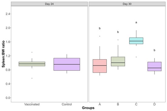
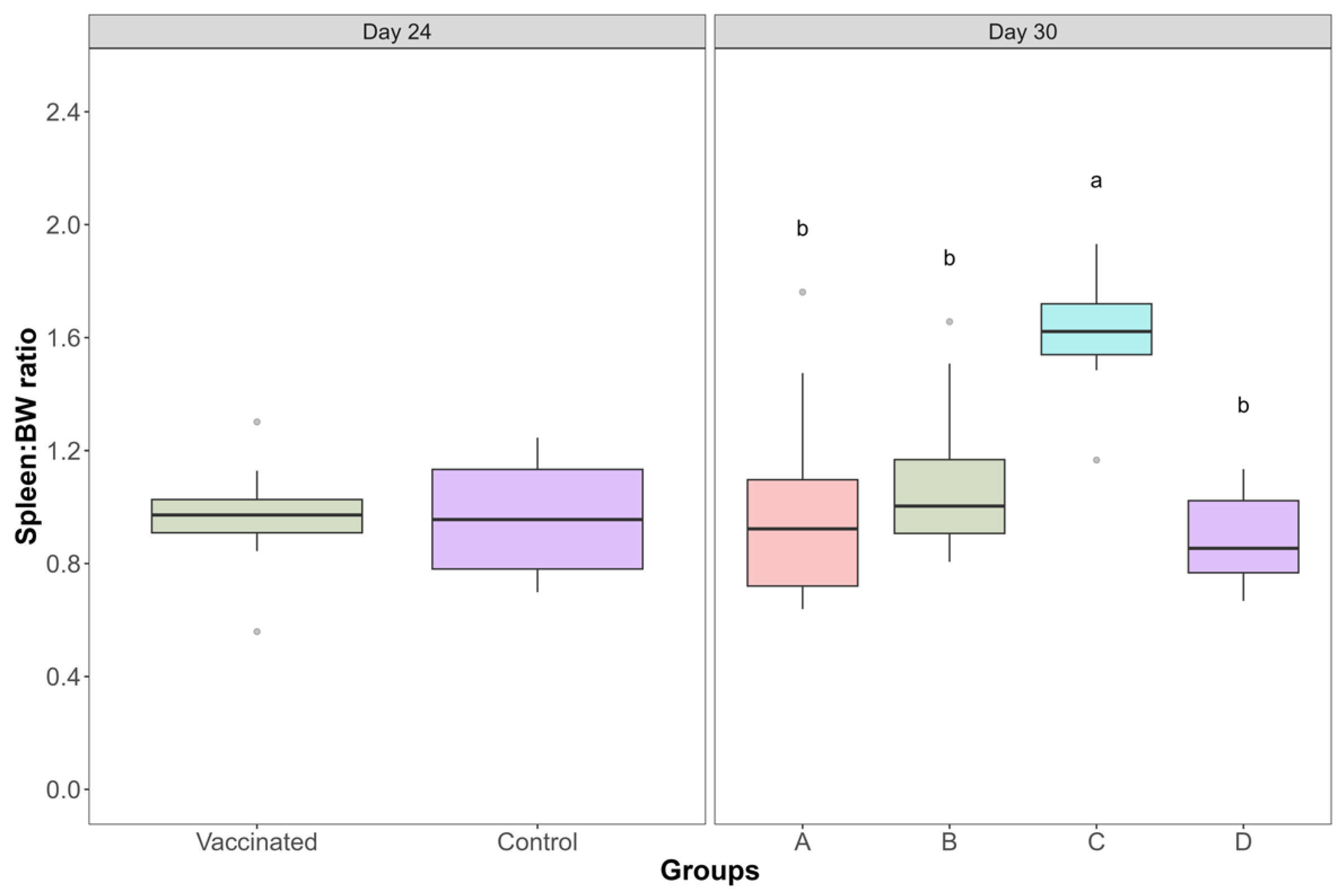
The spleen:BW ratios on day 24 and day 30 of the study are shown in Figure 4. On day 24, spleen:BW values were the same between vaccinated and control groups (0.96 ± 0.19), and no significant differences were detected (Mann–Whitney test, p = 0.974). On day 30, significant differences in spleen:BW values among experimental groups were observed (Kruskal–Wallis test, p = 0.0003). Birds in group C displayed a marked increase in the spleen:BW ratio (1.61 ± 0.21) compared with the remaining groups. In contrast, spleen:BW ratios in vaccinated groups A (1.00 ± 0.37) and B (1.09 ± 0.29), as well as in the non-challenged control group D (0.88 ± 0.16), remained within a similar range.
Figure 4.
Spleen:Body Weight (BF:BW) of each experimental group on different days of the study. Boxes indicate the interquartile range, the horizontal line within each box represents the median, whiskers indicate the range, and circles represent outliers. No significant differences were found on day twenty-four (Mann–Whitney test, p = 0.974). On day thirty, there were significant differences between groups (Kruskal–Wallis test, p = 0.0003; different letters indicate differences between groups by two-by-two comparisons). Group A (vaccinated and challenged), group B (vaccinated and non-challenged), group C (control and challenged), and group D (control and non-challenged).
3.2. Protection Against Microscopic Bursal Injury
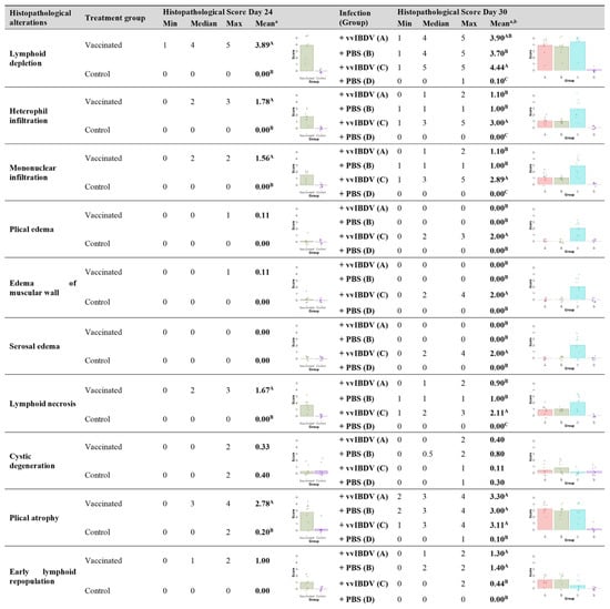
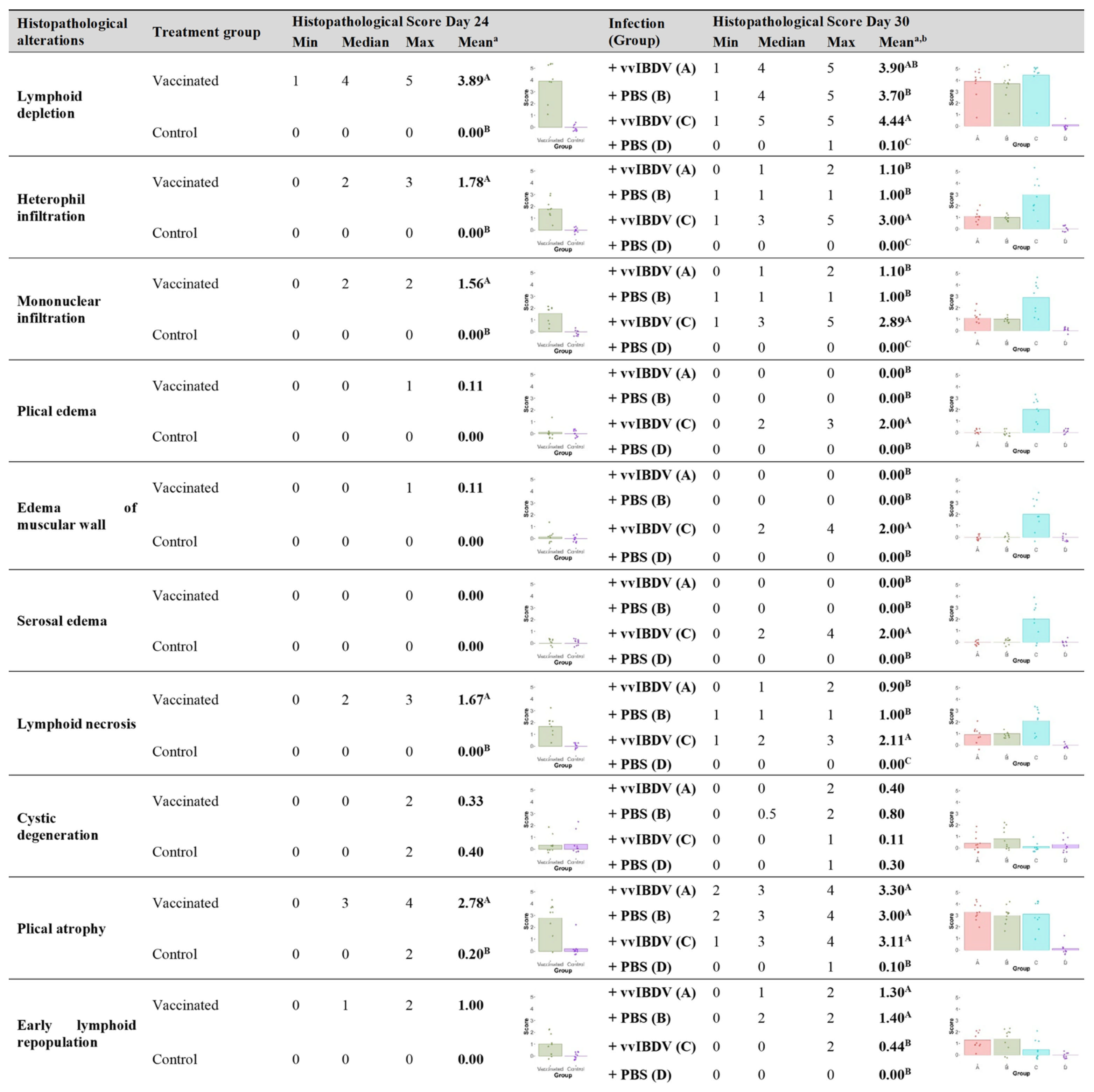
The summary of the BF histological scores is presented in Figure 5. Hemorrhage, luminal exudate and necrotic cysts were evaluated with score 0 in all bursa samples and, therefore, were excluded.
Figure 5.
Summary of histopathological scores of BF at necropsy for the different groups on different days of necropsy. Different superscripts represent significant differences (p < 0.05) using a Mann–Whitney/Kruskal–Wallis tests comparing scores of each group for each histopathological alteration. b Pairwise comparisons were obtained using Wilcoxon rank sum test comparing scores. Group A (vaccinated and challenged), group B (vaccinated and non-challenged), group C (control and challenged), and group D (control and non-challenged).
On day 24 (pre-challenge), bursae from vaccinated birds showed significantly moderate to severe lymphoid depletion and plical atrophy, accompanied by mild heterophil and mononuclear infiltration compared to control birds, which displayed normal histological architecture (p < 0.05).
On day 30, lymphoid depletion and plical atrophy was evident in all vaccinated or IBDV-challenged groups; however, significant differences among groups were observed in the associated lesional patterns. Group C showed significantly higher scores for heterophil and mononuclear infiltrates, plical, muscular and serosal edema, and lymphoid necrosis compared with all other groups (p < 0.05). In contrast, vaccinated groups A and B exhibited significantly lower inflammatory and necrotic scores, absence of edema, and significantly higher scores for early lymphoid repopulation.
3.3. Clinical Signs and Body-Weight Gain
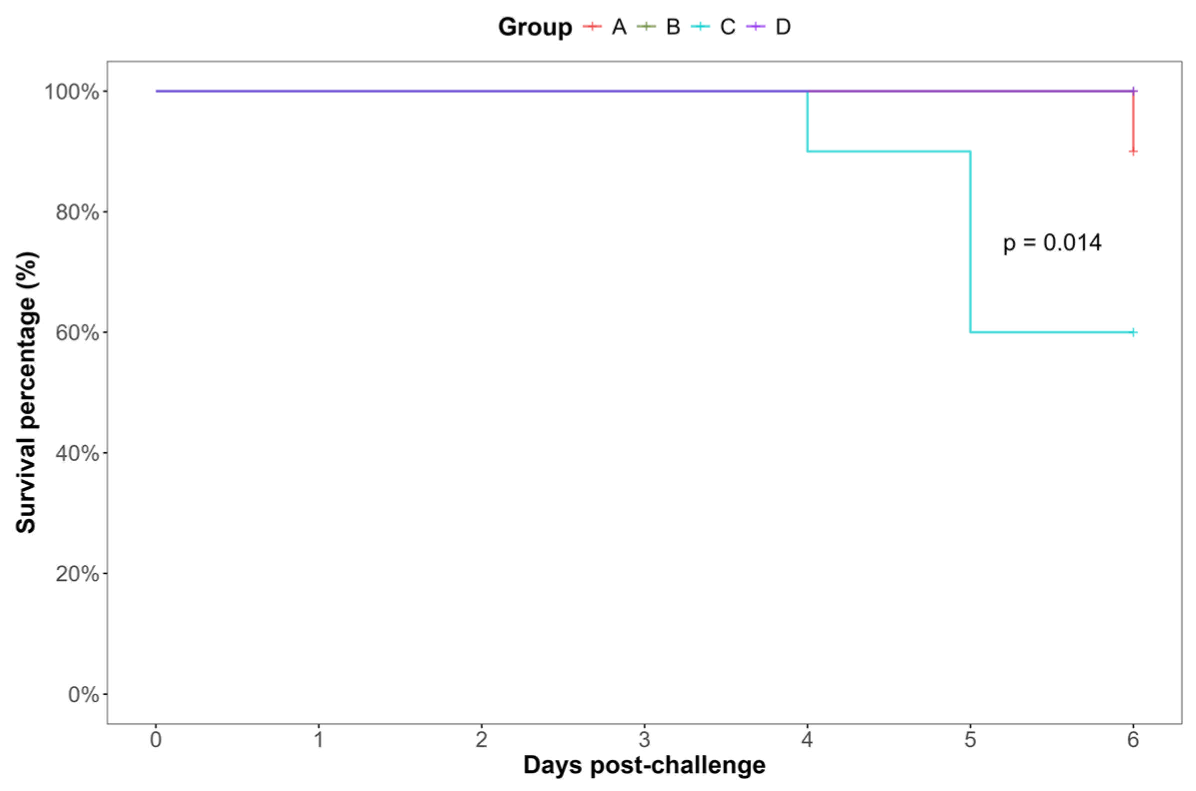
No clinical signs or mortality were observed in any group during the pre-challenge period (days 1–24). After vvIBDV challenge on day 24 of the study, no clinical signs or mortality were observed in vaccinated groups A and B, nor in the non-challenged control group D. In contrast, five out of ten birds in the control and challenged group C showed mild clinical signs compatible with IBD, including depression and ruffled feathers.
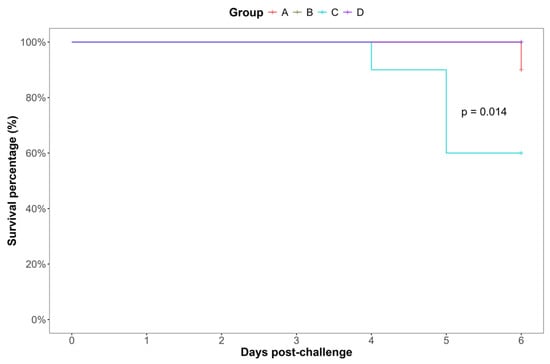
Figure 6 shows the Kaplan–Meier survival curves for the time until appearance of IBD clinical signs during the post-challenge period. A significant difference in the time to onset of clinical signs was observed between group C and the remaining groups (log-rank test, p = 0.014), with survival percentages remaining close to 90% for groups A, B and D throughout the post-challenge period.
Figure 6.
Survival curves for appearance of IBD clinical signs. Differences between curves are significant (p = 0.014, Mantel–Haenszel test comparing Kaplan–Meier estimates of survival). Group A (vaccinated and challenged), group B (vaccinated and non-challenged), group C (control and challenged), and group D (control and non-challenged).
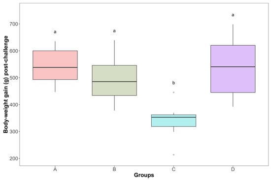
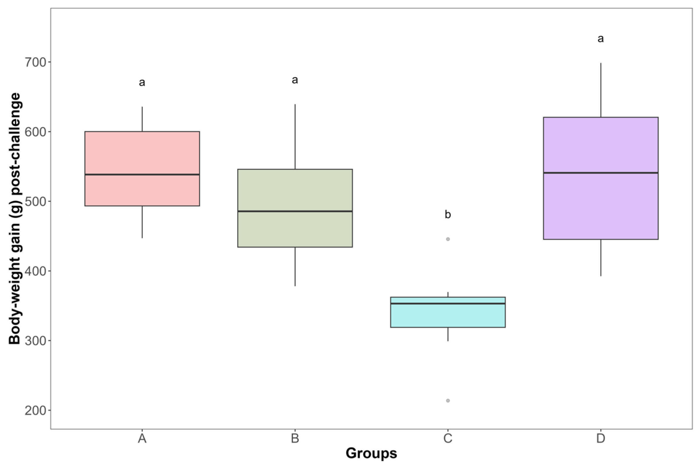
Body-weight gain during the post-challenge period (from day 23, one day before the challenge, to day 30) are shown in Figure 7. Mean body-weight gain values were 541.38 ± 67.10 g for group A, 493.05 ± 79.91 g for group B, 339.85 ± 59.35 g for group C, and 533.91 ± 107.39 g for group D. Pairwise comparisons revealed significant differences between group C and the remaining groups, whereas no significant differences were observed among groups A, B, and D (ANOVA, p = 4.36 × 10⁻⁶).
Figure 7.
Body-weight gain of each experimental group post-challenge (from day twenty-three to day thirty of study). Boxes indicate the interquartile range, the horizontal line within each box represents the median, whiskers indicate the range, and circles represent outliers. There were significant differences between groups (ANOVA test p = 4.36 × 10⁻⁶; different letters indicate differences between groups using two-by-two comparisons). Group A (vaccinated and challenged), group B (vaccinated and non-challenged), group C (control and challenged), and group D (control and non-challenged).
4. Discussion
The present study demonstrates the protective efficacy of an in ovo immune-complex IBD vaccine against challenge with a heterologous vvIBDV strain in broiler chickens. Vaccinated challenged birds were protected against the acute pathological and clinical consequences of vvIBDV infection, including characteristic bursal lesions, splenomegaly, clinical disease, and impaired post-challenge body-weight gain. In contrast, non-vaccinated challenged birds developed the expected acute disease pattern, with swollen bursae and gelatinous serosal edema, increased relative bursal and spleen weights, inflammatory and necrotic bursal lesions, clinical signs compatible with IBD, and reduced body-weight gain. These findings are consistent with previous experimental observations in broilers challenged with virulent IBDV strains [2,8,15].
As expected, the histopathological examination of the BF indicated that lymphoid depletion and plical atrophy were observed in all groups exposed to IBDV, either via vaccination or challenge. However, these parameters alone were insufficient to distinguish acute vvIBDV-induced lesions from vaccine-associated bursal alterations, consistent with previous observations indicating that lymphoid depletion alone does not necessarily correlate with functional impairment of the bursa [7,16].
In contrast, control birds challenged with vvIBDV showed a distinct acute histopathological lesion profile characterized by moderate to severe heterophil and mononuclear inflammatory infiltrates, marked plical, muscular and serosal edema affecting the full thickness of the bursae, and increased lymphoid necrosis. These lesions were accompanied by an absence of early lymphoid repopulation, denoting an acute, dysfunctional stage of bursal damage consistent with the severe inflammatory and necrotic lesions induced by highly virulent vvIBDV strains, as previously described [15,16]. Importantly, such lesions were exclusive of the control non-vaccinated group and were not observed in vaccinated birds, regardless of challenge status, nor in non-challenged controls.
Vaccinated groups displayed a markedly different histopathological pattern. Although moderate to severe lymphoid depletion and plical atrophy were evident, indicative of active replication of the live vaccine virus in the BF, these alterations were observed with only minimal inflammatory infiltrates, absence of edema, low necrosis scores, and the presence of early lymphoid repopulation. This pattern of lesions is consistent with a post-vaccinal stage, in which initial bursal structural damage is followed by regenerative processes and bursal functional recovery [12,17,18]. Similar distinctions between acute vvIBDV-induced lesions and vaccine-associated changes have been described in previous field and experimental studies [16]. However, the specific contribution of the present study is the independent semi-quantitative assessment of inflammatory, edematous, necrotic, and regenerative lesions alongside lymphoid depletion within the same experimental challenge model. This approach provides a more detailed pathological framework for distinguishing acute vvIBDV-induced damage from expected post-vaccination bursal changes.
Early lymphoid repopulation was identified in follicles showing a severe depletion together with a dense peripheral rim of small, actively proliferating lymphoid cells around its marginal area. Although this interpretation may be debated (as follicles suffering from a severe but subtotal lymphoid depletion can show similar histological features) significantly higher repopulation scores were observed in vaccinated groups compared with the control challenged group, supporting the presence of active and faster regenerative processes in vaccinated birds, as previously reported [19].
Moreover, cystic degeneration was observed at low levels (score 1) across all groups and generally corresponded to small cystic formations in the interfollicular epithelium lining the plicae. These lesions were considered non-specific, as they were also detected in the non-challenged control group, and may indicate individual cell degeneration due to cell replacement processes [20]. Slightly higher scores in vaccinated groups were likely reflect previous vaccine replication, whereas in the control and challenged group these changes were masked by the predominance of massive inflammatory lesions.
The macroscopic evaluation further supported the histopathological findings. Control and challenged birds frequently exhibited swollen bursae (~90% of BF), covered by a thin layer of gelatinous serosal edema, a hallmark of acute vvIBDV infection previously described in experimental studies [3,10]. This characteristic was not observed in vaccinated, non-challenged birds. These macroscopic changes were also reflected in the BF:BW ratio. While vaccinated birds showed reduced BF:BW ratio on day 30, consistent with bursal atrophy following vaccine virus replication, these values were not significantly different from those of non-challenged controls. In contrast, challenged control birds exhibited increased BF:BW ratios, reflecting acute inflammatory enlargement rather than preserved bursal function. Consequently, BF:BW values must be interpreted in conjunction with histopathological findings.
Similarly, splenomegaly, as evidenced by an increased spleen:BW ratio, was exclusively observed in the challenged control birds on day 30. On the contrary, no differences were detected in the spleen:BW ratio on day 24 (before challenge) between the vaccinated and control group. This finding represents an additional distinctive sign of IBD infection [2] and further supports the protective efficacy of the vaccine against the damage of acute vvIBDV infection.
Under field conditions, vvIBDV infections are known to cause clear clinical disease to broiler birds [2,3]. However, reproduction of consistent clinical signs under controlled experimental conditions, after inoculation of vvIBDV strains to broiler birds, remains challenging [9,10]. In the present study, despite the administration of a high infective dose, clinical signs remained mild and were exclusively observed in challenged control birds during the post-challenge period. These signs, including ruffled feathers or mild depression, were compatible with Gumboro disease.
Although mild, the presence of clinical signs was associated with a significant reduction in body-weight gain in the challenged control birds, indicating a measurable impact of acute vvIBDV infection on animal performance. In contrast, vaccinated birds, whether challenged or not, maintained body-weight gain comparable to non-challenged controls, consistent with the absence of acute systemic disease.
A strength of this study is the combined clinical, macroscopic, and detailed histopathological assessment of bursal lesions, including the independent evaluation of inflammatory, edematous, necrotic, and regenerative changes. The study was designed to assess the acute response to vvIBDV challenge at the expected onset of vaccine-induced protection. While this approach was suitable for detecting the main clinical and pathological differences observed between groups, the relatively small sample size and the 6-day post-challenge observation period should be considered when interpreting the findings. Moreover, virological and immunological parameters, such as challenge-virus load, infectious virus detection, or antibody responses, were not assessed. Therefore, the present results demonstrate clinical and pathological protection but do not allow conclusions regarding reduction in challenge-virus replication, duration of immunity, or longer-term production performance.
5. Conclusions
The experimental study demonstrates that the in ovo immune-complex vaccine protected broiler chickens against the clinical signs, impaired post-challenge growth, and acute bursal pathological consequences of challenge with the heterologous vvIBDV strain VG-248. Importantly, the independent assessment of inflammatory, edematous, necrotic, and regenerative lesions provided additional discriminatory value beyond lymphoid depletion alone, allowing a clearer distinction between expected vaccine-associated bursal changes and acute vvIBDV-induced damage.
Author Contributions
Conceptualization, G.B.-S., I.T.-R. and E.S.-P.; methodology, G.B.-S. and E.S.-P.; validation, G.B.-S., I.T.-R. and E.S.-P.; formal analysis, G.B.-S.; investigation, G.B.-S. and E.S.-P.; data curation, G.B.-S.; writing—original draft preparation, G.B.-S.; writing—review and editing, G.B.-S., I.T.-R. and E.S.-P.; visualization, G.B.-S.; supervision, E.S.-P.; project administration, E.S.-P. All authors have read and agreed to the published version of the manuscript.
Funding
This research received no external funding.
Institutional Review Board Statement
All procedures were approved by the Ethical Committee at Hipra Scientific S.L.U and by the competent authority (Department of Agriculture, Generalitat de Catalunya: code 11601 and date of approval 15 March 2022), fulfilling European regulations on animal welfare (2010/63/EU).
Informed Consent Statement
Not applicable.
Data Availability Statement
The data presented in this study are available from the corresponding author upon reasonable request.
Acknowledgments
The authors would like to express their gratitude to Jaume Altimira (Histovet laboratory) for performing the histological analyses and for providing the microscopic images. The authors would also like to thank Marina Solé for providing medical writing support during manuscript preparation.
Conflicts of Interest
G.B.-S., I.T.-R. and E.S.-P. are employees of HIPRA, the manufacturer of the vaccines evaluated in this study. The authors declare no other commercial or financial relationships that could be construed as a potential conflict of interest.
Abbreviations
The following abbreviations are used in this manuscript:
| IBD | Infectious bursal disease |
| IBDV | Infectious bursal disease virus |
| vvIBDV | Very virulent infectious bursal disease virus |
| BF | Bursa of Fabricius |
| BW | Body weight |
| BF:BW | Bursa of Fabricius-to-body-weight ratio |
| PBS | Phosphate-buffered saline |
| ELISA | Enzyme-linked immunosorbent assay |
| EID50 | 50% egg infectious dose |
| IgY | Immunoglobulin Y |
| Ph. Eur. | European Pharmacopoeia |
References
- van den Berg, T.P.; Eterradossi, N.; Toquin, D.; Meulemans, G. Infectious Bursal Disease (Gumboro Disease). Rev. Sci. Tech. 2000, 19, 509–543. [Google Scholar]
- Eterradossi, N.; Saif, Y.M. Infectious Bursal Disease. In Diseases of Poultry; John Wiley & Sons, Ltd.: Hoboken, NJ, USA, 2020; pp. 257–283. ISBN 978-1-119-37119-9. [Google Scholar]
- Ingrao, F.; Rauw, F.; Lambrecht, B.; van den Berg, T. Infectious Bursal Disease: A Complex Host-Pathogen Interaction. Dev. Comp. Immunol. 2013, 41, 429–438. [Google Scholar] [CrossRef] [Scilit]
- Alkie, T.N.; Rautenschlein, S. Infectious Bursal Disease Virus in Poultry: Current Status and Future Prospects. Vet. Med. 2016, 7, 9–18. [Google Scholar] [CrossRef] [Scilit]
- Müller, H.; Mundt, E.; Eterradossi, N.; Islam, M.R. Current Status of Vaccines against Infectious Bursal Disease. Avian Pathol. 2012, 41, 133–139. [Google Scholar] [CrossRef] [Scilit]
- Bech, G.; Marzo, E.; Blake-Dyke, C.; Busquet, M.; Woodward, M.; Alech, M.; Solé, M.; Sánchez, A.; Sebastià, E. Identification of a Reassortant Infectious Bursal Disease Virus (Genotype A3B1) in the United Kingdom: Pathogenicity and Immune Complex Vaccine Efficacy. Avian Pathol. 2026, 1–13. [Google Scholar] [CrossRef] [Scilit]
- Aihara, N.; Horiuchi, N.; Hikichi, N.; Ochiai, M.; Hosoda, Y.; Ishikawa, Y.; Shimazaki, Y.; Oishi, K. Immunoreactivity and Morphological Changes of Bursal Follicles in Chickens Infected with Vaccine or Wild-Type Strains of the Infectious Bursal Disease Virus. J. Vet. Med. Sci. 2015, 77, 913–918. [Google Scholar] [CrossRef] [Scilit]
- Williams, A.E.; Davison, T.F. Enhanced Immunopathology Induced by Very Virulent Infectious Bursal Disease Virus. Avian Pathol. 2005, 34, 4–14. [Google Scholar] [CrossRef] [Scilit]
- Tippenhauer, M.; Heller, D.E.; Weigend, S.; Rautenschlein, S. The Host Genotype Influences Infectious Bursal Disease Virus Pathogenesis in Chickens by Modulation of T Cells Responses and Cytokine Gene Expression. Dev. Comp. Immunol. 2013, 40, 1–10. [Google Scholar] [CrossRef] [Scilit]
- Aricibasi, M.; Jung, A.; Heller, E.D.; Rautenschlein, S. Differences in Genetic Background Influence the Induction of Innate and Acquired Immune Responses in Chickens Depending on the Virulence of the Infecting Infectious Bursal Disease Virus (IBDV) Strain. Vet. Immunol. Immunopathol. 2010, 135, 79–92. [Google Scholar] [CrossRef] [Scilit]
- EDQM Monograph, Avian Infectious Bursal Disease Vaccine (Live) (0587). In European Pharmacopoeia; 13, Supplement 13.3; European Directorate for the Quality of Medicine & Health Care of the Council of Europe (EDQM): Strasbourg, France, 2026; pp. 1–3. Available online: https://pheur-online.edqm.eu/home/ (accessed on 21 October 2026).
- Ateş, M.B.; Özdemir, Ö.; Çelik, Z.; Salik, R.; Dağar, O.; Bulut, A.; Tural, A. Histopathological Evaluation of the Effects of Live Infectious Bursal Disease Vaccine Originated from WF2512 Strain on Bursa Fabricius in the Broilers. J. Adv. VetBio Sci. Tech. 2022, 7, 250–258. [Google Scholar] [CrossRef] [Scilit]
- Majó, N.; El-Attrache, J.; Banda, A.; Villegas, P.; Ramis, A.; Pagès, A.; Ikuta, N. Molecular Characterization of Spanish Infectious Bursal Disease Virus Field Isolates. Avian Dis. 2002, 46, 859–868. [Google Scholar] [CrossRef] [Scilit]
- Sharma, J.M.; Dohms, J.E.; Metz, A.L. Comparative Pathogenesis of Serotype 1 and Variant Serotype 1 Isolates of Infectious Bursal Disease Virus and Their Effect on Humoral and Cellular Immune Competence of Specific-Pathogen-Free Chickens. Avian Dis. 1989, 33, 112–124. [Google Scholar] [CrossRef] [Scilit]
- Huang, X.; Liu, W.; Zhang, J.; Liu, Z.; Wang, M.; Wang, L.; Zhou, H.; Jiang, Y.; Cui, W.; Qiao, X.; et al. Very Virulent Infectious Bursal Disease Virus-Induced Immune Injury Is Involved in Inflammation, Apoptosis, and Inflammatory Cytokines Imbalance in the Bursa of Fabricius. Dev. Comp. Immunol. 2021, 114, 103839. [Google Scholar] [CrossRef] [Scilit]
- Dolz, R.; Majó, N.; Ordóñez, G.; Porta, R. Viral Genotyping of Infectious Bursal Disease Viruses Isolated from the 2002 Acute Outbreak in Spain and Comparison with Previous Isolates. Avian Dis. 2005, 49, 332–339. [Google Scholar] [CrossRef] [Scilit]
- Castro, M.; Saume, E.; Díaz, C.; García, J. Bursal Restoration after Intermediate and Intermediate plus Infectious Bursal Disease Virus Vaccination. Rev. Cient. 2009, 19, 119–123. [Google Scholar]
- Iván, J.; Nagy, N.; Magyar, A.; Kacskovics, I.; Mészáros, J. Functional Restoration of the Bursa of Fabricius Following In Ovo Infectious Bursal Disease Vaccination. Vet. Immunol. Immunopathol. 2001, 79, 235–248. [Google Scholar] [CrossRef] [Scilit]
- Kim, I.J.; Gagic, M.; Sharma, J.M. Recovery of Antibody-Producing Ability and Lymphocyte Repopulation of Bursal Follicles in Chickens Exposed to Infectious Bursal Disease Virus. Avian Dis. 1999, 43, 401–413. [Google Scholar] [CrossRef] [Scilit]
- Singh, J.; Banga, H.S.; Brar, R.S.; Singh, N.D.; Sodhi, S.; Leishangthem, G.D. Histopathological and Immunohistochemical Diagnosis of Infectious Bursal Disease in Poultry Birds. Vet. World 2015, 8, 1331–1339. [Google Scholar] [CrossRef] [Scilit]
Disclaimer/Publisher’s Note: The statements, opinions and data contained in all publications are solely those of the individual author(s) and contributor(s) and not of MDPI and/or the editor(s). MDPI and/or the editor(s) disclaim responsibility for any injury to people or property resulting from any ideas, methods, instructions or products referred to in the content. |
© 2026 by the authors. Licensee MDPI, Basel, Switzerland. This article is an open access article distributed under the terms and conditions of the Creative Commons Attribution (CC BY) license.