


Abstract
Background: Chronic hepatitis B virus (HBV) infection (CHB) affects nearly 300 million individuals globally and remains incurable with current antiviral therapies, which suppress viral replication but rarely achieve a functional cure, defined by sustained loss of hepatitis B surface antigen (HBsAg). CHB is characterized by profound virus-induced immune tolerance that limits the efficacy of conventional therapeutic vaccination strategies. Objective: To evaluate the therapeutic efficacy and immunological mechanisms of HEPLISAV-B, a CpG-1018 adjuvanted HBsAg vaccine, in breaking immune tolerance and inducing functional cure-like responses in a murine model of CHB. Design: Using the adeno-associated virus-HBV (AAV-HBV) mouse model, mice with high levels of persistent HBV viremia were vaccinated with two doses of HEPLISAV-B. Virological outcomes in the blood and liver, immune responses and mechanisms were assessed. Results: HEPLISAV-B induced rapid and durable HBsAg clearance, markedly reduced circulating and intrahepatic HBV DNA and RNA, and suppressed viral replication without hepatocellular injury. Vaccination elicited robust, sustained anti-HBs IgG1 and IgA responses, enhanced HBsAg-specific T- and B-cell immunity, reduced CD4 regulatory T cells, and decreased PD-1 expression on CD4 T cells. Therapeutic efficacy was strictly dependent on CD4 T cells and the CD40/CD40L signaling pathway, but independent of CD8 T cells, indicating a CD4-driven, non-cytolytic antiviral mechanism critical for HEPLISAV-B-induced HBV control. Conclusions: HEPLISAV-B effectively breaks HBV-induced immune tolerance and restores coordinated antiviral immunity through a CD4 T cell/CD40L-dependent pathway. The findings support its potential as a therapeutic vaccine in CHB patients.
1. Introduction
Chronic hepatitis B virus (HBV) infection remains a major global health issue, affecting approximately 296 million people worldwide [1]. It can lead to severe liver complications, including cirrhosis and hepatocellular carcinoma. Current nucleos(t)ide analog treatments, while effective in suppressing viral replication, rarely achieve a functional cure, defined as sustained hepatitis B surface antigen (HBsAg) and HBV DNA loss after discontinuation of HBV medications [2,3].
People with resolved acute HBV infections typically exhibit robust systemic antibody, CD4, and CD8 T-cell responses against HBV [4,5,6]. In contrast, individuals with chronic HBV infection fail to produce effective anti-HBs antibodies [7] and have dysfunctional HBV-specific CD4 and CD8 T cells, resulting in HBV-specific immune tolerance [8], characterized by high HBV viral load with minimal liver inflammation. High levels of systemic viral antigens induce immune tolerance, impairing dendritic cells, natural killer cells, and T- and B-cell functions, thereby contributing to HBV persistence [9,10,11,12,13,14]. Overcoming this tolerance to elicit effective anti-HBV immune responses remains a major challenge in developing HBV therapeutic vaccines. Therefore, novel treatments, including therapeutic vaccines capable of boosting anti-HBV immunity, are urgently needed to effectively eradicate chronic HBV infection and improve patients’ lives.
Our group and others have shown that optimizing immune adjuvants is a promising strategy to overcome HBV-induced immune tolerance [15,16,17,18,19,20,21,22,23,24]. CpG oligodeoxynucleotides (CpG ODNs), potent TLR9 agonists, have shown powerful adjuvant properties that kickstart a strong immune response capable of addressing the inherent immune hypo-responsiveness in chronic HBV patients (CHB). By activating and maturing Antigen Presenting Cells (especially pDCs), TLR9 agonists ensure that co-administered HBV antigens are efficiently processed and presented to naive and exhausted T cells, thereby initiating or boosting a specific anti-HBV immune response [25,26].
We have previously demonstrated that CpG oligodeoxynucleotide 1826 (CpG-1826), as an adjuvant to HBV vaccines, shows promise in breaking immune tolerance and achieving viral clearance in the AAV-HBV mouse model [15]. However, that study focused on short-term effects, leaving the long-term durability of the immune response, the risk of viral rebound, and potential adverse effects largely unexplored. Furthermore, CpG-1826 vaccination failed to elicit responses in mice with high HBV viremia [15].
CpG-1018, a Dynavax-developed CpG, is used as an adjuvant in HEPLISAV-B, the novel FDA-approved vaccine for inducing protective HBV immunity in populations who fail to respond to conventional HBV vaccines such as Engerix-B [27,28]. The HEPLISAV-B vaccine has demonstrated superior immunogenicity compared to alum-adjuvanted vaccines, even in populations with traditionally poor vaccine responses (e.g., people with HIV, diabetes, or chronic kidney disease) [29,30,31]. In clinical trials, CpG-1018 adjuvanted vaccines induced higher seroconversion rates, faster immune responses, and stronger T- and B-cell memory [29,30,31].
In the present study, we evaluated the therapeutic efficacy of the HEPLISAV-B vaccine in a murine model of chronic HBV infection. In contrast to conventional HBsAg vaccination, which failed to overcome established immune tolerance and exhibited no antiviral activity, HEPLISAV-B induced rapid and sustained clearance of circulating HBsAg, markedly reduced intrahepatic HBV DNA, and suppressed viral replication through a predominantly non-cytolytic mechanism.
HEPLISAV-B also elicited robust and durable anti-HBs antibody responses and was associated with a reduction in CD4 regulatory T cells and decreased PD-1 expression on CD4 T cells. The therapeutic effect of HEPLISAV-B was dependent on CD4 T cells and CD40L, but not CD8 T cells. Together, these findings establish HEPLISAV-B as a potent immunotherapeutic capable of breaking CHB immune tolerance and inducing coordinated antiviral immunity, supporting its potential use as a therapeutic vaccine for achieving a functional cure in patients with chronic HBV infection.
2. Materials and Methods
2.1. Animal Husbandry and Treatments
Male C57BL/6 mice were selected for this study due to their reported propensity to develop high HBV viremia and increased susceptibility to HBV-related liver complications following AAV-HBV inoculation. All mice were housed in a temperature-controlled environment (23 ± 2 °C) with a 12 h light/dark cycle and ad libitum access to water and food. All animal procedures were approved by the University of Maryland School of Medicine IACUC (AUP-00000478) [15].
Eight to nine weeks old animals were intravenously administered with recombinant AAV-HBV (5 × 10¹⁰ genome copies [gc]/mouse). HBV viremia and HBsAg levels in plasma were monitored every 2 to 4 weeks for up to 24 weeks post-infection (wpi).
In the first immunization experiment, HBV-positive and HBV-negative control mice received two subcutaneous doses, four weeks apart, of HBsAg (20 ug) emulsified in Montanide ISA 720 VG (25% final volume), along with intramuscular injections of pVAX1-coHBcAg (100 ug) and pORF-mIL-12 (20 ug) DNAs per mouse.
In a separate cohort, mock-infected and HBV-positive mice received two intramuscular doses of HEPLISAV-B, administered 4 weeks apart, at three dose levels of recombinant HBsAg and CpG-1018: 0.08 μg/12 μg, 0.4 μg/60 μg, and 2 μg/300 μg, respectively. To investigate the immune mechanisms underlying vaccine efficacy, HEPLISAV-B-treated HBV-positive mice received depleting antibodies against CD4+ T cells (InVivoMAb anti-mouse CD4, clone GK1.5, #BE0003-1; Bio X Cell) or CD8+ T cells (InVivoMAb anti-mouse CD8α, clone 53-6.7, #BP0004-1; Bio X Cell), or a blocking antibody against CD40L (InVivoMAb anti-mouse CD40L, clone MR-1, #BE0017-1; Bio X Cell) to inhibit CD40L–CD40 signaling. Antibodies were administered intraperitoneally before each vaccine dose. Specifically, mice received anti-CD4 (300 μg/mouse), anti-CD8 (50 μg/mouse), or anti-CD40L (300 μg/mouse) 24 h before each vaccination, followed by a second antibody administration 2 h before each vaccination at doses of 150 μg/mouse for anti-CD4 and anti-CD40L and 50 μg/mouse for anti-CD8.
2.2. HBV Viremia Analysis
HBV DNA was extracted from 25 µL of plasma using the QIAamp DNA Blood Mini Kit (Qiagen, Germantown, MD, USA, #51106). Plasma HBV DNA levels were quantified by qPCR using Power SYBR Green PCR Master Mix (Applied Biosystems, Frederick, MD, USA) on a CFX Opus 384 Real-Time PCR System (Bio-Rad, Hercules, CA, USA) with HBV-specific mouse primers (Table 1). Absolute HBV DNA copy numbers were calculated using standard curves generated from an HBV genome-containing plasmid, as previously described [32].
Table 1.
Primers sequences for qPCR/RT-qPCR.
2.3. DNA/RNA Extraction and Real-Time Quantitative PCR
Total RNA was extracted from cells (RNeasy Plus Kit, QIAGEN, Germantown, MD, USA) and from liver tissues using QIAzol Lysis Reagent and gDNA Eliminator Solution (RNeasy Plus Universal Kits, QIAGEN, Germantown, MD, USA) and quantified using Nanodrop. In total, 0.2–2 μg of total RNAs were used for cDNA preparation by reverse transcription with random primers and SuperScript III (Invitrogen, Frederick, MD, USA), according to the manufacturer’s instructions. DNA was extracted from 50 mg of liver using MasterPure™ Complete DNA and RNA Purification Kit (Biosearch Technologies, Novato, CA, USA). Two microliters of diluted (1:10) cDNA and DNA were used for quantification with the Power SyBR Green PCR Master Mix (Applied Biosystems, Frederick, MD, USA) on the CFX Opus 384 Real-Time PCR System from Bio-Rad, using specific mouse primers (Table 1). The comparative Ct method was used for the analysis of real-time PCR data. Relative expression levels were calculated using the comparative Ct method, normalized to mouse GAPDH, and expressed relative to control animals [33,34,35,36].
2.4. Protein Extraction and Immunoblot
Total protein was extracted from snap-frozen liver tissue using RIPA buffer (Thermo Scientific, Frederick, MD, USA) supplemented with protease and phosphatase inhibitor cocktails (Pierce, Frederick, MD, USA) and quantified by BCA assay. Plasma (3 µL) and liver protein extracts (5–20 µg) were separated by SDS–PAGE (4–12%), transferred to nitrocellulose membranes, and probed with primary antibodies against HBs (1:1000; BIOSYNTH, #20-HR20) and HBc (1:1000; ZETA Corp, Sierra Madre, CA, USA, #Z2085RL-R). Horseradish peroxidase-conjugated anti-mouse or anti-rabbit secondary antibodies were used, and HRP-conjugated β-actin (1:5000; Sigma/Millipore, Rockville, MD, USA, A3854-200 UL) served as a loading control. Signals were detected by ECL (Millipore, Rockville, MD, USA, or Invitrogen, Frederick, MD, USA) using a Bio-Rad CCD camera and quantified with ImageJ software (version 1.54k).
2.5. Histology and Immunofluorescence Staining
For histological and immunofluorescence analyses, formalin-fixed, paraffin-embedded mouse liver sections (5 µm) were stained with hematoxylin and eosin (H&E) or subjected to antigen retrieval and immunostained with anti-HBs (BIOSYNTH, Gardner, MA, USA, #20-HR20) and anti-HBc (ZETA Corp, Sierra Madre, CA, USA, #Z2085RL-R) antibodies, followed by Alexa Fluor 488-conjugated anti-rabbit secondary antibody (1:500; Invitrogen, Frederick, MD, USA). Apoptotic cells were detected using a TUNEL assay kit (Roche, Rockville, MD, USA) according to the manufacturer’s instructions. Nuclei were counterstained with Hoechst 33342 (Sigma, Rockville, MD, USA), and images were acquired using a 20× objective.
2.6. Immune Cell Isolation, Treatment and Flow Cytometry
Immune cells were isolated from the liver as described previously with slight modifications. Briefly, livers were perfused through the portal vein with a wash buffer (PBS 1×, 2% fetal bovine serum, 1% penicillin/streptomycin, and 0.1 mg/mL DNase I) and then minced with gentleMACS Dissociator in a Liver Digestion Medium (#17703034, ThermoFisher, Frederick, MD, USA) followed with incubation for 30 min at 37 °C. The digested liver was mixed with Percoll (40% final) and loaded onto 80% Percoll followed with centrifugation for 30 min at 800× g, at room temperature with brake off. Immune cells were collected at the interphase and washed. Spleens were also processed into splenocytes with gentleMACS Dissociator. Red blood cells were removed with ACK lysis buffer (Invitrogen, Frederick, MD, USA) and resuspended in wash buffer for downstream analysis.
Ex vivo T-cell stimulation was performed for 8 h with phorbol myristate acetate (PMA) and ionomycin with 6 h block with brefeldin A. Briefly, cells were incubated with anti-FcR (CD16/32, Biolegend, San Diego, CA, USA) for 15 min, followed by specific fluorescence-labeled antibodies at 4 °C for 30 min. First, extracellular staining was carried out, followed by several washes, fixation and permeabilization of the cells using the FOXP3 Fixation/Permeabilization buffer (eBioscience, San Diego, CA, USA) for intracellular staining using the following antibodies (Table 2). Samples were acquired on a spectral Aurora UV flow cytometer (Cytiva/Cytek, Bethesda, MD, USA).
Table 2.
Flow cytometry antibodies.
2.7. ELISPOT Assays
ELISPOT assays were performed on splenocytes isolated from mock- and AAV-HBV infected mice, with or without HEPLISAV-B treatment. IFN-γ–producing cells were assessed after 48 h stimulation with HBsAg (20 µg/mL) in the presence of CD28/CD49d co-stimulatory antibodies (BD Biosciences, San Diego, CA, USA, #347690); CD3/CD28 microbeads served as positive controls. For total and HBsAg-specific IgG detection, splenocytes were pre-stimulated with mouse B-Poly-S™ reagent for 5 days, then transferred to pre-activated ELISPOT plates coated with Igκ/λ or HBsAg (10 µg/mL) and incubated for 16 h. Assays were performed according to the manufacturer’s instructions, and plates were analyzed using a CTL ImmunoSpot® Analyzer and ImmunoSpot® Software (version 5.3), with quality control conducted by an experienced ELISPOT specialist.
2.8. Quantification of Secreted Proteins by Enzyme Linked Immunosorbent Assay (ELISA)
Plasma levels of alanine aminotransferase (ALT; Elabscience®, Houston, TX, USA, #E-BC-K235-M), HBsAg (Alphadiagnostic International, San Antonio, TX, USA, #4110), HBeAg (Ig Biotechnology, Burlingame, CA, USA, SKUs CL18004 and CL0312-2), and mouse anti-HBs IgG (Alphadiagnostic International, San Antonio, TX, USA, #4210) were quantified by ELISA according to the manufacturers’ instructions. HBsAg-specific IgG1, IgG2b, and IgA were measured using HBsAg-coated plates in combination with mouse IgG1/2b and IgA ELISA kits (ThermoFisher Scientific, Frederick, MD, USA, #88-50410-22, #88-50430-22, and #88-50450-22).
2.9. Sex as a Biological Variable
Our study examined male mice because male animals exhibited higher susceptibility to HBV infection and HBV-associated liver disease compared with female animals [15].
2.10. Statistical Analysis
Statistical significance was assessed by two-tailed unpaired Student t tests (used to analyze means in normally distributed populations) or one- and two-way analyses of variance (used to analyze ≥ 3 variables) using Prism 10 (GraphPad Software).
3. Results
3.1. HEPLISAV-B Breaks Tolerance and Suppresses HBV Production in Chronic HBV-Carrier Mice
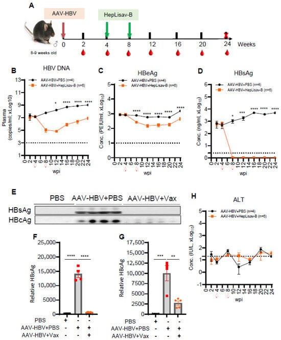
Consistent with prior reports that alum-adjuvanted Engerix-B fails to elicit anti-HBs in AAV-HBV-carrier mice [27,28], Montanide ISA 720 VG with co-administration of HBcAg/IL-12 DNA induced no seroconversion nor reduced viremia (Figure S1A–G). HBsAg and HBcAg remained unchanged in the liver (Figure S1E–G), confirming that conventional adjuvants do not overcome HBV-induced tolerance. We next evaluated HEPLISAV-B. Dose escalation in HBV-naïve mice identified dose 3 (HBsAg/CpG-1018: 2 µg/300 µg) as optimal, inducing the highest sustained anti-HBs titers after two injections (Figure S2A,B). In chronic AAV-HBV mice vaccinated at 4 and 8 wpi (Figure 1A), HEPLISAV-B produced a 1000-fold reduction in serum HBV DNA at 8–12 wpi (Figure 1B), with levels remaining significantly lower than controls through 24 wpi despite a partial rebound at 16 wpi. HBeAg declined significantly (Figure 1C), and, strikingly, HBsAg became undetectable, whereas it persisted in controls (Figure 1D). Immunoblotting confirmed marked depletion of HBsAg and HBcAg in plasma of vaccinated mice at 24 wpi (Figure 1E–G), while ALT remained unchanged (Figure 1H), indicating non-cytolytic viral control.
Figure 1.
HEPLISAV-B suppresses HBV replication in chronic HBV mice. (A) Experimental design: C57BL/6 mice were inoculated with AAV-HBV and treated with PBS or HEPLISAV-B at 4 and 8 weeks post-infection (wpi). Blood was collected from 2 to 24 wpi; mice were euthanized at 24 wpi. (B) Plasma HBV DNA quantified by qPCR. Plasma (C) HBeAg and (D) HBsAg measured by ELISA. (E) Immunoblot analysis of plasma HBsAg and HBcAg at 24 wpi with (F,G) densitometric quantification. (H) Plasma ALT levels measured by ELISA. Data are mean ± SEM. Statistics: Two-way ANOVA with Tukey post hoc test; * p < 0.05, ** p < 0.005, *** p < 0.0005, **** p < 0.00005. Dash line represents the kinetic of the study with X, the time of animals termination. Arrows indicated weeks of HEPLISAV-B administration.
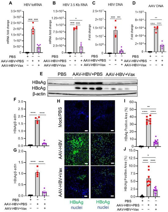
At termination, HEPLISAV-B markedly reduced hepatic HBV total RNA and 3.5 kb RNA (~4-fold; Figure 2A,B), total intrahepatic HBV DNA (~10-fold; Figure 2C), and AAV-HBV vector ITR DNA (~4-fold; Figure 2D). Concordant reductions in hepatic HBs and HBc protein were confirmed by immunoblot and immunofluorescence (Figure 2E–J). Normalization to AAV-HBV template DNA showed proportional loss of HBV RNA and rcDNA (Figure S3A–C), consistent with non-cytolytic depletion of AAV-HBV episome DNA, a functional surrogate for HBV cccDNA in this model (Figure S3D).
Figure 2.
HEPLISAV-B reduces intrahepatic HBV RNA, DNA, and protein. (A–D) RT-qPCR or qPCR was per-formed to measure the levels of (A) HBV total RNA (totRNA), (B) HBV pregenomic RNA (3.5Kb RNA/pgRNA), (C) HBV DNA, and (D) AAV Inverted Terminal Repeats (ITR) DNA (AAV DNA) in the livers of mice at 24 wpi with PBS or AAV-HBV and treated or not with HEPLISAV-B. (E) Immunoblot analysis of HBsAg and HBcAg levels in livers at 24 wpi and (F,G) quantification by ImageJ. (H) Immunofluorescence staining of intrahepatic HBsAg and HBcAg and (I,J) quantification by ImageJ. All images were acquired with 20x magnification lens. Bars in histograms represent the SEM. Statistical analysis was performed with two-way ANOVA and Tukey post hoc test; * p < 0.05; ** p < 0.005; *** p < 0.0005; **** p < 0.00005.
3.2. HEPLISAV-B Induces Strong Anti-HBs IgG1 and IgA Responses in Chronic HBV Carrier Mice
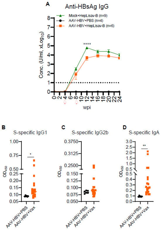
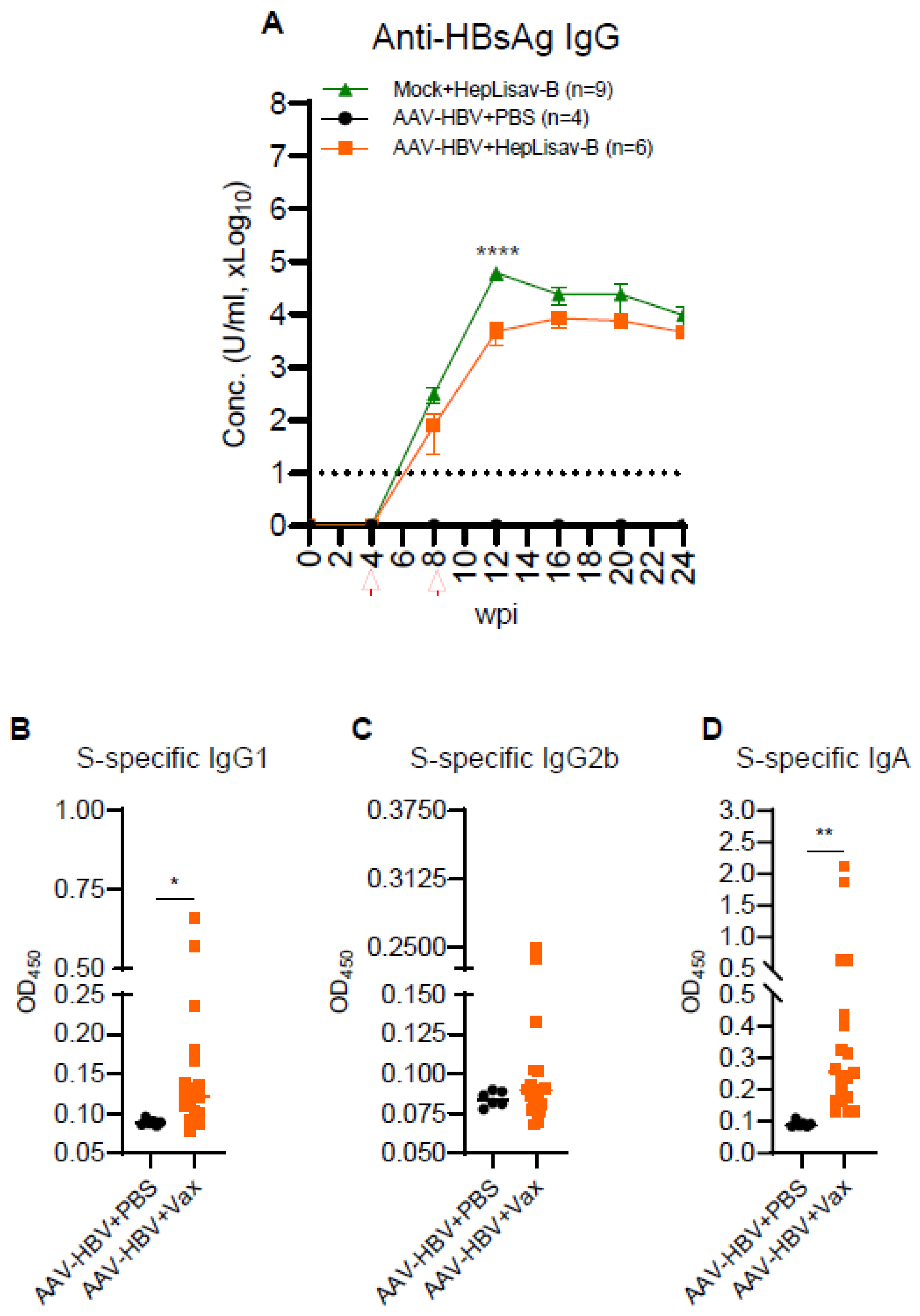
We next sought to determine if HEPLISAV-B-mediated HBV inhibition was associated with HBsAg seroconversion and the development of persistent and functional HBs-specific antibodies in chronic HBV infection. To assess this, we employed ELISA to quantify plasma levels of total anti-HBs IgG and S-specific IgG1, IgG2b, and IgA in vaccinated and control animals. As expected, HEPLISAV-B induced a robust and sustained anti-HBs IgG response in HBV-naïve mice. Importantly, HEPLISAV-B treatment in HBV-infected mice resulted in elevated and persistent anti-HBs IgG levels (≥104 U/mL for over 16 weeks post-vaccination), comparable to vaccinated HBV-naïve mice (Figure 3A). Interestingly, the significantly higher S-specific IgG1 and IgA observed in vaccinated HBV mice (Figure 3B,C) indicated that HEPLISAV-B promotes Th2-biased S-specific responses in the context of chronic HBV infection. The increased levels of S-specific IgA in vaccinated HBV mice (Figure 3D) suggest that HEPLISAV-B also elicits mucosal immunity against HBV.
Figure 3.
HEPLISAV-B induces robust anti-HBs humoral responses. (A) anti-HBsAg IgG levels were measured in plasmas by ELISA from 0 to 24 wpi. Dash line indicates level of detection. Plasma HBsAg-specific (B) IgG1 and (C) IgG2b assessment by ELISA. (D) Plasma HBsAg-specific IgA assessment by ELISA. Data was presented as optical density (OD) at 450 nm. Bars in the scatter plot represent the median value or SEM. Statistical analysis was performed with two-tailed unpaired t test or two-way ANOVA and Tukey post hoc test; * p < 0.05; ** p < 0.005; **** p < 0.00005.
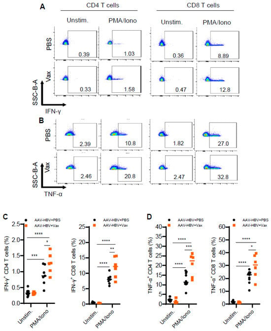
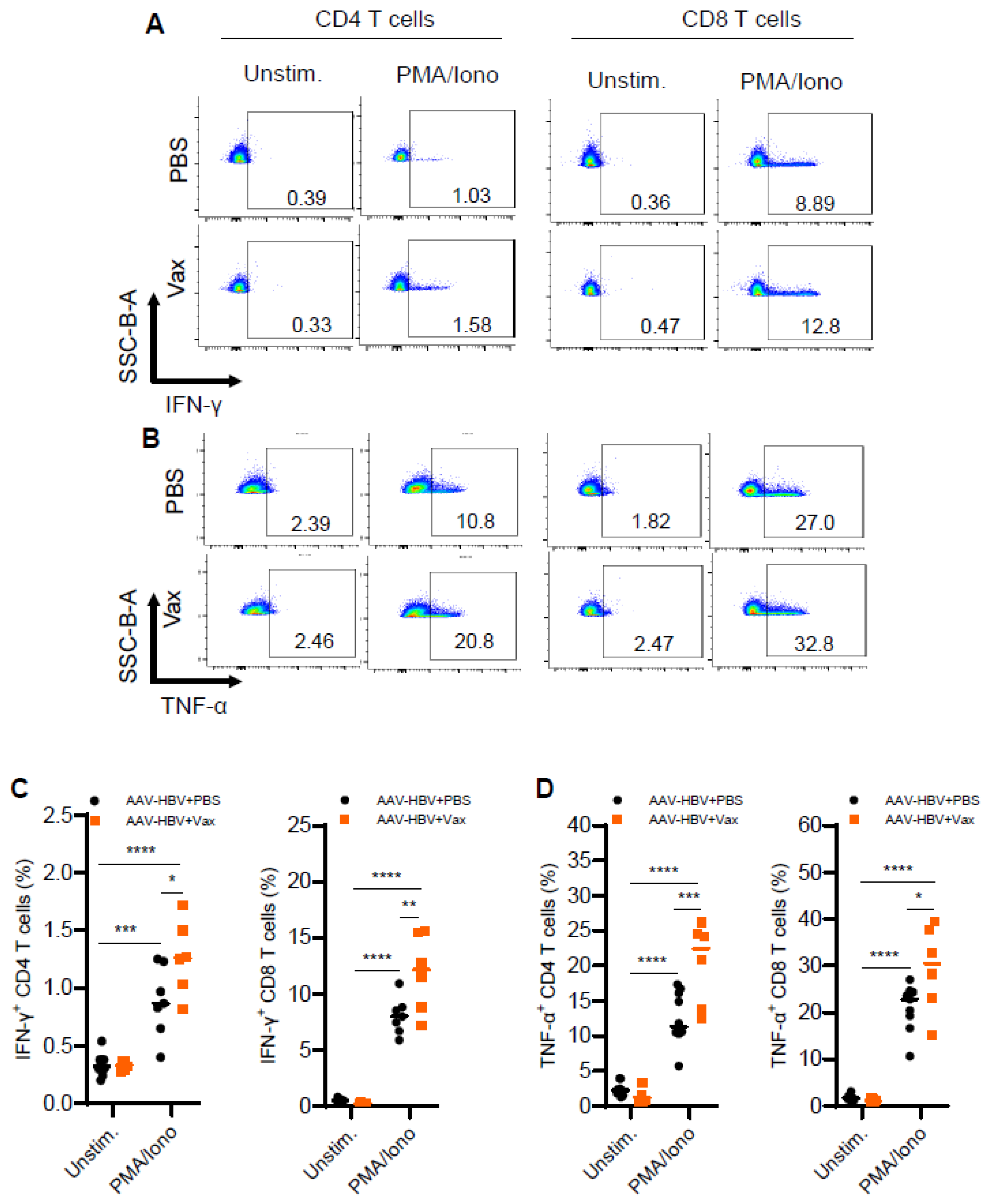
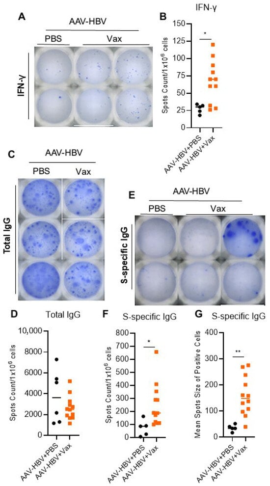
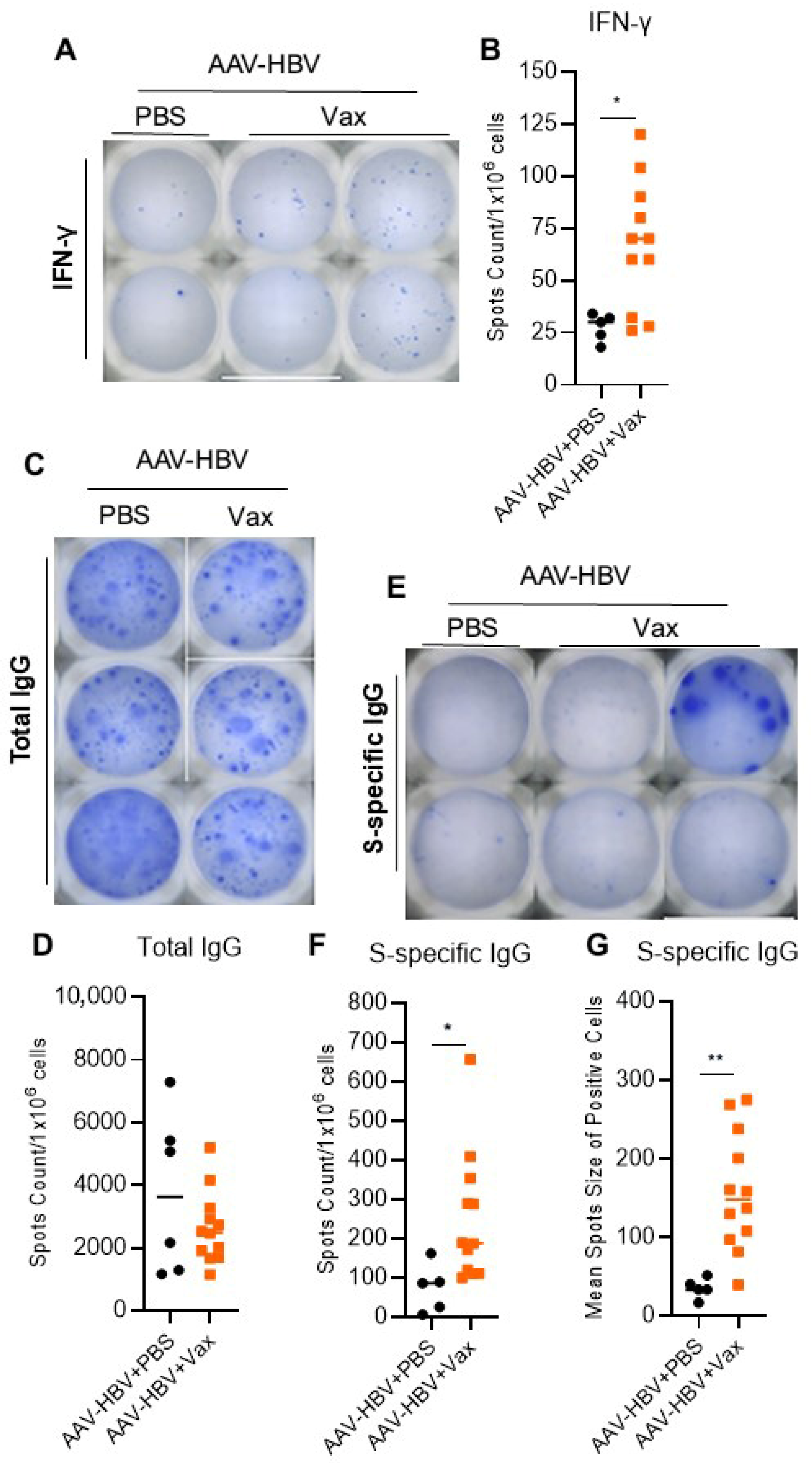
We also analyzed the activation of T cells from splenocytes isolated from vaccinated AAV-HBV and control mice. Ex vivo stimulation of splenocytes with PMA and ionomycin revealed enhanced production of both IFN-γ and TNF-α by CD4 and CD8 T cells from vaccinated HBV mice (Figure 4). Consistent with this, HBs-specific IFN-γ ELISPOT assays demonstrated a greater number of IFN-γ+ spots in splenocytes from vaccinated HBV mice compared to those from PBS-treated HBV mice (Figure 5A,B). Although total IgG ELISPOT analysis showed no significant differences across groups (Figure 5C,D), HEPLISAV-B-treated HBV mice displayed a higher HBs-specific IgG spot count and larger average spot size compared to untreated HBV mice (Figure 5E–G). Taken together, these findings suggest that HEPLISAV-B effectively overcame immune tolerance to HBsAg and induced potent anti-HBs T-cell and B-cell immune responses.
Figure 4.
HEPLISAV-B enhances cytokine production by T cells. Splenocytes from chronic HBV mice treated or not with HEPLISAV-B at 24 wpi were stimulated in vitro with phorbol myristate acetate (PMA) and ionomycin in presence of brefeldin A and analyzed by flow cytometry. (A,B) Representative FACS plots of IFN-γ+ and TNF-α+ CD4 and CD8 T cells. (B–D) Summarized data showing the frequency of IFN-γ+ and TNF-α+ CD4 and CD8 T cells. Bars in the scatter plots represent the median value. Statistical analysis was performed with two-way ANOVA and Tukey post hoc test; * p < 0.05; ** p < 0.005; *** p < 0.0005; **** p < 0.00005.
Figure 5.
HEPLISAV-B restores HBsAg-specific T and B cell responses. (A) Representative ELISPOT wells showing IFN-γ producing splenocytes (blue spots) after 48 h stimulation with HBsAg and (B) Summarized data of IFN-γ spots count analysis. (C,D) Representative ELISPOT wells of total IgG from splenocytes (blue spots) and quantification summary. (E) Representative ELISPOT wells of HBsAg-specific IgG produced by splenocytes (blue spots) after 16 h stimulation with HBsAg and (F,G) detection of spots counts and mean spots size. ELISPOT plates were acquired and analyzed by CTL ImmunoSpot software. Data were QC and normalized to million live cells. Bars in the scatter plots represent the median value. Statistical analysis was per-formed with a two-tailed unpaired t test, * p < 0.05; ** p < 0.005.
3.3. Therapeutic Efficacy of HEPLISAV-B Requires CD4 T Cells but Not CD8 T Cells
To elucidate the immune mechanisms underlying HEPLISAV-B’s therapeutic effect in chronic HBV-carrier mice, we analyzed immune responses in the liver and spleen. At 12 wpi (4 weeks after the second dose), vaccinated HBV mice exhibited an increased spleen and liver size compared to controls (Figure S4A–C). Histological analysis (H&E staining) revealed elevated intrahepatic immune infiltration in HEPLISAV-B-treated mice. Consistent with no elevation of ALT (Figure 1H), the proportion of TUNEL-positive liver cells (apoptotic cell death) was not increased by vaccination (Figure S4D,E). Furthermore, RT-qPCR analysis of hepatic gene expression at 12 wpi showed that HEPLISAV-B treatment did not significantly alter the intrahepatic transcription levels of IFN-I and ISGs (IFN-α2, IFN-β, IFN-γ, OAS1, IP-10), pro-inflammatory cytokines and chemokines (TNF-α, CCL-2, CCL-3, CXCL-9), and immune exhaustion markers (PD-1, PDL-1). Interestingly, HEPLISAV-B treatment significantly reduced hepatic mRNA levels of LY6D, IL-1β, IL-6, TGF-β, and IL-21, while notably increasing the expression of IL-27 and CXCL-11 (Figure S5).
Next, splenic immune profiling revealed an increased frequency of CD45 cells following HEPLISAV-B treatment (Figure S6A). While the proportions of CD4 and CD8 T cells remained similar across all groups, vaccinated HBV mice exhibited a significant increase in B-cell frequency and a reduction in NK cell proportion in the spleen (Figure S6B–E). The frequencies of dendritic cells (DCs) and monocytes/macrophages in the spleen were not altered by vaccination (Figure S6F,G). However, HEPLISAV-B treatment led to a significant reduction in the frequency of regulatory CD4 T cells (Tregs: both CD25+FOXP3+ and CD25−FOXP3+ subsets) (Figure S7A,B), and a significant decrease in PD-1 expression on CD4 T cells (Figure S7C,D). In contrast, PD-1 levels on CD8 T cells were unchanged (Figure S7E). To assess HEPLISAV-B-mediated immediate T cell immunity in chronic HBV+ mice, we analyzed effector memory T cells (TEM: CCR7−CD44+CD62L− CD4 or CD8 T cells) and central memory T cells (TCM: CCR7+CD44+CD62L+ CD4+ or CD8+ T cells) in the spleen. Interestingly, vaccinated HBV mice displayed an increase in TCM CD4 T cells, TEM CD8 T cells and a higher ratio of TCM/TEM CD4 T cells compared with the control group (Figure S7A–F).
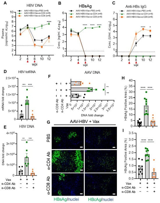
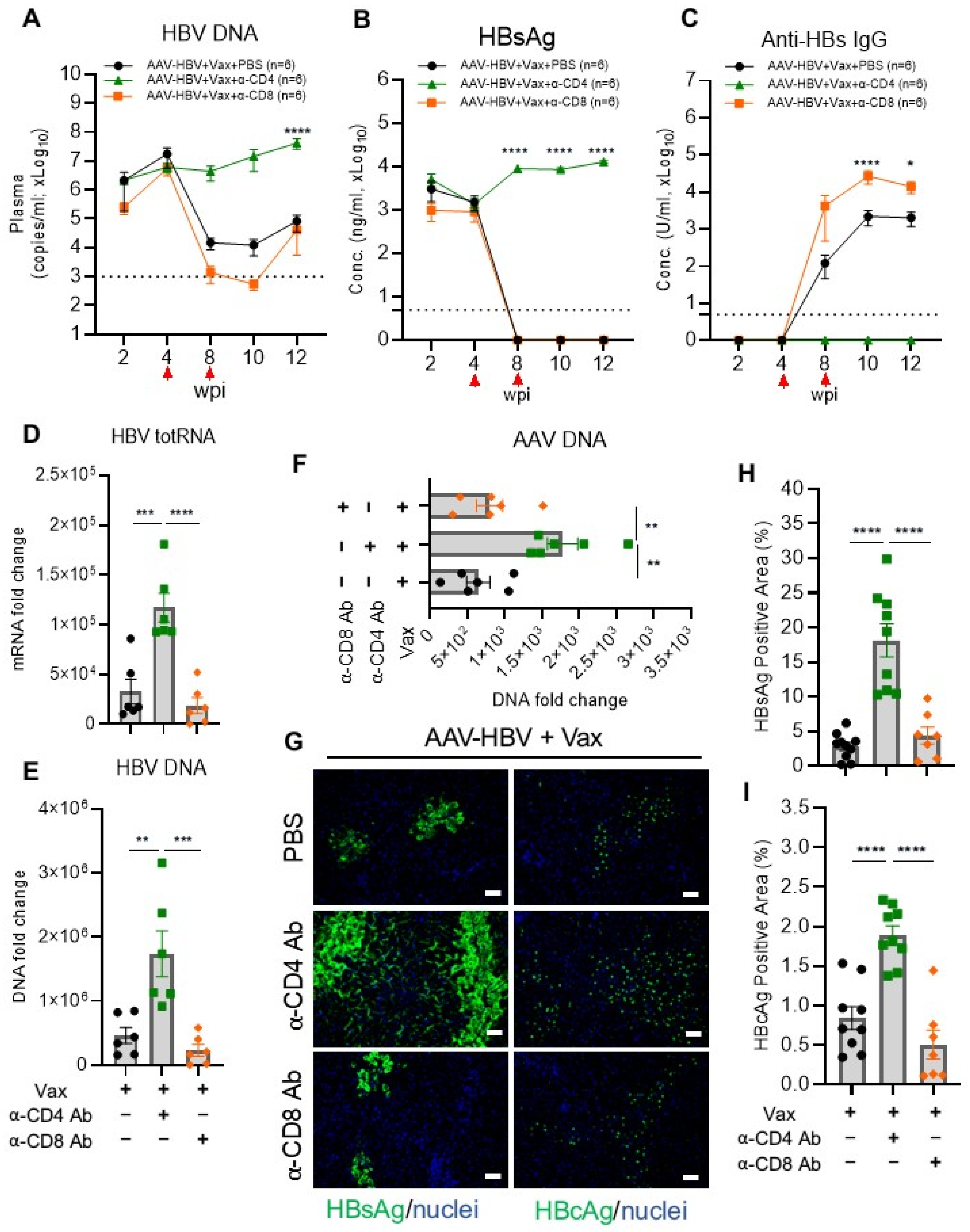
Finally, to investigate the specific contributions of CD4 and CD8 T cells to HEPLISAV-B’s therapeutic effect, HBV-carrier mice were vaccinated and administered with PBS, anti-CD4 antibody (Ab), or anti-CD8 Ab to deplete CD4 and CD8 T cells, respectively (Figure S8A). Effective T-cell depletion was monitored and confirmed in the blood 24 to 48 h post-antibody treatment (Figure S8B–D). While HEPLISAV-B led to significant reduction in HBV DNA and HBsAg clearance in PBS-treated and CD8 T cell-depleted HBV mice, interestingly, HEPLISAV-B’s therapeutic effect was abolished in CD4 T cell-depleted mice (Figure 6A,B). Consistently, HEPLISAV-B treatment failed to induce anti-HBs IgG in CD4 T cell-depleted HBV mice. In addition, HEPLISAV-B-induced HBV DNA lost (HBV totRNA, HBV DNA and AAV DNA) was abrogated in mice treated with anti-CD4 Ab (Figure 6D–F). Furthermore, HEPLISAV-B failed to reduce both HBsAg and HBcAg viral proteins in the liver of CD4 T cell-depleted HBV mice compared with HBV mice treated with PBS or depleted CD8 T cells (Figure 6G–I). Interestingly, vaccinated HBV mice with depleted CD8 T cells showed even higher anti-HBs IgG levels (Figure 6A,C), suggesting that CD8 T-cell depletion ameliorated HEPLISAV-B’s therapeutic effect in chronic HBV infection. Intrahepatic HBsAg and HBcAg levels were similar in vaccinated HBV mice treated with PBS and those depleted of CD8 T cells (Figure 6G–I). Taken together, these data demonstrate that HEPLISAV-B’s therapeutic effect in chronic HBV infection was associated with a decrease in Tregs and PD-1 expression on CD4 T cells. Importantly, this effect depended on CD4 T cells, while depletion of CD8 T cells enhanced HEPLISAV-B’s induction of anti-HBs IgG.
Figure 6.
CD4 T cells are required for HEPLISAV-B–mediated HBV clearance. Chronic HBV carrier mice with HEPLISAV-B received either PBS or anti-CD4 and anti-CD8 antibodies to deplete T cells. (A) HBV DNA detection in plasmas by qPCR. ELISA detection of (B) HBsAg and (C) anti-HBsAg IgG levels in plasmas. Intrahepatic detection of (D) HBV total RNA, (E) HBV DNA and (F) AAV DNA. (G) Immunofluorescence staining of intrahepatic HBsAg and HBcAg and (H,I) quantification by ImageJ. Bars in the scatter plot and his-tograms represent the standard error of the mean. Statistical analysis was performed with two-way ANOVA and Tukey post hoc test; * p < 0.05; ** p < 0.005; *** p < 0.0005; **** p < 0.00005.
3.4. Therapeutic Efficacy of HEPLISAV-B Is Dependent on CD40/CD40L Signaling
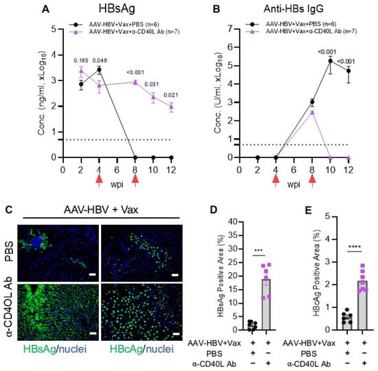
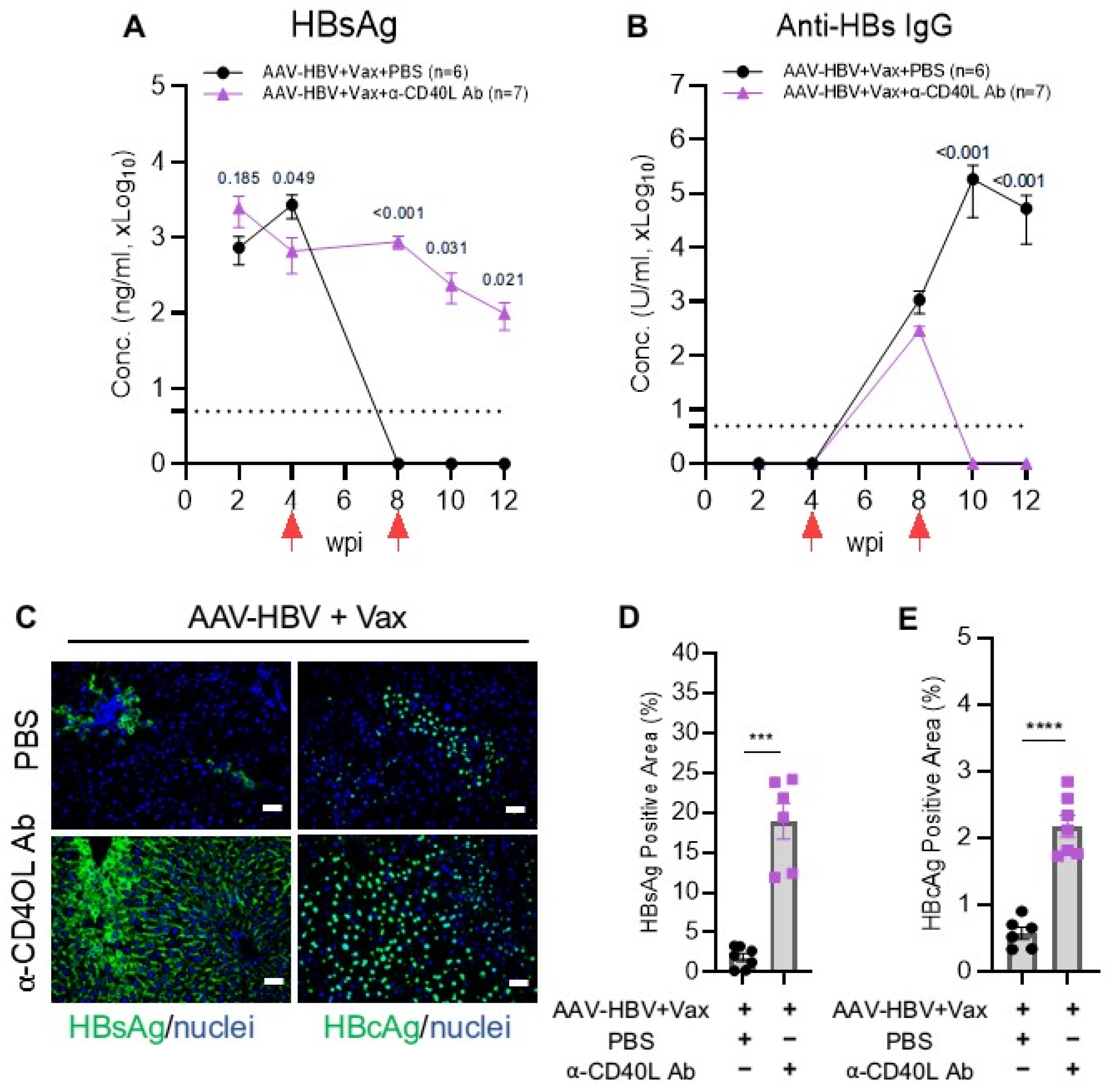
The interaction between CD40 on B cells and its ligand CD40L (CD154) on activated CD4 T cells is a central pathway mediating T cell help, driving B cell activation, clonal expansion, germinal center formation, and antibody production [25,26]. To define the contribution of CD40/CD40L signaling to HEPLISAV-B-induced HBV suppression and antibody induction, HBV-carrier mice were treated with an anti-CD40L blocking antibody in conjunction with HEPLISAV-B (Figure 7). Blockade of CD40/CD40L signaling completely abrogated HEPLISAV-B induced HBsAg clearance, anti-HBs induction (Figure 7A,B), and hepatic reduction in HBs and HBc (Figure 7C–E), establishing the critical role of the CD40/CD40L pathway in HEPLISAV-B mediated antiviral immunity.
Figure 7.
CD40/CD40L signaling is essential for HEPLISAV-B efficacy. Chronic HBV carrier mice with HEPLISAV-B received either PBS or anti-CD40L antibody. ELISA detection of (A) HBsAg and (B) anti-HBs IgG levels in plasmas. (C–E) Immunofluorescence staining of HBsAg and HBcAg levels in the liver, and their quantifications by ImageJ. Bars in the scatter plot and histograms represent the SEM. Statistical analysis was performed with either a two-tailed unpaired t test or two-way ANOVA and Tukey post hoc test; *** p < 0.0005; **** p < 0.00005.
4. Discussion
In this study, we demonstrate that therapeutic vaccination with HEPLISAV-B, comprising recombinant hepatitis B surface antigen (HBsAg) and the Toll-like receptor 9 (TLR9) agonist CpG-1018, overcomes HBV-associated immune dysfunction and induces HBsAg clearance in the AAV-HBV mouse model despite high-level viremia. HEPLISAV-B outperformed alum- and Montanide/IL-12-adjuvanted HBV vaccines, highlighting the importance of adjuvant selection in therapeutic vaccination against persistent HBV infection. Virological control was accompanied by substantial reductions in hepatic HBV transcripts and HBV DNA without detectable evidence of hepatocellular injury. Notably, intrahepatic AAV-HBV vector DNA, quantified as inverted terminal repeat (ITR)-positive episomes, also declined markedly. Because these episomes serve as the transcriptional template for HBV production in this model, their reduction suggests that HEPLISAV-B decreases the abundance of HBV-producing templates through a predominantly non-cytolytic process. Together, these findings demonstrate the therapeutic potential of HEPLISAV-B for controlling HBV antigenemia and reducing intrahepatic viral products in an immunocompetent model of HBV persistence.
HEPLISAV-B elicited a strong and durable anti-HBs antibody response, with titers reaching or exceeding 104 U/mL and enrichment of IgG1 and IgA. These humoral responses were accompanied by increased frequencies of IFN-γ- and TNF-α-producing CD4+ and CD8+ T cells following polyclonal and antigen-specific stimulation, as well as restoration of HBsAg-specific IFN-γ-producing T-cell responses. Vaccination also reduced regulatory T-cell (Treg) frequencies and PD-1 expression on CD4+ T cells while increasing CD4+ central memory and CD8+ effector memory populations [37]. Changes in cytokine and chemokine profiles further suggested a shift away from immunoregulation toward improved T-cell recruitment and function [38,39]. Collectively, these findings indicate that HEPLISAV-B induces coordinated humoral and cellular immune responses while modifying regulatory and memory-associated immune compartments. These effects are consistent with the capacity of CpG-1018 to activate TLR9-dependent innate immune pathways and provide an inflammatory context that supports adaptive immune activation despite persistent HBV antigen exposure.
Mechanistic studies identified CD4+ T cells as essential for vaccine-induced antiviral activity. CD4+ T-cell depletion abrogated anti-HBs induction and HBsAg clearance, prevented suppression of circulating HBV DNA, and impaired the reduction in intrahepatic HBsAg and HBcAg. In contrast, CD8+ T-cell depletion did not impair therapeutic efficacy and instead enhanced anti-HBs titers. This unexpected observation raises the possibility that CD8+ T cells may constrain vaccine-induced humoral responses under the conditions examined. However, the underlying mechanisms remain unknown, and these findings do not establish a generally suppressive role for CD8+ T cells in HBV control. Their effects may depend on cellular functional states and interactions with other immune populations during persistent antigen exposure. Further investigation is needed to determine whether specific CD8+ T-cell subsets directly regulate vaccine responses or whether their depletion indirectly favors CD4+ T-cell help and B-cell activation.
The requirement for CD4+ T cells was closely linked to CD40L–CD40 signaling. Blocking CD40L completely abrogated vaccine-induced anti-HBs antibody production and eliminated antiviral efficacy, demonstrating that this pathway is critical for coupling CD4+ T-cell activity to effective humoral and virological responses. The observation that B cells are the principal downstream mediators of the therapeutic effects associated with CD40L blockade further supports the importance of CD4+ T cell-dependent B-cell activation. These findings are consistent with the established role of CD40 signaling in B-cell activation and antibody production and suggest that innate immune stimulation alone is insufficient to sustain therapeutic control when this pathway is disrupted. Thus, the CD4+ T-cell/CD40L–CD40/B-cell axis provides a mechanistic basis for the antibody response and antiviral efficacy elicited by HEPLISAV-B, consistent with emerging evidence that effective CD4+ T-cell priming is important for therapeutic vaccine responses.
The reduction in intrahepatic AAV-HBV vector DNA provides additional insight into the nature of the antiviral response. HEPLISAV-B reduced hepatic HBV transcripts, total HBV DNA, and ITR-positive episomes without detectable increases in serum ALT or hepatocyte death by TUNEL staining. This pattern supports a predominantly non-cytolytic mechanism rather than widespread hepatocyte destruction. Nevertheless, the decline in vector DNA should be interpreted within the limitations of the model. Because AAV-HBV does not reproduce authentic HBV infection or the formation and maintenance of covalently closed circular DNA (cccDNA), changes in AAV episomes cannot establish that HEPLISAV-B directly eliminates HBV cccDNA in naturally infected hepatocytes. Instead, these findings demonstrate a reduction in the abundance of episomal HBV-producing templates in an immunocompetent host.
The transient rebound of circulating HBV DNA despite sustained HBsAg loss further suggests that these virological markers may reflect distinct biological processes. Residual HBV DNA could originate from defective or noninfectious viral particles, extracellular nucleic acids, or continued production from residual templates. The current data do not distinguish between these possibilities. Future studies should characterize the molecular forms, particle association, and temporal dynamics of residual circulating HBV DNA to clarify its sources and determine whether sustained HBsAg loss corresponds to durable control of intrahepatic HBV-associated nucleic acids.
Several limitations warrant consideration. First, the AAV-HBV model relies on AAV-derived episomal DNA rather than authentic HBV cccDNA. Consequently, reductions in ITR-positive episomes provide an indirect measure of the loss of HBV-producing templates and cannot be equated with cccDNA elimination. The identity and kinetics of residual circulating HBV DNA also remain unresolved. In addition, the durability of virological control beyond the study period and the safety of repeated innate immune stimulation require further investigation. Validation in models that more closely reproduce authentic HBV infection and cccDNA persistence will be necessary to establish the translational relevance of these findings.
Second, species-specific differences in TLR9 expression may influence the mechanisms underlying vaccine responses. In humans, TLR9 expression is concentrated primarily in B cells and plasmacytoid dendritic cells (pDCs), whereas mice exhibit broader TLR9 expression across immune cell populations. Accordingly, the cellular pathways responsible for vaccine-induced immune activation in mice may not fully reflect those operating in humans. Although our depletion and blockade experiments identify key downstream adaptive immune pathways, they do not establish which TLR9-expressing populations directly respond to CpG-1018 or how these populations initiate the subsequent antiviral response. Future studies should define the contributions of B cells, pDCs, and other antigen-presenting populations to vaccine-induced immunity.
Third, the absence of a CpG-1018-alone treatment group limits our ability to distinguish the contributions of adjuvant-mediated innate immune activation from those of HBsAg-specific immune priming. Our study was designed to evaluate the therapeutic potential of the FDA-approved HEPLISAV-B formulation, which was administered consistently to HBV-carrier and HBV-negative control mice, rather than to dissect the individual contributions of its components. Therefore, the observed effects cannot be attributed exclusively to HBsAg, CpG-1018, or their interaction. Future comparisons of HEPLISAV-B with recombinant HBsAg alone and CpG-1018 alone will be important for defining the relative contributions of antigen-specific and adjuvant-mediated responses.
5. Conclusions
This study demonstrates that therapeutic vaccination with HEPLISAV-B, a licensed hepatitis B surface antigen vaccine adjuvanted with the Toll-like receptor 9 agonist CpG-1018, can overcome established immune tolerance and induce functional cure–like virological control in mice with chronic, high-level HBV viremia. Two doses elicited rapid and durable HBsAg clearance, markedly reduced circulating and intrahepatic HBV DNA and RNA, and decreased intrahepatic viral replication and episomal AAV-HBV DNA without evident hepatocellular injury. These effects were accompanied by sustained anti-HBs IgG1 and IgA responses, enhanced HBV-specific T- and B-cell immunity, reduced regulatory T-cell frequencies, and decreased PD-1 expression on CD4 T cells, indicating coordinated restoration of antiviral humoral and cellular immunity. Mechanistically, vaccine efficacy depended on CD4 T cells and CD40–CD40L signaling but was independent of CD8 T cells, identifying a CD4-driven, non-cytolytic pathway of HBV control. Although the AAV-HBV model does not recapitulate authentic HBV covalently closed circular DNA (cccDNA) persistence [32], and the respective contributions of HBsAg and CpG-1018 remain to be defined, these findings provide a strong mechanistic rationale for evaluating HEPLISAV-B as a therapeutic vaccine for chronic hepatitis B. CD4 T-cell help and CD40–CD40L signaling emerge as key mechanisms and potential immune correlates for future studies. Further evaluation in clinically relevant models and clinical trials, including combination strategies with nucleos(t)ide analogues and other immunomodulators, will be essential to determine whether this approach can translate into functional cure in patients.
Supplementary Materials
The following supporting information can be downloaded at: https://www.mdpi.com/article/10.3390/vaccines14110928/s1, Figure S1: HBV-induced immune tolerance in chronic HBV carrier mice; Figure S2: Titration of HEPLISAV-B vaccine in HBV naïve mice; Figure S3: Effect of HEPLISAV-B in the liver of HBV carrier mice; Figure S4: HEPLISAV-B vaccine induces HBV clearance through a non-cytolytic mechanism; Figure S5: HEPLISAV-B vaccination leads to altered cytokines expression in the liver; Figure S6: HEPLISAV-B induces a reduction of Treg cells and PD-1 expression in CD4 T cells in the spleen of chronic HBV mice; Figure S7: TEM and TCM induction in the spleen of chronic HBV mice after HEPLISAV-B treatment; Figure S8: Effect of CD4 and CD8 T cells in HEPLISAV-B-induced HBV clearance.
Author Contributions
Conceptualization: J.A. and L.S. Methodology: J.A. and L.S. Investigation: J.A., J.W. and M.F. Visualization: J.A., J.W. and L.S. Resources: L.T., S.K. and L.S. Supervision: L.S. Writing—original draft: J.A. Writing—review and editing: J.A. and L.S. All authors have read and agreed to the published version of the manuscript.
Funding
This work was supported by the National Institutes of Health (NIH) grants AI138797 and AI175800 (to L.S.), and by the National Cancer Institute/NIH—Cancer Center Support Grant (CCSG)—P30CA134274.
Institutional Review Board Statement
The animal study protocol was approved by the Institutional Review Board of NAME OF the University of Maryland School of Medicine IACUC (protocol code AUP-00000478, 26 February 2024).
Informed Consent Statement
All procedures were approved by the University of Maryland School of Medicine IACUC (AUP-00000478-2/26/2024).
Data Availability Statement
The data presented in this study are available on request from the corresponding author.
Acknowledgments
We thank Weirong Yuan, Yichen Lu, and Sahra Sharifi for their technical support and acknowledge the University of Maryland School of Medicine Animal Facility Core and the Histology and Flow Cytometry Shared Services of the University of Maryland Marlene and Stewart Greenebaum Comprehensive Cancer.
Conflicts of Interest
The authors declare no conflicts of interest.
References
- Mahmood, F.; Xu, R.; Awan, M.U.N.; Song, Y.; Han, Q.; Xia, X.; Wei, J.; Xu, J.; Peng, J.; Zhang, J. HBV Vaccines: Advances and Development. Vaccines 2023, 11, 1862. [Google Scholar] [CrossRef] [Scilit] [PubMed]
- Lok, A.S.; Zoulim, F.; Dusheiko, G.; Ghany, M.G. Hepatitis B cure: From discovery to regulatory approval. Hepatology 2017, 66, 1296–1313. [Google Scholar] [CrossRef] [Scilit] [PubMed]
- Trepo, C.; Chan, H.L.; Lok, A. Hepatitis B virus infection. Lancet 2014, 384, 2053–2063. [Google Scholar] [CrossRef] [Scilit] [PubMed]
- Asabe, S.; Wieland, S.F.; Chattopadhyay, P.K.; Roederer, M.; Engle, R.E.; Purcell, R.H.; Chisari, F.V. The size of the viral inoculum contributes to the outcome of hepatitis B virus infection. J. Virol. 2009, 83, 9652–9662. [Google Scholar] [CrossRef] [Scilit] [PubMed]
- Chisari, F.V.; Isogawa, M.; WielandSF. Pathogenesis of hepatitis B virus infection. Pathol. Biol. 2010, 58, 258–266. [Google Scholar] [CrossRef] [Scilit] [PubMed]
- Yang, P.L.; Althage, A.; Chung, J.; Maier, H.; Wieland, S.; Isogawa, M.; Chisari, F.V. Immune effectors required for hepatitis B virus clearance. Proc. Natl. Acad. Sci. USA 2010, 107, 798–802. [Google Scholar] [CrossRef] [Scilit] [PubMed]
- Burton, A.R.; Pallett, L.J.; McCoy, L.E.; Suveizdyte, K.; Amin, O.E.; Swadling, L.; Alberts, E.; Davidson, B.R.; Kennedy, P.T.; Gill, U.S.; et al. Circulating and intrahepatic antiviral B cells are defective in hepatitis B. J. Clin. Investig. 2018, 128, 4588–4603. [Google Scholar] [CrossRef] [Scilit] [PubMed]
- Gehring, A.J.; Protzer, U. Targeting Innate and Adaptive Immune Responses to Cure Chronic HBV Infection. Gastroenterology 2019, 156, 325–337. [Google Scholar] [CrossRef] [Scilit] [PubMed]
- Duan, X.Z.; Wang, M.; Li, H.W.; Zhuang, H.; Xu, D.; Wang, F.S. Decreased frequency and function of circulating plasmocytoid dendritic cells (pDC) in hepatitis B virus infected humans. J. Clin. Immunol. 2004, 24, 637–646. [Google Scholar] [CrossRef] [Scilit] [PubMed]
- Mueller, S.N.; Ahmed, R. High antigen levels are the cause of T cell exhaustion during chronic viral infection. Proc. Natl. Acad. Sci. USA 2009, 106, 8623–8628. [Google Scholar] [CrossRef] [Scilit] [PubMed]
- Shi, B.; Ren, G.; Hu, Y.; Wang, S.; Zhang, Z.; Yuan, Z. HBsAg inhibits IFN-alpha production in plasmacytoid dendritic cells through TNF-alpha and IL-10 induction in monocytes. PLoS ONE 2012, 7, e44900. [Google Scholar] [CrossRef] [Scilit] [PubMed]
- Han, Q.; Lan, P.; Zhang, J.; Zhang, C.; Tian, Z. Reversal of hepatitis B virus-induced systemic immune tolerance by intrinsic innate immune stimulation. J. Gastroenterol. Hepatol. 2013, 28, 132–137. [Google Scholar] [CrossRef] [Scilit] [PubMed]
- Michler, T.; Zillinger, J.; Hagen, P.; Cheng, F.; Festag, J.; Kosinska, A.; Protzer, U. The lack of HBsAg secretion does neither facilitate induction of antiviral T cell responses nor Hepatitis B Virus clearance in mice. Antivir. Res. 2024, 226, 105896. [Google Scholar] [CrossRef] [Scilit] [PubMed]
- Rexhouse, C.; Bailey, J.T.; Moshkani, S.; Cimino, J.L.; Robek, M.D. High antigen burden drives CD8+ T cell dysfunction in a mouse model of chronic hepatitis B virus infection. J. Virol. 2025, 99, e0071125. [Google Scholar] [CrossRef] [Scilit] [PubMed]
- Yang, D.; Liu, L.; Zhu, D.; Peng, H.; Su, L.; Fu, Y.X.; Zhang, L. A mouse model for HBV immunotolerance and immunotherapy. Cell. Mol. Immunol. 2014, 11, 71–78. [Google Scholar] [CrossRef] [Scilit] [PubMed]
- Backes, S.; Jager, C.; Dembek, C.J.; Kosinska, A.D.; Bauer, T.; Stephan, A.S.; Dišlers, A.; Mutwiri, G.; Busch, D.H.; Babiuk, L.A.; et al. Protein-prime/modified vaccinia virus Ankara vector-boost vaccination overcomes tolerance in high-antigenemic HBV-transgenic mice. Vaccine 2016, 34, 923–932. [Google Scholar] [CrossRef] [Scilit] [PubMed]
- Bian, Y.; Zhang, Z.; Sun, Z.; Zhao, J.; Zhu, D.; Wang, Y.; Fu, S.; Guo, J.; Liu, L.; Su, L.; et al. Vaccines targeting preS1 domain overcome immune tolerance in hepatitis B virus carrier mice. Hepatology 2017, 66, 1067–1082. [Google Scholar] [CrossRef] [Scilit] [PubMed]
- Zhu, D.; Liu, L.; Yang, D.; Fu, S.; Bian, Y.; Sun, Z.; He, J.; Su, L.; Zhang, L.; Peng, H.; et al. Clearing Persistent Extracellular Antigen of Hepatitis B Virus: An Immunomodulatory Strategy To Reverse Tolerance for an Effective Therapeutic Vaccination. J. Immunol. 2016, 196, 3079–3087. [Google Scholar] [CrossRef] [Scilit] [PubMed]
- Su, J.; Brunner, L.; Ates Oz, E.; Sacherl, J.; Frank, G.; Kerth, H.A.; Thiele, F.; Wiegand, M.; Mogler, C.; Aguilar, J.C.; et al. Activation of CD4 T cells during prime immunization determines the success of a therapeutic hepatitis B vaccine in HBV-carrier mouse models. J. Hepatol. 2023, 78, 717–730. [Google Scholar] [CrossRef] [Scilit] [PubMed]
- Bailey, J.T.; Cangialosi, S.; Moshkani, S.; Rexhouse, C.; Cimino, J.L.; Robek, M.D. CD40 stimulation activates CD8+ T cells and controls HBV in CD4-depleted mice. JHEP Rep. 2024, 6, 101121. [Google Scholar] [CrossRef] [Scilit] [PubMed]
- Conceicao-Neto, N.; Pierson, W.; Vacca, M.; Beyens, M.; De Clerck, B.; Aerts, L.; Voeten, B.; De Pooter, D.; Verschueren, L.; Dockx, K.; et al. Sustained Liver HBsAg Loss and Clonal T- and B-Cell Expansion upon Therapeutic DNA Vaccination Require Low HBsAg Levels. Vaccines 2023, 11, 1825. [Google Scholar] [CrossRef] [Scilit] [PubMed]
- Kosinska, A.D.; Moeed, A.; Kallin, N.; Festag, J.; Su, J.; Steiger, K.; Michel, M.L.; Protzer, U.; Knolle, P.A. Synergy of therapeutic heterologous prime-boost hepatitis B vaccination with CpG-application to improve immune control of persistent HBV infection. Sci. Rep. 2019, 9, 10808. [Google Scholar] [CrossRef] [Scilit] [PubMed]
- Sacherl, J.; Kosinska, A.D.; Kemter, K.; Kachele, M.; Laumen, S.C.; Kerth, H.A.; Öz, E.A.; Wolff, L.S.; Su, J.; Essbauer, S.; et al. Efficient stabilization of therapeutic hepatitis B vaccine components by amino-acid formulation maintains its potential to break immune tolerance. JHEP Rep. 2023, 5, 100603. [Google Scholar] [CrossRef] [Scilit] [PubMed]
- Ye, X.; Chen, X.; Liu, H.; Jiang, Y.; Yang, C.; Xu, T.; Chen, Z.; Wang, Y.; Chen, F.; Liu, X.; et al. HBsAg and TLR7/8 dual-targeting antibody-drug conjugates induce sustained anti-HBV activity in AAV/HBV mice: A preliminary study. Antib. Ther. 2024, 7, 249–255. [Google Scholar] [CrossRef] [Scilit] [PubMed]
- Krieg, A.M. CpG motifs in bacterial DNA and their immune effects. Annu. Rev. Immunol. 2002, 20, 709–760. [Google Scholar] [CrossRef] [Scilit] [PubMed]
- Scheiermann, J.; Klinman, D.M. Clinical evaluation of CpG oligonucleotides as adjuvants for vaccines targeting infectious diseases and cancer. Vaccine 2014, 32, 6377–6389. [Google Scholar] [CrossRef] [Scilit] [PubMed]
- Campbell, J.D. Development of the CpG Adjuvant 1018: A Case Study. Methods Mol. Biol. 2017, 1494, 15–27. [Google Scholar] [CrossRef] [Scilit] [PubMed]
- Kumru, O.S.; Bajoria, S.; Kaur, K.; Hickey, J.M.; Van Slyke, G.; Doering, J.; Berman, K.; Richardson, C.; Lien, H.; Kleanthous, H.; et al. Effects of aluminum-salt, CpG and emulsion adjuvants on the stability and immunogenicity of a virus-like particle displaying the SARS-CoV-2 receptor-binding domain (RBD). Hum. Vaccines Immunother. 2023, 19, 2264594. [Google Scholar] [CrossRef] [Scilit] [PubMed]
- Girndt, M.; Houser, P.; Manllo-Karim, R.; Ervin, J.E.; Charytan, C.; Chow, S.; Symonian-Silver, M.; Lehrner, L.; Linfert, D.; Shemin, D.; et al. Long-term immunogenicity and safety of the hepatitis B vaccine HepB-CpG (HEPLISAV-B) compared with HepB-Eng (Engerix-B) in adults with chronic kidney disease. Vaccine 2023, 41, 3224–3232. [Google Scholar] [CrossRef] [Scilit] [PubMed]
- Reilly-Evans, B.; Dudzik, B.; Costlow, D.J.; Hartmann, C.; Khalsa, A.M.; Kassis, C.; Zmarlicka, M.T. Observational Study Evaluating the Seroprotection of HepB-alum Vaccine and HepB-CpG Vaccine in People With HIV. Open Forum Infect. Dis. 2023, 10, ofad267. [Google Scholar] [CrossRef] [Scilit] [PubMed]
- Marks, K.M.; Kang, M.; Umbleja, T.; Cox, A.; Vigil, K.J.; Ta, N.T.; Omoz-Oarhe, A.; Perazzo, H.; Kosgei, J.; Hatlen, T.; et al. HepB-CpG vs HepB-Alum Vaccine in People With HIV and Prior Vaccine Nonresponse: The BEe-HIVe Randomized Clinical Trial. JAMA 2025, 333, 295–306. [Google Scholar] [PubMed]
- Ahodantin, J.; Lekbaby, B.; Bou Nader, M.; Soussan, P.; Kremsdorf, D. Hepatitis B virus X protein enhances the development of liver fibrosis and the expression of genes associated with epithelial-mesenchymal transitions and tumor progenitor cells. Carcinogenesis 2020, 41, 358–367. [Google Scholar] [CrossRef] [Scilit] [PubMed]
- Ahodantin, J.; Nio, K.; Funaki, M.; Zhai, X.; Wilson, E.; Kottilil, S.; Cheng, L.; Li, G.; Su, L. Type I interferons and TGF-beta cooperate to induce liver fibrosis during HIV-1 infection under antiretroviral therapy. JCI Insight 2022, 7, e152738. [Google Scholar] [CrossRef] [Scilit] [PubMed]
- Ahodantin, J.; Wu, J.; Funaki, M.; Flores, J.; Wang, X.; Zheng, P.; Liu, Y.; Su, L. Siglec-H(−/−) Plasmacytoid Dendritic Cells Protect Against Acute Liver Injury by Suppressing IFN-gamma/Th1 Response and Promoting IL-21(+) CD4 T Cells. Cell. Mol. Gastroenterol. Hepatol. 2024, 18, 101367. [Google Scholar] [CrossRef] [Scilit] [PubMed]
- Strickland, A.B.; Sun, D.; Sun, P.; Chen, Y.; Liu, G.; Shi, M. IL-27 Signaling Promotes Th1 Responses and Is Required to Inhibit Fungal Growth in the Lung during Repeated Exposure to Aspergillus fumigatus. Immunohorizons 2022, 6, 78–89. [Google Scholar] [CrossRef] [Scilit] [PubMed]
- Wing, K.; Onishi, Y.; Prieto-Martin, P.; Yamaguchi, T.; Miyara, M.; Fehervari, Z.; Nomura, T.; Sakaguchi, S. CTLA-4 control over Foxp3+ regulatory T cell function. Science 2008, 322, 271–275. [Google Scholar] [CrossRef] [Scilit] [PubMed]
- Sakaguchi, S.; Wing, K.; Onishi, Y.; Prieto-Martin, P.; Yamaguchi, T. Regulatory T cells: How do they suppress immune responses? Int. Immunol. 2009, 21, 1105–1111. [Google Scholar] [CrossRef] [Scilit] [PubMed]
- Ahodantin, J.; Li, F.; Su, L. Modeling HBV Infection and Therapy in Immunodeficient NOD-Rag1-/-IL2RgammaC-null (NRG) Fumarylacetoacetate Hydrolase (FAH) Knockout Mice with Human Chimeric Liver. Methods Mol. Biol. 2024, 2837, 199–206. [Google Scholar] [CrossRef] [Scilit] [PubMed]
- Ahodantin, J.; Bou-Nader, M.; Cordier, C.; Megret, J.; Soussan, P.; Desdouets, C.; Kremsdorf, D. Hepatitis B virus X protein promotes DNA damage propagation through disruption of liver polyploidization and enhances hepatocellular carcinoma initiation. Oncogene 2019, 38, 2645–2657. [Google Scholar] [CrossRef] [Scilit] [PubMed]

Disclaimer/Publisher’s Note: The statements, opinions and data contained in all publications are solely those of the individual author(s) and contributor(s) and not of MDPI and/or the editor(s). MDPI and/or the editor(s) disclaim responsibility for any injury to people or property resulting from any ideas, methods, instructions or products referred to in the content. |
© 2026 by the authors. Licensee MDPI, Basel, Switzerland. This article is an open access article distributed under the terms and conditions of the Creative Commons Attribution (CC BY) license.