


Abstract
The potency of urate, an abundant human plasma antioxidant, in preventing oxidative damage caused by the carbonate radical anion CO3•−, was studied using quantum chemical calculations. The influence of microhydration of CO3•−/CO32− and urate−/urate• couples on the thermodynamic and kinetics of the one-electron oxidation process was investigated. Depending on the degree of microhydration, the estimated rate constant for one-electron transfer is in the range of 2.0–7.3 × 10⁹ M−1 s−1, in good agreement with the experimental value of 1.3 × 10⁹ M−1 s−1. Modeling using vertical detachment energy and electron affinity, the driving forces of single electron transfer revealed urate(H2O)6− and CO3(H2O)9•− clusters as the most likely existing species in water. Molecular docking revealed a favorable interaction of urate with the catalytic pocket of SOD1. Urate binds more strongly to the anionic active center of SOD1 than the reference inhibitor LSC-1, indicating its potency to prevent HCO3−-supported CO3•− formation. In contrast, the known OGG1 inhibitor TH13264 shows substantially stronger binding than urate, indicating urate’s weaker affinity toward the DNA repair enzyme catalytic pocket. The molecular dynamics data indicate that urate binding does not destabilize either SOD1 or OGG1. In light of increasing evidence that the major source of oxidative stress could be CO3•−, rather than the commonly assumed hydroxyl radical HO•, the obtained results indicate the inherent ability of plasma to combat oxidative stress induced by this selective, milder oxidant. Such an ability with respect to the non-selective, highly reactive HO• does not exist in vivo.
1. Introduction
Reactive oxygen and nitrogen species (ROS/RNS) overproduced by endogenous or exogenous stimuli are the major contributors to oxidative stress conditions [1]. The literature is continuously filled with theoretical discoveries of highly potent scavengers of ROS/RNS. Presumed scavengers are mainly natural phenolic compounds with very low concentration in biological sources of the human diet, usually much below 1 μM (and thus with poor bioaccessibility and bioavailability), and mostly with scarce water solubility. Consequently, their effective in vivo activity is questionable because physiological concentration could be a more important limitation than kinetics. Those facts have been reviewed and emphasized a decade ago [2], but are commonly overlooked or ignored by theoreticians. Earlier, radical scavenging was synonymous with antioxidant action. Free radicals were considered only as ‘bad gays’, and their elimination by scavenging seemed to be a key for suppressing oxidative stress [3]. Also, direct scavenging was designated as ‘primary’ antioxidant activity. Now, it is well-known that a plethora of antioxidant pathways exists, and their underlying mechanisms are far from completely elucidated, as well as the possible ranking of their in vivo physiological significance [4]. Moreover, enzyme reactions usually enable antioxidant defense rather than nonenzymatic direct radical scavenging [2].
On the other hand, despite the progress made, there are many challenges regarding activities and modes of action of existing low-molecular-weight antioxidants present in high concentration in the human body. One such compound is uric acid, the final product of purine metabolism. In aqueous environments, uric acid is present as urate (monovalent anion). The daily production and excretion of uric acid in males amounts to 600–700 mg [5]. The disruption of this homeostasis has been associated with the risk of gout and associated medical conditions such as cardiovascular diseases [6]. Normal urate concentrations in human plasma and serum are very high, 150–450 μM [7] and 155–430 μM [8], respectively, and somewhat lower in intracellular and other body fluids. Urate is the main contributor to the antioxidant capacity of human plasma (40–55%), overwhelming ascorbate (8–15%), α-tocopherol (<10%), and flavonoids (<2%) [7,9]. Thus, it is an important endogenous antioxidant capable of suppressing oxidative stress conditions by protecting the cell membrane, DNA, enzymes, and other biological molecules against oxidation by ROS/RNS. For example, experimental and theoretical evidence exist that uric acid can be efficient for repairing damaged protein residues [10,11,12].
One of the ROS is the carbonate radical anion, CO3•−, a species which attracts increased attention due to its presumed substantial involvement in oxidative stress genesis [13,14,15]. For years, it has been believed that the major cause of oxidative stress is the hydroxyl radical, HO• [16]. This extremely reactive species non-selectively reacts at the place of its generation with any nearby biomolecule, mainly with diffusion-controlled rates [17]. Because of that, HO•-effective in vivo direct scavenging is impossible: there are no enzymatic systems and scavengers capable of eliminating it [2,18]. Increasing evidence suggests that at physiological conditions, CO3•−, not HO•, is the major source of oxidative stress because in vivo in the HCO3−/CO32− buffering system CO3•− is produced, rather than HO• [19]. CO3•− is a powerful oxidant but milder than HO•: its reactions with various biological molecules are several orders of magnitude slower than the reactions of HO• [20,21]. CO3•− can diffuse away from the place of its generation and selectively oxidize critical biomolecules, e.g., amino acids, proteins, enzymes, lipids, and DNA. Unlike HO•, it could be in vivo eliminated by plasma antioxidants by direct scavenging.
When considering free radical scavenging as an antioxidant mechanism in physiological water environment, several common pathways could be operative: formal hydrogen atom transfer (fHAT), sequential proton loss followed by electron transfer (SPLET), single electron transfer (SET), single electron transfer followed by proton transfer (SET-PT), and radical adduct formation (RAF) [22]. Amongst them, it has been found that the SET is the most favorable mechanism for scavenging CO3•−. For example, SET from 2′-deoxyguanosine to CO3•− proceeds faster than fHAT and RAF [23]. Also, SET as the operative pathway was considered in scavenging of CO3•− by urate, ascorbate, and caffeate [24], α-tocopherol, α-CEHC, and gallic acid [25], and Trolox [26].
In water environments, both urate and CO3•− are anticipated to be strongly solvated and can form the water clusters urate(H2O)n− [27] and CO3(H2O)n•− [28,29,30,31]. The formation of a hydration shell via hydrogen bonds with water molecules alters their molecular structure and physicochemical properties. The number of water molecules in urate(H2O)n− and CO3(H2O)n•− clusters has not been unequivocally defined so far.
The first goal of this research is to theoretically estimate the potency of urate for direct scavenging of CO3•− under physiological conditions, as well as to determine the size of existing urate(H2O)n− and CO3(H2O)n•− clusters. This is achieved by examining the thermodynamics and kinetics underlying the SET mechanism and by analyzing the relationships between the VDE (vertical detachment energy) and AEA (adiabatic electron affinity), the main driving forces of electron transfer, and the reaction rate.
The second goal of this study is to ascertain the ability of urate to inhibit the peroxidase activity of the cytosolic isoform of superoxide dismutase (SOD) SOD1, i.e., Cu,Zn-SOD. SOD is known as an antioxidant enzyme that catalyzes the dismutation of O22− to generate H2O2 and O2, i.e., inhibits the damage triggered by O22−. On the other hand, SOD-1 may act as a prooxidant [32]. Namely, at physiological conditions in the presence of H2O2 and HCO3−, SOD-1 peroxidase activity produces CO3•−. It is proposed that at the active site of SOD1, hydrogen peroxide forms a copper-bound oxidant [33] which oxidizes HCO3−, anchored at anionic binding site, to CO3•− or inactivates the enzyme by oxidation of adjacent histidine residues [34]. CO3•− is also capable of oxidizing histidine residues as well as diffusing out of the active site to induce oxidation of surrounding biomolecules. In vitro evidence suggests that urate may prevent H2O2-induced inactivation of SOD1, but the underlying mechanism is not fully elucidated [35].
Also, the potential of urate to inhibit or activate the DNA repair enzyme 8-oxoguanine DNA glycosylase 1 (OGG1) was investigated. OGG1 is a promising multifunctional therapeutic target for the treatment of oxidative DNA damage-related diseases, such as malignant tumors, age-related disorders, and incurable inflammation [36].
2. Materials and Methods
2.1. DFT Computation
The density functional theory (DFT) represents a powerful tool for studying free radical scavenging mechanisms of natural compounds [22]. The M06-2X functional and 6–311++G(d,p) basis set was used to perform the optimization and frequency calculation of urate and CO3•− species involved in the SET mechanism. The M06-2X functional has been chosen because it is designed for main-group chemistry and recommended for thermochemical kinetics [37]. It has been recognized as particularly suitable for the estimation of thermodynamics and kinetics of reactions involving free radicals [22]. Explicitly hydrated reactants are immersed in the solvent continuum modeled by the SMD solvation approach [38]. For open-shell systems, unrestricted calculations were performed. The spin operator ⟨S2⟩ values for radical species, before and after the annihilation of the first spin contaminant, have been checked to avoid possible untrue results. Spin contamination can be considered negligible if the value of ⟨S2⟩ differs from the correct value (⟨S2⟩ = 0.75 for a pure doublet) by less than 10%. All computations were performed using the Gaussian 09 program package [39].
2.2. Estimations of Thermodynamic and Kinetic Data
The Eyringpy program [40] was used for calculations of thermodynamic and kinetic data. The Gibbs free energy of activation, , of investigated SET reactions was estimated by using Marcus theory [41]. This theory is based on the transition state theory (TST) and enables calculating the barrier of any SET reaction from two thermodynamic parameters: the free energy of the reaction, , and the nuclear reorganization energy, λ:
is the nonadiabatic energy difference between reactants and vertical products for SET.
The TST rate constant for the SET reaction, kTST is calculated as
kB is the Boltzmann constant, T is the temperature, h is the Planck constant, and R is the gas constant.
If kTST is close to the diffusion limit (kTST > 109 M−1 s−1), the apparent rate constant, kapp, and rate constant for an irreversible bimolecular diffusion-controlled reaction, kD, were calculated:
RAB is the reaction distance, and DAB is the mutual diffusion coefficient of the reactants A and B, and NA is the Avogadro constant.
kD = 4πRABDABNA
The rate constant for the SET mechanism at pH 7.4 was calculated involving kapp and the molar fraction of both reactants, i.e., Mfurate = 0.9862 for urate, and MfCO3•− = 1 for carbonate radical anion:
Molar fractions were estimated from experimentally determined pKa values. MfCO3•− amounts to 1 because CO3•− is a conjugated base of a strong acid, HCO3• (pKa < 0 [42]). Such a calculated rate constant is directly related to the experimentally determined one under the same conditions.
The vertical detachment energy (VDE) is defined as the difference in electronic energy of urate and its corresponding radical, both in the optimized geometry of urate [43].
The electron affinity (EA) is the difference in electronic energy of CO3•−–water cluster and CO32−–water cluster in their respective optimized geometry [44].
2.3. Molecular Docking and Binding Affinity Evaluation
To explore the interaction profile and binding affinity of urate toward SOD1 and OGG1, a detailed molecular docking analysis was performed using AutoDock Tools 1.5.7 in combination with AutoDock 4.2.6 [45]. A structured and validated docking protocol was applied to ensure the reliability and reproducibility of the obtained results [46,47,48]. The urate structure was reoptimized at the B3LYP-D3BJ/6-311+G(d,p) level of theory, providing an accurate description of the ligand’s geometrical and electronic properties [49,50]. The crystal structures of SOD1 (PDB ID: 1CB4) and OGG1 (PDB ID: 8BVX) were retrieved from the Protein Data Bank [51]. Protein preparation was conducted using BIOVIA Discovery Studio 2021 [52], including the removal of co-crystallized ligands, water molecules, and non-essential heteroatoms to avoid artificial interactions during docking simulations. For SOD1, a grid box of 40 × 40 × 40 Å was centered at coordinates (10.41, 87.88, 18.62 Å) in order to encompass the known catalytic/electrostatic channel of SOD1, located in the vicinity of the Cu/Zn metal binding region and amino acid residues relevant for substrate access and stabilization, whereas for OGG1, a grid box of 29 × 29 × 29 Å was defined with the grid center positioned at (−8.70, −15.91, 12.50 Å), based on the selected binding regions. Docking calculations were performed using the Lamarckian Genetic Algorithm (LGA), which enables efficient sampling of flexible ligand conformations within a rigid protein framework [53]. The following docking parameters were applied: population size of 150, maximum number of 2,500,000 energy evaluations, 27,000 generations, mutation rate of 0.02, and crossover rate of 0.8. For each protein–ligand system, 100 independent docking runs were carried out to ensure statistically significant sampling and identification of the most probable binding conformations.
2.4. Molecular Dynamics Study
To evaluate the structural stability and interaction dynamics of the urate-SOD1 and urate–OGG1 complexes, molecular dynamics (MD) simulations were performed using the AMBER 22 simulation package [54]. The ff14SB force field was employed for the protein component. In contrast, ligand parameters and system preparation were generated through the CHARMM-GUI platform, enabling the construction of AMBER-compatible topology and coordinate files [55,56]. The Cu and Zn ions were retained and treated using standard non-bonded ion parameters from the CHARMM toppar libraries, without additional bonded or QM/MM treatment of the metal coordination sphere. The docked complexes exhibiting the most favorable binding modes were embedded in a truncated octahedral periodic box filled with explicit TIP3P water molecules, maintaining a minimum distance of 10 Å between the solute and the box boundary [57]. System neutrality and physiological ionic strength (0.15 M) were achieved by introducing appropriate numbers of K+ and Cl− counterions. Prior to the production phase, a two-step energy minimization protocol was applied. Initially, solvent molecules and ions were relaxed while positional restraints were maintained on the solute (2500 steps of steepest descent followed by 2500 steps of conjugate gradient). Subsequently, the entire system was subjected to unrestrained minimization. Thermal equilibration was then carried out in sequential stages: a 1 ns NVT simulation at 300 K using the Langevin thermostat (collision frequency of 2 ps−1), followed by a 1 ns NPT equilibration at 1 atm with pressure control implemented via the Berendsen barostat [58]. Covalent bonds involving hydrogen atoms were constrained using the SHAKE algorithm, allowing an integration time step of 2 fs [59]. The final production simulation was conducted for 150 ns under NPT conditions, and trajectory coordinates were recorded every 10 ps for subsequent structural and energetic analyses.
3. Results and Discussion
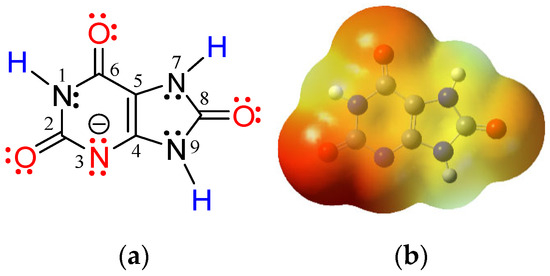
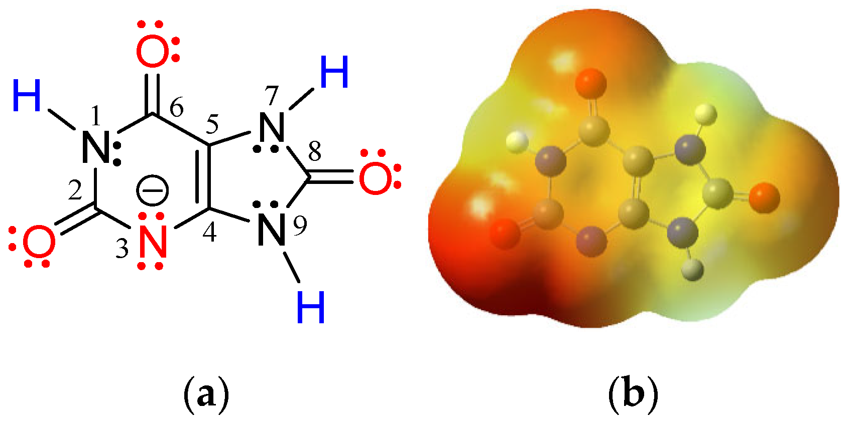
In neutral aqueous environments, uric acid is present almost completely as urate, a monoanion species formed by deprotonation of the imino-hydrogen atom in position 3-N [60]. Lewis structure of urate ascribes a negative charge to the 3-N atom (Figure 1a), but a more reliable picture arises from the electron density map of urate [61]: the distribution of negative charge is mainly located on carbonyl oxygens (Figure 1b).
Figure 1.
(a) Lewis structure and atom numbering in urate. H-bond donor sites (in blue) and H-bond acceptor sites (in red) are assigned. (b) Electron density map of urate. The red regions indicate the negative area.
Experimentally determined pKa values of uric acid amount to 5.4 and 9.8 [62], which implies that at a physiological pH of 7.4, molar fractions of uric acid, monovalent anion (urate), and divalent anion are 0.009862, 0.9862, and 0.003926, respectively. Because the electron-donating/accepting ability of urate(H2O)n− and CO3(H2O)n•− clusters undoubtedly depends on their geometry, minimal energy clusters are required for the estimation of thermodynamics and kinetics of the underlying SET mechanism.
3.1. Conformational Analysis
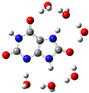
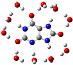
Urate has seven hydrogen bond binding sites available to chelate water molecules: oxygens of three carbonyl groups and nitrogen of the deprotonated imino group may act as a H-atom acceptor, while three imino groups may act as a H-atom donor (Figure 1a). On the other hand, each water molecule may form up to four H-bonds, two via donation of H-atoms and two via the H-atom accepting oxygen atom. For uric acid, it has been shown that water molecules are H-bonded to adjacent oxygen and hydrogen atoms (as well as between themselves), rather than to an isolated oxygen or hydrogen [27]. It could be expected that water molecules in the first hydration shell of urate follow this arrangement. Undoubtedly, the H-bond network is dynamic and alternates due to thermal excitation: H-bonds may be broken and rearranged in a picosecond to nanosecond time scale [63].
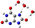
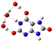
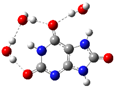
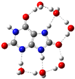
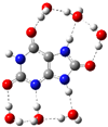
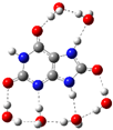
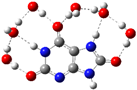
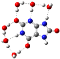
Based on the above-mentioned results of Cai et al. [27] twenty initial guess structures for the urate(H2O)3− cluster and thirty for the urate(H2O)6− cluster were generated, and full geometry optimizations and frequency calculations were performed. The hydration energy (Ehydr) of all clusters was calculated by using Equation (7) [64]:
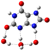
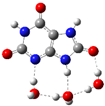
where Eurate(H2O)n−, EH2O, and Eurate− are the total energy of the cluster, the single water molecule, and the free urate, respectively. Ehydr relates to the interaction energy of urate with its hydrated shell of n H2O molecules and increases with the increase of the number of H-bonded H2O molecules [27]. The negative sign of Ehydr indicates spontaneity of urate hydration and the dominant conformer possesses the lowest Ehydr. Equilibrium population (in %) was predicted by the Boltzmann distribution based on the total energy (E in a.u.) of all conformers. Five minimal energy cluster structures of urate hydrated by 3 and 6 water molecules are presented in Table 1. Complete results are presented in Tables S1 and S2 in Supplementary Material.
Ehydr = Eurate(H2O)n− − (nEH2O + Eurate−)
Table 1.
The fully optimized minimal energy structures of: (a) urate(H2O)3− clusters, and (b) urate(H2O)6− clusters, calculated at SMD/M06-2X/6-311++G(d,p) level of theory. Hydration energy Ehydr (kcal mol−1), the total energy of the cluster E (a.u.), and Boltzmann population (%).
Revealed minimal energy conformer of urate(H2O)3− cluster and urate(H2O)6− cluster, along with free and fully hydrated urate, were used in the estimation of their scavenging potency. Their Cartesian coordinates are given in Tables S3 and S4.
Comprehensive conformational analysis of CO3(H2O)n•− clusters, including n = 1–8, has been performed by Pathak et al. [28]. We used global minimum energy structures, i.e., conformers for n = 4 and 6 from this work. Dooley and Vyas [31] used 0 to 30 explicit water molecules and different DFT functionals to reproduce the aqueous reduction potential of CO3•−. Minimum energy clusters of CO3(H2O)n•− (n = 9, 12, 15, and 24) from this work were taken. All used conformers were reoptimized at the applied SMD/M06-2X/6-311++G(d,p) level of theory. Corresponding Cartesian coordinates are given in Tables S5–S8. Conformer with n = 6 was used by Hebert and Schlegel [30] in a study of oxidation of guanine. Conformers with n = 6 and n = 9 were recently used by us in the estimation of antiradical potency of four phenolic antioxidants [25,26].
As already noted, the spin operator ⟨S2⟩ values for all of the urate and CO3•− radical species have been checked. In all cases, the deviations before and after annihilation of the first spin contaminant from the ideal value for a pure doublet (⟨S2⟩ = 0.75) were lower than 1.60% and 0.01%, indicating that the obtained energy values of radical species studied in this work are reliable (Table S9).
3.2. Estimation of Urate(H2O)n− Cluster Potency in the Scavenging of the CO3(H2O)n•− Cluster
The obtained results for SET from urate(H2O)n− clusters (n = 0, 3, 6 and 10) to CO3(H2O)n•− clusters (n = 0, 4, 6, 9, 12, 15 and 24) in water at pH = 7.4 are presented in Table 2 (selected data) and in Tables S9–S12 (complete data). Altogether 28 reactions are modeled to ascertain the potency of urate to inactivate CO3•− at physiological conditions, as well as to predict the most reliable size of reactants. All modeled reactions are thermodynamically feasible (ΔrG < 0), except the reaction of fully hydrated urate with free CO3•−, and diffusion-controlled kTST > 109 M−1 s−1, i.e., in the range of 2.7 × 10⁹ M−1 s−1 to 6.2 × 10¹² M−1 s−1. In accordance with the Evans–Polanyi principle [65], for the reaction of CO3(H2O)n•− clusters (n = 0, 4, 6, and 9) with all urate(H2O)n− clusters, the logarithms of the kTST constants are inversely fully correlated to the ΔrG (r ≥ −0.99), Figure S1. However, if the number of H2O molecules in the CO3(H2O)n•− cluster was enlarged to 12, 15, and 24, a linear trend was interrupted. The calculated AEA for CO3(H2O)n•− clusters indicate that increased hydration produces stronger oxidizing agents. On the other hand, VDE of urate(H2O)n− clusters increases with the number of hydrated water molecules, indicating less reactivity of more hydrated clusters.
Table 2.
SET from urate(H2O)n− clusters to CO3(H2O)n•− clusters in water at pH = 7.4. Reaction Gibbs free energy ΔrG in kcal/mol, Gibbs free energy of activation ΔG≠ in kcal/mol, transition state theory rate constant kTST in M−1 s−1, and rate constant including molar fractions in M−1 s−1. Vertical and adiabatic detachment energy VDE and ADE in eV of urate(H2O)n− clusters, and adiabatic and vertical electron affinity AEA and VEA in eV of CO3(H2O)n•− clusters are also given.
The data presented in Table 2 show that an increased number of explicit water molecules in the CO3•− hydration shell increases exergonicity, decreases reaction barrier height, and increases the TST rate constant of the SET mechanism, kTST. Because all kTST are larger than the diffusion rate, they lack physical meaning [22]. To be comparable with experimentally determined rate constant, kTST should be corrected by diffusion-controlled rate kD, thus giving apparent rate constant kapp (Tables S9–S12). Multiplying kapp with the molar fractions of reactants at a given pH produces the rate constant which should match the assayed one. All predicted are nearly equal due to the levelling effect of kD on kapp. is in the narrow range of 2.0–7.6 × 10⁹ M−1 s−1 (Table 2) and reveals the high potency of urate to scavenge CO3•−. Obtained result is in good agreement with the experimentally determined value at pH = 7.4, which amounts to 1.3 (±0.4) × 109 M−1 s−1 [66].
Electron transfer from urate to CO3•− is mainly driven by the electron donating ability of urate and the electron accepting ability of CO3•− [24]. Appropriate indices of those effects are VDE and AEA, respectively: lower VDE of urate clusters and higher AEA of CO3•− clusters favor electron transfer. The question that arises is which number of explicit water molecules in CO3•− and urate clusters satisfy reliable prediction of reaction kinetics. For example, Zilberg et al. [29] and Hebert and Schlegel [30] suggested that in aqueous solution CO3(H2O)6•− cluster exists, while Dooley and Vyas [31] accurately predicted the reduction potential of CO3•− by using the CO3(H2O)9•− cluster.
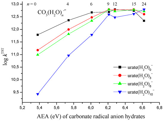
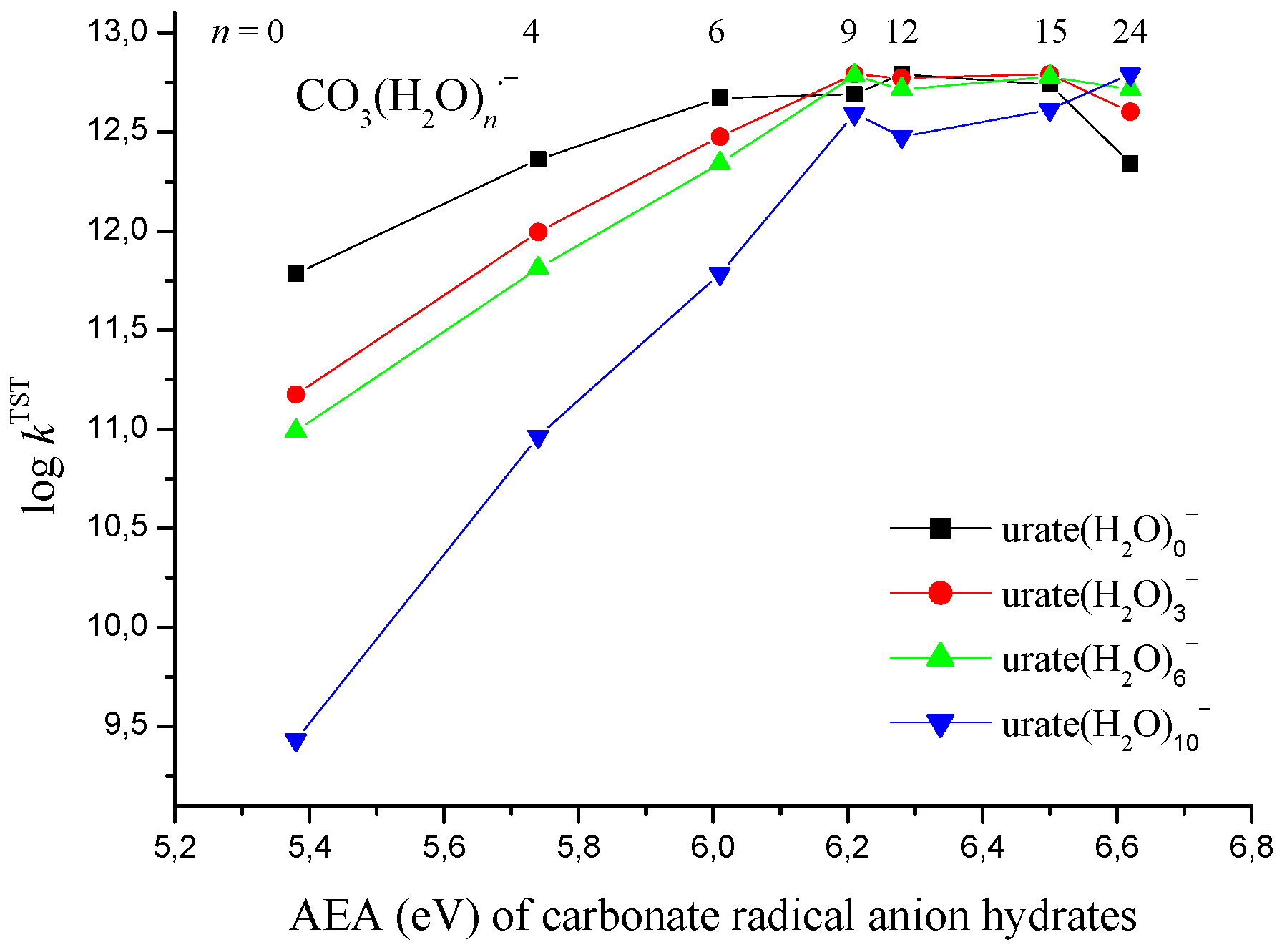
To determine the number of explicit water molecules in the CO3•− cluster, we analyzed the relationship between the AEA of the CO3•− clusters and the reaction rate, i.e., log kTST. AEA of CO3•− hydrated by 0, 4, 6, and 9 water molecules amounts to 5.38, 5.74, 6.01, and 6.21 eV, respectively, i.e., an increased number of water molecules in the first hydration shell facilitates the electron-accepting ability of CO3•−. Strong correlation exists between those AEAs and log kTST: correlation coefficient r amounts to 0.966, 0.997, 0.999, and 0.998 for the reaction of CO3•− clusters with urate hydrated by 0, 3, 6, and 10 explicit water molecules, respectively. Results presented in Figure 2 and Table S13 showed that an increased number of explicit water molecules in the hydration shell of CO3•− increases AEA, i.e., enables a faster reaction rate, while the increased microhydration of urate decreases reaction rate with CO3•− clusters holding 0, 4, 6, and 9 water molecules. For any urate cluster (Tables S9–S12), CO3•− hydrated by 9 water molecules produces nearly equal reaction rate, i.e., 9 explicit water molecules lead to a convergence of log kTST. An additional increase in CO3•− hydration with 12, 15, and 24 water molecules has no significant impact on the reaction rate.
Figure 2.
Plot of adiabatic electron affinity (AEA in eV) of CO3(H2O)n•− clusters vs. logarithm of transition state theory rate constant (log kTST) for SET reaction of urate(H2O)n− clusters with CO3(H2O)n•− clusters.
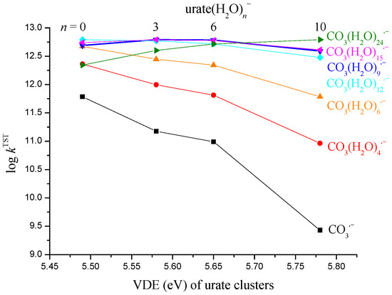
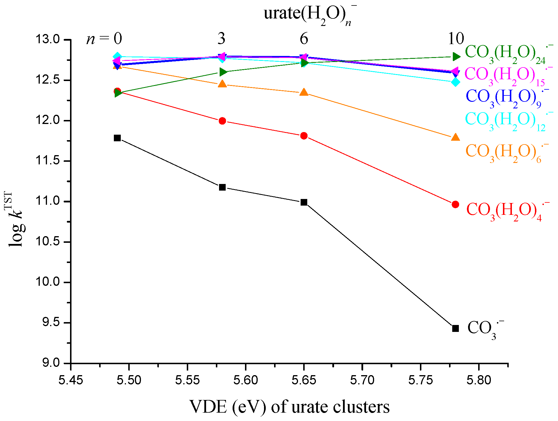
On the other hand, the VDE may serve as an index to ascertain the number of water molecules in the urate cluster, which leads to a convergence of reaction rate. The VDE of urate clusters increases with the increased number of explicit water molecules. For urate hydrated by 0, 3, 6, and 10 water molecules, VDE amounts to 5.49, 5.58, 5.65, and 5.78 eV, respectively. As already noted, the lower VDE enables a faster reaction rate. As Figure 3 and Table S14 show, this is valid for the reaction of urate clusters with CO3•− hydrated by 0, 4, and 6 water molecules. Strong correlation exists between VDE and log kTST: r amounts to −0.971, −0.984, and −0.980. CO3•− clusters with 9 and more water molecules lack such a relationship (except for n = 12), and all react with urate(H2O)6− cluster by nearly equal rate, i.e., log kTST ≈ 12.7. Thus, it appears that urate chelated by six water molecules is the most probable cluster in a water environment. The reaction of urate(H2O)6− cluster with CO3(H2O)9•− cluster proceeds with kTST = 6.1 × 10¹² M−1 s−1 (log kTST = 12.785), i.e., corresponds to the result obtained by using AEA for estimating the most probable CO3•−–water cluster.
Figure 3.
Plot of vertical detachment energy (VDE in eV) of urate clusters vs. logarithm of rate constant (log kTST) for SET reaction of urate(H2O)n− clusters with CO3(H2O)n•− clusters.
For urate(H2O)n− and CO3(H2O)n•− clusters, both vertical and adiabatic values of detachment energy and electron affinity were calculated (Table 2). The correlation between VDE and ADE of urate(H2O)n− clusters amounts to r = 0.985, and between VEA and AEA of CO3(H2O)n•− clusters amounts to r = 0.971. This indicates that the geometric relaxation of the studied species does not significantly modify the trends observed from the vertical values.
In summary, the results presented in this section suggest that at physiological pH = 7.4 both urate and CO3•− exist as hydrated species: performed analysis reveals urate(H2O)6− and CO3(H2O)9•− clusters as the most probable amongst eleven investigated. SET reaction between them is diffusion-controlled and proceeds with estimated rate constant of 7.3 × 10⁹ M−1 s−1, Table 2. The potency of urate as CO3•− scavenger is similar to that predicted for α-CEHC ( = 7.5 × 10⁹ M−1 s−1 [25]), and Trolox ( = 7.5 × 10⁹ M−1 s−1 [26]), and higher than that of α-tocopherol ( = 1.5 × 10⁷ M−1 s−1 [25]) and gallate ( = 3.5 × 10⁸ M−1f s−1 [25]). However, α-CEHC has a low concentration in plasma (<1 μM [67]), indicating its reduced potency in CO3•− scavenging, while Trolox is a synthetic water-soluble analogue of vitamin E.
3.3. Molecular Docking Study of Urate in Different Positions of the SOD1 and OGG1 Proteins
The molecular docking results indicate that urate interacts with both investigated enzymes, SOD1 and OGG1, with moderate binding affinity (Table 3). For SOD1, the calculated binding free energy is −5.25 kcal mol−1, corresponding to an inhibition constant of 0.14 mM. The binding is predominantly stabilized by intermolecular interactions (ΔGinter = −5.25 kcal mol−1), mainly originating from van der Waals, hydrogen bonding, and desolvation contributions (−5.13 kcal mol−1), while the electrostatic component is relatively small (−0.12 kcal mol−1).
Table 3.
Calculated thermodynamic and binding parameters of the most stable urate conformers within the active sites of SOD1 and OGG1, including the following: inhibition constants (Ki, µM) and binding free energy (ΔGbind, kcal mol−1) values obtained from various energy components such as total internal energy (ΔGtotal), torsional free energy (ΔGtor), unbound system’s energy (ΔGunb), electrostatic energy (ΔGelec), and the sum of dispersion and repulsion (ΔGvdw), hydrogen bond (ΔGhbond), and desolvation (ΔGdesolv).
A somewhat weaker interaction is observed for OGG1, where urate exhibits a binding free energy of −3.99 kcal mol−1 with a calculated inhibition constant of 1.19 mM. Similarly to the SOD1 system, stabilization is largely governed by dispersion and hydrogen bonding interactions (−3.94 kcal mol−1), whereas electrostatic contributions remain minimal (−0.05 kcal mol−1).
Comparison with reference inhibitors reveals that urate binds more strongly to SOD1 than the standard inhibitor LSC-1 (ΔGbind = −3.62 kcal mol−1), suggesting a relatively favorable interaction within the SOD1 active site. In contrast, the known OGG1 inhibitor TH13264 shows substantially stronger binding (ΔGbind = −7.44 kcal mol−1) than urate, indicating that the investigated ligand exhibits weaker affinity toward the OGG1 catalytic pocket.
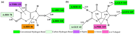
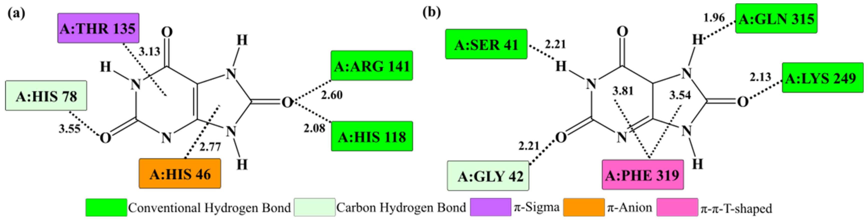
The analysis of intermolecular interactions indicates that urate establishes several stabilizing contacts within the active sites of both enzymes (Figure 4). In the SOD1 active site, the ligand forms multiple hydrogen bonds with key amino acid residues, including ARG 141 (2.60 Å) and HIS 118 (2.08 Å), which play an important role in positioning the ligand within the catalytic pocket. Additional interactions, such as a π–anion interaction with HIS 46 (2.77 Å) and a carbon–hydrogen bond with HIS 78 (3.55 Å), further stabilize the ligand orientation. Moreover, a π–σ interaction with THR 135 was observed, indicating that, in addition to hydrogen bonding, aromatic interactions also contribute to the stabilization of the complex.
Figure 4.
Two-dimensional map of key intermolecular interactions between urate and amino acid residues in the active sites of SOD1 (a) and OGG1 (b) obtained from molecular docking analysis. Interatomic distances are given in Å, while colors indicate different types of noncovalent interactions.
As noted in the Introduction section, SOD1 can be a source of oxidative damage via peroxidase activity. By using H2O2 as a substrate, the enzyme–copper-bound oxidant (usually designated as Cu2+- •OH) is formed at the active site of SOD1, which may inactivate the enzyme by damaging nearby histidine residues. In vitro experiments revealed that physiological levels of urate completely prevented the inactivation of SOD1. It has been suggested that urate restores the dismutase activity of SOD1 by the inactivation of Cu2+- •OH at the active site of enzyme, accompanied by urate radical formation [34,35].
On the other hand, HCO3− enhances SOD1 peroxidase activity via the production of CO3•− [68]. HCO3− is present in mM concentrations in cells and tissues and could be anchored at the anion-binding site of SOD1 (ARG 141) adjacent to the active site, enhancing oxidative damage via CO3•− production [69,70,71]. Our results suggest that urate may establish transient interactions with ARG 141, which could potentially influence HCO3− binding and CO3•− formation, and this will be further investigated through molecular dynamics simulations. Thus, urate has a twofold role in suppressing peroxidase activity of SOD1: it may neutralize bound oxidant at the enzyme active site, and suppress HCO3− dependent CO3•− production by occupying anion-binding site of the enzyme.
In the OGG1 active site, urate forms hydrogen bonds with residues SER 41 (2.21 Å), GLN 315 (1.96 Å), and LYS 249 (2.13 Å), creating a network of polar contacts that stabilize the ligand within the binding pocket. In addition, a π–π T-shaped interaction with PHE 319 further contributes to the stabilization of the complex.
Overall, the interaction pattern suggests that hydrogen bonding represents the dominant stabilization factor in both enzyme–ligand complexes, while additional π interactions further contribute to the stability and proper accommodation of the ligand within the binding pockets of the enzymes.
3.4. Molecular Dynamics Study of Urate in Different Positions of the SOD1 and OGG1 Proteins
Molecular dynamics simulations were conducted to evaluate the structural stability and conformational behavior of SOD1 and OGG1 in their apo forms and in complex with urate. The obtained Root Mean Square Deviation (RMSD), Radius of Gyration (Rg), and Root Mean Square Fluctuation (RMSF) parameters collectively provide insight into global stability, compactness, and local flexibility of both systems.
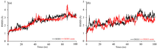
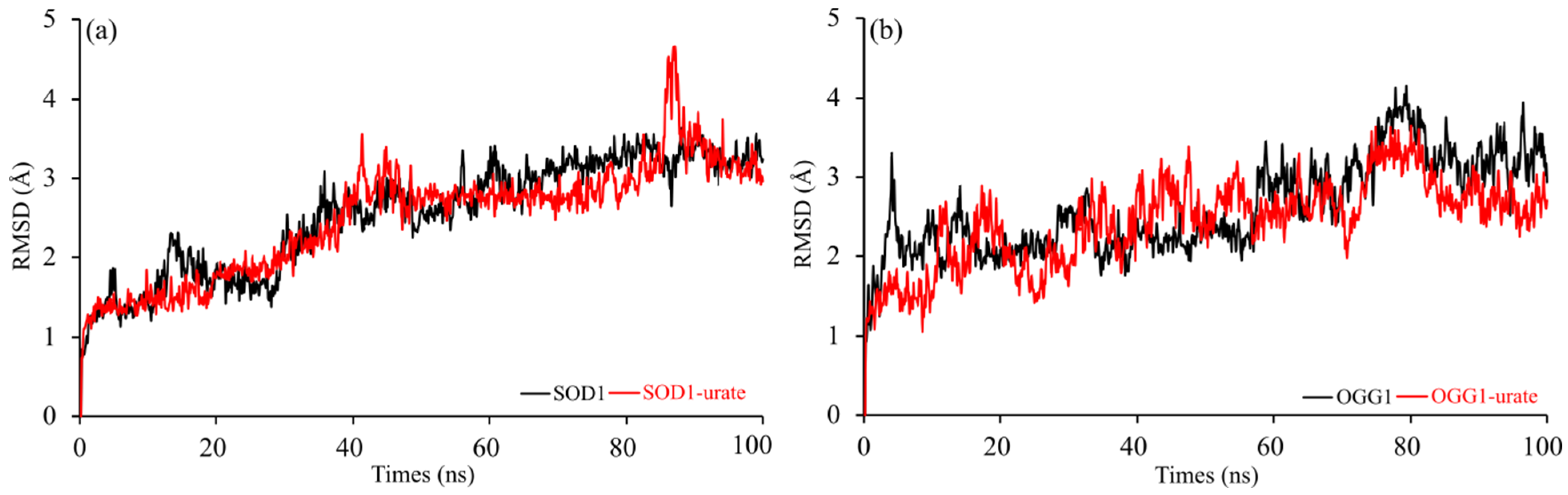
The backbone RMSD values indicate that both proteins remain structurally stable throughout the simulation, regardless of ligand presence (Figure 5). For SOD1, the average RMSD slightly decreased from 2.55 ± 0.70 Å in the apo form to 2.50 ± 0.71 Å in the urate-bound system. This minimal reduction, accompanied by nearly identical standard deviations, suggests that urate binding does not induce significant conformational perturbations. Instead, the structural fold of SOD1 is preserved, indicating that the ligand is accommodated without destabilizing the protein scaffold. A comparable trend was observed for OGG1, where RMSD decreased from 2.61 ± 0.58 Å in the free state to 2.44 ± 0.53 Å in complex with urate. The reduction in both the mean deviation and fluctuation amplitude suggests a modest stabilizing effect of urate on the OGG1 backbone dynamics. RMSD values within the range of approximately 2.4–2.6 Å further confirm that both systems reached equilibrium and maintained conformational consistency during the trajectory.
Figure 5.
Comparative analysis of 100 ns molecular dynamics (MD) simulations showing RMSD (root mean square deviation) of the C–Cα–N backbone for the SOD1 (a) and OGG1 (b) enzyme with and without urate in active sites.
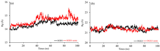
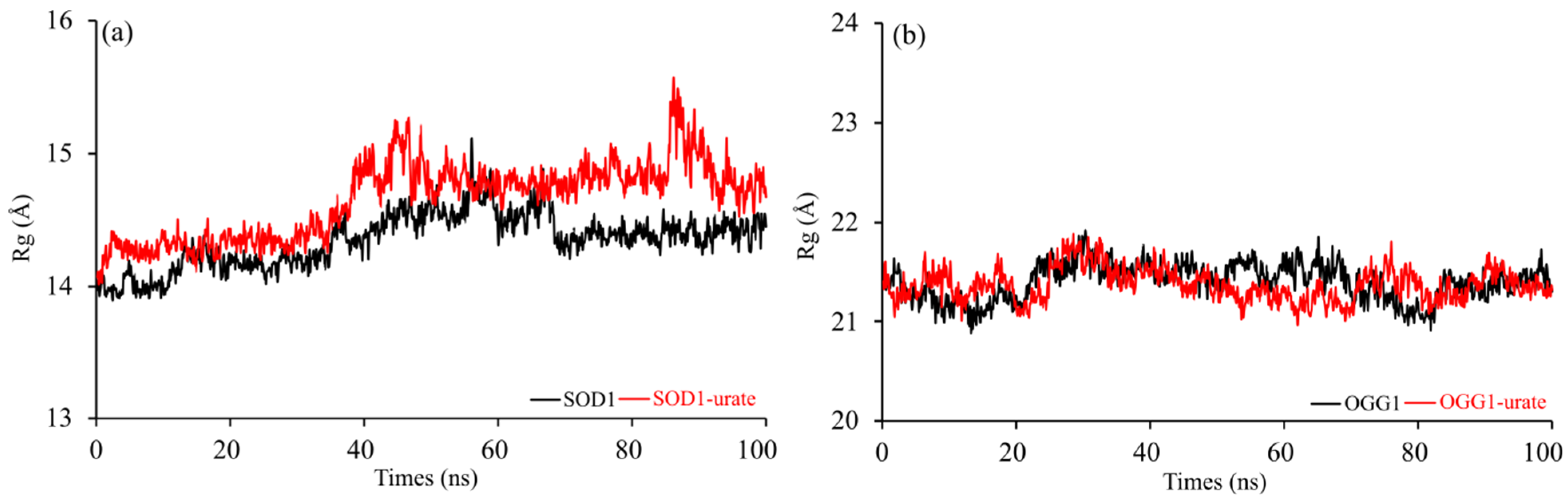
The radius of gyration analysis supports these observations by revealing only subtle differences in overall compactness (Figure 6). In the case of SOD1, Rg increased from 14.35 ± 0.20 Å to 14.65 ± 0.29 Å upon ligand binding. This slight expansion may reflect minor rearrangements of surface loops or adaptive breathing motions rather than large-scale conformational transitions. Importantly, the magnitude of change remains small, indicating preserved structural integrity. For OGG1, Rg values were nearly identical in the free (21.40 ± 0.19 Å) and bound (21.37 ± 0.16 Å) forms, suggesting that urate does not affect the global compactness of the enzyme. The consistency of Rg values reinforces the conclusion that no unfolding or structural collapse occurs in the presence of the ligand.
Figure 6.
Comparative analysis of 100 ns molecular dynamics (MD) simulations showing Rg (radius of gyration) of the C–Cα–N backbone for the SOD1 (a) and OGG1 (b) enzyme with and without urate in active sites.
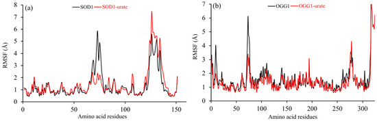
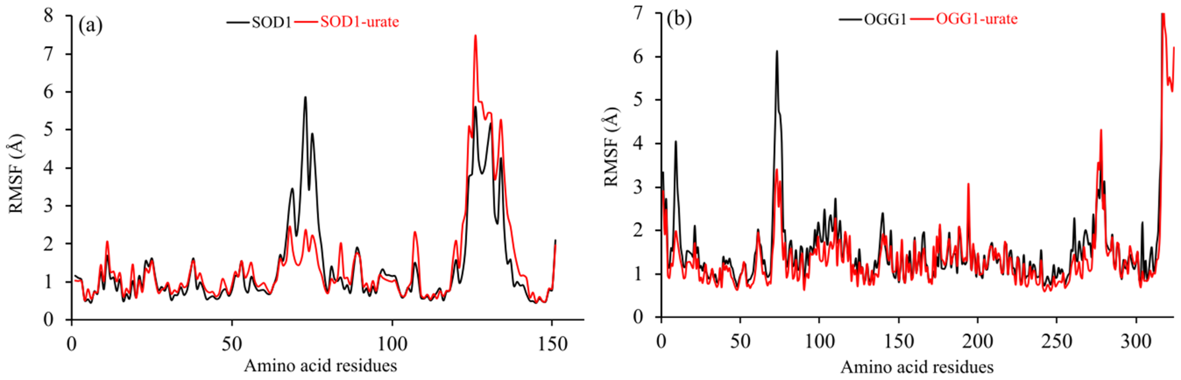
Residue-level flexibility, evaluated through RMSF analysis, reveals subtle differences between the two proteins (Figure 7). SOD1 exhibited a slight increase in average RMSF from 1.38 ± 1.14 Å to 1.47 ± 1.22 Å upon urate binding, indicating a marginal rise in local fluctuations. Such behavior may be attributed to adaptive adjustments in flexible surface regions while maintaining a stable core structure. In contrast, OGG1 displayed a decrease in average RMSF from 1.77 ± 1.62 Å in the apo state to 1.56 ± 1.21 Å in the complex, suggesting that ligand binding dampens local motions. This reduction in flexibility, together with the observed decrease in RMSD, implies a stabilizing interaction that restricts excessive backbone movement, particularly in regions proximal to the binding site.
Figure 7.
Comparative analysis of 100 ns molecular dynamics (MD) simulations showing RMSF (root mean square fluctuation) of the C–Cα–N backbone residues of SOD1 (a) and OGG1 (b) with and without urate in the active sites.
Additionally, to further examine the proposed interaction between urate and the ARG 141 region of SOD1, hydrogen-bond occupancy, contact persistence, and distance analyses were performed along the MD trajectory. The obtained results showed that the urate–ARG 141 interaction was predominantly transient in nature, with low hydrogen-bond occupancy values and gradual increase of the intermolecular distance during the simulation. Contact analysis indicated that interactions between urate and ARG 141 were mainly present during the early stages of the trajectory, while becoming significantly less frequent in later simulation periods. These findings suggest that urate may transiently access the ARG 141 region within the SOD1 catalytic channel; however, the interaction does not appear sufficiently persistent to support stable competition with HCO3− under the simulated conditions.
Taken together, the molecular dynamics data indicate that urate binding does not destabilize either SOD1 or OGG1. Structural deviations remain within a stable range, global compactness is preserved, and local fluctuations show only moderate adjustments. While SOD1 undergoes a slight expansion accompanied by marginally increased flexibility, OGG1 demonstrates reduced backbone deviation and dampened residue-level mobility, suggesting a more pronounced stabilizing effect in the latter case. The MD results suggest that urate forms stable complexes with both enzymes without inducing structural destabilization. The observed reduction of RMSD and RMSF values in the OGG1 system indicates partial rigidification of the protein backbone, particularly near the binding region, which may be associated with inhibitory behavior.
4. Conclusions
The results of this study highlight the multifunctional antioxidant potential of urate by exploring its ability to directly scavenge CO3•− and interact with both CO3•−-generating and DNA repair enzymes. This topic is of particular interest because of recent evidence that, under physiological conditions, instead of the commonly assumed HO•, CO3•− could be the major source of oxidative damage. The reaction kinetics of CO3•− scavenging by urate, an abundant plasma antioxidant, were estimated by using explicitly hydrated reactants immersed in a water continuum modeled by the SMD approach. Both urate and CO3•− in an aqueous environment tend to be hydrated. The number of explicit water molecules in their first hydration shell is determined by analyzing the relationship between log kTST and VDE and AEA, respectively. It was found that the urate(H2O)6− cluster and the CO3(H2O)9•− cluster are existing species at physiological pH 7.4. Obtained results indicate urate as a potent in vivo scavenger of CO3•−, more potent than α-tocopherol, α-CEHC, gallate, and Trolox. Molecular docking results indicate that urate can establish favorable interactions with the ARG 141 region within the catalytic channel of SOD1, while molecular dynamics simulations suggest that these interactions are predominantly transient in nature. Although the obtained computational results do not confirm definitive competition with HCO3−, they indicate a possible modulatory effect of urate on HCO3−-associated CO3•− formation. In contrast, urate exhibits weaker binding toward OGG1 and does not impair its catalytic function. Molecular dynamics simulations further confirm the structural stability of both SOD1 and OGG1 in their apo forms and in complex with urate, indicating that ligand binding does not induce destabilization of either enzyme. Overall, these findings provide mechanistic insight into the dual antioxidant role of urate, combining efficient radical scavenging with modulation of enzyme-mediated oxidative pathways.
Supplementary Materials
The following supporting information can be downloaded at: https://www.mdpi.com/article/10.3390/antiox15060761/s1, Tables S1–S8: Optimized geometry and Cartesian coordinates of investigated clusters; Table S9: Spin-squared values ⟨S2⟩; Tables S10–S13: Complete results of modeled SET reactions; Figure S1 and Tables S14 and S15: additional data. Reference [72] is cited in the Supplementary Material.
Author Contributions
All authors contributed substantially to the work. Conceptualization, A.A. and Ž.M.; methodology, A.A. and Ž.M.; software, Ž.M. and D.M.C.; validation, A.A., Ž.M. and D.M.C.; investigation, A.A., Ž.M. and D.M.C.; resources, Ž.M. and D.M.C.; writing—original draft preparation, A.A. and Ž.M.; writing—review and editing, A.A., Ž.M. and D.M.C.; visualization, Ž.M. All authors have read and agreed to the published version of the manuscript.
Funding
This research was funded from the 5801-PU.
Data Availability Statement
The original contributions presented in this study are included in the article/Supplementary Material. Further inquiries can be directed to the corresponding author.
Acknowledgments
This work was supported by the Croatian Ministry of Science and Education, the Slovak Grant Agency (VEGA 1/0461/21), and the Ministry of Science, Technological Development and Innovation of the Republic of Serbia (agreement no. 451-03-47/2023-01/200378). We are grateful to the HPC Center at the Slovak University of Technology in Bratislava, which is a part of the Slovak Infrastructure for High Performance Computing (SIVVP project, ITMS code 26230120002, funded by the European Region Development Fund, ERDF), for the computational time and resources made available.
Conflicts of Interest
The authors declare no conflicts of interest.
References
- Halliwell, B. Reactive species and antioxidants. Redox biology is a fundamental theme of aerobic life. Plant Physiol. 2006, 141, 312–322. [Google Scholar] [CrossRef] [Scilit] [PubMed]
- Forman, H.J.; Davies, K.J.A.; Ursini, F. How do nutritional antioxidants really work: Nucleophilic tone and para-hormesis versus free radical scavenging in vivo. Free Radic. Biol. Med. 2014, 66, 24–35. [Google Scholar] [CrossRef] [Scilit] [PubMed]
- Halliwell, B. The wanderings of a free radical. Free Radic. Biol. Med. 2009, 46, 531–542. [Google Scholar] [CrossRef] [Scilit] [PubMed]
- Fraga, C.G.; Croft, K.D.; Kennedy, D.O.; Tomás-Barberán, F.A. The effects of polyphenols and other bioactives on human health. Food Funct. 2019, 10, 514–528. [Google Scholar] [CrossRef] [Scilit] [PubMed]
- de Oliveira, E.P.; Burini, R.C. High plasma uric acid concentration: Causes and consequences. Diabetol. Metab. Syndr. 2012, 4, 12. [Google Scholar] [CrossRef] [Scilit] [PubMed]
- Dalbeth, N.; Stamp, L. Hyperuricaemia and gout: Time for a new staging system? Ann. Rheum. Dis. 2014, 73, 1598–1600. [Google Scholar] [CrossRef] [Scilit] [PubMed]
- Benzie, I.F.F.; Strain, J.J. The ferric reducing ability of plasma (FRAP) as a measure of “antioxidant power”: The FRAP assay. Anal. Biochem. 1996, 239, 70–76. [Google Scholar] [CrossRef] [Scilit] [PubMed]
- Benn, C.L.; Dua, P.; Gurrell, R.; Loudon, P.; Pike, A.; Storer, R.I.; Vangjeli, C. Physiology of hyperuricemia and urate-lowering treatments. Front. Med. 2018, 5, 160. [Google Scholar] [CrossRef] [Scilit] [PubMed]
- Hollman, P.C.H. Unravelling of the health effects of polyphenols is a complex puzzle complicated by metabolism. Arch. Biochem. Biophys. 2014, 559, 100–105. [Google Scholar] [CrossRef] [Scilit] [PubMed]
- Hoey, B.M.; Butler, J. The repair of oxidized amino acids by antioxidant. Biochim. Biophys. Acta Protein Struct. Mol. Enzymol. 1984, 791, 212–218. [Google Scholar] [CrossRef] [Scilit] [PubMed]
- Domazou, A.S.; Zhu, H.; Koppenol, W.H. Fast repair of protein radicals by urate. Free Radic. Biol. Med. 2012, 52, 1929–1936. [Google Scholar] [CrossRef] [Scilit] [PubMed]
- Muñoz-Rugeles, L.; Galano, A.; Alvarez-Idaboy, J.R. The role of acid–base equilibria in formal hydrogen transfer reactions: Tryptophan radical repair by uric acid as a paradigmatic case. Phys. Chem. Chem. Phys. 2017, 19, 15296–15309. [Google Scholar] [CrossRef] [Scilit] [PubMed]
- Illés, E.; Mizrahi, A.; Marks, V.; Meyerstein, D. Carbonate-radical-anions, and not hydroxyl radicals, are the products of the Fenton reaction in neutral solutions containing bicarbonate. Free Radic. Biol. Med. 2019, 131, 1–6. [Google Scholar] [CrossRef] [Scilit] [PubMed]
- Fleming, A.M.; Burrows, C.J. On the irrelevancy of hydroxyl radical to DNA damage from oxidative stress and implications for epigenetics. Chem. Soc. Rev. 2020, 49, 6524–6528. [Google Scholar] [CrossRef] [Scilit] [PubMed]
- Meyerstein, D. Re-examining Fenton and Fenton-like reactions. Nat. Rev. Chem. 2021, 5, 595–597. [Google Scholar] [CrossRef] [Scilit] [PubMed]
- Halliwell, B.; Adhikary, A.; Dingfelder, M.; Dizdaroglu, M. Hydroxyl radical is a significant player in oxidative DNA damage in vivo. Chem. Soc. Rev. 2021, 50, 8355–8360. [Google Scholar] [CrossRef] [Scilit] [PubMed]
- Galano, A.; Tan, D.X.; Reiter, R.J. Melatonin as a natural ally against oxidative stress: A physicochemical examination. J. Pineal Res. 2011, 51, 1–16. [Google Scholar] [CrossRef] [Scilit] [PubMed]
- Repine, J.E.; Eaton, J.W.; Anders, M.W.; Hoidal, J.R.; Fox, R.B. Generation of hydroxyl radical by enzymes, chemicals, and human phagocytes in vitro. Detection with the anti-inflammatory agent, dimethyl sulfoxide. J. Clin. Investig. 1979, 64, 1642–1651. [Google Scholar] [CrossRef] [Scilit] [PubMed]
- Fleming, A.M.; Burrows, C.J. Why the ROS matters: One-electron oxidants focus DNA damage and repair on G-quadruplexes for gene regulation. DNA Repair 2025, 145, 103789. [Google Scholar] [CrossRef] [Scilit] [PubMed]
- Chen, S.N.; Hoffman, M.Z. Rate constants for the reaction of the carbonate radical with compounds of biochemical interest in neutral aqueous solution. Radiat. Res. 1973, 56, 40–47. [Google Scholar] [CrossRef] [Scilit]
- Wojnárovits, L.; Tóth, T.; Takács, E. Rate constants of carbonate radical anion reactions with molecules of environmental interest in aqueous solution: A review. Sci. Total Environ. 2020, 717, 137219. [Google Scholar] [CrossRef] [Scilit] [PubMed]
- Galano, A.; Alvarez-Idaboy, J.R. A computational methodology for accurate predictions of rate constants in solution: Application to the assessment of primary antioxidant activity. J. Comput. Chem. 2013, 34, 2430–2445. [Google Scholar] [CrossRef] [Scilit] [PubMed]
- Wang, Y.; An, P.; Li, S.; Zhou, L. The oxidation mechanism and kinetics of 2′-deoxyguanosine by carbonate radical anion. Chem. Phys. Lett. 2020, 739, 136982. [Google Scholar] [CrossRef] [Scilit]
- McPherson, P.A.C. Evaluating theoretical rate constants for bimolecular antioxidant processes involving ionic species under physiological conditions. Comput. Theor. Chem. 2024, 1239, 114794. [Google Scholar] [CrossRef] [Scilit]
- Amić, A.; Milanović, Ž.; Mastiľák Cagardová, D. Protective roles of α-tocopherol, α-CEHC, and gallic acid against CO3•−-induced DNA damage and their effect on OGG1 and pro-oxidant enzymes. J. Mol. Liq. 2025, 438, 128847. [Google Scholar] [CrossRef] [Scilit]
- Amić, A.; Mastil’ák Cagardová, D. Role of explicit hydration in scavenging of CO3•− by Trolox: A DFT study. Int. J. Mol. Sci. 2025, 26, 11342. [Google Scholar] [CrossRef] [Scilit] [PubMed]
- Cai, Z.; Zhu, C.; Chen, G.; Wu, Y.; Gu, J.; Ma, C.; Gao, H.; Li, L.; Guo, S. Study on intermolecular hydrogen bond of uric acid water-clusters. Chem. Phys. Lett. 2023, 818, 140424. [Google Scholar] [CrossRef] [Scilit]
- Pathak, A.K.; Mukherjee, T.; Maity, D.K. Theoretical study on the spectroscopic properties of CO3•−.nH2O clusters: Extrapolation to bulk. ChemPhysChem 2008, 9, 2259–2264. [Google Scholar] [CrossRef] [Scilit] [PubMed]
- Zilberg, S.; Mizrahi, A.; Meyerstein, D.; Kornweitz, H. Carbonate and carbonate anion radicals in aqueous solutions exist as CO3(H2O)62− and CO3(H2O)6•− respectively: The crucial role of the inner hydration sphere of anions in explaining their properties. Phys. Chem. Chem. Phys. 2018, 20, 9429–9435. [Google Scholar] [CrossRef] [Scilit] [PubMed]
- Hebert, S.P.; Schlegel, H.B. Computational study of the pH-dependent competition between carbonate and thymine addition to the guanine radical. Chem. Res. Toxicol. 2019, 32, 195–210. [Google Scholar] [CrossRef] [Scilit] [PubMed]
- Dooley, M.R.; Vyas, S. Role of explicit solvation and level of theory in predicting the aqueous reduction potential of carbonate radical anion by DFT. Phys. Chem. Chem. Phys. 2025, 27, 6867–6874. [Google Scholar] [CrossRef] [Scilit] [PubMed]
- Figueroa, J.D.; Fuentes-Lemus, E.; Dorta, E.; Melin, V.; Cortés-Ríos, J.; Faúndez, M.; Contreras, D.; Denicola, A.; Álvarez, B.; Davies, M.J.; et al. Quantification of carbonate radical formation by the bicarbonate-dependent peroxidase activity of superoxide dismutase 1 using pyrogallol red bleaching. Redox Biol. 2019, 24, 101207. [Google Scholar] [CrossRef] [Scilit] [PubMed]
- Goss, S.P.A.; Singh, R.J.; Kalyanaraman, B. Bicarbonate enhances the peroxidase activity of Cu,Zn-superoxide dismutase. Role of carbonate anion radical. J. Biol. Chem. 1999, 274, 28233–28239. [Google Scholar] [CrossRef] [Scilit] [PubMed]
- Hink, H.U.; Santanam, N.; Dikalov, S.; McCann, L.; Nguyen, A.D.; Parthasarathy, S.; Harrison, D.G.; Fukai, T. Peroxidase properties of extracellular superoxide dismutase. Role of uric acid in modulating in vivo activity. Arterioscler. Thromb. Vasc. Biol. 2002, 22, 1402–1408. [Google Scholar] [CrossRef] [Scilit] [PubMed]
- Hodgson, E.K.; Fridovich, I. The interaction of bovine erythrocyte superoxide dismutase with hydrogen peroxide: Inactivation of the enzyme. Biochemistry 1975, 14, 5294–5299. [Google Scholar] [CrossRef] [Scilit] [PubMed]
- Zhong, Y.; Zhang, X.; Feng, R.; Fan, Y.; Zhang, Z.; Zhang, Q.-W.; Wan, J.-B.; Wang, Y.; Yu, H.; Li, G. OGG1: An emerging multifunctional therapeutic target for the treatment of diseases caused by oxidative DNA damage. Med. Res. Rev. 2024, 44, 2825–2848. [Google Scholar] [CrossRef] [Scilit] [PubMed]
- Zhao, Y.; Truhlar, D.G. The M06 suite of density functionals for main group thermochemistry, thermochemical kinetics, noncovalent interactions, excited states, and transition elements: Two new functionals and systematic testing of four M06-class functionals and 12 other functionals. Theor. Chem. Acc. 2008, 120, 215–241. [Google Scholar] [CrossRef] [Scilit]
- Marenich, A.V.; Cramer, C.J.; Truhlar, D.G. Universal solvation model based on solute electron density and on a continuum model of the solvent defined by the bulk dielectric constant and atomic surface tensions. J. Phys. Chem. B 2009, 113, 6378–6396. [Google Scholar] [CrossRef] [Scilit] [PubMed]
- Frisch, M.J.; Trucks, G.W.; Schlegel, H.B.; Scuseria, G.E.; Robb, M.A.; Cheeseman, J.R.; Scalmani, G.; Barone, V.; Mennucci, B.; Petersson, G.A.; et al. Gaussian 09, Revision D.01; Gaussian, Inc.: Wallingford, CT, USA, 2013. [Google Scholar]
- Dzib, E.; Cabellos, J.L.; Ortiz-Chi, F.; Pan, S.; Galano, A.; Merino, G. Eyringpy: A program for computing rate constants in the gas phase and in solution. Int. J. Quantum Chem. 2019, 119, e25686. [Google Scholar] [CrossRef] [Scilit]
- Marcus, R.A. Electron transfer reactions in chemistry. Theory and experiment. Angew. Chem. Int. Ed. 1993, 32, 1111–1121. [Google Scholar] [CrossRef] [Scilit]
- Czapski, G.; Lymar, S.V.; Schwarz, H.A. Acidity of the carbonate radical. J. Phys. Chem. A 1999, 103, 3447–3450. [Google Scholar] [CrossRef] [Scilit]
- Makurat, S.; Yuan, Q.; Czub, J.; Chomicz-Manka, L.; Cao, W.; Wang, X.-B.; Rak, J. Guanosine dianions hydrated by one to four water molecules. J. Phys. Chem. Lett. 2022, 13, 3230–3236. [Google Scholar] [CrossRef] [Scilit] [PubMed]
- Mishra, P.M. Electron affinity calculation for selected PAHs using DFT: Effect of cyclopenta ring fusion and aromaticity. Comput. Theor. Chem. 2015, 1068, 165–171. [Google Scholar] [CrossRef] [Scilit]
- Morris, G.M.; Huey, R.; Lindstrom, W.; Sanner, M.F.; Belew, R.K.; Goodsell, D.S.; Olson, A.J. AutoDock4 and AutoDockTools4: Automated docking with selective receptor flexibility. J. Comput. Chem. 2009, 30, 2785–2791. [Google Scholar] [CrossRef] [Scilit] [PubMed]
- Milanović, Ž. Structural properties of newly 4,7-dihydroxycoumarin derivatives as potential inhibitors of XIIa, Xa, IIa factors of coagulation. J. Mol. Struct. 2024, 1298, 137049. [Google Scholar] [CrossRef] [Scilit]
- Milanović, Ž. Exploring enzyme inhibition and comprehensive mechanisms of antioxidant/prooxidative activity of natural furanocoumarin derivatives: A comparative kinetic DFT study. Chem. Biol. Interact. 2024, 396, 111034. [Google Scholar] [CrossRef] [Scilit] [PubMed]
- Amić, A.; Mastil’ák Cagardová, D.; Milanović, Ž. Theoretical study of antioxidant and prooxidant potency of protocatechuic aldehyde. Int. J. Mol. Sci. 2025, 26, 404. [Google Scholar] [CrossRef] [Scilit] [PubMed]
- Becke, A.D.; Johnson, E.R. A density-functional model of the dispersion interaction. J. Chem. Phys. 2005, 123, 154101. [Google Scholar] [CrossRef] [Scilit] [PubMed]
- Zhao, Y.; Schultz, N.E.; Truhlar, D.G. Design of density functionals by combining the method of constraint satisfaction with parametrization for thermochemistry, thermochemical kinetics, and noncovalent interactions. J. Chem. Theory Comput. 2006, 2, 364–382. [Google Scholar] [CrossRef] [Scilit] [PubMed]
- Noinaj, N.; Easley, N.C.; Oke, M.; Mizuno, N.; Gumbart, J.; Boura, E.; Steere, A.N.; Zak, O.; Aisen, P.; Tajkhorshid, E.; et al. Structural basis for iron piracy by pathogenic Neisseria. Nature 2012, 483, 53–58. [Google Scholar] [CrossRef] [Scilit] [PubMed]
- BIOVIA, Dassault Systèmes. Discovery Studio Modeling Environment; Dassault Systèmes: San Diego, CA, USA, 2021. [Google Scholar]
- Morris, G.M.; Goodsell, D.S.; Halliday, R.S.; Huey, R.; Hart, W.E.; Belew, R.K.; Olson, A.J. Automated docking using a Lamarckian genetic algorithm and an empirical binding free energy function. J. Comput. Chem. 1998, 19, 1639–1662. [Google Scholar]
- Case, D.A.; Aktulga, H.M.; Belfon, K.; Cerutti, D.S.; Cisneros, G.A.; Cruzeiro, V.W.D.; Forouzesh, N.; Giese, T.J.; Götz, A.W.; Gohlke, H.; et al. AmberTools. J. Chem. Inf. Model. 2023, 63, 6183–6191. [Google Scholar] [CrossRef] [Scilit] [PubMed]
- Jo, S.; Kim, T.; Iyer, V.G.; Im, W. CHARMM-GUI: A web-based graphical user interface for CHARMM. J. Comput. Chem. 2008, 29, 1859–1865. [Google Scholar] [CrossRef] [Scilit] [PubMed]
- Vanommeslaeghe, K.; Hatcher, E.; Acharya, C.; Kundu, S.; Zhong, S.; Shim, J.; Darian, E.; Guvench, O.; Lopes, P.; Vorobyov, I.; et al. CHARMM general force field: A force field for drug-like molecules compatible with the CHARMM all-atom additive biological force fields. J. Comput. Chem. 2010, 31, 671–690. [Google Scholar] [CrossRef] [Scilit] [PubMed]
- Zielkiewicz, J. Structural properties of water: Comparison of the SPC, SPCE, TIP4P, and TIP5P models of water. J. Chem. Phys. 2005, 123, 104501. [Google Scholar] [CrossRef] [Scilit] [PubMed]
- Berendsen, H.J.; Postma, J.V.; Van Gunsteren, W.F.; DiNola, A.R.H.J.; Haak, J.R. Molecular dynamics with coupling to an external bath. J. Chem. Phys. 1984, 81, 3684–3690. [Google Scholar] [CrossRef] [Scilit]
- Ryckaert, J.-P.; Ciccotti, G.; Berendsen, H.J. Numerical integration of the cartesian equations of motion of a system with constraints: Molecular dynamics of n-alkanes. J. Comput. Phys. 1977, 23, 327–341. [Google Scholar] [CrossRef] [Scilit]
- León-Carmona, J.R.; Galano, A. Uric and 1-methyluric acids: Metabolic wastes or antiradical protectors? J. Phys. Chem. B 2011, 115, 15430–15438. [Google Scholar] [CrossRef] [Scilit] [PubMed]
- Amić, A.; Marković, Z.; Dimitrić Marković, J.M.; Lučić, B.; Stepanić, V.; Amić, D. The 2H+/2e− free radical scavenging mechanisms of uric acid: Thermodynamics of N–H bond cleavage. Comput. Theor. Chem. 2016, 1077, 2–10. [Google Scholar] [CrossRef] [Scilit]
- Simic, M.G.; Jovanovic, S.V. Antioxidant mechanisms of uric acid. J. Am. Chem. Soc. 1989, 111, 5778–5782. [Google Scholar] [CrossRef] [Scilit]
- Laage, D.; Elsaesser, T.; Hynes, J.T. Water dynamics in the hydration shells of biomolecules. Chem. Rev. 2017, 117, 10694–10725. [Google Scholar] [CrossRef] [Scilit] [PubMed]
- Pathak, A.K.; Mukherjee, T.; Maity, D.K. IR spectra of carbonate-water clusters. Synth. React. Inorg. Met. Org. Chem. 2008, 38, 76–83. [Google Scholar] [CrossRef] [Scilit]
- Evans, M.G.; Polanyi, M. Inertia and driving force of chemical reactions. Trans. Faraday Soc. 1938, 34, 11–24. [Google Scholar] [CrossRef] [Scilit]
- Karunakaran, C.; Zhang, H.; Joseph, J.; Antholine, W.E.; Kalyanaraman, B. Thiol oxidase activity of copper, zinc superoxide dismutase stimulates bicarbonate-dependent peroxidase activity via formation of a carbonate radical. Chem. Res. Toxicol. 2005, 18, 494–500. [Google Scholar] [CrossRef] [Scilit] [PubMed]
- Torquato, P.; Marinelli, R.; Bartolini, D.; Giusepponi, D.; Cruciani, G.; Siragusa, L.; Galarini, R.; Sebastiani, B.; Gioiello, A.; Galli, F. Vitamin E: Metabolism and molecular aspects. In Molecular Nutrition: Vitamins; Patel, V.B., Ed.; Elsevier Inc.: Amsterdam, The Netherlands, 2020; pp. 487–518. [Google Scholar] [CrossRef] [Scilit]
- Zhang, H.; Joseph, J.; Felix, C.; Kalyanaraman, B. Bicarbonate enhances the hydroxylation, nitration, and peroxidation reactions catalyzed by copper, zinc superoxide dismutase. Intermediacy of carbonate anion radical. J. Biol. Chem. 2000, 275, 14038–14045. [Google Scholar] [CrossRef] [Scilit] [PubMed]
- Sankarapandi, S.; Zweier, J.L. Bicarbonate is required for the peroxidase function of Cu,Zn-superoxide dismutase at physiological pH. J. Biol. Chem. 1999, 274, 1226–1232. [Google Scholar] [CrossRef] [Scilit] [PubMed]
- Elam, J.S.; Malek, K.; Rodriguez, J.A.; Doucette, P.A.; Taylor, A.B.; Hayward, L.J.; Cabelli, D.E.; Valentine, J.S.; Hart, P.J. An alternative mechanism of bicarbonate-mediated peroxidation by copper-zinc superoxide dismutase. Rates enhanced via proposed enzyme-associated peroxycarbonate intermediate. J. Biol. Chem. 2003, 278, 21032–21039. [Google Scholar] [CrossRef] [Scilit] [PubMed]
- Andrekopoulos, C.; Zhang, H.; Joseph, J.; Kalivendi, S.; Kalyanaraman, B. Bicarbonate enhances α-synuclein oligomerization and nitration: Intermediacy of carbonate radical anion and nitrogen dioxide radical. Biochem. J. 2004, 378, 435–447. [Google Scholar] [CrossRef] [Scilit] [PubMed]
- Young, D.C. Computational Chemistry: A Practical Guide for Applying Techniques to Real-World Problems; John Wiley & Sons: New York, NY, USA, 2001; p. 228. [Google Scholar] [CrossRef] [Scilit]






Disclaimer/Publisher’s Note: The statements, opinions and data contained in all publications are solely those of the individual author(s) and contributor(s) and not of MDPI and/or the editor(s). MDPI and/or the editor(s) disclaim responsibility for any injury to people or property resulting from any ideas, methods, instructions or products referred to in the content. |
© 2026 by the authors. Licensee MDPI, Basel, Switzerland. This article is an open access article distributed under the terms and conditions of the Creative Commons Attribution (CC BY) license.