


Abstract
Alcohol-associated Hepatitis (AH) is a rare acute injury caused by alcohol consumption, which can lead to one of the most severe manifestations of liver disease. It is part of the alcohol-related liver diseases (ArLD) spectrum, which represents a major global health burden, with oxidative stress and inflammation serving as central, interconnected pathogenic mechanisms. Chronic alcohol (ethanol) consumption induces hepatic reactive oxygen species (ROS) generation through multiple pathways, including cytochrome P450 2E1 (CYP2E1) induction, mitochondrial dysfunction, and NADPH oxidase activation. These oxidative insults trigger a cascade of cellular damage encompassing lipid peroxidation, protein adduct formation, DNA damage, and endoplasmic reticulum stress, ultimately leading to hepatocyte dysfunction and multiple forms of cell death, including apoptosis, necroptosis, pyroptosis, and ferroptosis. The inflammatory response, orchestrated primarily by Kupffer cells and infiltrating neutrophils through Toll-like receptor (TLR) signalling and inflammasome activation, not only amplifies hepatic injury but also promotes fibrogenesis through hepatic stellate cell activation. Neutrophils, characterised by elevated lipocalin-2 expression and spontaneous NETosis in AH, exhibit a paradoxical role by driving both tissue damage and repair. Current therapeutic strategies include corticosteroids, which remain the first-line treatment for severe AH, while emerging therapies targeting the gut–liver axis, hepatic regeneration, and specific molecular targets show promise in clinical trials. This review comprehensively examines the molecular crosstalk between oxidative stress and inflammation in the pathogenesis of AH to highlight current and investigational therapeutic approaches targeting these interconnected pathways.
1. Introduction
Alcohol-associated Hepatitis (AH), previously termed Alcoholic Hepatitis, represents one of the most challenging disease manifestations within the spectrum of alcohol-related liver diseases (ArLD). This is due to its acute presentation, challenges in confirmatory diagnosis and disease severity. The most acute florid severe form is characterised by high short-term mortality up to 50% within three months of diagnosis [1]. Recent evidence has highlighted the interplay between hepatocyte injury and oxidative stress, systemic inflammatory response and gut–liver axis dysfunction [2,3,4,5,6,7,8,9,10,11] in AH. Currently, European and American clinical practice guidelines recommend corticosteroid therapy in selected patients with a Maddrey’s Discriminant Function (MDF) score > 32 or a Model for End-Stage Liver Disease (MELD) score > 20, with recent evidence reporting the greatest benefit for those with MELD scores between 25 and 29 [12,13,14,15]. Corticosteroids are, however, not associated with long-term survival benefit [16] and in eligible cases, liver transplantation remains the only therapeutic option for patients unresponsive to medical treatment [14,17,18]. The aim of this review is to analyse the role of oxidative stress in the pathophysiology of AH and to highlight the recent therapeutic innovations proposed to treat this life-threatening condition.
2. Alcohol-Associated Hepatitis: Definition, Prevalence and Natural History
The definition of AH has recently been revised by the National Institute on Alcohol Abuse and Alcoholism (NIAAA)–AH Consortium, which has categorised AH into definite, probable, and possible diagnosis [19]. AH is defined as a clinical syndrome characterised by the association of new onset of jaundice and alteration of liver blood tests (AST < 500 IU/mL, ALT < 300 IU/mL within three months from clinical presentation and with total serum bilirubin > 80 μmol) in female/male patients with more than 60/80 g of daily alcohol use with less of 60 days of alcohol cessation [16]. Other potential causes of liver disease must be excluded, and liver biopsy may be performed in the presence of potential confounding factors, depending on local practice. A definitive diagnosis can be established with histologic confirmation (Figure 1), although the findings of steatohepatitis can be similar to those described in metabolic-associated steatohepatitis (MASH). Parker et al. [20] reported on a systematic review from 7528 biopsy-proven ArLD participants, of which 25% had steatohepatitis. These patients had a higher mortality when compared to those with simple steatosis alone due to alcohol. Although no single hallmark has been identified, features such as megamitochondria, pericellular fibrosis, macrovesicular steatosis, cholestasis and Mallory Denk bodies are more frequently observed in patients with AH than in MASH [21,22]. Thus, whilst helpful, histological interpretation should always be integrated with the clinical context. The prevalence of AH is equally variable across territories [13] but is increasing in line with alcohol consumption. Incidence is estimated to have increased during the COVID pandemic and is now more common among female and young patients [23,24]. Data from Denmark showed a rise in AH incidence in both men and women, with the increase being particularly pronounced among middle-aged female patients [25]. Similarly, UK data from the reported an increase of 31% in the number of deaths related to alcohol use from 2019 to 2022 [26]. These data are in line with those from the United States, where AH hospital admissions rose from 0.66% of total hospital admissions in 2002 to 0.83% in 2010 [27].
Figure 1.
Diagnostic flowchart of acute alcoholic hepatitis.
The clinical diagnosis is particularly challenging, as the disease manifestation in the context of sustained alcohol exposure is clinically similar to acute-on-chronic liver failure triggered by excessive alcohol consumption. In fact, real-world data from cohorts of patients transplanted for severe acute alcoholic hepatitis have revealed the presence of previously unrecognised cirrhosis in explanted livers in both American and European cohorts [28,29,30].
Hence, alcohol-related liver disease in general and alcohol-related hepatitis and ACLF caused by excess alcohol intake account for a significant proportion of global liver morbidity and mortality, which are linked to changes in patterns of alcohol intake.
3. Oxidative Stress Injury in Liver Diseases
Oxidative balance and homeostasis are crucial for physiological function. Reactive oxidant species (ROS) have dual roles as important signalling molecules regulating metabolism and cell survival in health, and driving protein, nuclear and lipid injury during pathogenic processes [31,32,33]. The liver, due to its role in metabolism, is particularly exposed to reactive oxygen species production, which causes potential damage when the oxidative balance is disrupted [34]. The unique cellular complexity of the liver means there are multiple different hepatic cell populations with distinct susceptibilities to oxidant stress (Figure 2). The hepatocyte population (80% of liver cells) performs essential functions, including detoxification, protein and lipid secretion, hormone and bile production [34,35,36]. Cholangiocytes, comprising 3–5% of liver cells, participate in bile synthesis, modification and excretion [37]. Non-parenchymal cells include liver sinusoidal endothelial cells (15–20% of liver cells), hepatic stellate cells (HSC), Kupffer cells (resident macrophages, KC), and natural killer (NK) cells [38,39], which influence the hepatic microenvironment and govern inflammatory cell recruitment, retention and fibrogenesis.
Figure 2.
The susceptibility of hepatic cell types to oxidative stress. Created in BioRender. D’Arcangelo, F. (2026) https://BioRender.com (accessed on 3 March 2026).
Reactive oxygen species (ROS) are produced mainly as a result of mitochondrial respiration, which generates superoxide radicals, although other sources of production have been identified as peroxisomes, xanthine oxidases, cytochrome P450 oxidases and the NADPH oxidases (NOXs) systems [40]. ROS can be subsequently categorised as unstable free radicals, such as superoxide radical and hydroxyl radical, which have potentially toxic effects, and stable non-radical oxidants like hydrogen peroxide and peroxynitrite. These non-radical oxidants have a fundamental role as messengers in different physiological pathways when oxidative balance is preserved [41]. However, NOX enzymes are considered responsible for ROS production, which enhances HSC and KC activation, leading to fibrosis progression via activation of the TGF-ß beta pathway [42,43,44]. Hepatocytes possess robust antioxidant mechanisms encompassing enzymatic components (superoxide dismutase, glutathione peroxidases, catalase, peroxiredoxins) and non-enzymatic molecules (glutathione, vitamins E, A, and C, ubiquinone) [45,46]. The enzymatic components act to metabolise ROS, while the non-enzymatic molecules mainly mitigate the damage caused by free radicals [47]. The nuclear factor erythroid 2-related factor 2 (NRF2)/antioxidant response element (ARE) pathway constitutes the master regulator of antioxidant defence. Under basal conditions, Kelch-like ECH-associated protein 1 (KEAP1) sequesters NRF2 in the cytoplasm, promoting its degradation. Oxidative stress modifies KEAP1 cysteine residues, thereby liberating NRF2 for nuclear translocation, where it activates transcription of cytoprotective genes such as glutathione S-transferases, NAD(P)H quinone oxidoreductase, SOD, CAT and GPX [48,49].
Oxidative stress contributes to the majority of liver pathologies [34]. In drug-induced liver injury, exemplified by acetaminophen toxicity, CYP450-mediated metabolism generates reactive intermediates that deplete glutathione stores, causing hepatocyte death [50,51,52]. Moreover, in metabolic dysfunction-associated steatotic liver disease (MASLD), lipid accumulation combined with mitochondrial dysfunction, lipid peroxidation, and oxidative DNA damage drives progression from simple steatosis to steatohepatitis (MASH) [53,54,55]. Chronic oxidative stress initiates a cascade of events promoting liver fibrosis [56,57]. Damaged hepatocytes release damage-associated molecular patterns and ROS that activate Kupffer cells, which secrete profibrogenic cytokines, including TGF-β [58]. This triggers hepatic stellate cell trans-differentiation into myofibroblasts, the principal collagen-producing cells in fibrotic liver [59]. The TGF-β/NOX axis amplifies this process through positive feedback mechanisms [44,60]. Paradoxically, while adaptive NRF2 activation protects against early liver disease, sustained NRF2 overexpression in established tumours promotes cancer cell proliferation and drug resistance [61,62].
4. Pathophysiology of Acute Alcohol-Associated Hepatitis
The cascade of events leading to the development of AH is incompletely described. These include events which are clearly attributable to arising oxidative stress and overwhelming of antioxidant protective mechanisms, and features which are independent of oxidative injury. These are considered in the sections below. Although most patients presenting with acute AH have a long history of alcohol use, it is unclear why some develop chronic disease while others develop an acute liver failure-type clinical presentation (even in the absence of signs of chronic damage), and others develop an acute chronic liver failure (ACLF) picture with high levels of systemic inflammatory response (SIRS). Genetic and epigenetic differences certainly play a role, with women—especially younger women—showing greater susceptibility to alcohol-induced injury compared to men [10,63]. The underlying cause is a dysfunction and death of hepatocytes due to alcohol metabolism in the liver, to which is added the role of chronic inflammation resulting from bacterial translocation caused by increased intestinal permeability, and the response to hepatocyte damage [10].
The majority of patients with AH develop the disease following episodes of major binge drinking. Preclinical mouse models using intragastric alcohol administration have demonstrated two distinct patterns of liver damage depending on the presence of binge alcohol use [64]. Mice with chronic alcohol exposure showed features of alcoholic steatohepatitis (ASH) characterised by macrophage-predominant inflammation and liver fibrosis, whereas mice exposed to binge drinking exhibited a shift toward neutrophil-predominant inflammation with clinical characteristics typical of AH [64,65]. One proposed theory to explain this phenomenon is that excessive alcohol consumption leads to massive hepatocyte damage with the release of damage-associated molecular patterns (DAMPs) and the development of SIRS [66]. This also occurs in the context of chronic alcohol consumption, which causes gut dysbiosis and translocation of pathogen-associated molecular patterns (PAMPs) from the gastrointestinal tract into the portal circulation. This translocation enhances the production of pro-inflammatory cytokines, exacerbating the SIRS and potentially leading to multi-organ failure [67]. Woodward Hopf et al. have analysed three different patterns of alcohol use: recurrent binge drinking, single binge drinking event and moderate alcohol consumption [68]. They observed increased levels of CYP2E1 expression and ADH mRNA, accompanied by higher concentrations of cytokine IL-1β in the recurrent binge drinking group [68]. Both ADH and CYP2E1 metabolise ethanol through oxidative pathways, and their up-regulation may further exacerbate oxidative stress associated with excessive alcohol intake. Moreover, increased levels of triglycerides were observed along with up-regulation of SREBP-1c mRNA and 4-hydroxynonenal (4-HNE), a marker of oxidative stress generated during ethanol metabolism [69], further supporting the presence of alcohol-induced oxidative damage in recurrent binge drinking mice [68].
4.1. Oxidative Stress and Cellular Dysfunction in Alcohol Liver Disease
Ethanol oxidation proceeds through two principal enzymatic pathways, which are used differentially depending on the nature, time course, and extent of alcohol consumption. Alcohol dehydrogenase (ADH) and the microsomal ethanol-oxidising system (MEOS), particularly cytochrome P450 2E1 (CYP2E1) [70,71]. ADH-mediated metabolism converts ethanol to acetaldehyde while reducing NAD+ to NADH, significantly altering the hepatic NAD+/NADH ratio and disrupting metabolic homeostasis [72,73]. This redox imbalance impairs fatty acid β-oxidation, promotes lipogenesis through SREBP-1c activation, and contributes to the characteristic steatosis observed in early ArLD [74]. CYP2E1 expression is induced 4- to 10-fold by chronic alcohol consumption and represents a major source of hepatic ROS. This enzyme exhibits loose coupling between substrate oxidation and electron transfer, resulting in substantial electron leakage and generation of superoxide anions, hydrogen peroxide, and hydroxyl radicals [6,75]. Furthermore, CYP2E1 displays elevated expression not only in hepatocytes but also in adipose tissue during chronic ethanol exposure, contributing to systemic oxidative stress through increased lipid peroxidation products, including 4-hydroxynonenal (4-HNE) and decreased glutathione ratios [3].
NADPH oxidases (NOXs) constitute another critical source of ROS during ethanol metabolism in the liver. Among the seven NOX family members, NOX1, NOX2, and NOX4 are predominantly expressed in liver cells [76]. NOX4 expression is significantly increased in the mitochondrial fraction following chronic alcohol exposure, establishing a direct link between NOX activity and mitochondrial oxidative stress [77]. NOX2, primarily expressed in Kupffer cells and infiltrating neutrophils, generates ROS in response to lipopolysaccharide (LPS) stimulation, linking gut-derived endotoxin exposure and ROS generation, leading to inflammatory liver injury [78]. Studies using NOX1/NOX4 inhibitors such as GKT137831 have demonstrated attenuation of hepatic fibrosis and apoptosis, validating these enzymes as therapeutic targets [79,80]. Hepatocytes possess robust antioxidant defence mechanisms comprising enzymatic components, including superoxide dismutase (SOD), glutathione peroxidases (GPX), catalase (CAT), peroxiredoxins (PRX), and thioredoxins (TRX), as well as non-enzymatic antioxidants such as glutathione (GSH), vitamin E, vitamin C, and coenzyme Q10 [81]. The NRF2/Kelch-like ECH-associated protein 1 (KEAP1) pathway represents the master regulator of cellular antioxidant defence; however, alcohol consumption impairs its signalling, leading to GSH depletion and creating a pro-oxidant environment that perpetuates hepatocyte injury [48,49].
ROS-mediated injury is particularly evident in mitochondria, as they serve as both major sources and targets of ROS in hepatocytes. The electron transport chain, particularly complexes I and III, generates superoxide radicals under normal physiological conditions [82]. Chronic alcohol exposure amplifies mitochondrial ROS production through multiple mechanisms, including electron transport chain dysfunction, CYP2E1 induction, and impaired mitochondrial DNA (mtDNA) repair [82,83]. The resulting oxidative damage compromises mitochondrial membrane integrity, leading to mitochondrial permeability transition (MPT) and subsequent release of pro-apoptotic factors, including cytochrome c and increased capsase-3 [84,85]. Moreover, mitochondrial DNA (mtDNA) is particularly vulnerable to oxidative damage due to its proximity to the electron transport chain and limited repair mechanisms [86]. Oxidatively damaged mtDNA released into the cytoplasm acts as a DAMP, activating the NLRP3 inflammasome through TLR9 signalling and triggering the STING (stimulator of interferon genes) pathway to induce type I interferon responses [87]; hence, MtDNA-mediated inflammation represents a critical link between mitochondrial oxidative stress and hepatic inflammatory injury [88].
Recently, endoplasmic reticulum (ER) stress has been recognised to play a role in hepatocyte dysfunction since redox homeostasis is essential for proper protein folding [89]. Chronic alcohol exposure activates the unfolded protein response (UPR) as evidenced by increased expression of glucose-regulated proteins GRP78 and GRP94, CHOP, and caspase-12, which contributes to hepatic steatosis through activation of SREBP-1c and SREBP-2 transcription factors that promote lipogenesis [74]. This pathway seems to enhance the innate immune response, enhancing hepatic inflammation and cell damage [90]. Oxidative stress activates multiple regulated cell death pathways in hepatocytes. Apoptosis, mediated through both intrinsic (mitochondrial) and extrinsic (death receptor) pathways, involves cytochrome c release, apoptosome formation with APAF1, and caspase cascade activation [91]. Subsequently, the balance between pro-apoptotic (BAX, BAK) and anti-apoptotic (Bcl-2, Bcl-xL) proteins determines hepatocyte susceptibility to apoptotic stimuli [11]. This mechanism is not the only one involved in hepatocyte death, with Ferroptosis, a recently characterised iron-dependent cell death mechanism, increasingly recognised in ArLD pathogenesis [92]. Ferroptosis is characterised by overwhelming lipid peroxidation in an iron-dependent manner, leading to membrane rupture distinct from apoptotic cell shrinkage [93]. GSH depletion and GPX4 inactivation remove critical antioxidant defences against lipid peroxidation, rendering hepatocytes susceptible to ferroptotic death [94]. Finally, Necroptosis, a programmemed form of necrosis mediated by RIPK1/RIPK3/MLKL signalling, and pyroptosis, involving NLRP3 inflammasome activation and gasdermin D-mediated membrane pore formation, represent additional death pathways activated by oxidative stress in ArLD [95].
ROS and lipid peroxidation products also directly activate hepatic stellate cells (HSCs), inducing their transformation from quiescent vitamin A-storing cells to proliferative myofibroblasts [58]. Activated HSCs lose their vitamin A content, express α-smooth muscle actin (α-SMA), and produce excessive extracellular matrix (ECM) components, including type I collagen, fibronectin, and proteoglycans [56]. TGF-β, the most potent pro-fibrogenic cytokine, activates HSCs through SMAD2/3-dependent pathways and directly induces COL1A1 and COL1A2 gene transcription [96]. ROS amplify TGF-β signalling, while TGF-β reciprocally upregulates NOX4 expression, creating a self-perpetuating fibrogenic cycle [60]. PDGF released by activated HSCs increases tissue inhibitor of metalloproteinase (TIMP) expression, inhibiting collagenase activity and promoting ECM accumulation [97].
ROS is also considered an indicator and driver of senescence, which is defined as a state of cell cycle blockage in which apoptosis is arrested [98,99]. Rodrigo-Torres et al. performed RNA sequencing and bioinformatics analysis in patients affected by AH [100]. Their longitudinal analysis revealed a reduction in senescence-related gene expression in the liver during clinical resolution of AH after acute injury [100]. Bioinformatic profiling further uncovered a transcriptomic pattern involving senescence, apoptosis, and proliferation that becomes progressively more disrupted as ALD severity increases, with marked upregulation of senescence markers in AH. Although transcriptomic data suggest partial reversibility of this response during recovery, senescence-associated proteins remained elevated after 28 days, indicating persistence of senescent cells [100]. The authors’ hypothesis that senescence amplifies mitochondrial dysfunction in alcohol-related injury [100].
4.2. SIRS
In patients who develop AH, inflammatory mediators play a central role in driving systemic inflammation alongside parenchymal cell damage. As previously discussed, when hepatocyte function becomes severely compromised due to excessive alcohol intake, PAMPs crossing the intestinal barrier amplify cellular injury. This inflammatory cascade involves TNF-α signalling and increased expression of multiple chemokines, including IL-8, CXCL-5, Gro-γ, CXCL-6, IL-1, osteopontin, and MCP1/CCL2 [101,102,103]. Recently, IL-8 levels have been reported to be particularly elevated in patients with sAH [104].
Beyond the inflammatory component, impaired hepatic regeneration likely represents another critical factor underlying liver decompensation in AH patients. Examination of explanted livers from AH patients after liver transplantation showed that individuals resistant to medical management exhibited decreased levels of regeneration-associated cytokines (tumour necrosis factor α and interleukin-6), along with no markers of hepatocyte proliferation [105]. Furthermore, AH patients frequently display marked proliferation of liver progenitor cells (LPCs), manifesting as a ductular reaction. However, in this context, LPCs are not able to mature into functional hepatocytes, and their presence correlates directly with disease severity and early mortality risk in this patient population [106].
Neutrophil infiltration represents a hallmark of AH and is tightly integrated with oxidative stress through bidirectional signalling. In this context, the innate immune system, particularly Kupffer cells and neutrophils, orchestrates the inflammatory response that amplifies hepatic injury while also participating in tissue repair [7]. The degree of hepatic neutrophil infiltration correlates with MELD score and predicts mortality, as per the neutrophil-to-lymphocyte ratio, which is also acknowledged as a prognostic biomarker in severe AH [7]. Neutrophil recruitment involves a coordinated chemokine response: activated Kupffer cells release CCL2 and CXCL2, hepatic sinusoidal endothelial cells (HSECs) upregulate CXCL1 and CXCL8, and activated stellate cells contribute additional CXCL1 and CXCL8. The HSEC are particularly sensitive to ROS and oxidants generated by KC activation, which lead to enhanced chemokine-dependent neutrophil recruitment across the sinusoids [103,107,108,109]. The IL-17 pathway plays a central role in amplifying neutrophil recruitment. Th17 cells recruited by CCL2 release IL-17, which activates stellate cells to produce neutrophil-attracting chemokines [110]. Moreover, the inflammatory response and oxidative stress seem to be related through IL-6 trans-signalling by the IL-6/sIL-6R complex, which regulates neutrophilic infiltration in AH and enhances ROS generation through p47phox [111,112]. MicroRNA-223 (miR-223), which normally protects against oxidative stress by inhibiting the IL-6-p47phox pathway, is downregulated in AH neutrophils, increasing their susceptibility to oxidative damage [113]. Moreover, circulating neutrophils in AH display baseline activation predisposing to spontaneous NETosis (neutrophil extracellular trap formation) and release of ROS and proteases, all of which amplify local tissue damage [7]. Recently, Schnabl et al. [114] applied tandem mass tag (TMT)-based proteomic profiling to examine faecal proteins in patients with severe alcohol-associated hepatitis (sAH), comparing them with healthy controls and individuals with alcohol use disorder. Their analysis showed that the proteins altered in sAH are predominantly associated with neutrophil granules and the neutrophil degranulation pathway. Myeloperoxidase (MPO), a key granule-derived neutrophil marker, was strongly linked to disease severity and served as a predictor of 60-day mortality. In an independent validation cohort, the authors verified that faecal MPO concentrations were associated with short-term (60-day) survival. These findings underscore the central involvement of neutrophils in the pathogenesis of sAH [114].
Neutrophils in AH exhibit phenotypic and functional abnormalities in addition to their changes in basal activation. Upon density gradient centrifugation, neutrophils separate into high-density neutrophils (HDNs) with classical morphology and low-density neutrophils (LDNs) with altered functional capacity [115]. An increase in LDN populations has been observed in AH patients, with these cells having an impaired response to ROS and LPS, leading to a reduction in phagocytosis [116,117]. However, the potential pleiotropic roles of neutrophils make them a challenging target for potential treatments. As an example, neutrophils in AH promote hepatocyte regeneration via secretion of hepatocyte growth factor (HGF) [117], and so depletion of neutrophils is detrimental in late stages of injury. This may be linked to the role of neutrophil-derived oxidants in promoting macrophage polarisation to the restorative phenotype, which drives organ healing [114,118].
4.3. Gut–Liver Axis Dysfunction
Chronic alcohol consumption disrupts intestinal barrier integrity through iNOS-dependent ROS production in enterocytes and alterations in tight junction proteins [119]. Alcohol-induced dysbiosis, characterised by decreased Faecalibacterium prausnitzii (butyrate producers) and increased proteobacteria, further compromises barrier function [120,121]. The resulting translocation of lipopolysaccharide (LPS) and other pathogen-associated molecular patterns (PAMPs) to the liver via the portal circulation activates Kupffer cells through TLR4 signalling [122]. NADPH oxidase-derived ROS in Kupffer cells mediate NF-κB activation and subsequent cytokine production, establishing another direct link between oxidative stress and inflammation [123]. In fact, NF-κB leads to an increase in TNF-α, which decreases the activity of the PPAR gamma gene, resulting in an increase in beta oxidation with consequent steatosis [124]. Moreover, TNF-α stimulates Kupffer cells to produce IL-8/CXCL-8, which are involved in neutrophil mobilisation and correlate with patient survival in AH [103,125]. Alcohol-induced dysbiosis has been reported by several authors who have examined the microbiome of patients with AH, revealing increased prevalence of pathogenic taxa, such as Enterobacteriaceae, Streptococcaceae, and Enterococcus [119]. Fouts and Bernd Schnabl et al. [126] observed in patients with AH an increase in E. faecalis, which correlated with liver disease severity and with mortality in patients with AH [126].
5. Therapies—Current and Experimental
Despite significant advances in understanding ArLD pathogenesis, effective pharmacological therapies remain limited and there is no FDA-approved drug treatment specifically for acute-associated hepatitis. Current treatments focus primarily on supportive care (e.g., nutritional and decompensation of cirrhosis management) and alcohol abstinence, with corticosteroids serving as the only established pharmacological intervention for severe AH. However, several emerging therapies targeting oxidative stress, inflammation, and hepatic regeneration are under investigation. These are summarised in Table 1. Corticosteroids remain the first-line pharmacological treatment for severe AH (Maddrey Discriminant Function ≥ 32 or MELD > 20, with a significant survival benefit in patients with MELD score between 25 and 39, with no benefit in patients with MELD > 51 [13,127]. A recent meta-analysis of 52 randomised controlled trials (5121 participants) demonstrated that corticosteroids reduce 28-day mortality compared to placebo (RR 0.62; 95% CI 0.41–0.95) [128]. However, available data suggest this does not extend survival beyond 28 days and concerns regarding increased infection susceptibility persist [129,130]. Concern around the prevalence of bacterial and fungal infection, for patients on steroid therapy, is around 16% and associated with a consequent high risk of mortality [131].
Among antioxidant therapies, N-acetylcysteine (NAC), which is the treatment of choice for paracetamol hepatotoxicity, restores hepatic glutathione stores and reduces endoplasmic reticulum (ER) stress [132]. However, NAC monotherapy did not demonstrate survival benefit in AH, whereas the combination therapy with corticosteroids reduced 28-day mortality compared to corticosteroids alone (RR 0.35; 95% CI 0.16–0.78), though this benefit was not maintained out to six months [128]. S-adenosyl-L-methionine (SAMe), which plays a role in GSH synthesis, increasing hepatic glutathione content, was also suggested as a promising antioxidant therapy. However, patients treated with SAMe exhibited similar biochemical and histological scores when compared with placebo in randomised controlled trials [133,134]. Other agents look more promising, with Metadoxine, which accelerates acetaldehyde clearance and possesses antioxidant properties, showing benefit both at 28 days (RR 0.47; 95% CI 0.25–0.90) and 90 days when combined with corticosteroids in a recent meta-analysis [128]. Vitamin E, a lipid-soluble antioxidant, has demonstrated efficacy in NAFLD/NASH with reductions in ALT and AST levels and is recommended by AASLD guidelines [135] for MASLD patients. Although vitamin E nano-emulsions targeting CYP2E1-induced ER stress showed protective effects [136,137], studies in patients with mild to moderate alcohol-related hepatitis suggest limited benefit [138]. Finally, natural products with antioxidant properties have demonstrated hepatoprotective effects in preclinical studies [139]. In this context, curcumin from turmeric reduces MDA and inhibits NF-κB activation; Resveratrol from grapes enhances antioxidant enzyme activities and modulates inflammatory pathways; and Green tea catechins increase GSH and SOD activities while reducing MDA in chronic alcohol-fed animals, though rigorous clinical trials are needed to establish efficacy and safety [140,141,142].
The gut–liver axis also presents new targets for therapy. Gut dysbiosis is linked to hepatocyte and Kupffer cell activation, NADPH oxidase activation and mitochondrial dysfunction, and so restoration of microbial homeostasis has obvious benefit. In this context, faecal microbiota transplantation (FMT) addresses alcohol-induced dysbiosis and has emerged as a promising approach [10]. A randomised trial comparing FMT to prednisolone in severe AH demonstrated a reduction in 90-day mortality RR 0.58 (95% CI 0.37–0.92) [128]. A possible explanation could be that FMT restored microbial diversity and reduced intestinal permeability in treated patients. There is also evidence that healthy microbial metabolites, such as short-chain fatty acids and alpha linolenic acid, have antioxidant properties that are beneficial in alcohol-related injury [143]. However, standardisation of donor screening, preparation protocols, and delivery methods remains challenging [2]. Currently, results are awaited from several clinical trials targeting antibiotics and probiotic use, FMT, and gut barrier function [7].
Among therapies targeting hepatic regeneration, Granulocyte colony-stimulating factor (G-CSF), which mobilises bone marrow stem cells and promotes hepatocyte proliferation, has shown promising results in combination with pentoxifylline vs. pentoxifylline alone at 28-day mortality and at 90-day mortality in combination with steroids vs. steroids alone [128,144]. This could be linked to the recruitment of neutrophils and subsequent polarisation of restorative macrophages as discussed previously. Certainly, in acute paracetamol toxicity, administration of GCSF has been linked to a reduction in oxidative damage [145] but this may not be the case in acute alcohol injury. Attempting to modify the inflammatory consequences of tissue injury in alcohol exposure by modifying cytokine function has yielded disappointing results [146]. Targeting IL-22, a cytokine with hepatoprotective and regenerative properties, the IL-22 agonist F-652 showed safety and efficacy signals in a phase-2 dose-escalating study, resulting in inflammatory cytokine down-regulation and improvements in MELD scores and bilirubin levels [147]. The ISAIAH trial evaluated canakinumab, an anti-IL-1β monoclonal antibody, in AH patients with mDF ≥ 32 and MELD ≤ 27. Every patient underwent liver biopsy before and after 28 days of treatment, despite histological improvement in mononuclear infiltrate, no clinical benefit was observed [148]. Similarly, Canakinumab (IL-1 β receptor antagonist) has been associated with histological improvement without improving disease severity score and clinical outcomes [148]. In fact, the randomised trial of anakinra plus zinc vs. prednisone was stopped early due to worsening MELD score and uncontrolled infection events [149]. Finally, conflicting results have been reported in clinical trials investigating the role of G-CSF in stimulating neutrophil migration from bone marrow without a difference in survival or infection risk in European and American cohorts [150,151]. As described earlier, the pleiotropic role of neutrophils, which are involved in both inflammatory and repair pathways, makes it difficult to target their effects in a single, predictable way.
Molecular-targeted approaches under investigation include NOX inhibitors such as GKT137831, which attenuated fibrosis and apoptosis in preclinical models [79]. Mitochondria-targeted antioxidants, including MitoQ concentrate, an ubiquinone derivative, which accumulates within mitochondria acting as an antioxidant, were able to prevent lipid peroxidation in an experimental model of ALD [152]. Strategies to limit hepatic neutrophil infiltration through blockade of adhesion molecules (ICAM-1, E-selectin, CD44) and chemotaxis are limited [153,154,155]. Recent evidence has explored the link between mitochondrial dysfunction and damage to reveal new pathways for potential treatments, including MicroRNA-based therapies [156]. Recently, Rodríguez-Agudo et al. [156] observed an increased expression of miR-873-5p in hepatocytes of ArLD patients. This is a potential key regulator of NAD metabolism and SIRT1 deacetylase activity [141]. The authors were able to restore SIRT1 expression and bile acid homeostasis by utilising anti-miR-873-5p, thereby reducing mitochondrial ROS, ER stress, and hepatocyte death in experimental models. Rodrigo-Torres et al. [100], using transcriptomic datasets together with bioinformatic approaches, recently documented that cellular senescence undergoes dynamic modulation in the context of injury. During the recovery phase of AH, senescence-associated markers in the liver decline compared to their levels at the moment of injury. The analyses also revealed a broader gene-expression program involving senescence, apoptosis, and cell proliferation, which becomes increasingly disrupted as ArLD progresses. Notably, senescence-related genes are strongly elevated in AH. These insights indicate that therapeutic strategies aimed at senescence pathways in the early stages of ArLD may help reduce the intensity of acute disease flares [100].
Another line of research has been investigated by Goikoetxea-Usandizaga et al. [157] who downregulated the methylation-controlled J protein (MCJ), also known as DnaJC15, which is an endogenous negative regulator of mitochondrial activity. The authors found reduced survival in whole-body knockout of MCJ after alcohol exposure, with observed increased hepatic steatosis as a result of increased lipid peroxidation and reduced mitochondrial function, with no difference in terms of immune infiltrate when compared with normal mice. Moreover, augmented intestinal permeability and translocation of bacterial products were reported along with hyperglycaemia due to pancreatic beta-cell dysfunction. Promising results have been observed using MCJ siRNA with reduced lipid deposit, oxidative stress and inflammatory response [157]. Finally, Artru et al., [158] explored the potential role of endogenous bioactive liver mediators through untargeted lipidomics from a large cohort of patients with sAH in order to identify lipid mediators involved in the pathogenesis of sAH. The authors observed high levels of acylcarnitine, a long-chain fatty acid considered a mitochondrial dysfunction marker, in the plasma of patients with sAH. Moreover, Acylcarnitines were found to correlate with MELD, pro-inflammatory cytokine levels, and hepatocyte ballooning on histology [158]. Experimental therapies are summarised in Table 1.
Table 1.
Current and experimental treatments for AH.
6. Conclusions
Oxidative stress and inflammation represent intertwined pathogenic mechanisms in ArLD and AH that perpetuate hepatocyte injury and drive disease progression. The molecular crosstalk between ROS generation, mitochondrial dysfunction, ER stress, inflammasome activation, and multiple cell death pathways creates a complex network that defies simple therapeutic intervention. Neutrophils, while contributing to tissue damage through ROS and protease release, also participate in hepatic repair, necessitating meticulous therapeutic approaches. Current pharmacological options remain limited to corticosteroids for sAH and liver transplantation in selected patients. However, understanding of the contribution of the cellular balance of oxidant signalling in alcohol-related disease does raise the possibility of novel therapies. However, despite recent advances in our pathophysiological understanding, significant gaps in the literature persist. In particular, it remains unclear how alcohol exposure can lead to such a wide spectrum of clinical manifestations, ranging from simple steatosis to chronic liver disease and severe conditions such as acute-on-chronic liver failure and acute hepatitis. Understanding interindividual and gender-specific variation in response to similar alcohol exposure, the impact of comorbidities such as steatotic liver disease and the interplay between nutritional status, genetic background and microbiome constituents will be important for future developments in this space. Novel insights are expected to arise from pre-clinical studies currently investigating different potential new therapies. Molecular-targeted approaches, including NOX inhibitors and drugs to correct mitochondrial dysfunction, represent an exciting frontier, as they address upstream mechanisms of hepatocyte injury rather than downstream inflammatory consequences alone.
The integration of transcriptomic, lipidomic, and bioinformatic approaches into clinical trial design holds particular promise for uncovering novel therapeutic targets and accelerating the translation of preclinical findings into personalised clinical practice. Finally, we believe that the key challenge moving forward will be to integrate these diverse molecular pathways into a unified and clinically applicable therapeutic strategy, or alternatively, to identify the most appropriate target based on individual patient characteristics in the era of precision medicine. In this context, novel biochemical and clinical biomarkers may prove valuable in the future, potentially helping clinicians to tailor treatments to individual patients, beyond the use of conventional prognostic scores based solely on clinical features.
Author Contributions
F.D. writing—original draft preparation N.R. and P.F.L. writing—review, editing and supervision. All authors have read and agreed to the published version of the manuscript.
Funding
This work is partly funded by the National Institute for Health and Care Research (NIHR) Birmingham Biomedical Research Centre (BRC). The views expressed are those of the authors and not necessarily those of the NIHR or the Department of Health and Social Care.
Data Availability Statement
Please add the corresponding content.
Conflicts of Interest
The authors declare no conflicts of interest.
References
- Singal, A.K.; Mathurin, P. Diagnosis and Treatment of Alcohol-Associated Liver Disease: A Review. JAMA 2021, 326, 165–176. [Google Scholar] [CrossRef] [Scilit]
- Lai, W.; Zhang, J.; Sun, J.; Min, T.; Bai, Y.; He, J.; Cao, H.; Che, Q.; Guo, J.; Su, Z. Oxidative stress in alcoholic liver disease, focusing on proteins, nucleic acids, and lipids: A review. Int. J. Biol. Macromol. 2024, 278, 134809. [Google Scholar] [CrossRef] [Scilit] [PubMed]
- Yang, Y.M.; Cho, Y.E.; Hwang, S. Crosstalk between Oxidative Stress and Inflammatory Liver Injury in the Pathogenesis of Alcoholic Liver Disease. Int. J. Mol. Sci. 2022, 23, 774. [Google Scholar] [CrossRef] [Scilit]
- Lieber, C.S. Alcoholic fatty liver: Its pathogenesis and mechanism of progression to inflammation and fibrosis. Alcohol 2004, 34, 9–19. [Google Scholar] [CrossRef] [Scilit]
- Gao, B.; Bataller, R. Alcoholic liver disease: Pathogenesis and new therapeutic targets. Gastroenterology 2011, 141, 1572–1585. [Google Scholar] [CrossRef] [Scilit]
- Zhu, H.; Jia, Z.; Misra, H.; Li, Y.R. Oxidative stress and redox signaling mechanisms of alcoholic liver disease: Updated experimental and clinical evidence. J. Dig. Dis. 2012, 13, 133–142. [Google Scholar] [CrossRef] [Scilit] [PubMed]
- Khan, R.S.; Lalor, P.F.; Thursz, M.; Newsome, P.N. The role of neutrophils in alcohol-related hepatitis. J. Hepatol. 2023, 79, 1037–1048. [Google Scholar] [CrossRef] [Scilit] [PubMed]
- LeFort, K.R.; Rungratanawanich, W.; Song, B.J. Contributing roles of mitochondrial dysfunction and hepatocyte apoptosis in liver diseases through oxidative stress, post-translational modifications, inflammation, and intestinal barrier dysfunction. Cell Mol. Life Sci. 2024, 81, 34. [Google Scholar] [CrossRef] [Scilit]
- Chen, P.; Stärkel, P.; Turner, J.R.; Ho, S.B.; Schnabl, B. Dysbiosis-induced intestinal inflammation activates tumor necrosis factor receptor I and mediates alcoholic liver disease in mice. Hepatology 2015, 61, 883–894. [Google Scholar] [CrossRef] [Scilit]
- Bataller, R.; Arab, J.P.; Shah, V.H. Alcohol-Associated Hepatitis. N. Engl. J. Med. 2022, 387, 2436–2448. [Google Scholar] [CrossRef] [Scilit]
- Gao, B.; Ahmad, M.F.; Nagy, L.E.; Tsukamoto, H. Inflammatory pathways in alcoholic steatohepatitis. J. Hepatol. 2019, 70, 249–259. [Google Scholar] [CrossRef] [Scilit] [PubMed]
- Morales-Arráez, D.; Ventura-Cots, M.; Altamirano, J.; Abraldes, J.G.; Cruz-Lemini, M.; Thursz, M.R.; Atkinson, S.R.; Sarin, S.K.; Kim, W.; Chavez-Araujo, R.; et al. The MELD Score Is Superior to the Maddrey Discriminant Function Score to Predict Short-Term Mortality in Alcohol-Associated Hepatitis: A Global Study. Am. J. Gastroenterol. 2022, 117, 301–310. [Google Scholar] [CrossRef] [Scilit]
- Arab, J.P.; Díaz, L.A.; Baeza, N.; Idalsoaga, F.; Fuentes-López, E.; Arnold, J.; Ramirez, C.A.; Morales-Arraez, D.; Ventura-Cots, M.; Alvarado-Tapias, E.; et al. Identification of optimal therapeutic window for steroid use in severe alcohol-associated hepatitis: A worldwide study. J. Hepatol. 2021, 75, 1026–1033. [Google Scholar] [CrossRef] [Scilit]
- Crabb, D.W.; Im, G.Y.; Szabo, G.; Mellinger, J.L.; Lucey, M.R. Diagnosis and Treatment of Alcohol-Associated Liver Diseases: 2019 Practice Guidance from the American Association for the Study of Liver Diseases. Hepatology 2020, 71, 306–333. [Google Scholar] [CrossRef] [Scilit]
- European Association for the Study of the Liver; Thursz, M.; Gual, A.; Lackner, C.; Mathurin, P.; Moreno, C.; Spahr, L.; Sterneck, M.; Cortez-Pinto, H. EASL Clinical Practice Guidelines: Management of Alcohol-Related Liver Disease. J. Hepatol. 2018, 69, 154–181. [Google Scholar] [CrossRef] [Scilit]
- Thursz, M.R.; Richardson, P.; Allison, M.; Austin, A.; Bowers, M.; Day, C.P.; Downs, N.; Gleeson, D.; MacGilchrist, A.; Grant, A.; et al. Prednisolone or pentoxifylline for alcoholic hepatitis. N. Engl. J. Med. 2015, 372, 1619–1628. [Google Scholar] [CrossRef] [Scilit]
- Louvet, A.; Naveau, S.; Abdelnour, M.; Ramond, M.J.; Diaz, E.; Fartoux, L.; Dharancy, S.; Texier, F.; Hollebecque, A.; Serfaty, L.; et al. The Lille model: A new tool for therapeutic strategy in patients with severe alcoholic hepatitis treated with steroids. Hepatology 2007, 45, 1348–1354. [Google Scholar] [CrossRef] [Scilit]
- European Association for the Study of the Liver; Samuel, D.; DeMartin, E.; Berg , T.; Berengeur, M.; Burra, P.; Fondevilla, C.; Heimbach, J.K.; Pageaux, G.P.; Sanchez-Fueyeo, A. EASL Clinical Practice Guidelines on Liver Transplantation. J. Hepatol. 2024, 81, 1040–1086. [Google Scholar] [CrossRef] [Scilit] [PubMed]
- Crabb, D.W.; Bataller, R.; Chalasani, N.P.; Kamath, P.S.; Lucey, M.; Mathurin, P.; McClain, C.; McCullough, A.; Mitchell, M.C.; Morgan, T.R.; et al. Standard Definitions and Common Data Elements for Clinical Trials in Patients with Alcoholic Hepatitis: Recommendation from the NIAAA Alcoholic Hepatitis Consortia. Gastroenterology 2016, 150, 785–790. [Google Scholar] [CrossRef] [Scilit]
- Parker, R.; Aithal, G.P.; Becker, U.; Gleeson, D.; Masson, S.; Wyatt, J.I.; Rowe, I.A. Natural history of histologically proven alcohol-related liver disease: A systematic review. J. Hepatol. 2019, 71, 586–593. [Google Scholar] [CrossRef] [Scilit] [PubMed]
- Lackner, C.; Stauber, R.E.; Davies, S.; Denk, H.; Dienes, H.P.; Gnemmi, V.; Guido, M.; Miquel, R.; Paradis, V.; Schirmacher, P.; et al. Development and prognostic relevance of a histologic grading and staging system for alcohol-related liver disease. J. Hepatol. 2021, 75, 810–819. [Google Scholar] [CrossRef] [Scilit]
- Jensen, K.; Gluud, C. The Mallory body: Morphological, clinical and experimental studies (Part 1 of a literature survey). Hepatology 1994, 20, 1061–1077. [Google Scholar] [CrossRef] [Scilit]
- Singal, A.K.; Arsalan, A.; Dunn, W.; Arab, J.P.; Wong, R.J.; Kuo, Y.F.; Kamath, P.S.; Shah, V.H. Alcohol-associated liver disease in the United States is associated with severe forms of disease among young, females and Hispanics. Aliment. Pharmacol. Ther. 2021, 54, 451–461. [Google Scholar] [CrossRef] [Scilit]
- Bloom, P.P.; Fontana, R.J. With Alcohol as the Fuel, COVID Is the Match: Liver Transplantation for Alcohol-Associated Liver Disease Is Increasing in the United States. Hepatology 2021, 74, 2948–2951. [Google Scholar] [CrossRef] [Scilit] [PubMed]
- Sandahl, T.D.; Jepsen, P.; Thomsen, K.L.; Vilstrup, H. Incidence and mortality of alcoholic hepatitis in Denmark 1999-2008: A nationwide population based cohort study. J. Hepatol. 2011, 54, 760–764. [Google Scholar] [CrossRef] [Scilit]
- Office for Health Improvement and Disparities. Liver Disease Profile, April 2024 Update. 2024. Available online: https://www.gov.uk/government/statistics/liver-disease-profile-april-2024-update/liver-disease-profile-april-2024-update (accessed on 1 November 2025).
- Jinjuvadia, R.; Liangpunsakul, S.; Translational Research and Evolving Alcoholic Hepatitis Treatment Consortium. Trends in Alcoholic Hepatitis-related Hospitalizations, Financial Burden, and Mortality in the United States. J. Clin. Gastroenterol. 2015, 49, 506–511. [Google Scholar] [CrossRef] [Scilit]
- Lee, B.P.; Chen, P.H.; Haugen, C.; Hernaez, R.; Gurakar, A.; Philosophe, B.; Dagher, N.; Moore, S.A.; Li, Z.; McCameron, A.M. Three-year Results of a Pilot Program in Early Liver Transplantation for Severe Alcoholic Hepatitis. Ann. Surg. 2017, 265, 20–29. [Google Scholar] [CrossRef] [Scilit]
- Lee, B.P.; Mehta, N.; Platt, L.; Gurakar, A.; Rice, J.P.; Lucey, M.R.; Im, G.Y.; Therapondos, G.; Han, H.; Victor, D.W.; et al. Outcomes of Early Liver Transplantation for Patients with Severe Alcoholic Hepatitis. Gastroenterology 2018, 155, 422–430.e1. [Google Scholar] [CrossRef] [Scilit] [PubMed]
- Germani, G.; Angrisani, D.; Addolorato, G.; Merli, M.; Mazzarelli, C.; Tarli, C.; Lattanzi, B.; Panariello, A.; Prandoni, P.; Craxi, L.; et al. Liver transplantation for severe alcoholic hepatitis: A multicenter Italian study. Am. J. Transplant. 2022, 22, 1191–1200. [Google Scholar] [CrossRef] [Scilit] [PubMed]
- Ursini, F.; Maiorino, M.; Forman, H.J. Redox homeostasis: The Golden Mean of healthy living. Redox Biol. 2016, 8, 205–215. [Google Scholar] [CrossRef] [Scilit]
- Raut, S.K.; Khullar, M. Oxidative stress in metabolic diseases: Current scenario and therapeutic relevance. Mol. Cell Biochem. 2023, 478, 185–196. [Google Scholar] [CrossRef] [Scilit]
- Le Daré, B.; Lagente, V.; Gicquel, T. Ethanol and its metabolites: Update on toxicity, benefits, and focus on immunomodulatory effects. Drug Metab. Rev. 2019, 51, 545–561. [Google Scholar] [CrossRef] [Scilit] [PubMed]
- Allameh, A.; Niayesh-Mehr, R.; Aliarab, A.; Sebastiani, G.; Pantopoulos, K. Oxidative Stress in Liver Pathophysiology and Disease. Antioxidants 2023, 12, 1653. [Google Scholar] [CrossRef] [Scilit]
- Schulze, R.J.; Schott, M.B.; Casey, C.A.; Tuma, P.L.; McNiven, M.A. The cell biology of the hepatocyte: A membrane trafficking machine. J. Cell Biol. 2019, 218, 2096–2112. [Google Scholar] [CrossRef] [Scilit]
- Boyer, J.L. Bile formation and secretion. Compr. Physiol. 2013, 3, 1035–1078. [Google Scholar] [CrossRef] [Scilit]
- Banales, J.M.; Huebert, R.C.; Karlsen, T.; Strazzabosco, M.; LaRusso, N.F.; Gores, G.J. Cholangiocyte pathobiology. Nat. Rev. Gastroenterol. Hepatol. 2019, 16, 269–281. [Google Scholar] [CrossRef] [Scilit]
- Gracia-Sancho, J.; Caparrós, E.; Fernández-Iglesias, A.; Francés, R. Role of liver sinusoidal endothelial cells in liver diseases. Nat. Rev. Gastroenterol. Hepatol. 2021, 18, 411–431. [Google Scholar] [CrossRef] [Scilit]
- Trivedi, P.; Wang, S.; Friedman, S.L. The Power of Plasticity-Metabolic Regulation of Hepatic Stellate Cells. Cell Metab. 2021, 33, 242–257. [Google Scholar] [CrossRef] [Scilit]
- Jaeschke, H.; Ramachandran, A. Reactive oxygen species in the normal and acutely injured liver. J. Hepatol. 2011, 55, 227–228. [Google Scholar] [CrossRef] [Scilit] [PubMed]
- Sies, H.; Jones, D.P. Reactive oxygen species (ROS) as pleiotropic physiological signalling agents. Nat. Rev. Mol. Cell Biol. 2020, 21, 363–383. [Google Scholar] [CrossRef] [Scilit] [PubMed]
- Li, S.; Tan, H.Y.; Wang, N.; Zhang, Z.J.; Lao, L.; Wong, C.W.; Feng, Y. The Role of Oxidative Stress and Antioxidants in Liver Diseases. Int. J. Mol. Sci. 2015, 16, 26087–26124. [Google Scholar] [CrossRef] [Scilit] [PubMed]
- Lan, T.; Kisseleva, T.; Brenner, D.A. Deficiency of NOX1 or NOX4 Prevents Liver Inflammation and Fibrosis in Mice through Inhibition of Hepatic Stellate Cell Activation. PLoS ONE 2015, 10, e0129743. [Google Scholar] [CrossRef] [Scilit] [PubMed]
- Herranz-Itúrbide, M.; Peñuelas-Haro, I.; Espinosa-Sotelo, R.; Bertran, E.; Fabregat, I. The TGF-β/NADPH Oxidases Axis in the Regulation of Liver Cell Biology in Health and Disease. Cells 2021, 10, 2312. [Google Scholar] [CrossRef] [Scilit]
- Halliwell, B. Antioxidant defence mechanisms: From the beginning to the end (of the beginning). Free Radic. Res. 1999, 31, 261–272. [Google Scholar] [CrossRef] [Scilit]
- Casas-Grajales, S.; Muriel, P. Antioxidants in liver health. World J. Gastrointest. Pharmacol. Ther. 2015, 6, 59–72. [Google Scholar] [CrossRef] [Scilit] [PubMed]
- Lü, J.M.; Lin, P.H.; Yao, Q.; Chen, C. Chemical and molecular mechanisms of antioxidants: Experimental approaches and model systems. J. Cell Mol. Med. 2010, 14, 840–860. [Google Scholar] [CrossRef] [Scilit]
- Ma, Q. Role of nrf2 in oxidative stress and toxicity. Annu. Rev. Pharmacol. Toxicol. 2013, 53, 401–426. [Google Scholar] [CrossRef] [Scilit]
- Galicia-Moreno, M.; Lucano-Landeros, S.; Monroy-Ramirez, H.C.; Silva-Gomez, J.; Gutierrez-Cuevas, J.; Santos, A.; Armendariz-Borunda, J. Roles of Nrf2 in Liver Diseases: Molecular, Pharmacological, and Epigenetic Aspects. Antioxidants 2020, 9, 980. [Google Scholar] [CrossRef] [Scilit]
- Villanueva-Paz, M.; Morán, L.; López-Alcántara, N.; Freixo, C.; Andrade, R.J.; Lucena, M.I.; Cubero, F.J. Oxidative Stress in Drug-Induced Liver Injury (DILI): From Mechanisms to Biomarkers for Use in Clinical Practice. Antioxidants 2021, 10, 390. [Google Scholar] [CrossRef] [Scilit]
- Cardin, R.; Bizzaro, D.; Russo, F.P.; D’Arcangelo, F.; Ideo, F.; Pelizzaro, F.; Carlotto, C.; Minotto, M.; Farinati, F.; Burra, P.; et al. Drug-Induced Liver Injury: Role of Circulating Liver-Specific microRNAs and Keratin-18. Gastroenterol. Insights 2024, 15, 1093–1105. [Google Scholar] [CrossRef] [Scilit]
- Athersuch, T.J.; Antoine, D.J.; Boobis, A.R.; Coen, M.; Daly, A.K.; Possamai, L.; Nicholson, J.K.; Wilson, I.D. Paracetamol metabolism, hepatotoxicity, biomarkers and therapeutic interventions: A perspective. Toxicol. Res. 2018, 7, 347–357. [Google Scholar] [CrossRef] [Scilit]
- Rolo, A.P.; Teodoro, J.S.; Palmeira, C.M. Role of oxidative stress in the pathogenesis of nonalcoholic steatohepatitis. Free Radic. Biol. Med. 2012, 52, 59–69. [Google Scholar] [CrossRef] [Scilit]
- Koliaki, C.; Szendroedi, J.; Kaul, K.; Jelenik, T.; Nowotny, P.; Jankowiak, F.; Herder, C.; Cartensen, M.; Krausch, M.; Knoefel, W.T.; et al. Adaptation of hepatic mitochondrial function in humans with non-alcoholic fatty liver is lost in steatohepatitis. Cell Metab. 2015, 21, 739–746. [Google Scholar] [CrossRef] [Scilit] [PubMed]
- Seki, S.; Kitada, T.; Yamada, T.; Sakaguchi, H.; Nakatani, K.; Wakasa, K. In situ detection of lipid peroxidation and oxidative DNA damage in non-alcoholic fatty liver diseases. J. Hepatol. 2002, 37, 56–62. [Google Scholar] [CrossRef] [Scilit]
- Kisseleva, T.; Brenner, D. Molecular and cellular mechanisms of liver fibrosis and its regression. Nat. Rev. Gastroenterol. Hepatol. 2021, 18, 151–166. [Google Scholar] [CrossRef] [Scilit]
- Sánchez-Valle, V.; Chávez-Tapia, N.C.; Uribe, M.; Méndez-Sánchez, N. Role of oxidative stress and molecular changes in liver fibrosis: A review. Curr. Med. Chem. 2012, 19, 4850–4860. [Google Scholar] [CrossRef] [Scilit] [PubMed]
- Tsuchida, T.; Friedman, S.L. Mechanisms of hepatic stellate cell activation. Nat. Rev. Gastroenterol. Hepatol. 2017, 14, 397–411. [Google Scholar] [CrossRef] [Scilit] [PubMed]
- Acharya, P.; Chouhan, K.; Weiskirchen, S.; Weiskirchen, R. Cellular Mechanisms of Liver Fibrosis. Front. Pharmacol. 2021, 12, 671640. [Google Scholar] [CrossRef] [Scilit] [PubMed]
- Liu, R.M.; Desai, L.P. Reciprocal regulation of TGF-β and reactive oxygen species: A perverse cycle for fibrosis. Redox Biol. 2015, 6, 565–577. [Google Scholar] [CrossRef] [Scilit]
- Rojo de la Vega, M.; Chapman, E.; Zhang, D.D. NRF2 and the Hallmarks of Cancer. Cancer Cell. 2018, 34, 21–43. [Google Scholar] [CrossRef] [Scilit]
- Zhang, M.; Zhang, C.; Zhang, L.; Yang, Q.; Zhou, S.; Wen, Q.; Wang, J. Nrf2 is a potential prognostic marker and promotes proliferation and invasion in human hepatocellular carcinoma. BMC Cancer 2015, 15, 531. [Google Scholar] [CrossRef] [Scilit]
- Bizzaro, D.; Becchetti, C.; Trapani, S.; Lavezzo, B.; Zanetto, A.; D’Arcangelo, F.; Merli, M.; Lapenna, L.; Invernizzi, F.; Taliani, G.; et al. Influence of sex in alcohol-related liver disease: Pre-clinical and clinical settings. United Eur. Gastroenterol. J. 2023, 11, 218–227. [Google Scholar] [CrossRef] [Scilit]
- Mandrekar, P.; Bataller, R.; Tsukamoto, H.; Gao, B. Alcoholic hepatitis: Translational approaches to develop targeted therapies. Hepatology 2016, 64, 1343–1355. [Google Scholar] [CrossRef] [Scilit]
- Lazaro, R.; Wu, R.; Lee, S.; Zhu, N.L.; Chen, C.L.; French, S.W.; Xu, J.; Machida, K.; Tsukamoto, H. Osteopontin deficiency does not prevent but promotes alcoholic neutrophilic hepatitis in mice. Hepatology 2015, 61, 129–140. [Google Scholar] [CrossRef] [Scilit] [PubMed]
- Mandrekar, P.; Szabo, G. Signalling pathways in alcohol-induced liver inflammation. J. Hepatol. 2009, 50, 1258–1266. [Google Scholar] [CrossRef] [Scilit] [PubMed]
- Bala, S.; Marcos, M.; Gattu, A.; Catalano, D.; Szabo, G. Acute binge drinking increases serum endotoxin and bacterial DNA levels in healthy individuals. PLoS ONE 2014, 9, e96864. [Google Scholar] [CrossRef] [Scilit] [PubMed]
- Wegner, S.A.; Pollard, K.A.; Kharazia, V.; Darevsky, D.; Perez, L.; Roychowdhury, S.; Xu, A.; Ron, D.; Nagy, L.E.; Hopf, F.W. Limited Excessive Voluntary Alcohol Drinking Leads to Liver Dysfunction in Mice. Alcohol Clin. Exp. Res. 2017, 41, 345–358. [Google Scholar] [CrossRef] [Scilit]
- Roychowdhury, S.; McMullen, M.R.; Pritchard, M.T.; Li, W.; Salomon, R.G.; Nagy, L.E. Formation of gamma-ketoaldehyde-protein adducts during ethanol-induced liver injury in mice. Free Radic. Biol. Med. 2009, 47, 1526–1538. [Google Scholar] [CrossRef] [Scilit]
- Cederbaum, A.I. Alcohol metabolism. Clin. Liver Dis. 2012, 16, 667–685. [Google Scholar] [CrossRef] [Scilit]
- Leung, T.M.; Nieto, N. CYP2E1 and oxidant stress in alcoholic and non-alcoholic fatty liver disease. J. Hepatol. 2013, 58, 395–398. [Google Scholar] [CrossRef] [Scilit]
- Lieber, C.S. ALCOHOL: Its metabolism and interaction with nutrients. Annu. Rev. Nutr. 2000, 20, 395–430. [Google Scholar] [CrossRef] [Scilit]
- Zima, T.; Kalousová, M. Oxidative stress and signal transduction pathways in alcoholic liver disease. Alcohol Clin. Exp. Res. 2005, 29, 110S–115S. [Google Scholar] [CrossRef] [Scilit]
- Ji, C.; Chan, C.; Kaplowitz, N. Predominant role of sterol response element binding proteins (SREBP) lipogenic pathways in hepatic steatosis in the murine intragastric ethanol feeding model. J. Hepatol. 2006, 45, 717–724. [Google Scholar] [CrossRef] [Scilit]
- Lu, Y.; Zhuge, J.; Wang, X.; Bai, J.; Cederbaum, A.I. Cytochrome P450 2E1 contributes to ethanol-induced fatty liver in mice. Hepatology 2008, 47, 1483–1494. [Google Scholar] [CrossRef] [Scilit]
- Jiang, J.X.; Török, N.J. NADPH Oxidases in Chronic Liver Diseases. Adv. Hepatol. 2014, 2014, 742931. [Google Scholar] [CrossRef] [Scilit]
- Paik, Y.H.; Iwaisako, K.; Seki, E.; Inokuchi, S.; Schnabl, B.; Osterreicher, C.H.; Kisseleva, T.; Brenner, D.A. The nicotinamide adenine dinucleotide phosphate oxidase (NOX) homologues NOX1 and NOX2/gp91(phox) mediate hepatic fibrosis in mice. Hepatology 2011, 53, 1730–1741. [Google Scholar] [CrossRef] [Scilit] [PubMed]
- Kono, H.; Rusyn, I.; Yin, M.; Gäbele, E.; Yamashina, S.; Dikalova, A.; Kadiiska, M.B.; Connor, H.D.; Mason, R.P.; Segal, B.H. NADPH oxidase-derived free radicals are key oxidants in alcohol-induced liver disease. J. Clin. Invest. 2000, 106, 867–872. [Google Scholar] [CrossRef] [Scilit]
- Aoyama, T.; Paik, Y.H.; Watanabe, S.; Laleu, B.; Gaggini, F.; Fioraso-Cartier, L.; Molango, S.; Heitz, F.; Merlot, C.; Szyndralewiez, C.; et al. Nicotinamide adenine dinucleotide phosphate oxidase in experimental liver fibrosis: GKT137831 as a novel potential therapeutic agent. Hepatology 2012, 56, 2316–2327. [Google Scholar] [CrossRef] [Scilit]
- Jiang, J.X.; Chen, X.; Serizawa, N.; Szyndralewiez, C.; Page, P.; Schröder, K.; Brandes, R.P.; Devaraj, S.; Torok, N.J. Liver fibrosis and hepatocyte apoptosis are attenuated by GKT137831, a novel NOX4/NOX1 inhibitor in vivo. Free Radic. Biol. Med. 2012, 53, 289–296. [Google Scholar] [CrossRef] [Scilit] [PubMed]
- Forman, H.J.; Zhang, H. Targeting oxidative stress in disease: Promise and limitations of antioxidant therapy. Nat. Rev. Drug Discov. 2021, 20, 689–709. [Google Scholar] [CrossRef] [Scilit] [PubMed]
- Murphy, M.P. How mitochondria produce reactive oxygen species. Biochem. J. 2009, 417, 1–13. [Google Scholar] [CrossRef] [Scilit]
- Mantena, S.K.; King, A.L.; Andringa, K.K.; Eccleston, H.B.; Bailey, S.M. Mitochondrial dysfunction and oxidative stress in the pathogenesis of alcohol- and obesity-induced fatty liver diseases. Free Radic. Biol. Med. 2008, 44, 1259–1272. [Google Scholar] [CrossRef] [Scilit] [PubMed]
- Benavides, G.A.; Liang, Q.; Dodson, M.; Darley-Usmar, V.; Zhang, J. Inhibition of autophagy and glycolysis by nitric oxide during hypoxia-reoxygenation impairs cellular bioenergetics and promotes cell death in primary neurons. Free Radic. Biol. Med. 2013, 65, 1215–1228. [Google Scholar] [CrossRef] [Scilit] [PubMed]
- Pastorino, J.G.; Shulga, N.; Hoek, J.B. TNF-α-induced cell death in ethanol-exposed cells depends on p38 MAPK signaling but is independent of Bid and caspase-8. Am. J. Physiol.-Gastrointest. Liver Physiol. 2003, 285, G503–G516. [Google Scholar] [CrossRef] [Scilit] [PubMed]
- Yakes, F.M.; Van Houten, B. Mitochondrial DNA damage is more extensive and persists longer than nuclear DNA damage in human cells following oxidative stress. Proc. Natl. Acad. Sci. USA 1997, 94, 514–519. [Google Scholar] [CrossRef] [Scilit]
- Shimada, K.; Crother, T.R.; Karlin, J.; Dagvadorj, J.; Chiba, N.; Chen, S.; Ramanujan, V.K.; Wolf, A.J.; Vergnes, L.; Ojcius, D.M.; et al. Oxidized mitochondrial DNA activates the NLRP3 inflammasome during apoptosis. Immunity 2012, 36, 401–414. [Google Scholar] [CrossRef] [Scilit]
- Ambade, A.; Mandrekar, P. Oxidative stress and inflammation: Essential partners in alcoholic liver disease. Int. J. Hepatol. 2012, 2012, 853175. [Google Scholar] [CrossRef] [Scilit] [PubMed]
- Na, M.; Yang, X.; Deng, Y.; Yin, Z.; Li, M. Endoplasmic reticulum stress in the pathogenesis of alcoholic liver disease. PeerJ. 2023, 11, e16398. [Google Scholar] [CrossRef] [Scilit]
- Martinon, F.; Glimcher, L.H. Regulation of innate immunity by signaling pathways emerging from the endoplasmic reticulum. Curr. Opin. Immunol. 2011, 23, 35–40. [Google Scholar] [CrossRef] [Scilit]
- Feldstein, A.E.; Gores, G.J. Apoptosis in alcoholic and nonalcoholic steatohepatitis. Front Biosci. 2005, 10, 3093–3099. [Google Scholar] [CrossRef] [Scilit]
- Harrison-Findik, D.D.; Schafer, D.; Klein, E.; Timchenko, N.A.; Kulaksiz, H.; Clemens, D.; Fein, E.; Andriopoulos, B.; Pantopoulos, K.; Gollan, J. Alcohol metabolism-mediated oxidative stress down-regulates hepcidin transcription and leads to increased duodenal iron transporter expression. J. Biol. Chem. 2006, 281, 22974–22982. [Google Scholar] [CrossRef] [Scilit] [PubMed]
- Dixon, S.J.; Lemberg, K.M.; Lamprecht, M.R.; Skouta, R.; Zaitsev, E.M.; Gleason, C.E.; Patel, D.N.; Bauer, A.J.; Cantley , A.M.; Yang, W.S.; et al. Ferroptosis: An iron-dependent form of nonapoptotic cell death. Cell 2012, 149, 1060–1072. [Google Scholar] [CrossRef] [Scilit]
- Yang, W.S.; SriRamaratnam, R.; Welsch, M.E.; Shimada, K.; Skouta, R.; Viswanathan, V.S.; Cheah, J.H.; Clemons, P.A.; Shamji, A.F.; Clish, C.B.; et al. Regulation of ferroptotic cancer cell death by GPX4. Cell 2014, 156, 317–331. [Google Scholar] [CrossRef] [Scilit]
- Weinlich, R.; Oberst, A.; Beere, H.M.; Green, D.R. Necroptosis in development, inflammation and disease. Nat. Rev. Mol. Cell. Biol. 2017, 18, 127–136. [Google Scholar] [CrossRef] [Scilit]
- Shi, Y.; Massagué, J. Mechanisms of TGF-beta signaling from cell membrane to the nucleus. Cell 2003, 113, 685–700. [Google Scholar] [CrossRef] [Scilit]
- Ying, H.Z.; Chen, Q.; Zhang, W.Y.; Zhang, H.H.; Ma, Y.; Zhang, S.Z.; Fang, J.; Yu, C.H. PDGF signaling pathway in hepatic fibrosis pathogenesis and therapeutics (Review). Mol. Med. Rep. 2017, 16, 7879–7889. [Google Scholar] [CrossRef] [Scilit]
- Nousis, L.; Kanavaros, P.; Barbouti, A. Oxidative Stress-Induced Cellular Senescence: Is Labile Iron the Connecting Link? Antioxidants 2023, 12, 1250. [Google Scholar] [CrossRef] [Scilit]
- Ferreira-Gonzalez, S.; Rodrigo-Torres, D.; Gadd, V.L.; Forbes, S.J. Cellular Senescence in Liver Disease and Regeneration. Semin. Liver Dis. 2021, 41, 50–66. [Google Scholar] [CrossRef] [Scilit]
- Rodrigo-Torres, D.; Kilpatrick, A.M.; Ferreira-Gonzalez, S.; Aird, R.E.; Atkinson, S.R.; Gadd, V.L.; Man, Y.T.; Tyson, L.D.; Dhondalay, G.K.R.; Vergis, N.; et al. Longitudinal paired liver biopsies and transcriptome profiling in alcohol-associated hepatitis reveal dynamic changes in cellular senescence. Gut 2025, 74, 1500–1513. [Google Scholar] [CrossRef] [Scilit] [PubMed]
- Mandrekar, P.; Ambade, A.; Lim, A.; Szabo, G.; Catalano, D. An essential role for monocyte chemoattractant protein-1 in alcoholic liver injury: Regulation of proinflammatory cytokines and hepatic steatosis in mice. Hepatology 2011, 54, 2185–2197. [Google Scholar] [CrossRef] [Scilit] [PubMed]
- Petrasek, J.; Bala, S.; Csak, T.; Lippai, D.; Kodys, K.; Menashy, V.; Barrieau, M.; Min, S.Y.; Kurt-Jones, E.A.; Szabo, G. IL-1 receptor antagonist ameliorates inflammasome-dependent alcoholic steatohepatitis in mice. J. Clin. Invest. 2012, 122, 3476–3489. [Google Scholar] [CrossRef] [Scilit]
- Dominguez, M.; Miquel, R.; Colmenero, J.; Moreno, M.; García-Pagán, J.C.; Bosch, J.; Arroya, V.; Gines, P.; Caballeria, J.; Bataller, R. Hepatic expression of CXC chemokines predicts portal hypertension and survival in patients with alcoholic hepatitis. Gastroenterology 2009, 136, 1639–1650. [Google Scholar] [CrossRef] [Scilit]
- Guan, Y.; Peiffer, B.; Feng, D.; Parra, M.A.; Wang, Y.; Fu, Y.; Shah, V.H.; Cameron, A.M.; Sun, Z.; Gao, B. IL-8+ neutrophils drive inexorable inflammation in severe alcohol-associated hepatitis. J. Clin. Invest. 2024, 134, e178616. [Google Scholar] [CrossRef] [Scilit]
- Dubuquoy, L.; Louvet, A.; Lassailly, G.; Truant, S.; Boleslawski, E.; Artru, F.; Maggiotto, F.; Gantier, E.; Buob, B.; Leteurtre, E.; et al. Progenitor cell expansion and impaired hepatocyte regeneration in explanted livers from alcoholic hepatitis. Gut 2015, 64, 1949–1960. [Google Scholar] [CrossRef] [Scilit]
- Sancho-Bru, P.; Altamirano, J.; Rodrigo-Torres, D.; Coll, M.; Millán, C.; José Lozano, J.; Miquel, R.; Arroyo, V.; Caballeria, J.; Gines, P.; et al. Liver progenitor cell markers correlate with liver damage and predict short-term mortality in patients with alcoholic hepatitis. Hepatology 2012, 55, 1931–1941. [Google Scholar] [CrossRef] [Scilit]
- Mooli, R.G.R.; Mukhi, D.; Ramakrishnan, S.K. Oxidative Stress and Redox Signaling in the Pathophysiology of Liver Diseases. Compr. Physiol. 2022, 12, 3167–3192. [Google Scholar] [CrossRef] [Scilit]
- Ruart, M.; Chavarria, L.; Campreciós, G.; Suárez-Herrera, N.; Montironi, C.; Guixé-Muntet, S.; Bosch, J.; Friedman, S.L.; Garcia-Pagan, J.C.; Hernandez-Gea, V. Impaired endothelial autophagy promotes liver fibrosis by aggravating the oxidative stress response during acute liver injury. J. Hepatol. 2019, 70, 458–469. [Google Scholar] [CrossRef] [Scilit]
- Shibuya, H.; Ohkohchi, N.; Tsukamoto, S.; Satomi, S. Tumor necrosis factor-induced, superoxide-mediated neutrophil accumulation in cold ischemic/reperfused rat liver. Hepatology 1997, 26, 113–120. [Google Scholar] [CrossRef] [PubMed]
- Meng, F.; Wang, K.; Aoyama, T.; Grivennikov, S.I.; Paik, Y.; Scholten, D.; Cong, M.; Iwaisako, K.; Liu, X.; Zhang, M.; et al. Interleukin-17 signaling in inflammatory, Kupffer cells, and hepatic stellate cells exacerbates liver fibrosis in mice. Gastroenterology 2012, 143, 765–776.e3. [Google Scholar] [CrossRef] [Scilit] [PubMed]
- Schmidt-Arras, D.; Rose-John, S. IL-6 pathway in the liver: From physiopathology to therapy. J. Hepatol. 2016, 64, 1403–1415. [Google Scholar] [CrossRef] [Scilit]
- Li, M.; He, Y.; Zhou, Z.; Ramirez, T.; Gao, Y.; Ross, R.A.; Cao, H.; Cai, Y.; Xu, M.; Feng, D.; et al. MicroRNA-223 ameliorates alcoholic liver injury by inhibiting the IL-6-p47Phox oxidative stress pathway in neutrophils. Gut 2017, 66, 705–715. [Google Scholar] [CrossRef] [Scilit]
- Haneklaus, M.; Gerlic, M.; O’Neill, L.A.; Masters, S.L. miR-223: Infection, inflammation and cancer. J. Intern. Med. 2013, 274, 215–226. [Google Scholar] [CrossRef] [Scilit]
- Kreimeyer, H.; Gonzalez, C.G.; Fondevila, M.F.; Hsu, C.L.; Hartmann, P.; Zhang, X.; Starkel, P.; Bosques-Padilla, F.; Verna, E.C.; Abraldes, J.G.; et al. Faecal proteomics links neutrophil degranulation with mortality in patients with alcohol-associated hepatitis. Gut 2024, 74, 103–115. [Google Scholar] [CrossRef] [Scilit]
- Pillay, J.; Kamp, V.M.; van Hoffen, E.; Visser, T.; Tak, T.; Lammers, J.W.; Ulfman, L.H.; Leenen, L.P.; Pickkers, P.; Koenderman, L. A subset of neutrophils in human systemic inflammation inhibits T cell responses through Mac-1. J. Clin. Invest. 2012, 122, 327–336. [Google Scholar] [CrossRef] [Scilit]
- Cho, Y.; Bukong, T.N.; Tornai, D.; Babuta, M.; Vlachos, I.S.; Kanata, E.; Catalano, D.; Szabo, G. Neutrophil extracellular traps contribute to liver damage and increase defective low-density neutrophils in alcohol-associated hepatitis. J. Hepatol. 2023, 78, 28–44. [Google Scholar] [CrossRef] [Scilit]
- Taïeb, J.; Delarche, C.; Paradis, V.; Mathurin, P.; Grenier, A.; Crestani, B.; Dehoux, M.; Thabut, D.; Gougerot-Pocidalo, M.A.; Poynard, T.; et al. Polymorphonuclear neutrophils are a source of hepatocyte growth factor in patients with severe alcoholic hepatitis. J. Hepatol. 2002, 36, 342–348. [Google Scholar] [CrossRef] [Scilit] [PubMed]
- Cartwright, J.A.; Potey, P.M.D.; Livingstone, E.; Campana, L.; Starkey Lewis, P.J.; Oremek, M.E.M.; Gachanja, N.N.; Rinaldi, G.; Aird, R.E.; Man, T.Y.; et al. Temporal dichotomy of neutrophil function in acute liver injury and repair. JHEP Rep. 2025, 7, 101417. [Google Scholar] [CrossRef] [Scilit] [PubMed]
- Fairfield, B.; Schnabl, B. Gut dysbiosis as a driver in alcohol-induced liver injury. JHEP Rep. 2021, 3, 100220. [Google Scholar] [CrossRef] [Scilit] [PubMed]
- Leclercq, S.; Matamoros, S.; Cani, P.D.; Neyrinck, A.M.; Jamar, F.; Stärkel, P.; Windey, K.; Tremaroli, V.; Backhed, F.; Verbeke, K.; et al. Intestinal permeability, gut-bacterial dysbiosis, and behavioral markers of alcohol-dependence severity. Proc. Natl. Acad. Sci. USA 2014, 111, E4485–E4493. [Google Scholar] [CrossRef] [Scilit]
- Mutlu, E.A.; Gillevet, P.M.; Rangwala, H.; Sikaroodi, M.; Naqvi, A.; Engen, P.A.; Kwasny, M.; Lau, C.K.; Keshavarzian, A. Colonic microbiome is altered in alcoholism. Am. J. Physiol. Gastrointest. Liver Physiol. 2012, 302, G966–G978. [Google Scholar] [CrossRef] [Scilit]
- Wheeler, M.D. Endotoxin and Kupffer cell activation in alcoholic liver disease. Alcohol Res. Health 2003, 27, 300–306. [Google Scholar]
- Park, H.S.; Jung, H.Y.; Park, E.Y.; Kim, J.; Lee, W.J.; Bae, Y.S. Cutting edge: Direct interaction of TLR4 with NAD(P)H oxidase 4 isozyme is essential for lipopolysaccharide-induced production of reactive oxygen species and activation of NF-kappa B. J. Immunol. 2004, 173, 3589–3593. [Google Scholar] [CrossRef] [Scilit] [PubMed]
- Zhou, M.; Wu, R.; Dong, W.; Jacob, A.; Wang, P. Endotoxin downregulates peroxisome proliferator-activated receptor-gamma via the increase in TNF-alpha release. Am. J. Physiol. Regul. Integr. Comp. Physiol. 2008, 294, R84–R92. [Google Scholar] [CrossRef] [Scilit]
- Colmenero, J.; Bataller, R.; Sancho-Bru, P.; Bellot, P.; Miquel, R.; Moreno, M.; Jares, P.; Bosch, J.; Arroyo, V.; Caballeria, C.; et al. Hepatic expression of candidate genes in patients with alcoholic hepatitis: Correlation with disease severity. Gastroenterology 2007, 132, 687–697. [Google Scholar] [CrossRef] [Scilit] [PubMed]
- Duan, Y.; Llorente, C.; Lang, S.; Brandl, K.; Chu, H.; Jiang, L.; Hernandez-Morales, A.; Lessor, L.; Rahman, I.R.; Miyamoto, Y.; et al. Bacteriophage targeting of gut bacterium attenuates alcoholic liver disease. Nature 2019, 575, 505–511. [Google Scholar] [CrossRef] [Scilit]
- Germani, G.; Mathurin, P.; Lucey, M.R.; Trotter, J. Early liver transplantation for severe acute alcohol-related hepatitis after more than a decade of experience. J. Hepatol. 2023, 78, 1130–1136. [Google Scholar] [CrossRef] [Scilit]
- Islam, A.H.; Alvizuri, C.; Desalegn, H.; Stephenson, E.; Idalsoaga, F.; Diaz, L.A.; MacDonald, J.K.; Im, G.Y.; Singhal, A.K.; Jairath, V.; et al. Pharmacological Strategies for the Management of Severe Alcohol-associated Hepatitis: A Systematic Review and Meta-analysis. Clin. Gastroenterol. Hepatol. 2025, 25, 606–620. [Google Scholar] [CrossRef] [Scilit]
- Louvet, A.; Thursz, M.R.; Kim, D.J.; Labreuche, J.; Atkinson, S.R.; Sidhu, S.S.; O’Grady, J.G.; Akriviadis, E.; Sinakos, E.; Cairithers, R.L.; et al. Corticosteroids Reduce Risk of Death Within 28 Days for Patients with Severe Alcoholic Hepatitis, Compared with Pentoxifylline or Placebo-a Meta-analysis of Individual Data from Controlled Trials. Gastroenterology 2018, 155, 458–468.e8. [Google Scholar] [CrossRef] [Scilit]
- Vergis, N.; Atkinson, S.R.; Knapp, S.; Maurice, J.; Allison, M.; Austin, A.; Forrest, E.H.; Masson, S.; McCune, A.; Patch, D.; et al. In Patients with Severe Alcoholic Hepatitis, Prednisolone Increases Susceptibility to Infection and Infection-Related Mortality, and Is Associated with High Circulating Levels of Bacterial DNA. Gastroenterology 2017, 152, 1068–1077.e4. [Google Scholar] [CrossRef] [Scilit] [PubMed]
- Gustot, T.; Maillart, E.; Bocci, M.; Surin, R.; Trépo, E.; Degré, D.; Lucidi, V.; Taccone, F.S.; Delforge, M.L.; Vincent, J.L.; et al. Invasive aspergillosis in patients with severe alcoholic hepatitis. J. Hepatol. 2014, 60, 267–274. [Google Scholar] [CrossRef] [Scilit]
- Smilkstein, M.J.; Knapp, G.L.; Kulig, K.W.; Rumack, B.H. Efficacy of oral N-acetylcysteine in the treatment of acetaminophen overdose. Analysis of the national multicenter study (1976 to 1985). N. Engl. J. Med. 1988, 319, 1557–1562. [Google Scholar] [CrossRef] [Scilit]
- Mato, J.M.; Cámara, J.; Fernández de Paz, J.; Caballería, L.; Coll, S.; Caballero, A.; Garcia-Buey, L.; Beltran, J.; Benita, V.; Caballeria, J.; et al. S-adenosylmethionine in alcoholic liver cirrhosis: A randomized, placebo-controlled, double-blind, multicenter clinical trial. J. Hepatol. 1999, 30, 1081–1089. [Google Scholar] [CrossRef] [Scilit]
- Medici, V.; Virata, M.C.; Peerson, J.M.; Stabler, S.P.; French, S.W.; Gregory, J.F.; Albanese, A.; Bowlus, C.L.; Devaraj, S.; Panacek, E.A.; et al. S-adenosyl-L-methionine treatment for alcoholic liver disease: A double-blinded, randomized, placebo-controlled trial. Alcohol Clin. Exp. Res. 2011, 35, 1960–1965. [Google Scholar] [CrossRef] [Scilit] [PubMed]
- Rinella, M.E.; Neuschwander-Tetri, B.A.; Siddiqui, M.S.; Abdelmalek, M.F.; Caldwell, S.; Barb, D.; Kleiner, D.E.; Loomba, R. AASLD Practice Guidance on the clinical assessment and management of nonalcoholic fatty liver disease. Hepatology 2023, 77, 1797–1835. [Google Scholar] [CrossRef] [Scilit]
- Chalasani, N.; Younossi, Z.; Lavine, J.E.; Charlton, M.; Cusi, K.; Rinella, M.; Harrison, S.A.; Brunt, E.M.; Sanyal, A.J. The diagnosis and management of nonalcoholic fatty liver disease: Practice guidance from the American Association for the Study of Liver Diseases. Hepatology 2018, 67, 328–357. [Google Scholar] [CrossRef] [Scilit]
- Shi, Y.; Liu, Y.; Wang, S.; Huang, J.; Luo, Z.; Jiang, M.; Lu, Y.; Lin, Q.; Liu, H.; Cheng, N.; et al. Endoplasmic reticulum-targeted inhibition of CYP2E1 with vitamin E nanoemulsions alleviates hepatocyte oxidative stress and reverses alcoholic liver disease. Biomaterials 2022, 288, 121720. [Google Scholar] [CrossRef] [Scilit]
- Mezey, E.; Potter, J.J.; Rennie-Tankersley, L.; Caballeria, J.; Pares, A. A randomized placebo controlled trial of vitamin E for alcoholic hepatitis. J. Hepatol. 2004, 40, 40–46. [Google Scholar] [CrossRef] [Scilit]
- Sharma, P.; Nandave, M.; Nandave, D.; Yadav, S.; Vargas-De-La-Cruz, C.; Singh, S.; Tandon, R.; Ramniwas, S.; Behi, T. Reactive oxygen species (ROS)-mediated oxidative stress in chronic liver diseases and its mitigation by medicinal plants. Am. J. Transl. Res. 2023, 15, 6321–6341. [Google Scholar] [PubMed]
- Lee, H.Y.; Kim, S.W.; Lee, G.H.; Choi, M.K.; Chung, H.W.; Lee, Y.C.; Kim, H.R.; Kwon, H.J.; Chae, H.J. Curcumin and Curcuma longa L. extract ameliorate lipid accumulation through the regulation of the endoplasmic reticulum redox and ER stress. Sci. Rep. 2017, 7, 6513. [Google Scholar] [CrossRef] [Scilit] [PubMed]
- Ajmo, J.M.; Liang, X.; Rogers, C.Q.; Pennock, B.; You, M. Resveratrol alleviates alcoholic fatty liver in mice. Am. J. Physiol. Gastrointest. Liver Physiol. 2008, 295, G833–G842. [Google Scholar] [CrossRef] [Scilit]
- Arteel, G.E.; Uesugi, T.; Bevan, L.N.; Gäbele, E.; Wheeler, M.D.; McKim, S.E.; Thurman, R.G. Green tea extract protects against early alcohol-induced liver injury in rats. Biol. Chem. 2002, 383, 663–670. [Google Scholar] [CrossRef] [Scilit]
- Su, R.; Ma, J.; Li, J.; Liu, Y.; Ma, T.; Wang, J.; Mai, Q.; Ma, Q.; Wang, J.; Wang, H.; et al. Fecal microbiota transplantation ameliorates alcohol-associated liver disease through coordinated restoration of short-chain fatty acid and α-linolenic acid signaling. Front. Microbiol. Sec. Microorg. Vertebr. Dig. Syst. 2026, 17, 1744446. [Google Scholar] [CrossRef] [Scilit]
- Mishra, A.K.; Shasthry, S.M.; Vijayaraghavan, R.; Kumar, G.; Sarin, S.K. Granulocyte Colony-Stimulating Factor Improves Prednisolone Responsiveness and 90-Day Survival in Steroid-Eligible Severe Alcohol-Associated Hepatitis: The GPreAH Study a Randomized Trial. Am. J. Gastroenterol. 2025, 120, 1087–1097. [Google Scholar] [CrossRef] [Scilit]
- Ahmed, E.A.; Abd-Eldayem, A.M.; Ahmed, E. Can granulocyte colony stimulating factor (G-CSF) ameliorate acetaminophen-induced hepatotoxicity? Hum. Exp. Toxicol. 2021, 40, 1755–1766. [Google Scholar] [CrossRef] [Scilit]
- Ortega-Ribera, M.; Zhuang, Y.; Brezani, V.; Thevkar Nagesh, P.; Joshi, R.S.; Babuta, M.; Wang, Y.; Szabo, G. G-CSF increases calprotectin expression, liver damage and neuroinflammation in a murine model of alcohol-induced ACLF. Front. Cell Dev. Biol. 2024, 12, 1347395. [Google Scholar] [CrossRef] [Scilit] [PubMed]
- Arab, J.P.; Sehrawat, T.S.; Simonetto, D.A.; Verma, V.K.; Feng, D.; Tang, T.; Dreyer, K.; Yan, X.; Daley, W.L.; Sanyal, A. An Open-Label, Dose-Escalation Study to Assess the Safety and Efficacy of IL-22 Agonist F-652 in Patients with Alcohol-associated Hepatitis. Hepatology 2020, 72, 441–453. [Google Scholar] [CrossRef] [Scilit] [PubMed]
- Vergis, N.; Patel, V.; Bogdanowicz, K.; Czyzewska-Khan, J.; Keshinro, R.; Fiorentino, F.; Day, E.; Middleton, P.; Atkinson , S.; Tranah, T.; et al. IL-1 Signal Inhibition in Alcohol-Related Hepatitis: A Randomized, Double-Blind, Placebo-Controlled Trial of Canakinumab. Clin. Gastroenterol. Hepatol. 2025, 23, 797–807.e5. [Google Scholar] [CrossRef] [Scilit] [PubMed]
- Gawrieh, S.; Dasarathy, S.; Tu, W.; Kamath, P.S.; Chalasani, N.P.; McClain, C.J.; Bataller, R.; Szabo, G.; Tang, Q.; Radaeva, S.; et al. Randomized trial of anakinra plus zinc vs. prednisone for severe alcohol-associated hepatitis. J. Hepatol. 2024, 80, 684–693. [Google Scholar] [CrossRef] [Scilit]
- Tayek, J.A.; Stolz, A.A.; Nguyen, D.V.; Fleischman, M.W.; Donovan, J.A.; Alcorn, J.M.; Chao, D.C.K.; Asgher, A.; Morgan, T.R. A phase II, multicenter, open-label, randomized trial of pegfilgrastim for patients with alcohol-associated hepatitis. EClinicalMedicine 2022, 54, 101689. [Google Scholar] [CrossRef] [Scilit]
- Marot, A.; Singal, A.K.; Moreno, C.; Deltenre, P. Granulocyte colony-stimulating factor for alcoholic hepatitis: A systematic review and meta-analysis of randomised controlled trials. JHEP Rep. 2020, 2, 100139. [Google Scholar] [CrossRef] [Scilit]
- Kelso, G.F.; Porteous, C.M.; Coulter, C.V.; Hughes, G.; Porteous, W.K.; Ledgerwood, E.C.; Smith, R.A.; Murphy, M.P. Selective targeting of a redox-active ubiquinone to mitochondria within cells: Antioxidant and antiapoptotic properties. J. Biol. Chem. 2001, 276, 4588–4596. [Google Scholar] [CrossRef] [Scilit] [PubMed]
- Kono, H.; Uesugi, T.; Froh, M.; Rusyn, I.; Bradford, B.U.; Thurman, R.G. ICAM-1 is involved in the mechanism of alcohol-induced liver injury: Studies with knockout mice. Am. J. Physiol. Gastrointest. Liver Physiol. 2001, 280, G1289–G1295. [Google Scholar] [CrossRef] [Scilit] [PubMed]
- Bertola, A.; Park, O.; Gao, B. Chronic plus binge ethanol feeding synergistically induces neutrophil infiltration and liver injury in mice: A critical role for E-selectin. Hepatology 2013, 58, 1814–1823. [Google Scholar] [CrossRef] [Scilit]
- McDonald, B.; McAvoy, E.F.; Lam, F.; Gill, V.; de la Motte, C.; Savani, R.C.; Kubes, P. Interaction of CD44 and hyaluronan is the dominant mechanism for neutrophil sequestration in inflamed liver sinusoids. J. Exp. Med. 2008, 205, 915–927. [Google Scholar] [CrossRef] [Scilit] [PubMed]
- Rodríguez-Agudo, R.; González-Recio, I.; Serrano-Maciá, M.; Bravo, M.; Petrov, P.; Blaya, D.; Herranz, J.M.; Mercado-Gomez, M.; Rejano-Gordillo, C.M.; Lachiondo-Ortega, S.; et al. Anti-miR-873-5p improves alcohol-related liver disease by enhancing hepatic deacetylation via SIRT1. JHEP Rep. 2024, 6, 100918. [Google Scholar] [CrossRef] [Scilit]
- Goikoetxea-Usandizaga, N.; Bravo, M.; Egia-Mendikute, L.; Abecia, L.; Serrano-Maciá, M.; Urdinguio, R.G.; Clos-Garcia, M.; Rodriguez-Agudo, R.; Araujo-Legido, R.; Lopez-Bermudo, L.; et al. The outcome of boosting mitochondrial activity in alcohol-associated liver disease is organ-dependent. Hepatology 2023, 78, 878–895. [Google Scholar] [CrossRef] [Scilit]
- Artru, F.; Atkinson, S.; Trovato, F.; Tyson, L.D.; Patel, V.C.; Vergis, N.; Kano, N.; Goldin, R.; Quaglia, A.; Pechlivanis, A.; et al. Lipidomics-based plasma signature of alcohol-related hepatitis linked to short-term mortality. JHEP Rep. 2025, 7, 101367. [Google Scholar] [CrossRef] [Scilit]
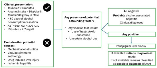
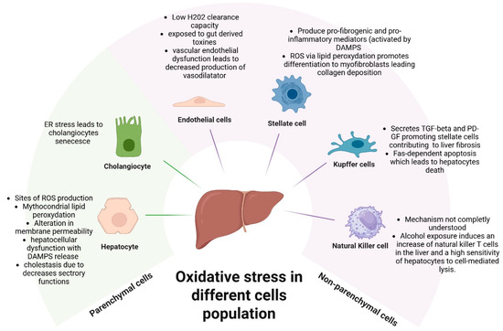
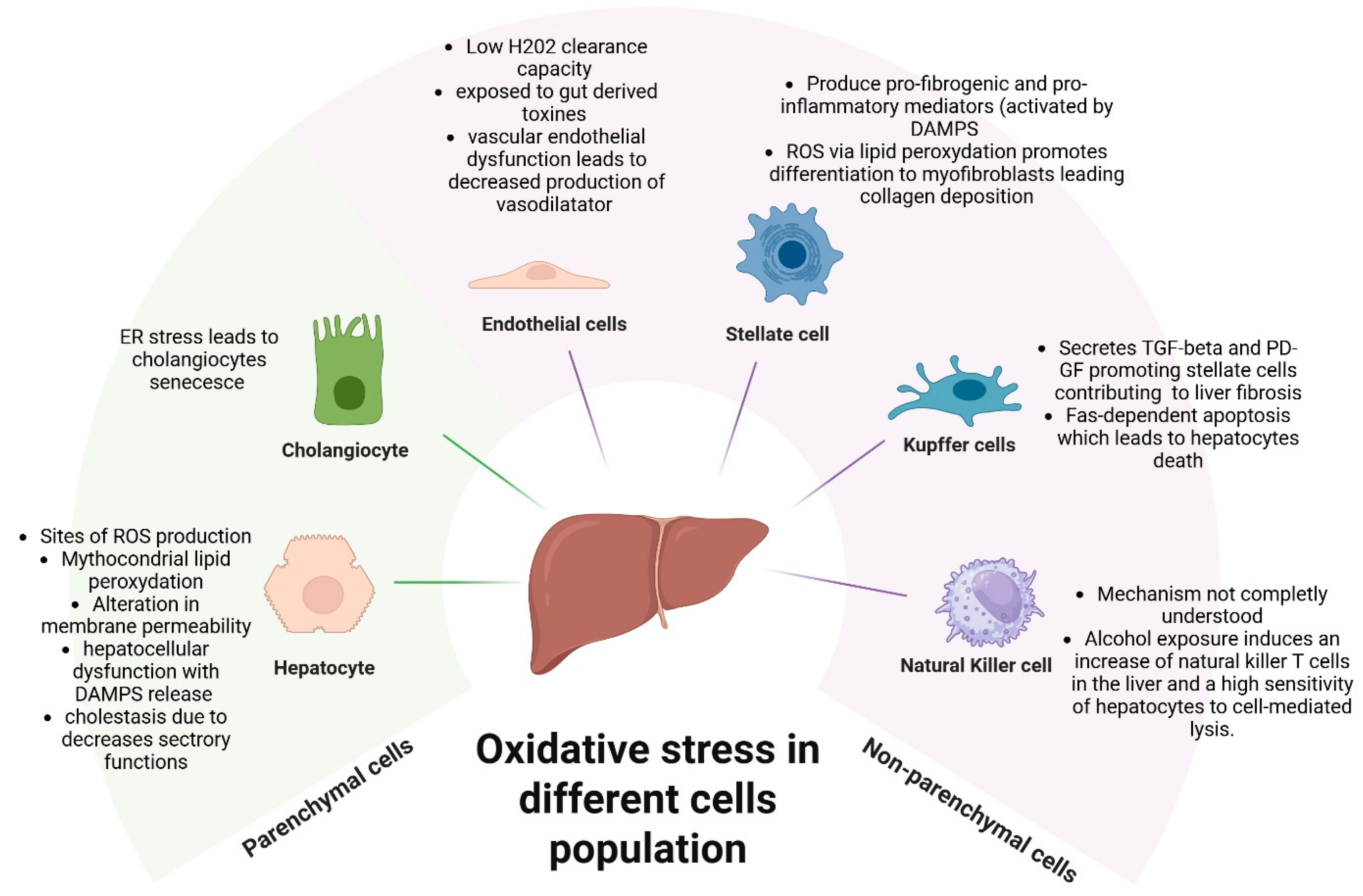
Disclaimer/Publisher’s Note: The statements, opinions and data contained in all publications are solely those of the individual author(s) and contributor(s) and not of MDPI and/or the editor(s). MDPI and/or the editor(s) disclaim responsibility for any injury to people or property resulting from any ideas, methods, instructions or products referred to in the content. |
© 2026 by the authors. Licensee MDPI, Basel, Switzerland. This article is an open access article distributed under the terms and conditions of the Creative Commons Attribution (CC BY) license.