


Abstract
Ionizing radiation from diagnostic imaging can induce DNA damage and oxidative stress, prompting interest in antioxidants as potential radioprotective agents. This systematic review evaluated whether antioxidant interventions reduce radiation-induced DNA damage and oxidative stress following diagnostic-dose X-ray exposure. This systematic review was registered in PROSPERO (CRD420261506735) and conducted according to PRISMA 2020 guidelines. PubMed, Embase, and Cochrane CENTRAL were systematically searched. Of 1888 records identified, 12 studies met the eligibility criteria and were included in the qualitative synthesis, comprising in vitro, animal, and human investigations. Eligible studies exposed in vitro models, animals, or humans to X-ray doses representative of diagnostic imaging and compared antioxidant-treated groups with irradiated untreated or placebo controls, assessing radiation-induced genetic damage and oxidative stress-related outcomes. Overall, most studies reported reduced radiation-induced DNA damage, primarily assessed by γH2AX, following antioxidant administration. N-acetylcysteine showed the most consistent protective effect in direct comparisons, although its superiority over other antioxidants could not be established. Antioxidant administration before irradiation, particularly approximately one hour before exposure, was generally associated with greater protection. Animal studies also suggested beneficial effects on oxidative stress and endogenous antioxidant defenses, whereas no eligible human study assessed oxidative stress biomarkers. Substantial heterogeneity, methodological limitations, and the predominance of short-term surrogate outcomes limit the strength of the evidence. Current findings support the biological plausibility of antioxidant-mediated radioprotection but do not establish a clinically meaningful benefit or justify routine antioxidant administration in diagnostic imaging. Further well-designed clinical studies are needed to determine the safety, efficacy, and clinical relevance of this approach.
1. Introduction
Exposure to ionizing radiation (IR), which includes X-rays, gamma rays, alpha and beta particles, can induce cellular damage, with DNA being one of the most critical biological targets [1]. IR-induced DNA damage may occur through both direct and indirect mechanisms [2,3,4]. Direct damage is estimated to account for approximately 30% of total DNA damage, although this proportion may vary depending on radiation quality, oxygenation levels, and the cellular environment [5,6]. It results from the direct interaction of radiation with DNA. At the molecular level, this process generates radical cation sites and promotes rapid electron transfer between adjacent DNA subunits. These unstable intermediates can cause structural alterations, including chemical modifications of DNA bases and lesions of the sugar–phosphate backbone, such as single-strand breaks (SSBs) and double-strand breaks (DSBs) [7].
The majority of IR-induced DNA damage (approximately 60–70%) occurs indirectly through oxidative stress (OS), initiated by water radiolysis following radiation exposure [1,8]. Water radiolysis produces a variety of reactive species through the interaction of IR with water molecules [9]. The primary products include hydroxyl radicals (•OH), hydrated electrons (e−aq), hydrogen atoms (H•), hydrogen molecules (H2), and hydrogen peroxide (H2O2) [10]. Among these, •OH and H2O2 are classified as reactive oxygen species (ROS), whereas e−aq and H• are reducing species. In the presence of dissolved molecular oxygen (O2), secondary reactions involving hydrated electrons and hydrogen atoms lead to the formation of superoxide anion radicals (O2•−) and hydroperoxyl radicals (HO2•), respectively, both of which are ROS. Among these ROS, hydroxyl radicals are particularly reactive and are considered major contributors to radiation-induced oxidative damage [11]. They can interact with nearby biomolecules, inducing oxidative DNA lesions such as 7,8-dihydro-8-oxoguanine (8-oxoG), thymine glycol (TG), single-strand breaks (SSBs), and double-strand breaks (DSBs) [12,13,14]. Because DSBs are difficult to repair accurately, they are among the most biologically relevant forms of radiation-induced DNA damage and may contribute to chromosomal aberrations, genomic instability, and carcinogenesis [15]. Closely spaced ROS-induced lesions may also generate clustered or complex DNA damage, further challenging DNA repair mechanisms [16].
Current radiobiological models generally assume a linear dose–response relationship for stochastic radiation effects in radiation protection [17]. Within this framework, even low-dose exposures are considered potentially contributory to stochastic health risks, although the magnitude of biological effects and the dose–response relationship at very low doses remain under investigation [18,19]. Over the past decades, population exposure to low doses of IR has substantially increased, largely due to the widespread use of diagnostic imaging, particularly computed tomography (CT) [20,21]. In 2016, more than 84 million CT scans were performed in the United States alone [22]. Although diagnostic imaging is indispensable in modern clinical practice, its increasing use has raised concerns about the cumulative biological effects of repeated radiation exposure, particularly in radiosensitive populations such as children. Epidemiological estimates suggest that diagnostic IR procedures may account for approximately 0.6 to 3% of cancers in developed countries [23]. However, these estimates are model-based and depend on assumptions regarding exposure distributions, latency, and radiation-associated risk; they should, therefore, be interpreted as population-level estimates rather than as direct evidence of causality in individual patients [21].
To minimize radiation-related risks, considerable efforts have focused on reducing patient exposure through technological improvements in imaging systems, including high-pitch acquisition, low tube voltage, prospective electrocardiographic (ECG) gating and iterative reconstruction algorithms [24]. Alongside these physical dose-reduction strategies, pharmacological approaches aimed at mitigating the biological consequences of radiation exposure have received increasing attention [25,26,27,28,29,30].
Among these interventions, antioxidants have attracted particular interest because they target the oxidative component of radiation-induced damage [31,32,33]. Their potential radioprotective effects involve several complementary mechanisms, including direct free radical scavenging, inhibition of oxidative chain reactions, chelation of transition metals involved in ROS generation, and enhancement of endogenous antioxidant defenses such as superoxide dismutase (SOD), catalase, and glutathione peroxidase (GPx) [34,35,36,37]. However, the contribution of direct radical scavenging depends on reaction kinetics, antioxidant concentrations, and the spatial and temporal distribution of radiolytic species, as illustrated by Monte Carlo simulations in aqueous systems [38]. In particular, the earliest stages of water radiolysis occur on extremely short timescales, during which radical-radical reactions compete with reactions involving dissolved solutes [9,38]. Therefore, the biological effects of antioxidants cannot solely be attributed primarily to the direct scavenging of primary radiolytic radicals. Rather, indirect cellular mechanisms, including the preservation or replenishment of endogenous antioxidant defenses and the maintenance of intracellular redox homeostasis, may also play an important role. For example, N-acetylcysteine (NAC) can act as a direct radical scavenger through its thiol (-SH) group while also supporting intracellular glutathione (GSH) synthesis by providing cysteine, thereby reinforcing endogenous antioxidant defenses [39,40].
For the purposes of this review, the term “antioxidant intervention” was used broadly to include classical antioxidant compounds as well as selected compounds or formulations with direct free-radical-scavenging or radioprotective activity. This broader definition was adopted to capture interventions that may mitigate radiation-induced oxidative or molecular damage even when their pharmacological classification does not strictly correspond to that of a conventional antioxidant.
Some antioxidants may also exert pro-oxidant effects under specific conditions [41,42]. For example, high concentrations of vitamin C (1000 mg/kg of body weight) in the presence of transition metals such as iron or copper may promote hydroxyl radical formation through Fenton-type reactions [43,44]. Therefore, radioprotective efficacy may depend on the specific molecule, dose, timing of administration, and experimental context. Accordingly, increased antioxidant exposure cannot necessarily be assumed to result in proportionally greater radioprotection.
A growing body of evidence supports the radioprotective potential of antioxidants, particularly in radiotherapy, where they have been investigated as agents to reduce radiation-induced toxicity while preserving treatment efficacy [45,46,47,48]. However, radiotherapy differs substantially from diagnostic imaging, not only because therapeutic exposures involve radiation doses several orders of magnitude higher, but also due to its highly localized nature. While diagnostic procedures often involve systemic or broad-area exposures aimed at imaging, radiotherapy delivers targeted, high-dose radiation confined to specific tumor volumes, raising distinct challenges regarding the selective protection of normal tissues vs. cancer cells [49,50]. Findings obtained under radiotherapy conditions therefore cannot be directly extrapolated to low-dose diagnostic exposures, where biological responses may differ both qualitatively and quantitatively. This distinction is particularly relevant because the present review focuses specifically on radiation exposures representative of diagnostic imaging rather than therapeutic irradiation.
Despite increasing interest in antioxidant-based radioprotection, evidence regarding its effects on DNA damage and oxidative stress following diagnostic X-ray exposures remains fragmented. Available studies span substantially different experimental levels, ranging from cellular models [51,52] and animal experiments [53] to controlled human studies [54,55] and human in vivo/in vitro investigations [56,57]. These designs provide complementary but not interchangeable information on biological plausibility, preclinical efficacy, and clinical applicability. To our knowledge, no previous systematic review has comprehensively synthesized evidence focusing exclusively on antioxidant interventions in the context of diagnostic-dose X-ray exposure. Given the continuing growth in medical imaging and the need for evidence-based strategies to minimize radiation-induced biological damage, a systematic evaluation of the available literature is warranted.
The principal aim of this systematic review was to evaluate whether antioxidant interventions reduce radiation-induced DNA damage and oxidative stress following X-ray exposure representative of diagnostic imaging, while considering evidence from in vitro models, animal studies, and human investigations separately. Furthermore, a secondary objective was to assess methodological limitations and the risk of bias and to determine whether current evidence supports the clinical translation of antioxidant-based radioprotection.
2. Materials and Methods
2.1. Study Design
This systematic review was conducted in accordance with the PRISMA (Preferred Reporting Items for Systematic Reviews and Meta-Analyses) guidelines [58]. The review methods were defined a priori according to a structured eligibility framework, whereas study selection, data extraction, and risk-of-bias assessment procedures were prespecified. This systematic review was registered in PROSPERO (CRD420261506735), accessed on 14 September 2026 and conducted according to PRISMA 2020. The completed PRISMA 2020 checklist is provided as Supplementary Table S1.
2.1.1. Eligibility Criteria
Eligibility criteria were defined according to the Population, Intervention, Comparator, Outcome and Study Design framework (PICOS) [59].
Eligible populations included established cell lines, primary cells, animal models and human subjects exposed to X-rays representative of diagnostic radiology. Because diagnostic exposure varies by modality, anatomical region, and setting, no single universal numerical dose threshold was used. Instead, the diagnostic relevance was assessed using prespecified criteria based on the exposure parameters and clinical or experimental context of exposure. Studies were included if explicitly linked to a diagnostic X-ray procedure or within published diagnostic ranges, considering imaging modality, anatomical region, tube voltage (kVp), tube current–time product (mAs), dose, and CT metrics (CTDIvol, DLP). For studies involving cells or animal models, exposure parameters were compared against established clinical ranges alongside the authors’ rationale. No parameter was evaluated in isolation. To minimize subjectivity, two reviewers (LN and AV) independently assessed each study. Reviewers recorded the radiation parameters and exposure context in the Supplementary Table (Supplementary Table S2). Studies with insufficient data were classified as unclear and discussed with a third reviewer (AG) to resolve any disagreements.
Studies describing exposure solely as “low-dose,” without an explicit diagnostic link or sufficient exposure parameters were excluded, as were those exceeding typical diagnostic doses or using non-X-ray radiation. For studies with multiple irradiation conditions, eligibility was determined at the exposure level; at least one condition had to meet the criteria, with its outcome data separately identifiable. The intervention of interest was the administration of antioxidants before and/or after exposure. Eligible interventions included classical antioxidants and compounds with recognized free-radical-scavenging or radioprotective activity, administered alone or in combination, without restrictions on timing (pre-, co-, or post-treatment).
Eligible comparators included irradiated untreated controls or placebo-treated irradiated controls. Studies without an appropriate comparator were excluded.
The outcomes of interest encompassed X-ray-induced molecular and cellular damage, specifically DNA damage (e.g., γH2AX foci, micronuclei, comet assay parameters, AP sites) and OS (e.g., ROS measures, oxidative damage, or antioxidant defense biomarkers) with no restrictions on assessment timing.
Original research articles without sufficient methodological or outcome data, reviews, editorials, commentaries, conference abstracts and non-original articles were excluded.
Only articles written in English and published within the 20 years preceding the final literature search were considered eligible for inclusion.
2.1.2. Search Strategy
A comprehensive search of three electronic bibliographic databases, i.e., PubMed, Embase, and Cochrane CENTRAL, was performed. The cut-off date for all databases was 24 March 2026. For each database, a structured search strategy was developed using a combination of controlled vocabulary terms, keywords and text words with different spellings related to antioxidants, diagnostic X-ray exposure and cellular damage. Search terms were identified and refined using database-specific controlled vocabulary tools, including MeSH (Medical Subject Headings), the NLM controlled vocabulary thesaurus used for indexing articles in PubMed and Emtree, and the controlled vocabulary used in Embase. These tools were used to identify appropriate subject headings, synonyms and related terms to improve the sensitivity and relevance of the search strategy.
The complete electronic search strategies for all databases, including controlled vocabulary, free-text terms, Boolean operators, truncation and proximity operators, where applicable and database-specific limits, are reported in Supplementary Table S3.
The search strategy was validated by testing its ability to retrieve eved a set of known eligible studies identified before the final search. These studies addressed antioxidant-mediated mitigation of cellular or DNA damage following diagnostic dose X-rays. Keywords, synonyms, MeSH and Emtree terms from these records were used to refine the search. No formal peer review using the PRESS checklist was performed.
Additionally, reference lists of included studies were manually screened, applying the same eligibility criteria used to identify further relevant articles.
2.2. Study Selection
Search results were imported into EndNote 20.6, and duplicate records were automatically detected and removed using bibliographic metadata, including title, publication year, author names, and DOI. The articles remaining were uploaded to Rayyan software (web application) [60] for reference management and manual title/abstract screening. Two independent reviewers (LN and AV) screened the remaining records by title and abstract according to the predefined PICO-based eligibility criteria, resolving disagreements by consensus. Full texts of potentially eligible studies were then retrieved and independently assessed by both reviewers. Additional records were identified by manually screening reference lists of included articles using the same criteria. No machine learning classifier or automation tools were used for eligibility decisions, except for initial duplicate detection.
2.3. Data Collection
Data from the included reports were extracted by one reviewer (LN) using a predefined Microsoft Excel data extraction form and cross-checked by a second reviewer (AV), with disagreements resolved by consensus or a third reviewer (AG).
The data extraction form was structured according to the PICO framework [59]. Extracted information included population or experimental models, type and dose of X-ray exposure, intervention characteristics, i.e., antioxidants, their dose or concentration, route or mode of administration, and timing of administration relative to radiation exposure. Comparator groups were categorized as placebo-treated controls or untreated irradiated controls.
Outcomes were extracted as reported and converted into descriptive percentage reduction relative to the irradiated comparator. When data were reported only graphically or were insufficiently described, they were recorded as unclear or not available unless numerical values could be reliably extracted from the text, tables, or Supplementary Materials. Any conversion of reported outcome data was documented in the extraction form, and no imputation of missing numerical data was performed unless explicitly described.
Due to the high heterogeneity among the included studies, results were categorized by experimental model into three categories: in vitro, in vivo animals, and in vivo humans. In vitro and in vivo animal studies involved direct radiation exposure and antioxidant administration to the experimental model. In contrast, in human studies, the antioxidant treatment was always administered to humans, whereas irradiation could have been performed either in vivo on the patient (clinical trials) or in vitro on peripheral blood mononuclear cells (PBMCs) isolated from blood samples (in vivo/in vitro studies). Moreover, results were further stratified by outcome, specifically DNA damage and OS. If the results were not already expressed as a percentage of damage reduction in the selected articles, the quantitative results were, whenever possible, converted into a uniform format and expressed as percentages of damage reduction according to the following formula:
This calculation was applied only when the direction and scale of the outcome allowed a meaningful interpretation. Percentage changes were not used to rank interventions across different biomarkers or experimental models.
Given the heterogeneity of the included studies, the results were not combined via meta-analysis but were synthesized descriptively. The decision not to pool results was based on several issues including statistical heterogeneity, substantial clinical, biological, and methodological heterogeneity in experimental models, X-ray exposure protocols, antioxidant interventions, doses and timing, comparators, outcome definitions, and outcome-assessment time points. Although several studies assessed γH2AX, these differences were considered sufficiently substantial to preclude a single pooled estimate with clear biological or clinical interpretability.
No predefined threshold was adopted to classify the magnitude of the effect.
2.4. Risk of Bias
Due to experimental heterogeneity, no single risk-of-bias tool was applied; instead, study-specific or domain-based approaches were used.
In vitro studies [51,61,62] and in vitro components of mixed studies [56,57] were evaluated using a prespecified domain-based framework adapted from principles of the SYRCLE Risk of Bias Tool [63] and informed by the general principles of methodological transparency and reporting completeness described in the ARRIVE 2.0 guidelines [64], in accordance with the approach reported in [65]. ARRIVE 2.0 was not used as a risk-of-bias assessment tool. The framework was developed a priori for the purposes of this review and was not considered a validated risk-of-bias instrument. It was therefore used as a structured approach to assess potential sources of bias and methodological limitations across the included in vitro studies, rather than as a formally validated instrument. The framework included six domains: selection bias, performance bias, detection bias, attrition bias, reporting bias, and mechanistic/translational validity. Each domain was classified as having a low, moderate, or high risk of bias according to prespecified criteria based on the reporting and methodological characteristics of each study. Because no universally validated risk-of-bias tool specifically designed for the diverse range of in vitro experiments included in this review was identified, the assessment was explicitly treated as a prespecified adapted framework rather than as a formally validated instrument. Because the in vitro framework was specifically developed for this review and incorporated an overall qualitative judgment to facilitate the interpretation of the domain-level assessments, an overall risk-of-bias judgment was reported for the in vitro studies. In contrast, for animal studies assessed using SYRCLE, no overall risk-of-bias score was calculated, in accordance with the recommendations for the use of the original tool.
In vivo animal studies [66,67,68,69] were evaluated using SYRCLE’s Risk of Bias Tool [63], retaining its domains and terminology without calculating an overall score. The evaluated domains included random sequence generation, allocation concealment, baseline characteristics, random housing, blinding of caregivers/investigators, random outcome assessment, blinding of outcome assessors, incomplete outcome data, selective reporting, and other potential sources of bias. Judgements were classified as “low risk”, “unclear”, or “high risk”.
Randomized clinical trials [70,71] were evaluated using the updated version of the Cochrane Risk of Bias 2 (RoB 2) tool [72], in accordance with the Cochrane Handbook for Systematic Reviews of Interventions (version 6.5, 2024) [73]. RoB 2 assesses risk of bias in five domains: the randomization process, deviations from intended interventions, missing outcome data, measurement of the outcome, and selection of the reported result. Judgements were classified as “low risk”, “some concerns”, or “high risk” according to the RoB 2 framework. The risk of bias for the two clinical trials [70,71] was assessed with reference to the DNA damage outcome.
The in vivo/in vitro study [74] and the in vivo/in vitro component of mixed studies [56,57] were evaluated using the original 2016 versioni of the Risk of Bias in Non-randomized Studies of Interventions (ROBINS-I) tool [72]. The seven ROBINS-I domains were considered: bias due to confounding, selection of participants, classification of interventions, deviations from intended interventions, missing data, measurement of outcomes, and selection of the reported result. Judgements were classified as “low risk”, “moderate”, or “serious risk”. Similarly to clinical trials, the risk of bias for the in vivo/in vitro study and the in vivo/in vitro component of mixed studies was assessed with reference to the DNA damage outcome.
Risk-of-bias assessment was performed independently by two reviewers (LN and AV) and disagreements were resolved through discussion and, when necessary, consultation with a third reviewer (AG). The results were plotted using the robvis tool [75].
3. Results
3.1. Study Selection
The literature search identified a total of 1888 records: 1496 from PubMed, 340 from Embase, and 52 from Cochrane CENTRAL. After removing 108 duplicates, 1780 records underwent title and abstract screening. Of these, 1644 were excluded because they did not meet the eligibility criteria, while the full text was sought for the remaining 136. The full text could not be located for 30 articles; therefore, 106 articles were fully evaluated for eligibility. Following the evaluation of the full text, 94 articles were excluded for the following reasons: publication type not meeting the predefined criteria (n = 29); outcomes not relevant to the assessment of cellular damage (n = 22); radiation dose not representative of typical exposures in diagnostic radiology (n = 41); and study population not meeting the eligibility criteria (n = 2). Overall, 12 studies met the selection criteria and were included in the qualitative synthesis. The study selection process for the systematic review is detailed in Figure 1.
Figure 1.
Flow chart of the study selection process for inclusion studies in the systematic review. Note: PRISMA 2020 distinguishes records (bibliographic entries), reports (documents describing studies), and studies (the underlying investigations). “Reports of included studies” refers to the documents describing the studies included in this review; a single study may have multiple reports.
3.2. Study Characteristics
Twelve studies fulfilled the eligibility criteria and were included in this systematic review. As illustrated in Table 1, the studies comprised three in vitro investigations [51,61,62], four in vivo animal studies [66,67,68,69], three in vivo human studies [70,71,74] and two mixed studies [56,57] involving human participants and including both an in vitro and in vivo/in vitro component. Among the human studies, two were clinical trials, in which both antioxidant administration and X-ray exposure occurred directly in patients undergoing diagnostic imaging [70,71], whereas one adopted an in vivo/in vitro design [74], in which antioxidants were administered to healthy volunteers before irradiation of isolated PBMCs. This study was considered separately from clinical studies because the irradiation was not performed directly on the participant during a diagnostic procedure.
Table 1.
Characteristics of included studies.
DNA damage was primarily assessed using γH2AX foci formation (9 studies [51,56,57,61,62,66,70,71,74]), whereas the micronucleus assay, comet assay, and quantification of AP sites were used less frequently [66,67,69]. Oxidative stress outcomes were evaluated by measuring oxidative burden via intracellular ROS production [51], total oxidative status (TOS) [68], malondialdehyde (MDA) lipid peroxidation [67,68], antioxidant defenses, assessed through total antioxidant capacity (TAC) [66], total antioxidant status (TAS) [68], and GSH [67] and SOD activities [67]. In addition, tumor necrosis factor-α (TNF-α) was also quantified as a biomarker of the inflammatory response associated with OS [68].
The included studies showed substantial heterogeneity in the experimental model, antioxidant intervention, formulation, dose or concentration, timing of administration, X-ray exposure protocol, outcome definition, and timing of outcome assessment. Therefore, quantitative meta-analysis was not considered appropriate and findings were synthesized narratively.
3.3. Risk of Bias Assessment
3.3.1. Risk of Bias for In Vitro Studies and In Vitro Component of Mixed Studies
The domains related to selection (D1), performance (D2), and reporting (D5) were judged to be at moderate risk in all studies, primarily due to the lack of description of randomization, operator blinding, and experimental protocols. The risk associated with outcome measurement (detection bias, D3) was low in the studies by Kuefner et al. [56] and Brand et al. [57], in which the counting of foci was performed in a blinded manner, and moderate in the studies by Bicheru et al. [61], Jermusek et al. [62], and Cervelli et al. [51], since these failed to report whether the outcome assessors were blinded to the experimental conditions. Attrition bias (D4) was judged to be low in all studies except in Bicheru et al. [61], where it was moderate, due to uncertainties regarding sample size and the independence of replicates. Mechanistic and translational validity (D6) was judged to be at moderate risk of bias in all selected studies, except in Bicheru et al. [61], which carried a higher risk of bias due to the lack of non-irradiated controls treated with antioxidants. In addition, the overall risk of bias was also considered moderate in all in vitro studies, except in Bicheru et al. [61] (Figure 2).
Figure 2.
Risk of bias assessment of included in vitro studies [51,56,57,61,62].
3.3.2. Risk of Bias for In Vivo Animal Studies
All studies were judged to be at low risk of bias regarding the comparability of baseline characteristics (D2). Several other domains, including sequence generation (D1), allocation concealment (D3), random housing (D4), outcome assessment (D6), and blinding of the outcome assessor (D7), were rated as having an unclear risk of bias because the corresponding procedures, including assessor blinding, were insufficiently reported. However, the main methodological limitation was the consistently high risk of performance bias related to the blinding of caregivers and investigators (D5). Blinding was either not feasible or not adequately implemented owing to the absence of comparable blinded sham or vehicle procedures.
Risk of bias related to incomplete outcome data (D8) was judged to be low in three studies [66,67,68] and unclear in another [69]. Selective outcome reporting (D9) was rated as low risk in two studies [66,68] and unclear in another two [67,69]. The greatest uncertainty regarding reporting was observed in [69], owing to discrepancies in the reported total number of animals, the lack of group-specific sample sizes, and incomplete reporting of some analyses.
An additional source of bias (D10) was identified in [67], where the absence of non-irradiated control groups precluded distinguishing a specific radioprotective effect from a general effect of watermelon juice. In the study [69], treatment was administered at the cage level through drinking water, with each condition apparently represented by a single cage. The analysis of individual animals as independent observations may therefore have introduced pseudoreplication, as the cage rather than the individual animal represented the experimental unit. In accordance with SYRCLE recommendations, an overall risk-of-bias score was not calculated for each study (Figure 3).
Figure 3.
Risk of bias assessment of included animal studies [66,67,68,69].
3.3.3. Risk of Bias for In Vivo Human Studies and for In Vivo/In Vitro Component of Mixed Studies
Bias arising from the randomization process (D1) and in the selection of the reported result (D5) raised some concerns in both clinical trials [70,71]. For allocation concealment (D1), it was inadequately described in both studies, while Stheli et al. [70] also provided insufficient information on sequence generation. For D5, the absence of sufficiently detailed prespecified statistical analysis plans prevented exclusion of selective reporting. Bias due to deviations from intended interventions (D2) was low in both studies. For outcome measurement (D4), Stheli et al. [70] was at low risk of bias, whereas S.M. Tao et al. [71] raised some concerns because blinding of outcome assessors was not reported. Regarding missing outcome data (D3), the risk of bias was low in S.M. Tao et al. [71] but high in Stheli et al. [70] owing to a discrepancy between the number of participants reported in the publication and the registered enrolment. Overall, the risk of bias was judged as having some concerns in S.M. Tao et al. [71] and as high in Stheli et al. [70], mainly due to the high risk associated with missing outcome data (Figure 4).
Figure 4.
Risk of bias assessment of clinical trials [70,71].
In vivo/in vitro study [74] and the in vivo/in vitro component (human) of two mixed studies [56,57] were judged to have a serious overall risk of bias, principally because of confounding arising from their uncontrolled single-arm before–after design, which precluded the separation of treatment effects from temporal physiological or procedural changes. The domains related to participant selection (D2), intervention classification (D3), deviations from the intended interventions (D4), and missing data (D5) were rated at low risk, while outcome measurement (D6) ranged from low to moderate risk depending on assessor blinding. The selection of reported outcomes (D7) was judged to be at moderate risk in all studies, because no registered protocols or analysis plans were available (Figure 5).
Figure 5.
Risk of bias assessment of included in vivo/in vitro studies [56,57,74].
3.4. Primary Outcome: DNA Damage
3.4.1. In Vitro Evidence
Five studies (3 in vitro and 2 mixed studies) investigated DNA damage in vitro, all using γH2AX foci as the primary endpoint (Table 2). Overall, all studies reported a reduction in radiation-induced γ-H2AX foci following antioxidant administration, although the magnitude of protection varied according to the antioxidant compound, concentration, and timing of administration.
Table 2.
Characteristics and main findings of in vitro studies evaluating the effects of antioxidants on X-ray-induced DNA damage.
Among the evaluated compounds, NAC consistently demonstrated the greatest radioprotective effect [57,61]. In studies directly comparing multiple antioxidants under identical experimental conditions, NAC produced the greatest reduction in γH2AX foci, followed by vitamin C, vitamin E, β-carotene [57,61], coenzyme Q10 and selenium [57], whereas zinc, Trolox and α-lipoic acid did not significantly reduce DNA damage under any tested condition [57]. These within-study comparisons suggest greater activity of NAC under the specific experimental conditions evaluated. RiduROS, a mixture of compounds with antioxidant activity, reduced radiation-induced γH2AX foci by 41% [51], whereas PrC-210, an aminothiol radioprotective compound, restored γH2AX levels close to baseline values following irradiation [62].
The efficacy of antioxidant treatment was strongly influenced by the timing of administration. Maximum protection was generally achieved after approximately one hour of pre-incubation, whereas shorter exposure times resulted in smaller reductions as shown by different groups [56,57,61]. Post-irradiation administration did not confer measurable protection under the conditions tested [56,57]. Thus, among the administration schedules evaluated, pretreatment approximately one hour before irradiation generally produced the greatest reduction in radiation-induced γH2AX formation.
Similarly, a concentration-dependent effect was observed for most compounds. Increasing concentrations generally enhanced the radioprotective effect up to an apparent plateau, although NAC and vitamin C continued to show improved efficacy at the highest tested concentrations [57,61]. Three studies evaluated antioxidant mixtures. Although combined formulations significantly reduced γH2AX formation, none demonstrated a clear superiority over the most effective single antioxidant [57,61]. In particular, NAC alone achieved protection comparable to combinations containing NAC, β-carotene, vitamin C and vitamin E [61], or NAC, vitamin C, vitamin E and selenium [57].
3.4.2. In Vivo Evidence from Animal Models
The three animal studies evaluated antioxidant-mediated protection against radiation-induced DNA damage using complementary biomarkers, including γH2AX foci [66], micronucleus assay [66,69], comet assay [69] and AP-site quantification [67] (Table 3). All studies reported an association between antioxidant administration and reduced genetic damage following diagnostic-dose X-ray exposure.
Table 3.
Characteristics and main findings of animal studies evaluating the effects of antioxidants on X-ray-induced DNA damage.
As observed in vitro, the magnitude of protection differed according to the antioxidant investigated [66]. In the only comparison between individual compounds, NAC showed a greater reduction in γH2AX foci (14.3%) compared with vitamin C (10.3%), while the reduction in micronucleus frequency was 34.9% for NAC and 28.6% for vitamin C [66]. However, although differences between the two compounds were observed for both γH2AX foci and micronucleus frequency, they did not reach statistical significance.
Multicomponent antioxidant formulations also demonstrated protective activity. Genuine Essiac tea markedly reduced DNA damage assessed by the comet assay and decreased micronucleus formation, although the latter did not reach statistical significance [69]. Vitamin C showed a more modest, non-significant reduction in DNA damage measured by the comet assay (31.9%), while micronucleus analysis revealed a non-significant increase in damage [69]. Watermelon juice attenuated oxidative DNA lesions measured as AP sites in several organs [67]. The findings from animal studies therefore indicate a generally protective direction of effect, but the evidence is not uniform across biomarkers or interventions.
3.4.3. Human Evidence
Five studies involving human participants evaluated the effects of antioxidant administration on IR DNA damage (Table 4). These comprised two clinical trials [70,71] and three in vivo/in vitro studies [56,57,74]. All five studies assessed DNA damage by quantifying γH2AX foci in PBMCs. Because the latter studies did not involve direct irradiation of participants during a diagnostic procedure, their findings were considered separately from those of the clinical trials when interpreting clinical applicability.
Table 4.
Characteristics and main findings of human studies evaluating the effects of antioxidants on X-ray-induced DNA damage.
Overall, the human evidence was consistent with both in vitro and animal findings, showing that antioxidant administration reduced radiation-induced γH2AX formation.
Vitamin C [70,71] and NAC [57,70] were the compounds most extensively investigated in patients undergoing diagnostic CT. Both agents reduced excess γH2AX foci, although statistical significance was achieved only in studies [57,71]. The largest reduction was observed following oral vitamin C administration before abdominal CT, where DNA damage decreased by approximately 61% [71].
Similarly to the in vitro and in vivo animal models, multicomponent antioxidant formulations also demonstrated protective activity. BioShield-Radiation showed a significant reduction in γ-H2AX foci (58%) when orally administered before irradiation [56].
The efficacy of antioxidant treatment as a function of administration timing was evaluated in four studies [56,57,71,74]. Across all studies, the greatest protective effect was observed when the antioxidant was administered 1 h before irradiation.
3.5. Secondary Outcome: Oxidative Stress
3.5.1. In Vitro Evidence
Only one in vitro study evaluated oxidative stress outcomes [51] (Table 5). Pretreatment with an antioxidant mixture (RiduROS) markedly reduced intracellular ROS production following irradiation. The protective effect was both time- and concentration-dependent: 24 h pretreatment resulted in a substantially greater reduction in ROS levels (97%) than 1 h pretreatment (18%), while treatment with 1 μg/mL achieved a 98% reduction compared with 65% at 0.1 μg/mL. The combined formulation was also more effective than its individual antioxidant components.
Table 5.
Characteristics and main findings of in vitro studies evaluating the effects of antioxidants on X-ray-induced OS damage.
3.5.2. In Vivo Evidence from Animal Models
Three in vivo animal studies evaluated the effects of antioxidant treatment on radiation-induced oxidative stress [66,67,68] (Table 6). Overall, antioxidant administration reduced oxidative burden while enhancing endogenous antioxidant defenses. Protective effects included reductions in MDA, TOS and TNF-α, together with increases in TAC, TAS, SOD activity and GSH levels.
Table 6.
Characteristics and main findings of animal studies evaluating the effects of antioxidants on X-ray-induced OS damage.
MDA levels decreased by 65.4% after amifostine treatment [68] and by 15.9% and 21.2% in the lungs and liver, respectively, following watermelon juice supplementation [67], whereas no significant reduction was observed in the brain. Similarly, TOS decreased by 71.5% after amifostine administration [68].
TAC increased by 68.2% following vitamin C and by 152.3% following NAC administration compared with untreated irradiated animals [66], although irradiation itself did not significantly alter TAC relative to non-irradiated controls.
Amifostine increased ovarian tissue TAS by 40.9% one month after irradiation [68], whereas watermelon juice increased SOD activity in the brain, lungs, and liver (15.8–90.0%) and GSH levels in the brain (157.1%) and liver (50.0%); the increase in lung GSH did not reach statistical significance [67]. In the study by Can et al. [68], amifostine also reduced TNF-α levels by 31% at 3 h post-irradation, although no significant differences in MDA, TOS, or TAS were observed at this earlier time point [68].
3.5.3. Human Evidence
No eligible human studies evaluated oxidative stress biomarkers following diagnostic-dose X-ray exposure. Consequently, despite the central mechanistic role attributed to oxidative stress in indirect radiation-induced damage, there is currently insufficient human evidence to determine whether antioxidant administration actually modifies systemic or tissue-level oxidative stress following diagnostic imaging.
4. Discussion
The present systematic review synthesizes the available evidence on the potential of antioxidant interventions to mitigate biological damage induced by diagnostic-dose X-ray exposure. Overall, the available evidence indicates a generally protective direction of effect, particularly for radiation-induced DNA damage, across in vitro, animal and human studies. However, the certainty and clinical relevance of this evidence remain limited by the small number of eligible studies, substantial heterogeneity in experimental models and intervention protocols, methodological limitations and the predominance of short-term surrogate biomarkers. Importantly, the evidence currently supports a potential biological radioprotective effect rather than an established clinical benefit.
A generally consistent direction of effect was observed across the different experimental settings, with antioxidant administration associated with a reduction in radiation-induced γH2AX formation. This finding was observed in cellular models, animal experiments and human investigations, although the magnitude and statistical significance of the effect varied considerably between studies. The overall consistency of this signal provides biological plausibility for antioxidant-mediated radioprotection, but it should be interpreted cautiously. The different experimental levels address distinct questions and are not interchangeable: in vitro studies primarily assess cellular responses under controlled conditions and can provide evidence of direct molecular activity, animal studies incorporate systemic and tissue-specific effects and human studies provide greater translational relevance but were few and included both in vivo human exposure and exposure of isolated human PBMCs. Therefore, the apparent consistency of the overall signal should not be interpreted as evidence of equivalent efficacy across these settings.
Among the evaluated interventions, NAC showed the most consistent protective signal. In direct head-to-head comparisons performed under the same experimental conditions, NAC produced greater reductions in γH2AX foci than several other individual antioxidants. However, these comparisons were limited to a small number of studies and were conducted using specific concentrations, exposure protocols and experimental models. Therefore, the available evidence does not establish NAC as universally superior to other antioxidants. Rather, NAC may represent a promising candidate for further investigation, particularly because protective effects were observed across more than one experimental setting. Vitamin C also demonstrated potentially protective effects, although findings were less consistent. The non-significant increase in micronucleus frequency reported in one animal experiment highlights the context dependence of antioxidant effects and cautions against assuming that antioxidant supplementation is invariably beneficial.
Multicomponent antioxidant formulations generally reduced radiation-induced biological damage; however, available head-to-head comparisons did not demonstrate a clear additional benefit over the most effective individual compound, particularly NAC. This does not establish that combination therapy is inferior to single-agent treatment, as differences in composition, dose, bioavailability, pharmacokinetics, and mechanisms of action limit direct comparisons between formulations. Future studies should therefore determine whether specific combinations provide additive or synergistic effects rather than assuming that increasing the number of antioxidant components necessarily increases radioprotection.
The timing of administration emerged as another potentially important determinant of efficacy. Across the experimental schedules evaluated, administration approximately one hour before irradiation generally produced the greatest reduction in radiation-induced DNA damage. A plausible explanation is that pretreatment allows sufficient systemic or intracellular availability of the intervention at the time of radiation exposure, when the generation of reactive species is expected to occur rapidly [76]. However, the available evidence does not establish one hour as a universally optimal biological or clinical pretreatment interval. Differences in the antioxidant, dose, exposure protocol, and outcome-assessment timing indicate that the apparent advantage of one-hour pretreatment may reflect the experimental conditions tested rather than a general pharmacological principle. Moreover, requiring pretreatment may represent an important practical limitation in routine diagnostic imaging, where examinations are frequently performed urgently or with limited preparation time.
The translational relevance of the observed effects is further constrained by differences between experimental concentrations and clinically achievable exposures. Concentrations that produce substantial radioprotection in vitro may not be attainable in human plasma or target tissues without substantially increasing the administered dose. Pharmacokinetics, tissue distribution, metabolism, intracellular availability and the timing of peak concentrations are therefore critical determinants of whether experimental findings can be reproduced safely in humans. Future translational studies should integrate pharmacokinetic and dose-finding assessments rather than extrapolating effective in vitro concentrations directly to clinical supplementation.
Another important consideration regarding the clinical relevance of the included studies is the extent to which the radiation doses and irradiation conditions reflect those encountered in diagnostic imaging. Although all selected studies investigated radiation exposures within the context of diagnostic X-ray applications, substantial differences may exist in terms of absorbed dose, dose rate, irradiation geometry, and exposure protocols. In particular, in vitro and animal studies may employ irradiation conditions that do not fully reproduce those experienced by patients during routine diagnostic procedures. Furthermore, dosimetric comparisons across different experimental models require caution, as absorbed dose (Gy) and effective dose (mSv) represent different quantities and are not directly interchangeable. Consequently, although these studies provide valuable insights into the biological effects of antioxidant interventions following X-ray exposure, differences in radiation dose and irradiation conditions may limit the direct extrapolation of their findings to clinical diagnostic imaging. These aspects should therefore be considered when interpreting the potential clinical relevance of antioxidant-based radioprotection.
Antioxidant activity should not be equated with an invariably protective biological effect. Reactive oxygen species are involved in physiological signaling as well as oxidative damage and the effects of antioxidant compounds depend on concentration, cellular redox status, tissue environment and the presence of transition metals or other reactive species [77,78]. Under specific conditions, some compounds may display pro-oxidant properties [42]. The inconsistent findings observed for vitamin C in animal models illustrate the context dependence of antioxidant effects [65], although the present evidence is insufficient to attribute the observed result specifically to a pro-oxidant mechanism. More broadly, increasing antioxidant exposure should not be assumed to produce a proportional increase in radioprotection. Defining the therapeutic window and safety profile of candidate interventions is therefore essential before clinical implementation can be considered.
The most important question for clinical translation is whether the magnitude of the observed biological effect is sufficient to justify administering an additional intervention to patients undergoing diagnostic imaging. This is particularly relevant because diagnostic examinations generally involve relatively low radiation doses, and the absolute clinical risk associated with an individual examination is generally low, although it varies according to patient characteristics, examination type and exposure parameters. Consequently, even a measurable reduction in a surrogate biomarker may not correspond to a clinically meaningful reduction in cancer or other radiation-related health outcomes.
The human studies primarily assessed γH2AX foci in PBMCs, an early biomarker of the cellular DNA damage response [79]. Although γH2AX is biologically relevant and widely used as a marker of radiation-induced DNA damage, its reduction cannot be assumed to translate directly into fewer persistent mutations, chromosomal abnormalities, genomic instability, cancer events, or other long-term health outcomes [80]. Indeed, γH2AX foci represent an early biomarker of the cellular response to DNA damage, and their reduction does not necessarily reflect a corresponding decrease in persistent DNA lesions or demonstrate long-term radioprotection [80]. Furthermore, PBMCs may not fully represent radiation responses in all tissues exposed during diagnostic imaging. Thus, the current human evidence supports a potential mechanistic radioprotective effect but does not establish a clinically meaningful health benefit. To address these translational limitations, future research could incorporate non-invasive, long-term systemic markers, such as micronucleus or chromosomal aberration assays, to verify whether early DNA repair translates into genuine clinical health benefits.
Any potential benefit of antioxidant administration must also be considered in relation to its potential risks and practical burdens. These may include dose-dependent adverse effects, gastrointestinal or systemic intolerance, drug interactions, uncertain effects of long-term or repeated supplementation and context-dependent pro-oxidant activity. Antioxidant administration may also introduce logistical complexity if an adequate biological concentration must be achieved before imaging. Therefore, the risk–benefit balance cannot be determined by the magnitude of γH2AX reduction alone. Before clinical implementation can be considered, candidate interventions should demonstrate a reproducible protective effect at a safe and feasible dose, together with acceptable tolerability and a clinically meaningful benefit that outweighs their potential harms and costs.
The distinction between human exposure for diagnostic purposes and irradiation of human isolated PBMCs is also important for interpreting human evidence. The two clinical trials involved patients undergoing diagnostic X-ray exposure, whereas the other human studies administered antioxidants to participants and subsequently irradiated in vitro isolated PBMCs. Although the latter design provides useful information on the biological response of circulating cells after antioxidant exposure, it does not reproduce the complete physiological context of a diagnostic examination. Differences in tissue exposure, systemic responses, cellular trafficking and pharmacokinetics limit the direct extrapolation of these findings to clinical imaging. To overcome these physiological gaps pragmatically, future research could conduct prospective trials on consenting patients already scheduled for routine imaging who are administered safe, widely used antioxidants. Furthermore, parallel studies might utilize fresh whole-blood samples to preserve natural plasma–cell interactions, complemented by physiologically based pharmacokinetic (PBPK) modeling to simulate real-world tissue distribution.
Evidence concerning oxidative stress is particularly limited. Only one eligible in vitro study evaluated intracellular ROS, while three animal studies assessed a range of oxidative stress or antioxidant-defense biomarkers. These studies generally suggested reductions in oxidative burden and enhancement of endogenous antioxidant defenses following antioxidant or radioprotective treatment. However, MDA, TOS, TAC, TAS, SOD, and GSH reflect different aspects of redox biology and should not be regarded as interchangeable measures of oxidative stress [81]. Effects also varied according to tissue, intervention, and timing of assessment. Most importantly, no eligible human study evaluated oxidative stress biomarkers following direct diagnostic-dose X-ray exposure. Therefore, the proposed mechanistic pathway linking antioxidant administration, reduced oxidative stress, and reduced DNA damage remains incompletely characterized in humans.
The methodological limitations identified in the risk-of-bias assessment further reduce confidence in the available evidence. In vitro studies often provided insufficient information regarding randomization, blinding, replication, and experimental procedures, while animal studies frequently lacked adequate reporting of randomization, allocation procedures, and assessor blinding. One animal study also raised concerns regarding pseudoreplication because treatment was administered at the cage level while individual animals appeared to be analyzed as independent observations. In human studies, uncontrolled before–after designs were particularly vulnerable to confounding by temporal, physiological, or procedural changes, and selective reporting was a concern in one clinical trial. These limitations do not demonstrate that the reported biological effects are false; rather, they reduce confidence that alternative explanations for the observed effects have been adequately excluded.
The absence of quantitative synthesis should also be interpreted in light of this methodological and biological heterogeneity. Differences in antioxidant compounds and formulations, concentrations, administration schedules, radiation doses, biological models, outcome measures, and follow-up intervals precluded meaningful pooling of effect estimates. Under these circumstances, a meta-analysis could yield a summary estimate with limited biological interpretability and potentially obscure important sources of heterogeneity. Narrative synthesis was therefore considered more appropriate for the current evidence base. Future research should aim to reduce this heterogeneity through standardized diagnostic exposure protocols, predefined antioxidant regimens, harmonized outcome measures, and adequately powered study designs.
5. Conclusions
Current evidence supports the biological plausibility of antioxidant-mediated radioprotection but does not currently justify routine antioxidant administration to patients undergoing diagnostic imaging. The evidence is strongest for short-term modulation of DNA damage biomarkers and weaker for oxidative stress and clinical outcomes. Future research must therefore determine if early biomarker reductions (e.g., γH2AX) translate to persistent genomic protection and clinically relevant benefits, while explicitly evaluating safety, tolerability, pharmacokinetics, dose–response relationships, and the risk–benefit balance of antioxidant administration.
Future clinical trials should ideally be adequately powered, randomized, and prospectively registered, with standardized antioxidant formulations and dosing schedules and well-characterized diagnostic radiation exposures. They should simultaneously assess DNA damage, oxidative stress, safety and, where feasible, persistent indicators of genomic damage. Particular attention should be given to patient populations potentially more vulnerable to cumulative radiation exposure, while avoiding unnecessary pharmacological intervention when risks outweigh benefits. Overall, the present evidence suggests that antioxidants may reduce early biological responses to diagnostic-dose X-rays, particularly DNA damage. However, clinical benefit remains unproven. Antioxidant radioprotection should therefore be considered an investigational strategy rather than a routine clinical intervention, complementing rather than replacing established radiation protection principles.
Supplementary Materials
The following supporting information can be downloaded at https://www.mdpi.com/article/10.3390/antiox15111519/s1: Table S1: PRISMA 2020 checklist [58]. Table S2: Reported Parameters for establishing the diagnostic validity of X-ray procedures used in included studies [51,56,57,61,62,66,67,68,69,70,71,74]. Table S3: Database-specific search strategies.
Author Contributions
Conceptualization, methodology, investigation, L.N., A.V. (Alessandra Verdina) and C.M.; data curation, L.N. and A.V. (Alessandro Valenti); validation, A.V. (Alessandra Verdina); writing—original draft preparation, writing—review and editing, L.N. and A.V. (Alessandra Verdina); visualization, V.B.; supervision, S.S., M.C.F., A.G.; project administration, I.F. and M.C.F. All authors have read and agreed to the published version of the manuscript.
Funding
The authors are supported by the Italian Ministry of Health (Ricerca Corrente ISG 2026) and by a generous donation from “Associazione Tempi Moderni, solidarietà e amicizia Claudio Caterini” to A.Verdina.
Institutional Review Board Statement
Not applicable.
Informed Consent Statement
Not applicable.
Data Availability Statement
All data generated or analyzed during this study are included in this published article and its Supplementary Materials. Study characteristics and outcome data are summarized in the tables of the article. Additional details on X-ray exposure parameters are provided in the Supplementary Materials. The systematic review protocol is publicly available on PROSPERO (CRD420261506735).
Conflicts of Interest
The authors declare no conflicts of interest.
Abbreviations
The following abbreviations are used in this manuscript:
| 8-oxoG | 7,8-Dihydro-8-oxoguanine |
| AP | Apurinic/apyrimidinic |
| ARRIVE | Animal Research: Reporting of In Vivo Experiments |
| PBMCs CENTRAL | Peripheral Blood Mononuclear Cells Cochrane Central Register of Controlled Trials |
| CT | Computed tomography |
| CTDIvol | Volume computed tomography dose index |
| DLP | Dose-length product |
| DNA | Deoxyribonucleic acid |
| DSBs | Double-strand breaks |
| ECG | Electrocardiographic |
| GPx | Glutathione peroxidase |
| GSH | Glutathione |
| IR | Ionizing radiation |
| MDA | Malondialdehyde |
| MeSH | Medical Subject Headings |
| NAC | N-Acetylcysteine |
| NLM | National Library of Medicine |
| OS | Oxidative stress |
| PBPK | Physiologically based pharmacokinetic |
| PICO | Population, Intervention, Comparator, Outcome |
| PICOS | Population, Intervention, Comparator, Outcome, and Study Design |
| PRESS | Peer Review of Electronic Search Strategies |
| PRISMA | Preferred Reporting Items for Systematic Reviews and Meta-Analyses |
| RoB 2 | Risk of Bias 2 |
| ROBINS-I | Risk of Bias in Non-randomized Studies of Interventions |
| ROS | Reactive oxygen species |
| SOD | Superoxide dismutase |
| SSBs | Single-strand breaks |
| SYRCLE | SYstematic Review Centre for Laboratory animal Experimentation |
| TAC | Total antioxidant capacity |
| TAS | Total antioxidant status |
| TG | Thymine glycol |
| TNF-α | Tumor necrosis factor alpha |
| TOS | Total oxidative status |
| γH2AX | Phosphorylated histone H2AX |
References
- Wei, W.; Ren, Y.; Lan, J.; Yi, J.; Wang, M.; Zhang, Y.; Wang, S.; Xu, Y.; Han, G.; Fu, Y.; et al. Ionizing Radiation: Molecular Mechanisms, Biological Effects, and Therapeutic Targets. Mol. Biomed. 2026, 7, 3. [Google Scholar] [CrossRef] [Scilit] [PubMed]
- Ibáñez, B.; Melero, A.; Montoro, A.; San Onofre, N.; Soriano, J.M. Molecular Insights into Radiation Effects and Protective Mechanisms: A Focus on Cellular Damage and Radioprotectors. CIMB 2024, 46, 12718–12732. [Google Scholar] [CrossRef] [Scilit] [PubMed]
- Alizadeh, E.; Orlando, T.M.; Sanche, L. Biomolecular Damage Induced by Ionizing Radiation: The Direct and Indirect Effects of Low-Energy Electrons on DNA. Annu. Rev. Phys. Chem. 2015, 66, 379–398. [Google Scholar] [CrossRef] [Scilit] [PubMed]
- Saini, S.; Gurung, P. A Comprehensive Review of Sensors of Radiation-induced Damage, Radiation-induced Proximal Events, and Cell Death. Immunol. Rev. 2025, 329, e13409. [Google Scholar] [CrossRef] [Scilit] [PubMed]
- Hayes, D.P. Non-Problematic Risks from Low-Dose Radiation-Induced DNA Damage Clusters. Dose-Response 2008, 6, 30–52. [Google Scholar] [CrossRef] [Scilit]
- Kuefner, M.; Brand, M.; Engert, C.; Schwab, S.; Uder, M. Radiation Induced DNA Double-Strand Breaks in Radiology. Fortschr. Röntgenstr. 2015, 187, 872–878. [Google Scholar] [CrossRef] [Scilit] [PubMed]
- Chatterjee, N.; Walker, G.C. Mechanisms of DNA Damage, Repair, and Mutagenesis. Environ. Mol. Mutagen. 2017, 58, 235–263. [Google Scholar] [CrossRef] [Scilit] [PubMed]
- Ramos, B.; Gómez-Cayupán, J.; Aranis, I.; García Tapia, E.; Coghlan, C.; Ulloa, M.-J.; Gelerstein Claro, S.; Urbina, K.; Espinoza, G.; De Grazia, J.; et al. Oxidative Stress-Mediated DNA Damage Induced by Ionizing Radiation in Modern Computed Tomography: Evidence for Antioxidant-Based Radioprotective Strategies. Antioxidants 2025, 14, 1085. [Google Scholar] [CrossRef] [Scilit] [PubMed]
- Jay-Gerin, J.-P. Fundamentals of Water Radiolysis. Encyclopedia 2025, 5, 38. [Google Scholar] [CrossRef] [Scilit]
- Reisz, J.A.; Bansal, N.; Qian, J.; Zhao, W.; Furdui, C.M. Effects of Ionizing Radiation on Biological Molecules—Mechanisms of Damage and Emerging Methods of Detection. Antioxid. Redox Signal. 2014, 21, 260–292. [Google Scholar] [CrossRef] [Scilit]
- Halliwell, B.; Adhikary, A.; Dingfelder, M.; Dizdaroglu, M. Hydroxyl Radical Is a Significant Player in Oxidative DNA Damage in Vivo. Chem. Soc. Rev. 2021, 50, 8355–8360. [Google Scholar] [CrossRef] [Scilit] [PubMed]
- Wang, J.; Li, C.; Han, J.; Xue, Y.; Zheng, X.; Wang, R.; Radak, Z.; Nakabeppu, Y.; Boldogh, I.; Ba, X. Reassessing the Roles of Oxidative DNA Base Lesion 8-oxoGua and Repair Enzyme OGG1 in Tumorigenesis. J. Biomed. Sci. 2025, 32, 1. [Google Scholar] [CrossRef] [Scilit] [PubMed]
- Bellon, S.; Shikazono, N.; Cunniffe, S.; Lomax, M.; O’Neill, P. Processing of Thymine Glycol in a Clustered DNA Damage Site: Mutagenic or Cytotoxic. Nucleic Acids Res. 2009, 37, 4430–4440. [Google Scholar] [CrossRef] [Scilit] [PubMed]
- Sedelnikova, O.A.; Redon, C.E.; Dickey, J.S.; Nakamura, A.J.; Georgakilas, A.G.; Bonner, W.M. Role of Oxidatively Induced DNA Lesions in Human Pathogenesis. Mutat. Res./Rev. Mutat. Res. 2010, 704, 152–159. [Google Scholar] [CrossRef] [Scilit] [PubMed]
- Rothkamm, K.; Kühne, M.; Jeggo, P.A.; Löbrich, M. Radiation-Induced Genomic Rearrangements Formed by Nonhomologous End-Joining of DNA Double-Strand Breaks. Cancer Res. 2001, 61, 3886–3893. [Google Scholar] [PubMed]
- Nickoloff, J.A.; Sharma, N.; Taylor, L. Clustered DNA Double-Strand Breaks: Biological Effects and Relevance to Cancer Radiotherapy. Genes 2020, 11, 99. [Google Scholar] [CrossRef] [Scilit] [PubMed]
- Laurier, D.; Billarand, Y.; Klokov, D.; Leuraud, K. The Scientific Basis for the Use of the Linear No-Threshold (LNT) Model at Low Doses and Dose Rates in Radiological Protection. J. Radiol. Prot. 2023, 43, 024003. [Google Scholar] [CrossRef] [Scilit] [PubMed]
- Bosch De Basea, M.; Thierry-Chef, I.; Harbron, R.; Hauptmann, M.; Byrnes, G.; Bernier, M.-O.; Le Cornet, L.; Dabin, J.; Ferro, G.; Istad, T.S.; et al. Author Correction: Risk of Hematological Malignancies from CT Radiation Exposure in Children, Adolescents and Young Adults. Nat. Med. 2023, 31, 2452. [Google Scholar] [CrossRef] [Scilit] [PubMed]
- Han, S.; Soh, J.; Nah, S.; Han, K.; Jung, J.-H.; Park, J.; Hwang, Y.; Lee, C.; Hong, J.-Y. Pediatric Computed Tomography Scan and Subsequent Risk of Malignancy: A Nationwide Population-Based Cohort Study in Korea Using National Cancer Institute Dosimetry System for Computed Tomography (NCICT). BMC Med. 2025, 23, 355. [Google Scholar] [CrossRef] [Scilit] [PubMed]
- Uruma, T. Current Trends in Computed Tomography Practices in Japan and the Role of Referring Physicians in Radiation Exposure Management. Intern. Med. 2025, 64, 3343–3348. [Google Scholar] [CrossRef] [Scilit] [PubMed]
- Smith-Bindman, R.; Chu, P.W.; Azman Firdaus, H.; Stewart, C.; Malekhedayat, M.; Alber, S.; Bolch, W.E.; Mahendra, M.; Berrington De González, A.; Miglioretti, D.L. Projected Lifetime Cancer Risks From Current Computed Tomography Imaging. JAMA Intern. Med. 2025, 185, 710. [Google Scholar] [CrossRef] [Scilit] [PubMed]
- Mettler, F.A.; Mahesh, M.; Bhargavan-Chatfield, M.; Chambers, C.E.; Elee, J.G.; Frush, D.P.; Miller, D.L.; Royal, H.D.; Milano, M.T.; Spelic, D.C.; et al. Patient Exposure from Radiologic and Nuclear Medicine Procedures in the United States: Procedure Volume and Effective Dose for the Period 2006–2016. Radiology 2020, 295, 418–427. [Google Scholar] [CrossRef] [Scilit] [PubMed]
- De González, A.B.; Darby, S. Risk of Cancer from Diagnostic X-Rays: Estimates for the UK and 14 Other Countries. Lancet 2004, 363, 345–351. [Google Scholar] [CrossRef] [Scilit] [PubMed]
- Richards, C.E.; Obaid, D.R. Low-Dose Radiation Advances in Coronary Computed Tomography Angiography in the Diagnosis of Coronary Artery Disease. CCR 2019, 15, 304–315. [Google Scholar] [CrossRef] [Scilit] [PubMed]
- Li, X.; Yu, L.; Xu, H.; Xing, X.; Wu, W.; Feng, Y.; Ma, L.; Zhou, Z.; Li, B.; He, Y. Research on the Mechanisms of Natural Products in Radiation Protection. Front. Bioeng. Biotechnol. 2025, 13, 1640682. [Google Scholar] [CrossRef] [Scilit] [PubMed]
- Palkar, P.A.; John, G.; Chekuri, G.; Chatterjee, A.; Kunwar, A.; Goda, J.S. Radioprotectors & Mitigators in Radiation Therapy: Harnessing Diverse Pathways for Optimizing Clinical Outcomes. Indian. J. Med. Res. 2025, 162, 343–366. [Google Scholar] [CrossRef] [Scilit] [PubMed]
- Obrador, E.; Estrela, J.M.; López-Blanch, R.; Moreno-Murciano, P.; Montoro, A.; Oriol-Caballo, M. Radiomitigators: Breakthroughs in Post-Radiation Recovery. Antioxidants 2026, 15, 381. [Google Scholar] [CrossRef] [Scilit] [PubMed]
- Athanassiou, H.; Antonadou, D.; Coliarakis, N.; Kouveli, A.; Synodinou, M.; Paraskevaidis, M.; Sarris, G.; Georgakopoulos, G.R.; Panousaki, K.; Karageorgis, P.; et al. Protective Effect of Amifostine during Fractionated Radiotherapy in Patients with Pelvic Carcinomas: Results of a Randomized Trial. Int. J. Radiat. Oncol. Biol. Phys. 2003, 56, 1154–1160. [Google Scholar] [CrossRef] [Scilit] [PubMed]
- Upadhyaya, A.; Zhou, P.; Meng, Z.; Wang, P.; Zhang, G.; Jia, Q.; Tan, J.; Li, X.; Hu, T.; Liu, N.; et al. Radioprotective Effect of Vitamin E on Salivary Glands after Radioiodine Therapy for Differentiated Thyroid Cancer: A Randomized-Controlled Trial. Nucl. Med. Commun. 2017, 38, 891–903. [Google Scholar] [CrossRef] [Scilit] [PubMed]
- Brzóska, K.; Carpenter, A.D.; Petrus, S.A.; Singh, V.K. Radiation Countermeasure Gamma-Tocotrienol Inhibits Accumulation of Lipid Peroxidation Products in the Serum of Nonhuman Primates Exposed to Partial- or Total-Body Radiation—A Hallmark of Inhibition of Irradiation-Induced Ferroptosis? Int. J. Mol. Sci. 2026, 27, 3387. [Google Scholar] [CrossRef] [Scilit] [PubMed]
- Shelkovskaia, O.; Chernikov, A.V.; Serov, D.A.; Burmistrov, D.E.; Trutnev, Y.A.; Sarimov, R.M.; Simakin, A.V.; Konchekov, E.M.; Abdullaev, S.A.; Karmanova, E.E.; et al. 2R,3R-Trans-Dihydroquercetin Has Powerful Antioxidant Properties, Prevents DNA and Protein Damage, and Protects Mice from Injury Caused by Radiation-Induced Oxidative Stress. Antioxidants 2026, 15, 423. [Google Scholar] [CrossRef] [Scilit] [PubMed]
- Alcaraz, M.; Achel, D.G.; Garcia-Gamuz, J.A.; Ruiz-Gómez, M.J.; Mercado-Díaz, A.; Alcaraz-Saura, M.; Navarro-Fernández, J.L.; Olivares, A. Different Effects of Antioxidants Against Ionizing Radiation: An Experimental Model of Micronuclei. Curr. Issues Mol. Biol. 2026, 48, 364. [Google Scholar] [CrossRef] [Scilit] [PubMed]
- Baralić, K.; Živanović, J.; Božović, P.; Kržanović, N.; Šćepanović, N.; Petrović, J.; Đukić-Ćosić, D. Protective Effects of Curcumin and Sulforaphane Against Ionising Radiation–Induced Oxidative Stress and Inflammatory Responses in Rat Lung Tissue. Antioxidants 2026, 15, 255. [Google Scholar] [CrossRef] [Scilit] [PubMed]
- Novotný, P.; Laknerová, I.; Jakubek, M.; Petrusová, J. Dietary Antioxidants Influence IER5 Activation and DNA Repair: Implications for Radioprotection and Healthy Aging. Antioxidants 2025, 14, 1357. [Google Scholar] [CrossRef] [Scilit] [PubMed]
- Khan, F.A.; Maalik, A.; Murtaza, G. Inhibitory Mechanism against Oxidative Stress of Caffeic Acid. J. Food Drug Anal. 2016, 24, 695–702. [Google Scholar] [CrossRef] [Scilit] [PubMed]
- Lu, Y.; Wang, K.; Hu, L. Advancements in Delivery Systems for Dietary Polyphenols in Enhancing Radioprotection Effects: Challenges and Opportunities. npj Sci. Food 2025, 9, 51. [Google Scholar] [CrossRef] [Scilit] [PubMed]
- Mahioğlu, Ş.İ.; Aras, S.; Erdem, E.; Hacımustafaoğlu, F.; Ünal, D. Comparative Evaluation of Flattening Filter (FF) and Flattening Filter-Free (FFF) Radiotherapy Beams: Melatonin’s Radioprotective Role on Thyroid and Laryngeal Tissues in Rat Model. J. Radiat. Res. 2025, 66, 617–626. [Google Scholar] [CrossRef] [Scilit] [PubMed]
- Penabeï, S.; Meesungnoen, J.; Jay-Gerin, J.-P. Assessment of Cystamine’s Radioprotective/Antioxidant Ability under High-Dose-Rate Irradiation: A Monte Carlo Multi-Track Chemistry Simulation Study. Antioxidants 2023, 12, 776. [Google Scholar] [CrossRef] [Scilit] [PubMed]
- Aruoma, O.I.; Halliwell, B.; Hoey, B.M.; Butler, J. The Antioxidant Action of N-Acetylcysteine: Its Reaction with Hydrogen Peroxide, Hydroxyl Radical, Superoxide, and Hypochlorous Acid. Free Radic. Biol. Med. 1989, 6, 593–597. [Google Scholar] [CrossRef] [Scilit] [PubMed]
- Banks, M.F.; Stipanuk, M.H. The Utilization of N-Acetylcysteine and 2-Oxothiazolidine-4-Carboxylate by Rat Hepatocytes Is Limited by Their Rate of Uptake and Conversion to Cysteine. J. Nutr. 1994, 124, 378–387. [Google Scholar] [CrossRef] [Scilit] [PubMed]
- Robaszkiewicz, A.; Balcerczyk, A.; Bartosz, G. Antioxidative and Prooxidative Effects of Quercetin on A549 Cells. Cell Biol. Int. 2007, 31, 1245–1250. [Google Scholar] [CrossRef] [Scilit] [PubMed]
- Sotler, R. Prooxidant Activities of Antioxidants and Their Impact on Health. ACC 2019, 58, 726–736. [Google Scholar] [CrossRef] [Scilit] [PubMed]
- Seo, M.-Y.; Lee, S.-M. Protective Effect of Low Dose of Ascorbic Acid on Hepatobiliary Function in Hepatic Ischemia/Reperfusion in Rats. J. Hepatol. 2002, 36, 72–77. [Google Scholar] [CrossRef] [Scilit] [PubMed]
- Urbański, N.K.; Beresewicz, A. Generation of *OH Initiated by Interaction of Fe2+ and Cu+ with Dioxygen; Comparison with the Fenton Chemistry. Acta Biochim. Pol. 2000, 47, 951–962. [Google Scholar] [CrossRef] [Scilit] [PubMed]
- Gholami, M.; Ahmadi, A.A.; Yusofvand, R.; Khanchoupan, M.; Hajimazdarany, S.; Najibi, R. Radioprotective Effects of Vitamin C, Cimetidine, and Famotidine on Lipid Peroxidase and Hepatic Glutathione Levels in Mouse Liver. Int. J. Cell Biol. 2025, 2025, 1106920. [Google Scholar] [CrossRef] [Scilit] [PubMed]
- Prasad, N.R.; Srinivasan, M.; Pugalendi, K.V.; Menon, V.P. Protective Effect of Ferulic Acid on γ-Radiation-Induced Micronuclei, Dicentric Aberration and Lipid Peroxidation in Human Lymphocytes. Mutat. Res./Genet. Toxicol. Environ. Mutagen. 2006, 603, 129–134. [Google Scholar] [CrossRef] [Scilit] [PubMed]
- Bairati, I.; Meyer, F.; Gélinas, M.; Fortin, A.; Nabid, A.; Brochet, F.; Mercier, J.-P.; Têtu, B.; Harel, F.; Abdous, B.; et al. Randomized Trial of Antioxidant Vitamins to Prevent Acute Adverse Effects of Radiation Therapy in Head and Neck Cancer Patients. JCO 2005, 23, 5805–5813. [Google Scholar] [CrossRef] [Scilit] [PubMed]
- Margalit, D.N.; Kasperzyk, J.L.; Martin, N.E.; Sesso, H.D.; Gaziano, J.M.; Ma, J.; Stampfer, M.J.; Mucci, L.A. Beta-Carotene Antioxidant Use During Radiation Therapy and Prostate Cancer Outcome in the Physicians’ Health Study. Int. J. Radiat. Oncol. Biol. Phys. 2012, 83, 28–32. [Google Scholar] [CrossRef] [Scilit] [PubMed]
- Fuziama, C.D.H.; Borges-Oliveira, A.C.; Santos, L.F.; Lopes, S.L.P.D.C.; Costa, A.L.F. Head and Neck Radiotherapy and Dentomaxillofacial Diagnostic Imaging: Biological Interactions and Protective Approaches. Biomedicines 2025, 13, 3046. [Google Scholar] [CrossRef] [Scilit] [PubMed]
- Limbrunner, J.; Doerfler, J.; Pietschmann, K.; Buentzel, J.; Scharpenberg, M.; Huebner, J. The Influence of Antioxidant Supplementation on Adverse Effects and Tumor Interaction during Radiotherapy: A Systematic Review. Clin. Exp. Med. 2025, 25, 258. [Google Scholar] [CrossRef] [Scilit] [PubMed]
- Cervelli, T.; Panetta, D.; Navarra, T.; Gadhiri, S.; Salvadori, P.; Galli, A.; Caramella, D.; Basta, G.; Picano, E.; Del Turco, S. A New Natural Antioxidant Mixture Protects against Oxidative and DNA Damage in Endothelial Cell Exposed to Low-Dose Irradiation. Oxidative Med. Cell. Longev. 2017, 2017, 9085947. [Google Scholar] [CrossRef] [Scilit] [PubMed]
- Shimada, M.; Matsumoto, Y. Antioxidants Ameliorates Ionizing Radiation-Induced Microcephaly in Cerebral Organoid Derived from Human-Induced Pluripotent Stem Cells. Radiat. Res. 2025, 203, 410–420. [Google Scholar] [CrossRef] [Scilit] [PubMed]
- Niska, K.; Bloch, P.; Kowalczyk, P.K.; Zima, K.; Gramatyka, M.; Cichoń, T.; Dobkowski, M.; Lemke, K.; Khaidakov, B. Radioprotective Potential of a Polyphenol-Rich Extract Blend: Preclinical Evaluation in Female Balb/c Mice Exposed to Ionizing Radiation. Int. J. Mol. Sci. 2025, 26, 9972. [Google Scholar] [CrossRef] [Scilit] [PubMed]
- Velauthapillai, N.; Barfett, J.; Jaffer, H.; Mikulis, D.; Murphy, K. Antioxidants Taken Orally Prior to Diagnostic Radiation Exposure Can Prevent DNA Injury. J. Vasc. Interv. Radiol. 2017, 28, 406–411. [Google Scholar] [CrossRef] [Scilit] [PubMed]
- Gündüz, A.M.; Demir, H.; Toprak, N.; Akdeniz, H.; Demir, C.; Arslan, A.; Göya, C. The Effect of Computed Tomography on Oxidative Stress Level and Some Antioxidant Parameters. Acta Radiol. 2021, 62, 260–265. [Google Scholar] [CrossRef] [Scilit] [PubMed]
- Kuefner, M.A.; Brand, M.; Ehrlich, J.; Braga, L.; Uder, M.; Semelka, R.C. Effect of Antioxidants on X-Ray–Induced γ-H2AX Foci in Human Blood Lymphocytes: Preliminary Observations. Radiology 2012, 264, 59–67. [Google Scholar] [CrossRef] [Scilit] [PubMed]
- Brand, M.; Sommer, M.; Ellmann, S.; Wuest, W.; May, M.S.; Eller, A.; Vogt, S.; Lell, M.M.; Kuefner, M.A.; Uder, M. Influence of Different Antioxidants on X-Ray Induced DNA Double-Strand Breaks (DSBs) Using γ-H2AX Immunofluorescence Microscopy in a Preliminary Study. PLoS ONE 2015, 10, e0127142. [Google Scholar] [CrossRef] [Scilit] [PubMed]
- Page, M.J.; McKenzie, J.E.; Bossuyt, P.M.; Boutron, I.; Hoffmann, T.C.; Mulrow, C.D.; Shamseer, L.; Tetzlaff, J.M.; Akl, E.A.; Brennan, S.E.; et al. The PRISMA 2020 Statement: An Updated Guideline for Reporting Systematic Reviews. BMJ 2021, 372, n71. [Google Scholar] [CrossRef] [Scilit] [PubMed]
- Amir-Behghadami, M.; Janati, A. Population, Intervention, Comparison, Outcomes and Study (PICOS) Design as a Framework to Formulate Eligibility Criteria in Systematic Reviews. Emerg. Med. J. 2020, 37, 387. [Google Scholar] [CrossRef] [Scilit] [PubMed]
- Ouzzani, M.; Hammady, H.; Fedorowicz, Z.; Elmagarmid, A. Rayyan—A Web and Mobile App for Systematic Reviews. Syst. Rev. 2016, 5, 210. [Google Scholar] [CrossRef] [Scilit] [PubMed]
- Bicheru, N.S.; Haidoiu, C.; Călborean, O.; Popa, A.; Porosnicu, I.; Hertzog, R. Effect of Different Antioxidants on X-Ray Induced DNA Double-Strand Breaks Using γ-H2AX in Human Blood Lymphocytes. Health Phys. 2020, 119, 101–108. [Google Scholar] [CrossRef] [Scilit] [PubMed]
- Jermusek, F.; Benedict, C.; Dreischmeier, E.; Brand, M.; Uder, M.; Jeffery, J.J.; Ranallo, F.N.; Fahl, W.E. Significant Suppression of CT Radiation-Induced DNA Damage in Normal Human Cells by the PrC-210 Radioprotector. Radiat. Res. 2018, 190, 133. [Google Scholar] [CrossRef] [Scilit] [PubMed]
- Hooijmans, C.R.; Rovers, M.M.; De Vries, R.B.; Leenaars, M.; Ritskes-Hoitinga, M.; Langendam, M.W. SYRCLE’s Risk of Bias Tool for Animal Studies. BMC Med. Res. Methodol. 2014, 14, 43. [Google Scholar] [CrossRef] [Scilit] [PubMed]
- Percie Du Sert, N.; Hurst, V.; Ahluwalia, A.; Alam, S.; Avey, M.T.; Baker, M.; Browne, W.J.; Clark, A.; Cuthill, I.C.; Dirnagl, U.; et al. The ARRIVE Guidelines 2.0: Updated Guidelines for Reporting Animal Research. PLoS Biol. 2020, 18, e3000410. [Google Scholar] [CrossRef] [Scilit] [PubMed]
- Greenblatt, Z.L.; Tork, A.; Cruz, I.; Fausak, E.D.; Tran, H.; Krovidy, M.; Giulivi, C. Direct Effects of Glucagon-like Peptide-1 Receptor Agonists on Mitochondrial Function in Human-Derived in Vitro Models: A Systematic Review and Meta-Analysis. Metab. Open 2026, 31, 100484. [Google Scholar] [CrossRef] [Scilit] [PubMed]
- Tao, S.; Tao, H.; Liu, Y.; Wang, S.; Li, M.; Wang, J.; Lu, G.; Zhang, L.; Gu, H. Intragastric Administration of Vitamin C and N-Acetylcysteine Mitigates Computed Tomography Radiation-Induced Biological Damage in Rats. Int. J. Radiat. Biol. 2026, 102, 145–152. [Google Scholar] [CrossRef] [Scilit] [PubMed]
- Mohammad, M.K.A.; Mohamed, M.I.; Zakaria, A.M.; Abdul Razak, H.R.; Saad, W.M.M. Watermelon (Citrullus Lanatus (Thunb.) Matsum. and Nakai) Juice Modulates Oxidative Damage Induced by Low Dose X-Ray in Mice. BioMed Res. Int. 2014, 2014, 1–6. [Google Scholar] [CrossRef] [Scilit] [PubMed]
- Can, B.; Atilgan, R.; Pala, S.; Kuloğlu, T.; Kiray, S.; Ilhan, N. Examination of the Effect of Ovarian Radiation Injury Induced by Hysterosalpingography on Ovarian Proliferating Cell Nuclear Antigen and the Radioprotective Effect of Amifostine: An Experimental Study. DDDT 2018, 12, 1491–1500. [Google Scholar] [CrossRef] [Scilit] [PubMed]
- Martinez Marignac, V.; Mondragon, L.; Gloria, O.; Cervantes, L.; Cantero, F.; Favant, J. Preclinical Study of Genuine Essiac Formula: A Cancer Treatment Eight-Herbs’ Tea Minimizes DNA Insult of X-Rays. Clin. Cancer Investig. J. 2020, 9, 126. [Google Scholar] [CrossRef] [Scilit] [PubMed]
- Stehli, J.; Fuchs, T.A.; Ghadri, J.R.; Gaemperli, O.; Fiechter, M.; Kaufmann, P.A. Antioxidants Prevent DNA Double-Strand Breaks From X-Ray–Based Cardiac Examinations. J. Am. Coll. Cardiol. 2014, 64, 117–118. [Google Scholar] [CrossRef] [Scilit] [PubMed]
- Tao, S.M.; Zhou, F.; Joseph Schoepf, U.; Fischer, A.M.; Giovagnoli, D.; Lin, Z.X.; Zhou, C.S.; Lu, G.M.; Zhang, L.J. The Effect of Prophylactic Oral Vitamin C Use on DNA Double-Strand Breaks after Abdominal Contrast-Enhanced CT: A Preliminary Study. Eur. J. Radiol. 2019, 117, 69–74. [Google Scholar] [CrossRef] [Scilit] [PubMed]
- Sterne, J.A.; Hernán, M.A.; Reeves, B.C.; Savović, J.; Berkman, N.D.; Viswanathan, M.; Henry, D.; Altman, D.G.; Ansari, M.T.; Boutron, I.; et al. ROBINS-I: A Tool for Assessing Risk of Bias in Non-Randomised Studies of Interventions. BMJ 2016, 355, i4919. [Google Scholar] [CrossRef] [Scilit] [PubMed]
- Higgins, J.P.T.; Savović, J.; Page, M.J.; Elbers, R.G.; Sterne, J.A.C. Chapter 8: Assessing risk of bias in a randomized trial. In Cochrane Handbook for Systematic Reviews of Interventions; Cochrane: London, UK, 2024. [Google Scholar]
- Esmaely, F.; Mahmoudzadeh, A.; Cheki, M.; Shirazi, A. The Radioprotective Effect of Melatonin against Radiation-Induced DNA Double-Strand Breaks in Radiology. J. Can. Res. Ther. 2019, 16, 59. [Google Scholar] [CrossRef] [Scilit] [PubMed]
- McGuinness, L.A.; Higgins, J.P.T. Risk-of-bias VISualization (Robvis): An R Package and Shiny Web App for Visualizing Risk-of-bias Assessments. Res. Synth. Methods 2021, 12, 55–61. [Google Scholar] [CrossRef] [Scilit] [PubMed]
- Weiss, J.F.; Landauer, M.R. Protection against Ionizing Radiation by Antioxidant Nutrients and Phytochemicals. Toxicology 2003, 189, 1–20. [Google Scholar] [CrossRef] [Scilit] [PubMed]
- Bouayed, J.; Bohn, T. Exogenous Antioxidants—Double-Edged Swords in Cellular Redox State: Health Beneficial Effects at Physiologic Doses versus Deleterious Effects at High Doses. Oxidative Med. Cell. Longev. 2010, 3, 228–237. [Google Scholar] [CrossRef] [Scilit] [PubMed]
- Sies, H.; Jones, D.P. Reactive Oxygen Species (ROS) as Pleiotropic Physiological Signalling Agents. Nat. Rev. Mol. Cell Biol. 2020, 21, 363–383. [Google Scholar] [CrossRef] [Scilit] [PubMed]
- Valdiglesias, V.; Giunta, S.; Fenech, M.; Neri, M.; Bonassi, S. γH2AX as a Marker of DNA Double Strand Breaks and Genomic Instability in Human Population Studies. Mutat. Res./Rev. Mutat. Res. 2013, 753, 24–40. [Google Scholar] [CrossRef] [Scilit] [PubMed]
- Valente, D.; Gentileschi, M.P.; Guerrisi, A.; Bruzzaniti, V.; Morrone, A.; Soddu, S.; Verdina, A. Factors to Consider for the Correct Use of γH2AX in the Evaluation of DNA Double-Strand Breaks Damage Caused by Ionizing Radiation. Cancers 2022, 14, 6204. [Google Scholar] [CrossRef] [Scilit] [PubMed]
- Frijhoff, J.; Winyard, P.G.; Zarkovic, N.; Davies, S.S.; Stocker, R.; Cheng, D.; Knight, A.R.; Taylor, E.L.; Oettrich, J.; Ruskovska, T.; et al. Clinical Relevance of Biomarkers of Oxidative Stress. Antioxid. Redox Signal. 2015, 23, 1144–1170. [Google Scholar] [CrossRef] [Scilit] [PubMed]
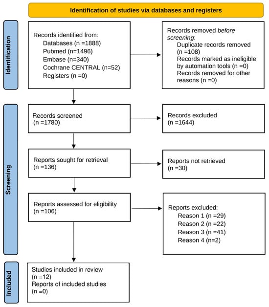
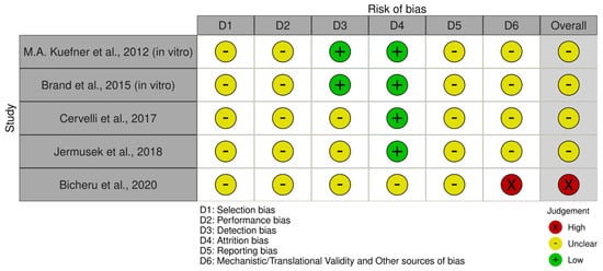
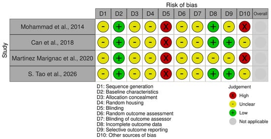
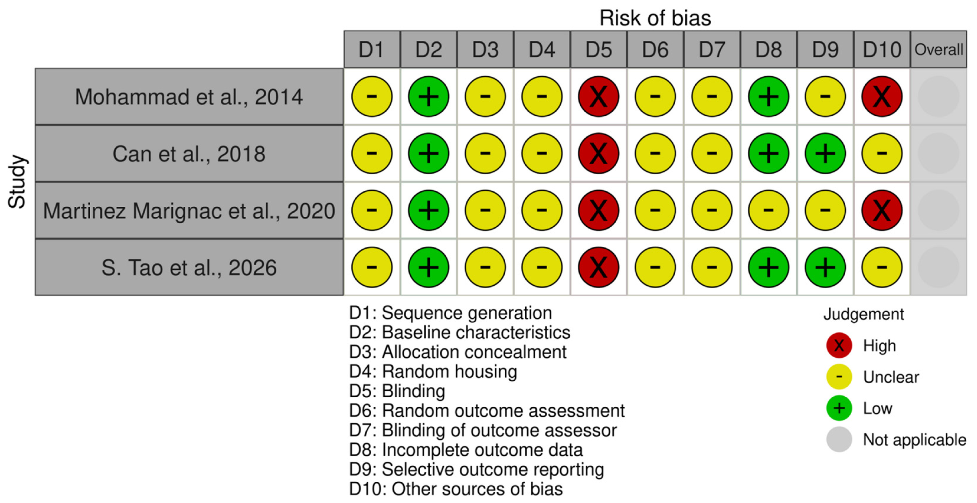
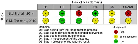
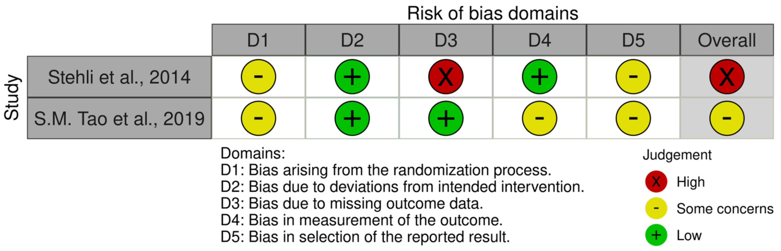
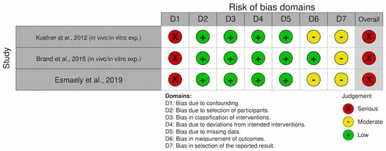
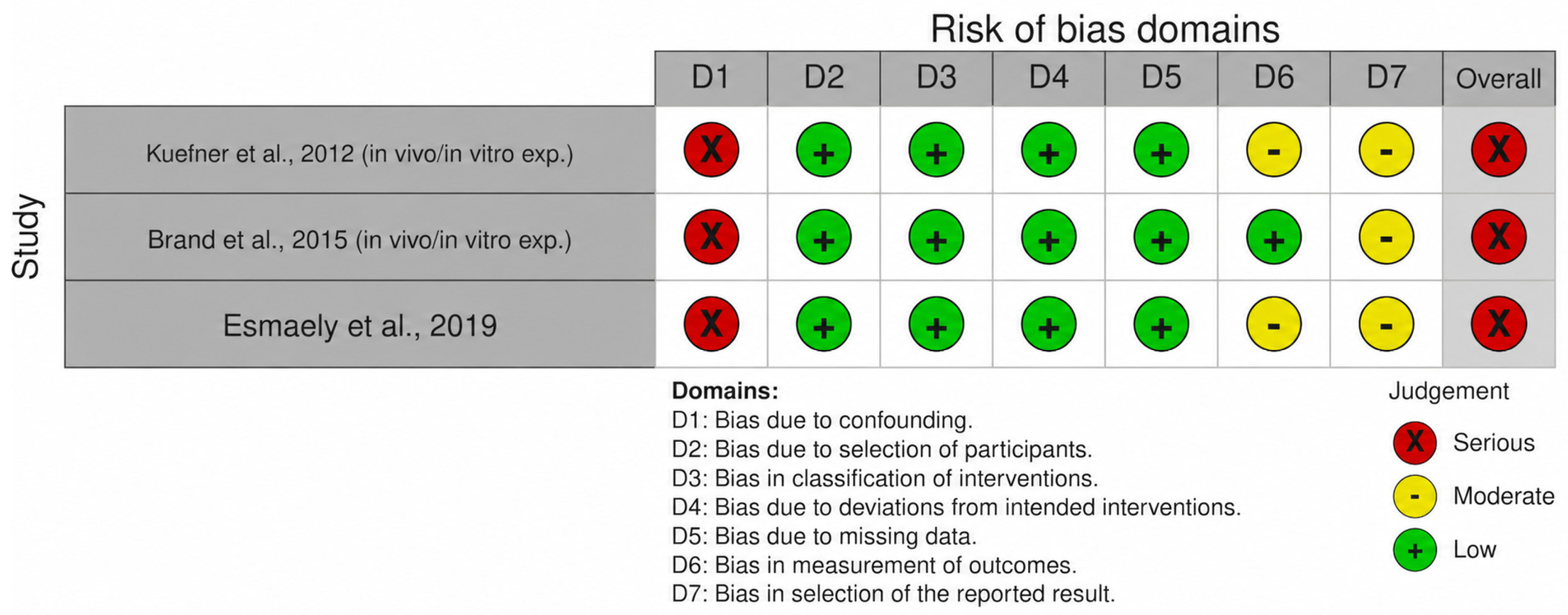

Disclaimer/Publisher’s Note: The statements, opinions and data contained in all publications are solely those of the individual author(s) and contributor(s) and not of MDPI and/or the editor(s). MDPI and/or the editor(s) disclaim responsibility for any injury to people or property resulting from any ideas, methods, instructions or products referred to in the content. |
© 2026 by the authors. Licensee MDPI, Basel, Switzerland. This article is an open access article distributed under the terms and conditions of the Creative Commons Attribution (CC BY) license.